Charles Darwin University Factors affecting the ...
21
Charles Darwin University Factors affecting the electrocardiographic QT interval in malaria A systematic review and meta-analysis of individual patient data Chan, Xin Hui S.; Win, Yan Naung; Haeusler, Ilsa L.; Tan, Jireh Y.; Loganathan, Shanghavie; Saralamba, Sompob; Chan, Shu Kiat S.; Ashley, Elizabeth A.; Barnes, Karen I.; Baiden, Rita; Bassi, Peter U.; Djimde, Abdoulaye; Dorsey, Grant; Duparc, Stephan; Hanboonkunupakarn, Borimas; Ter Kuile, Feiko O.; Lacerda, Marcus V.G.; Nasa, Amit; Nosten, François H.; Onyeji, Cyprian O.; Pukrittayakamee, Sasithon; Siqueira, André M.; Tarning, Joel; Taylor, Walter R.J.; Valentini, Giovanni; van Vugt, Michèle; Wesche, David; Day, Nicholas P.J.; Huang, Christopher L.H.; Brugada, Josep; Price, Ric N.; White, Nicholas J. Published in: PLoS Medicine DOI: 10.1371/journal.pmed.1003040 Published: 05/03/2020 Document Version Publisher's PDF, also known as Version of record Link to publication Citation for published version (APA): Chan, X. H. S., Win, Y. N., Haeusler, I. L., Tan, J. Y., Loganathan, S., Saralamba, S., Chan, S. K. S., Ashley, E. A., Barnes, K. I., Baiden, R., Bassi, P. U., Djimde, A., Dorsey, G., Duparc, S., Hanboonkunupakarn, B., Ter Kuile, F. O., Lacerda, M. V. G., Nasa, A., Nosten, F. H., ... White, N. J. (2020). Factors affecting the electrocardiographic QT interval in malaria: A systematic review and meta-analysis of individual patient data. PLoS Medicine, 17(3), 1-20. [e1003040]. https://doi.org/10.1371/journal.pmed.1003040 General rights Copyright and moral rights for the publications made accessible in the public portal are retained by the authors and/or other copyright owners and it is a condition of accessing publications that users recognise and abide by the legal requirements associated with these rights. • Users may download and print one copy of any publication from the public portal for the purpose of private study or research. • You may not further distribute the material or use it for any profit-making activity or commercial gain • You may freely distribute the URL identifying the publication in the public portal
Transcript of Charles Darwin University Factors affecting the ...

Charles Darwin University
Factors affecting the electrocardiographic QT interval in malaria
A systematic review and meta-analysis of individual patient data
Chan, Xin Hui S.; Win, Yan Naung; Haeusler, Ilsa L.; Tan, Jireh Y.; Loganathan, Shanghavie;Saralamba, Sompob; Chan, Shu Kiat S.; Ashley, Elizabeth A.; Barnes, Karen I.; Baiden, Rita;Bassi, Peter U.; Djimde, Abdoulaye; Dorsey, Grant; Duparc, Stephan; Hanboonkunupakarn,Borimas; Ter Kuile, Feiko O.; Lacerda, Marcus V.G.; Nasa, Amit; Nosten, François H.; Onyeji,Cyprian O.; Pukrittayakamee, Sasithon; Siqueira, André M.; Tarning, Joel; Taylor, WalterR.J.; Valentini, Giovanni; van Vugt, Michèle; Wesche, David; Day, Nicholas P.J.; Huang,Christopher L.H.; Brugada, Josep; Price, Ric N.; White, Nicholas J.Published in:PLoS Medicine
DOI:10.1371/journal.pmed.1003040
Published: 05/03/2020
Document VersionPublisher's PDF, also known as Version of record
Link to publication
Citation for published version (APA):Chan, X. H. S., Win, Y. N., Haeusler, I. L., Tan, J. Y., Loganathan, S., Saralamba, S., Chan, S. K. S., Ashley, E.A., Barnes, K. I., Baiden, R., Bassi, P. U., Djimde, A., Dorsey, G., Duparc, S., Hanboonkunupakarn, B., TerKuile, F. O., Lacerda, M. V. G., Nasa, A., Nosten, F. H., ... White, N. J. (2020). Factors affecting theelectrocardiographic QT interval in malaria: A systematic review and meta-analysis of individual patient data.PLoS Medicine, 17(3), 1-20. [e1003040]. https://doi.org/10.1371/journal.pmed.1003040
General rightsCopyright and moral rights for the publications made accessible in the public portal are retained by the authors and/or other copyright ownersand it is a condition of accessing publications that users recognise and abide by the legal requirements associated with these rights.
• Users may download and print one copy of any publication from the public portal for the purpose of private study or research. • You may not further distribute the material or use it for any profit-making activity or commercial gain • You may freely distribute the URL identifying the publication in the public portal

RESEARCH ARTICLE
Factors affecting the electrocardiographic QT
interval in malaria: A systematic review and
meta-analysis of individual patient data
Xin Hui S. ChanID1,2*, Yan Naung WinID
1,3, Ilsa L. HaeuslerID4,5, Jireh Y. Tan1,
Shanghavie Loganathan1,6, Sompob Saralamba1, Shu Kiat S. Chan1,7, Elizabeth
A. AshleyID2,8, Karen I. BarnesID
9,10, Rita Baiden11, Peter U. Bassi12, Abdoulaye DjimdeID13,
Grant DorseyID14, Stephan DuparcID
15, Borimas HanboonkunupakarnID1,16, Feiko O. ter
KuileID17, Marcus V. G. Lacerda18,19, Amit Nasa20, Francois H. NostenID
2,21, Cyprian
O. Onyeji22, Sasithon Pukrittayakamee1,16,23, Andre M. SiqueiraID18,24, Joel TarningID
1,2,4,
Walter R. J. Taylor1,2, Giovanni Valentini25, Michèle van Vugt26, David Wesche27, Nicholas
P. J. DayID1,2, Christopher L-H HuangID
28, Josep Brugada29, Ric N. PriceID1,2,30, Nicholas
J. WhiteID1,2*
1 Mahidol-Oxford Tropical Medicine Research Unit, Faculty of Tropical Medicine, Mahidol University,
Bangkok, Thailand, 2 Centre for Tropical Medicine and Global Health, Nuffield Department of Medicine,
University of Oxford, Oxford, United Kingdom, 3 Health and Diseases Control Unit, Naypyidaw, Myanmar,
4 WorldWide Antimalarial Research Network, Centre for Tropical Medicine and Global Health, Nuffield
Department of Medicine, University of Oxford, Oxford, United Kingdom, 5 University College London Great
Ormond Street Institute of Child Health, London, United Kingdom, 6 Christ Church College, University of
Oxford, Oxford, United Kingdom, 7 Singapore Armed Forces Medical Corps, Singapore, 8 Lao-Oxford-
Mahosot Hospital-Wellcome Trust Research Unit, Vientiane, Lao PDR, 9 Division of Clinical Pharmacology,
Department of Medicine, University of Cape Town, Cape Town, South Africa, 10 WorldWide Antimalarial
Resistance Network, Cape Town, South Africa, 11 INDEPTH Network Secretariat, Accra, Ghana,
12 Department of Internal Medicine, Faculty of Clinical Sciences, College of Health Sciences, University of
Abuja, Abuja, Nigeria, 13 Malaria Research and Training Center, Department of Epidemiology of Parasitic
Diseases, Faculty of Pharmacy, University of Science Techniques and Technologies of Bamako, Bamako,
Mali, 14 Department of Medicine, University of California San Francisco, San Francisco, California, United
States of America, 15 Medicines for Malaria Venture, Geneva, Switzerland, 16 Department of Clinical
Tropical Medicine, Faculty of Tropical Medicine, Mahidol University, Bangkok, Thailand, 17 Department of
Clinical Sciences, Liverpool School of Tropical Medicine, Liverpool, United Kingdom, 18 Fundacão de
Medicina Tropical Dr Heitor Vieira Dourado, Manaus, Brazil, 19 Instituto Leonidas e Maria Deane (FIOCRUZ-
Amazonas), Fundacão Oswaldo Cruz, Manaus, Brazil, 20 Sun Pharmaceutical Industries Ltd, Gurgaon,
Haryana, India, 21 Shoklo Malaria Research Unit, Mahidol-Oxford Tropical Medicine Research Unit, Faculty
of Tropical Medicine, Mahidol University, Mae Sot, Thailand, 22 Faculty of Pharmacy, Obafemi Awolowo
University, Ile-Ife, Nigeria, 23 The Royal Society of Thailand, Dusit, Bangkok, Thailand, 24 Instituto Nacional
de Infectologia Evandro Chagas, Fundacão Oswaldo Cruz, Rio de Janeiro, Brazil, 25 Corporate R&D
Department, Alfasigma S.p.A., Rome, Italy, 26 Amsterdam University Medical Centers, Location Academic
Medical Center, University of Amsterdam, Amsterdam, the Netherlands, 27 Certara, Princeton, New Jersey,
United States of America, 28 Physiological Laboratory, University of Cambridge, Cambridge, United
Kingdom, 29 Cardiovascular Institute, Hospital Clinic, University of Barcelona, Barcelona, Spain, 30 Global
and Tropical Health Division, Menzies School of Health Research, Charles Darwin University, Darwin, NT,
Australia
* [email protected] (XHSC); [email protected] (NJW)
Abstract
Background
Electrocardiographic QT interval prolongation is the most widely used risk marker for ven-
tricular arrhythmia potential and thus an important component of drug cardiotoxicity
PLOS Medicine | https://doi.org/10.1371/journal.pmed.1003040 March 5, 2020 1 / 20
a1111111111
a1111111111
a1111111111
a1111111111
a1111111111
OPEN ACCESS
Citation: Chan XHS, Win YN, Haeusler IL, Tan JY,
Loganathan S, Saralamba S, et al. (2020) Factors
affecting the electrocardiographic QT interval in
malaria: A systematic review and meta-analysis of
individual patient data. PLoS Med 17(3):
e1003040. https://doi.org/10.1371/journal.
pmed.1003040
Academic Editor: James G. Beeson, Burnet
Institute, AUSTRALIA
Received: August 16, 2019
Accepted: February 5, 2020
Published: March 5, 2020
Copyright: © 2020 Chan et al. This is an open
access article distributed under the terms of the
Creative Commons Attribution License, which
permits unrestricted use, distribution, and
reproduction in any medium, provided the original
author and source are credited.
Data Availability Statement: The data are available
upon request to the Mahidol Oxford Tropical
Medicine Research Unit Data Access Committee
(http://www.tropmedres.ac/data-sharing) for
researchers and following the Mahidol Oxford
Tropical Medicine Research Unit data access policy
(http://www.tropmedres.ac/_asset/file/data-
sharing-policy-v1-1.pdf). Queries and applications
for datasets should be directed to Rita
Chanviriyavuth ([email protected]).
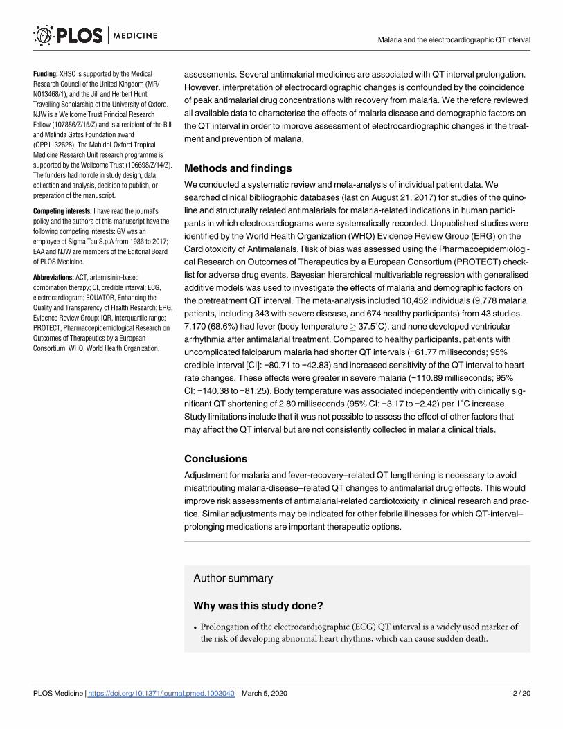
assessments. Several antimalarial medicines are associated with QT interval prolongation.
However, interpretation of electrocardiographic changes is confounded by the coincidence
of peak antimalarial drug concentrations with recovery from malaria. We therefore reviewed
all available data to characterise the effects of malaria disease and demographic factors on
the QT interval in order to improve assessment of electrocardiographic changes in the treat-
ment and prevention of malaria.
Methods and findings
We conducted a systematic review and meta-analysis of individual patient data. We
searched clinical bibliographic databases (last on August 21, 2017) for studies of the quino-
line and structurally related antimalarials for malaria-related indications in human partici-
pants in which electrocardiograms were systematically recorded. Unpublished studies were
identified by the World Health Organization (WHO) Evidence Review Group (ERG) on the
Cardiotoxicity of Antimalarials. Risk of bias was assessed using the Pharmacoepidemiologi-
cal Research on Outcomes of Therapeutics by a European Consortium (PROTECT) check-
list for adverse drug events. Bayesian hierarchical multivariable regression with generalised
additive models was used to investigate the effects of malaria and demographic factors on
the pretreatment QT interval. The meta-analysis included 10,452 individuals (9,778 malaria
patients, including 343 with severe disease, and 674 healthy participants) from 43 studies.
7,170 (68.6%) had fever (body temperature� 37.5˚C), and none developed ventricular
arrhythmia after antimalarial treatment. Compared to healthy participants, patients with
uncomplicated falciparum malaria had shorter QT intervals (−61.77 milliseconds; 95%
credible interval [CI]: −80.71 to −42.83) and increased sensitivity of the QT interval to heart
rate changes. These effects were greater in severe malaria (−110.89 milliseconds; 95%
CI: −140.38 to −81.25). Body temperature was associated independently with clinically sig-
nificant QT shortening of 2.80 milliseconds (95% CI: −3.17 to −2.42) per 1˚C increase.
Study limitations include that it was not possible to assess the effect of other factors that
may affect the QT interval but are not consistently collected in malaria clinical trials.
Conclusions
Adjustment for malaria and fever-recovery–related QT lengthening is necessary to avoid
misattributing malaria-disease–related QT changes to antimalarial drug effects. This would
improve risk assessments of antimalarial-related cardiotoxicity in clinical research and prac-
tice. Similar adjustments may be indicated for other febrile illnesses for which QT-interval–
prolonging medications are important therapeutic options.
Author summary
Why was this study done?
• Prolongation of the electrocardiographic (ECG) QT interval is a widely used marker of
the risk of developing abnormal heart rhythms, which can cause sudden death.
Malaria and the electrocardiographic QT interval
PLOS Medicine | https://doi.org/10.1371/journal.pmed.1003040 March 5, 2020 2 / 20
Funding: XHSC is supported by the Medical
Research Council of the United Kingdom (MR/
N013468/1), and the Jill and Herbert Hunt
Travelling Scholarship of the University of Oxford.
NJW is a Wellcome Trust Principal Research
Fellow (107886/Z/15/Z) and is a recipient of the Bill
and Melinda Gates Foundation award
(OPP1132628). The Mahidol-Oxford Tropical
Medicine Research Unit research programme is
supported by the Wellcome Trust (106698/Z/14/Z).
The funders had no role in study design, data
collection and analysis, decision to publish, or
preparation of the manuscript.
Competing interests: I have read the journal’s
policy and the authors of this manuscript have the
following competing interests: GV was an
employee of Sigma Tau S.p.A from 1986 to 2017;
EAA and NJW are members of the Editorial Board
of PLOS Medicine.
Abbreviations: ACT, artemisinin-based
combination therapy; CI, credible interval; ECG,
electrocardiogram; EQUATOR, Enhancing the
Quality and Transparency of Health Research; ERG,
Evidence Review Group; IQR, interquartile range;
PROTECT, Pharmacoepidemiological Research on
Outcomes of Therapeutics by a European
Consortium; WHO, World Health Organization.

• Several antimalarial drugs are associated with QT interval prolongation, motivating sys-
tematic ECG safety monitoring in many malaria clinical studies.
• Malaria illness itself may also affect the heart and QT interval, but these disease effects
are not well-understood.
• Interpretation of ECG changes after antimalarial treatment is complicated by the coinci-
dence of peak antimalarial drug concentrations with malaria recovery.
What did the researchers do and find?
• We performed a systematic review and meta-analysis of individual patient data to inves-
tigate the effect of malaria disease factors on the ECG QT interval.
• We included pretreatment data from 10,452 individuals (9,778 malaria patients and 674
healthy participants) from 43 studies in 20 countries.
• Malaria was associated with QT interval shortening and increased sensitivity of the QT
interval to heart rate changes, and these effects increased with malaria severity.
• Body temperature increase (fever) was associated independently with clinically signifi-
cant QT shortening.
What do these findings mean?
• To improve cardiac safety assessments of antimalarial medicines, adjustment for
malaria recovery- and defervescence-related QT interval prolongation is needed to
avoid misattributing QT changes solely to drugs.
• These findings are relevant to drug developers, healthcare providers, clinical trialists,
and policymakers who use QT interval safety data to decide which antimalarials to
develop and use and at what dose.
• Further study of the QT interval in other infections and inflammatory disorders that
cause fever and are treated with QT-prolonging medicines is recommended in order to
understand the role of disease factors in QT interval changes.
Introduction
Malaria remains the most important parasitic disease of humans. Over a thousand people—
mostly children in Africa—still die of the disease every day. Decades of progress in prevention
and control have now stagnated [1]. Strategic use of all available tools is essential to prevent
reversal of these hard-won gains.
Antimalarial medicines are central to malaria control efforts. They are given both to prevent
malaria and to treat it. The artemisinin-based combination therapies (ACTs) are now the gold
standard oral treatment for malaria and the first-line antimalarial treatment in >80 malaria-
endemic countries [2]. ACTs contain a rapidly acting artemisinin derivative combined with a
more slowly eliminated partner drug. Most of the partner drugs in use and several of those in
development are structurally related quinoline or quinoline-like compounds, some of which
Malaria and the electrocardiographic QT interval
PLOS Medicine | https://doi.org/10.1371/journal.pmed.1003040 March 5, 2020 3 / 20

prolong the electrocardiographic QT interval. Drug-related QT interval prolongation is a
widely used [3] yet nonspecific surrogate risk marker for repolarisation-related cardiotoxicity
in the form of torsade de pointes, a potentially fatal polymorphic ventricular tachycardia. QT
interval prolongation has been the most common reason for drug withdrawal and relabelling
[4]. As part of ongoing safety assessments of population-based use of ACTs and other quino-
line- or structurally related compound-containing combinations for malaria control and elimi-
nation in both acutely unwell patients and healthy people at risk of symptomatic disease, there
has been renewed interest in the evaluation of antimalarial effects on the ECG to guide antima-
larial selection and dosage [5–7].
Malaria is characterised by red blood cell parasitisation, fever, and anaemia [8]. Malaria ill-
ness itself may affect the heart and in particular the QT interval [9,10], although these disease
effects and their possible interaction with other factors known to affect the QT interval [11]
are not well-understood [12]. Because malaria illness and antimalarial drug concentrations
change over the course of malaria treatment, it is important to characterise the independent
contributions of disease and demographic factors on the QT interval in order to avoid misat-
tributing changes solely to drug effects [12].
To address this, we conducted a systematic review and meta-analysis of individual patient
data from malaria clinical trials to characterise the disease and demographic factors that inde-
pendently affect the electrocardiographic QT interval in malaria.
Methods
Search strategy and selection criteria
We performed a systematic literature search on October 22, 2015 (updated on August 21,
2017) of the databases MEDLINE, Embase, and Global Health for primary clinical studies of
the quinoline and structurally related antimalarials for malaria-related indications in which
electrocardiograms (ECGs) were recorded before and after drug administration (Search Strat-
egy in S1 Appendix). These published and additional unpublished studies were identified as
part of the work of the World Health Organization (WHO) Evidence Review Group (ERG) on
the Cardiotoxicity of Antimalarials [5].
Studies were eligible for inclusion in the review if they were prospective randomised-con-
trolled trials or cohort studies published from 1988 onwards in which 5 or more participants
were given a quinoline or structurally related antimalarial drug—amodiaquine, chloroquine,
halofantrine, lumefantrine, mefloquine, piperaquine, primaquine, pyronaridine, or quinine—
either as monotherapy or as part of an ACT. Studies that coadministered other drugs with QT-
prolonging potential (e.g., azithromycin) as part of the trial intervention were excluded.
Study authors were contacted with a request for clinical study reports and protocols as well
as anonymised individual patient-level data sets of the following prespecified variables identi-
fied from expert consultation [5]: age, weight, sex, body temperature, parasitaemia, haemoglo-
bin or haematocrit, heart rate or RR interval duration, uncorrected QT interval duration, ECG
abnormalities, and other cardiovascular adverse events. Studies were included in this meta-
analysis if individual patient-level data were available for all requested variables from the
screening or a baseline time point before antimalarial drug administration.
All included individual patient-level data were obtained in accordance with appropriate
ethical approvals from countries and institutions of origin. Additional ethical approval for this
systematic review and meta-analysis of fully anonymised individual patient data was not
deemed necessary in keeping with University of Oxford Central University Research Ethics
Committee guidance.
Malaria and the electrocardiographic QT interval
PLOS Medicine | https://doi.org/10.1371/journal.pmed.1003040 March 5, 2020 4 / 20

Data extraction and standardisation
At least two independent reviewers (from XHSC, YNW, ILH, and SKSC) screened titles,
abstracts, full texts, trial documentation, and anonymised data sets and agreed on study eligi-
bility. From study publications, reports, and protocols, we extracted study-level characteristics,
including location, antimalarial treatment indication, inclusion and exclusion criteria, temper-
ature measurement method, and ECG measurement methodology (Data Extraction in S1
Appendix), into a standardised database. Where required, trial registry records and study
investigators were consulted for further information.
Manual data entry was undertaken for data sets available only in printed format. Once digi-
tised, individual patient-level data sets were converted into a standard file format using Stat/
Transfer [13] version 13.3, then standardised and checked according to a prespecified data dic-
tionary (Data Standardisation in S1 Appendix). For studies of repeated treatments, only data
from the first treatment episode were extracted. Individual patient records were excluded if
data for any requested variables were missing at the selected time point before drug adminis-
tration (Data Integrity Checks in S1 Appendix).
Data analysis
We performed Bayesian hierarchical multivariable regression with generalised additive mod-
els. The QT interval was the response variable, and individual study ID was the varying inter-
cept. The square-root–transformed RR interval (ffiffiffiffiffiffiRRp
), sex, body temperature, and malaria
type/antimalarial treatment indication were the linear predictors. Age was modelled with sepa-
rate smooths for females and males because of the known sex-hormone–related QT interval
changes around puberty [11]. Weight was omitted because of its collinearity with age in a pre-
dominantly paediatric population. Haemoglobin was considered an intermediate variable and
omitted. Variable selection was based on directed acyclic graphs of proposed causal relation-
ships among collected variables (Fig B in S1 Appendix) identified from literature review and
expert consultation [5].
Four models were fitted to the data, in which different combinations of malaria-disease–
related terms were added to known factors affecting the QT interval: the first contained only
heart rate and demographic terms, the second added to the first a term for body temperature,
the third included a further term for malaria type (species, severity), and the fourth added to
the third an interaction term for malaria type andffiffiffiffiffiffiRRp
. We carried out Pareto smoothed
importance-sampling leave-one-out cross-validation for model comparison (Data Analysis in
S1 Appendix). We did not publish or preregister this analysis plan.
As sensitivity analyses, we compared the model with the best expected predictive perfor-
mance to a model with an additional binary variable for whether a participant was in a study
that excluded at screening individuals with one or more torsade de pointes risk factors, a
model with a linear predictor for haemoglobin, and another model with a cube root (Frideri-
cia-like) instead of square root (Bazett-like) transformation of the RR interval. In the subgroup
of malaria patients only, we added a linear predictor for log parasitaemia to the best model
(Data Analysis in S1 Appendix).
All statistical analyses and data visualisation were done in R [14] version 3.5.0. Bayesian
regression was done using the brms [15] package version 2.6.0 and the probabilistic program-
ming language Stan [16] version 2.18.0.
The risk of bias of individual studies at the outcome level was assessed using the PROTECT
[17] checklist for systematic reviews on adverse drug events.
Malaria and the electrocardiographic QT interval
PLOS Medicine | https://doi.org/10.1371/journal.pmed.1003040 March 5, 2020 5 / 20

Results
Individual patient-level data were sought from 159 clinical studies (137 published and 22
unpublished at the time of the literature search). Data from 11,109 participants in 53 studies
were shared, of which data from 10,452 participants in 43 studies (28 published [18–45], 5 sub-
sequently published [46–50], and 10 unpublished) were suitable for inclusion in the meta-anal-
ysis (Fig 1 and Tables A and B in S1 Appendix).
Overall, 93.6% (9,778/10,452) of included individuals had microscopy-confirmed Plasmo-dium falciparum or P. vivax malaria, of whom 89.7% (8,769/9,778) had uncomplicated P. fal-ciparum mono- or mixed infection (Table 1). The remaining 674 individuals were healthy
participants, the majority of whom (78.8%; 531/674) were enrolled in healthy volunteer phar-
macokinetic studies (Table 1). The median age of the 10,452 included individuals was 13.3
years (interquartile range [IQR] 4.6–26.0; range 6 months to 84 years), with 26.8% (2,803)
aged 5 to<15 years and 26.3% (2,751) aged<5 years. The healthy group were almost entirely
adult participants from Europe, North America, and urban Asia and Africa, while malaria
patients were more likely to be children or adolescents from rural Africa, Asia, and South
America and to have fever (temperature� 37.5˚C), anaemia, and tachycardia (Table 1 and
Figs C–E in S1 Appendix).
60.5% (26/43) of studies listed as exclusion criteria one or more risk factors for torsade de
pointes, such as a personal or family history of clinically significant arrhythmias, pre-existing
conditions or concomitant medications that prolong the QT interval or increase antimalarial
drug concentrations, a baseline corrected QT interval of more than 450 milliseconds, and elec-
trolyte imbalances including hypokalaemia and hypomagnesaemia. These 26 studies that
excluded patients with risk factors for torsade de pointes enrolled 73% (7,633/10,452) of indi-
viduals providing data.
Almost all (99.3%; 10,381/10,452) participants, including all malaria patients, had ECG
intervals measured manually by cardiologists (75.3%; 7,872/10,452) or other trained personnel.
In addition, 68.6% (7,170/10,452) of participants in 30.2% (13/43) of studies had ECGs sent to
a centralised facility where specialist staff read ECGs, and in the remainder, ECGs were read at
the study site. Only two studies, both of piperaquine in healthy volunteers [48,51], had
24-hour continuous ECG recordings at baseline (Tables C and D in S1 Appendix).
None of the 10,452 participants included had a baseline uncorrected QT interval of more
than 500 milliseconds.
Compared to included studies, a higher proportion of excluded studies were conducted
before 2007, did not specifically exclude torsade de pointes risk factors, and had unclear or
high risk of bias (Tables E and F in S1 Appendix), reflecting quality of measurement and
reporting methods of safety outcomes. The characteristics of included and excluded studies
were otherwise comparable (Table E in S1 Appendix). As with the excluded studies, most of
the 260 participants who were excluded for missing data were malaria patients in studies con-
ducted before 2007 with available characteristics similar to those of the included population
(Table G in S1 Appendix). There were no cases of sudden cardiac death, life-threatening ven-
tricular tachyarrhythmias (ventricular fibrillation or ventricular tachycardia), or torsade de
pointes documented for any of the 23,708 participants in the 159 studies from which individ-
ual patient-level data were sought.
Effect of malaria on the QT interval
Adjustment for malaria disease variables of body temperature and malaria type improved
model performance (Table H in S1 Appendix). Results are presented from the best model,
Malaria and the electrocardiographic QT interval
PLOS Medicine | https://doi.org/10.1371/journal.pmed.1003040 March 5, 2020 6 / 20

Fig 1. Study selection flow chart. ECG, electrocardiogram.
https://doi.org/10.1371/journal.pmed.1003040.g001
Malaria and the electrocardiographic QT interval
PLOS Medicine | https://doi.org/10.1371/journal.pmed.1003040 March 5, 2020 7 / 20

Table 1. Demographics and population characteristics.
Healthy Participants (n = 674) Malaria Patients (n = 9,778) Overall (n = 10,452)
Antimalarial Treatment Indication
Severe/complicated malaria 343 (3.5%) 343 (3.3%)
Uncomplicated malaria 9,435 (96.5%) 9,435 (90.3%)
P. falciparum mono- or mixed infection 8,769 (89.7%) 8,769 (83.9%)
P. vivax monoinfection 666 (6.8%) 666 (6.4%)
IPT 143 (21.2%) 143 (1.4%)
Pregnancy (IPTp) 125 (18.5%) 125 (1.2%)
Infancy (IPTi) 18 (2.7%) 18 (0.2%)
Healthy volunteer pharmacokinetics 531 (78.8%) 531 (5.1%)
Age (years)
Median (IQR) 28.9 (23.0–37.0) 12.1 (4.2–24.5) 13.3 (4.6–26.0)
<15 18 (2.7%) 5,536 (56.7%) 5,554 (53.1%)
<1 18 (2.7%) 193 (2.0%) 211 (2.0%)
1 to <5 0 2,540 (26.0%) 2,540 (24.3%)
5 to <15 0 2,803 (28.7%) 2,803 (26.8%)
�15 656 (97.3%) 4,242 (43.4%) 4,898 (46.9%)
�35 209 (31.0%) 1,296 (13.3%) 1,505 (14.4%)
�50 0 40 (0.41%) 40 (0.38%)
Sex
Female 343 (50.9%) 3,909 (40.0%) 4,252 (40.7%)
Pregnant 125 (18.5%) 9 (0.09%) 134 (1.3%)
Male 331 (49.1%) 5,869 (60.0%) 6,200 (59.3%)
Temperature (˚C)
Mean (SD) 36.8 (0.4) 38.2 (1.1) 38.2 (1.1)
�37.5 23 (3.4%) 7,147 (73.1%) 7,170 (68.6%)
Parasitaemia (parasites/μL)
Median (IQR) N/A 14,080 (2,851–45,219) 14,080 (2,851–45,219)
�10,000 N/A 5,500 (56.2%) 5,500 (52.6%)
�50,000 N/A 2,191 (22.4%) 2,191 (20.9%)
�100,000 N/A 905 (9.3%) 905 (8.7%)
�250,000 N/A 175 (1.8%) 175 (1.7%)
Heart Rate (beats per minute)
Mean (SD) 68 (17) 108 (30) 106 (31)
�140 0 1,537 (15.7%) 1,537 (14.7%)
120–139 16 (2.4%) 1,603 (16.4%) 1,619 (15.5%)
100–119 22 (3.3%) 2,356 (24.1%) 2,378 (22.8%)
80–99 79 (11.7%)) 2,560 (26.2%) 2,639 (25.2%)
60–79 320 (47.4%) 1,537 (15.7%) 1,857 (17.8%)
<60 237 (35.2%) 185 (1.9%) 422 (4.0%)
Torsade de Pointes Risk Factors
Excluded from the individual study 669 (99.3%) 6,964 (71.2%) 7,633 (73.0%)
Not excluded from the individual study 5 (0.7%) 2,814 (28.8%) 2,819 (27.0%)
Geographical Region
Africa 172 (25.5%) 6,363 (65.0%) 6,535 (62.5%)
Asia 147 (21.8%) 3,065 (31.3%) 3,212 (30.7%)
Americas 15 (2.2%) 350 (3.6%) 365 (3.5%)
Europe 340 (50.4%) 0 340 (3.3%)
Abbreviations: ECG, electrocardiogram; IPT, intermittent preventive therapy; IQR, interquartile range; N/A, not applicable; SD, standard deviation.
https://doi.org/10.1371/journal.pmed.1003040.t001
Malaria and the electrocardiographic QT interval
PLOS Medicine | https://doi.org/10.1371/journal.pmed.1003040 March 5, 2020 8 / 20

which had heart rate (asffiffiffiffiffiffiRRp
), age, sex, body temperature, and malaria type (both as an inde-
pendent term and an interaction term with heart rate) as predictors.
From the meta-analysis of all included participants (n = 10,452), body temperature had an
independent effect on the QT interval, with a mean shortening of the QT interval by 2.80 milli-
seconds (95% credible interval [CI]: 2.42 to 3.17) per 1˚C rise in temperature (Table 2 and Fig
2). When compared to healthy participants (n = 674) and adjusting for other predictors, QT
shortening increased with malaria severity: patients with severe malaria (n = 343) had the
shortest QT intervals (mean difference: −110.89 milliseconds; 95% CI: −140.38 to −81.25),
followed by patients with uncomplicated falciparum malaria (n = 8,769) (mean difference:
−61.77 milliseconds; 95% CI: −80.71 to −42.83). Patients with uncomplicated vivax malaria
(n = 666) also had shorter QT intervals than healthy participants, but the 95% CI included zero
(mean difference: −11.77 milliseconds; 95% CI −37.30 to 14.72). Sensitivity of the QT interval
to changes in heart rate also increased with malaria severity: the additional increase in the QT
interval per unit increase offfiffiffiffiffiffiRRp
(i.e., with decreasing heart rate) was higher in severe malaria
patients (mean difference: 4.89 milliseconds; 95% CI: 3.85 to 5.91) than patients with uncom-
plicated falciparum malaria (mean difference: 2.24 milliseconds; 95% CI: 1.65 to 2.83). These
values compared with a mean increase of 9.16 milliseconds (95% CI: 8.59 to 9.73) in healthy
Table 2. Factors affecting the QT interval in malaria.
Predictor Number of
Participants
Estimate (95% CI)/Smooth Description Clinically
Significant?
Improved
Model?†
ffiffiffiffiffiffiRRp
interval, perp
millisecond increase�
(healthy participants)
10,452 9.16 (8.59, 9.73) milliseconds Yes N/A
ffiffiffiffiffiffiRRp
interval, perp
millisecond increase�
(by malaria type versus healthy
participants)
10,452 Yes Yes
Healthy participants 674 Reference
Uncomplicated vivax malaria 666 0.62 (−0.11, 1.34) milliseconds
Uncomplicated falciparum malaria 8,769 2.24 (1.65, 2.83) milliseconds
Severe/complicated malaria 343 4.89 (3.85, 5.91) milliseconds
Age 10,452 Yes N/A
Female 4,252 Lengthens by approximately 8 milliseconds over childhood, then
lengthens more gradually by another approximately 5 milliseconds
in adulthood
Male 6,200 Lengthens by approximately 8 milliseconds over childhood, then
shortens by approximately 10 milliseconds around puberty before
gradually lengthening by approximately 10 milliseconds in
adulthood
Sex 10,452 Yes N/A
Female 4,252 Reference
Male 6,200 −4.22 (-5.00, −3.43) milliseconds
Body temperature, per 1˚C increase 10,452 −2.80 (-3.17, −2.42) milliseconds Yes Yes
Malaria Type 10,452 Yes Yes
Healthy participants 674 Reference
Uncomplicated vivax malaria 666 −11.77 (−37.30, 14.72) milliseconds
Uncomplicated falciparum malaria 8,769 −61.77 (−80.71, −42.83) milliseconds
Severe/complicated malaria 343 −110.89 (−140.38, −81.25) milliseconds
�Electrocardiographic RR interval in milliseconds = 60,000/(heart rate in beats per minute).†Improved expected predictive accuracy as estimated by the standard error of the difference in expected log predictive density. Multivariable regression results from
hierarchical generalised additive model. Abbreviations: CI, credible interval; N/A, not applicable.
https://doi.org/10.1371/journal.pmed.1003040.t002
Malaria and the electrocardiographic QT interval
PLOS Medicine | https://doi.org/10.1371/journal.pmed.1003040 March 5, 2020 9 / 20

participants. Again, uncomplicated vivax malaria patients had a slightly larger increase in the
QT interval with decreasing heart rate than healthy participants, but the 95% CI contained
zero (mean difference: 0.62 milliseconds; 95% CI: −0.11 to 1.34) (Table 2 and Fig 3).
The demographic variables of age and sex had clinically significant effects: the QT interval
lengthened by a mean of approximately 8 milliseconds over childhood before shortening by
approximately 10 milliseconds in males, but not in females, around puberty, then gradually
lengthened by approximately 5–10 milliseconds in both sexes over adulthood, although there
were few data for participants aged�50 years (n = 40); males (n = 6,200) also had overall
Fig 2. Body temperature and the QT interval in malaria. Independent effect of body temperature on the QT interval from hierarchical generalised
additive model adjusting for heart rate/RR interval (asffiffiffiffiffiffiRRp
), age, sex, malaria type, and individual study. Shaded area represents 95% CIs, and circles
represent original data points without adjustment. CI, credible interval.
https://doi.org/10.1371/journal.pmed.1003040.g002
Malaria and the electrocardiographic QT interval
PLOS Medicine | https://doi.org/10.1371/journal.pmed.1003040 March 5, 2020 10 / 20

Fig 3. Malaria type, RR interval, and the QT interval. Interaction between malaria type and the RR interval (on the square root scale) and
conditional effect on the QT interval (on the linear scale) from a hierarchical generalised additive model adjusting for age, sex, body temperature, and
individual study. Shaded areas represent 95% CIs, and circles represent original data points without adjustment. CI, credible interval.
https://doi.org/10.1371/journal.pmed.1003040.g003
Malaria and the electrocardiographic QT interval
PLOS Medicine | https://doi.org/10.1371/journal.pmed.1003040 March 5, 2020 11 / 20

shorter QT intervals than females (n = 4,252) (mean difference: −4.21 milliseconds; 95% CI:
−4.99 to −3.44) (Table 2 and Fig 4).
From this model, a 25-year–old male patient with uncomplicated falciparum malaria
admitted with a heart rate of 100 beats per minute and a temperature of 38.5˚C, whose heart
rate slows to 60 beats per minute and who defervesces to a temperature of 36.5˚C in recovery,
would be predicted to have a 22 millisecond or 25% greater QT interval lengthening than an
age- and sex-matched healthy afebrile participant with the same heart rate reduction indepen-
dent of any drug treatment (Table 3).
Fig 4. Age, sex, and the QT interval in malaria. Interaction between age and sex, and conditional effect on the QT interval, from a
hierarchical generalised additive model adjusting for heart rate/RR interval (asffiffiffiffiffiffiRRp
), malaria type, body temperature, and individual study.
Shaded areas represent 95% CIs, and rug marks represent age distribution of original data points. CI, credible interval.
https://doi.org/10.1371/journal.pmed.1003040.g004
Table 3. Predicted QT intervals at baseline and in recovery from malaria and fever.
Healthy Uncomplicated Vivax Uncomplicated
Falciparum
Severe Malaria
QT interval at baseline, milliseconds (95% PI) [HR = 100 bpm] 329 (285–371)
[T = 36.5˚C]
327 (281–370)
[T = 38.5˚C]
317 (273–358)
[T = 38.5˚C]
332 (287–377)
[T = 38.5˚C]
QT interval in recovery, milliseconds (95% PI) [HR = 60 bpm] 394 (351–435)
[T = 36.5˚C]
403 (356–446)
[T = 36.5˚C]
404 (360–445)
[T = 36.5˚C]
438 (392–484)
[T = 36.5˚C]
QT lengthening from baseline, milliseconds 65 76 87 106
Additional QT lengthening from baseline compared to healthy
participant, milliseconds
0 11 22 41
Malaria-related QT lengthening from baseline, % 0 14 25 39
Abbreviations: bpm, beats per minute; HR, heart rate; PI, prediction interval; T, body temperature. Predicted values for a 25-year–old male from a hierarchical
generalised additive model adjusting for heart rate/RR interval (asffiffiffiffiffiffiRRp
), age, sex, malaria type, body temperature, and individual study effects.
https://doi.org/10.1371/journal.pmed.1003040.t003
Malaria and the electrocardiographic QT interval
PLOS Medicine | https://doi.org/10.1371/journal.pmed.1003040 March 5, 2020 12 / 20

Sensitivity analyses
The QT intervals of participants in studies that screened for and excluded individuals with tor-
sade de pointes risk factors (n = 7,633) were not significantly different from those in studies
without documented risk factor screening (n = 2,819) (mean difference: −0.78 milliseconds;
95% CI −9.69 to 7.88) once other predictors had been adjusted for. There were no clinically
significant changes in predictor estimates when a cube root (Fridericia-like) instead of square
root (Bazett-like) transformation of the RR interval was used (Tables I–L in S1 Appendix).
The small effects associated with haemoglobin (−0.51 milliseconds per g/dL increase; 95%
CI: −0.72 to −0.30) and parasitaemia (0.65 milliseconds per 10-fold increase in parasite density;
95% CI: 0.17 to 1.14) were of unclear clinical significance, and these additional terms did not
improve model performance (Tables M–P, S1 Appendix).
Discussion
To our knowledge, this is the most extensive study to date of the factors affecting the electro-
cardiographic QT interval in malaria. We pooled individual patient data before treatment
from 10,452 adults and children (9,778 malaria patients, including 343 with severe disease, and
674 healthy participants) in antimalarial drug trials identified through a comprehensive sys-
tematic review and expert consultation. This allowed formal evaluation of the independent
effects of malaria disease (type, temperature, and parasitaemia) and patient demographics (age
and sex) on the QT interval without confounding from antimalarial drug effects through
meta-analysis using hierarchical generalised additive models. Of these, malaria type, body tem-
perature, age, and sex were found or confirmed to have clinically relevant effects on the QT
interval.
Malaria has important effects on the QT interval that are proportional to disease severity:
the marked QT interval shortening and increased sensitivity to changes in heart rate seen in
malaria are greater in severe than uncomplicated disease and greater in P. falciparum than in
P. vivax infection. These effects occur independently of temperature and parasitaemia, sug-
gesting further unmeasured disease factors may be responsible. Possible candidates include
increasing stimulation of the sympathetic nervous system and acid-base abnormalities result-
ing from microvascular sequestration with increasing malaria severity. Parasite density is a
poor predictor of sequestered parasite burden in falciparum malaria [8]. Higher levels of
proinflammatory cytokines such as interleukin-6 during acute infection in less immune
populations [52] and in severe malaria [53] may also contribute through inhibition of cardio-
myocyte ion channel function [54,55]. Yet, for a disease with a wide range of systemic compli-
cations causing multiple organ dysfunction, the heart is relatively spared in acute malaria:
clinically significant arrhythmias are rare [7], and even in severe malaria, in which there may
be extensive sequestration of parasitised erythrocytes in the myocardial microvasculature, car-
diac performance is maintained [56,57].
Fever, a cardinal sign of malaria illness, was found to have an independent effect on the QT
interval: the QT interval shortens as body temperature increases and lengthens correspond-
ingly as temperature decreases. In other words, fever shortens the QT interval, and recovery
from fever lengthens it. This is supported by the only prospective study, to our knowledge, of
fever and the QT interval [58] without confounding from drug treatment, which measured
ECGs in 27 otherwise healthy young Finnish male soldiers before and after self-limiting
uncomplicated febrile illness of bacterial, viral, or undefined aetiology. That study found that
the QT interval was significantly shorter during fever than after recovery (measurement of QT
intervals at specific heart rates of 60, 80, and 100 beats per minute obviated the need for heart
rate correction). Further prospective evaluation of the QT interval in febrile illness would be
Malaria and the electrocardiographic QT interval
PLOS Medicine | https://doi.org/10.1371/journal.pmed.1003040 March 5, 2020 13 / 20
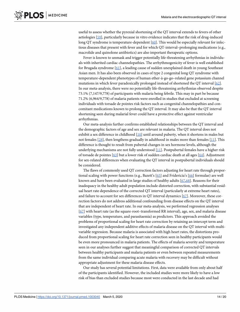
useful to assess whether the pyrexial shortening of the QT interval extends to fevers of other
aetiologies [59], particularly because in vitro evidence indicates that the risk of drug-induced
long QT syndrome is temperature-dependent [60]. This would be especially relevant for infec-
tious diseases that present with fever and for which QT-interval–prolonging medications (e.g.,
macrolide and quinolone antibiotics) are also important therapeutic options.
Fever is known to unmask and trigger potentially life-threatening arrhythmias in individu-
als with inherited cardiac channelopathies. The arrhythmogenicity of fever is well established
for Brugada syndrome [61], a leading cause of sudden unexplained death in young Southeast
Asian men. It has also been observed in cases of type 2 congenital long QT syndrome with
temperature-dependent phenotypes of human ether-à-go-go–related gene potassium channel
mutations in which fever paradoxically prolonged instead of shortened the QT interval [62].
In our meta-analysis, there were no potentially life-threatening arrhythmias observed despite
73.1% (7,147/9,778) of participants with malaria being febrile. This may in part be because
71.2% (6,964/9,778) of malaria patients were enrolled in studies that excluded at screening
individuals with torsade de pointes risk factors such as congenital channelopathies and con-
comitant medications known to prolong the QT interval. It may also be that the QT interval
shortening seen during malarial fever could have a protective effect against ventricular
arrhythmias.
Our meta-analysis further confirms established relationships between the QT interval and
the demographic factors of age and sex are relevant in malaria. The QT interval does not
exhibit a sex difference in childhood [10] until around puberty, when it shortens in males but
not females [28], then lengthens gradually in adulthood in males more than females [11]. This
difference is thought to result from pubertal changes in sex hormone levels, although the
underlying mechanisms are not fully understood [11]. Postpubertal females have a higher risk
of torsade de pointes [63] but a lower risk of sudden cardiac death at all ages [64]. Adjustment
for sex-related differences when evaluating the QT interval in postpubertal individuals should
be considered.
The flaws of commonly used QT correction factors adjusting for heart rate through propor-
tional scaling with power functions (e.g., Bazett’s [65] and Fridericia’s [66] formulae) are well
known and have been evaluated in large studies of healthy adults [67,68]. Reasons for their
inadequacy in the healthy adult population include distorted correction, with substantial resid-
ual heart rate dependence of the corrected QT interval (particularly at extreme heart rates),
and failure to account for sex differences in QT interval dynamics [67]. Moreover, these cor-
rection factors do not address additional confounding from disease effects on the QT interval
that are independent of heart rate. In our meta-analysis, we performed regression analyses
[67] with heart rate (as the square-root–transformed RR interval), age, sex, and malaria disease
variables (type, temperature, and parasitaemia) as predictors. This approach avoided the
problems of proportional scaling for heart rate correction by retaining an intercept term and
investigated any independent additive effects of malaria disease on the QT interval with multi-
variable regression. Because malaria is associated with high heart rates, the distortions pro-
duced from proportional scaling for heart rate correction seen in healthy participants would
be even more pronounced in malaria patients. The effects of malaria severity and temperature
seen in our analyses further suggest that meaningful comparison of corrected QT intervals
between healthy participants and malaria patients or even between repeated measurements
from the same individual comparing acute malaria with recovery may be difficult without
appropriate adjustment for these malaria disease effects.
Our study has several potential limitations. First, data were available from only about half
of the participants identified. However, the included studies were more likely to have a low
risk of bias than excluded studies because most were conducted in the last decade and had
Malaria and the electrocardiographic QT interval
PLOS Medicine | https://doi.org/10.1371/journal.pmed.1003040 March 5, 2020 14 / 20

more comprehensive measurement and reporting methods, reflecting the increased regulatory
interest in the cardiac safety of antimalarials. Second, it was not possible to assess directly the
effects of other factors known to contribute to the intrinsic variability of the QT interval such
as circadian rhythm, activity level, postural changes, and food ingestion [3], as well as those
that may alter cardiomyocyte electrophysiology during systemic illness such as inflammatory
biomarkers [54], because these data are not usually collected in malaria clinical trials. Most
hospitalised malaria patients are supine and anorexic. Third, we have considered interindivid-
ual measurements from a single time point before drug administration, an approach that
allowed us to consider data from a large number of patients with malaria and a smaller number
of healthy participants without confounding from drug therapy. While assessments of repeated
measurements from patients undergoing treatment for malaria with drugs not known to pro-
long the QT interval would be valuable, these data are few [7] and were not available to us.
Evaluation of QT interval prolongation after treatment with quinoline and structurally
related antimalarials has been the major motivation for ECG monitoring in malaria. ECG
monitoring is an operational challenge in the resource-limited settings where malaria is
endemic and would severely limit the use of any drug for which monitoring is mandatory. In
this large study of the QT interval in malaria, we have found malaria shortens the QT interval
and increases its sensitivity to changes in heart rate. These differences are greater in severe
malaria [9]. In addition, fever shortens the QT, and recovery from fever lengthens it. In acute
uncomplicated malaria, there is usually an irregular fever with an appropriate rise in heart
rate. As the illness and fever resolve, the heart rate declines to normal. This often coincides
with the highest blood concentrations of slowly eliminated quinoline antimalarial drugs,
which usually peak on the third day of treatment. The QT interval lengthening seen with
recovery from malaria after antimalarial therapy results from resolution of disease effects in
addition to any drug effects. Comparisons of predrug QT interval measurements with those at
peak drug concentrations in malaria studies should take into account malaria- and fever-
recovery–related QT lengthening; this would avoid excessive attribution of QT prolongation
to the antimalarial treatment and improve risk assessments of potential antimalarial-related
cardiotoxicity. This could avoid unnecessary discontinuation of new drug development and
reduce the need for unnecessary adjustment or withdrawal of antimalarial treatment in
response to malaria-related QT changes during research trials and clinical care. Similar adjust-
ments may also be indicated for other febrile illnesses for which QT-interval–prolonging med-
ications are important therapeutic options.
Supporting information
S1 Checklist. PRISMA-IPD checklist. PRISMA-IPD, Preferred Reporting Items for System-
atic Reviews and Meta-analyses of Individual Patient Data.
(DOCX)
S1 Appendix. Supplementary methods and results.
(DOCX)
Acknowledgments
The authors wish to express their gratitude to all investigators and participants of the studies
included in this analysis, as well as Sanofi Pasteur for sharing trial databases. We thank Nia
Roberts of the Bodleian Libraries of the University of Oxford and Shona Kirtley of the Enhanc-
ing the Quality and Transparency of Health Research (EQUATOR) Network for their expert
support with the literature searches; Catrin Moore and Michael Chipeta of the Big Data
Malaria and the electrocardiographic QT interval
PLOS Medicine | https://doi.org/10.1371/journal.pmed.1003040 March 5, 2020 15 / 20

Institute of the University of Oxford for guidance with spatial visualisation; and Mak Yiing
Chau, Laura Mawer, and Naomi Waithira for their kind assistance with data preparation. We
also thank Ronald Geskus, Sue Jean Lee, Mavuto Mukaka, Le Thanh Hoang Nhat, and Lisa
White for their statistical modelling advice, as well as the panel members of the WHO ERG on
the Cardiotoxicity of Antimalarial Medicines for valuable discussions.
Author Contributions
Conceptualization: Xin Hui S. Chan, Christopher L-H Huang, Josep Brugada, Nicholas J.
White.
Data curation: Xin Hui S. Chan, Yan Naung Win, Ilsa L. Haeusler, Jireh Y. Tan, Shanghavie
Loganathan, Shu Kiat S. Chan.
Formal analysis: Xin Hui S. Chan.
Funding acquisition: Xin Hui S. Chan, Nicholas P. J. Day, Nicholas J. White.
Investigation: Xin Hui S. Chan, Yan Naung Win, Ilsa L. Haeusler, Shu Kiat S. Chan, Elizabeth
A. Ashley, Karen I. Barnes, Rita Baiden, Peter U. Bassi, Abdoulaye Djimde, Grant Dorsey,
Stephan Duparc, Borimas Hanboonkunupakarn, Feiko O. ter Kuile, Marcus V. G. Lacerda,
Amit Nasa, Francois H. Nosten, Cyprian O. Onyeji, Sasithon Pukrittayakamee, Andre M.
Siqueira, Joel Tarning, Walter R. J. Taylor, Giovanni Valentini, Michèle van Vugt, David
Wesche, Nicholas P. J. Day, Ric N. Price, Nicholas J. White.
Methodology: Xin Hui S. Chan, Jireh Y. Tan, Sompob Saralamba, Nicholas P. J. Day.
Resources: Sompob Saralamba.
Software: Xin Hui S. Chan, Yan Naung Win, Jireh Y. Tan, Shu Kiat S. Chan.
Validation: Joel Tarning, Christopher L-H Huang.
Visualization: Xin Hui S. Chan, Sompob Saralamba.
Writing – original draft: Xin Hui S. Chan.
Writing – review & editing: Xin Hui S. Chan, Yan Naung Win, Ilsa L. Haeusler, Jireh Y. Tan,
Shanghavie Loganathan, Sompob Saralamba, Shu Kiat S. Chan, Elizabeth A. Ashley, Karen
I. Barnes, Rita Baiden, Peter U. Bassi, Abdoulaye Djimde, Grant Dorsey, Stephan Duparc,
Borimas Hanboonkunupakarn, Feiko O. ter Kuile, Marcus V. G. Lacerda, Amit Nasa, Fran-
cois H. Nosten, Cyprian O. Onyeji, Sasithon Pukrittayakamee, Andre M. Siqueira, Joel
Tarning, Walter R. J. Taylor, Giovanni Valentini, Michèle van Vugt, David Wesche, Nicho-
las P. J. Day, Christopher L-H Huang, Josep Brugada, Ric N. Price, Nicholas J. White.
References1. World Health Organization. World Malaria Report 2019. Geneva, Switzerland: 2019.
2. World Health Organization. Guidelines for the Treatment of Malaria. 3rd ed. Geneva, Switzerland.
2015.
3. ICH Harmonised Tripartite Guideline E14. The Clinical Evaluation of QT/QTc Interval Prolongation and
Proarrhythmic Potential for Non-Antiarrhythmic Drugs. Internet. 2005 [cited 2019 Dec 3]. Available
from: http://www.ich.org/fileadmin/Public_Web_Site/ICH_Products/Guidelines/Efficacy/E14/E14_
Guideline.pdf.
4. Roden DM. Drug-induced prolongation of the QT interval. N Engl J Med. 2004; 350(10):1013–22.
https://doi.org/10.1056/NEJMra032426 PMID: 14999113.
5. World Health Organization. WHO Evidence Review Group on the Cardiotoxicity of Antimalarial Medi-
cines. Geneva, Switzerland: 2017.
Malaria and the electrocardiographic QT interval
PLOS Medicine | https://doi.org/10.1371/journal.pmed.1003040 March 5, 2020 16 / 20

6. Chan XHS, Win YN, Mawer LJ, Tan JY, Brugada J, White NJ. Risk of sudden unexplained death after
use of dihydroartemisinin-piperaquine for malaria: a systematic review and Bayesian meta-analysis.
Lancet Infect Dis. 2018; 18(8):913–23. https://doi.org/10.1016/S1473-3099(18)30297-4 PMID:
29887371; PubMed Central PMCID: PMC6060085.
7. Haeusler IL, Chan XHS, Guerin PJ, White NJ. The arrhythmogenic cardiotoxicity of the quinoline and
structurally related antimalarial drugs: a systematic review. BMC Med. 2018; 16(1):200. https://doi.org/
10.1186/s12916-018-1188-2 PMID: 30400791.
8. White NJ, Pukrittayakamee S, Hien TT, Faiz MA, Mokuolu OA, Dondorp AM. Malaria. Lancet. 2014;
383(9918):723–35. https://doi.org/10.1016/S0140-6736(13)60024-0 PMID: 23953767.
9. Roggelin L, Pelletier D, Hill JN, Feldt T, Hoffmann S, Ansong D, et al. Disease-associated QT-shortage
versus quinine associated QT-prolongation: age dependent ECG-effects in Ghanaian children with
severe malaria. Malar J. 2014; 13:219. https://doi.org/10.1186/1475-2875-13-219 PMID: 24902591;
PubMed Central PMCID: PMC4067506.
10. von Seidlein L, Jaffar S, Greenwood B. Prolongation of the QTc interval in African children treated for
falciparum malaria. Am J Trop Med Hyg. 1997; 56(5):494–7. https://doi.org/10.4269/ajtmh.1997.56.494
PMID: 9180596.
11. Vink AS, Clur SB, Wilde AAM, Blom NA. Effect of age and gender on the QTc-interval in healthy individ-
uals and patients with long-QT syndrome. Trends Cardiovasc Med. 2018; 28(1):64–75. https://doi.org/
10.1016/j.tcm.2017.07.012 PMID: 28869094.
12. White NJ. Cardiotoxicity of antimalarial drugs. Lancet Infect Dis. 2007; 7(8):549–58. https://doi.org/10.
1016/S1473-3099(07)70187-1 PMID: 17646028.
13. Circle Systems Inc. Stat/Transfer: Data Conversion Software Utility. Seattle, Washington: Circle Sys-
tems; 2017.
14. R Core Team. R: A language and environment for statistical computing. Vienna, Austria: R Foundation
for Statistical Computing; 2018.
15. Burkner P-C. brms: An R Package for Bayesian Multilevel Models Using Stan. J StatSoftw. 2017; 80
(1):1–28. https://doi.org/10.18637/jss.v080.i01
16. Carpenter B, Gelman A, Hoffman MD, Lee D, Goodrich B, Betancourt M, et al. Stan: A probabilistic pro-
gramming language. J Stati Softw. 2017;76(1). https://doi.org/10.18637/jss.v076.i01.
17. Faillie JL, Ferrer P, Gouverneur A, Driot D, Berkemeyer S, Vidal X, et al. A new risk of bias checklist
applicable to randomized trials, observational studies, and systematic reviews was developed and vali-
dated to be used for systematic reviews focusing on drug adverse events. J Clin Epidemiol. 2017;
86:168–75. https://doi.org/10.1016/j.jclinepi.2017.04.023 PMID: 28487158.
18. Siqueira AM, Alencar AC, Melo GC, Magalhaes BL, Machado K, Alencar Filho AC, et al. Fixed-Dose
Artesunate-Amodiaquine Combination vs Chloroquine for Treatment of Uncomplicated Blood Stage P.
vivax Infection in the Brazilian Amazon: An Open-Label Randomized, Controlled Trial. Clin Infect Dis.
2016; 64(2):166–174. https://doi.org/10.1093/cid/ciw706 PMID: 27988484.
19. Valecha N, Savargaonkar D, Srivastava B, Rao BH, Tripathi SK, Gogtay N, et al. Comparison of the
safety and efficacy of fixed-dose combination of arterolane maleate and piperaquine phosphate with
chloroquine in acute, uncomplicated Plasmodium vivax malaria: a phase III, multicentric, open-label
study. Malar J. 2016; 15(1):42. https://doi.org/10.1186/s12936-016-1084-1 PMID: 26818020; PubMed
Central PMCID: PMC4728808.
20. Toure OA, Valecha N, Tshefu AK, Thompson R, Krudsood S, Gaye O, et al. A Phase 3, Double-Blind,
Randomized Study of Arterolane Maleate-Piperaquine Phosphate vs Artemether-Lumefantrine for Fal-
ciparum Malaria in Adolescent and Adult Patients in Asia and Africa. Clin Infect Dis. 2016; 62(8):964–
971. https://doi.org/10.1093/cid/ciw029 PMID: 26908796.
21. Sagara I, Beavogui AH, Zongo I, Soulama I, Borghini-Fuhrer I, Fofana B, et al. Safety and efficacy of re-
treatments with pyronaridine-artesunate in African patients with malaria: a substudy of the WANECAM
randomised trial. Lancet Infect Dis. 2016; 16(2):189–98. https://doi.org/10.1016/S1473-3099(15)
00318-7 PMID: 26601738; PubMed Central PMCID: PMC4726763.
22. Kredo T, Mauff K, Workman L, Van der Walt JS, Wiesner L, Smith PJ, et al. The interaction between
artemether-lumefantrine and lopinavir/ritonavir-based antiretroviral therapy in HIV-1 infected patients.
BMC Infect Dis. 2016; 16:30. https://doi.org/10.1186/s12879-016-1345-1 PMID: 26818566; PubMed
Central PMCID: PMC4728832.
23. Kakuru A, Jagannathan P, Muhindo MK, Natureeba P, Awori P, Nakalembe M, et al. Dihydroartemisi-
nin-Piperaquine for the Prevention of Malaria in Pregnancy. N Engl J Med. 2016; 374(10):928–39.
https://doi.org/10.1056/NEJMoa1509150 PMID: 26962728; PubMed Central PMCID: PMC4847718.
24. Toure OA, Rulisa S, Anvikar AR, Rao BS, Mishra P, Jalali RK, et al. Efficacy and safety of fixed dose
combination of arterolane maleate and piperaquine phosphate dispersible tablets in paediatric patients
Malaria and the electrocardiographic QT interval
PLOS Medicine | https://doi.org/10.1371/journal.pmed.1003040 March 5, 2020 17 / 20

with acute uncomplicated Plasmodium falciparum malaria: a phase II, multicentric, open-label study.
Malar J. 2015; 14(1):469. https://doi.org/10.1186/s12936-015-0982-y PMID: 26608469.
25. Darpo B, Ferber G, Siegl P, Laurijssens B, Macintyre F, Toovey S, et al. Evaluation of the QT effect of a
combination of piperaquine and a novel anti-malarial drug candidate OZ439, for the treatment of
uncomplicated malaria. Br J Clin Pharmacol. 2015; 80(4):706–15. https://doi.org/10.1111/bcp.12680
PMID: 25966781; PubMed Central PMCID: PMC4594707.
26. Baiden R, Oduro A, Halidou T, Gyapong M, Sie A, Macete E, et al. Prospective observational study to
evaluate the clinical safety of the fixed-dose artemisinin-based combination Eurartesim(R) (dihydroarte-
misinin/piperaquine), in public health facilities in Burkina Faso, Mozambique, Ghana, and Tanzania.
Malar J. 2015; 14:160. https://doi.org/10.1186/s12936-015-0664-9 PMID: 25885858; PubMed Central
PMCID: PMC4405867.
27. Pukrittayakamee S, Tarning J, Jittamala P, Charunwatthana P, Lawpoolsri S, Lee SJ, et al. Pharmaco-
kinetic interactions between primaquine and chloroquine. Antimicrob Agents Chemother. 2014; 58
(6):3354–9. https://doi.org/10.1128/AAC.02794-13 PMID: 24687509; PubMed Central PMCID:
PMC4068454.
28. Ogutu B, Juma E, Obonyo C, Jullien V, Carn G, Vaillant M, et al. Fixed dose artesunate amodiaquine—
a phase IIb, randomized comparative trial with non-fixed artesunate amodiaquine. Malar J. 2014;
13:498. https://doi.org/10.1186/1475-2875-13-498 PMID: 25515698; PubMed Central PMCID:
PMC4302156.
29. Hanboonkunupakarn B, Ashley EA, Jittamala P, Tarning J, Pukrittayakamee S, Hanpithakpong W,
et al. Open-label crossover study of primaquine and dihydroartemisinin-piperaquine pharmacokinetics
in healthy adult thai subjects. Antimicrob Agents Chemother. 2014; 58(12):7340–6. https://doi.org/10.
1128/AAC.03704-14 PMID: 25267661; PubMed Central PMCID: PMC4249579.
30. Valecha N, Krudsood S, Tangpukdee N, Mohanty S, Sharma SK, Tyagi PK, et al. Arterolane maleate
plus piperaquine phosphate for treatment of uncomplicated Plasmodium falciparum malaria: a compar-
ative, multicenter, randomized clinical trial. Clin Infect Dis. 2012; 55(5):663–71. https://doi.org/10.1093/
cid/cis475 PMID: 22586253.
31. Ndiaye JL, Faye B, Gueye A, Tine R, Ndiaye D, Tchania C, et al. Repeated treatment of recurrent
uncomplicated Plasmodium falciparum malaria in Senegal with fixed-dose artesunate plus amodiaquine
versus fixed-dose artemether plus lumefantrine: a randomized, open-label trial. Malar J. 2011; 10:237.
https://doi.org/10.1186/1475-2875-10-237 PMID: 21838909; PubMed Central PMCID: PMC3171378.
32. Kredo T, Mauff K, Van der Walt JS, Wiesner L, Maartens G, Cohen K, et al. Interaction between arte-
mether-lumefantrine and nevirapine-based antiretroviral therapy in HIV-1-infected patients. Antimicrob
Agents Chemother. 2011; 55(12):5616–23. https://doi.org/10.1128/AAC.05265-11 PMID: 21947399;
PubMed Central PMCID: PMC3232823.
33. Valecha N, Phyo AP, Mayxay M, Newton PN, Krudsood S, Keomany S, et al. An open-label, rando-
mised study of dihydroartemisinin-piperaquine versus artesunate-mefloquine for falciparum malaria in
Asia. PLoS ONE. 2010; 5(7):e11880. https://doi.org/10.1371/journal.pone.0011880 PMID: 20689583;
PubMed Central PMCID: PMC2912766.
34. Krudsood S, Looareesuwan S, Tangpukdee N, Wilairatana P, Phumratanaprapin W, Leowattana W,
et al. New fixed-dose artesunate-mefloquine formulation against multidrug-resistant Plasmodium falcip-
arum in adults: a comparative phase IIb safety and pharmacokinetic study with standard-dose nonfixed
artesunate plus mefloquine. Antimicrob Agents Chemother. 2010; 54(9):3730–7. https://doi.org/10.
1128/AAC.01187-09 PMID: 20547795; PubMed Central PMCID: PMC2935027.
35. Navaratnam V, Ramanathan S, Wahab MS, Siew Hua G, Mansor SM, Kiechel JR, et al. Tolerability and
pharmacokinetics of non-fixed and fixed combinations of artesunate and amodiaquine in Malaysian
healthy normal volunteers. Eur J Clin Pharmacol. 2009; 65(8):809–21. https://doi.org/10.1007/s00228-
009-0656-1 PMID: 19404632; PubMed Central PMCID: PMC2714898.
36. Bassat Q, Mulenga M, Tinto H, Piola P, Borrmann S, Menendez C, et al. Dihydroartemisinin-pipera-
quine and artemether-lumefantrine for treating uncomplicated malaria in African children: a randomised,
non-inferiority trial. PLoS ONE. 2009; 4(11):e7871. https://doi.org/10.1371/journal.pone.0007871
PMID: 19936217; PubMed Central PMCID: PMC2776302.
37. Mytton OT, Ashley EA, Peto L, Price RN, La Y, Hae R, et al. Electrocardiographic safety evaluation of
dihydroartemisinin piperaquine in the treatment of uncomplicated falciparum malaria. Am J Trop Med
Hyg. 2007; 77(3):447–50. PMID: 17827358.
38. Bassi PU, Onyeji CO, Ukponmwan OE. Effects of tetracycline on the pharmacokinetics of halofantrine
in healthy volunteers. Br J Clin Pharmacol. 2004; 58(1):52–5. https://doi.org/10.1111/j.1365-2125.2004.
02087.x PMID: 15206992; PubMed Central PMCID: PMC1884545.
39. Abernethy DR, Wesche DL, Barbey JT, Ohrt C, Mohanty S, Pezzullo JC, et al. Stereoselective halofan-
trine disposition and effect: concentration-related QTc prolongation. Br J Clin Pharmacol. 2001; 51
Malaria and the electrocardiographic QT interval
PLOS Medicine | https://doi.org/10.1371/journal.pmed.1003040 March 5, 2020 18 / 20
(3):231–7. https://doi.org/10.1046/j.1365-2125.2001.00351.x PMID: 11298069; PubMed Central
PMCID: PMC2015022.
40. van Vugt M, Looareesuwan S, Wilairatana P, McGready R, Villegas L, Gathmann I, et al. Artemether-
lumefantrine for the treatment of multidrug-resistant falciparum malaria. Trans R Soc Trop Med Hyg.
2000; 94(5):545–8. https://doi.org/10.1016/s0035-9203(00)90082-8 PMID: 11132386.
41. van Vugt M, Wilairatana P, Gemperli B, Gathmann I, Phaipun L, Brockman A, et al. Efficacy of six
doses of artemether-lumefantrine (benflumetol) in multidrug-resistant Plasmodium falciparum malaria.
Am J Trop Med Hyg. 1999; 60(6):936–42. https://doi.org/10.4269/ajtmh.1999.60.936 PMID: 10403324.
42. Tran TH, Day NP, Nguyen HP, Nguyen TH, Tran TH, Pham PL, et al. A controlled trial of artemether or
quinine in Vietnamese adults with severe falciparum malaria. N Engl J Med. 1996; 335(2):76–83.
https://doi.org/10.1056/NEJM199607113350202 PMID: 8649493.
43. Price RN, Nosten F, Luxemburger C, Kham A, Brockman A, Chongsuphajaisiddhi T, et al. Artesunate
versus artemether in combination with mefloquine for the treatment of multidrug-resistant falciparum
malaria. Trans R Soc Trop Med Hyg. 1995; 89(5):523–7. https://doi.org/10.1016/0035-9203(95)90094-
2 PMID: 8560531.
44. Nosten F, ter Kuile FO, Luxemburger C, Woodrow C, Kyle DE, Chongsuphajaisiddhi T, et al. Cardiac
effects of antimalarial treatment with halofantrine. Lancet. 1993; 341(8852):1054–6. https://doi.org/10.
1016/0140-6736(93)92412-m PMID: 8096959.
45. White NJ, Miller KD, Churchill FC, Berry C, Brown J, Williams SB, et al. Chloroquine treatment of severe
malaria in children. Pharmacokinetics, toxicity, and new dosage recommendations. N Engl J Med.
1988; 319(23):1493–500. https://doi.org/10.1056/NEJM198812083192301 PMID: 3054558.
46. Hanboonkunupakarn B, van der Pluijm RW, Hoglund R, Pukrittayakamee S, Winterberg M, Mukaka M,
et al. Sequential Open-Label Study of the Safety, Tolerability, and Pharmacokinetic Interactions
between Dihydroartemisinin-Piperaquine and Mefloquine in Healthy Thai Adults. Antimicrob Agents
Chemother. 2019; 63(8):e00060–19. https://doi.org/10.1128/AAC.00060-19 PMID: 31182525; PubMed
Central PMCID: PMC6658739.
47. Ahmed R, Poespoprodjo JR, Syafruddin D, Khairallah C, Pace C, Lukito T, et al. Efficacy and safety of
intermittent preventive treatment and intermittent screening and treatment versus single screening and
treatment with dihydroartemisinin-piperaquine for the control of malaria in pregnancy in Indonesia: a
cluster-randomised, open-label, superiority trial. Lancet Infect Dis. 2019; 19(9):973–87. https://doi.org/
10.1016/S1473-3099(19)30156-2 PMID: 31353217; PubMed Central PMCID: PMC6715823.
48. Funck-Brentano C, Bacchieri A, Valentini G, Pace S, Tommasini S, Voiriot P, et al. Effects of Dihydroar-
temisinin-Piperaquine Phosphate and Artemether-Lumefantrine on QTc Interval Prolongation. Sci Rep.
2019; 9(1):777. https://doi.org/10.1038/s41598-018-37112-6 PMID: 30692558.
49. Macintyre F, Adoke Y, Tiono AB, Duong TT, Mombo-Ngoma G, Bouyou-Akotet M, et al. A randomised,
double-blind clinical phase II trial of the efficacy, safety, tolerability and pharmacokinetics of a single
dose combination treatment with artefenomel and piperaquine in adults and children with uncomplicated
Plasmodium falciparum malaria. BMC Med. 2017; 15(1):181. https://doi.org/10.1186/s12916-017-
0940-3 PMID: 28988541; PubMed Central PMCID: PMC5632828.
50. Natureeba P, Kakuru A, Muhindo M, Littmann E, Ochieng T, Ategeka J, et al. Intermittent Preventive
Treatment with Dihydroartemisinin-piperaquine for the Prevention of Malaria among HIV-infected Preg-
nant Women. J Infect Dis. 2017; 216(1):29–35. https://doi.org/10.1093/infdis/jix110 PMID: 28329368.
51. European Medicines Agency. Eurartesim 160/20mg Tablets: Summary of Product Characteristics.
Internet. 2011 [cited 2019 Dec 3]. Available from: http://www.ema.europa.eu/docs/en_GB/document_
library/EPAR_-_Product_Information/human/001199/WC500118113.pdf.
52. Farrington L, Vance H, Rek J, Prahl M, Jagannathan P, Katureebe A, et al. Both inflammatory and regu-
latory cytokine responses to malaria are blunted with increasing age in highly exposed children. Malar
J. 2017; 16(1):499. https://doi.org/10.1186/s12936-017-2148-6 PMID: 29284469; PubMed Central
PMCID: PMC5747142.
53. Oyegue-Liabagui SL, Bouopda-Tuedom AG, Kouna LC, Maghendji-Nzondo S, Nzoughe H, Tchitoula-
Makaya N, et al. Pro- and anti-inflammatory cytokines in children with malaria in Franceville, Gabon.
Am J Clin Exp Immunol. 2017; 6(2):9–20. PMID: 28337387; PubMed Central PMCID: PMC5344990.
54. Lazzerini PE, Laghi-Pasini F, Bertolozzi I, Morozzi G, Lorenzini S, Simpatico A, et al. Systemic inflam-
mation as a novel QT-prolonging risk factor in patients with torsades de pointes. Heart. 2017; 103
(22):1821–9. https://doi.org/10.1136/heartjnl-2016-311079 PMID: 28490617.
55. Aromolaran AS, Srivastava U, Ali A, Chahine M, Lazaro D, El-Sherif N, et al. Interleukin-6 inhibition of
hERG underlies risk for acquired long QT in cardiac and systemic inflammation. PLoS ONE. 2018; 13
(12):e0208321. https://doi.org/10.1371/journal.pone.0208321 PMID: 30521586; PubMed Central
PMCID: PMC6283635.
Malaria and the electrocardiographic QT interval
PLOS Medicine | https://doi.org/10.1371/journal.pmed.1003040 March 5, 2020 19 / 20

56. Bethell DB, Phuong PT, Phuong CX, Nosten F, Waller D, Davis TM, et al. Electrocardiographic monitor-
ing in severe falciparum malaria. Trans R Soc Trop Med Hyg. 1996; 90(3):266–9. https://doi.org/10.
1016/s0035-9203(96)90241-2 PMID: 8758072.
57. World Health Organization. Severe Malaria. Trop Med Int Health. 2014; 19 Suppl 1:7–131. https://doi.
org/10.1111/tmi.12313_2 PMID: 25214480.
58. Karjalainen J, Viitasalo M. Fever and cardiac rhythm. Arch Intern Med. 1986; 146(6):1169–71. PMID:
2424378.
59. Drew D, Baranchuk A, Hopman W, Brison RJ. The impact of fever on corrected QT interval. J Electro-
cardiol. 2017; 50(5):570–5. https://doi.org/10.1016/j.jelectrocard.2017.04.006 PMID: 28465023
60. Lee W, Windley MJ, Vandenberg JI, Hill AP. In Vitro and In Silico Risk Assessment in Acquired Long
QT Syndrome: The Devil Is in the Details. Front Physiol. 2017; 8:934. https://doi.org/10.3389/fphys.
2017.00934 PMID: 29201009; PubMed Central PMCID: PMC5696636.
61. Webster G, Berul CI. An update on channelopathies: from mechanisms to management. Circulation.
2013; 127(1):126–40. https://doi.org/10.1161/CIRCULATIONAHA.111.060343 PMID: 23283857.
62. Amin AS, Herfst LJ, Delisle BP, Klemens CA, Rook MB, Bezzina CR, et al. Fever-induced QTc pro-
longation and ventricular arrhythmias in individuals with type 2 congenital long QT syndrome. J Clin
Invest. 2008; 118(7):2552–61. https://doi.org/10.1172/JCI35337 PMID: 18551196; PubMed Central
PMCID: PMC2423868.
63. Sauer AJ, Newton-Cheh C. Clinical and genetic determinants of torsade de pointes risk. Circulation.
2012; 125(13):1684–94. https://doi.org/10.1161/CIRCULATIONAHA.111.080887 PMID: 22474311;
PubMed Central PMCID: PMC3347483.
64. Deo R, Albert CM. Epidemiology and genetics of sudden cardiac death. Circulation. 2012; 125(4):620–
37. https://doi.org/10.1161/CIRCULATIONAHA.111.023838 PMID: 22294707; PubMed Central
PMCID: PMC3399522.
65. Bazett HC. An Analysis of the Time Relations of Electrocardiograms. Heart. 1920; 7:353–70.
66. Fridericia LS. The duration of systole in the electrocardiogram of normal subjects and of patients with
heart disease. Acta Med Scand. 1920; 53:489–506.
67. Rautaharju PM, Zhang ZM. Linearly scaled, rate-invariant normal limits for QT interval: eight decades of
incorrect application of power functions. J Cardiovasc Electrophysiol. 2002; 13(12):1211–8. https://doi.
org/10.1046/j.1540-8167.2002.01211.x PMID: 12521335.
68. Rabkin SW, Szefer E, Thompson DJS. A New QT Interval Correction Formulae to Adjust for Increases
in Heart Rate. JACC Clin Electrophysiol. 2017; 3(7):756–66. https://doi.org/10.1016/j.jacep.2016.12.
005 PMID: 29759542.
Malaria and the electrocardiographic QT interval
PLOS Medicine | https://doi.org/10.1371/journal.pmed.1003040 March 5, 2020 20 / 20