
characterized by next generation sequencing Exodontia ...
21
Page 1/21 Exodontia-associated bacteremia in horses characterized by next generation sequencing Kile Townsend University of Missouri Philip Johnson ( [email protected] ) University of Missouri Alison LaCarrubba University of Missouri Lynn Martin University of Missouri Aaron Ericsson University of Missouri Research Article Keywords: horses, exodontia-associated bacteremia, surgery, next generation sequencing Posted Date: December 9th, 2020 DOI: https://doi.org/10.21203/rs.3.rs-117954/v1 License: This work is licensed under a Creative Commons Attribution 4.0 International License. Read Full License Version of Record: A version of this preprint was published at Scientic Reports on March 18th, 2021. See the published version at https://doi.org/10.1038/s41598-021-85484-z.
Transcript of characterized by next generation sequencing Exodontia ...

Page 1/21
Exodontia-associated bacteremia in horsescharacterized by next generation sequencingKile Townsend
University of MissouriPhilip Johnson ( [email protected] )
University of MissouriAlison LaCarrubba
University of MissouriLynn Martin
University of MissouriAaron Ericsson
University of Missouri
Research Article
Keywords: horses, exodontia-associated bacteremia, surgery, next generation sequencing
Posted Date: December 9th, 2020
DOI: https://doi.org/10.21203/rs.3.rs-117954/v1
License: This work is licensed under a Creative Commons Attribution 4.0 International License. Read Full License
Version of Record: A version of this preprint was published at Scienti�c Reports on March 18th, 2021. Seethe published version at https://doi.org/10.1038/s41598-021-85484-z.

Page 2/21
AbstractBacteremia resulting from dental surgery is increasingly recognized as a health risk, especially in olderand immunocompromised patients. Dentistry-associated bacteremic showering can lead to remoteinfections, as exempli�ed by valvular endocarditis. Emerging evidence points to a novel role played byuncultivable oral cavity commensals in the pathogenesis of diabetes, respiratory disease, cardiovasculardisease, and adverse pregnancy outcomes. Whether dental extraction, a commonly-undertaken procedurein old horses, causes bacteremic showering has not been reported extensively. In a prospective clinicalstudy using next generation sequencing (based on bacterial 16S rRNA), the circulating blood microbiomewas characterized before and at 1 hour following extraction of incisor, canine or cheek teeth from 29adult horses with dental disease. 16S rRNA sequencing results from the blood microbiome werecompared with those from gingival swab samples obtained prior to extraction at the location of thediseased tooth. Bacteremic showering by translocated gingival commensals was demonstrated in horsesundergoing exodontia and was, in some cases, still evident one hour post-operatively.
IntroductionExodontia (tooth extraction) and periodontal disease are associated with bacteremic showering, aphenomenon that may lead to adverse health outcomes [1–12]. Numerous reports over the last 50 yearshave documented the pathophysiological association between periodontal disease and systemicconditions such as diabetes mellitus, atherosclerotic cardiovascular disease, metabolic syndrome,chronic diseases (i.e., rheumatoid arthritis, cancer, Alzheimer’s disease), and adverse pregnancy outcomes[1, 3, 4, 8, 13]. Whereas exodontia-associated bacteremia is independent of the number of extracted teeth,it is associated with concurrent odontogenic infection in affected people [10]. Post-exodontia bacteremiahas also been associated with infection at distant sites such as the heart (valvular endocarditis),respiratory tract, joints, and brain [4, 14].
Recently, veterinarians have also recognized associations between periodontal disease and systemichealth in canine and feline species [15–17], leading to broader recognition of the importance of routineoral examination and preventive dentistry in veterinary clinical practice. Additionally, the extent to whichveterinarians provide advanced dental care for horses has increased substantially in recent years as aresult of parallel increases in both the availability of improved dental surgical equipment and educationregarding the technical skills associated with this discipline [18]. Veterinary work with old horses hasincreased considerably in recent years and clinical problems resulting from age-associated dentalattrition represent an especially common component of equine veterinary work in geriatric horses [18–20]. Remarkably, the incidence of periodontal disease in horses has been estimated to be as high as 75%,with the incidence increasing with advancing age [21].
Dental diseases in horses, such as those associated with equine odontoclastic tooth reabsorption andhypercementosis (EOTRH) syndrome or apical infections, are often unrecognized until relatively latestages, at which time dental extraction is commonly undertaken. Post-exodontia endocarditis, a potential

Page 3/21
complication of bacteremic showering, has been described in both dogs and horses [22–24]. Oral cavitymicroorganisms have been implicated in a few case reports in which fatal bacterial infections(endocarditis, meningitis, and pneumonia) developed following exodontia in horses [25, 26]. The extent towhich bacteremic showering leads to numerous complications in other species suggests that its role inequidae may be under-appreciated and deserving of further investigation [4–6, 10, 14]. These potentialcomplications are important when one considers that exodontia is frequently performed in older horsesthat are at substantially higher risk of disseminated infection as a result of underlying immune-debilitating comorbidities such as pituitary pars intermedia dysfunction (PPID) and immunosenescence[27–30].
Post-exodontia bacterial showering was recently reported by Kern et al. (2017), occurring in 18 out of 20horses using conventional bacteriological culturing methods [31]. Although this was a noteworthy �nding,it was likely limited by the method (conventional microbiological culturing) used to detect blood-bornebacteria, which relies on laboratory cultivation of bacteria present in the circulation. Conventionalbacterial culturing is limiting because most oral cavity bacteria are uncultivable [32]. An alternativemethod, such as 16S rRNA amplicon sequencing allows for identi�cation of not only cultivable bacteriabut also bacteria that might be present in small numbers or uncultivable via standard techniques [32–36].
Therefore, the primary aim of this study was to determine the extent to which dental extraction results inpost-procedural bacteremic showering and to characterize bacterial DNA present in the circulation beforeand following exodontia in adult horses with dental disease using 16S rRNA amplicon sequencing. Asecondary aim was to compare gingival swab microbiomes to blood microbiomes before and followingexodontia.
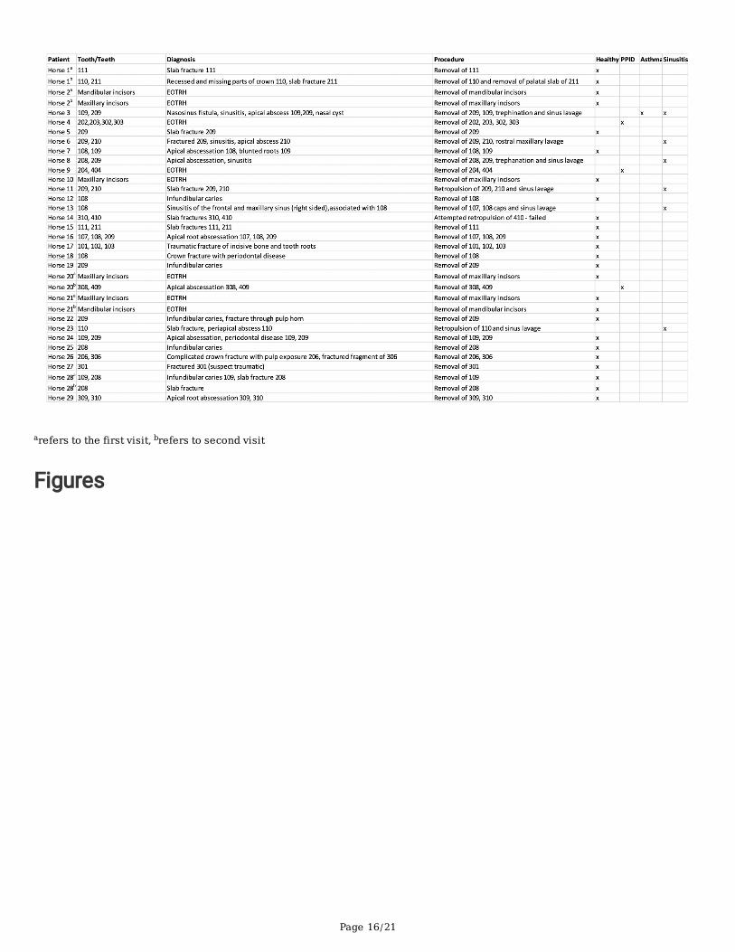
ResultsOf the 29 horses, most were determined to be healthy (n = 24) or affected with paranasal sinusitis (n = 6),PPID (n = 3), or asthma (n = 1). Procedures included those requiring cheek tooth extraction (n = 25) andthose requiring incisor or canine tooth extraction (n = 9). Justi�cation for exodontia included: apical toothroot abscessation (n = 9), slab fracture (n = 9), equine odontoclastic tooth reabsorption andhypercementosis (EOTRH) syndrome (n = 8), infundibular caries (n = 5), crown fractures (n = 3), andfractured incisive bone (n = 1). A total of 34 procedures were performed on 29 horses (some horsesreturned for a second exodontia procedure at least one month after completion of the �rst). Retropulsionof teeth was needed for extraction in three cases and standard intra-oral tooth extraction was performedin the remaining 31 cases. Sinus lavage was performed post-procedurally in all cases with comorbidsinusitis. Detailed signalment and procedural information are presented in Table 1.
To qualitatively assess the validity of microbial signatures detected via 16S rRNA sequencing, DNAampli�cation (quanti�ed by total number of reads for a given sample among a shared sequencing �owcell) was compared between pre- and post-exodontia blood samples, gingival swabs, negative reagentcontrols, and a commercially available bacterial community standard. As anticipated, the swabs yielded

Page 4/21
higher sequence numbers than either group of blood samples, and the mock community standard yieldedhigher coverage, by an order of magnitude, than negative reagent controls and most blood samples(Fig. 1). Notably however, �ve blood samples collected post-exodontia yielded unexpectedly deepcoverage, ranging from 187,130 to 669,731 sequences per sample. While sequencing coverage is notabsolutely quantitative of starting microbial biomass, these results suggested the presence of increasedbacterial biomass in a subset of blood samples collected post-exodontia. Moreover, the validity of theremaining blood samples and a few swabs samples that ampli�ed poorly was brought into question.
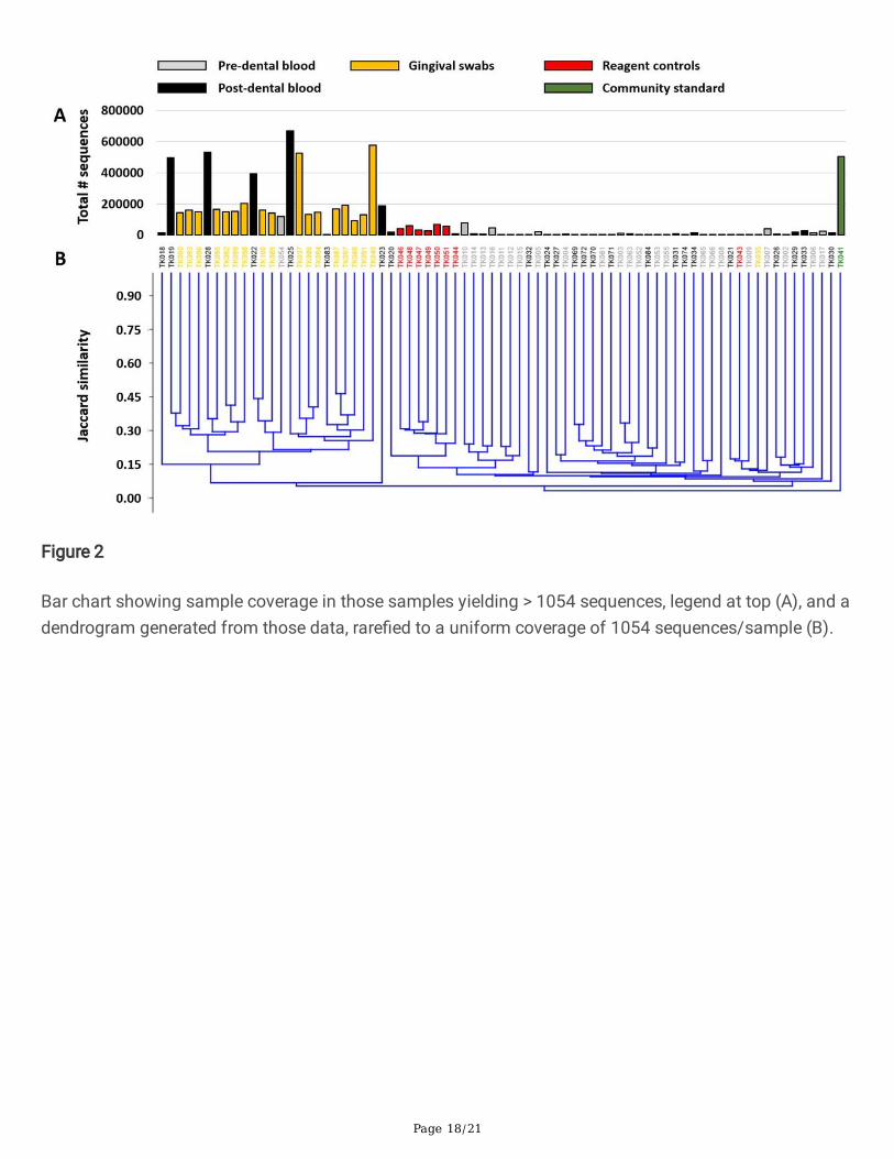
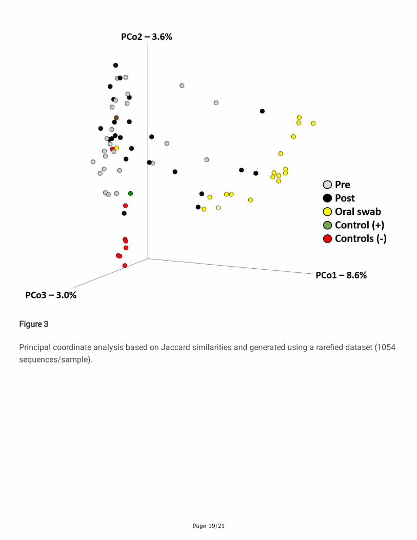
Recognizing that the differences in sample coverage would likely skew comparisons of bacterialcomposition, all samples yielding fewer than 1055 sequences were removed from the following analyses,and the remaining data were rare�ed randomly to a uniform read depth of 1054 reads/sample. Theoriginal sequencing coverage of those samples (Fig. 2A) is re�ective in the hierarchical clustering ofsamples based on the rare�ed dataset, with those same �ve highly ampli�ed post-exodontia bloodsamples clustering with the gingival swabs, along with two other post-exodontia and one pre-exodontiablood samples with lower coverage (Fig. 2B). These relationships were also visualized using principalcoordinate analysis (PCoA), which demonstrated a similar pattern with the same post-exodontia bloodsamples clustering close to the gingival swabs (Fig. 3). One-way permutational multivariate ANOVAcon�rmed signi�cant differences between swabs and pre-exodontia blood (p ≤ 0.0001, F = 6.6), swabsand post-exodontia blood (p ≤ 0.0001, F = 4.7), and between pre- and post-exodontia blood (p = 0.039, F = 1.3). All three groups were signi�cantly different from negative reagent controls (p ≤ 0.0001; F = 2.6, 2.6,and 6.2 for pre- and post-exodontia blood and swabs, respectively). Collectively, we interpreted theseresults to indicate compositional similarities between the microbial communities present on the gingivaand those detected in post-exodontia blood in a subset of horses, including those whose samples yieldedhigh sequence counts.
To identify the taxonomies contributing to the differences between swabs and pre- and post-exodontiablood, data from control samples were removed, and serial ANOVA testing was performed on all detectedAmplicon Sequence Variants (ASV). Based on those ASVs returning the 50 lowest p values, hierarchicalclustering was repeated and visualized using a heatmap (Fig. 4). The same post-exodontia bloodsamples clustered with the gingival swabs, due to the shared presence of multiple taxa associated withthe oral cavity including members of the genera Actinobacillus, Fusobacterium, Leptotrichia,Porphyromonas, Prevotella, Streptococcus, and Veillonella. Notably, these same taxa linking a subset ofpost-exodontia blood samples to the gingival microbiota, represent the dominant taxa in the gingivalmicrobiota (Fig. 5). Thus, we interpret the extremely high coverage selectively observed in a subset ofpost-exodontia samples, and compositional similarities between those samples and the oral cavitymicrobiota, as compelling evidence of bacteremic showering of the gingival microbiota in horsesundergoing exodontia procedures.
Discussion

Page 5/21
To the authors’ knowledge, there have been no previous characterizations of an equine blood microbiome,either in health or disease. The present study is the �rst to provide information about the equine bloodmicrobiome in adult horses before and after exodontia. 16S rRNA gene pro�ling has consistently yieldedgreater microbial diversity in samples with an anticipated low microbial biomass (such as amniotic �uidand blood) than appreciated based on culture-dependent methods [4]. We adopted an approach that hadbeen successfully employed to improve 16S rRNA sequencing in several types of samples, includingmurine blood [37]. The method entailed increasing the PCR cycle number during library preparation from25 to 40 cycles and was highly effective in the present study, yielding detection of many ASVs in blood ofhorses both before and after exodontia. While the requisite reagent controls yielded greater coverage thanmany of the blood samples, the marked increases observed in a subset of post-exodontia samples, alongwith the compositional similarity to oral microbiota in those same samples, demonstrate the utility ofincreased cycle number for similar low microbial biomass samples.
Post-exodontia bacteremic showering has been well documented in non-equine species and is associatedwith various potential health complications [4–6, 8, 10, 13, 15, 17, 31, 38, 39]. Results of earlier studieshave varied based on the speci�c surgical treatment undertaken, method used for bacterial identi�cation,immune system responsiveness, and whether antimicrobial drugs were present in sampled blood at thetime of collection [5]. In most instances, distant site bacterial infections (such as bacterial endocarditis)were attributed to bacteria originating from the oral microbiome (including Streptococcus mitis andStreptococcus oralis in the human medical context) [36]. Although there have been few reports of distantsite infections associated with post-exodontia bacterial showering in horses, implicated pathogenicbacteria were also likely derived from oral cavity microbiota [25, 26]. Results of the present study showthat the 16S rRNA signatures of bacteria present at the gingiva in proximity to an extracted (diseased)tooth are similar to those detected in the blood following exodontia. Moreover, the results of the presentstudy indicate that bacteremic showering by oral commensal bacteria is still evident one hour followingexodontia.
Whereas the human oral microbiome (reportedly the most extensively studied human micro�ora) hasbeen extensively characterized [36, 40], only a few descriptions of the equine oral microbiome have beenpublished [21, 33, 41, 42]. Approximately 600 prevalent bacterial species have been identi�ed in thehuman oral cavity based on bacteriological culturing [36]. However, using culture-independent 16S rRNAgene clonal analyses, a majority of bacterial species present in the oral cavity are uncultivable [40, 43,44]. Earlier investigations of the equine oral cavity microbiota using bacteriological culturing methodsshowed that Gram positive cocci (mainly Streptococci, Micrococci, and starch hydrolysers) representprevalent colonizers in healthy horses [33, 45, 46]. Both Gemella spp. and Actinobacillus spp. are alsofrequently associated with periodontal health in horses [31, 33, 42]. Corynebacterium spp. and Moraxellaspp. have also been identi�ed in the oral cavity of healthy horses [33]. In another study, Actinobacillusspp. and an unclassi�ed Pasteurellaceae sp. were the most abundant taxa present in healthy subgingivalplaque samples from horses [41]. In that study, Gammaproteobacteria, Firmicutes, and Bacteroidetes(with Treponema, Tannerella, and Porphyromonas species detected at low levels) represented thepredominant bacterial phyla identi�ed in the healthy equine subgingival microbiome [21, 41].

Page 6/21
16S rRNA gene sequencing was used to show that periodontitis is associated with disruption of the oralcavity microbiota (dysbiosis) in horses [21]. Whereas bacteria in the healthy oral cavity includedPrevotella spp., Veillonella spp., Gemella spp., and Actinobacillus spp., both Tannerella and Treponemagenera were signi�cantly increased when periodontitis was identi�ed [21]. 16S rRNA PCR was also usedto show that acidogenic and aciduric bacteria, including Streptococcus species, are associated withperipheral caries in horses, as has been reported in other species [32]. Novel red complex bacteria,Treponema and Tannerella species, were also identi�ed through their DNA signatures from the gingiva ofEOTRH-affected horses [42]. In another study, 18 of 20 horses developed positive blood cultures followingexodontia and, in some of those horses, gingival elevation alone resulted in bacteremia [31]. The mostcommonly identi�ed bacteria on blood culture in that study were Streptococcus spp., Actinomyces spp.,Fusobacterium spp., and Prevotella spp.; bacterial genera isolated from swab samples of extracted teethwere similar to those detected in the blood, emphasizing that bacteremia resulted from translocation oforal cavity bacteria [31]. However, it should also be noted that results of bacteriological culturingunderestimate the extent of bacteremic showering because most bacteria are uncultivable [34].
Collectively, these studies demonstrate commonalities in oral microbiota composition between diversespecies (human, canine, and feline) and that the equine oral microbiome appears to be broadly similar atthe taxonomic level of genus and higher [21, 41]. Consistent with previous publications, predominantgenera that were identi�ed in the oral cavity of horses in the present study included Actinobacillus,Fusobacterium, Leptotrichia, Porphyromonas, Prevotella, Streptococcus, and Veillonella. Moreover, thesesame taxa were identi�ed in post-exodontia blood samples the �ve blood samples that yieldedunexpectedly deep coverage (prolonged bacterial DNA presence). Four of those horses were also affectedwith sinusitis, suggesting that post-exodontia bacteremia may be more signi�cant when exodontia isundertaken in horses with comorbid sinusitis.
The use of 16S rRNA gene cloning and sequencing methods has led to the discovery that many diversebacterial phyla that were previously unrecognized or considered unimportant do play a signi�cant role insome diseases [35]. It is becoming increasingly evident that uncultivable commensal bacteria from theoral cavity microbiome are important in the pathogenesis of post-exodontia complications in peoplefollowing dental surgery [10, 34, 38, 40, 44]. Although 16S rRNA gene cloning and sequencing methods donot differentiate living bacteria from residual bacterial nucleic acid, even residual microbial DNA (in theabsence of viable bacterial cells) can serve as an in�ammatory signal via innate immune mechanismsincluding various Toll-like receptors [47]. In light of the fact that a majority of identi�ed bacteria areuncultivable, it is not possible to conclude which, if any, of the identi�ed bacteria are playing a clinicallyimportant role in the pathogenesis of exodontia-associated disease based on 16S rRNA signatures [34].
It has long been recognized that bacteremic showering resulting from either dental infection or dentalsurgery can lead to distant infection (such as bacterial endocarditis), especially in immunocompromisedindividuals [6, 14, 22–24, 30]. Disruption of the gingival-blood barrier as a result of disease or surgicalintervention potentially facilitates translocation of bacteria and bacterial products into the circulation,potentially leading to systemic diseases [1]. Moreover, there is emerging realization that uncultivable
Page 7/21
anaerobic commensal bacteria from the oral cavity might, given access to the circulation, play a role inthe pathogenesis of a remarkable and diverse inventory of extra-oral diseases. Various (human) diseasesthat have been attributed to this phenomenon include diabetes mellitus, respiratory disease,cardiovascular disease, and atheroma [3, 48, 49]. Of special interest in this regard is Fusobacteriumnucleatum, which has been associated with dental disease, various adverse pregnancy outcomes(chorioamnionitis, preterm birth, stillbirth, neonatal sepsis, and preeclampsia), neoplastic andin�ammatory gastrointestinal diseases, and various other infections in human patients [3]. Although itremains to be seen whether uncultivable oral cavity commensals might contribute to systemic disease ina hitherto unrecognized manner in horses, the fact that periodontal disease is very common in aginghorses and that Fusobacteria were prominently identi�ed in post-exodontia blood in the present studysuggests that parallel equine studies should be undertaken [50].
The extent to which post-procedural bacteremic showering persists has not been extensively reported. Inone (human) investigation it was reported that viridans group streptococci were rapidly (within 10minutes) eliminated from 42 of 46 patients undergoing various oral surgical procedures [5]. In one equinestudy, two blood samples yielded positive cultures following exodontia (samples obtained 10 minutesafter the termination of surgery), providing evidence for short term persistence of bacteremia [31]. Thoseauthors speculated that persistence of bacteremic showering could have resulted from a greater numberof bacteria (quantitative bacterial counts were not performed) or a result of immune function variationsbetween individual horses (two horses in that study were bacteremic prior to the surgical procedure) [31].Results of earlier work in other species suggests that intravascular bacteria are rapidly cleared from thecirculation by the reticuloendothelial system (within 10–20 minutes) [51]. Our results show thatsigni�cant post-exodontia bacteremia is still evident at 60 minutes following conclusion of surgery insome horses. The immune status of the horses in this study was not examined, but future investigationscould incorporate an evaluation of the immune system for horses receiving exodontia surgery. Furtherstudies might also evaluate additional time points beyond one hour for evidence of longer-persistingbacteremia.
The use of prophylactic antimicrobials peri-operatively is restricted to more invasive dental procedures inhuman dentistry, especially for individuals affected with immunocompromising comorbidities or thosewith orthopedic implants [52]. Antimicrobials are used under the assumption that they do not preventbacteremia but inhibit bacterial propagation and bacterial adherence to tissues/implants [52]. Speci�cguidelines for antimicrobial use in horses receiving exodontia have not been published. Results of thepresent study showing marked post-exodontia bacteremia persisting for at least one hour suggest thatantimicrobial use might be important in this setting, especially for immunocompromised horses.
Using only a solitary time point for blood sampling post-exodontia (one hour post-operatively) was alimitation of this study and the results imply signi�cant post-procedural bacteremia may persist beyondthis timeframe and is deserving of further investigation. Although time expended with each exodontiawas not measured, it is reasonable to assume that di�cult extractions requiring more time could beassociated with increased post-procedural bacteremic showering when compared with more

Page 8/21
expeditiously concluded procedures. Other limitations include the limited number of cases and the lack ofage-matched controls. Blood microbiome results do not necessarily re�ect a normal population as allrecruited horses were affected with dental disease necessitating exodontia and pre-exodontia bloodmicrobiomes may have been in�uenced by the presence of dental infection. It should be emphasized that16S rRNA gene sequencing results are relative, meaning that the actual quantity of bacteria in a givensample is uncertain [53]. It is also possible that each 16S rRNA gene may not amplify with equale�ciency during PCR reactions due to differential primer a�nity and GC content and taxonomyassignment is conditional upon the completeness of reference databases [53].
The results of this study a�rm that bacteremic showering by oral cavity commensals occurs in horsesfollowing dental extraction. Additionally, post-exodontia bacteremia is still evident in some individuals forup to one-hour, which is much longer than had been previously documented. These results include the�rst extensive documentation of a blood microbiome based on 16S rRNA gene sequencing in adulthorses. The extent of post-exodontia bacteremic showering, especially as pertains to uncultivablecommensal bacteria and their propensity to contribute to extra-oral disease, is deserving of furtherinvestigation in horses.
Materials And MethodsAnimals
The study group consisted of 29 adult horses, including 22 geldings and 12 mares, with a mean ± s.d.age of 19.4 ± 5.6 years (range: 3 to 32 years) and mean ± s.d. weight of 479.3 ± 107.1 kg (range: 99.0 to621.0 kg), presented to the University of Missouri Veterinary Health Center for dental examination anddental extraction. There were a variety of breeds, including 7 Thoroughbreds, 7 American Quarter Horses,3 American Paint Horses, 2 Hanoverians, and one each of the following breeds: Tennessee WalkingHorse, Standardbred, Saddlebred, Oldenburg, American Miniature Horse, Ha�inger, National Show Horse,Welsh pony, Missouri Fox Trotting Horse, and Arabian. None of the horses had received antimicrobialdrugs for at least 1 week prior to presentation. All horses received both a physical examination and anoral cavity examination. Oral endoscopic and radiographic examinations were used, if indicated.
Preparation and medication
Horses were placed in stocks. The left jugular vein was subjected to aseptic preparation by clipping andscrubbing with 4% chlorhexidine gluconate that was rinsed using 70% isopropanol. Immediatelyfollowing skin disinfection, a blood sample (20 mL) was collected from the left jugular vein using avacutainer needle and immediately transferred into two 10 mL tubes containingethylenediaminetetraacetic acid (EDTA). Subsequently, an indwelling intravenous catheter was placedinto the left jugular vein for drug administration and secured with mono�lament suture material. Forsedation, horses were given a bolus of detomidine hydrochloride (Orion Pharma Orion Corporation, Espoo,Finland) at 0.01 mg/kg bwt i.v. and butorphanol tartrate (Zoetis Manufacturing and Research, Spain, S.L.,Girona, Spain) at 0.01 mg/kg bwt i.v. followed by a constant rate infusion (CRI) of detomidine

Page 9/21
hydrochloride at 0.005 mg/kg bwt/hr i.v./butorphanol tartrate at 0.005 mg/kg bwt/hr i.v. in saline. Prior toadministration of local anesthesia, the gingiva adjacent to both the lingual and buccal aspects ofextracted teeth was sampled using a sterile cotton swab that was then placed into a semi‐solid transportmedium (Remel, Lenexa, KS, USA). Additionally, anesthesia of the mental, infraorbital, mandibular, ormaxillary nerves (as appropriate for location of tooth to be extracted) and local in�ltration of the gingivasurrounding the diseased tooth were performed using 2% lidocaine HCl (Hospira, Inc., Lake Forest, IL,USA.). No antimicrobials were given prior to or during extractions.
The oral extraction of cheek, canine, or incisor teeth was performed in a standardized manner asdescribed elsewhere [54-56]. One hour following delivery of the last tooth and cessation of all surgicalmanipulations, blood was aseptically drawn from the left jugular catheter. The �rst 10 mL of blood werediscarded, and the next 20 mL were collected and transferred into two 10 mL tubes containing EDTA. Allblood samples and gingival swabs collected were immediately frozen until further processing. All dentalprocedures were performed by the same veterinarian. Horse-owners gave informed consent for theiranimals’ inclusion in this study, which was approved by the institutional Animal Care and Use Committee(MU ACUC# 9233).
DNA extraction
DNA was extracted from 750 µL whole blood and dental/gingival swabs using PowerFecal kits (Qiagen)according to the manufacturer’s instructions, with the exception that, rather than performing the initialhomogenization of samples using the vortex adapter described in the protocol, samples werehomogenized in the provided bead tubes using a TissueLyser II (Qiagen, Venlo, Netherlands) for threeminutes at 30/second, before proceeding according to the protocol and eluting with 100 µL of elutionbuffer (Qiagen). DNA yields were quanti�ed via �uorometry (Qubit 2.0, Invitrogen, Carlsbad, CA) usingquant-iT BR dsDNA reagent kits (Invitrogen). As negative and positive controls respectively, blankreagents (n = 10) and one mock bacterial community standard (ZymoBIOMICS, #D6300) were processedalongside experimental samples.
16S rRNA library preparation and sequencing
Extracted blood and gingival swab DNA was processed at the University of Missouri DNA Core Facility.Bacterial 16S rRNA amplicons were constructed via ampli�cation of the V4 region of the 16S rRNA genewith universal primers (U515F/806R) previously developed against the V4 region, �anked by Illuminastandard adapter sequences [57,58]. Oligonucleotide sequences are available at proBase [59]. Dual-indexed forward and reverse primers were used in all reactions. PCR was performed in 50 µL reactionscontaining 100 ng metagenomic DNA, primers (0.2 µM each), dNTPs (200 µM each), and Phusion high-�delity DNA polymerase (1 U). Ampli�cation parameters were 98°C(3 min) + [98°C(15 sec) + 50°C(30 sec) +72°C(30 sec)] × 40 cycles +72°C(7 min). Amplicon pools (5 µL/reaction) were combined, thoroughly mixed,and then puri�ed by addition of Axygen Axyprep MagPCR clean-up beads to an equal volume of 50 µL ofamplicons and incubated for 15 minutes at room temperature. Products were then washed multiple times

Page 10/21
with 80% ethanol, and the dried pellet was resuspended in 32.5 µL EB buffer, incubated for two minutes atroom temperature, and then placed on the magnetic stand for �ve minutes. The �nal amplicon pool wasevaluated using the Advanced Analytical Fragment Analyzer automated electrophoresis system,quanti�ed using quant-iT HS dsDNA reagent kits, and diluted according to Illumina’s standard protocol forsequencing on the MiSeq instrument.
Bioinformatics analysis
The DNA sequences were assembled and annotated at the MU Informatics Research Core Facility.Primers were designed to match the 5' ends of the forward and reverse reads. Cutadapt (version 2.6;https://github.com/marcelm/cutadapt) was used to remove the primer from the 5' end of the forwardread [60]. If found, the reverse complement of the primer to the reverse read was then removed from theforward read as were all bases downstream. Thus, a forward read could be trimmed at both ends if theinsert was shorter than the amplicon length. The same approach was used on the reverse read, but withthe primers in the opposite roles. Read pairs were rejected if one read or the other did not match a 5'primer, and an error-rate of 0.1 was allowed. Two passes were made over each read to ensure removal ofthe second primer. A minimal overlap of three with the 3' end of the primer sequence was required forremoval.
The Qiime2 [61] DADA2 [62] plugin (version 1.10.0) was used to denoise, de-replicate, and count ASVs,incorporating the following parameters: 1) forward and reverse reads were truncated to 150 bases, 2)forward and reverse reads with number of expected errors higher than 2.0 were discarded, and 3)chimeras were detected using the "consensus" method and removed. R version 3.5.1 and Biom version2.1.7 were used in Qiime2. Taxonomies were assigned to �nal sequences using the Silva.v132 database[63], using the classify-sklearn procedure.
Hierarchical clustering was performed using an unweighted pair group method with arithmetic mean(UPGMA) approach based on unweighted Jaccard similarities. Similarly, principal coordinate analysiswas performed using Jaccard similarities. Clustering approaches were executed using Past3 software[64], downloaded on August 20, 2019.
All methods were carried out in accordance with relevant guidelines and regulations.
Statistical analysis
Univariate data were �rst tested for normality using the Shapiro-Wilk method. Non-normally distributeddata were then tested using a Kruskal-Wallis analysis of variance (ANOVA) on ranks, followed by post hocpairwise comparisons using Dunn’s method, with signi�cance de�ned by p < 0.05. Multivariate data werecompared using permutational multivariate ANOVA (PERMANOVA) based on Jaccard similarities, usingPast3 software [64].
Declarations
Page 11/21
Acknowledgments
This work was supported by the USDA National Institute of Food and Agriculture, Animal Health project1025109.
Author contributions
K.S.T. contributed to the study design, data collection, study execution, data analysis, and interpretation.P.J.J. and A.C.E. contributed to the study design, data analysis, and interpretation. A.M.L. and L.M.M.contributed to the study design and execution. All authors contributed to the preparation of themanuscript and gave �nal approval of the manuscript.
Additional information (including a Competing Interests Statement)
The authors declare no competing interests.
Data Availability
The datasets generated and analyzed during the current study are available in the National Center forBiotechnology Information (NCBI) Sequence Read Archive (SRA), as BioProject ID PRJNA674326.
Ethical Animal Research
MU ACUC# 9233 for opportunistic blood sample acquisition from client owned horses. Horse-ownersgave informed consent for their animals’ inclusion in the study.
References1. Gulati, M. et al. Essentials of periodontal medicine in preventive medicine. Int. J. Prev. Med. 4, 988–
994 (2013).
2. Hajishengallis, G. et al. Low-abundance bio�lm species orchestrates in�ammatory periodontaldisease through the commensal microbiota and complement. Cell Host Microbe 10, 497–506,doi:10.1016/j.chom.2011.10.006 (2011).
3. Han, Y. W. Fusobacterium nucleatum: a commensal-turned pathogen. Curr. Opin. Microbiol. 23, 141–147, doi:10.1016/j.mib.2014.11.013 (2015).
4. Han, Y. W. & Wang, X. Mobile microbiome: oral bacteria in extra-oral infections and in�ammation. J.Dent. Res. 92, 485–491, doi:10.1177/0022034513487559 (2013).
5. Heimdahl, A. et al. Detection and quantitation by lysis-�ltration of bacteremia after different oralsurgical procedures. J. Clin. Microbiol. 28, 2205–2209, doi:10.1128/jcm.28.10.2205-2209.1990(1990).
�. Ito, H. O. Infective endocarditis and dental procedures: evidence, pathogenesis, and prevention. J.Med. Invest. 53, 189–198, doi:10.2152/jmi.53.189 (2006).

Page 12/21
7. Kinane, D. F., Stathopoulou, P. G. & Papapanou, P. N. Periodontal diseases. Nat. Rev. Dis. Primers 3,17038, doi:10.1038/nrdp.2017.38 (2017).
�. Meurman, J. H. & Hämäläinen, P. Oral health and morbidity–implications of oral infections on theelderly. Gerodontology 23, 3–16, doi:10.1111/j.1741-2358.2006.00102.x (2006).
9. Okell, C. & Elliott, T. S. Bacteriaemia and oral sepsis with special reference to aetiology of subacuteendocarditis. Lancet 226, 869–872 (1935).
10. Takai, S., Kuriyama, T., Yanagisawa, M., Nakagawa, K. & Karasawa, T. Incidence and bacteriology ofbacteremia associated with various oral and maxillofacial surgical procedures. Oral Surg. Oral Med.Oral Pathol. Oral Radiol. Endod. 99, 292–298, doi:10.1016/j.tripleo.2004.10.022 (2005).
11. Yamashita, Y. & Takeshita, T. The oral microbiome and human health. J. Oral Sci. 59, 201–206,doi:10.2334/josnusd.16-0856 (2017).
12. Zarrinpar, A. et al. Antibiotic-induced microbiome depletion alters metabolic homeostasis byaffecting gut signaling and colonic metabolism. Nat. Commun. 9, 2872, doi:10.1038/s41467-018-05336-9 (2018).
13. Genco, R. J. & Sanz, M. Clinical and public health implications of periodontal and systemic diseases:An overview. Periodontol. 2000 83, 7–13, doi:10.1111/prd.12344 (2020).
14. Parahitiyawa, N. B., Jin, L. J., Leung, W. K., Yam, W. C. & Samaranayake, L. P. Microbiology ofodontogenic bacteremia: beyond endocarditis. Clin. Microbiol. Rev. 22, 46–64, Table of Contents,doi:10.1128/cmr.00028-08 (2009).
15. DeBowes, L. J. The effects of dental disease on systemic disease. Vet. Clin. North Am. Small Anim.Pract 28, 1057–1062, doi:10.1016/s0195-5616(98)50102-7 (1998).
1�. Marshall, M. D. et al. A longitudinal assessment of periodontal disease in 52 Miniature Schnauzers.BMC Vet. Res. 10, 166, doi:10.1186/1746-6148-10-166 (2014).
17. Rawlinson, J. E., Goldstein, R. E., Reiter, A. M., Attwater, D. Z. & Harvey, C. E. Association of periodontaldisease with systemic health indices in dogs and the systemic response to treatment of periodontaldisease. J. Am. Vet. Med. Assoc. 238, 601–609, doi:10.2460/javma.238.5.601 (2011).
1�. Schiesser, E., Geyer, H., Kummer, M. & Jackson, M. [Equine dentistry: Survey on Swiss horse owners].Schweiz. Arch. Tierheilkd 159, 437–444, doi:10.17236/sat00125 (2017).
19. Ireland, J. L. et al. Disease prevalence in geriatric horses in the United Kingdom: Veterinary clinicalassessment of 200 cases. Equine Vet. J. 44, 101–106, doi:10.1111/j.2042-3306.2010.00361.x(2012).
20. Ireland, J. L., McGowan, C. M., Clegg, P. D., Chandler, K. J. & Pinchbeck, G. L. A survey of health careand disease in geriatric horses aged 30 years or older. Vet. J. 192, 57–64,doi:10.1016/j.tvjl.2011.03.021 (2012).
21. Kennedy, R. et al. The microbiome associated with equine periodontitis and oral health. Vet. Res. 47,49, doi:10.1186/s13567-016-0333-1 (2016).

Page 13/21
22. Semedo-Lemsaddek, T., Tavares, M., São Braz, B., Tavares, L. & Oliveira, M. Enterococcal InfectiveEndocarditis following Periodontal Disease in Dogs. PLoS One 11, e0146860,doi:10.1371/journal.pone.0146860 (2016).
23. Maxson, A. D. & Reef, V. B. Bacterial endocarditis in horses: ten cases (1984–1995). Equine Vet. J. 29,394–399, doi:10.1111/j.2042-3306.1997.tb03146.x (1997).
24. Glickman, L. T., Glickman, N. W., Moore, G. E., Goldstein, G. S. & Lewis, H. B. Evaluation of the risk ofendocarditis and other cardiovascular events on the basis of the severity of periodontal disease indogs. J. Am. Vet. Med. Assoc. 234, 486–494, doi:10.2460/javma.234.4.486 (2009).
25. Bartmann, C., Peters, M., Amtsberg, G. & Deegen, E. Dentogene Sinusitis durch gramnegativeAnaerobier beim Pferd. Tierärztliche Praxis. Ausgabe G, Grosstiere/Nutztiere 30, 178–183 (2002).
2�. Verdegaal, E. J. M. M., Heer, N. d., Meertens, N. M., Maree, J. T. M. & van Oldruitenborgh-Oosterbaan,M. M. S. A right-sided bacterial endocarditis of dental origin in a horse. Equine Vet. Educ. 18, 191–195, doi:10.1111/j.2042-3292.2006.tb00444.x (2006).
27. Horn, R. et al. Factors associated with survival, laminitis and insulin dysregulation in horsesdiagnosed with equine pituitary pars intermedia dysfunction. Equine Vet. J. 51, 440–445,doi:10.1111/evj.13041 (2019).
2�. McGowan, T. W., Pinchbeck, G. P. & McGowan, C. M. Prevalence, risk factors and clinical signspredictive for equine pituitary pars intermedia dysfunction in aged horses. Equine Vet. J. 45, 74–79,doi:10.1111/j.2042-3306.2012.00578.x (2013).
29. Muirhead, T. L. et al. The effect of age on serum antibody titers after rabies and in�uenzavaccination in healthy horses. J. Vet. Intern. Med. 22, 654–661, doi:10.1111/j.1939-1676.2008.0091.x (2008).
30. Fülöp, T., Dupuis, G., Witkowski, J. M. & Larbi, A. The Role of Immunosenescence in the Developmentof Age-Related Diseases. Rev. Invest. Clin. 68, 84–91 (2016).
31. Kern, I., Bartmann, C. P., Verspohl, J., Rohde, J. & Bienert-Zeit, A. Bacteraemia before, during and aftertooth extraction in horses in the absence of antimicrobial administration. Equine Vet. J. 49, 178–182,doi:10.1111/evj.12581 (2017).
32. Tyler, A. D., Smith, M. I. & Silverberg, M. S. Analyzing the human microbiome: a "how to" guide forphysicians. Am. J. Gastroenterol. 109, 983–993, doi:10.1038/ajg.2014.73 (2014).
33. Borkent, D., Reardon, R. J. M., Mc, L. G., Glendinning, L. & Dixon, P. M. A microbiome analysis ofequine peripheral dental caries using next generation sequencing. Equine Vet. J. 52, 67–75,doi:10.1111/evj.13126 (2020).
34. Epstein, S. S. The phenomenon of microbial uncultivability. Curr. Opin. Microbiol. 16, 636–642,doi:10.1016/j.mib.2013.08.003 (2013).
35. Rappé, M. S. & Giovannoni, S. J. The Uncultured Microbial Majority. Annu. Rev. Microbiol. 57, 369–394, doi:10.1146/annurev.micro.57.030502.090759 (2003).
3�. Aas, J. A., Paster, B. J., Stokes, L. N., Olsen, I. & Dewhirst, F. E. De�ning the normal bacterial �ora ofthe oral cavity. J. Clin. Microbiol. 43, 5721–5732, doi:10.1128/jcm.43.11.5721-5732.2005 (2005).

Page 14/21
37. Witzke, M. C. et al. In�uence of PCR cycle number on 16S rRNA gene amplicon sequencing of lowbiomass samples. J. Microbiol. Methods 176, 106033, doi:10.1016/j.mimet.2020.106033 (2020).
3�. Olsen, I. Update on bacteraemia related to dental procedures. Transfus. Apher. Sci. 39, 173–178,doi:10.1016/j.transci.2008.06.008 (2008).
39. Potgieter, M., Bester, J., Kell, D. B. & Pretorius, E. The dormant blood microbiome in chronic,in�ammatory diseases. FEMS Microbiol. Rev. 39, 567–591, doi:10.1093/femsre/fuv013 (2015).
40. Chen, T. et al. The Human Oral Microbiome Database: a web accessible resource for investigatingoral microbe taxonomic and genomic information. Database (Oxford) 2010, baq013,doi:10.1093/database/baq013 (2010).
41. Gao, W. et al. In-depth snapshot of the equine subgingival microbiome. Microb. Pathog. 94, 76–89,doi:10.1016/j.micpath.2015.11.002 (2016).
42. Sykora, S. et al. Isolation of Treponema and Tannerella spp. from equine odontoclastic toothresorption and hypercementosis related periodontal disease. Equine Vet. J. 46, 358–363,doi:10.1111/evj.12115 (2014).
43. Dewhirst, F. E. et al. The human oral microbiome. J. Bacteriol. 192, 5002–5017,doi:10.1128/jb.00542-10 (2010).
44. Curtis, M. A., Diaz, P. I. & Van Dyke, T. E. The role of the microbiota in periodontal disease.Periodontol. 2000 83, 14–25, doi:10.1111/prd.12296 (2020).
45. Baker, G. J. Some aspects of equine dental decay. Equine Vet. J. 6, 127–130, doi:10.1111/j.2042-3306.1974.tb03945.x (1974).
4�. Lundström, T., Lingström, P., Wattle, O., Carlén, A. & Birkhed, D. Equine saliva components duringmastication, and in vivo pH changes in the oral bio�lm of sound and carious tooth surfaces aftersucrose exposure. Acta. Vet. Scand. 62, 21, doi:10.1186/s13028-020-00518-2 (2020).
47. Crump, K. E. & Sahingur, S. E. Microbial Nucleic Acid Sensing in Oral and Systemic Diseases. J. Dent.Res. 95, 17–25, doi:10.1177/0022034515609062 (2016).
4�. Allen-Vercoe, E., Strauss, J. & Chadee, K. Fusobacterium nucleatum: an emerging gut pathogen? GutMicrobes 2, 294–298, doi:10.4161/gmic.2.5.18603 (2011).
49. Field, C. A., Gidley, M. D., Preshaw, P. M. & Jakubovics, N. Investigation and quanti�cation of keyperiodontal pathogens in patients with type 2 diabetes. J. Periodontal. Res. 47, 470–478,doi:10.1111/j.1600-0765.2011.01455.x (2012).
50. Nuttall, H. E. & Ravenhill, P. J. Prevalence and analysis of equine periodontal disease, diastemata andperipheral caries in a �rst-opinion horse population in the UK. Vet. J. 246, 98–102,doi:10.1016/j.tvjl.2019.02.005 (2019).
51. Silver, J. G., Martin, L. & McBride, B. C. Recovery and clearance rates of oral microorganismsfollowing experimental bacteraemias in dogs. Arch. Oral Biol. 20, 675–679, doi:10.1016/0003-9969(75)90136-3 (1975).
Page 15/21
52. Wilson, W. et al. Prevention of infective endocarditis: guidelines from the American HeartAssociation: a guideline from the American Heart Association Rheumatic Fever, Endocarditis andKawasaki Disease Committee, Council on Cardiovascular Disease in the Young, and the Council onClinical Cardiology, Council on Cardiovascular Surgery and Anesthesia, and the Quality of Care andOutcomes Research Interdisciplinary Working Group. J. Am. Dent. Assoc. 139 Suppl, 3 s-24 s,doi:10.14219/jada.archive.2008.0346 (2008).
53. Jo, J. H., Kennedy, E. A. & Kong, H. H. Research Techniques Made Simple: Bacterial 16S RibosomalRNA Gene Sequencing in Cutaneous Research. J. Invest. Dermatol. 136, e23-e27,doi:10.1016/j.jid.2016.01.005 (2016).
54. Tremaine, W. H. & Schumacher, J. Exodontia. In Equine Dentistry. Third Edition edn, 319–344(Saunders Ltd., 2011).
55. Tremaine, W. H. Oral extraction of equine cheek teeth. Equine Vet. Educ. 16, 151–158,doi:10.1111/j.2042-3292.2004.tb00287.x (2004).
5�. Caramello, V. et al. Equine cheek tooth extraction: Comparison of outcomes for �ve extractionmethods. Equine Vet. J. 52, 181–186, doi:10.1111/evj.13150 (2020).
57. Walters, W. A. et al. PrimerProspector: de novo design and taxonomic analysis of barcodedpolymerase chain reaction primers. Bioinformatics 27, 1159–1161,doi:10.1093/bioinformatics/btr087 (2011).
5�. Caporaso, J. G. et al. Global patterns of 16S rRNA diversity at a depth of millions of sequences persample. Proc. Natl. Acad. Sci. U.S.A. 108 Suppl 1, 4516–4522, doi:10.1073/pnas.1000080107(2011).
59. Loy, A., Maixner, F., Wagner, M. & Horn, M. probeBase–an online resource for rRNA-targetedoligonucleotide probes: new features 2007. Nucleic Acids Res. 35, D800-804, doi:10.1093/nar/gkl856(2007).
�0. Martin, M. Cutadapt removes adapter sequences from high-throughput sequencing reads. 2011 17, 3J. EMBnet, doi:10.14806/ej.17.1.200 (2011).
�1. Bolyen, E. et al. Reproducible, interactive, scalable and extensible microbiome data science usingQIIME 2. Nat. Biotechnol. 37, 852–857, doi:10.1038/s41587-019-0209-9 (2019).
�2. Callahan, B. J. et al. DADA2: High-resolution sample inference from Illumina amplicon data. NatureMethods 13, 581–583, doi:10.1038/nmeth.3869 (2016).
�3. Quast, C. et al. The SILVA ribosomal RNA gene database project: improved data processing and web-based tools. Nucleic Acids Res. 41, D590-596, doi:10.1093/nar/gks1219 (2013).
�4. Hammer, O., Harper, D. A. T. & Ryan, P. D. Past: Paleontological Statistics Software Package forEducation and Data Analysis. Palaeontol. Electron. 4, 9 (2001).
TableTable depicting individual horse information including teeth extracted, diagnosis, surgical procedure, and health designation(healthy, affected with PPID, equine asthma, sinusitis). Teeth were numbered using the Modified Triadan System.
Page 16/21
arefers to the first visit, brefers to second visit
Figures

Page 17/21
Figure 1
Dot plots showing the total number of 16S rRNA amplicon sequences resulting from ampli�cation andsequencing on a shared �ow cell, of negative (-) and positive (+) controls, peripheral blood collectedaseptically pre- and post-exodontia procedure, and dental/gingival swabs.
Page 18/21
Figure 2
Bar chart showing sample coverage in those samples yielding > 1054 sequences, legend at top (A), and adendrogram generated from those data, rare�ed to a uniform coverage of 1054 sequences/sample (B).
Page 19/21
Figure 3
Principal coordinate analysis based on Jaccard similarities and generated using a rare�ed dataset (1054sequences/sample).

Page 20/21
Figure 4
Heatmap generated via hierarchical clustering of samples based on the relative abundance of the 50ASVs yielding the lowest p values following ANOVA of all ASVs comparing pre- and post- exodontia bloodand swabs.

Page 21/21
Figure 5
Pie chart showing the mean relative abundance of ASVs detected in the gingival swabs, with dominantgenera labeled. The grey portion represents a total of 8544 rare ASVs, comprising roughly 10% of anygiven sample.


