Leukemia virus long terminal repeat activates NF B pathway ...

69VERA AL. Biol Res 41, 2008, 69-80Biol Res 41: 69-80, 2008 BRCharacterization of the long-terminal repeat single-strandtail-binding site of Moloney-MuLV integrase bycrosslinking
JORGE VERA1, BEATRIZ VALENZUELA1, MÓNICA J ROTH2 and ÓSCAR LEÓN1*
1 Programa de Virología. Instituto de Ciencias Biomédicas. Facultad de Medicina, Universidad de Chile,Independencia 1027, Santiago2 Department of Biochemistry, University of Medicine and Dentistry of New Jersey-Robert Wood JohnsonMedical School, 675 Hoes Lane, Piscataway, New Jersey 08854 USA
ABSTRACT
Processing of viral DNA by retroviral integrase leaves a dinucleotide single-strand overhang in theunprocessed strand. Previous studies have stressed the importance of the 5’ single-stranded (ss) tail in theintegration process. To characterize the ss-tail binding site on M-MuLV integrase, we carried outcrosslinking studies utilizing a disintegration substrate that mimics the covalent intermediate formed duringintegration. This substrate carried reactive groups at the 5’ ss tail. A bromoacetyl derivative with a sidechain of 6 Å was crosslinked to the mutant IN 106-404, which lacks the N-terminal domain, yielding acrosslinked complex of 50 kDa. Treatment of IN 106-404 with N-ethylmaleimide (NEM) preventedcrosslinking, suggesting that Cys209 was involved in the reaction. The reactivity of Cys209 was confirmedby crosslinking of a more specific derivative carrying maleimide groups that spans 8Å approximately. Incontrast, WT IN was not reactive, suggesting that the N-terminal domain modifies the reactivity of theCys209 or the positioning of the crosslinker side chain. A similar oligonucleotide-carrying iodouridine atthe 5’ss tail reacted with both IN 106-404 and WT IN upon UV irradiation. This reaction was alsoprevented by NEM, suggesting that the ss-tail positions near a peptide region that includes Cys209.
Key terms: integrase, retrovirus, crosslinking.
* Corresponding Author: Oscar Leon, Programa de Virología, ICBM, Facultad de Medicina, Universidad de Chile,Independencia 1027, Independencia, Santiago, Chile
Received: August 2, 2007. In Revised form: February 2, 2008. Accepted: March 18, 2008
INTRODUCTION
Integration of the reverse transcribedretroviral genome into the host chromosomeis an essential step in the life cycle ofretroviruses (Asante-Appiah and Skalka,1999; Hindmarsh and Leis, 1999). This stepis catalyzed by the viral-encoded integrase(IN) and requires the DNA sequencespresent in the long-terminal repeats (LTR) ofthe viral DNA (reviewed by Craigie, R.2001). After reverse transcription of the viralRNA genome in the cytoplasm by reversetranscriptase, the linear DNA is cleaved atthe 3’ end of each strand, releasing twonucleotides and creating a two nucleotide 5’overhang. The 5’ single-strand overhangs (5’ss-tail) on the LTRs have been implicated in
the stabilization of the IN-LTR complexes invitro (Ellison and Brown, 1994; Vink et al.,1994). This end-processing exposes theconserved CA sequence at the 3’ ends, foundamong all retroviruses and retrotransposons.The processed viral DNA migrates to thenucleus as a protein-DNA complex where itis integrated into the chromosome by anisoenergetic, staggered transesterification ofthe LTR termini into the target DNA (strandtransfer) (Engelman et al., 1991). Strandtransfer in Moloney murine leukemia virus(M-MuLV) IN joins each viral DNA end tosites on opposite strands that are separatedby four bases, in a coordinated event,resulting in a gapped intermediate thatgenerates short direct repeats flanking theprovirus upon repair.
VERA AL. Biol Res 41, 2008, 69-8070
Both end-processing and strand-transferreactions have been reconstituted in vitroutilizing synthetic oligonucleotidescontaining the LTR termini (Craigie et al.,1990; Katzman et al., 1989; Sherman andFyfe, 1990) and recombinant integrases.Assays for the concerted two-endintegration have also been developed(Hindmarsh et al., 1999; Yang et al., 1999).The retroviral IN is also able to catalyze thedisintegration of an intermediate containingLTR and target sequences (Chow et al.,1992). The integration and disintegrationactivity have been characterized for the M-MuLV IN proteins produced in bacteria as aGST fusion (Chow and Brown, 1994),renatured (Jonsson et al., 1993), and solubleforms (Villanueva et al., 2003).
Comparison of the amino acid sequencesbetween different retroviral species showtwo regions of high similarity: a zinc-binding motif or HHCC region in the aminoterminus and a central core region containingthe DD(35)E motif. A third region located atthe C-terminus is the least conserved amongretroviruses. Mutational analysis of thehuman immunodeficiency virus (HIV-1)(reviewed by Chiu and Davies, 2004), avianretrovirus (Bushman and Wang, 1994; Katzet al., 1990; Khan et al., 1991; Kulkosky etal., 1992), and M-MuLV (Jonsson et al.,1996) indicate that all three regions arerequired for in vitro and in vivo activities.The N-terminal domain (HHCC) thatcoordinates a Zn2+ cation has been expressedas a separate domain. The functional role ofthis domain in M-MuLV IN has beendelineated by chemical modification andcomplementation analysis (Jonsson et al.,1996). This domain can complement an N-terminal-deleted mutant (IN 106-404) in 3’processing, strand transfer and concertedtwo-end integration (Yang and Roth, 2001).The N-terminal domain was also required forthe coordinated disintegration reactionscatalyzed by the deletion mutant IN 106-404on substrates lacking the 5’ overhangs of theLTRs (ss-tail) (Donzella et al., 1996).Chemical modification of the HHCC domainby NEM impaired complementation ofcoordinated disintegration of the untailedsubstrates. Similarly, disintegration ofuntailed dumbbell substrate by the N-
terminal-deleted mutant also required theaddition of the HHCC domain.
The central core of IN contains a triad ofacidic amino acids (DD(35)E) that isrequired for activity both in vitro and in vivo(Drelich et al., 1992; Engelman and Craigie,1992; Kulkosky et al., 1992; Leavitt, 1993;Vera et al., 2005). Crosslinking andmutagenesis studies indicate that nucleotidesat the end of the LTR interact with aminoacid residues close to the putative active site(Jenkins et al., 1997; Esposito and Craigie,1998). Crosslinking studies of the C-terminaldomain have identified peptides of thisdomain in close contact with substratescontaining LTR sequences (Heuer andBrown 1997, 1998; Lutzke and Plasterk,1998; Esposito and Craigie, 1998; Gao et al.,2001).
To understand the architecture of thecatalytic protein-DNA complex involved inconcerted integration, several models forHIV-1 IN have been proposed (reviewed byKarki et al., 2004; Wielens et al., 2005;Chen et al., 2006), based on the tetramerrepresenting the minimal oligomer to carryout the concerted two-end integration(Faure et al., 2005) and the distancebetween the active sites consistent with afive-base-pair separation of the cleavagesites in the target DNA. However, severaldifferences are noticed in the organizationof the N- and C-terminal domains. Forexample, in one of the models (Wielens etal., 2005), dimerization of either the N-terminal domains or C-terminal domain isnot observed, whereas in another model(Podtelezhnikov et al., 2003), the N-terminal domain dimerizes in one pair ofsubunits. The latest model (Chen et al.,2006) shows a more symmetricorganization. In this case, the N-terminaldomains of all subunits dimerize. Thismodel predicts the existence of twoperpendicular grooves able to accommodatetarget and viral DNAs. It has beensuggested that the flexible loop 140-150stabilizes the integration complex andregulates the appropriate positioning of the3’OH during strand transfer (Wielens et al.,2005). For HIV-1 IN, the mutation of Gly140 to Ala decreases the disintegrationactivity with an increase in the rigidity of

71VERA AL. Biol Res 41, 2008, 69-80
the loop (Greenwald et al., 1999). Diketoacids such as 1-(5-chloroindol-3-yl)-3-(tetrazoyl)-1,3-propandione-ene (5CITEP)that inhibit strand transfer bind near theflexible loop (Goldgur et al., 1999; Johnsonet al., 2006). The HIV-1 IN mutant G140Swas less sensitive to inhibition by diketoacids in agreement with that hypothesis(King et al., 2003). Alignment of the aminoacid sequences of HIV-1, ASV, and M-MuLV INs showed high similarities in thecore region that includes the flexible loop(Johnson et al., 1986).
In order to identify amino acid that arenear the ss-tail of the M-MuLV LTRs, wecarried out crosslinking studies, utilizingoligonucleotide derivatives carryingreactive groups at the 5’ terminus. In theexperiments described here, we useddisintegration substrate (dumbbell) carryingreactive groups directed to cysteine. Wefound that in the presence of Mn+2, amutant lacking the N-terminal domainproduced a crosslinked complex of 50,000MW consistent with the size of the protein(35 kDa) and the oligonucleotide (50nucleotides long). In contrast, under similarconditions, WT IN was not reactive. Furthercharacterization of the crosslinking reactionindicated that the core cysteine (Cys209)was the target of cysteine directedcrosslinkers used in this work.
MATERIALS AND METHODS
Oligonucleotides, plasmids and bacterialstrains
DNA oligonucleotides were prepared on anApplied Biosystems Model 380B DNASynthesizer by the UMDNJ BiochemistryDepartment DNA Synthesis Facility.Oligonucleotides were purified byelectrophoresis through 20% polyacrylamidedenaturing gels, eluted from gel slices in 500mM ammonium acetate, 10 mM magnesiumacetate at 37°C overnight and ethanolprecipitated. Oligonucleotide 6015, 5’-CATGAAAGCGTAAGCTTTCAACCTGCGTAAGCAGGTAGACCGTAAGGTCTwas used for chemical crosslinking.Iodouracil (X) containing oligonucleotides
were used in photocrosslinking 8161,XXTGAAAGCGTAAGCTTTCAACCTGCGTAAGCAGGTAGACCGTAAGGTCTand 2899, XATGAAAGCGTAAGCTTTCAACCTGCGTAAGCAGGTAGACCGTAAGGTCT; both of these oligonucleotides lackone nucleotide at the 3’end to avoiddisintegration. Other oligonucleotides usedin this work were: 4166, 5-AATGAAAGTTCTTTCACGCTGTCCTTGGAC; 4167, 59-AATGAAAGTTCTTTCAAGCGAGTCCTTGGAC; 5467, 59-AATGAAAGTTCTTTCACGCT; 5527, 5-TGAAAGTTCTTTCACGCT; 4985, 59-CGCTTACCTGTTTACAGGTA.
WT IN and IN 106-404 carrying ahexahistidine tag at the C-terminus werepurified by expression of previouslyreported plasmids in E. coli BL21(DE3)cells (Jonsson et al., 1993).
Modification of the 5’ terminal cytidine ofdumbbell 6015
The attachment of an aminoethyl side chainto a cytidine residue in the 5’ single-strandtail of oligo 6015 was performed aspreviously described (Schulman et al.,1981). Following gel purification andannealing (Donzella et al., 1998), 5 nmoleof the oligonucleotide were dissolved in 50μl sterile H2O. Then, 200 μl of an aqueoussolution containing 3 M NaHSO3 and 1.5 Methylenediamine pH 7.0 was added andincubated at 37ºC for 70 h. At the end ofthis reaction, the oligonucleotides wereseparated from reactants by centrifugationthrough a Sephadex G-25 spin column. 1 MTris pH 9.2 was then added to a 0.1 M finalconcentration and incubated at 37° for 8 h,followed by ethanol precipitation in 2 Mammonium acetate. All crosslinkingsubstrates were 5’-end 32P-labeled aspreviously described (Jonsson and Roth,1993), with the exception that removal ofthe unincorporated [γ 32P]-ATP wasperformed in a Sephadex G-25 spin columnequilibrated in 0.2 M HEPES pH 7.8, toallow for efficient coupling of thecrosslinking reagents. To determine theextent of the modification, 1 pmol of the32P-labeled aminoethylated dumbbell(AE6015) was heated at 95°C for 5 min,
VERA AL. Biol Res 41, 2008, 69-8072
cooled on ice, and subjected to hydrolysiswith P1 nuclease (1 U), in 20 mM sodiumacetate, pH 5.3 for 16 h at 37°C. A secondaliquot of nuclease was added after heatingthe digest at 70°C to ensure completehydrolysis. The resulting nucleotidesmonophosphates were analyzed by TLC onPEI cellulose plates, developed with 2 MLiCl and exposed to X-ray films.
Coupling of Bromoacetyl N-hydroxy-succinimide (BrAcNHS) or MaleimidoPropionyl N-hydroxysuccinimide to themodified cytidine
20 pmol of AE 6015 oligonucleotide in 20μl 0.2 M HEPES pH 7.8 were added to 20μl of 6 mg/ml BrAcNHS dissolved inDMSO. The mixture was incubated at 25ºCfor 15 min. The BrAcNHS coupledoligonucleotide was ethanol precipitated,washed extensively with 80% ethanol anddried at room temperature. To couplemaleimido acetyl N-hydroxysuccinimide tothe modified cytidine, the procedure abovewas followed with the exception that thereaction was protected from light.
Disintegration Assays
All disintegration reactions were performedin 15 μ l volume containing 20 mMpiperazine-N,N’-bis(2-ethanesulfonic acid)(PIPES) pH 6.4, 10 mM DTT, 25 mMMnCl2, 10 mM 3-[(3-cholamido-propyl)-dimethylammonio]-1-propane sulfonate(CHAPS), and 0.05% Nonidet P-40, at30°C for 1 hr, typically with 0.5-1.0 pmolof dumbbell substrate and 5 to 20 pmol ofM-MuLV IN.
Chemical crosslinking
Crosslinking reactions were performedunder the same conditions fordisintegration, with the exception that DTTwas omitted from the reaction buffer tolower its concentration. Reactions wereterminated by the addition of 10 μl stopbuffer (95% formamide, 1 mM EDTA),heated to 95°C for 3 to 5 min, and then runon 20% sequencing gels. Dried gels wereexposed to Kodak X-OMAT X-ray film.
RESULTS
The productive interaction of M-MuLV INwith a unimolecular dumbbelldisintegration substrate was investigatedthrough chemical crosslinking studies. Adumbbell substrate containing the sequence5’ CA at the LTR ss-tail instead of normal5’AA was synthesized to attach acrosslinker (6015, Fig. 1A). M-MuLV INmaintained high levels of disintegration onsubstrates bearing this mutation, efficientlyreleasing the 15-mer LTR product (Fig. 1B,lane 2). Ethylenediamine was coupled tothe terminal cytidine of the LTR 5’ ss-tailto provide reactive amino groups for theintroduction of bifunctional crosslinkingreagents, directed toward different aminoacid side chains. Thus, the relativeproximity of the LTR 5’ ss-tail to specificresidues or regions of M-MuLV IN couldbe delineated.
Synthesis of the crosslinking derivatives
The synthesis of the probes for crosslinkingwas carried out in two steps. In the firststep, the amino group of the single-strandedcytidine, at the 5’ end of the annealeddumbbell substrate (6015), was replaced byethylenediamine by a transaminationreaction catalyzed by sodium bisulfite(Schulman et al., 1981). TLC analysis ofthe P1 nuclease hydrolysis products showedthat under the conditions of themodification, more than 80% of the labelmigrated similarly to modified [32P]-dCMP(not shown). As expected, a minor spotmigrates in the position of [32P]-UMP, aside product of the modification withbisulfite, but no radioactivity was detectedat the position of dCMP. Modification ofthe terminal cytidine did not affectdisintegration, as shown in Figure 1B (lane4). The change in mobility of the substrate,readily visible in the released AE 15-merLTR product (Fig. 1B, lane 4), can beattributed to the introduction of a positivecharge by ethylenediamine. In a secondstep, bifunctional crosslinkers containingN-hydroxysuccinimide ester groups werecoupled to the reactive amino group of themodified cytosine (Fig. 1C).

73VERA AL. Biol Res 41, 2008, 69-80
Figure 1: Panel A. Oligonucleotides used in this work.Panel B. Disintegration activity of WT IN on the dumbbell substrate (6015) modified withethylendiamine/bisulfite. The assay was done as described in “Materials and Methods” using 1pmole of the [32 P]-labeled oligonucleotide and 15 pmoles of WT IN. Lanes 1 and 2: unmodifiedsubstrate. Lanes 3 and 4: modified substrate. Lanes 1 and 3: no enzyme. The position of thesubstrates and products of the reaction are indicated.Panel C. Scheme of the reactions for the coupling of N-hydroxysuccinimide esters to the 5’terminal cytidine of the oligonucleotide 6015.
VERA AL. Biol Res 41, 2008, 69-8074
Chemical crosslinking
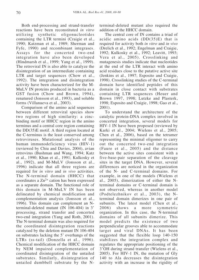
Bromoacetyl groups in the modifiedoligonucleotide are capable of crosslinkingto proteins mainly with appropriatelyoriented cysteine SH groups and histidine,although reactions with other amino acidshave also been reported (Hartman andBrown, 1976). This crosslinker is expectedto react at a distance of 6Å from the basewhen it is extended. Incubation of the [32P]-labeled-bromoacetyl dumbbell with amutant lacking the HHCC domain, IN 106-404 in the presence of Mn+2, resulted in arapid formation of a covalent complex ofprotein and nucleic acid, as determined bySDS-PAGE (Fig. 2). The reaction was overafter 5 min (lane 3). The size of apredominant band (50 kDa) matches theexpected molecular mass of a 1: 1 protein-nucleic acid complex. Under theseconditions, WT IN did not react (see resultsbelow).
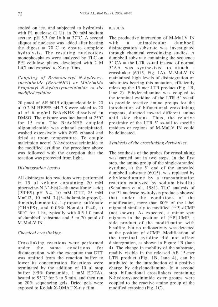
The specificity of the crosslinkingreaction was examined by determining theeffect of several unmodifiedoligonucleotides that are shown in Figure1A. As it is shown in Figure 3, thecrosslinking reaction was competed byunmodified dumbbell and oligonucleotidescontaining both a viral LTR and a targetsequence (4166 and 4167). Oligonucleotidescontaining the tailed (5467) or untailed(5527) LTR or the target sequence (4985)had no significant effect on the crosslinkingreaction. These results indicate thatsimultaneous binding of the LTR and targetsequences are required to displace thedumbbell substrate.
In order to eliminate the possibility ofnon-specific reaction of the bromoacetylgroups, crosslinking was performed in thepresence of lysine-quenched bromoacetylNHS. The addition of lysine-quenchedbromoacetyl NHS at concentrations up to50 times higher than the crosslinking probewas unable to prevent crosslinking,indicating that the reaction is dependent onDNA binding (not shown).
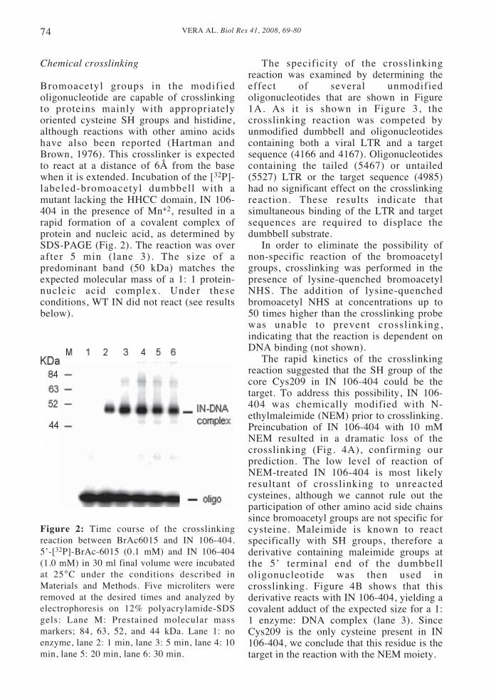
The rapid kinetics of the crosslinkingreaction suggested that the SH group of thecore Cys209 in IN 106-404 could be thetarget. To address this possibility, IN 106-404 was chemically modified with N-ethylmaleimide (NEM) prior to crosslinking.Preincubation of IN 106-404 with 10 mMNEM resulted in a dramatic loss of thecrosslinking (Fig. 4A), confirming ourprediction. The low level of reaction ofNEM-treated IN 106-404 is most likelyresultant of crosslinking to unreactedcysteines, although we cannot rule out theparticipation of other amino acid side chainssince bromoacetyl groups are not specific forcysteine. Maleimide is known to reactspecifically with SH groups, therefore aderivative containing maleimide groups atthe 5’ terminal end of the dumbbelloligonucleotide was then used incrosslinking. Figure 4B shows that thisderivative reacts with IN 106-404, yielding acovalent adduct of the expected size for a 1:1 enzyme: DNA complex (lane 3). SinceCys209 is the only cysteine present in IN106-404, we conclude that this residue is thetarget in the reaction with the NEM moiety.
Figure 2: Time course of the crosslinkingreaction between BrAc6015 and IN 106-404.5’-[32P]-BrAc-6015 (0.1 mM) and IN 106-404(1.0 mM) in 30 ml final volume were incubatedat 25°C under the conditions described inMaterials and Methods. Five microliters wereremoved at the desired times and analyzed byelectrophoresis on 12% polyacrylamide-SDSgels: Lane M: Prestained molecular massmarkers; 84, 63, 52, and 44 kDa. Lane 1: noenzyme, lane 2: 1 min, lane 3: 5 min, lane 4: 10min, lane 5: 20 min, lane 6: 30 min.
75VERA AL. Biol Res 41, 2008, 69-80
Figure 3: Competition of crosslinking between BrAc-6015 and IN 106-404 by DNAoligonucleotides containing target and LTR sequences. These experiments were carried out asdescribed in Fig. 2, except that unlabeled oligonucleotides at 1 mM were included in the reaction.The oligonucleotides used are described in Fig. 1A and are indicated by number. After 30 min, theproducts of the reaction were separated by electrophoresis on 12% acrylamide gels with SDS. Therelative amounts of the protein-DNA complex was determined in a phosphorimager.
Figure 4: Panel A. Effect of NEM on the crosslinking of BrAc6015 to IN 106-404. Crosslinkingwas carried out as described in Fig. 2. For NEM treatment, IN 106-404 was preincubated with 20mM NEM for 30 min on ice in the disintegration reaction buffer before adding the reactiveoligonucleotide. The products of the reaction were separated by electrophoresis on 12% acrylamidegels with SDS and quantified in a phosphoimager.Panel B. Crosslinking of NEM-Ac6015 to WT IN and IN 106-404. The proteins and NEM-Ac6015were incubated in the disintegration conditions. Lanes 1: no enzyme. Lane 2: WT IN. Lane 3: IN106-404. Positions of the molecular mass standards are marked on the left.
VERA AL. Biol Res 41, 2008, 69-8076
In contrast, no crosslinked product wasobserved in the presence of WT IN (lane 2),indicating that this cysteine is not availablefor reaction in the full-length enzyme. Oneexplanation for this result is that thecrosslinker side chain is sterically blockedand cannot assume the appropriateorientation to target the cysteine.
Photocrosslinking
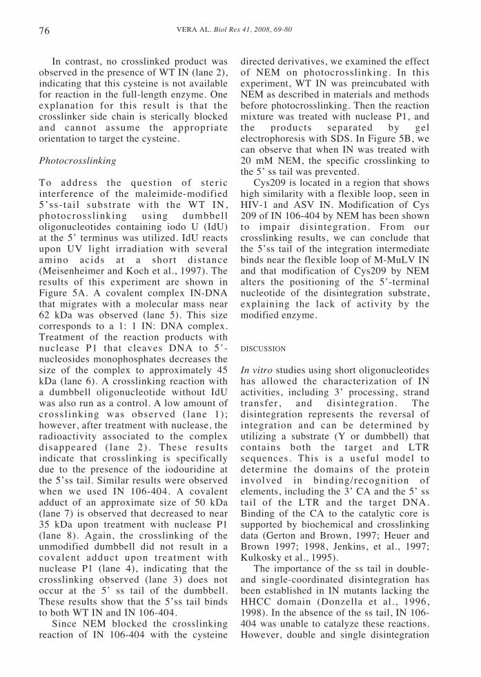
To address the quest ion of s ter icinterference of the maleimide-modified5’ss-tai l substrate with the WT IN,photocrossl inking using dumbbelloligonucleotides containing iodo U (IdU)at the 5’ terminus was utilized. IdU reactsupon UV light irradiation with severalamino acids at a short distance(Meisenheimer and Koch et al., 1997). Theresults of this experiment are shown inFigure 5A. A covalent complex IN-DNAthat migrates with a molecular mass near62 kDa was observed (lane 5). This sizecorresponds to a 1: 1 IN: DNA complex.Treatment of the reaction products withnuclease P1 that cleaves DNA to 5’-nucleosides monophosphates decreases thesize of the complex to approximately 45kDa (lane 6). A crosslinking reaction witha dumbbell oligonucleotide without IdUwas also run as a control. A low amount ofcrossl inking was observed ( lane 1);however, after treatment with nuclease, theradioactivity associated to the complexdisappeared ( lane 2) . These resultsindicate that crosslinking is specificallydue to the presence of the iodouridine atthe 5’ss tail. Similar results were observedwhen we used IN 106-404. A covalentadduct of an approximate size of 50 kDa(lane 7) is observed that decreased to near35 kDa upon treatment with nuclease P1(lane 8). Again, the crosslinking of theunmodified dumbbell did not result in acovalent adduct upon treatment withnuclease P1 (lane 4), indicating that thecrosslinking observed (lane 3) does notoccur at the 5’ ss tail of the dumbbell.These results show that the 5’ss tail bindsto both WT IN and IN 106-404.
Since NEM blocked the crosslinkingreaction of IN 106-404 with the cysteine
directed derivatives, we examined the effectof NEM on photocrosslinking. In thisexperiment, WT IN was preincubated withNEM as described in materials and methodsbefore photocrosslinking. Then the reactionmixture was treated with nuclease P1, andthe products separated by gelelectrophoresis with SDS. In Figure 5B, wecan observe that when IN was treated with20 mM NEM, the specific crosslinking tothe 5’ ss tail was prevented.
Cys209 is located in a region that showshigh similarity with a flexible loop, seen inHIV-1 and ASV IN. Modification of Cys209 of IN 106-404 by NEM has been shownto impair disintegration. From ourcrosslinking results, we can conclude thatthe 5’ss tail of the integration intermediatebinds near the flexible loop of M-MuLV INand that modification of Cys209 by NEMalters the positioning of the 5’-terminalnucleotide of the disintegration substrate,explaining the lack of activity by themodified enzyme.
DISCUSSION
In vitro studies using short oligonucleotideshas allowed the characterization of INactivities, including 3’ processing, strandtransfer, and disintegration. Thedisintegration represents the reversal ofintegration and can be determined byutilizing a substrate (Y or dumbbell) thatcontains both the target and LTRsequences. This is a useful model todetermine the domains of the proteininvolved in binding/recognition ofelements, including the 3’ CA and the 5’ sstail of the LTR and the target DNA.Binding of the CA to the catalytic core issupported by biochemical and crosslinkingdata (Gerton and Brown, 1997; Heuer andBrown 1997; 1998, Jenkins, et al., 1997;Kulkosky et al., 1995).
The importance of the ss tail in double-and single-coordinated disintegration hasbeen established in IN mutants lacking theHHCC domain (Donzella et al., 1996,1998). In the absence of the ss tail, IN 106-404 was unable to catalyze these reactions.However, double and single disintegration

77VERA AL. Biol Res 41, 2008, 69-80
Figure 5: Panel A. Photocrosslinking of dumbbell oligonucleotides to WT IN and IN 106-404. 5’-[32 P] 8161 (0.1 mM) and IN (1.0 mM) in 30 ml of disintegration buffer were irradiated at 300 nmfor 30 min. 15 ml samples were run on a 12% acrylamide gel with 0.1% SDS (lanes 1, 3, 5, and 7),15 ml of the samples was treated with 2 units of nuclease P1 in buffer 20 mM sodium acetate pH6.0, 1 mM zinc chloride for 16 h at 37°C (lanes 2, 4, 6, and 8). Lanes 1 to 4 show the crosslinkingwith the unmodified dumbbell substrate 6015 to wt IN (lanes 1 and 2) and to IN 106-404 (lanes 3and 4). Lanes 5 to 8 show the crosslinking of the IdU modified oligonucleotide to WT IN (lanes 5to 6) and IN 106-404 (lanes 7 and 8). Positions of the bands after nuclease digestion are indicatedby arrows.Panel B. Effect of NEM on crosslinking of WT IN with the IdU containing oligonucleotide.Photocrosslinking was carried out as described in panel A. To determine the effect of NEM, theenzyme was preincubated with 20 mM NEM on ice for 30 min and quenched with DTT, asdescribed in Methods. Samples were digested with 2 units of nuclease P1 for 16 hrs and subjectedto electrophoresis in a 12% polyacrylamide-SDS gel. Positions of the molecular mass standards aremarked on the left.
of untailed crossbones can be restored byadding the HHCC domain (IN 1-105),suggesting a functional relationshipbetween the ss-tail and the HHCC domain(Donzella et al., 1993). Disintegration ofthe untailed dumbbell requires both the coreand the HHCC domain. Modification of IN106-404 by NEM blocks complementationby the unmodified HHCC domain (Donzellaet al. 1998; Yang et al., 1999).
In this work, photo and chemicalcrosslinking studies were carried out to
determine the peptide regions near the ssDNA tail-binding site. In this approach, wesynthesized an oligonucleotide carrying anon-specific crosslinker capable of reactingwith several functional groups. Thisderivative reacted with IN 106-404 in arapid fashion, and the crosslinking reactionwas blocked by NEM, suggesting Cys209as the main target. This conclusion wasconfirmed by using a specific crosslinkeron the substrate directed towards cysteine,such as NEM.

VERA AL. Biol Res 41, 2008, 69-8078
A model for the structure of the regioncomprising residues P178 to S238 of M-MuLV IN (residues Val110 to Glu170 inHIV-1 IN, PDB, 1BL3) was generatedusing the program Swiss Model Prot (Fig.6). Cys 209 is located in a region called theflexible loop (His 208-Ser 216). The role ofthis region in the catalytic activity of IN isnot clear, however, it has been suggestedthat it is involved in the regulation of thestrand transfer step during concertedintegration (Wielens et al., 2005; Chen etal. , 2006). Photocrosslinking studiesbetween HIV-1 and an LTR substrate locatethe 5’ end of the non-processed strand closeto the residues Tyr143 and Gln148(Esposito and Craigie, 1998). Johnson et al.(2006) proposed that in HIV-1, a specificinteraction takes place between the secondC located at the 5’end of the unprocessedstrand of the LTR and Gln 148. Chen et al.,(2006) have proposed that the flexible loopcould be involved in regulating binding ofthe target DNA. The results presented inthis work indicate that in M-MuLV IN, theterminal base of the 5’ ss tail localizes
within 6 Å of Cys 209, suggesting aninteraction between the flexible loop andthe 5’ single-strand region. The lack ofreactivity of Cys209 in WT IN to thecysteine directed crosslinkers could beexplained by a modification of theenvironment of this residue in the presenceof the HHCC domain.
Alternatively, the orientation of thecrosslinker reactive groups in the assembledDNA-enzyme complex could be altered in thefull-length IN. However, photocrosslinkingoccurs since DNA binding is not affected.Photocrosslinking studies reported in thiswork indicate that the 5’ ss tail of thedumbbell oligonucleotide has access to thecore and that modification of Cys 209 byNEM blocked photocrosslinking at the 5’ sstail. Whether this effect is due to a directsteric interference of the 5’ ss-tail binding orto a conformational change of the flexibleloop remains to be defined.
Collectively, the results presented herebyindicate that the flexible loop of the coredomain of M-MuLV IN would be involvedin positioning of the ss-DNA tail. These
Figure 6: Ribbon structures of a predicted model of catalytic domain of M-MuLV IN (right),comprising residues P178 to S238, in comparison with the structure of the catalytic domain of HIV-1 IN (left), comprising residues V110 to E170, (PDB format: 1BL3), generated by Swiss ModelProt Program. Energy Minimization was done by the program MODELLER. The residues of theputative active site (D116 and E152 in HIV-1 IN and D184 and E220 in M-MuLV IN) and residuesof the loop (I141, Y143, N148 in HIV-IN and H208, C209, Y211 and S215 in M-MuLV IN) arelabeled to show the proximity and spatial relationship to each other and some more distant residues(K159 in HIV-1 IN and I226 in M-MuLV IN).

79VERA AL. Biol Res 41, 2008, 69-80
observations agree with the suggestion thatthis loop may have an important role in theseparation of both LTRs through binding ofthe ss tail during concerted integration andstabilization of the preintegration complexes.Mutation of His 208 to Ala in M-MuLV INyields a phenotype that is similar to the druginhibition pattern seen in HIV-1 IN (King etal., 2003), since strand transfer anddisintegration were completely abolished,although processing was reduced by 90% (O.Leon, unpublished results). This resultssuggest that 5’ss tail could bind to IN indifferent contexts throughout the catalysisinvolving conformational changes of theflexible loop. A more systematic study of theflexible loop in M-MuLV is currentlyunderway in our laboratory.
ACKNOWLEDGEMENTS
This work was funded by grants Fondecyt1040409 (O.L.) and National Institutes ofHealth RO1 GM070837 (M.J.R.)
REFERENCES
ASANTE-APPIAH E, SKALKA AM (1999) HIV-1integrase: Structural organization conformationalchanges and catalysis. Adv Virus Res 52: 351-69
BUSHMAN FD, WANG B (1994) Rous sarcoma virusintegrase protein: Mapping functions for catalysis andsubstrate binding. J Virol 68: 2215-23
CHEN A, WEBER IT, HARRISON RW, LEIS J (2006)Identification of amino acids in HIV-1 and aviansarcoma virus integrase subsites required for specificrecognition of the long terminal repeat ends. J BiolChem 281: 4173-82
CHIU TK, DAVIES DR (2004) Structure and function ofHIV-1 integrase. Curr Top Med Chem 4: 965-77
CHOW SA, BROWN PO (1994) Juxtaposition of two viralDNA ends in a bimolecular disintegration reactionmediated by multimers of human immunodeficiencyvirus type 1 or murine leukemia virus integrase. J Virol68: 7869-78
CHOW SA, VINCENT KA, ELLISON V, BROWN PO(1992) Reversal of integration and DNA splicingmediated by integrase of human immunodeficiencyvirus. Science 255: 723-6
CRAIGIE R (2001) HIV integrase, A brief overview fromchemistry to therapeutics. J Biol Chem 276: 23213-6
CRAIGIE R, FUJIWARA T, BUSHMAN F (1990) The INprotein of Moloney murine leukemia virus processesthe viral DNA ends and accomplishes their integrationin vitro. Cell 62: 829-37
DONZELLA GA, JONSSON CB, ROTH MJ (1996)Coordinated disintegration reactions mediated byMoloney murine leukemia virus integrase. J Virol 70:3909-21
DONZELLA GA, JONSSON CB, ROTH MJ (1993)Influence of substrate structure on disintegrationactivity of Moloney murine leukemia virus integrase. JVirol 67: 7077-87
DONZELLA GA, LEÓN O, ROTH MJ (1998) Implicationof a central cysteine residue and the HHCC domain ofMoloney murine leukemia virus integrase protein infunctional multimerization. J Virol 72: 1691-8
DRELICH M, WILHELM R, MOUS J (1992) Identificationof amino acid residues critical for endonuclease andintegration activities of HIV-1 IN protein in vitro.Virology 188: 459-68
DYDA F, HICKMAN AB, JENKINS TM, ENGELMAN A,CRAIGIE R, DAVIES DR (1994) Crystal structure ofthe catalytic domain of HIV-1 integrase: Similarity toother polynucleotidyl transferases. Science 266: 1981-6
ELLISON V, BROWN PO (1994) A stable complexbetween integrase and viral DNA ends mediates humanimmunodeficiency virus integration in vitro. Proc NatlAcad Sci U S A 91: 7316-20
ENGELMAN A, CRAIGIE R (1992) Identification ofconserved amino acid residues critical for humanimmunodeficiency virus type-1 integrase function invitro. J Virol 66: 6361-9
ENGELMAN A HICKMAN AB, CRAIGIE R (1994) Thecore and carboxyl-terminal domains of the integraseprotein of human immunodeficiency virus type-1 eachcontribute to nonspecific DNA binding. J Virol 68:5911-7
ENGELMAN A MIZUUCHI K, CRAIGIE R (1991) HIV-1DNA integration: Mechanism of viral DNA cleavageand DNA strand transfer. Cell 67: 1211-21
ESPOSITO D, CRAIGIE R (1998) Sequence specificity ofviral end DNA binding by HIV-1 integrase revealscritical regions for protein-DNA interaction. EMBO J17: 5832-43
FAURE A, CALMELS C, DESJOBERT C,CASTROVIEJO M, CAUMONT-SARCOS A,TARRAGO-LITVAK L, LITVAK S, PARISSI V(2005) HIV-1 integrase crosslinked oligomers areactive in vitro. Nucleic Acids Res 33: 977-86
GAO K, BUTLER S L, AND BUSHMAN F (2001) Humanimmunodeficiency virus type-1 integrase: Arrangementof protein domains in active cDNA complexes. EMBOJ 20: 3565-76
GERTON JL, BROWN PO (1997) The core domain ofHIV-1 integrase recognizes key features of its DNAsubstrates. J Biol Chem 272: 25809-15
GOLDGUR Y, CRAIGIE R, COHEN GH, FUJIWARA T,YOSHINAGA T, FUJISHITA T, SUGIMOTO H,ENDO T, MURAI H, DAVIES DR (1999) Structure ofthe HIV-1 integrase catalytic domain complexed withan inhibitor: A platform for antiviral drug design. ProcNatl Acad Sci USA 96: 13040-3
GREENWALD J, BUTLER SL, BUSHMAN FD, CHOE S(1999) The mobility of an HIV-1 integrase active siteloop is correlated with catalytic activity. Biochemistry38: 8892-8
HARTMAN FC, BROWN J P (1976) Affinity labeling of apreviously undetected essential lysyl residue in class-Ifructose bisphosphate aldolase. J Biol Chem 251: 3057-62
HEUER TS, BROWN PO (1997) Mapping features of HIV-1 integrase near selected sites on viral and target DNAmolecules in an active enzyme-DNA complex byphotocrosslinking. Biochemistry 36: 10655-65
HEUER TS, BROWN PO (1998) Photocrosslinking studiessuggest a model for the architecture of an active humanimmunodeficiency virus type-1 integrase DNAcomplex. Biochemistry 37: 6667-78

VERA AL. Biol Res 41, 2008, 69-8080
HINDMARSH P, LEIS J (1999) Retroviral DNAintegration. Microbiol Mol Biol Rev 63: 836-43
HINDMARSH P, RIDKY T, REEVES R, ANDRAKE M,SKALKA AM, LEIS J (1999) HMG protein familymembers stimulate human immunodeficiency virustype-1 and avian sarcoma virus concerted DNAintegration in vitro. J Virol 73: 2994-3003
JENKINS TM, ESPOSITO D, ENGELMAN A, CRAIGIER (1997) Critical contacts between HIV-1 integraseand viral DNA identified by structure-based analysisand photocrosslinking. EMBO J 16: 6849-59
JOHNSON AA, SANTOS W, PAIS GC, MARCHAND C,AMIN R, BURKE TR, JR, VERDINE G, POMMIER Y(2006) Integration requires a specific interaction of thedonor DNA terminal 5’-cytosine with glutamine 148 ofthe HIV-1 integrase flexible loop. J Biol Chem 281:461-7
JOHNSON MS, MCCLURE MA, FENG DF, GRAY J,DOOLITTLE RF (1986) Computer analysis ofretroviral pol genes: Assignment of enzymaticfunctions to specific sequences and homologies withnonviral enzymes. Proc Natl Acad Sci USA 83: 7648-52
JONSSON C B, DONZELLA G A, GAUCAN E, SMITHCM, ROTH MJ (1996) Functional domains of Moloneymurine leukemia virus integrase defined by mutationand complementation analysis. J Virol 70: 4585-97
JONSSON CB, DONZELLA GA, ROTH MJ (1993)Characterization of the forward and reverse integrationreactions of the Moloney murine leukemia virusintegrase protein purified from Escherichia coli. J BiolChem 268: 1462-9
JONSSON CB, ROTH MJ (1993) Role of the His-Cysfinger of Moloney murine leukemia virus integraseprotein in integration and disintegration. J Virol 67:5562-71
KARKI R, TANG GY, BURKE TR, JR, NICKLAUS MC(2004) Model of full- length HIV-1 integrasecomplexed with viral DNA as template for anti-HIVdrug design. J Comput Aided Mol Des 18: 739-60
KATZ RA, MERKEL G, KULKOSKY J, LEIS J, SKALKAAM (1990) The avian retroviral IN protein is bothnecessary and sufficient for integrative recombinationin vitro. Cell 63: 87-95
KATZMAN M, KATZ RA, SKALKA AM, LEIS J (1989)The avian retroviral integration protein cleaves theterminal sequences of linear viral DNA at the in vivosites of integration. J Virol 63: 5319-27
KHAN E, MACK JP, KATZ RA, KULKOSKY J,SKALKA AM (1991) Retroviral integrase domains:DNA binding and the recognition of LTR sequences.Nucleic Acids Res 19: 851-60
KING PJ, LEE DJ, REINKE RA, VICTORIA JG, BEALEK, ROBINSON WE, JR, (2003) Humanimmunodeficiency virus type-1 integrase containing aglycine to serine mutation at position 140 is attenuatedfor catalysis and resistant to integrase inhibitors.Virology 306: 147-61
KULKOSKY J, JONES KS, KATZ RA, MACK JP,SKALKA AM (1992) Residues critical for retroviral
integrative recombination in a region that is highlyconserved among retroviral/retrotransposon integrasesand bacterial insertion sequence transposases. Mol CellBiol 12: 2331-8
KULKOSKY J, KATZ RA, MERKEL G, SKALKA AM(1995) Activities and substrate specificity of theevolutionarily conserved central domain of retroviralintegrase. Virology 206: 448-56
LEAVITT A, SHIUE DL, VARMUS HE (1993) Site-directed mutagenesis of HIV-1 integrase demonstratesdifferential effects on integrase functions in vitro. JBiol Chem 268: 2113-9
LUTZKE RA, PLASTERK R H (1998) Structure-basedmutational analysis of the C-terminal DNA-bindingdomain of human immunodeficiency virus type-1integrase: Critical residues for protein oligomerizationand DNA binding. J Virol 72: 4841-8
MEISENHEIMER KM, KOCH TH (1997)Photocrosslinking of nucleic acids to associatedproteins. Crit Rev Biochem Mol Biol 32: 101-40
PODTELEZHNIKOV AA, GAO K, BUSHMAN FD,MCCAMMON JA (2003) Modeling HIV-1 integrasecomplexes based on their hydrodynamic properties.Biopolymers 68: 110-20
SCHULMAN LH, VALENZUELA D, PELKA H (1981)Reversible inactivation of Escherichia coli methionyl-tRNA synthetase by covalent at tachment offormylmethionine tRNA to the tRNA binding site witha cleavable crosslinker. Biochemistry 20: 6018-23
SHERMAN PA, FYFE, JA (1990) Humanimmunodeficiency virus integration protein expressedin Escherichia coli possesses selective DNA cleavingactivity. Proc Natl Acad Sci USA 87: 5119-23
VERA J, PARISSI V, GARCÍA A, ZÚÑIGA R,ANDREOLA ML, CAUMONT-SARCOS A,TARRAGO-LITVAK L, LEÓN O (2005) Yeast systemas a model to study Moloney murine leukemia virusintegrase: Expression mutagenesis and search foreukaryotic partners. J Gen Virol 86: 2481-8
VILLANUEVA RA, JONSSON CB, JONES J,GEORGIADIS MM, ROTH MJ
(2003) Differential multimerization of Moloney murineleukemia virus integrase purified under nondenaturingconditions. Virology 316: 146-160
VINK C, LUTZKE RA, PLASTERK RH (1994) Formationof a stable complex between the humanimmunodeficiency virus integrase protein and viralDNA. Nucleic Acids Res 22: 4103-10
WIELENS J, CROSBY IT, CHALMERS DK (2005) Athree-dimensional model of the humanimmunodeficiency virus type 1 integration complex. JComput Aided Mol Des 19: 301-17
YANG F, LEÓN O, GREENFIELD NJ, ROTH MJ (1999)Functional interactions of the HHCC domain ofmoloney murine leukemia virus integrase revealed bynonoverlapping complementation and zinc-dependentdimerization. J Virol 73: 1809-17
YANG F, ROTH MJ (2001) Assembly and catalysis ofconcerted two-end integration events by Moloneymurine leukemia virus integrase. J Virol 75: 9561-70