Characterization of the FHA domain involved in ...
77
Characterization of the FHA domain involved in Saccharomyces cerevisiae dNTP regulation by Geburah Straker A thesis presented to the University of Waterloo in fulfillment of the thesis requirement for the degree of Master of Science in Biology Waterloo, Ontario, Canada, 2018 ©Geburah Straker 2018
Transcript of Characterization of the FHA domain involved in ...

Characterization of the FHA domain
involved in Saccharomyces cerevisiae
dNTP regulation
by
Geburah Straker
A thesis
presented to the University of Waterloo
in fulfillment of the
thesis requirement for the degree of
Master of Science
in
Biology
Waterloo, Ontario, Canada, 2018
©Geburah Straker 2018

ii
AUTHOR'S DECLARATION
I hereby declare that I am the sole author of this thesis. This is a true copy of the thesis, including
any required final revisions, as accepted by my examiners.
I understand that my thesis may be made electronically available to the public.

iii
Abstract
Over 2000 Forkhead-associated (FHA) domain-containing proteins exhibiting diverse functions,
such as kinases, phosphatases, and transcription factors, have been identified to date in both eukaryotic
and prokaryotic organisms. Initially characterized as the only known protein-protein interaction motif
with phosphothreonine (pThr)-binding specificity, research from the Duncker Lab that characterized the
minimal interaction surfaces between the Rad53 FHA1 domain and the Dbf4 H-BRCT domain in the
model organism Saccharomyces cerevisiae demonstrated the existence and importance of a conserved
non-canonical binding surface on Rad53 FHA1 that does not rely on the phosphothreonine-binding patch.
Recent analysis by the Duncker Lab of a Rad53 paralog called DNA damage UNinducible (Dun1), a
budding yeast cell cycle checkpoint kinase involved in regulating dNTP synthesis, identified another
instance of a conserved non-canonical FHA domain lateral surface interaction patch, located on the Dun1
FHA domain, similar to that of Rad53 FHA1. Continued examination of the Dun1 FHA domain lateral
surface interaction patch and pThr-binding site suggested the existence of a differential requirement for
the non-canonical FHA domain lateral surface interaction patch and the canonical pThr-binding site
during interactions between Dun1 and some of its ligands.
The research presented in this thesis aimed to study the prevalence of protein-protein interactions
in S. cerevisiae that operate using this novel non-canonical lateral surface interaction patch of FHA
domains as well as their functional significance in cell growth and survival mechanisms in response to
genotoxic stress, using the Dun1 FHA domain as an example. In order to investigate the existence and
importance of Dun1 non-canonical FHA domain-based protein-protein interactions, bioinformatics
analysis was used to identify candidate conserved residues on the FHA domain lateral surface interaction
patch, site-directed mutagenesis was used to alter select amino acids and yeast two-hybrid assays were
used to compare disruptions and/or the abrogation of protein-protein interactions between wild type and

iv
mutant Dun1 FHA domains with ligands involved in dNTP regulation. Analysis of the interaction
between the FHA domain of Dun1 and proteins involved in the dNTP regulation pathway illustrated a
differential requirement of the FHA domain lateral surface interaction patch and the pThr-binding site as
well as a contribution of the kinase domain for the establishment of at least two interactions. Highly
conserved residues of the Dun1 FHA domain lateral surface interaction patch contributed to FHA
domain-based protein-protein interactions and slight but reproducible genotoxic sensitivity was observed
for Dun1 FHA domain mutants. The interaction between Dun1 and Damage-regulated Import Facilitator
(Dif1) suggested a contribution of the kinase activity of the Dun1 kinase domain to the establishment of a
maximal interaction. In order to observe any interaction between Dun1 and Suppressor of mec1 lethality
(Sml1), both the FHA and kinase domains needed to be present. Mutation of the conserved arginine 60
(R60A) residue of the canonical pThr-binding site completely disrupted the interaction between Dun1 and
Dif1 whereas it only weakened the interaction between Dun1 and Sml1. Single mutations of the
conserved asparagine 121 (N121A) and leucine 134 (L134A) of the non-canonical FHA domain lateral
surface interaction patch did not affect the interaction between Dun1 and Dif1, but mutation of the
conserved lysine 136 (K136A) within the context of just the Dun1 FHA domain increased the strength of
the interaction between Dun1 and Dif1. The N121A single mutation had no affect on the interaction
between Dun1 and Sml1 while both the L134A and K136A single mutations decreased the strength of the
interaction between Dun1 and Sml1. The R60A, N121A, L134A, and K136A single mutants showed
decreased growth in response to genotoxic stress, illustrating the importance of Dun1 interactions that
utilize conserved residues of the non-canonical FHA domain lateral surface interaction patch and the
canonical pThr-binding site to genotoxic stress responses in budding yeast.

v
Acknowledgements
I would like to thank my supervisor, Dr. Bernard Duncker, for giving me the opportunity to work
in his lab. As a supervisor, his expectations give his students the ability to learn how to work efficiently
and successfully while also having fun with their research. I would like to thank my committee members
Dr. Brendan McConkey and Dr. Christine Dupont. Thank you for all of your guidance and suggestions.
You have both been extremely easy to work with, especially for scheduling committee meetings, and I
greatly appreciate your interest in my work. I would also like to thank all of the faculty and staff of the
Department of Biology who have in any way contributed to my success as a student here at the University
of Waterloo.
Thank you to all of the Duncker lab members for being both accommodating and helpful. I would
not have learned as much as I have without all of you. Thank you for keeping me 'on my toes' and for
making every day interesting. I wish everyone the best of luck in their future endeavors. Special thanks to
Darryl Jones, Damir Mingaliev, Aaron Robertson, and Alison Guitor who have all made contributions to
the topic of FHA domain research in the Duncker Lab.
Above all, I'd like to thank my parents for all of the support and guidance that has led me to
where I am today and where I will be tomorrow.

vi
Table of Contents
Author’s Declaration ................................................................................................................. ii
Abstract.....................................................................................................................................iii
Acknowledgements ................................................................................................................... v
Table of Contents ..................................................................................................................... vi
List of Figures ........................................................................................................................ viii
List of Tables ........................................................................................................................... ix
List of Abbreviations ................................................................................................................ x
Chapter 1: Introduction and Research Objectives .................................................................... 1
1.1 Saccharomyces cerevisiae and the Cell Cycle ................................................................ 1
1.1.1 Budding Yeast Model Organism .............................................................................. 1
1.1.2 The Cell Cycle .......................................................................................................... 3
1.1.3 Cell Cycle Checkpoints ............................................................................................ 7
1.2 Forkhead Associated Domains ........................................................................................ 9
1.2.1 FHA Domain Discovery and Characteristics ........................................................... 9
1.2.2 Non-canonical FHA Domain Lateral Surface Interaction Patch Identification and
Characterization ............................................................................................................... 10
1.3 DNA damage UNinducible (DUN1) ............................................................................. 11
1.4 Research Objectives ...................................................................................................... 16
Chapter 2: Materials and Methods .......................................................................................... 18
2.1 Yeast Strains .................................................................................................................. 18
2.2 Genomic DNA Isolation................................................................................................ 18
2.3 Bioinformatics Analysis ................................................................................................ 19
2.4 Site-directed Mutagenesis ............................................................................................. 21
2.5 Plasmid Construction .................................................................................................... 21
2.6 Yeast Transformation .................................................................................................... 24
2.7 Yeast two-Hybrid Assay ............................................................................................... 24
2.8 Yeast Whole Cell Extract and Western Blotting ........................................................... 26
2.9 Spot Plate Assay ............................................................................................................ 27

vii
2.10 Statistical Analysis ...................................................................................................... 28
Chapter 3: Dun1-Dif1 and Dun1-Sml1 interactions utilize the conserved non-canonical FHA
domain lateral surface interaction patch ................................................................................. 29
3.1 Introduction ................................................................................................................... 29
3.2 Results ........................................................................................................................... 30
3.2.1 dNTP regulation pathway interactions exhibit different requirements...................30
3.2.2 Highly conserved residues of the pThr-binidng site and lateral surface interaction
patch influence detectable genomic level protein expression ......................................... 38
3.2.3 K136-central projection of the Dun1 non-canonical FHA domain lateral surface
interaction patch contributes to interactions involved in dNTP regulation ..................... 49
3.3 Discussion ..................................................................................................................... 52
Chapter 4: General Conclusions and Future Directions ......................................................... 57
4.1 The Dun1 kinase domain and the non-canonical FHA domain lateral surface interaction
patch influence protein-protein interactions involved in dNTP regulation ......................... 57
4.2 Future Directions ........................................................................................................... 58
4.3 Impact and Relevance of FHA Domain Research ........................................................ 60
Bibliography ........................................................................................................................... 62

viii
List of Figures
Figure 1.1: Eukaryotic Cell Cycle and Saccharomyces cerevisiae ........................................... 5
Figure 1.2: Cyclin-CDK Complexes and S. cerevisiae Cell Cycle Phase Progression ............ 7
Figure 1.3: Structured Regions and Positions of Dun1........................................................... 12
Figure 1.4: Dun1 and the dNTP Regulation Pathway............................................................. 13
Figure 2.1: Schematic Diagram of the Yeast two-hybrid Assay ............................................ 25
Figure 3.1: MUSCLE Multiple Sequence Alignment of Dun1 Homologs ............................. 31
Figure 3.2: Conservation Mapping of the Dun1 FHA Domain .............................................. 32
Figure 3.3: Analysis of the Interaction Between Dun1 and the Rad53, Wtm1, and Crt1 Ligands
................................................................................................................................................. 34
Figure 3.4: Analysis of the Interaction Between Dun1 and the Sml1 and Dif1 Ligands ....... 36
Figure 3.5: Conserved Surface Residues of the Dun1 FHA Domain Canonical pThr-binding Site
and Non-canonical FHA Domain Lateral Surface Interaction Patch ...................................... 38
Figure 3.6: Analysis of the Interaction Between Dun1 FHA Domain Mutants and Dif1 ...... 40
Figure 3.7: Dun1 FHA - Dif1 Interaction Analysis for the N108A and K136A Mutations ... 42
Figure 3.8: Analysis of the K136A Mutation in Full-length Dun1 and the Dun1 FHA Domain
................................................................................................................................................. 43
Figure 3.9: Functional Complementation of dun1Δ and Genotoxic Sensitivity Assessment for
Dun1 FHA Mutants................................................................................................................. 45
Figure 3.10: Assessment of Genotoxic Sensitivity with Increased Temperature or Other
Genotoxins .............................................................................................................................. 48
Figure 3.11: Illustration of the K136-central Projection on the Dun1 FHA Domain Lateral
Surface Interaction Patch ........................................................................................................ 49
Figure 3.12: Analysis of the K136-central Projection Substitution Mutants for the Dun1 - Dif1
and Dun1 - Sml1 Interactions ................................................................................................. 51
Figure 3.13: Genotoxic Sensitivity Analysis for K136-central Projection Mutants ............... 52

ix
List of Tables
Table 2.1: List of Yeast Strains............................................................................................... 18
Table 2.2: Protein BLAST Query for Dun1 Homologs .......................................................... 20
Table 2.3: List of Antibodies .................................................................................................. 27
Table 2.4: List of Genotoxic Agents ....................................................................................... 28
x
List of Abbreviations
Cdc: Cell division cycle
CDK: Cyclin dependent kinase
DDK: Dbf4 dependent kinase
DNA: Deoxyribonucleic acid
dNTP: Deoxyribonucleoside triphosphate
DTT: Dithiothreitol
EDTA: Ethylenediaminetetraacetic acid
FHA: Forkhead-associated
FL: Full-length
GAL/RAF: Galactose/Raffinose
HA: Hemagglutinin
H-BRCT: Helix-BRCA 1 Carboxy-terminal
HU: Hydroxyurea
Kb: kilobases
kDa: kilodalton
MMS: Methyl methanesulfonate
MYC: Myelocytamatosis
ONPG: 2-Nitrophenyl-β-D-galactopyranoside
PCR: Polymerase chain reaction
PMSF: Phenylmethylsulphonyl fluoride
RNR: Ribonucleotide reductase
SC: Synthetic complete
SDS: Sodium dodecyl sulfate
WCE: Whole cell extract
WT: Wild-type
YPD: Yeast extract, peptone, dextrose

1
Chapter 1
Introduction and Research Objectives
1.1 Saccharomyces cerevisiae and the Cell Cycle
Model organisms are and have consistently been fundamental tools for studying biological
processes, as proxies for studying the same or similar processes in higher eukaryotes. The most effective
model organisms are those that are easy to grow, control, manipulate and maintain. One model organism
prominent for its use in studying the molecular biology of the cell cycle and cell cycle checkpoints,
largely contributing to the breadth of knowledge acquired under the 'umbrella' of cancer research, are
yeasts.
1.1.1 Budding Yeast Model Organism
The taxonomic definition of yeast, of which there are thousands of species, is that yeasts are
single-celled eukaryotic micro-organisms of the fungus kingdom that can grow and function with or
without oxygen (reviewed in Mortimer, 2000). These facultative anaerobes utilize fermentation to break
down sugars into carbon dioxide and ethanol (reviewed in Mortimer, 2000). In general, yeasts are easy to
grow and maintain, with a typical doubling time of approximately 90 minutes under an optimal
laboratory-constructed environment of sugar and nitrogen-based media, and they are easy to manipulate
physically and genetically (reviewed in Duina et al., 2014, Gershon and Gershon, 2000, Herskowitz,
1988, and Mortimer and Johnston, 1986). Saccharomyces cerevisiae, budding yeast, is one of two main
species of yeast used extensively for cell cycle research, and was used throughout the research presented
in this thesis (reviewed in Gershon and Gershon, 2000). The 'budding yeast' common name describes a
distinguishing characteristic of this yeast where cell proliferation is achieved through an asymmetrical

2
division of a 'mother cell' generating a 'bud' that pinches off from the 'mother cell' (reviewed in Duina et
al., 2014).
Budding yeast exhibit many of the features that make a model organism useful. In addition to
being unicellular with a relatively short generation time, the relatively small genome of budding yeast has
been completely sequenced and extensively mapped at just over 12, 000 kilo bases (Kb) organized as 16
chromosomes in the haploid genome (Goffeau et al., 1996; reviewed in Gershon and Gershon, 2000).
Budding yeast characteristics have also been useful in the design of a wide variety of molecular biology
tools for studying genetics, cell biology and the cell cycle (reviewed in Gershon and Gershon, 2000). The
intricate control over cell cycle progression and the ability to switch between mitosis and meiosis have
allowed for the development of tools such as the ability to add chemicals or mating factors to a cell
culture to stall cell cycle progression in order to synchronize progression for a culture of cells as well as
generating and maintaining cells in a haploid state to be studied (reviewed in Gershon and Gershon,
2000). The mechanisms of genetic recombination in budding yeast have been exploited to develop a large
collection of genomic knockout yeast strains used by many yeast researchers (reviewed in Gershon and
Gershon, 2000). Additionally, the budding yeast genome contains a large number of orthologs to human
disease-causing genes and genes involved in similar, if not conserved, biological pathways (reviewed in
Gershon and Gershon, 2000).
Budding yeast can exist as any of three possible cell types: (1) Mating type a (MATa) haploid
cells, (2) Mating type α (MATα) haploid cells, or (3) Mating type a/α (MATa/α) diploid cells (reviewed in
Herskowitz, 1988 and Duina et al., 2014). Located on chromosome III, the MAT locus will have one of
the two nonhomologous MAT alleles, either MATa or MATα, containing the genetic information that
determines the mating type (reviewed in Duina et al., 2014). The two haploid cells each secrete mating
type-specific peptides as mating factors, mating pheromones, which allow the MATa and MATα haploid
cells to mate with each other to form the MATa/α diploid (reviewed in Herskowitz, 1988 and Duina et al.,
2014). MATa and MATα haploid cells undergo mitosis for cellular proliferation or fuse to form a MATa/α

3
diploid cell which also undergo mitosis for cellular proliferation or meiosis for gamete production during
times of nutrient starvation (reviewed in Herskowitz, 1988 and Duina et al., 2014).
Both heterothallic and homothallic yeast strains were found on rotting figs during the late 1800s -
early 1900s and were used by yeast geneticists, Ojvind Winge and Carl Lindergren, to pioneer the use of
yeast for research (Mortimer and Johnston, 1986; reviewed in Mortimer, 2000 and Duina et al., 2014).
Heterothallism refers to the inability of a haploid yeast cell to switch mating types due to a lack of
functional HO endonuclease, encoded by the HO ("Homothallism") gene, which is responsible for the
transposition of an HMLα or HMRa genetic cassette from a location where it is silenced to the location of
the MAT locus where it will be expressed (Mortimer and Johnston, 1986; reviewed in Herskowitz, 1988,
Mortimer, 2000 and Duina et al., 2014). Homothallic cells, for which the HO gene and endonuclease were
named, are able to switch mating types because they do have functional HO endonuclease (reviewed in
Herskowitz, 1988 and Duina et al., 2014). The HMLα and HMRa genetic cassettes contain the same
genetic information expressed at the MATα and MATa loci respectively (reviewed in Herskowitz, 1988).
Most yeast strains used for research, including the research presented in this thesis, were derived from a
heterothallic yeast strain isolated by a pioneer of yeast genetics, Emil Mrak in 1891 (reviewed in
Mortimer and Johnston, 1986 and Duina et al., 2014).
1.1.2 The Cell Cycle
The cell cycle refers to the progression of a cell through stages of growth, replication, and
division. Typical eukaryotic cells exhibit a consistent pattern of progression through the cell cycle;
however the precise steps that occur in each stage may differ among organisms (reviewed in Herskowitz,
1988). That consistent pattern of cell cycle progression entails a Gap 1 (G1), Synthesis (S), Gap 2 (G2),
and mitosis phase (Figure 1.1). During G1 phase, cells exhibit continual growth to prepare for ensuing
deoxyribonucleic acid (DNA) replication during the S phase (reviewed in Herskowitz, 1988).
Additionally during G1, eukaryotic cells regulate the association of multiple DNA replication initiation
4
factors at sites of DNA replication initiation referred to as origins of replication in a process called origin
licensing (Shirahige et al., 1998; Duncker et al., 2002; reviewed in Matthews et al., 2012). The G0 phase
represents a quiescent stage of the cell cycle where cells in G1 phase can rest if conditions are not optimal
for commitment to DNA replication and cellular division such as poor nutrient availability, present
compatible mating pheromones, or inadequate cell size (reviewed in Herskowitz, 1988; Figure 1.1). The
START point of the cell cycle, highly regulated and influenced by nutrient availability, presence of
compatible mating pheromones, and cell size, represents the point during late G1 phase when cells commit
to entrance into S phase (reviewed in Hiroshima et al., 2013 and Herskowitz, 1988; Figure 1.1). The S
phase represents the stage of the cell cycle during which DNA is replicated via semi-conservative
replication at 'licensed' origins of replication which have accumulated all of the required replication
initiation factors (reviewed in Hiroshima et al., 2013). The G2 phase is a secondary period of growth for
most eukaryotic cells during which they truly prepare to separate all duplicated cellular components in
addition to dividing replicated DNA (reviewed in Herskowitz, 1988). The M phase primarily features the
physical segregation of replicated genetic material over the duration of four stages: Prophase, Metaphase,
Anaphase, and Telophase which can be mostly identified by the physical appearance of the DNA and
cellular components (reviewed in Herskowitz, 1988). Typically, the actual physical separation of the
resulting daughter cell(s) and their cytoplasm occurs after the completion of mitosis, and is referred to as
Cytokinesis (reviewed in Herskowitz, 1988). In eukaryotic cells, mitosis features the segregation of
genetic content to opposite poles of the parent cell via the spindle microtubules (reviewed in
Balasubramanian et al., 2004). The formation of a contractile actomyosin ring at the 'division plane'
between the two poles orchestrates the cytoplasmic separation seen during cytokinesis, and continual
contraction of the actin ring pinches the cytoplasm between the two poles until two distinct daughter cells
are formed (reviewed in Balasubramanian et al., 2004).

5
Figure 1.1: Eukaryotic Cell Cycle and Saccharomyces cerevisiae
Depiction of the mitotic cell cycle illustrating the order and approximate duration of each phase of the cell
cycle: Gap1 (G1), Synthesis (S), Gap 2 (G2), and Mitosis (M) as well as bud formation from the mother
cell generating the smaller daughter cell. Resting stage (G0) refers to a quiescent stage during which the
cell cycle is arrested. Pink and yellow circles represent yeast cell(s) and nuclei, respectively. The solid
black line indicates the START point of S phase entry commitment (adapted from Herskowitz, 1988).
In contrast to the general eukaryotic mitotic cell cycle, the budding yeast cell cycle does not
feature an actual G2 phase due to the formation of the mitotic spindle during S phase (Gershon and
Gershon, 2000; Figure 1.2). Additionally, since bud formation (shown in Figure 1.1) results in a daughter
cell that is smaller than the mother cell, the duration of the G1 phase is extended to compensate for the
additional time required for the daughter cell to grow to its 'adult' size in preparation for its own entrance
into cell division and in order to get past the START checkpoint (Gershon and Gershon, 2000). Unlike
animal cells, budding yeast cells possess a cell wall and do not exhibit nuclear envelope breakdown

6
(reviewed in Balasubramanian et al., 2004). As a result, budding yeast cells feature the formation of
spindle pole bodies within the nuclear envelope and a septum of cell wall material at the 'division plane'
during cytokinesis following the formation of the actomyosin contractile ring (reviewed in
Balasubramanian et al., 2004). The spindle pole bodies influence the segregation of replicated genetic
material to opposite poles of the nucleus generating two 'daughter nuclei' (reviewed in Balasubramanian
et al., 2004). The passage of one of the two 'daughter nuclei' through the 'bud neck' contributes to mitotic
exit and cytokinesis in budding yeast (reviewed in Balasubramanian et al., 2004).
The natural progression of the cell cycle is highly regulated by cyclin-dependent protein kinases
(CDKs) and their associated cyclin subunits (Nasmyth, 1996; Morgan, 1997). The levels of cyclins tend
to oscillate in a manner that mostly matches cell cycle progression and, as a result of their required
association with CDKs for kinase activation and signal transduction, contribute to various cell stage-
specific processes that influence cell cycle progression (Nasmyth, 1996; Koivomagi et al., 2011; reviewed
in Hiroshima et al., 2013). There are five major CDKs in budding yeast: Cdc28, Pho85, Kin28, Srb10,
and Ctk1, but only Cdc28 influences proliferation via its control of the cell cycle (Morgan, 1997;
reviewed in Hiroshima et al., 2013). Activities associated with G1 phase are largely controlled by the
associations of Cdc28 with the Cln1, Cln2, and Cln3 cyclins (Figure 1.2, Cln3 not shown; Nasmyth,
1996; reviewed in Morgan, 1997 and Koivomagi et al., 2011). S phase is mostly coordinated by Cdc28
associated with the Clb5 and Clb6 cyclins while mitosis is governed primarily by Cdc28 associated with
Clb1, Clb2, Clb3, and Clb4 (Figure 1.2; Nasmyth, 1996; reviewed in Morgan, 1997 and Koivomagi et al.,
2011). The associations of Cdc28 with specific cyclins as well as the order in which those associations
occur generate activities that are mostly phase-specific, however, there is extensive overlap between the
timings of cyclin-Cdc28 couplings and the duration of their functions across the cell cycle phases which
contributes to the pattern of cell cycle progression in budding yeast (Nasmyth, 1996; reviewed in Morgan,
1997 and Koivomagi et al., 2011).
7
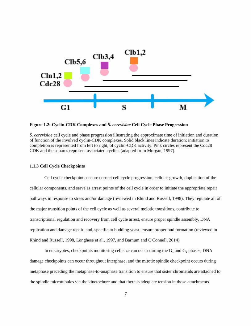
Figure 1.2: Cyclin-CDK Complexes and S. cerevisiae Cell Cycle Phase Progression
S. cerevisiae cell cycle and phase progression illustrating the approximate time of initiation and duration
of function of the involved cyclin-CDK complexes. Solid black lines indicate duration; initiation to
completion is represented from left to right, of cyclin-CDK activity. Pink circles represent the Cdc28
CDK and the squares represent associated cyclins (adapted from Morgan, 1997).
1.1.3 Cell Cycle Checkpoints
Cell cycle checkpoints ensure correct cell cycle progression, cellular growth, duplication of the
cellular components, and serve as arrest points of the cell cycle in order to initiate the appropriate repair
pathways in response to stress and/or damage (reviewed in Rhind and Russell, 1998). They regulate all of
the major transition points of the cell cycle as well as several meiotic transitions, contribute to
transcriptional regulation and recovery from cell cycle arrest, ensure proper spindle assembly, DNA
replication and damage repair, and, specific to budding yeast, ensure proper bud formation (reviewed in
Rhind and Russell, 1998, Longhese et al., 1997, and Barnum and O'Connell, 2014).
In eukaryotes, checkpoints monitoring cell size can occur during the G1 and G2 phases, DNA
damage checkpoints can occur throughout interphase, and the mitotic spindle checkpoint occurs during
metaphase preceding the metaphase-to-anaphase transition to ensure that sister chromatids are attached to
the spindle microtubules via the kinetochore and that there is adequate tension in those attachments

8
(reviewed in Barnum and O'Connell, 2014). The DNA damage response (DDR), triggered by DNA
damage, is fundamental to genome stability (reviewed in Longhese et al., 1997). Errors in cell cycle
progression and DNA replication, especially errors that are not successfully repaired, often lead to
genomic instability and compounding mutations that can manifest in the form of various complex
diseases such as cancer (reviewed in Longhese et al., 1997).
In budding yeast, there are four main DNA damage checkpoints that typically deal with DNA
damage repair and recovery: (1) the G1/S checkpoint, (2) the intra-S phase checkpoint, (3) the S/M
checkpoint, and (4) the metaphase or G2/M checkpoint (Siede et al., 1993, 1994; Paulovich and Hartwell,
1995; Weinert and Hartwell, 1988; reviewed in Longhese et al., 1997 and Barnum and O'Connell, 2014).
The intra-S phase checkpoint specifically focuses on the stabilization of the complex of DNA polymerase
and associated replication factors when replication forks have been stalled due to replication stress or
damage and is controlled by the Rad53 and Dun1 sensor kinases in budding yeast (reviewed in Barnum
and O'Connell, 2014). The following genes are known to be involved in the coordination of the DNA
damage checkpoints: RAD9, RAD17, RAD24, MEC3, DDC1, MEC1, and RAD53 (Weinert et al., 1994;
Weinert and Hartwell, 1988; Longhese et al., 1997; Allen et al., 1994; reviewed in Rhind and Russell,
1988). RAD17, RAD24, MEC3, and DDC1 are referred to as the RAD24 epistasis group and, in
conjunction with RAD9, MEC1, and RAD53, constitute the group of genes whose gene products are
required for the metaphase checkpoint in budding yeast, while only MEC1 and RAD53 are required for
the S/M checkpoint (Weinert et al., 1994; Weinert and Hartwell, 1988; Longhese et al., 1997; Allen et al.,
1994; reviewed in Rhind and Russell, 1998). MEC1 and RAD53, essential genes required for the S/M
checkpoint and the metaphase checkpoint, slow entry into mitosis when the S phase is inhibited for the
S/M checkpoint and mediate signal transduction pathways for other DNA damage checkpoints (reviewed
in Longhese et al., 1997). The non-essential genes, RAD9 and the RAD24 epistasis group, contribute to all
other known DNA damage checkpoints except the S/M checkpoint (reviewed in Longhese et al., 1997).

9
1.2 Forkhead-associated Domains
Forkhead-associated (FHA) domains have historically been studied for the purpose of evaluating
protein-protein interactions established between FHA domain-containing proteins and their binding
partners via phosphorylated threonine residue-based recognition of ligands by the phosphothreonine-
binding site of the FHA domain (reviewed in Mahajan et al., 2008). However, recent research has
identified a novel method of FHA domain ligand recognition that does not rely on this canonical method
of phosphothreonine motif binding (Matthews et al., 2014).
1.2.1 FHA Domain Discovery and Characteristics
During a 1995 bioinformatics-based study of the family of forkhead transcription factors, the
Forkhead-associated (FHA) domain was discovered (Hofmann and Bucher, 1995). FHA domains have
since been identified in over 2000 proteins, both eukaryotic and prokaryotic, commonly functioning as
regulatory proteins, kinases, phosphatases, or transcription factors (Hofmann and Bucher, 1995; Mahajan
et al., 2008; reviewed in Mohammad and Yaffe, 2009). The FHA domain is currently the only known
protein-protein interaction motif that specifically recognizes phosphorylated threonine (pThr) residues,
using the canonical pThr-binding site located on an apical surface of the domain, and will often do so via
the recognition of a pattern in the residues flanking the phosphorylated threonine residue (reviewed in
Mahajan et al., 2008). Little amino acid sequence homology exists between the large number of known
FHA domains; however, they do exhibit structural homology (reviewed in Mahajan et al., 2008). In
general, FHA domains are 80-100 amino acids in length and fold into two beta sheets, forming an 11-
stranded beta sandwich with connecting coiled loops (Durocher and Jackson, 2002; Mahajan et al., 2008;
reviewed in Mohammad and Yaffe, 2009). FHA domains and the proteins in which they are found often
have important roles relevant to human diseases, especially those that feature DNA damage responses,
abnormal cell growth, and/or impaired cell cycle regulation (Mahajan et al., 2008).

10
1.2.2 Non-canonical FHA Domain Lateral Surface Interaction Patch Identification and
Characterization
RADiation sensitive (Rad53) is a budding yeast cell cycle checkpoint kinase often used to study
DNA damage-based checkpoint signalling (Shirahige et al., 1998; reviewed in Durocher and Jackson,
2002). One heavily studied pathway in yeast is that of DNA replication initiation and checkpoint
activation (Shirahige et al., 1998). In this pathway, Rad53 acts in conjunction with the Dbf4-dependent
kinase (DDK) to regulate the firing of licensed origins of replication during replication initiation
(Shirahige et al., 1998; Duncker et al., 2002; reviewed in Matthews et al., 2012). DDK is a complex that
consists of the yeast proteins Cdc7 and Dbf4 which function as the kinase and regulatory subunits,
respectively (reviewed in Matthews et al., 2012 and Matthews et al., 2014). DDK-dependent
phosphorylation of proteins located at licensed origins triggers DNA replication, whereas activated Rad53
effector kinase delays the entry of cells into the mitosis phase of the cell cycle in response to genotoxic
stress (reviewed in Matthews et al., 2012). A unique feature of Rad53 is its possession of two FHA
domains, FHA1 and FHA2 (reviewed in Duncker et al., 2002). However, through studying the interaction
between Rad53 and one of its ligands, Dbf4, it was determined that the Rad53 and Dbf4 interaction relies
primarily on FHA1 (Duncker et al., 2002).
While characterizing the minimal region of Dbf4 required for its interaction with the FHA1
domain of Rad53, a BRCA1 C-terminal (BRCT) domain housing an additional alpha-helix within its core
domain, referred to as an H-BRCT domain, was found within the N-terminal region of Dbf4 (Matthews et
al., 2012). This H-BRCT domain was then analyzed using site-directed mutagenesis and yeast two-hybrid
assays to determine the phosphoepitope that was expected to be bound by the canonical pThr-binding site
of the Rad53 FHA1 domain (Matthews et al., 2014). However, individual mutagenesis of each of the
threonine residues within the H-BRCT domain of Dbf4 was not able to disrupt the interaction between the
H-BRCT domain and FHA1 (Matthews et al., 2014). As a result, the presence of an alternative interface
11
for the Rad53-Dbf4 interaction was hypothesized (Matthews et al., 2014). Via the use of nuclear magnetic
resonance, bioinformatics and yeast two-hybrid analysis, a set of conserved residues located on the lateral
surface of the Rad53 FHA1 domain was found to mediate the Rad53-Dbf4 interaction (Matthews et al.,
2014). This discovery has expanded the scope of studying FHA domain-based protein-protein
interactions, introducing the potential for discovering novel protein-protein interactions that may have
been previously overlooked by studies that focused solely on the canonical pThr-binding site (Matthews
et al., 2014).
1.3 DNA damage UNinducible (DUN1)
DNA damage UNinducible (DUN1) is a non-essential budding yeast gene that codes for the Dun1
checkpoint kinase, a protein involved in signal transduction pathways for DNA damage and replication
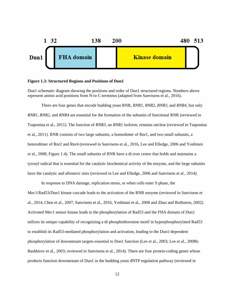
stress as well as natural progression through S phase (reviewed in Tsaponina et al., 2011). Dun1 is 513
amino acids long and includes two functional domains, the N-terminal FHA domain and the C-terminal
kinase domain (reviewed in Sanvisens et al., 2016; Figure 1.3). The primary function of Dun1 is to
regulate the levels of deoxyribonucleoside triphosphates (dNTPs), the fundamental building blocks of
DNA, via the transcriptional, spatial, and functional regulation of ribonucleotide reductase (RNR), the
tetrameric protein complex responsible for catalyzing the dNTP synthesis rate-limiting step of converting
ribonucleoside diphosphates into their deoxy form (reviewed in Chen et al., 2007, Sanvisens et al., 2016,
Yoshitani et al., 2008, Lee and Elledge, 2006 and Zhao and Rothstein, 2002; Figure 1.4).
12
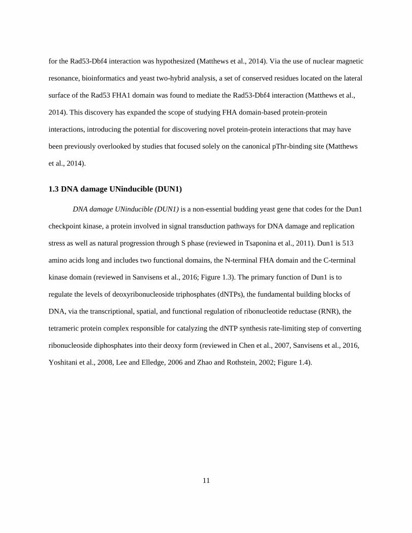
Figure 1.3: Structured Regions and Positions of Dun1
Dun1 schematic diagram showing the positions and order of Dun1 structured regions. Numbers above
represent amino acid positions from N-to C-terminus (adapted from Sanvisens et al., 2016).
There are four genes that encode budding yeast RNR, RNR1, RNR2, RNR3, and RNR4, but only
RNR1, RNR2, and RNR4 are essential for the formation of the subunits of functional RNR (reviewed in
Tsaponina et al., 2011). The function of RNR3, an RNR1 isoform, remains unclear (reviewed in Tsaponina
et al., 2011). RNR consists of two large subunits, a homodimer of Rnr1, and two small subunits, a
heterodimer of Rnr2 and Rnr4 (reviewed in Sanvisens et al., 2016, Lee and Elledge, 2006 and Yoshitani
et al., 2008; Figure 1.4). The small subunits of RNR have a di-iron centre that holds and maintains a
tyrosyl radical that is essential for the catalytic biochemical activity of the enzyme, and the large subunits
have the catalytic and allosteric sites (reviewed in Lee and Elledge, 2006 and Sanvisens et al., 2014).
In response to DNA damage, replication stress, or when cells enter S phase, the
Mec1/Rad53/Dun1 kinase cascade leads to the activation of the RNR enzyme (reviewed in Sanvisens et
al., 2014, Chen et al., 2007, Sanvisens et al., 2016, Yoshitani et al., 2008 and Zhao and Rothstein, 2002).
Activated Mec1 sensor kinase leads to the phosphorylation of Rad53 and the FHA domain of Dun1
utilizes its unique capability of recognizing a di-phosphothreonine motif in hyperphosphorylated Rad53
to establish its Rad53-mediated phosphorylation and activation, leading to the Dun1-dependent
phosphorylation of downstream targets essential to Dun1 function (Lee et al., 2003; Lee et al., 2008b;
Bashkirov et al., 2003; reviewed in Sanvisens et al., 2014). There are four protein-coding genes whose
products function downstream of Dun1 in the budding yeast dNTP regulation pathway (reviewed in

13
Sanvisens et al., 2016; Figure 1.4). WTM1 encodes a protein that anchors Rnr2 and Rnr4 to the nucleus,
SML1 encodes an inhibitor of RNR large subunit activity, DIF1 encodes a protein responsible for the
nuclear import of the Rnr2 and Rnr4 subunits, and CRT1 encodes a transcriptional repressor for the Rnr2
and Rnr4 genes (reviewed in Sanvisens et al., 2016). Mec1/Rad53 phosphorylation-dependent activation
of Dun1 results in the phosphorylation of Crt1, phosphorylation and degradation of Dif1 and Sml1, and
the hypothesized phosphorylation of Wtm1 resulting in the release of Rnr2 and Rnr4 transcriptional
repression, the release of Rnr activity inhibition, and the re-localization of Rnr2 and Rnr4 to the
cytoplasm in order to form a functional RNR holoenzyme (Sanvisens et al., 2016; Figure 1.4).
Figure 1.4: Dun1 and the dNTP Regulation Pathway
Depiction of the Dun1 dNTP regulation pathway, illustrating the role of the activated Dun1 kinase, the
proteins responsible for activating Dun1 kinase, and the proteins that are regulated by activated Dun1 in
the dNTP regulation pathway (adapted from Sanvisens et al., 2016).
Wtm1 is a member of the WTM (WD repeat-containing Transcriptional Modulator) family of
WD40-repeat proteins, a protein family that exhibits many diverse functions such as transcriptional
control, signal transduction, autophagy and apoptosis (Pemberton and Blobel, 1997; reviewed in Lee and

14
Elledge, 2006). WTM1, WTM2, and WTM3, identified in the WTM gene family in budding yeast, are
homologs that likely arose due to gene duplication and all three of them encode nuclear proteins
(Pemberton and Blobel, 1997; Huh et al., 2003; reviewed in Lee and Elledge, 2006). Of the three genes,
WTM3 is the most divergent (Pemberton and Blobel, 1997). Both WTM1 and WTM2 encode proteins that
bind to themselves as well as each other (Pemberton and Blobel, 1997) and despite the fact that both
Wtm1 and Wtm2 are able to bind to the Rnr2 and Rnr4 subunits of RNR, it is Wtm1 that is responsible
for the localization of the Rnr2 and Rnr4 subunits via its action as a nuclear anchor (Lee and Elledge,
2006). The association of Rnr2-Rnr4 subunits with the Wtm1 nuclear protein holds Rnr2 and Rnr4 in the
nucleus (Lee and Elledge, 2006; reviewed in Sanvisens et al., 2016). Proposed phosphorylation of Wtm1
by Dun1 in response to DNA damage disrupts the association between Wtm1 and Rnr2-Rnr4 releasing
Rnr2-Rnr4 from the nucleus to the cytoplasm where its association with Rnr1 subunits form a functional
RNR holoenzyme that can catalyze dNTP synthesis (Lee and Elledge, 2006; reviewed in Sanvisens et al.,
2016).
Suppressor of mec1 lethality (SML1), named after the observation that the addition of a sml1
mutation could restore viability to lethal mec1 mutants, encodes a small and largely disordered suppressor
protein that negatively affects dNTP synthesis by binding to the Rnr1 subunit and inhibiting its activity
(Zhao et al., 1998, reviewed in Andreson et al., 2010). Mec1 and Rad53 are required to remove that
inhibition during S phase in order to facilitate DNA replication (Zhao et al., 1998). The carboxyl terminus
of Rnr1 associates with the amino terminus of Rnr1 in order to regenerate the active site located on the
amino terminus using a cysteine pair located on the carboxyl terminus (Zhao et al., 1998, 2000; Chabes et
al., 1999; Zhang et al., 2007; reviewed in Sanvisens et al., 2014). Sml1 competes with the carboxyl
terminus for an association with the amino terminus and in doing so hinders Rnr1 active site regeneration,
preventing the catalytic activity of RNR (Zhao et al., 1998, 2000; Chabes et al., 1999; Zhang et al., 2007;
reviewed in Sanvisens et al., 2014). Sml1 phosphorylation at serine residues within its Sml domain by the
Mec1/Rad53/Dun1 kinase cascade results in a conformational change in Sml1 that causes it to dissociate

15
from Rnr1 (Andreson et al., 2010). Phosphorylated Sml1 is then recognized by the Rad6-Ubr2-Mub1
E2/E3 ligase complex, ubiquitinylated, and targeted for degradation by the 26S proteosome, allowing the
Rnr1 active site to be regenerated and dNTPs to be synthesized (Andreson et al., 2010).
Damage-regulated Import Facilitator (DIF1) is a paralog of SML1 (Lee et al., 2008a). DIF1,
SML1, and another yeast gene HUG1 arose following the duplication and divergence of an ancestral gene,
resulting in DIF1 on chromosome XII and both SML1 and HUG1 on chromosome XIII (Lee et al.,
2008a). Similar to SML1, DIF1 encodes a small and largely disordered protein (Lee et al., 2008a). Dif1
contributes to dNTP regulation during S-phase in a cell cycle-dependent manner when Dif1 levels peak
towards the end of S phase (Lee et al., 2008a; reviewed in Sanvisens et al., 2016). Dif1 shares a conserved
domain with Sml1, referred to as the Sml domain, and another conserved domain with Hug1, referred to
as the Hug domain (Lee et al., 2008a). Dif1 imports the Rnr2-Rnr4 small subunits from the cytoplasm to
the nucleus via an association between its Hug domain and the Rnr2-Rnr4 small subunits (Lee et al.,
2008a). Hypothetically, Dif1 carries out its import function either by acting as an adaptor or activating a
dormant nuclear localization signal on the Rnr2-Rnr4 complex (Lee et al., 2008a). During S phase and in
response to DNA damage when a rise in dNTP levels is required, Dif1 is phosphorylated and degraded in
order to prevent the import of Rnr2-Rnr4 subunits from the cytoplasm to the nucleus (Lee et al., 2008a;
reviewed in Sanvisens et al., 2016). The Sml domain of Dif1 is a phosphodegron and the direct
phosphorylation of serine residues within the Sml domain by Dun1 results in the degradation of Dif1 and
the maintenance of Rnr2-Rnr4 localization in the cytoplasm where it can associate with Rnr1 subunits to
form a functional RNR holoenzyme that can synthesize dNTPs to increase dNTP levels (Lee et al., 2008a;
reviewed in Sanvisens et al., 2016).
Constitutive RNR Transcription (CRT1) is one of many genes that encode regulators of DNA
damage inducibility (Huang et al., 1998). CRT1 encodes a transcriptional repressor specifically
responsible for the repression of the RNR2, RNR3, and RNR4 genes (Huang et al., 1998). Crt1 mediates
repression via the recruitment of two general repressors, Tup1 and Ssn6, to the promoters of target genes

16
(Huang and Elledge, 1997; Huang et al., 1998; reviewed in Tsaponina et al., 2011). RNR2 and RNR4
repression specifically prevents RNR holoenzyme formation and thereby its function, however, Dun1-
dependent phosphorylation of Crt1 prevents the transcriptional repression of RNR2, RNR3, and RNR4
allowing Rnr2 and Rnr4 production and formation of the RNR holoenzyme for dNTP synthesis (Huang
and Elledge, 1997; Huang et al., 1998; reviewed in Tsaponina et al., 2011 and Sanvisens et al., 2016).
1.4 Research Objectives
The imperative question, following the discovery of the non-canonical FHA domain lateral
surface interaction patch on the Rad53 FHA1 domain and its importance to the interaction between Rad53
and Dbf4, was whether or not other FHA domain-containing proteins possess a non-canonical FHA
domain lateral surface interaction patch and, if so, to what extent does it contributes to both previously
identified and novel protein-protein interactions. Previous work in the Duncker lab demonstrated that
mutations of the non-canonical FHA domain lateral surface interaction patch or the canonical pThr-
binding site of the Dun1 FHA domain were sufficient to reduce the interaction between the Dun1 FHA
domain and Sml1, only mutation of the pThr-binding site disrupted the interaction between the Dun1
FHA domain and Rad53, but that neither mutation of the Dun1 pThr-binding site nor of the lateral surface
interaction patch were sufficient to disrupt the interaction between the Dun1 FHA with Dif1 (Robertson,
2015; Guitor, 2016). Additionally, yeast two-hybrid analysis of the Dun1 FHA domain and Crt1
suggested a lack of a protein-protein interaction (Guitor, 2016). As a result, the objective of the research
presented in this thesis was to evaluate the contribution of the non-canonical FHA domain lateral surface
interaction patch identified on the Dun1 FHA domain, compared to that of the canonical pThr-binding
site, to protein-protein interactions involved in dNTP regulation in budding yeast. This objective was
executed via the use of (1) yeast two-hybrid assays to confirm Dun1 ligands, (2) bioinformatics, site-
directed mutagenesis and yeast two-hybrid assays to evaluate the interaction abrogation of Dun1 FHA

17
domain lateral surface interaction patch mutants compared to pThr-binding site mutants with Dun1
ligands, and (3) spot plate assays to assess the genotoxic sensitivity of Dun1 FHA mutants.

18
Chapter 2
Materials and Methods
2.1 Yeast Strains
The following yeast strains were used for genomic DNA isolation, yeast two-hybrid assays, and
spot plate assays (Table 2.1).
Table 2.1: List of Yeast Strains
Strain Genotype Source
DY-1 MATa, ade2-1, can1-100, trp1-1, his3-11, ura3-1, leu2-3, leu2-
112, pep4::LEU2
(Duncker et al. 2002)
DY-30 MATa, his3Δ1, leu2Δ0, met15Δ0, ura3Δ0 ATCC
DY-145 MATa, ade2-1, can1-100, his3-11,15, leu2-3, trp1-1, ura3-1,
SML1::HIS3 RAD5
(Tam et al. 2008)
DY-147 MATa, ade2-1, can1-100, his3-11,15, leu2-3, trp1-1, ura3-1,
SML1::HIS3, RAD5, RAD53::URA3
(Tam et al. 2008)
DY-351 MATa, his3Δ1, leu2Δ0, met15Δ0, ura3Δ0, Dun1Δ GE Healthcare
* The rad53Δ mutation is coupled to the smlΔ mutation in order to rescue rad53Δ lethality (reviewed in
Dohrmann and Sclafani, 2006).
2.2 Genomic DNA Isolation
Genomic DNA was isolated from the DY-1 yeast strain for use as template DNA for most PCR
reactions for plasmid construction. A working culture of DY-1, inoculated from a saturated culture grown
for 2 days at 30°C, was grown to a concentration of ~ 1 x 10⁷ cells/mL in 10 mL of YPD (10 % yeast
extract, 20 % peptone, and 20 % dextrose) media. Cells were centrifuged at 4000 rotations-per-minute
(rpm) for 5 minutes, re-suspended in 500 µL of sterile water and transferred to a sterile 2 mL screw-cap
tube. Re-suspended cells were centrifuged for 10 seconds at maximum speed, the supernatant decanted,
and then mixed at a low speed on a vortex to loosen the pellet. A 200 µL aliquot of 'genomic prep mix'
(2% Triton X-100, 1% SDS, 100 mM NaCl, 10 mM Tris-Cl pH 8, and 1 mM EDTA) was added to the
loosened pellet, followed by 200 µL of phenol:chloroform:isoamylalcohol (25:24:1) and 0.5 g of 0.5 mm

19
glass beads. All components were mixed for 3-4 minutes using a vortex and then 200 µL of 1X TE pH 8
was added and mixed. Following a 5 minute centrifugation at maximum speed, the top layer was
transferred to a new sterile 1.5 mL tube with 1 mL of 100% room temperature ethanol and mixed by
inversion. After another run of centrifugation for 2 minutes at maximum speed, the pellet was completely
re-suspended in 0.4 mL of 1X TE pH 8 to which 10 µL of 10 mg/mL RNase A (Sigma) was added before
incubating at 37°C for 10 minutes. After incubation, 10µL of 4 M ammonium acetate and 1 mL of room
temperature 100% ethanol were added and mixed by inversion. After a 2 minute centrifugation, the
supernatant was discarded and the pellet left to air dry before completely re-suspending the pellet in 50
µL of 1X TE pH 8. Genomic DNA was stored at -20°C.
2.3 Bioinformatics Analysis
Protein BLAST (BLASTp) searches were done using the full length amino acid sequence of
Dun1 (S288C, Saccharomyces Genome Database, https://www.yeastgenome.org/locus/S000002259) as
the input of a search against the non-redundant protein sequences (nr) database using the Position-
Specific Iterated BLAST (PSI-BLAST) algorithm in order to identify Dun1 homologs (National Center
for Biotechnology Information, NCBI). A selection of returned sequences with percentage identities of
approximately 30% were used to generate a multiple sequence alignment (MSA) in the ClustalW format
using the online MUltiple Sequence Comparison by Log-Expectation (MUSCLE) tool (European
Molecular Biology Laboratory, EMBL-EBI). The MUSCLE-generated MSA file was examined using the
Jalview software to identify and illustrate highly conserved residues within the in Dun1 FHA domain
using amino acid percent identity beyond a threshold of 30% to colour the residues in accordance with
their conservation for a range of blue for high conservation to white for low conservation
(http://www.jalview.org/). Using the UCSF Chimera protein model analysis software and its Multi-Align
viewer tool or the Consurf server (http://consurf.tau.ac.il/2016/), the MUSCLE-generated MSA was used
to map the amino acid sequence conservation of Dun1 homologs to the Dun1 FHA domain model (PDB
20
template ID: 2JQJ) using colouration to depict the level of conservation ranging from red residues
showing high conservation to blue residues showing low conservation. Chimera was also used to assess
the orientation of the amino acid side chains for highly conserved residues in order to identify highly
conserved surface residues that could contribute to protein-protein interaction surfaces for the canonical
pThr-binding site or non-canonical FHA domain lateral surface interaction patch
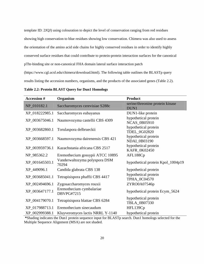
(https://www.cgl.ucsf.edu/chimera/download.html). The following table outlines the BLASTp query
results listing the accession numbers, organisms, and the products of the associated genes (Table 2.2).
Table 2.2: Protein BLAST Query for Dun1 Homologs
Accession # Organism Product
NP_010182.1 Saccharomyces cerevisiae S288c serine/threonine protein kinase
DUN1
XP_018222985.1 Saccharomyces eubayanus DUN1-like protein
XP_003675046.1 Naumovozyma castellii CBS 4309 hypothetical protein
NCAS_0B05910
XP_003682860.1 Torulaspora delbrueckii hypothetical protein
TDEL_0G02820
XP_003668597.1 Naumovozyma dairenensis CBS 421 hypothetical protein
NDAI_0B03190
XP_003959736.1 Kazachstania africana CBS 2517 hypothetical protein
KAFR_0K02450
NP_985362.2 Eremothecium gossypii ATCC 10895 AFL188Cp
XP_001645503.1 Vanderwaltozyma polyspora DSM
70294 hypothetical protein Kpol_1004p19
XP_449096.1 Candida glabrata CBS 138 hypothetical protein
XP_003685041.1 Tetrapisispora phaffii CBS 4417 hypothetical protein
TPHA_0C04570
XP_002494696.1 Zygosaccharomyces rouxii ZYRO0A07546p
XP_003647177.1 Eremothecium cymbalariae
DBVPG#7215 hypothetical protein Ecym_5624
XP_004179070.1 Tetrapisispora blattae CBS 6284 hypothetical protein
TBLA_0B07330
XP_017988713.1 Eremothecium sinecaudum HFL139Cp
XP_002999388.1 Kluyveromyces lactis NRRL Y-1140 hypothetical protein
*Shading indicates the Dun1 protein sequence input for BLASTp search. Dun1 homologs selected for the
Multiple Sequence Alignment (MSA) are not shaded.

21
2.4 Site-directed mutagenesis
Site-directed mutagenesis was used to generate mutations, primarily point mutations, within the
DUN1 coding sequence using the QuikChange II XL Site-Directed Mutagenesis Kit and its full protocol
which is available online: http://www.agilent.com/cs/library/usermanuals/public/200523.pdf (Agilent
Technologies). Mutagenic primers were designed using the QuikChange Site-Directed Mutagenesis
online interface (https://www.genomics.agilent.com/primerDesignProgram.jsp) and then utilized during
PCR to remove or replace the desired nucleotide(s). The PCR product was digested with provided Dpn1
restriction endonuclease in order to degrade parental copies of the plasmid containing the gene to be
mutagenized and then the product was transformed into the provided XL-10 Gold Ultracompetent cells
and plated on Luria-Bertani (LB) agar (1% NaCl, 1% tryptone, 0.5% yeast extract, and 2.4% agar)
containing the antibiotic ampicillin (BioShop). Colony PCR was used to confirm colonies that maintained
the integrity of both the insert and vector and then plasmid DNA from plasmid preps of 4 positive clones
was sent for sequencing at the Robarts Sequencing Facility.
2.5 Plasmid Construction
Plasmids were constructed using either the pEG202, pJG4-6, or pRS315 vectors (Ausubel et al.,
1994; Gyuris et al., 1993, reviewed in Duncker et al., 2002; Dobson et al., 2005). Plasmid constructs
made with the pEG202 and pJG4-6 vectors were designed for use in yeast two-hybrid assays, and
constructs made with the pRS315 vector were used for spot plate assays. The pEG202 and pJG4-6 vectors
are referred to as the bait and prey vectors in the context of yeast two-hybrid assays (Ausubel et al., 1994;
Gyuris et al., 1993, reviewed in Duncker et al., 2002). In addition to traditional cloning vector features,
the bait and prey vectors contain a selectable marker for the biosynthesis of histidine and tryptophan,
respectively, as well as the coding sequence of the DNA binding domain and transcriptional activation
domain of the transcription factor for the LacZ reporter located on the pSH18-34 reporter plasmid utilized
during yeast two-hybrid assays (Ausubel et al., 1994; Gyuris et al., 1994). In addition to other cloning

22
vector traditional features, the pSH18-34 plasmid contains a selectable marker for the biosynthesis of
uracil (Ausubel et al., 1994; Gyuris et al., 1994). The pRS315 vector, YCp or yeast centromeric plasmid
contains a selectable marker for the biosynthesis of leucine amongst its other traditional cloning vector
features. Due to the incorporation of yeast centromeric sequence within the plasmid, pRS315 is
maintained in yeast cells with a low copy number of usually 1-2 copies per cell (Clarke and Carbon,
1980; Ishii et al., 2009; Stearns et al., 1990). This feature was exploited in order to allow the expression of
gene(s) of interest at a level approximating that of genomic DNA.
The protein coding sequence of DUN1 for the region of the FHA domain, amino acids 21-145,
was previously cloned into the pJG4-6 vector in the Duncker Lab. The following Dun1 FHA domain
mutants were generated via site-directed mutagenesis using the pre-existing Dun1 FHA domain construct
as a template: pJG4-6 dun1-R60A-fha, pJG4-6 dun1-N108A-fha, pJG4-6 dun1-K136A-fha, pJG4-6 dun1-
2M-fha, pJG4-6 dun1-4M-fha, pJG4-6 dun1-N121A-fha, pJG4-6 dun1-L134A-fha, and pJG4-6 dun1-
KRA-fha. The full-length coding sequence for DUN1 was amplified from DY-1 genomic DNA via the
Polymerase Chain Reaction (PCR) using a commercially available High-Fidelity PCR kit (Roche) for the
construction of the pJG4-6 DUN1 plasmid. PCR products were purified using a commercial PCR clean up
kit that was also used for the clean-up of EcoRI and ApaI forward and reverse restriction enzyme digested
PCR products and vectors (Geneaid). All restriction enzymes were purchased from Thermo Fisher
Scientific and their sequences were incorporated into the design of the primers used for PCR
amplification. Ligation was completed using a T4 DNA ligase kit (BioBasic), and the ligated product was
transformed into calcium-chloride competent DH5α Escherichia coli (E. coli) cells grown and plated on
LB media (1% NaCl, 1% tryptone, 0.5% yeast extract, and 2.4% agar) containing the antibiotic ampicillin
(BioShop). Colony PCR was performed using a commercial colony PCR kit (New England Biolabs).
Plasmid DNA was isolated from saturated overnight cultures of DH5α for positive colonies using a
commercial plasmid prep kit (Geneaid). The full protocol for plasmid DNA purification is available
online: http://www.geneaid.com/sites/default/files/PD13.pdf. Plasmid DNA from 2-4 positive clones was

23
sent for sequencing of the full length of the insert at the Robarts Sequencing Facility to ensure the fidelity
of the cloned PCR product. Site-directed mutagenesis of the full-length DUN1 prey construct generated
the following Dun1 mutants in the context of full-length Dun1: pJG4-6 dun1-ΔFHA, pJG4-6 dun1-
N108A, pJG4-6 dun1-K136A, pJG4-6 dun1-2M, pJG4-6 dun1-4M, pJG4-6 dun1-KRA, pJG4-6 dun1-
N121A, pJG4-6 dun1-L134A, pJG4-6 dun1-D328A, pJG4-6 dun1-T380A, and pJG4-6 dun1-R60A.
The full-length DUN1 coding sequence along with its native promoter, 600 base pairs upstream
of the coding sequence, was amplified from DY-1 genomic DNA as described previously for the pJG4-6
DUN1 construct, cloned into a pCM190-myc13 vector and then moved, with or without the myc13 tag,
into the pRS315 plasmid using BamHI and NotI forward and reverse restriction enzymes in order to
generate the pRS315 DUN1 construct. Site-directed mutagenesis of the DUN1 construct generated the
following mutants in the context of full-length DUN1: pRS315 dun1-N108A, pRS315 dun1-K136A,
pRS315 dun1-2M, pRS315 dun1-4M, pRS315 dun1-KRA, pRS315 dun1-ΔFHA, pRS315 dun1-R60A,
pRS315 dun1-N121A, and pRS315 dun1-L134A.
All Dun1 constructs generated within the pJG4-6 vector were used to create the same constructs
in the pEG202 vector via EcoRI and XhoI forward and reverse restriction enzyme digests of the pJG4-6
constructs and ligation into the pEG202 vector: pEG202 DUN1-FHA, pEG202 dun1-R60A-fha, pEG202
dun1-N108A-fha, pEG202 dun1-K136A-fha, pEG202 dun1-2M-fha, pEG202 dun1-4M-fha, pEG202
dun1-N121A-fha, pEG202 dun1-L134A-fha, pEG202 dun1-KRA-fha, pEG202 DUN1, pEG202 dun1-
ΔFHA, pEG202 dun1-N108A, pEG202 dun1-K136A, pEG202 dun1-2M, pEG202 dun1-4M, pEG202
dun1-KRA, pEG202 dun1-N121A, pEG202 dun1-L134A, and pEG202 dun1-R60A.
The full-length coding sequences of DIF1, SML1, CRT1, and RAD53 had already been cloned
into the pEG202 vector along with full-length RAD53 in the pJG4-6 vector by members of the Duncker
Lab. The full-length coding sequence of WTM1 was amplified from DY-1 genomic DNA and cloned into
the pEG202 vector as described previously for the pJG4-6 DUN1 construct, and then the full-length
coding sequences of SML1, CRT1, and WTM1 were digested from the pEG202 vector using the EcoRI

24
forward and XhoI reverse restriction enzymes and inserted into the pJG4-6 vector to generate the
following constructs: pJG4-6 SML1, pJG4-6 CRT1, and pJG4-6 WTM1.
2.6 Yeast Transformation
Plasmid DNA containing the protein coding sequence for genes of interest was transformed into
the DY-1 yeast strain pre-transformed with the pSH18-34 reporter plasmid for yeast two-hybrid assays or
the DY-351 yeast strain for spot plate assays. A working culture of the appropriate yeast strain was grown
to a concentration of ~ 1 x 10⁷ cells/mL in 10 mL of either Synthetic Complete (SC) media (0.17 % yeast
nitrogen base, 0.5 % ammonium sulfate, 2 % glucose, 1X amino acid mix) lacking uracil for the selection
of the pSH18-34 reporter plasmid or YPD (10 % yeast extract, 20 % peptone, 20 % dextrose). Working
cultures were centrifuged for 5 minutes at 4000 rpm, resuspended in 1X Tris-EDTA (TE) solution pH 8.0,
centrifuged again, resuspended in 200 µL of Lithium acetate/TE solution (100 mM lithium acetate, 2.5
mL 1X TE), and then held at room temperature for 10 minutes. Approximately 0.4 µg of plasmid DNA
was added to 100 µL of the yeast suspension mix along with 100 µg of salmon sperm DNA, and then 300
µL of Lithium acetate/TE/PEG4000 solution (100 mM Lithium acetate, 2 g polyethylene glycol 4000, 0.5
mL 10X TE) was added. The solution was incubated at 30°C for 30 minutes before 40 µL of
dimethylsulfoxide (DMSO) was added. Cells were heat shocked at 42°C for 7 minutes, held on ice for 2
minutes and then plated on selective media and grown for 2-3 days.
2.7 Yeast two-Hybrid Assay
This technique was performed as outlined previously (Duncker et al., 2002). DY-1 yeast cells
pre-transformed with the pSH18-34 lacZ reporter plasmid were transformed with a pEG202-derived bait
construct and a pJG4-6-derived prey construct (Ausubel et al., 1994; Gyuris et al., 1993; reviewed in
Duncker et al., 2002). A physical interaction between the bait and prey proteins during the assay would
bring the DNA binding domain, fused to the gene of interest expressed from the bait vector, and the
25
transcriptional activation domain, fused to the gene of interest expressed from the prey vector, into close
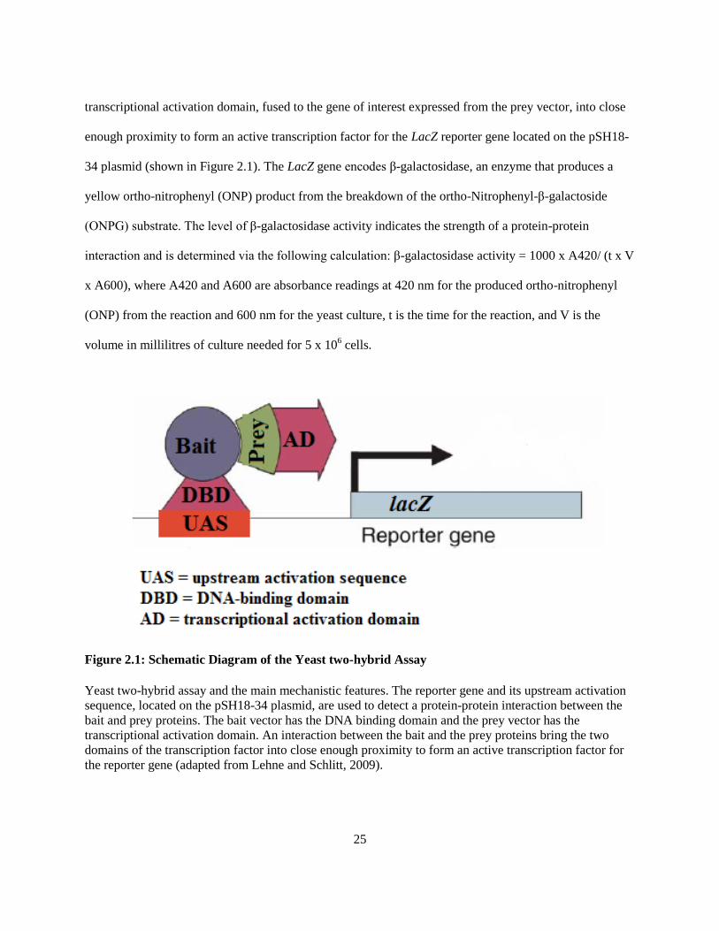
enough proximity to form an active transcription factor for the LacZ reporter gene located on the pSH18-
34 plasmid (shown in Figure 2.1). The LacZ gene encodes β-galactosidase, an enzyme that produces a
yellow ortho-nitrophenyl (ONP) product from the breakdown of the ortho-Nitrophenyl-β-galactoside
(ONPG) substrate. The level of β-galactosidase activity indicates the strength of a protein-protein
interaction and is determined via the following calculation: β-galactosidase activity = 1000 x A420/ (t x V
x A600), where A420 and A600 are absorbance readings at 420 nm for the produced ortho-nitrophenyl
(ONP) from the reaction and 600 nm for the yeast culture, t is the time for the reaction, and V is the
volume in millilitres of culture needed for 5 x 10⁶ cells.
Figure 2.1: Schematic Diagram of the Yeast two-hybrid Assay
Yeast two-hybrid assay and the main mechanistic features. The reporter gene and its upstream activation
sequence, located on the pSH18-34 plasmid, are used to detect a protein-protein interaction between the
bait and prey proteins. The bait vector has the DNA binding domain and the prey vector has the
transcriptional activation domain. An interaction between the bait and the prey proteins bring the two
domains of the transcription factor into close enough proximity to form an active transcription factor for
the reporter gene (adapted from Lehne and Schlitt, 2009).

26
Yeast cultures were grown in 10 mL of SC media without uracil, histidine, or tryptophan to a
concentration of ~ 1.0 x 10⁷ cells/mL. Following centrifugation for 5 minutes at 4000 rpm, cells were
washed in sterile water and incubated for six hours at 30°C and a speed of 200 rpm in 20 mL of
Galactose/Raffinose media (0.17 % yeast nitrogen base, 0.5 % ammonium sulfate, 2% galactose, 1%
raffinose) lacking uracil, histidine, and tryptophan in order to induce recombinant protein expression from
the prey vector. Following induction, cells were counted using a haemocytometer and then ~ 5 x 10⁶ cells
were harvested for the assay. The cells were centrifuged for 10 minutes at 16X gravity (g) and then
resuspended in 0.5 mL of Z buffer (60 mM Na2HPO4, 40 mM NaH2PO4, 10 mM KCl, 1 mM MgSO4, and
0.05M β mercaptoethanol). Using a P200 micropipette, 2 drops of chloroform and 1 drop of 0.1% sodium
dodecyl sulfate (SDS) were added to the resuspended cells which were then vortexed at maximum speed
for 10 seconds and then incubated for 5 minutes at 28°C. After the 5 minute incubation, 100 µL of ONPG
(4 mg/mL in 0.1 M potassium phosphate buffer, pH 7, Sigma) was added to begin the β-galactosidase
reaction. Based on the appearance of a yellow colour in the reaction solution, 250 µL of 1 M Na2CO3
(BioShop) was then added to stop the reaction. After a final centrifugation of 10 minutes at 16X g, the
OD600 and OD420 were measured in order to calculate the β-galactosidase activity.
2.8 Yeast Whole Cell Extract and Western Blotting
In order to verify appropriate protein expression, yeast whole cell extracts (WCE) were generated
for Western blotting. Approximately 20 mL of yeast culture (~ 1.0 X 10⁷ cells/mL) were spun down for 5
minutes at 4000 rpm to pellet cells, resuspended in 300 µL of lysis buffer (10 mM Tris-HCl pH 8.0, 140
mM NaCl, 1% Triton X-100, 1 mM EDTA, 100 µL of Fisher HALT! protease inhibitor and 1 mM PMSF)
and added to a 2 mL tube with 0.3 g of 0.5 mm glass beads before subjecting the samples to lysis at 4°C
via the use of the Biospec Mini Bead-Beater for eight cycles of 30 seconds of agitation and 30 seconds of
rest on ice. A total of 105 µL of supernatant (whole cell extract) was collected following the
centrifugation of lysed cells for 30 seconds at 13, 200 rpm in 4°C: 5 µL for the Bradford Assay and 100

27
µL for the SDS-PAGE. The Bio-Rad Bradford Assay was used to determine the protein concentrations of
each sample (Bio-Rad). Protein extracts were mixed with loading buffer (60% 4X buffer [15% SDS, 40%
glycerol, and 166 mM Tris base], 0.26 M DTT, 7% bromophenol blue) at a volume of one-half that of the
extract and then boiled for 10 minutes before loading 30 - 50 µg of protein onto SDS polyacrylamide gels
(10 % resolving gel and 5% stacking gel). After electrophoresis, proteins were transferred from the gel to
a nitrocellulose membrane using a wet transfer method (200 mM glycine, 25 mM Tris-base, 20 %
methanol, 0.054% SDS for the transfer buffer). Membranes were pre-stained with 0.1% Ponceau S,
imaged using an Epson scanner (any good quality scanner can be used), and then de-stained with 1X TEN
+ T (20 mM Tris-HCl, 1 mM EDTA, 0.14 NaCl, 0.05% Tween 20) before detection with antibodies
(Table 2.3). Antibody incubations were performed for 1-2 hours after 1 hour blocking in 1X TEN + T
with 5% skim milk powder. Three 10 minute washes were done using 1X TEN + T after each antibody
exposure and images were taken with a Pharos FX Plus imager (Bio-Rad).
Table 2.3: List of Antibodies
Antibody Dilution Source
Anti-HA (mouse monoclonal) -
pJG4-6
0.75:5000 in 3% bovine serum
albumin, BSA
Sigma
Anti-LexA (rabbit polyclonal) -
pEG202
1:5000 in 3% BSA Cedarlane
Anti-MYC (mouse monoclonal) -
pRS315
1:5000 in 3% BSA Sigma
AlexaFluor 488 anti-mouse 1:3000 in 5% skim milk Invitrogen
AlexaFluor 647 anti-rabbit 1:3000 in 5% skim milk Invitrogen
2.9 Spot Plate Assay
The spot-plate assay was used to determine the growth defects of yeast cells exposed to genotoxic
stress under the circumstances of reduced or abolished interactions between Dun1 and other proteins
involved in dNTP regulation: Rad53, Wtm1, Crt1, Dif1 and Sml1. Genotoxic agents used as stressors
were intended to cause DNA damage, an inability to repair DNA damage, or impair DNA synthesis. A
DUN1 genomic knockout yeast strain, DY-351, purchased from GE Healthcare was used for yeast

28
transformation of full length Dun1 expression vectors. Using a sterile 60-well plate, three 1:10 dilutions
were made from an initial dilution of 30 µL of saturated (~ 1 x 10⁸ cells/mL) culture diluted in 270 µL of
media. Aliquots of 5 µL of the four serial dilutions of yeast culture were plated onto 25 mL of solid agar,
either synthetic complete (SC) lacking leucine or yeast peptone dextrose (YPD), containing various levels
of genotoxic agents. Genotoxic agents were mixed into 25 mL aliquots of agar media prior to agar
solidification. Genotoxic compounds used and their concentrations were as listed (Table 2.4).
Table 2.4: List of Genotoxic Agents
Genotoxic Agent Action Concentrations Source
Hydroxyurea (HU) dNTP pool depletion 20 mM - 200 mM BioShop
Methane methylsulfonate (MMS) DNA methylation 0.005% - 0.025% Sigma
Bleomycin DNA breakage 1 µg/mL - 5 µg/mL Sigma
Phleomycin DNA breakage 1 µg/mL - 5 µg/mL Sigma
Camptothecin DNA topoisomerase I inhibition 5 µM - 20 µM Sigma
2.10 Statistical Analysis
The IBM SPSS software was used to ascertain the significance of the differences observed
amongst yeast two-hybrid assay samples (https://www.ibm.com/analytics/data-science/predictive-
analytics/spss-statistical-software). The One-way ANOVA test and its associated Tukey post-hoc test
were used to determine whether or not the there were significant differences in the mean β-galactosidase
activity for each yeast two-hybrid assay sample. The Levene's test and Kolmogorov-Smirov tests were
used to assess the assumptions for homoscedasticity and normality, respectively, in order to confirm the
appropriate use of the One-way ANOVA test. The acceptance threshold was set at 0.05. Means with
different letters are significantly different (Tukey's HSD, p < 0.05).

29
Chapter 3
Dun1-Dif1 and Dun1-Sml1 interactions utilize the conserved non-canonical
FHA domain lateral surface interaction patch
3.1 Introduction
A primary function of Dun1 is to increase the levels of dNTPs in a cell via its regulatory control
of the transcription, localization, and function of the RNR enzyme subunits involved in the catalysis of
the dNTP synthesis rate-limiting step (reviewed in Sanvisens et al., 2016). Mec1/Rad53-dependent
phosphorylation and activation of Dun1 leads to the phosphorylation of downstream Dun1 targets: Wtm1,
Sml1, Dif1, and Crt1, and the removal of their collective inhibition with respect to dNTP synthesis (Lee
and Elledge, 2006; Andreson et al., 2010; Lee et al., 2008a; Huang and Elledge, 1997; Huang et al., 1998;
reviewed in Sanvisens et al., 2016). The Dun1 protein consists of an N-terminal FHA domain and a C-
terminal kinase domain that contribute to phosphoepitope recognition for ligand binding and
phosphorylation of targets, respectively (reviewed in Sanvisens et al., 2016).
Mec1 sensor kinase activation results in the phosphorylation and activation of the Rad53
checkpoint kinase (reviewed in Lee et al., 2003). Dun1 recognition of hyperphosphorylated Rad53 via the
Dun1 FHA di-phosphothreonine recognition motif allows for Rad53-dependent phosphorylation and
activation of Dun1 (Lee et al., 2003; Lee et al., 2008b; Bashkirov et al., 2003; reviewed in Sanvisens et
al., 2016). Considering the identification of the non-canonical FHA domain lateral surface interaction
patch of the Rad53 FHA1 domain and its contribution to the FHA1 and H-BRCT interaction of Rad53
and Dbf4 (Matthews et al., 2014), the Dun1 FHA domain was assessed for the existence of a candidate
conserved non-canonical FHA domain lateral surface interaction patch four years ago by Damir
Mingaliev, an undergraduate student in the Duncker Lab. Using the candidate non-canonical FHA domain
lateral surface interaction patch identified by Damir, Dun1 FHA domain ligands and binding patterns

30
were evaluated by Aaron Robertson and Allison Guitor, two other students in the Duncker lab. Using
yeast two-hybrid assays, protein-protein interactions were observed between the Dun1 FHA domain and
ligands: Sml1, Rad53 and Dif1, whereas an interaction between the Dun1 FHA domain and Crt1 was not
observed (Robertson, 2015; Guitor, 2016). Yeast two-hybrid assays also showed the existence of different
patterns in the association of the Dun1 FHA domain with its various ligands with respect to the
requirement of the pThr-binding site and/or the lateral surface interaction patch (Robertson, 2015; Guitor,
2016).
The Dun1 FHA - Sml1 interaction appeared to require both the canonical and non-canonical
interaction interfaces due to the observation that neither mutation of the pThr-binding site nor the lateral
surface interaction patch led to complete abrogation of the interaction (Robertson, 2015). Analysis of the
Dun1 FHA and Rad53 interaction showed that mutation of the canonical interface was able to completely
disrupt the interaction whilst mutation of the non-canonical interface had no effect (Robertson, 2015). For
the analysis of the Dun1 FHA - Dif1 interaction, mutation of the lateral surface interaction patch appeared
to have no effect on the interaction, but mutation of the pThr-binding site appeared to increase the
interaction strength (Guitor, 2016). An interaction between the Dun1 FHA domain and Wtm1 had yet to
be tested, preliminary spot plate assays were not very conclusive, and the effects of a coupled pThr-
binding site and lateral surface interaction patch mutant had not been assessed (Robertson, 2015).
3.2 Results
3.2.1 dNTP regulation pathway interactions exhibit different requirements
A prominent feature of the FHA domain is the recognition of phosphorylated threonine residues
via the canonical pThr-binding site for the establishment of ligand recognition and binding for FHA
domain-containing proteins (reviewed in Mahajan et al., 2008). The research presented in Matthews et al.,
2014 initiated a quest for the discovery of other FHA domain-containing proteins besides Rad53 that
possess a non-canonical FHA domain lateral surface interaction patch in addition to the canonical pThr-

31
binding site. The Multiple Sequence Alignment (MSA) of Dun1 homologs edited in Jalview illustrates the
approximate regions of the canonical pThr-binding site and non-canonical FHA domain lateral surface
interaction patch as well as the conservation of the residues encompassed within each area (Figure 3.1).
Key residues previously identified as highly conserved residues of each area were used to demarcate
those regions on the MSA (reviewed in Sanvisens et al., 2016; Robertson et al., 2015).
Figure 3.1: MUSCLE Multiple Sequence Alignment of Dun1 Homologs
A segment of the Multiple Sequence Alignment generated using the MUSCLE algorithm shows amino
acid conservation for the Dun1 FHA domain, specifically the regions of the canonical pThr-binding site
and non-canonical FHA domain lateral surface interaction patch. Key residues for the pThr-binding site
and lateral surface patch were used to mark approximate borders for each region. Conservation was
coloured using a range of blue (high conservation) to white (low conservation) based on a 30% percent
identity threshold within the Jalview software. Single letters and numbers correspond with the amino acid
letter code and the amino acid position with respect to S. cerevisiae Dun1.
The MUSCLE-generated MSA was then used to map amino acid sequence conservation to the
surface of the Dun1 FHA domain model which illustrates the location of the pThr-binding site and the
candidate lateral surface interaction patch (Figure 3.2). The apical region of the domain, known to house
the pThr-binding site, is one large area of highly conserved residues. Another large area of highly
conserved residues, representing the candidate non-canonical FHA domain lateral surface interaction

32
patch, can be seen on a lateral surface of the FHA domain. A small region of highly conserved residues
near the apical region may or may not be a part of the canonical pThr-binding site.
Figure 3.2: Conservation Mapping of the Dun1 FHA Domain
3-D surface model of the Dun1 FHA domain, amino acids 1-145, (PDB template ID: 2JQJ) showing
amino acid conservation using colours ranging from red (high conservation) to blue (low conservation).
Surface model shows the location of the canonical phosphothreonine-binding site and the candidate non-
canonical FHA domain lateral surface interaction patch.
Preliminary re-assessment of previously confirmed Dun1 FHA domain binding partners revealed
a lack of RAD53 and CRT1 expression from pEG202 two-hybrid bait vector constructs (data not shown),
the same anomaly was observed with expression of WTM1 from the pEG202 bait vector (data not shown).
As a result CRT1 and WTM1 protein coding sequences were cloned into the pJG4-6 two-hybrid prey
vector, a pJG4-6 RAD53 construct was already available and functional in the Duncker Lab, where
successful and consistent protein expression was recovered. Yeast two-hybrid assays and corresponding
western blots were used to evaluate the necessity and/or sufficiency of the Dun1 FHA domain for
interactions between Dun1 and the Rad53, Wtm1, and Crt1 ligands by comparing the interaction strengths
of the ligands with full-length Dun1, just the FHA domain, or Dun1 without the FHA domain (Figure
3.3). Calculations of the mean β-galactosidase activity observed over three trials were illustrated in a

33
graph to show that the Dun1 FHA domain could interact with Rad53, Wtm1, and Crt1 (Figure 3.3A-C).
Unlike the Dun1 FHA domain, neither full-length Dun1 nor Dun1 without its FHA domain were able to
interact with Rad53, shown via the lack of β-galactosidase activity relative to the empty prey negative
control (Figure 3.3A). Full-length Dun1 had twice as much β-galactosidase activity as the empty bait
negative control for Wtm1 but only half of the β-galactosidase activity of the Dun1 FHA with Wtm1,
indicating that only full-length Dun1 and just the FHA domain of Dun1 are capable of interacting with
Wtm1. The lower β-galactosidase activity for full-length Dun1 is likely a result of lower full-length Dun1
protein expression (Figure 3.3B). For the analysis of Crt1, the empty bait negative control had an
unexpectedly high signal for β-galactosidase activity that did not persist in samples that had Crt1 in the
prey vector and a version of the Dun1 protein in the bait vector (Figure 3.3C). The lack of β-galactosidase
activity for Crt1 with full-length Dun1 and Dun1 without the FHA domain indicates a lack of an
observable interaction between Crt1 with full-length Dun1 and with Dun1 without the FHA domain
(Figure 3.3C). The corresponding western blot images show the protein expression of the several versions
of Dun1 as well as the protein expression for the Rad53, Wtm1, and Crt1 ligands across samples. Protein
expression of full-length Dun1 appears to be lower than that of Dun1 without the FHA domain and just
the FHA domain, while ligand expression levels are the same throughout (Figure 3.3A-C).
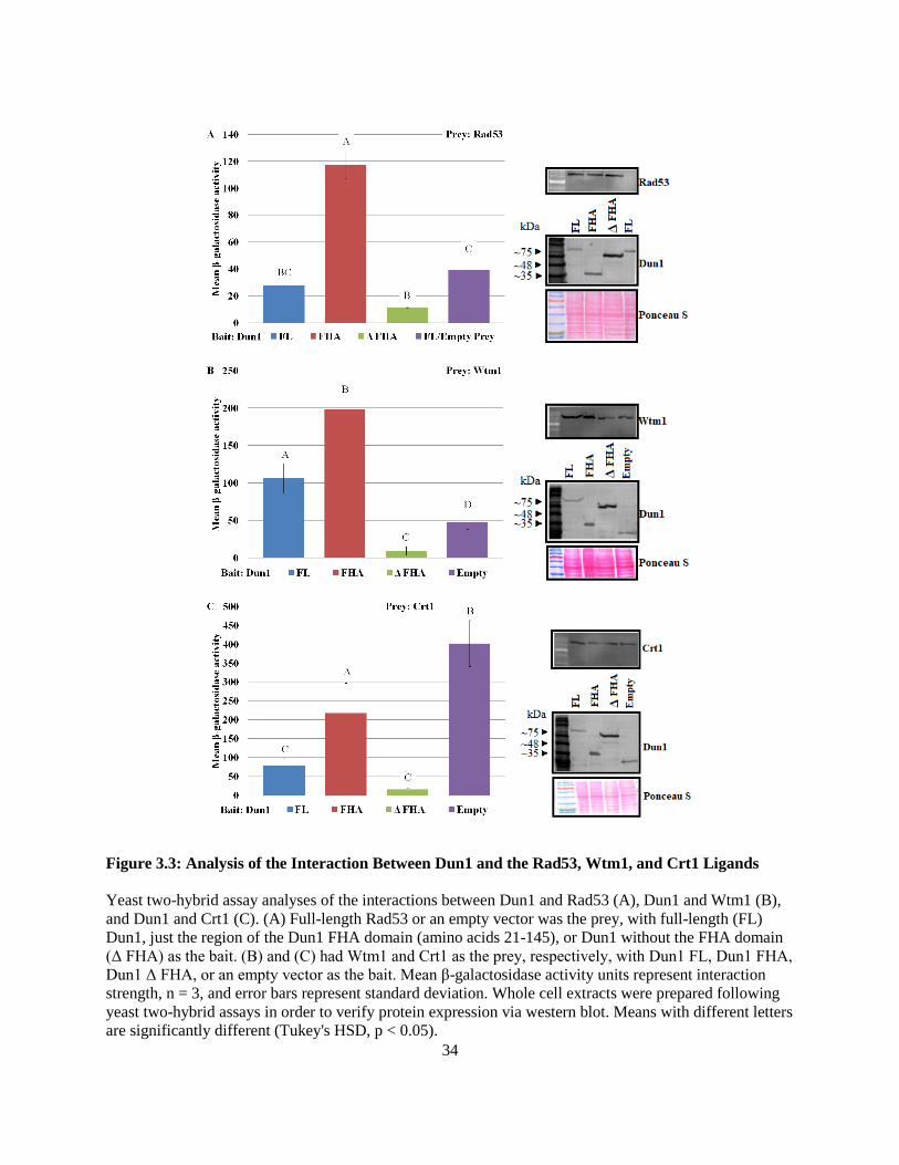
34
Figure 3.3: Analysis of the Interaction Between Dun1 and the Rad53, Wtm1, and Crt1 Ligands
Yeast two-hybrid assay analyses of the interactions between Dun1 and Rad53 (A), Dun1 and Wtm1 (B),
and Dun1 and Crt1 (C). (A) Full-length Rad53 or an empty vector was the prey, with full-length (FL)
Dun1, just the region of the Dun1 FHA domain (amino acids 21-145), or Dun1 without the FHA domain
(Δ FHA) as the bait. (B) and (C) had Wtm1 and Crt1 as the prey, respectively, with Dun1 FL, Dun1 FHA,
Dun1 Δ FHA, or an empty vector as the bait. Mean β-galactosidase activity units represent interaction
strength, n = 3, and error bars represent standard deviation. Whole cell extracts were prepared following
yeast two-hybrid assays in order to verify protein expression via western blot. Means with different letters
are significantly different (Tukey's HSD, p < 0.05).

35
The previous assessment of the interaction between Dun1 FHA and Sml1 completed by an
undergraduate student in the Duncker Lab (personal communication) revealed a systematic problem with
the yeast two-hybrid assay where Sml1 in the pEG202 bait vector activates transcription of the LacZ
reporter, independently of the prey construct's transcriptional activation domain, resulting in false
positives. As a result, SML1 was cloned into the pJG4-6 prey vector, effectively preventing the
occurrence of the false positives, and yeast two-hybrid assays and corresponding western blots were used
to evaluate Dun1 FHA domain necessity and sufficiency for the interaction with Sml1 (Figure 3.4A).
Calculations of average β-galactosidase activity over three trials were graphed to depict the interaction
strength between Dun1 and Sml1 (Figure 3.4A). Empty prey negative control levels of β-galactosidase
activity were observed for the Dun1 FHA domain and Dun1 without the FHA domain with Sml1 while
full-length Dun1 with Sml1 exhibited a high level of β-galactosidase activity (Figure 3.4A). This
indicates that only full-length Dun1 that had both the FHA and kinase domains was able to interact with
Sml1 (Figure 3.4A). Sequencing of Dun1 FHA domain prey constructs for FHA domain lateral surface
interaction patch and pThr-binding site mutants used previously in the Duncker Lab for the evaluation of
the Dun1 - Sml1 and Dun1 - Dif1 interactions revealed the absence of expected nucleotide substitutions
that were supposed to change the key amino acid residues of the lateral surface interaction patch and the
pThr-binding site. As a result, lateral surface interaction patch and pThr-binding site mutants were re-
created and the Dun1 - Dif1 interaction was re-assessed after evaluating the necessity and sufficiency of
the Dun1 FHA domain for the Dun1 - Dif1 interaction (Figure 3.4B - 3.6). Calculations of average β-
galactosidase activity over three trials were graphed to depict the interaction strength between Dun1 and
Dif1 (Figure 3.4B). Levels of β-galactosidase activity showed that a very strong interaction existed
between full-length Dun1 that had both the FHA and kinase domains and Dif1 while a weakened
interaction existed between Dif1 and just the FHA domain of Dun1 (Figure 3.4B). Without the FHA
domain, there was no interaction between Dun1 and Dif1 (Figure 3.4B).
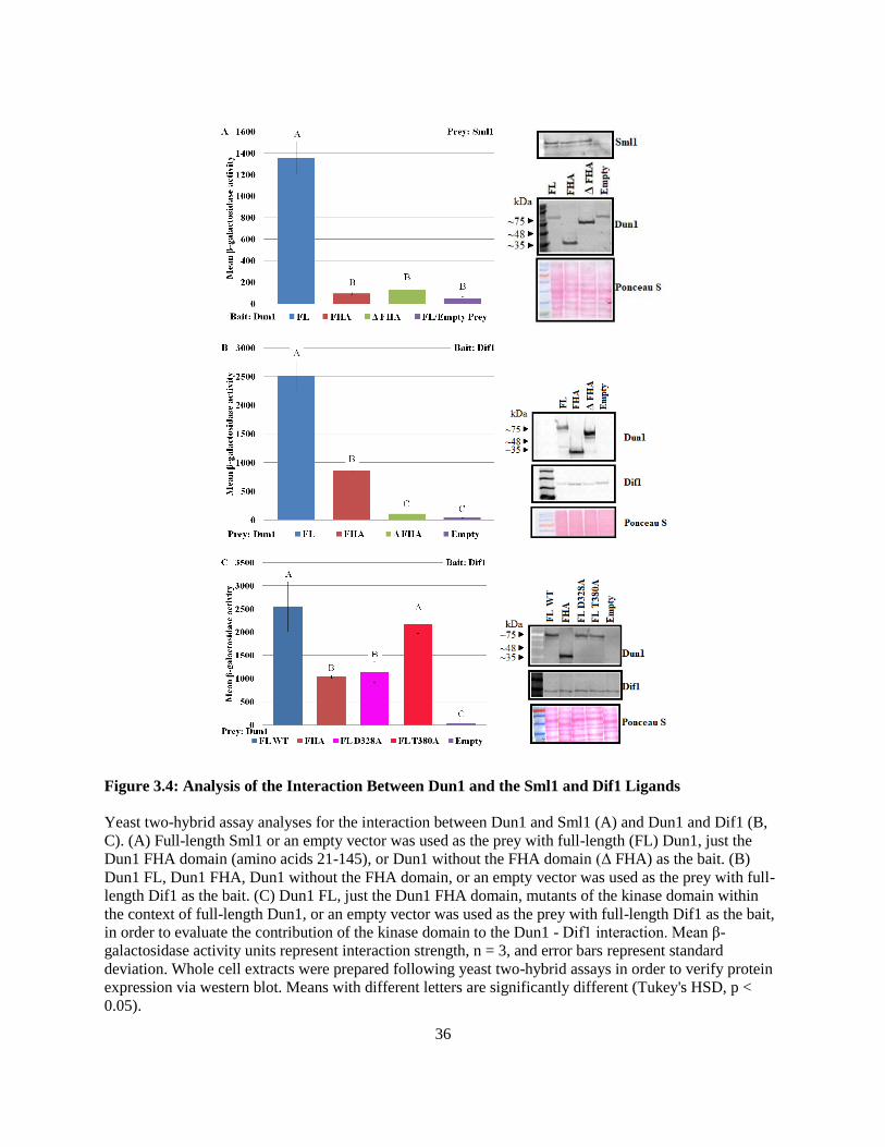
36
Figure 3.4: Analysis of the Interaction Between Dun1 and the Sml1 and Dif1 Ligands
Yeast two-hybrid assay analyses for the interaction between Dun1 and Sml1 (A) and Dun1 and Dif1 (B,
C). (A) Full-length Sml1 or an empty vector was used as the prey with full-length (FL) Dun1, just the
Dun1 FHA domain (amino acids 21-145), or Dun1 without the FHA domain (Δ FHA) as the bait. (B)
Dun1 FL, Dun1 FHA, Dun1 without the FHA domain, or an empty vector was used as the prey with full-
length Dif1 as the bait. (C) Dun1 FL, just the Dun1 FHA domain, mutants of the kinase domain within
the context of full-length Dun1, or an empty vector was used as the prey with full-length Dif1 as the bait,
in order to evaluate the contribution of the kinase domain to the Dun1 - Dif1 interaction. Mean β-
galactosidase activity units represent interaction strength, n = 3, and error bars represent standard
deviation. Whole cell extracts were prepared following yeast two-hybrid assays in order to verify protein
expression via western blot. Means with different letters are significantly different (Tukey's HSD, p <
0.05).

37
The involvement of Dun1 kinase activity and/or Rad53-dependent Dun1 phosphorylation and
activation in the Dun1 - Dif1 and Dun1 - Sml1 interactions was evaluated using yeast two-hybrid assays
that included the following Dun1 kinase domain mutants: D328A, a substitution of the aspartic acid at
position 328 to an alanine in order to eliminate Dun1 kinase activity from plasmid-expressed Dun1, and
T380A, a substitution of the threonine at position 380 to an alanine preventing Rad53-dependent
phosphorylation and activation of Dun1 (Zhou and Elledge, 1993; Zhao and Rothstein, 2002; Chen et al.,
2007; reviewed in Sanvisens et al., 2014). Without the kinase domain, Dun1 was unable to interact with
Sml1 (Figure 3.4A). However, absence of the kinase domain only weakened the interaction between
Dun1 and Dif1 (Figure 3.4B). The 'kinase dead' D328A Dun1 mutant exhibited the same level of β-
galactosidase activity when interacting with Dif1 as the Dun1 FHA domain while the T380A mutant of
Dun1 that could not be phosphorylated and activated by Rad53 exhibited roughly the same level of β-
galactosidase activity as full-length Dun1 when interacting with Dif1 (Figure 3.4C).

38
3.2.2 Highly conserved residues of the pThr-binding site and lateral surface interaction patch
influence detectable genomic level protein expression
The 3-D ribbon diagrams corresponding to the surface models for the Dun1 FHA domain shown
in Figure 3.2 are depicted in Figure 3.5. The ribbon diagrams show key residues that were selected for
mutation of the canonical pThr-binding site and the candidate non-canonical FHA domain lateral surface
interaction patch. Amino acid conservation colouring corresponds to Figure 3.2.
Figure 3.5: Conserved Surface Residues of the Dun1 FHA Domain Canonical pThr-binding Site
and Non-canonical FHA Domain Lateral Surface Interaction Patch
Ribbon diagrams of the Dun1 FHA domain (PDB template ID: 2JQJ) showing candidate highly
conserved residues selected for site-directed mutagenesis, using colours ranging from red (high
conservation) to blue (low conservation), in order to evaluate the involvement of the pThr-binding site
and/or lateral surface interaction patch of the Dun1 FHA domain in protein-protein interactions important
for dNTP regulation. Letters refer to the single letter amino acid code with numbers representing amino
acid position.
The Dun1 FHA domain has a di-phosphothreonine-binding motif at the pThr-binding site that is
necessary for Dun1 recognition of hyperphosphorylated Rad53 (Lee et al., 2003; Lee et al., 2008b;
Bashkirov et al., 2003; reviewed in Sanvisens et al., 2014). The arginine residue at position 60 (R60) is
necessary for Dun1 FHA domain recognition of the first phosphothreonine residue in Rad53 while lysine
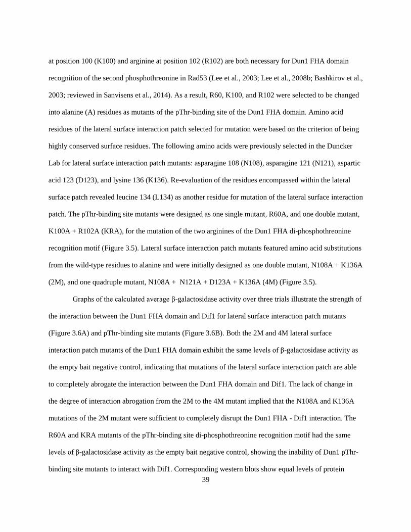
39
at position 100 (K100) and arginine at position 102 (R102) are both necessary for Dun1 FHA domain
recognition of the second phosphothreonine in Rad53 (Lee et al., 2003; Lee et al., 2008b; Bashkirov et al.,
2003; reviewed in Sanvisens et al., 2014). As a result, R60, K100, and R102 were selected to be changed
into alanine (A) residues as mutants of the pThr-binding site of the Dun1 FHA domain. Amino acid
residues of the lateral surface interaction patch selected for mutation were based on the criterion of being
highly conserved surface residues. The following amino acids were previously selected in the Duncker
Lab for lateral surface interaction patch mutants: asparagine 108 (N108), asparagine 121 (N121), aspartic
acid 123 (D123), and lysine 136 (K136). Re-evaluation of the residues encompassed within the lateral
surface patch revealed leucine 134 (L134) as another residue for mutation of the lateral surface interaction
patch. The pThr-binding site mutants were designed as one single mutant, R60A, and one double mutant,
K100A + R102A (KRA), for the mutation of the two arginines of the Dun1 FHA di-phosphothreonine
recognition motif (Figure 3.5). Lateral surface interaction patch mutants featured amino acid substitutions
from the wild-type residues to alanine and were initially designed as one double mutant, N108A + K136A
(2M), and one quadruple mutant, N108A + N121A + D123A + K136A (4M) (Figure 3.5).
Graphs of the calculated average β-galactosidase activity over three trials illustrate the strength of
the interaction between the Dun1 FHA domain and Dif1 for lateral surface interaction patch mutants
(Figure 3.6A) and pThr-binding site mutants (Figure 3.6B). Both the 2M and 4M lateral surface
interaction patch mutants of the Dun1 FHA domain exhibit the same levels of β-galactosidase activity as
the empty bait negative control, indicating that mutations of the lateral surface interaction patch are able
to completely abrogate the interaction between the Dun1 FHA domain and Dif1. The lack of change in
the degree of interaction abrogation from the 2M to the 4M mutant implied that the N108A and K136A
mutations of the 2M mutant were sufficient to completely disrupt the Dun1 FHA - Dif1 interaction. The
R60A and KRA mutants of the pThr-binding site di-phosphothreonine recognition motif had the same
levels of β-galactosidase activity as the empty bait negative control, showing the inability of Dun1 pThr-
binding site mutants to interact with Dif1. Corresponding western blots show equal levels of protein
40
expression for the Dun1 mutants and Dif1, with the exception of the 2M and 4M Dun1 FHA domain
mutants which appear to be lower than the WT Dun1 FHA domain level of protein expression.
Figure 3.6: Analysis of the Interaction Between Dun1 FHA Domain Mutants and Dif1
Yeast two-hybrid assays evaluating the importance of the non-canonical FHA domain lateral surface
interaction patch (A) and the canonical pThr-binding site (B) to the interaction between the Dun1 FHA
domain and Dif1. Wild-type (WT) Dun1 FHA, mutants of the lateral surface interaction patch or pThr-
binding site, or an empty vector was used as the prey with full-length Dif1 as the bait. Mean β-
galactosidase activity units represent interaction strength, n = 3, and error bars represent standard
deviation. Whole cell extracts were prepared following yeast two-hybrid assays in order to verify protein
expression via western blot. (A) Point mutations changed asparagine 108, lysine 136, asparagine 121, and
aspartic acid 123 to alanine. 2M and 4M denote a double and quadruple mutant of the non-canonical FHA
domain lateral surface interaction patch of Dun1 [2M = N108A + K136A and 4M = N108A + K136A +
N121A + D123A]. (B) Point mutations changed arginine 60, lysine 100 and arginine 102 to alanine.
R60A and KRA denote a single and double mutant of the canonical pThr-binding site of Dun1 [KRA =
K100A + R102A]. Means with different letters are significantly different (Tukey's HSD, p < 0.05).

41
Since the 2M mutations were enough to completely disrupt the interaction between the Dun1
FHA domain and Dif1, the mutations of the 2M mutant were split into an N108A single mutant and a
K136A single mutant (Figure 3.7). The graph of average β-galactosidase activity calculated from three
yeast two-hybrid trials shows that the N108A mutation of the lateral surface interaction patch reduces the
Dun1 FHA domain - Dif1 interaction to roughly one-third of the strength of the interaction seen with the
wild-type (WT) Dun1 FHA domain, a level similar to the empty vector negative control, which indicates
complete disruption of the interaction (Figure 3.7A). The 2M double mutant and R60A single mutant both
show a lack of interaction with Dif1 as seen in Figure 3.6A. The K136A single mutant exhibited an
increase in the level of β-galactosidase activity relative to the WT Dun1 FHA domain for the interaction
with Dif1, suggesting that the interaction between the Dun1 FHA domain and Dif1 is stronger with the
K136A mutation of the lateral surface interaction patch (Figure 3.7B). The protein expression of Dun1
and Dif1 were confirmed via western blotting. Western blots show that the protein levels for the N108A
and 2M Dun1 FHA mutants appear to be lower than that of the WT Dun1 FHA domain and the K136A
mutant. The Dun1 2M FHA - Dif1 sample for Figure 3.7A was not expressing the Dif1 protein like the
other samples.
Due to the unexpected increase in Dun1 FHA - Dif1 interaction strength for the Dun1 K136A
FHA mutant, additional yeast two-hybrid assays were conducted to evaluate the extent of the increase in
the interaction within the context of full-length Dun1 or in contrast to full-length WT Dun1 (Figure 3.8).
Average β-galactosidase activity was calculated as a percentage of the WT for the analysis of the K136A
mutation in full-length Dun1 over three trials (Figure 3.8A). The levels of β-galactosidase activity remain
the same between the WT full-length Dun1 and K136A full-length Dun1 mutant with Dif1 (Figure 3.8A).
The average β-galactosidase activity over three trials was graphed for the comparison of the K136A
mutation in just the Dun1 FHA domain to full-length WT Dun1 and shows that the K136A mutation in
the Dun1 FHA domain has a stronger interaction with Dif1 than full-length WT Dun1 (Figure 3.8B). The

42
corresponding western blots show relatively equal levels of protein expression with the exception of Dif1
in Figure 3.8A.
Figure 3.7: Dun1 FHA - Dif1 Interaction Analysis for the N108A and K136A Mutations
Yeast two-hybrid assays evaluating the effect of the (A) single N108A mutation, asparagine 108
converted to alanine, and the (B) single K136A mutation, lysine 136 converted to alanine, on the strength
of the interaction between the Dun1 FHA domain and Dif1. Wild-type (WT) Dun1 FHA, single or double
mutants of the non-canonical FHA domain lateral surface interaction patch, or an empty vector was used
as the prey with full-length Dif1 as the bait [2M = N108A + K136A, a non-canonical FHA domain lateral
surface interaction patch double mutant]. R60A is a single mutant of the canonical pThr-binding site
where arginine 60 was changed to an alanine. Mean β-galactosidase activity units represent interaction
strength, n = 3, and error bars represent standard deviation. Whole cell extracts were prepared following
yeast two-hybrid assays in order to verify protein expression via western blot. Means with different letters
are significantly different (Tukey's HSD, p < 0.05).

43
Figure 3.8: Analysis of the K136A Mutation in Full-length Dun1 and the Dun1 FHA Domain
Yeast two-hybrid assays evaluating the effect of the K136A, lysine 136 converted to alanine, mutation of
the non-canonical FHA domain lateral surface interaction patch of the Dun1 FHA domain on its
interaction with Dif1 in the context of full-length (FL) Dun1 (A) and just the FHA domain (B) compared
to full-length wild-type (FL WT) Dun1. Wild-type full-length Dun1, mutant full-length Dun1, or an
empty vector was used as the prey with full-length Dif1 as the bait (A) and wild-type full-length Dun1,
mutant Dun1 FHA, or an empty vector was used as the prey with Dif1 as the bait (B). The mean
percentage of the wild-type level of β-galactosidase activity units (A) and the mean β-galactosidase
activity units (B) represent interaction strength, n = 3, and error bars represent standard deviation. Whole
cell extracts were prepared following yeast two-hybrid assays in order to verify protein expression via
western blot. Means with different letters are significantly different (Tukey's HSD, p < 0.05).

44
Spot plate assays were used to determine whether mutations of the pThr-binding site or lateral
surface interaction patch would result in yeast cells being more or less sensitive to hydroxyurea, HU, or
methyl methanesulfonate, MMS (Figure 3.9). The dun1Δ yeast strain was provided with full-length
DUN1 or dun1 mutants harbouring a c-terminal myc13 tag, in order to monitor protein expression, from a
yeast centromeric pRS315 plasmid which can mimic genomic expression due to the single copy number
maintenance of the plasmid (Clarke and Carbon, 1980; Ishii et al., 2009; Stearns et al., 1990). Primary
spot plate assays were used to assess the functional complementation of plasmid supplied Dun1. The spot
plate assay shows that dun1Δ supplied with empty plasmid exhibits sensitivity to MMS and HU at a
minimum of 0.015% MMS and 60 mM HU (Figure 3.9A). The addition of DUN1 expressed from the
pRS315 vector is able to rescue the sensitivity of the dun1Δ knock out mutant regardless of the addition
of the c-terminal myc13 tag, confirming that not only is the pRS315 DUN1 construct able to complement
the dun1Δ genomic knockout, but that the addition of the c-myc13 tag for evaluating protein expression
does not hinder Dun1 function. The 'untreated' control plate that does not have any genotoxic chemicals
confirms the lack of any growth defects in the absence of genotoxic stress, indicating that any observed
sensitivities on treated plates are a result of the genotoxic stress. The western blot confirms the addition of
the c-myc13 tag and its usefulness in monitoring protein expression. Figure 3.9B shows the assessment of
the genotoxic sensitivities of the dun1Δ yeast strain supplied with DUN1 or dun1 mutants, where the FHA
domain has been mutated at the pThr-binding site or the lateral surface interaction patch, expressed from
the pRS315 plasmid. The 'untreated' control plate again confirms that all samples initially grow
successfully. At 80 mM HU all samples of dun1Δ supplied with plasmid-expressed dun1 for lateral
surface interaction patch mutations, with the exception of the R60A and K136A single mutations, begin
to show sensitivity to HU; sensitivity can also be observed at 0.015% MMS. The dun1Δ samples with
plasmid-expressed dun1 for the R60A and K136A mutations exhibit slight but reproducible sensitivity to
the highest concentrations of HU or MMS. The dun1Δ sample with plasmid-expressed dun1 for the
ΔFHA mutation shows sensitivity to HU, beginning at 80 mM, and to 0.015% MMS.

45
Figure 3.9: Functional Complementation of dun1Δ and Genotoxic Sensitivity Assessment for Dun1
FHA Mutants
Spot plate assays for (A) functional complementation of the dun1Δ genomic knockout with plasmid-
expressed c-myc13-tagged Dun1 and (B) genotoxic sensitivity of yeast cells with plasmid-expressed dun1
to hydroxyurea (HU) and methyl methanesulfonate (MMS). Whole cell extracts were prepared from
cultures used for spot plate assays in order to verify protein expression via western blot. Plates were
incubated for two days at 30°C. Mutants consisted of single, double, or quadruple mutations for select
residues: asparagine 108, lysine 136, asparagine 121, aspartic acid 123, arginine 60, lysine 100, and
arginine 102, changed to alanine or removal of the gene sequence for the FHA domain. Selective
synthetic complete media lacking leucine was used for plasmid maintenance. Each row of samples
represents one of four 10-fold serial dilutions spotted from the highest concentration of cells to the lowest.
46
The corresponding western blot shows that only the WT, K136A, and R60A versions of Dun1 are being
expressed at the same level. The N108A mutant exhibits compromised protein expression, as does the 2M
mutant, while the 4M, KRA and ΔFHA mutants are not visibly expressed.
Due to the slight genotoxic sensitivity observed for dun1Δ supplied with plasmid-expressed dun1
for the K136A or R60A mutations, temperature and other genotoxic agents were included as independent
variables for analysis (Figure 3.10). The Figure 3.9B spot plate assay was repeated, excluding the samples
for mutants that did not show WT levels of protein expression, using incubation temperatures of either
30°C, the regularly used optimal temperature, or 37°C (Figure 3.10A). The increase in temperature,
which can denature and destabilize proteins, coupled with HU or MMS stress was used in order to
potentially amplify subtle growth defects. All samples collectively exhibited decreased growth on the
plates incubated at 37°C compared to plates incubated at 30°C, but there was no effect on growth for the
K136A or R60A mutant expressing samples compared to WT due to the increase in incubation
temperature. The western blot confirmed equal expression of the WT, K136A, and R60A Dun1 proteins.
Since an increased temperature coupled with HU or MMS exposure did not reveal an increase in the
genotoxic sensitivity of the K136A or R60A mutant expressing samples, several other genotoxic
chemicals used previously in the Duncker Lab: Bleomycin, Phleomycin, and Camptothecin, were selected
for analysis in order to assess dun1 mutant sensitivities to other types of genotoxic stress (Figure 3.10B).
A preliminary spot plate assay was used to determine whether the dun1Δ genomic knockout strain was
sensitive to Bleomycin, Phleomycin, or Camptothecin via the comparison of the dun1Δ strain to the
highly sensitive rad53Δsml1Δ strain (Figure 3.10B). The lack of dun1Δ and rad53Δsml1Δ growth on the
Bleomycin and Phleomycin plates relative to the untreated plate and their positive controls on treated
plates demonstrates the observable sensitivity of the dun1Δ strain to Bleomycin and Phleomycin. The
decrease in growth of dun1Δ and rad53Δsml1Δ on the Camptothecin plates relative to the untreated plate
and their positive controls on treated plates demonstrates the observable sensitivity of the dun1Δ strain to
Camptothecin. There was no requirement for the maintenance of an external plasmid for the initial

47
assessment of dun1Δ sensitivity to Bleomycin, Phleomycin, and Camptothecin shown in Figure 3.10B, so
agar plates for that assay were made using YPD media instead of the selective SC drop out media used for
all other spot plate assays where the pRS315 plasmid needed to be maintained. After the establishment of
dun1Δ sensitivity to Bleomycin, Phleomycin and Camptothecin, the dun1Δ strain with plasmid-supplied
DUN1 or dun1 mutants was tested for sensitivity to Bleomycin, Phleomycin, and Camptothecin using
agar plates that were made with selective SC drop out media (data not shown). None of the samples
grown on the selective SC plates showed any sensitivity to Bleomycin, Phleomycin, or Camptothecin.
Most striking was the observation that even the dun1Δ strain supplied with an empty plasmid negative
control did not show any sensitivity to Bleomycin, Phleomycin, or Camptothecin when it was already
observed in Figure 3.10B that dun1Δ is sensitive to all three genotoxins. As a result, the spot plate assays
were repeated using non-selective YPD media instead of selective SC drop out media (data not shown).
The observations that the wild-type strain/empty plasmid sample exhibits growth on all YPD plates
containing Bleomycin, Phleomycin, or Camptothecin while the dun1Δ strain/empty plasmid sample has
compromised growth on YPD plates containing Bleomycin, Phleomycin, or Camptothecin indicate that
the type of media may influence the activity of Bleomycin, Phleomycin, and Camptothecin. As a result,
the analysis of dun1Δ with plasmid-supplied DUN1 or dun1 mutants remains inconclusive due to the lack
of selective pressure for plasmid maintenance with YPD media.
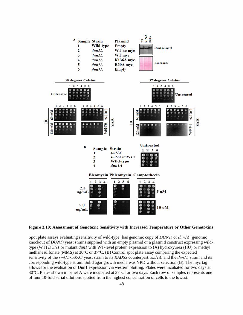
48
Figure 3.10: Assessment of Genotoxic Sensitivity with Increased Temperature or Other Genotoxins
Spot plate assays evaluating sensitivity of wild-type (has genomic copy of DUN1) or dun1Δ (genomic
knockout of DUN1) yeast strains supplied with an empty plasmid or a plasmid construct expressing wild-
type (WT) DUN1 or mutant dun1 with WT-level protein expression to (A) hydroxyurea (HU) or methyl
methanesulfonate (MMS) at 30°C or 37°C. (B) Control spot plate assay comparing the expected
sensitivity of the sml1Δrad53Δ yeast strain to its RAD53 counterpart, sml1Δ, and the dun1Δ strain and its
corresponding wild-type strain. Solid agar growth media was YPD without selection (B). The myc tag
allows for the evaluation of Dun1 expression via western blotting. Plates were incubated for two days at
30°C. Plates shown in panel A were incubated at 37°C for two days. Each row of samples represents one
of four 10-fold serial dilutions spotted from the highest concentration of cells to the lowest.

49
3.2.3 K136-central projection of the Dun1 non-canonical FHA domain lateral surface interaction
patch contributes to interactions involved in dNTP regulation
The re-assessment of the ribbon diagram showing highly conserved lateral surface interaction
patch residues revealed a side-chain 'projection' on the lateral surface of the Dun1 FHA domain (Figure
3.11). The largest contribution to this projection came from the side-chain of lysine 136. In addition to
lysine 136, the asparagine 121 (N121) and leucine 134 (L134) side-chains also contribute to this surface
projection. Due to the observation that the K136A Dun1 single mutant had a WT Dun1 level of protein
expression from the pRS315 plasmid, N121A and L134A were created as two Dun1 lateral surface
interaction patch single mutants to be included in yeast two-hybrid assays (Figure 3.12), evaluated for
stable protein expression, and then used to assess HU and MMS sensitivity (Figure 3.13).
Figure 3.11: Illustration of the K136-central Projection on the Dun1 FHA Domain Lateral Surface
Interaction Patch
Ribbon diagram of the 3-D model for the Dun1 FHA domain (PDB template ID: 2JQJ) showing
conservation of FHA domain residues with colours ranging from red (high conservation) to blue (low
conservation) and depicting the side-chain projections of the lysine 136 (K136) amino acid and two
neighbouring residues asparagine 121 (N121) and leucine 134 (L134). Side chains were coloured
according to the conservation level of their associated residue with a mesh outline illustrating the surface
of the residue.

50
The N121A and L134A single mutants were added as potential lateral surface interaction patch
mutants that could disrupt the interactions between the Dun1 FHA domain and Dif1 or Sml1. Yeast two-
hybrid assays were used to evaluate whether the N121A and L134A single mutants had any effect on the
interactions of Dun1 with Dif1 (Figure 3.12A) or Sml1 (Figure 3.12B). Calculations of the average β-
galactosidase activity over three trials were graphed to illustrate that neither the N121A mutation nor the
L134A mutation have any effect on the interaction strength between the Dun1 FHA domain and Dif1
(Figure 3.12A). As observed previously, the K136A mutation generated a large increase in the interaction
strength between the Dun1 FHA domain and Dif1 while the R60A mutation completely abrogated the
interaction. The corresponding western blot shows equal levels of Dun1 and Dif1 expression across
samples. The graph shown in Figure 3.12B shows the calculated averages of β-galactosidase activity over
three trials for the assessment of the Dun1 - Sml1 interaction. The N121A mutation has no affect on the
interaction between full-length Dun1 and Sml1 whereas the L134A mutation reduces the interaction
strength by approximately 50% (Figure 3.12B). In contrast to the Dun1 - Dif1 interaction, the K136A
mutation decreases the strength of the Dun1 - Sml1 interaction by approximately one-third, while the
R60A mutation reduces the interaction strength to less than half of the WT Dun1 - Sml1 interaction. The
corresponding western blot shows that both Dun1 and Sml1 exhibit equivalent levels of protein
expression across samples.
The R60A, N121A, L134A, and K136A Dun1 single mutants were then expressed for spot plate
assay assessments of HU and MMS genotoxic sensitivity in the dun1Δ yeast strain (Figure 3.13). Samples
of dun1Δ supplied with plasmid-expressed dun1 for the K136A or R60A mutations continued to only
show slight but reproducible sensitivity to the highest concentrations of HU or MMS relative to the wild-
type/empty plasmid positive control sample, while dun1Δ supplied with plasmid-expressed dun1 for the
N121A or L134A mutations exhibited stronger sensitivity to HU at 150 mM and MMS at 0.015%,
although still not as much as the dun1Δ/empty plasmid negative control sample. The western blot

51
confirmed equivalent levels of protein expression for all Dun1 mutant proteins relative to WT Dun1
(Figure 3.13).
Figure 3.12: Analysis of the K136-central Projection Substitution Mutants for the Dun1 - Dif1 and
Dun1 - Sml1 Interactions
Yeast two-hybrid assays evaluating the change in interaction strength between Dun1 and Dif1 (A) or
Sml1 (B) with a variety of Dun1 FHA domain mutants. (A) Wild-type (WT) Dun1 FHA, indicated
mutants of the FHA domain, or an empty vector was used as the prey with full-length Dif1 as the bait. (B)
Full-length Sml1 or an empty vector was used as the prey with WT full-length (FL) Dun1 or indicated
Dun1 FL mutants as the bait. Missense mutations were used to change the highly conserved asparagine
121 (N121), leucine 134 (L134), lysine 136 (K136), and arginine 60 (R60) residues into alanine in order
to mutate the non-canonical FHA domain lateral surface interaction patch (N121A, L134A, and K136A)
or the canonical pThr-binding site (R60A). Mean β-galactosidase activity units represent interaction
strength, n = 3, and error bars represent standard deviation. Whole cell extracts were prepared following
yeast two-hybrid assays in order to verify protein expression via western blot. Means with different letters
are significantly different (Tukey's HSD, p < 0.05).

52
Figure 3.13: Genotoxic Sensitivity Analysis for K136-central Projection Mutants
Spot plate assay evaluating the sensitivity of wild-type (has genomic copy of DUN1) or dun1Δ (genomic
knockout of DUN1) yeast strains supplied with an empty plasmid or a plasmid construct of wild-type
(WT) DUN1 or mutant dun1 with WT Dun1 levels of protein expression (verified via western blot using
the c-myc tag added to the c-terminus of the Dun1 coding sequence in the pRS315 plasmid) to
hydroxyurea (HU) or methyl methanesulfonate (MMS). Solid agar growth media was synthetic complete
(SC) media lacking leucine, for selection. All plates were incubated for two days at 30°C. Mutants
consisted of single missense mutations of the non-canonical FHA domain lateral surface interaction patch
(N121A, L134A, and K136A) and the canonical pThr-binding site (R60A). Highly conserved asparagine
121 (N121), leucine 134 (L134), lysine 136 (K136), and arginine 60 (R60) were changed to alanine. Each
row of samples represents one of four 10-fold serial dilutions spotted from the highest concentration of
cells to the lowest.
3.3 Discussion
The data described throughout this thesis both confirms the existence of the conserved non-
canonical FHA domain lateral surface interaction patch on the Dun1 FHA domains and illuminates its
contribution to protein-protein interactions involved in dNTP regulation. The yeast two-hybrid assays
shown in Figures 3.3 and 3.4 demonstrate the importance of the Dun1 FHA domain to Dun1-based
protein-protein interactions. Unique to the interaction between Dun1 and Sml1, both the FHA and kinase
53
domains were necessary for the interaction (Figure 3.4A). For the Dun1 - Dif1 interaction, the FHA
domain was necessary for the interaction but not fully sufficient (Figure 3.4B). Removal of the Dun1
kinase domain weakened the Dun1 - Dif1 interaction and the disruption of the kinase activity of plasmid-
expressed Dun1 weakened the Dun1 - Dif1 interaction to the same extent as removal of the kinase
domain, suggesting that Dun1 phosphorylation may contribute to protein-protein interactions between
Dun1 and its downstream ligands (Figure 3.4C). The FHA domain of Dun1 on its own was sufficient for
interaction with the Rad53, Wtm1, and Crt1 ligands (Figure 3.3). Since most of the Dun1 ligands exhibit
prior associations with other proteins as part of their regular inhibitory functions, it may be that the
interactions between Dun1 and its ligands are based on competitive binding (Lee and Elledge, 2006; Lee
et al., 2008a; Andreson et al., 2010; reviewed in Tsaponina et al., 2011 and Sanvisens et al., 2016). The
low β-galactosidase activity levels for the interactions between Dun1 without its FHA domain and the
Rad53, Wtm1, and Crt1 ligands relative to the empty vector negative control and/or other samples that
indicated a lack of detectable protein-protein interactions was unexpected. The current speculation
remains that removal of the entire FHA domain results in the decreased opportunity for chance
occurrences of bait and prey construct proximity as it was anticipated that their β-galactosidase activity
levels would be the same as the empty vector negative controls and other samples without observable
interactions.
The reduction or lack of protein expression for the N108A, 2M, 4M, Δ FHA, and KRA Dun1
mutants for the spot plate assay shown in Figure 3.9B demonstrated that the decrease in β-galactosidase
activity observed between the Dun1 N108A FHA mutant and Dif1 was likely not a result of interaction
abrogation but rather due to a decrease in Dun1 protein levels for the Dun1 N108A mutant (Figure 3.7A)
and that the interaction abrogation observed for the 2M, 4M, and KRA mutants were also likely due to
diminished protein expression (Figure 3.6). The same cannot be said of the Dun1 Δ FHA mutant seen
throughout Figures 3.3 and 3.4 because there was not an obvious decrease in the over-expression of the
Dun1 Δ FHA mutant protein like there was for the N108A, 2M, 4M, and KRA mutants during the yeast
54
two-hybrid assays. While the K136A and R60A Dun1 mutants did show WT levels of protein expression
in Figure 3.9B, there was only slight but reproducible sensitivity of dun1Δ expressing those mutant
versions of dun1 to the highest concentrations of HU or MMS (Figure 3.9B, 3.10A). Despite the fact that
little to no sequence homology exists amongst the vast collection of discovered FHA domains, the
structure is highly conserved (reviewed in Mahajan et al., 2008). The observation that mutating highly
conserved residues located within the lateral surface interaction patch and pThr-binding site caused
compromised protein expression, suggests that highly conserved residues within the lateral surface
interaction patch as well as the pThr-binding site may contribute to protein stability as well as the
establishment of protein-protein interactions.
The spot plate assays testing Bleomycin, Phleomycin, and Camptothecin sensitivity yielded an
unexpected result (data not shown). Whereas HU and MMS are able to cause genotoxic sensitivity to
dun1Δ on SC media lacking leucine, leucine drop out provides the selective pressure that ensures the
maintenance of the pRS315 plasmid, neither Bleomycin, Phleomycin, nor Camptothecin appear to
function on SC media lacking leucine (data not shown). As a result, no conclusions could be made about
the genotoxic sensitivity of dun1Δ expressing mutant versions of dun1. In order to properly assess the
effects of those genotoxins, the mutations of interest will need to be created within the genome as
opposed to being expressed from an external plasmid thereby removing the need to use selective drop out
media.
The most fruitful analyses were generated after the discovery of the K136-central projection on
the lateral surface interaction patch (Figure 3.11). The addition of the N121A and L134A Dun1 single
mutants to the collection of mutants included in the yeast two-hybrid assays (shown in Figure 3.12)
revealed a contrasting pattern to the interaction between Dun1 and Dif1 compared to Dun1 and Sml1, a
revelation that might help explain the slight HU and MMS sensitivity seen with dun1Δ samples
expressing the K136A and R60A dun1 mutants from the pRS315 plasmid shown in Figure 3.9B and
3.10A. For the Dun1 - Dif1 interaction, neither the N121A mutation nor the L134A mutation had any

55
effect (Figure 3.12A). The K136A mutation generated a huge increase in the strength of the interaction
while the R60A mutation completely disrupted the interaction (Figure 3.12A). The pattern is mostly
reversed for the Dun1 - Sml1 interaction (Figure 3.12B). The N121A mutation exhibited no effect on the
interaction strength between Dun1 and Sml1 whereas the L134A mutation decreased the strength of the
interaction (Figure 3.12B). The K136A mutation also reduced the interaction strength and, unlike the
Dun1 - Dif1 interaction, the R60A mutation reduced the interaction strength as opposed to completely
disrupting it, suggesting that both the pThr-binding site and the lateral surface interaction patch are
needed for the Dun1 - Sml1 interaction. The pThr-binding site appears to be the primary interface for the
Dun1 - Dif1 interaction with the lateral surface interaction patch functioning only to modulate the
strength of the interaction and/or the initial affinity between Dun1 and Dif1 (Figure 3.12B).
The Figure 3.13 spot plate assay shows that the N121A and L134A-expressing dun1Δ mutants
show sensitivity to HU and MMS while the K136A and R60A-expressing dun1Δ mutants only showed
slight sensitivity to the highest concentrations of HU and MMS. The slight sensitivity of the latter two
samples was initially surprising when it was observed in Figure 3.9B and again in Figure 3.10A, but
considering the pattern with which Dun1 interacts with Dif1 and Sml1, the lack of sensitivity may just be
a reflection of the complexity and/or multi-faceted nature of the Dun1 contribution to dNTP regulation
keeping in mind that the effects of the mutations have only been analyzed for the Dif1 and Sml1 ligands
so far. The K136A mutation exhibits no detrimental effect to the Dun1 - Dif1 interaction when in the
context of full-length Dun1, although it increases the interaction strength between the Dun1 FHA domain
and Dif1 beyond the level of the Dun1 FL - Dif1 interaction when in the context of just the FHA domain,
likely due to the isolation of the FHA domain and the change in the size of the side chain as a result of the
mutation (Figure 3.8). With respect to the Dun1 - Sml1 interaction, the K136A mutation only weakens the
strength of the interaction and not by more than 50% (Figure 3.12B). Considering dNTP regulation as a
whole, the K136A mutation alone would not likely have a very drastic defect on the ability of a cell to
still manage to regulate dNTP levels especially given the fact that there are multiple factors involved in

56
regulating dNTP synthesis. On the other hand, the R60A mutation completely disrupted the Dun1 - Dif1
interaction and weakened the Dun1 - Sml1 interaction but still did not show strong HU and MMS
sensitivity (Figure 3.12-3.13). One could speculate that the lack of strong sensitivity may be because the
R60A mutant is still able to interact with other Dun1 ligands, at least marginally, in order to relieve
enough of the collective inhibitory activities involved in dNTP regulation to still permit adequate dNTP
synthesis when cells are stressed, but confirmation would depend on an evaluation of the effects of the
R60A mutant on the other Dun1 interactions keeping in mind that the Dun1 FHA pThr-binding site has a
di-phosphothreonine-recognition motif, although it has currently only been shown to contribute to the
Rad53 - Dun1 interaction (Lee et al., 2003; Lee et al., 2008b; Bashkirov et al., 2003; reviewed in
Sanvisens et al., 2014). The observations that the N121A and L134A mutations generate, to a degree,
genotoxic sensitivity suggests that some mutations may disrupt more interactions than others and that by
doing so are more capable of interfering with dNTP regulation.

57
Chapter 4
General Conclusions and Future Directions
4.1 The Dun1 kinase domain and the non-canonical FHA domain lateral surface
interaction patch influence protein-protein interactions involved in dNTP regulation
The initial yeast two-hybrid assays confirming the existence of protein-protein interactions
between Dun1 and the other proteins involved in the dNTP regulation pathway demonstrated the
importance of the Dun1 FHA domain to those interactions. Establishment of the interactions between
Dun1 and two of its ligands, Dif1 and Sml1 depended on both the Dun1 FHA and kinase domains, while
only the Dun1 FHA domain seemed to be able to interact with the Rad53, Wtm1, and Crt1 ligands. The
unexpected observation that Crt1 could activate β-galactosidase activity on its own in the two-hybrid
assay was likely due to an interaction between Crt1 in the prey vector and the LexA operators on the
reporter vector in the absence of Dun1 as the bait. This implicated Dun1 as being a protein that utilizes
competitive binding, an implication supported by the fact that Dun1 is not the 'customary' binding partner
for other proteins involved in dNTP regulation as Wtm1, Crt1, Dif1, and Sml1 all carry out their normal
inhibitory functions via their associations with other proteins.
Yeast two-hybrid assays evaluating the Dun1 FHA domain mutants with respect to the Dun1-
Dif1 and Dun1-Sml1 interactions proved that the identified patch of conserved residues on the lateral
surface of the Dun1 FHA domain constitute a non-canonical FHA domain lateral surface interaction patch
that contributes to Dun1 protein-protein interactions both independently and in conjunction with the
canonical pThr-binding site. The collection of Dun1 mutants that were examined revealed that highly
conserved residues of the lateral surface interaction patch and the pThr-binding site participate in FHA

58
domain-based protein-protein interactions and that mutations of some conserved residues compromise
'genomic level' protein expression.
The multiple downstream targets of Dun1 each carry out individual yet interconnected functions.
Single mutants of the Dun1 FHA domain had different effects on the strength of the interactions between
Dun1 and its downstream targets. Spot plate assays examining Dun1 mutants showed that only some
cases of dun1Δ strain transformants expressing mutant dun1 from the pRS315 plasmid exhibited
sensitivity to HU and MMS. The observed instances of genotoxic sensitivity and the strength of the
sensitivity did not necessarily correspond with Dun1 FHA mutants that were able to disrupt Dun1
interactions, suggesting that the complexity of the dNTP regulation pathway allows cells to cope with the
loss of single parts of the pathway. Together the results of the yeast two-hybrid and spot plate assays
proved the involvement of the lateral surface interaction patch in the protein-protein interactions
orchestrating dNTP regulation and their importance to genotoxic stress resistance.
4.2 Future Directions
The data presented in this thesis presents an excellent general examination of the dNTP regulation
pathway and the Dun1 FHA domain-based protein-protein interactions that orchestrate it. The results also
demonstrate the value of investigating other FHA domain-containing proteins in budding yeast and
looking for the existence of additional conserved FHA domain lateral surface interaction patches.
However, in order to establish an understanding of Dun1 ligand recognition and binding as well as the
Dun1 FHA domain lateral surface interaction patch and its contribution to dNTP regulation that is both
cohesive and comprehensive, all remaining ambiguities need to be addressed. Upon discovering that the
kinase domain, the only other structured region of Dun1, was necessary for the Dun1-Sml1 interaction,
the contribution of the kinase domain was evaluated with respect to the Dun1-Dif1 interaction. D328A
and T380A kinase domain single mutants were incorporated into yeast two-hybrid assays evaluating the
interaction between Dun1 and Dif1 showing that the kinase activity of plasmid-expressed Dun1

59
contributed to the full-length Dun1 interaction with Dif1. The same mutants should be tested with Sml1 in
order to determine whether the Dun1-Sml1 interaction benefits from the kinase domain in a similar
fashion.
Protein-protein interactions were observed between the Dun1 FHA domain and the Rad53,
Wtm1, and Crt1 ligands, but the effects of Dun1 FHA mutations on those interactions have yet to be
tested. In order to make any strong conclusions about the frequency of disrupted protein-protein
interactions due to a particular mutation, all interactions need to be assessed. As such, yeast two-hybrid
assays should be used to examine whether the lateral surface interaction patch and pThr-binding site
mutants have any effect on the interactions between the Dun1 FHA domain and the Rad53, Wtm1, or
Crt1 ligands. A modification of the typical yeast two-hybrid assay that introduces another vector
containing the coding sequence for a protein that could compete with Dun1 for interactions with Rad53,
Wtm1, or Crt1 could be used to examine whether Dun1-based interactions rely on competitive binding to
induce dNTP synthesis. Mass spectroscopy and/or Nuclear Magnetic Resonance (NMR) of purified full-
length Dun1 or just the region of the Dun1 FHA domain could be used to determine the existence of any
post-translational modifications to the Dun1 FHA domain at or around the position of lysine 136 that
might explain the increase in Dun1 FHA-Dif1 interaction strength with the K136A mutation.
Neither mutants of the lateral surface interaction patch nor the pThr-binding site were able to
individually result in complete disruption of the Dun1-Sml1 interaction, but they did individually reduce
the strength of the interaction. A likely scenario, and one that needs to be tested, is whether a combined
mutant of both regions would completely disrupt the interaction between Dun1 and Sml1. Another
particularly useful examination would be to conduct a pull-down assay using purified Dun1, Dif1, and
Sml1 expressed from bacterial plasmids in Escherichia coli (E. coli) to determine whether the Dun1-Dif1
and Dun1-Sml1 interactions observed using yeast two-hybrid assays are direct physical interactions or
indirect. In the event that the interactions are direct, collaborating with a structural biology lab to solve
the co-crystal structure between the Dun1 FHA domain and full-length Dif1 and maybe full-length Dun1

60
and Sml1 would prove invaluable to a comprehensive analysis of the Dun1 lateral surface interaction
patch by depicting the exact residues involved in the interactions. Additionally, generating Dun1 FHA
domain mutations within the budding yeast genome instead of utilizing expression from an external
plasmid could be used to bypass the apparent inactivity of Bleomycin, Phleomycin, and Camptothecin on
selective SC media. Spot plate assays evaluating genotoxic sensitivity to other compounds besides HU
and MMS could reveal a more distinct phenotypic effect of Dun1 FHA mutants that have compromised
protein-protein interactions that are involved in the dNTP regulation pathway.
4.3 Impact and Relevance of FHA Domain Research
The canonical pThr-binding site is a prominent feature of FHA domains. Until recently, the scope
of research done on FHA domain-based protein-protein interactions did not extend beyond analyzing the
contribution of the pThr-binding site as the means of FHA domain ligand recognition (reviewed in
Mahajan et al., 2008). The discovery of the non-canonical FHA domain lateral surface interaction patch in
the FHA1 domain of budding yeast Rad53 and the characterization of its contribution to the interaction
between the FHA1 domain of Rad53 and the H-BRCT domain of Dbf4 paved the way for researchers to
broaden their vantage point when studying protein-protein interactions involving FHA domains
(Matthews et al., 2014). The research presented in this thesis confirming both the existence and
contribution of a conserved non-canonical FHA domain lateral surface interaction patch in another FHA
domain-containing protein, proves that the existence of a lateral surface interaction patch within an FHA
domain is not a unique feature of Rad53 FHA1. It demonstrates the importance of studying this non-
canonical FHA domain lateral surface interaction patch as a potential interaction interface in other FHA
domain-containing proteins. Since FHA domains are common amongst cell cycle regulation and
checkpoint proteins, a more comprehensive examination of FHA domains and the protein-protein
interactions that they mediate via the study of the occurrence and contribution of non-canonical FHA
domain lateral surface interaction patches in FHA domain-containing proteins could unveil the true

61
complexities of cell cycle regulation and checkpoints. Such revelations would advance our understanding
of cell cycle progression and regulation errors, key contributors to cancer and cancer-related diseases.
Such revelations will advance our techniques for preventative and post-diagnostic care for such diseases
by giving us the means to design targeted therapeutic tactics to attack the specific problems of disease
thereby avoiding the systemic damage that remains a prominent side-effect of treatments such as
chemotherapy and radiation.
62
Bibliography
Allen, J. B., Zhou, Z., Siede, W., Friedberg, E. C., & Elledge, S. J. (1994). The SAD1/RAD53 protein
kinase controls multiple checkpoints and DNA damage-induced transcription in yeast. Genes &
Development, 8(20), 2401-2415.
Ausubel, F. M., Brent, R., Kinston, R., Moore, D., Seidman, J. J., Smith, J., Struhl, K. eds. (1994).
Current Protocols in Molecular Biology. (Wiley, New York).
Andreson, B. L., Gupta, A., Georgieva, B. P., & Rothstein, R. (2010). The ribonucleotide reductase
inhibitor, Sml1, is sequentially phosphorylated, ubiquitylated and degraded in response to DNA
damage.38(19), 6490-6501.
Balasubramanian, M. K., Bi, E., & Glotzer, M. (2004). Comparative analysis of cytokinesis in budding
yeast, fission yeast and animal cells.
Barnum, K. J., & O'Connell, M. J. (2014). Cell cycle regulation by checkpoints. Methods in Molecular
Biology (Clifton, N.J.), 1170, 29-40.
Bashkirov, V. I., Bashkirova, E. V., Haghnazari, E., & Heyer, W. D. (2003). Direct kinase-to-kinase
signaling mediated by the FHA phosphoprotein recognition domain of the Dun1 DNA damage checkpoint
kinase. Molecular and Cellular Biology, 23(4), 1441-1452.
Chabes, A., Domkin, V., & Thelander, L. (1999). Yeast Sml1, a protein inhibitor of ribonucleotide
reductase. The Journal of Biological Chemistry, 274(51), 36679-36683.
Chen, S., Smolka, M. B., & Zhou, H. (2007). Mechanism of Dun1 activation by Rad53 phosphorylation
in saccharomyces cerevisiae. The Journal of Biological Chemistry, 282(2), 986-995.
Clarke, L., & Carbon, J. (1980). Isolation of a yeast centromere and construction of functional small
circular chromosomes. Nature, 287(5782), 504-509.

63
Dobson, M. J., Pickett, A. J., Velmurugan, S., Pinder, J. B., Barrett, L. A., Jayaram, M., & Chew, J. S. K.
(2005). The 2µm plasmid causes cell death in saccharomyces cerevisiae with a mutation in Ulp1
protease. Molecular and Cellular Biology, 25(10), 4299-4310.
Dohrmann, P. R., & Sclafani, R. A. (2006). Novel role for checkpoint Rad53 protein kinase in the
initiation of chromosomal DNA replication in saccharomyces cerevisiae. Genetics, 174(1), 87-99.
Duina, A. A., Miller, M. E., & Keeney, J. B. (2014). Budding yeast for budding geneticists: A primer on
the saccharomyces cerevisiae model system. Genetics, 197(1), 33-48.
Duncker, B. P., Shimada, K., Tsai-Pflugfelder, M., Pasero, P., & Gasser, S. M. (2002). An N-terminal
domain of Dbf4p mediates interaction with both origin recognition complex (ORC) and Rad53p and can
deregulate late origin firing. Proceedings of the National Academy of Sciences, 99(25), 16087-16092.
Durocher, D., Jackson, S. P. (2002). The FHA domain. FEBS Letters. 513: 58-66
Gershon, H., & Gershon, D. (2000). The budding yeast, saccharomyces cerevisiae, as a model for aging
research: A critical review. Mechanisms of Ageing and Development, 120(1-3), 1-22.
Goffeau, A., Barrell, B. G., Bussey, H., Davis, R. W., Dujon, B., Feldmann, H., Galibert, G., Hoheisel, J.
D., Johnston, M., Louis, E. J., Mewes, H. W., Murakami, Y., Philippsen, P., Tettelin, H., & Oliver, S. G.
(1996). Life with 6000 genes. Science (New York, N.Y.), 274(5287), 546, 563-7.
Guitor, A. Characterizing non-canonical forkhead-associated domain-binding interfaces in
Saccharomyces cerevisiae. [Senior Honours Thesis]. University of Waterloo; 2016. 55 p.
Gyuris, J., Golemis, E., Chertkov, H., & Brent, R. (1993). Cdi1, a human G1 and S phase protein
phosphatase that associates with Cdk2. Cell, 75(4), 791-803.
Harashima, H., Dissmeyer, N., & Schnittger, A. (2013). Cell cycle control across the eukaryotic
kingdom. Trends in Cell Biology, 23(7), 345-356.
Herskowitz, I. (1988). Life cycle of the budding yeast saccharomyces cerevisiae. Microbiological
Reviews, 52(4), 536-553.

64
Hofmann, K., Bucher, P. (1995). The FHA domain: a putative nuclear signalling domain found in protein
kinases and transcription factors. Trends Biochem Sci. 20(9): 347-349.
Huang, M., & Elledge, S. J. (1997). Identification of RNR4, encoding a second essential small subunit of
ribonucleotide reductase in saccharomyces cerevisiae. Molecular and Cellular Biology, 17(10), 6105-
6113.
Huang, M., Zhou, Z., & Elledge, S. J. (1998). The DNA replication and damage checkpoint pathways
induce transcription by inhibition of the Crt1 repressor. Cell, 94(5), 595-605.
Huh, W. K., Falvo, J. V., Gerke, L. C., Carroll, A. S., Howson, R. W., Weissman, J. S., & O'Shea, E. K.
(2003). Global analysis of protein localization in budding yeast. Nature, 425(6959), 686-691.
Ishii, J., Izawa, K., Matsumura, S., Wakamura, K., Tanino, T., Tanaka, T., Ogino, C., Fukuda, H., &
Kondo, A. (2009). A simple and immediate method for simultaneously evaluating expression level and
plasmid maintenance in yeast. Journal of Biochemistry, 145(6), 701-708.
Koivomagi, M., Valk, E., Venta, R., Iofik, A., Lepiku, M., Morgan, D., & Loog, M. (2011). Dynamics of
Cdk1 substrate specificity during the cell cycle. Molecular Cell, 42(5-4), 610-623.
Lee, H., Yuan, C., Hammet, A., Mahajan, A., Chen, E. S., Wu, M. R., Su, M. I., Heierhorst, J., & Tsai, M.
D. (2008). Diphosphothreonine-specific interaction between an SQ/TQ cluster and an FHA domain in the
Rad53-Dun1 kinase cascade. Molecular Cell, 30(6), 767-778.
Lee, S. J., Schwartz, M. F., Duong, J. K., & Stern, D. F. (2003). Rad53 phosphorylation site clusters are
important for Rad53 regulation and signaling. Molecular and Cellular Biology, 23(17), 6300-6314.
Lee, Y. D., & Elledge, S. J. (2006). Control of ribonucleotide reductase localization through an anchoring
mechanism involving Wtm1. Genes & Development, 20(3), 334-344.
Lee, Y. D., Wang, J., Stubbe, J. A., & Elledge, S. J. (2008). Dif1 is a DNA damage regulated facilitator of
nuclear import for ribonucleotide reductase. Molecular Cell, 32(1), 70-80.
Lehne, B., Schlitt, T. (2009). Protein-protein interaction databases: Keeping up with growing
interactomes. Human Genomics. 3(3): 291-297.

65
Longhese, M. P., Paciotti, V., Fraschini, R., Zaccarini, R., Plevani, P., & Lucchini, G. (1997). The novel
DNA damage checkpoint protein ddc1p is phosphorylated periodically during the cell cycle and in
response to DNA damage in budding yeast. The EMBO Journal, 16(17), 5216-5226.
Mahajan, A., Yuan, C., Lee, H., Chen, E. S. W., Wu, P., Tsai, M. (2008). Structure and Function of the
Phosphothreonine-Specific FHA Domain. Science Signaling. 1(51): 1-18.
Matthews, L. A., Jones, D. R., Prasad, A. A., Duncker, B. P., Guarne, A. (2012). Saccharomyces
cerevisiae Dbf4 Has Unique Fold Necessary for Interaction with Rad53 Kinase. J. Biol. Chem. 287(4):
2378-2387.
Matthews, L. A., Selvaratnam, R., Jones, D. R., Akimoto, M., McConkey, B. J., Melacini, G., Duncker,
B. P., Guarne, A. (2014). A Novel Non-canonical Forkhead-associated (FHA) Domain-binding Interface
Mediates the Interaction between Rad53 and Dbf4 Proteins. J. Biol. Chem. 289(5): 2589-2599.
Mohammad, D. H., Yaffe, M. B. (2009). 14-3-3 Proteins, FHA Domains and BRCT Domains in the DNA
Damage Response. DNA Repair (Amst). 8(9): 1009-1017.
Morgan, D. O. (1997). Cyclin-dependent kinases: Engines, clocks, and microprocessors. Annual Review
of Cell and Developmental Biology, 13, 261-291.
Mortimer, R. K. (2000). Evolution and variation of the yeast (saccharomyces) genome. Genome
Research, 10(4), 403-409.
Mortimer, R. K., & Johnston, J. R. (1986). Genealogy of principal strains of the yeast genetic stock
center. Genetics, 113(1), 35-43.
Nasmyth, K. (1996). At the heart of the budding yeast cell cycle. Trends in Genetics, 12(10), 405-412.
Paulovich, A. G., & Hartwell, L. H. (1995). A checkpoint regulates the rate of progression through S
phase in S. cerevisiae in response to DNA damage. Cell, 82(5), 841-847.
Pemberton, L. F., & Blobel, G. (1997). Characterization of the wtm proteins, a novel family of
saccharomyces cerevisiae transcriptional modulators with roles in meiotic regulation and
silencing. Molecular and Cellular Biology, 17(8), 4830-4841.

66
Rhind, N., & Russell, P. (1998). Mitotic DNA damage and replication checkpoints in yeast. Current
Opinion in Cell Biology, 10(6), 749-758.
Robertson, A. (2015). Characterization of novel FHA domain ligand binding motifs. [Master of Science
Thesis]. University of Waterloo. 122 p.
Sanvisens, N., Romero, A. M., An, X., Zhang, C., de Llanos, R., Martinez-Pastor, M. T., Bano, M. C.,
Huang, M., & Puig, S. (2014). Yeast Dun1 kinase regulates ribonucleotide reductase inhibitor Sml1 in
response to iron deficiency. Molecular and Cellular Biology, 34(17), 3259-3271.
Sanvisens, N., Romero, A. M., Zhang, C., Wu, X., An, X., Huang, M., Puig, S. (2016). Yeast Dun1
Kinase Regulates Ribonucleotide Reductase Small Subunit Localization in Response to Iron Deficiency.
J. Biol. Chem. 291(18): 9807-9817.
Shirahige, K., Hori, Y., Shiraishi, K., Yamashita, M., Takahashi, K., Obuse, C., Tsurimoto, T.,
Yoshikawa, H. (1998). Regulation of DNA-replication origins during cell cycle progression. Nature. 395:
618-621.
Siede, W., Friedberg, A. S., Dianova, I., & Friedberg, E. C. (1994). Characterization of G1 checkpoint
control in the yeast saccharomyces cerevisiae following exposure to DNA-damaging
agents. Genetics, 138(2), 271-281.
Siede, W., Friedberg, A. S., & Friedberg, E. C. (1993). RAD9-dependent G1 arrest defines a second
checkpoint for damaged DNA in the cell cycle of saccharomyces cerevisiae. Proceedings of the National
Academy of Sciences, 90(17), 7985-7989.
Stearns, T., Ma, H., & Botstein, D. (1990). Manipulating yeast genome using plasmid vectors. Methods in
Enzymology, 185, 280-297.
Tam, A. T., Pike, B. L., & Heierhorst, J. (2008). Location-specific functions of the two forkhead-
associated domains in Rad53 checkpoint kinase signaling. Biochemistry, 47(12), 3912-3916.
Tsaponina, O., Barsoum, E., Astrom, S. U., & Chabes, A. (2011). Ixr1 is required for the expression of
the ribonucleotide reductase Rnr1 and maintenance of dNTP pools. PLoS Genetics, 7(5), e1002061.
67
Weinert, T. A., & Hartwell, L. H. (1988). The RAD9 gene controls the cell cycle response to DNA
damage in saccharomyces cerevisiae. Science (New York, N.Y.), 241(4863), 317-322.
Weinert, T. A., Kiser, G. L., & Hartwell, L. H. (1994). Mitotic checkpoint genes in budding yeast and the
dependence of mitosis on DNA replication and repair. Genes & Development, 8(6), 652-665.
Yoshitani, A., Ling, F., Yoshida, M. (2008). The Yeast Checkpoint Kinase Dun1 Downregulates DIN7 in
the Absence of DNA Damage. Biosci. Biotechnol. Biochem. 72(6): 1630-1634.
Zhang, Z., Yang, K., Chen, C. C., Feser, J., & Huang, M. (2007). Role of the C terminus of the
ribonucleotide reductase large subunit in enzyme regeneration and its inhibition by Sml1. Proceedings of
the National Academy of Sciences of the United States of America, 104(7), 2217-2222.
Zhao, X., Georgieva, B., Chabes, A., Domkin, V., Ippel, J. H., Schleucher, J., Wijmenga, S., Thelander,
L., & Rothstein, R. (2000). Mutational and structural analyses of the ribonucleotide reductase inhibitor
Sml1 define its Rnr1 interaction domain whose inactivation allows suppression of mec1 and rad53
lethality. Molecular and Cellular Biology, 20(23), 9076-9083.
Zhao, X., Muller, E. G., & Rothstein, R. (1998). A suppressor of two essential checkpoint genes identifies
a novel protein that negatively affects dNTP pools. Molecular Cell, 2(3), 329-340.
Zhao, X., & Rothstein, R. (2002). The Dun1 checkpoint kinase phosphorylates and regulates the
ribonucleotide reductase inhibitor Sml1. Proceedings of the National Academy of Sciences of the United
States of America, 99(6), 3746-3751.
Zhou, Z., & Elledge, S. J. (1993). DUN1 encodes a protein kinase that controls the DNA damage
response in yeast. Cell, 75(6), 1119-1127.