Characterization of RNA-modifying enzymes and their roles ...
110
Characterization of RNA-modifying enzymes and their roles in diseases Dissertation for the award of the degree “Doctor rerum naturalium” (Dr. rer. nat.) of the Georg-August-Universität Göttingen within the doctoral program Molecular Biology of the Georg-August University School of Science (GAUSS) submitted by Ahmed Warda from Cairo, Egypt Göttingen 2017
Transcript of Characterization of RNA-modifying enzymes and their roles ...
Characterization of RNA-modifying enzymes and their roles in diseases
Dissertation
for the award of the degree
“Doctor rerum naturalium” (Dr. rer. nat.)
of the Georg-August-Universität Göttingen
within the doctoral program Molecular Biology
of the Georg-August University School of Science (GAUSS)
submitted by
Ahmed Warda from Cairo, Egypt
Göttingen 2017
2
Members of the Thesis Committee Prof. Dr. Markus Bohnsack (1st reviewer) Department of Molecular Biology, University Medical Center Göttingen Prof. Dr. Dirk Görlich (2nd reviewer) Department of Cellular Logistics, Max Planck Institute for Biophysical Chemistry, Göttingen Prof. Dr. Jörg Stülke Institute for Microbiology and Genetics, Georg-August-University Göttingen Further members of the Examination Board Prof. Dr. Peter Rehling Department of Cellular Biochemistry, University Medical Center Göttingen Prof. Dr. Detlef Doenecke Department of Molecular Biology, University Medical Center Göttingen Prof. Dr. Claudia Höbartner Institute for Organic and Biomolecular Chemistry, Georg-August-University Göttingen Institute for Organic Chemistry, University of Würzburg Date of the oral examination: 21st November, 2017

3
Declaration Herewith I declare that I prepared the PhD dissertation ‘’Characterization of RNA-
modifying enzymes and their roles in diseases’’ on my own and with no other sources and
aids than quoted.
Göttingen, September 2017
4
Acknowledgments
I would like to express my sincere gratitude and appreciation to my supervisor, Prof. Dr.
Markus Bohnsack, for the guidance, advice and motivation he has provided me
throughout the last four years. I have been extremely lucky to have his support when I
faced challenges, and to receive his continuous encouragement to tackle exciting and
competitive projects, which made me experience various topics in Molecular and Cell
Biology.
I would also like to thank the members of my thesis committee, Prof. Dr. Dirk Görlich and
Prof. Dr. Jörg Stülke, for their insightful advice and stimulating discussions throughout my
time as a PhD student. My sincere thanks go to the members of the extended thesis
committee, Prof. Dr. Peter Rehling, Prof. Dr. Detlef Doenecke and Prof. Dr. Claudia
Höbartner for their time and care.
I am very grateful to Dr. Katherine Sloan for sharing her theoretical and practical
experience in various topics in Molecular Biology with me on daily basis, which boosted
the progress of my work, and I would also like to thank her for critically reading this
dissertation and providing her valuable opinions on it. I am also grateful to Dr. Sara Haag
for supervising me when I started my PhD work and for teaching me new methods and
providing me with essential literature. I have also been blessed with a collaborative
environment from current and previous members of the Bohnsack lab, Philipp Hackert,
Jens Kretschmer, Charlotte Blessing, Benedikt Hübner, Manuel Günnigmann, Lukas
Brüning, Pryianka Choudhury, Indira Memet, Gerald Aquino and Jimena Davila.
I would also like to thank our collaborators, Prof. Dr. Dirk Görlich, Prof. Dr. Claudia
Höbartner, Prof. Dr. Peter Rehling, Prof. Dr. Marina Rodnina and Prof. Dr. Henning
Urlaub, and the members of their groups that contributed to this work, Dr. Bernard
Freytag, Jan Seikowski, Dr. Sven Dennerlein, Dr. Namit Ranjan and Dr. Cristoph Lenz.
My thanks also go to the coordinators of the International Max Planck Research School of
Molecular Biology in Göttingen, Dr. Steffen Burkhardt and Kerstin Grüniger for their
constant support during my master studies and my PhD time.
Finally, a huge thanks to my family members, who have been supporting and encouraging
me throughout my entire life, Dr. Samir Warda, Sanaa Abdelgawad, Dr. Noha Farag,
Mohamed Elsaadany and Hana Elsaadany, and just like my family, Maria Silva Garcia,
Mohamed Mostafa and Magdy Nasser were always present for me and provided me with
continuous support.
5
Table of Contents List of publications.……………………………………………………………………………..7 List of abbreviations…………………………………………………………………………….8 Abstract…………………………………………………………………………………………..11 Chapter One: Introduction…………………………………………………………………….13 1.1 Overview of RNA modifications………………………………………………………....13 1.2 rRNA modifications………………………………………………………………………..13
1.2.1 Ribosome biogenesis in eukaryotes……………………………………………….13
1.2.2 rRNA modifications introduced by snoRNPs and stand-alone enzymes………18
1.2.3 Functions of rRNA modifications………………………………...………………....20
1.2.4 Timing and regulation of rRNA modifications………………………………..……22
1.2.5 Ribosomopathies and diseases associated with rRNA modifications……….....24
1.3 tRNA modifications ……………………………...………………………………………..26 1.3.1 Overview of eukaryotic tRNA biogenesis…………...……………………………..26
1.3.2 Diversity of tRNA modifications……...……………………………………………..27
1.3.3 Functions of tRNA modifications……...…………………………………………....29
1.3.4 tRNA modifications and disease……………………………………………………32
1.4 Modifications in other RNA species………………………………………………..…..35
1.4.1 Transcriptome-wide mapping approaches………………………………………...35
1.4.2 Modifications in other non-coding RNAs……………………………………...…...36
1.4.3 mRNA modifications……………………………………………..…………………..38
Chapter Two: Aims of this work...…………………………………………………………...42 Chapter Three: Results………………………………………………………...……………...43 Manuscript 1……………………………………………………………………………………..43
‘’METTL16 in an N6-methyladenosine methyltransferase that targets pre-mRNAs and
various non-coding RNAs’’ Manuscript 2……………………………………………………………………………………..44
‘’NSUN6 is a human methyltransferase that catalyzes the formation of m5C72 in
specific tRNAs’’
Manuscript 3…………...…..……………………………………………………………………45 ‘’NSUN3 and ABH1 modify the wobble position of mt-tRNAMet to expand codon
recognition in mitochondrial translation’’
6
Manuscript 4………………..…………………..……………………………………………….47 ‘’Effects of the Bowen-Conradi syndrome mutation in EMG1 on its nuclear import,
stability and nucleolar recruitment’’ Chapter Four: Discussion……………………………………………………………………..48 4.1 Characterization of the novel m6A writer METTL16……………………………………..48
4.2 Characterization of the putative m5C methyltransferases NSUN6 and NSUN3……...53
4.3 Understanding the molecular basis of Bowen-Conradi syndrome……………………..59
4.4 RNA modifications and RNA modification enzymes………………………………….....63
Chapter Five: Bibliography……………………………………...……………………………67
7
List of publications
Warda AS*, Kretschmer J*, Hackert P, Lenz C, Urlaub H, Höbartner C, Sloan KE,
Bohnsack MT (2017) METTL16 is a N6-methyladenosine (m6A) methyltransferase that
targets pre-mRNAs and various non-coding RNAs. EMBO Rep, in press.
* These authors contributed equally to this work.
Haag S, Warda AS, Kretschmer J, Günnigmann MA, Höbartner C, Bohnsack MT (2015)
NSUN6 is a human RNA methyltransferase that catalyzes formation of m5C72 in specific
tRNAs. RNA 21: 1532-1543.
Haag S*, Sloan KE*, Ranjan N*, Warda AS*, Kretschmer J, Blessing C, Hübner B,
Seikowski J, Dennerlein S, Rehling P, Rodnina MV, Höbartner C, Bohnsack MT (2016)
NSUN3 and ABH1 modify the wobble position of mt-tRNAMet to expand codon recognition
in mitochondrial translation. EMBO J 35: 2104-2119.
* These authors contributed equally to this work.
Warda AS, Freytag B, Haag S, Sloan KE, Görlich D, Bohnsack MT (2016) Effects of the
Bowen-Conradi syndrome mutation in EMG1 on its nuclear import, stability and nucleolar
recruitment. Hum Mol Genet 25: 5353-5364.
8
List of Abbreviations 3’UTR 3’ untranslated region
5-aza 5-azacytidine
A-site Aminoacylated tRNA site
ac4C N4-acetylcytidine
acp 3-amino-3-carboxypropyl
Am 2’-O-methyladenosine
ASL Anticodon stem loop
BCS Bowen-Conradi syndrome
CAC Citric acid cycle
cDNA Complementary DNA
CRAC Cross linking and analysis of cDNA
Cm 2’-O-methylcytosine
CMCT N-cyclohexyl-N’-(2-morpholinoethyl)carbodiimide methyl-p-toluenesulfonate
D Dihydrouridine
DEAF Maternally inherited deafness
DMAPP Dimethylallylpyrophosphate
DNA Deoxyribonucleic acid
DBA Diamond Blackfan anaemia
DNMT2 DNA methyltransferase 2
DUS Dihydrouridine synthetase
ERISQ Excess ribosomal protein quality control
ETS External transcribed spacer
f5C 5-formylcytosine
FD Familial dysautonomia
Gm 2’-O-methylguanosine
GNAT Gcn5-related N-acetyltransferase
HCLA Hypertrophic cardiomyopathy and lactic acidosis
hm5C 5-hydroxymethylcytosine
I Inosine
i6A N6-Isopentenyladenosine
IMP Importin
ITS Internal transcribed spacer
lncRNA Long non-coding RNA
LSU Large subunit
m1A N1-methyladenosine
9
m1acp3Y N1-methyl-N3-aminocarboxypropylpseudouridine
m1G N1-methylguanosine
m1I 1-methylinosine
m2,2G N2,N2-dimethylguanosine
m2G N2-methylguanosine
m3C N3-methylcytosine
m3U N3-methyluridine
m5C C5-methylcytosine
m5U C5-methyluridine
m62A N6,N6-dimethyladenosine
m6A N6-methyladenosine
m6Am N6,2’-O-dimethyladenosine
m7G N7-methylguanosine
mcm5s2U 5-methoxycarbonylmethyl-2-thiouridine
mcm5U 5-methylcarbonylmethyluridine
ME Myoclonic epilepsy
MELAS Mitochondrial encephalomyopathy with lactic acidosis and stroke-like episode
MERRF Myoclonus epilepsy associated with ragged-red fibers
METTL Methyltransferase-like
miRNA MicroRNA
MLASA myopathy, lactic acidosis and sideroblastic anemia
MM Mitochondrial myophathy
mRNA Messenger RNA
ms2i6A 2-methylthio-N6-isopentenyladenosine
ms2t6A 2-methylthio-N6-threonylcarbamoyladenosine
mt Mitochondrial
ncm5U 5-carbamoylmethyluridine
ncm5Um 5-carbamoylmethyl-2'-O-methyluridine
ND2 NADH dehydrogenase 2
NES Nuclear export signal
NGS Next generation sequencing
NPC Nuclear pore complex
NSUN Nol1/Nop2/SUN
OXPHOS Oxidative phosphorylation complex
P-site Peptidyl tRNA site
PCM Pericentriolar matrix
PDB Protein Data Bank
10
PN Poikiloderma with neutropenia
pre-rRNA Precursor ribosomal RNA
pri-miRNA Primary microRNA
PUA Pseudouridine synthase and archaeosine transglycosylase
PUS Pseudouridine synthase
y Pseudouridine
PTC Peptidyltransferase centre
Q Queuosine
RIRCD reversible infantile respiratory chain deficiency
RNA Ribonucleic acid
RNAi RNA interference
RNP Ribonucleoprotein
RPL Ribosomal protein of the large subunit
RPS Ribosomal protein of the small subunit
RRM RNA recognition motif
rRNA Ribosomal RNA
RT Reverse transcription
s2U 2-thiouridine
SAM S-adenosylmethionine
scaRNAs Small Cajal body-specific RNA
snRNA Small nuclear RNA
snRNP Small nuclear ribonucleoprotein
snoRNA Small nucleolar RNA
snoRNP Small nucleolar ribonucleoprotein
SSU Small subunit
t6A 6-threonylcarbamoyl adenosine
TAP Tandem affinity purification
TCS Treacher Collins syndrome
TGTase tRNA-guanine transglycosylase
tm5s2U 5-taurinomethyl-2-thiouridine
tm5U 5-taurinomethyluridine
TMG 2,2,7 trimethylguanosine
tRNA Transfer RNA
Um 2’-O-methyluridine
vtRNA Vault RNA
X-DC X-linked dyskeratosis congenita
yW Wybutosine
11
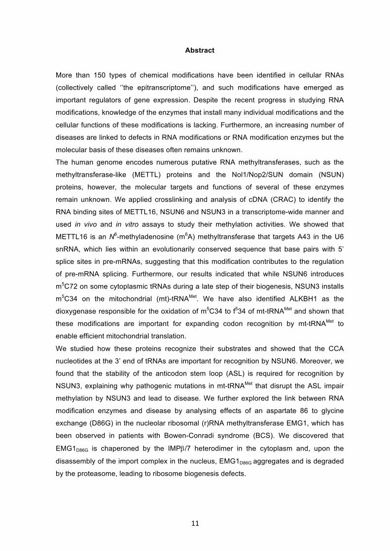
Abstract More than 150 types of chemical modifications have been identified in cellular RNAs
(collectively called ‘’the epitranscriptome’’), and such modifications have emerged as
important regulators of gene expression. Despite the recent progress in studying RNA
modifications, knowledge of the enzymes that install many individual modifications and the
cellular functions of these modifications is lacking. Furthermore, an increasing number of
diseases are linked to defects in RNA modifications or RNA modification enzymes but the
molecular basis of these diseases often remains unknown.
The human genome encodes numerous putative RNA methyltransferases, such as the
methyltransferase-like (METTL) proteins and the Nol1/Nop2/SUN domain (NSUN)
proteins, however, the molecular targets and functions of several of these enzymes
remain unknown. We applied crosslinking and analysis of cDNA (CRAC) to identify the
RNA binding sites of METTL16, NSUN6 and NSUN3 in a transcriptome-wide manner and
used in vivo and in vitro assays to study their methylation activities. We showed that
METTL16 is an N6-methyladenosine (m6A) methyltransferase that targets A43 in the U6
snRNA, which lies within an evolutionarily conserved sequence that base pairs with 5’
splice sites in pre-mRNAs, suggesting that this modification contributes to the regulation
of pre-mRNA splicing. Furthermore, our results indicated that while NSUN6 introduces
m5C72 on some cytoplasmic tRNAs during a late step of their biogenesis, NSUN3 installs
m5C34 on the mitochondrial (mt)-tRNAMet. We have also identified ALKBH1 as the
dioxygenase responsible for the oxidation of m5C34 to f534 of mt-tRNAMet and shown that
these modifications are important for expanding codon recognition by mt-tRNAMet to
enable efficient mitochondrial translation.
We studied how these proteins recognize their substrates and showed that the CCA
nucleotides at the 3’ end of tRNAs are important for recognition by NSUN6. Moreover, we
found that the stability of the anticodon stem loop (ASL) is required for recognition by
NSUN3, explaining why pathogenic mutations in mt-tRNAMet that disrupt the ASL impair
methylation by NSUN3 and lead to disease. We further explored the link between RNA
modification enzymes and disease by analysing effects of an aspartate 86 to glycine
exchange (D86G) in the nucleolar ribosomal (r)RNA methyltransferase EMG1, which has
been observed in patients with Bowen-Conradi syndrome (BCS). We discovered that
EMG1D86G is chaperoned by the IMPb/7 heterodimer in the cytoplasm and, upon the
disassembly of the import complex in the nucleus, EMG1D86G aggregates and is degraded
by the proteasome, leading to ribosome biogenesis defects.
12
Taken together, our studies characterized substrates of novel RNA-modifying enzymes
and provided insights into their cellular functions and the link between defects in these
enzymes and diseases.
13
Chapter One: Introduction
1.1 Overview of RNA modifications RNAs from all domains of life can be co- or post-transcriptionally modified by a collection
of more than 150 distinct chemical moieties, ranging from simple methylations to complex
modifications that are installed by the co-ordinated action of several, often highly
conserved, modification enzymes (Cantara et al., 2011; Machnicka et al., 2013). Such
modifications expand the chemical and topological properties of the four RNA nucleotides,
therefore influencing the structure, molecular interactions and biological roles of the RNAs
that carry them (Motorin and Helm, 2011).
Modified nucleotides are present in almost all types of cellular RNAs, and the most highly
modified species are transfer RNAs (tRNAs), with up to 17% of tRNA nucleotides being
modified, and ribosomal RNAs (rRNAs), which contain approximately 2% modified
nucleotides (reviewed in Jackman and Alfonzo, 2013; Sloan et al., 2017). Recently, high-
throughput sequencing approaches have been used to generate transcriptome-wide maps
of specific RNA modifications and this has revealed modified sites in messenger RNAs
(mRNAs) and several classes of non-coding RNA, such as long non-coding RNAs
(lncRNAs), small nucleolar RNAs (snoRNAs), small nuclear RNAs (snRNAs) and
microRNAs (miRNAs; reviewed in Roundtree et al., 2017). This complex landscape of
RNA modifications (collectively termed ‘’the epitranscriptome’’) represents an important
layer of gene expression regulation, and mutations in genes encoding RNA modification
enzymes are implicated in various human diseases such as malignancies and metabolic
disorders (reviewed in Sarin and Leidel, 2014). Nevertheless, many open questions
remain on the identity of the enzymes involved and the detailed mechanisms by which
RNA modifications are installed, regulated and exert their biological functions.
1.2 rRNA modifications 1.2.1 Ribosome biogenesis in eukaryotes Ribosomes are evolutionarily conserved molecular machines that are responsible for
cellular protein synthesis. The eukaryotic cytoplasmic, 80S ribosome is a complex
ribonucleoprotein (RNP) that comprises two subunits containing four different ribosomal
RNA (rRNA) molecules and around 80 ribosomal proteins (reviewed in Melnikov et al.,
2012). The large subunit (LSU; 60S) contains the 28S (in metazoans)/ 25S (in the yeast
Saccharomyces cerevisiae), 5S and 5.8S rRNAs assembled with 47 ribosomal proteins of
the large subunit (RPLs), and the small subunit (SSU; 40S) comprises the 18S rRNA
associated with 33 ribosomal proteins of the small subunit (RPSs; Ben-Shem et al., 2011;
Anger et al., 2013). The biogenesis of such complex molecular machines is one of the
14
most crucial and energy consuming processes in the cell (Warner et al., 1999). Our
current knowledge of ribosome biogenesis has been mainly obtained from studies in the
yeast Saccharomyces cerevisiae, due to the combination of powerful genetics and
biochemical methods and the relative simplicity of this organism compared to humans.
However, recent RNAi-based screens to identify human ribosome biogenesis factors and
individual studies on human ribosome biogenesis factors have revealed that the core
features of ribosome biogenesis are conserved from yeast to humans, but that several
conserved factors have extra or different functions (see for example, Badertscher et al.,
2015; Tafforeau et al., 2013; Wild et al., 2010). Furthermore, these screens have
uncovered the requirement for many additional factors for human ribosome assembly
compared to yeast. In humans, the process starts in the nucleolus with the RNA
polymerase I-mediated transcription of a long precursor ribosomal RNA (pre-rRNA) that
contains the mature 18S, 5.8S, and 28S rRNA sequences, separated by the internal
transcribed spacers 1 and 2 (ITS1 and ITS2) and flanked by the 5’ and 3’ external
transcribed spacers (5’-ETS and 3’-ETS; Mullineux and Lafontaine, 2012). These
additional pre-rRNA sequences are removed by an ordered series of endonucleolytic
cleavages and exonucleolytic processing steps to generate the mature 5’ and 3’ ends of
the rRNAs (Tomecki et al., 2017; Henras et al., 2015). The hierarchical assembly of
ribosomal proteins and biogenesis factors on the pre-rRNA forms a series of pre-
ribosomal particles, in which pre-rRNA processing steps and the modification of the rRNA
sequences take place (Fig. 1; reviewed in Henras et al., 2008). These maturation steps
require the assistance of small nucleolar ribonucleoprotein (snoRNP) complexes and
more than 200, mostly essential, trans-acting factors, such as RNA-modifying enzymes
(discussed below), nucleases, kinases, GTPases and RNA-remodelling enzymes, which
catalyse irreversible steps in the pathway (reviewed in Kressler et al., 2010). RNA-
remodelling enzymes include AAA-ATPases and DExD/H-box RNA helicases, which are
suggested to modulate the unidirectional transitions of pre-ribosomal structures by
unwinding or annealing RNA helices and/or facilitating the recruitment or release of RNA-
binding proteins during ribosome biogenesis (reviewed in Martin et al., 2013).
Additionally, ribosome assembly requires the import of most ribosomal proteins from the
cytoplasm to their incorporation sites on the nuclear pre-ribosomal particles. However, this
is a challenging task for the cell because of specific features of ribosomal proteins: they
contain unstructured extensions and highly basic regions that may form non-specific
interactions when not assembled into pre-ribosomes, leading to insolubility (Jäkel et al.,
2002). Therefore, there are several mechanisms for preventing aggregation of newly
synthesized ribosomal proteins in the cell (Pillet et al., 2016). Besides the general
ribosome-associated chaperones that assist the de novo folding of ribosomal proteins,
15
import receptors have been shown to protect ribosomal proteins from precipitation in the
cytoplasm by shielding their basic regions (Jäkel et al., 2002; Albanese et al., 2006, 2010).
Moreover, recent studies have uncovered a set of dedicated chaperones that bind
ribosomal proteins, often co-translationally, and escort them to appropriate pre-ribosomal
complexes (Pausch et al., 2015). Non-(pre-)-associated ribosomal proteins that are prone
to aggregation are targeted for degradation by the excess ribosomal protein quality control
(ERISQ), which specifically ubiquitinates lysine residues that are otherwise not accessible
after the assembly into pre-ribosomes (Sung et al., 2016).
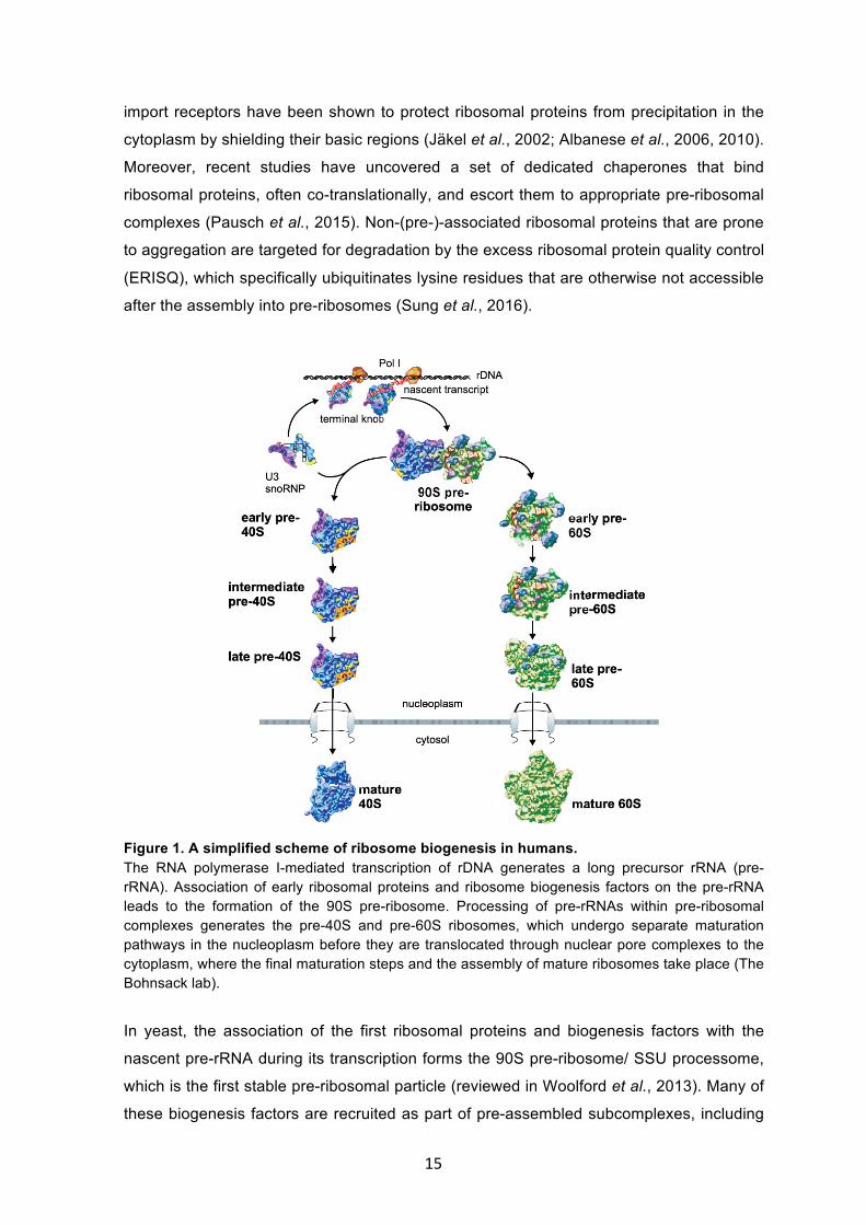
Figure 1. A simplified scheme of ribosome biogenesis in humans. The RNA polymerase I-mediated transcription of rDNA generates a long precursor rRNA (pre-rRNA). Association of early ribosomal proteins and ribosome biogenesis factors on the pre-rRNA leads to the formation of the 90S pre-ribosome. Processing of pre-rRNAs within pre-ribosomal complexes generates the pre-40S and pre-60S ribosomes, which undergo separate maturation pathways in the nucleoplasm before they are translocated through nuclear pore complexes to the cytoplasm, where the final maturation steps and the assembly of mature ribosomes take place (The Bohnsack lab).
In yeast, the association of the first ribosomal proteins and biogenesis factors with the
nascent pre-rRNA during its transcription forms the 90S pre-ribosome/ SSU processome,
which is the first stable pre-ribosomal particle (reviewed in Woolford et al., 2013). Many of
these biogenesis factors are recruited as part of pre-assembled subcomplexes, including
16
the U3 snoRNP and the UTP-A, UTP-B and UTP-C complexes, which sequentially
assemble on the 5’-ETS and 18S rRNA sequence (Kornprobst et al., 2016; Hunziker et al.,
2016). Following a pre-rRNA processing event in ITS1 (typically site A2 cleavage in yeast
or site 2 cleavage in humans), the early pre-40S particle is separated from the rest of the
pre-rRNA, which assembles with RPLs and biogenesis factors forming the early 60S pre-
ribosomal particles (reviewed in Henras et al., 2015). In yeast, it has been shown that
upon separation, the protein composition of the pre-40S particle changes dramatically as
most biogenesis factors are released and several RPSs are recruited (Schäfer et al.,
2003). This early pre-40S particle, which already displays the head, platform and body
structures of the mature SSU but not the characteristic beak structure, is rapidly exported
to the cytoplasm (see below; reviewed in Kressler et al., 2010). Formation of the beak
structure involves the incorporation of Rps3 (uS3), which is promoted by the
phosphorylation-dependent release of Ltv1 (Ghalei et al., 2015; Mitterer et al., 2016). Prior
to the final pre-rRNA processing step, Nob1-mediated cleavage to form the mature 3’ end
of the 18S RNA, pre-40S subunits undergo a translation-like cycle involving the GTPase
Fun12 (eIF5B), in which the pre-40S subunits bind mature 60S subunits (Lebaron et al.,
2012; Strunk et al., 2012; Turowski et al., 2014). This final quality control step prevents
aberrant or immature pre-40S particles from entering the pool of translating ribosomes.
In contrast to the relatively simple SSU assembly pathway in yeast, LSU assembly
appears more complex as it involves the formation of several successive intermediates
with different compositions (reviewed in Kressler et al., 2017). Several of these
intermediates have been characterised in yeast using tandem affinity purification (TAP) via
pre-60S bait proteins providing insight into the timing of recruitment and dissociation of
numerous trans-acting biogenesis factors. A key event during pre-60S biogenesis is the
integration of the 5S RNP. The 5S RNP is formed by the Syo1-facilitated association of
Rpl5 (uL5) and Rpl11 (uL18) with the 5S RNA, which is transcribed separately by RNA
polymerase III (Ciganda and Williams, 2011; Calvino et al., 2015). The incorporation of the
5S RNP into the pre-60S ribosome is then mediated by the Rpf2-Rrs1 heterodimer but in
humans, several other factors have also been linked to 5S RNP recruitment to pre-
ribosomal complexes (Zhang et al., 2007; Sloan et al., 2013; Kharde et al., 2015).
However, initially, the 5S RNP is rotated 180° from its final position and the
peptidyltransferase centre (PTC) is occupied by the GTPases Nog1 and Nog2, preventing
the recruitment of the export adaptor Nmd3 (Leidig et al., 2014; Wu et al., 2016). The
recruitment of the Rix1 subcomplex and the AAA-ATPase Rea1 coincides with the rotation
of the 5S RNP to its mature position and the activation and release of Nog2, allowing the
binding of Nmd3 to the pre-60S ribosome (Barrio-Garcia et al., 2016; Matsuo et al., 2014).
Rea1 and Nog2 are therefore considered checkpoint factors that monitor the maturation
17
status of pre-60S particles and licence their export to the cytoplasm. After export, the
release of biogenesis factors occurs in concert with the association of the final ribosomal
proteins. For example, the GTPase Nog1 is released by the AAA-ATPase Drg1, allowing
the incorporation of Rpl24 (eL24; Kappel et al., 2012). Moreover, the GTPase Lsg1
releases the export adaptor Nmd3 enabling the recruitment of Rpl10 (uL16; Hedges et al.,
2005). Finally, the release of Tif6 by the GTPase Efl1 and Sdo1 yields 60S subunits that
are competent to join 40S subunits (Weis et al., 2015; Ma et al., 2016).
During the maturation of the pre-40S and the pre-60S particles, numerous remodelling
events, nuclear export signal (NES)-containing export adaptors and additional export
factors have been proposed to mediate their translocation through nuclear pore
complexes (NPCs) into the cytoplasm (reviewed in Sloan et al., 2015). Both pre-40S and
pre-60S export rely on the exportin CRM1, which binds export adaptors associated with
the pre-ribosomal subunits in the presence of RanGTP to facilitate export of these
complexes (Hurt et al., 1999; Stage-Zimmermann et al., 2000). Additionally, the mRNA
export factor Mex67-Mtr2 was shown in yeast to contribute to the export of both pre-
ribosomal complexes, however, the loop insertions in the middle domain that are crucial
for this function are absent from its human homolog TAP-p15, questioning whether this
complex also contributes to pre-40S or pre-60S export in humans (Yao et al., 2007; Faza
et al., 2012). Several SSU biogenesis factors that contain classical NES motifs and shuttle
between the nucleus and the cytoplasm have been suggested to have a role in the export
of pre-40S particles. For example, the protein kinase human RIO2 was shown to directly
bind CRM1 in the presence of RanGTP via its NES and contribute to efficient pre-40S
export (Zemp et al., 2009). However, none of these factors is essential, suggesting the
presence of unidentified pre-40S export adaptors or that multiple factors play redundant
roles in the pre-40S export (reviewed in Sloan et al., 2015). In contrast, the pre-60S export
is dependent on the essential NES-containing export adaptor NMD3, which binds CRM1
in a RanGTP-dependent manner and facilitates the translocation of the pre-60S particles
to the cytoplasm (Thomas and Kutay, 2003; Bai et al., 2013). Alongside CRM1, in human
cells, Exportin 5 was similarly reported to bind the pre-60S particles and facilitate their
export, suggesting the presence of a second RanGTP-dependent pre-60S export pathway
(Wild et al., 2010). Interestingly, no role for Msn5, the yeast homolog of Exportin 5, in the
export of pre-ribosomal subunits has been observed.
Taken together, assembly of the ribosomal subunits is a complex and hierarchical process
that is orchestrated by a wealth of trans-acting factors and involves pre-rRNA processing
and folding and the concomitant assembly of ribosomal proteins, forming pre-ribosomes
that translocate from the nucleolus, through the nucleoplasm, to the cytoplasm as they
mature.
18
1.2.2 rRNA modifications introduced by snoRNPs and stand-alone enzymes During maturation of the pre-rRNAs, a significant portion of rRNA nucleotides
(approximately 210 sites in the human rRNAs) is modified by the action of snoRNPs or
stand-alone enzymes (reviewed in Sloan et al., 2017). Despite the relatively large fraction
of modified nucleotides in rRNA, the diversity of rRNA modifications is only limited to a set
of 12 different types of modifications. In contrast to prokaryotes where base methylations
are the most abundant modifications in rRNA, the majority of rRNA modifications in
eukaryotes are 2’-O-methylation of the ribose of any of the four nucleotides and
isomerization of uridine to pseudouridine (Y; Lestrade and Weber, 2006; Pienka-
Przybylska et al., 2008). These modifications are mostly installed by the box C/D and box
H/ACA snoRNPs respectively and, so far, approximately 100 of each modification are
documented in human rRNA. The only exceptions are the stand-alone enzymes Sbp1 and
Pus7, which have been reported in yeast to catalyse the 2’-O-methylation of G2922 of the
25S rRNA and pseudouridylation of U50 in the 5S rRNA respectively (Lapeyre et al.,
2004; Decatur and Schnare, 2008).
Eukaryotic snoRNPs, many of which are essential, contain a snoRNA that base pairs with
the pre-rRNA and guides the catalytic protein component of the snoRNP to modify a
specific target nucleotide (reviewed in Watkins and Bohnsack, 2012). Box C/D snoRNAs
contain a C/D motif at the 5’ and 3’ ends of the transcript respectively and an internal C’/D’
motif, and their extensive base pairing with the pre-rRNA adjacent to the D/D’ box is
facilitated by the association with the core proteins Nop58, Nop56 and Snu13 (15.5K in
humans). The catalytic subunit of box C/D snoRNPs, fibrillarin (Nop1 in yeast) is then
correctly positioned to modify the specific target nucleotide, five residues upstream of the
D/D’ box (Tollervey et al., 1993; van Nues et al., 2011). Box H/ACA snoRNAs contain a
conserved H box and an ACA sequence, and form a hairpin structure that contains the
‘’pseudouridylation pocket’’, where base pairing with the pre-rRNA takes place (Lafontaine
et al., 1998). The tertiary fold of the H/ACA box snoRNA, stabilized by the protein
components Nop10, Nhp2 and Gar1, leaves the target uridine non-base-paired and
correctly positioned in the pseudouridine synthase dyskerin (Cbf5 in yeast) active site
(Ganot et al., 1997). Interestingly, a subset of snoRNPs have been reported in yeast to
guide modifications of multiple sites on the same pre-rRNA (e.g. snR60 targets A817 and
G908 of the 25S rRNA) or on different pre-rRNAs (e.g. snR52 targets A420 of the 18S
rRNA and U2921 of the 25S rRNA; Kiss-Laszlo et al., 1996; Lowe et al., 1999; Petrov et
al., 2014). Conversely, redundancy between snoRNAs in targeting modification of a
particular site has also been documented. Examples include snoRNAs of the same class
(e.g. snR39 and snR59 modify A807 of the 25S rRNA), or even different classes (e.g. the
box C/D snoRNA snR65 and the box H/ACA snoRNA snR9 target U2347 of the 25S
19
rRNA; Taoka et al., 2016). Besides their function in rRNA modification, several snoRNAs,
including U14 and snR10, play additional roles in regulating pre-rRNA folding and
mediating long-range interactions within pre-ribosomal particles (Enright et al., 1996;
Martin et al., 2014).
Besides uridine isomerization, several rRNA bases at sites distributed between the LSU
and SSU are also modified (reviewed in Sharma and Lafontaine, 2015). In humans, the
28S rRNA of the LSU carries two C5-methylcytosine residues (m5C3761 and m5C4413/4),
one N1-methyladenosine residue (m1A1309) and one N3-methyluridine residue (m3U4500).
Modifications are more diverse on the 18S rRNA of the SSU, which contains two highly
conserved N6,N6-dimethyladenosine residues (m62A1850 and m6
2A1851), two acetylated
cytosine residues (ac4C1337 and ac4C1842), one N7-methylguanosine residue (m7G1639)
and one hypermodified N1-methyl-N3-aminocarboxypropylpseudouridine residue
(m1acp3Y1248; Pienka-Przybylska et al., 2008). The RNA methyltransferases involved in
the base modifications in human rRNA have been mostly identified: NSUN5/WBSCR20
(m5C3761; Schosserer et al., 2015), NSUN1/NOL1 (m5C4413/4; Bourgeois et al., 2015),
NML (m1A1309; Waku et al., 2016), DIMTL1 (m62A1850 and m6
2A1851; Zorbas et al., 2015)
and WBSCR22 (m7G1639; Haag et al., 2015). These RNA methyltransferases use S-
adenosylmethionine (SAM) as a methyl donor and display a classical Rossmann-like fold.
In contrast, the RNA methyltransferase EMG1, which participates in the m1acp3Y
hypermodification at position 1248 in the 18S rRNA, belongs to the SPOUT (alpha-beta
knot fold) family (Leulliot et al., 2008; Taylor et al., 2008; Thomas et al., 2010). The
chemically complex modification of U1248 is of particular interest because it requires
several strictly ordered steps by different factors in different subcellular compartments
(Brand et al., 1978). The first step, which has been described in yeast, is isomerization of
U1248 to Y in the nucleolus by the H/ACA box snoRNP snR35 (ACA 13 in humans;
Samarsky et al., 1995). This initial step generates the substrate for subsequent N1-
methylation of the residue by the essential nucleolar RNA methyltransferase EMG1
(Wurm et al., 2010). Finally, TSR3, which also has a SPOUT fold and utilizes SAM,
introduces the acp group in the cytoplasm (Meyer et al., 2016). Besides RNA
methyltransferases, the RNA acetyltransferase NAT10, which has a Gcn5-related N-
acetyltransferase (GNAT) domain, has been shown to be responsible for introducing
ac4C1337 and ac4C1842 in the 18S rRNA (Ito et al., 2014; Sharma et al., 2015).
Interestingly, the yeast homolog of NAT10, Kre33 was recently suggested to be guided to
its modification sites by snoRNAs snR4 and snR45 (Sharma et al., 2017).
Notably, several modifications have been reported in yeast and are not conserved in
humans, including one N1-methyladenosine residue (m1A2142) and two N3-methyluridine
residues (m3U2634 and m3U2843) in the 25S rRNA. Interestingly, the enzymes
20
responsible for installing these modifications (Bmt2 (Sharma et al., 2013), Bmt5 and Bmt6
(Sharma et al., 2014) respectively) are non-essential, suggesting that their roles in
ribosome structure or function may be less important than other enzymes. In contrast, all
other evolutionarily conserved enzymes involved in base modifications in rRNAs are either
essential or important for growth, and surprisingly, this is in most cases not because of
their RNA-modifying catalytic activity, but because their presence in pre-ribosomal
complexes is required for subunit assembly (reviewed in Sharma and Lafontaine, 2015).
For example, apart from its methylation function, Emg1 plays an additional essential role
in yeast ribosome biogenesis, which is proposed to be the recruitment of Rps19 (eS19)
into pre-40 particles (Meyer et al., 2011; Buchhaupt et al., 2006).
1.2.3 Functions of rRNA modifications The use of this minimal set of different modifications in rRNA in eukaryotes, despite the
plethora of modified RNA nucleotides in nature, suggests that their chemical nature is
particularly appropriate for their functions in rRNAs. Furthermore, rRNA modifications are
not randomly distributed over the ribosome, but they cluster in evolutionarily conserved
positions at the interface between the LSU and SSU and the inner cores of the subunits
(Fig. 2; Decatur and Fournier, 2002). These include functionally important regions, such
as the peptidyltransferase centre (PTC) in the LSU and the decoding site in the SSU,
suggesting that rRNA modifications might play important roles in optimizing ribosome
structure and function (Ben-Shem et al., 2011). Indeed, the two most abundant rRNA
modifications, 2’-O-methylation and pseudouridylation, are known to alter local
conformation and folding properties of RNA. 2’-O-methylation increases RNA rigidity by
promoting base stacking and can alter RNA folds (Prusiner et al., 1974; reviewed in Helm,
2006), while pseudouridine has increased hydrogen bonding capability compared to
uridine and stabilizes specific RNA structures (reviewed in Charette and Gray, 2000;
Hayrapetyan et al., 2009). However, rRNA modifications are generally thought to act in a
cumulative manner since loss of individual modifications causes only subtle defects in cell
growth, and significant phenotypes are only observed when clusters of modifications in
certain functional regions of the ribosome are concomitantly lacking (Baxter-Roshek et al.,
2007; Esguerra et al., 2008). For example, combined deletion of 5 box H/ACA snoRNAs
that guide modifications in the PTC was shown, in yeast, to cause synergistic effects on
ribosome structure and activity (King et al., 2003). Similarly, the base modifications
present in the rRNAs can serve to stabilise secondary and tertiary rRNA structures. For
example, loss of the conserved m5C2278 base modification in combination with the ribose
methylation at G2288 was shown to cause changes in the structure of the yeast 25S
rRNA, leading to dramatic ribosome instability (Gigova et al., 2014).
21
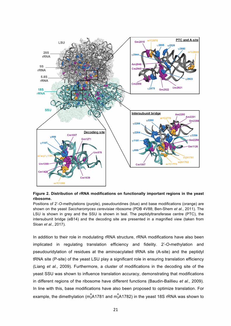
Figure 2. Distribution of rRNA modifications on functionally important regions in the yeast ribosome. Positions of 2’-O-methylations (purple), pseudouridines (blue) and base modifications (orange) are shown on the yeast Saccharomyces cerevisiae ribosome (PDB 4V88; Ben-Shem et al., 2011). The LSU is shown in grey and the SSU is shown in teal. The peptidyltransferase centre (PTC), the intersubunit bridge (eB14) and the decoding site are presented in a magnified view (taken from Sloan et al., 2017).
In addition to their role in modulating rRNA structure, rRNA modifications have also been
implicated in regulating translation efficiency and fidelity. 2’-O-methylation and
pseudouridylation of residues at the aminoacylated tRNA site (A-site) and the peptidyl
tRNA site (P-site) of the yeast LSU play a significant role in ensuring translation efficiency
(Liang et al., 2009). Furthermore, a cluster of modifications in the decoding site of the
yeast SSU was shown to influence translation accuracy, demonstrating that modifications
in different regions of the ribosome have different functions (Baudin-Baillieu et al., 2009).
In line with this, base modifications have also been proposed to optimize translation. For
example, the dimethylation (m62A1781 and m6
2A1782) in the yeast 18S rRNA was shown to
22
be required for translation, and cells expressing mutant forms of Dim1 or Emg1 display
hypersensitivity to aminoglycoside antibiotics, which affect translation (Lafontaine et al.,
1998; Liu and Thiele, 2001). Strikingly, certain rRNA modifications have been shown to
influence translation of specific subsets of mRNAs. For example, the loss of Rcm1 in
yeast, which introduces m5C2278 in the 25S rRNA, was shown to promote the recruitment
of a specific subset of oxidative stress-responsive mRNAs into polysomes (Schosserer et
al., 2015). In conclusion, rRNA modifications and the enzymes introducing them play
different roles in regulating ribosome biogenesis, structure and function. Interestingly,
some enzymes that perform rRNA modifications have been implicated in modification of
other classes of RNA. For example, NAT10 was shown to acetylate specific tRNAs
(Sharma et al., 2015) and fibrillarin was shown to mediate 2’-O-methylation in human
snRNAs (Tycowski et al., 1998; Jady and Kiss, 2001), suggesting possible crosstalk
between the biogenesis of different species of RNA.
1.2.4 Timing and regulation of rRNA modifications The observations that rRNA modifications are present in the core of the ribosomal
subunits, and that pre-ribosomal complexes are highly structured imply that factors
involved in rRNA modifications have limited access to their substrate residues during late
stages of ribosome maturation. It was therefore expected that the majority of rRNA
modifications are introduced in early stages, where pre-ribosomal complexes have more
open structures. Indeed, most 2’-O-methylations were shown in yeast to occur during
early stages of ribosome maturation, often co-transcriptionally (Kos and Tollervey, 2010;
Birkedal et al., 2015). It is similarly proposed that pseudouridylations occur early during
ribosome biogenesis, however, this remains to be documented. In contrast, rRNA base
modifications are thought to be introduced later during the process, however, the precise
timing of most base modifications has remained unclear so far. An exception is the N3-acp
modification of nucleotide 1248 of the 18S rRNA that is installed by TSR3 in the cytoplasm
and can therefore be clearly defined as a “late modification” (Meyer et al., 2016).
Furthermore, several rRNA modification enzymes are bound to early pre-ribosomal
complexes, but do not modify their substrate residues until later. For example, Dim1
associates with early nucleolar pre-ribosomes in yeast, but it only installs the m62A1850
and m62A1851 dimethylation after the export of the maturating pre-SSU to the cytoplasm
(Lafontaine et al., 1995). Remarkably, the corresponding dimethylation in humans is
introduced by DIMTL1 in the nucleus (Zorbas et al., 2015), but the significance of this
temporal difference is not yet understood.
The introduction of several rRNA modifications during late stages of ribosome biogenesis
suggests that RNA-remodelling enzymes, such as RNA helicases, may be required to
23
enable certain modifications to be installed. Late-acting snoRNPs, for example, were
proposed to gain access to their target sites in the human 18S rRNA by the coordinated
action of the RNA helicase DDX21 (Sloan et al., 2015). Similarly, several base
modification enzymes require co-factors with RNA-remodelling activities or even carry
such activities within their own sequence. For instance, the acetyltransferase NAT10 has
an ATPase-dependent helicase domain, which is suggested to facilitate the access of the
enzyme to its target site for acetylation (Sharma et al., 2015). Additionally, the yeast m7G
methyltransferase Bud23 requires the coactivator Trm112, which stabilizes Bud23 by
masking solvent-unfavourable hydrophobic surfaces (Figaro et al., 2012). The action of
the RNA helicase Dhr1 is then required for the Bud23-Trm112 complex to gain access to
its target site within the pre-40S particle (Sardana et al., 2014).
It has recently emerged that certain rRNA residues are differentially modified, i.e. some
rRNA molecules carry specific modifications while others do not (Birkedal et al., 2015). In
yeast, 18 modification sites were found to be modified on less than 85% of ribosomes
(Taoka et al., 2016). In line with this, in several human cell lines, around one-third of 2’-O-
methylation sites are partially modified (Krogh et al., 2016). Together, this suggests that
rRNA modifications contribute to the heterogeneity of the ribosomes. However, base
modifications installed by the conserved stand-alone enzymes appear to be constitutively
present. It has been speculated by Krogh and colleagues that such conserved
modifications are involved in essential aspects of rRNA folding and assembly of
ribosomes, while the fractionally modified residues play roles in fine-tuning translation.
The extent of specific 2’-O-methylations has been suggested to be determined by the
levels of the corresponding snoRNAs (Buchhaupt et al., 2014), but it is also possible that
alternative rRNA processing and folding pathways, or the selective removal of the
methylations by demethylases, also contribute to differences in rRNA modifications
(reviewed in Sloan et al., 2017). Excitingly, variations in the levels of specific rRNA
modifications in response to environmental signals have been reported. Diauxic shift and
heat-shock, for example, were observed to alter the modification levels of specific rRNA
residues in the yeast Saccharomyces cerevisiae (Schwartz et al., 2014; Carlile et al.,
2014). Similarly, changes in growth conditions of Schizosaccharomyces pombe cells were
shown to significantly affect the extent of modifications of specific sites in rRNA (Taoka et
al., 2015). In addition to their variation under physiological conditions, the extent of rRNA
modifications is also altered in several genetic disorders and cancers (see below).
24
1.2.5 Ribosomopathies and diseases associated with rRNA modifications Recently, a number of human diseases associated with defects in ribosome biogenesis
have been identified and collectively termed ‘’ribosomopathies’’ (reviewed in Narla and
Ebert, 2010; Khanna-Gupta, 2013). Ribosomopathies are caused by alterations in genes
encoding ribosomal proteins or ribosome biogenesis factors, and these alterations are in
most cases caused by congenital haploinsufficiency of the affected gene (mutation or
deletion of one copy of a gene, leaving the other copy functional; reviewed in McCann and
Baserga, 2013). For example, mutations in a number of ribosomal proteins, including
RPS19 (eS19), have been linked to Diamond-Blackfan anaemia (DBA; Draptchinskaia et
al., 1999; Gazda et al., 2006; Cmejla et al., 2007). Furthermore, mutations in the ribosome
biogenesis factor TCOF1/treacle have been found in Treacher Collins syndrome (TCS;
The Treacher Collins Syndrome Collaborative Group, 1996). Similarly, Schwachman-
Diamond syndrome patients have been found to carry biallelic mutations in SBDS, which
associates with late pre-60S complexes (Boocock et al., 2003). To date, the only reported
acquired ribosomopathy is the 5q-syndrome, which is caused by the deletion of
chromosome 5q including one allele of RPS14 (Ebert et al., 2008; Pellagatti et al., 2008).
Generally, many ribosomopathies display a number of common symptoms (bone marrow
failure, growth retardation, skeletal abnormalities and malignancies), however, each
ribosomopathy is clinically distinct. For example, patients with haploinsufficiency of RPSA
(uS2; isolated congenital asplenia) lack a spleen, but they have no other observable
anomalies (Bolze et al., 2013). Similarly, patients with a mutation in the ribosome
biogenesis factor hUTP4/Cirhin (North American Indian childhood cirrhosis) show one
main phenotype, which is biliary cirrhosis (Chagnon et al., 2002). The mechanism by
which such defects in a macromolecular complex as constitutive and essential as the
ribosome cause only tissue-specific phenotypes remains unclear. One possible
explanation is that certain defects in ribosomes could influence translation of specific
mRNAs, which might be essential for the affected cell type (reviewed in McCann and
Baserga, 2014). This model is supported by the observation that reduction of RPL40 in
humans impedes translation of specific vesicular stomatitis virus mRNAs (Lee et al.,
2013). An alternative explanation is that ribosome variants are produced in different cell
types, and ribosomes in the affected cell types are more sensitive to the defect that
causes the disease (Marcel et al., 2015).
Defects in ribosome biogenesis, such as those observed in ribosomopathies, cause the
5S RNP to bind the E3 ubiquitin ligase HDM2 and inhibits its activity, leading to induction
of the tumor suppressor p53 (Sloan et al., 2013; Donati et al., 2013). Such accumulation
of p53 arrests cell division and leads to apoptosis, which can be responsible for some of
the symptoms (McGowan et al., 2008; Fumagalli et al., 2009). In line, reduction of p53
25
activity rescued the craniofacial abnormalities in a mouse model for TCS (Jones et al.,
2008) and rescued the erythroid abnormalities in zebrafish models for DBA (Boultwood et
al., 2012). However, the accumulation of p53 when ribosome biogenesis is impeded is
surprising given the increased susceptibility to cancers observed in most ribosomopathies
(reviewed in Teng et al., 2013). One possible explanation for this is that the elevated p53
levels may lead to mutations or downregulation of the p53 pathway, resulting in
desensitizing patient cells to p53 (Pelava et al., 2016). Another possible explanation is that
defects in ribosomal proteins or ribosome biogenesis factors could produce ribosomes
with altered translation capacities that differentially translate distinct subpopulations of
mRNAs, leading to tumorigenesis. This hypothesis is supported by the finding that
depletion of RPL38 in mice impairs translation of a subset of Hox mRNAs, which could
lead to malignant transformation when dysregulated (Kondrashov et al., 2011).
Similarly, mutations in DKC1, which encodes the box H/ACA pseudouridine synthase
dyskerin, in patients with X-linked dyskeratosis congenita (X-DC) cause defects in IRES-
dependent translation of a subset of mRNAs including the ones encoding the tumor
suppressor p27 and the antiapoptotic factors Bcl-xL and XIAP (Yoon et al., 2006; Bellodi
et al., 2010). This suggests that changes in rRNA pseudouridylation pattern may result in
ribosomes with differential translation activities that promote tumorigenesis. In a similar
way, altered rRNA 2’-O-methylation profiles have been associated with tumorigenesis and
differences in the extent of modification at multiple sites have been reported in different
cancer cell lines (Krogh et al., 2016). The tumor suppressor p53 is suggested to regulate
the expression levels of fibrillarin, with overexpression of fibrillarin leading to changes in
the rRNA 2’-O-methylation pattern of the rRNAs and altered translation fidelity of
ribosomes (Marcel et al., 2013). Reciprocally, lack of fibrillarin has been reported to cause
an increase in the cap-independent translation of p53, independent from the 5SRNP-
HDM2 pathway (Su et al., 2014). Furthermore, the levels of other components of box C/D
snoRNPs including NOP56 (Cowling et al., 2014) and NOP58 (Nakamoto et al., 2001) are
elevated in different types of cancer, making them good markers of tumorigenesis (Liao et
al., 2010).
Defects in stand-alone rRNA base-modifying enzymes have also been linked to cancers
and genetic diseases. For example, WBSCR22 promotes survival and metastasis of tumor
cells (Nakazawa et al., 2011), and the high expression levels of NSUN1/NOL1 have been
correlated with growth of lung adenocarcinoma (Sato et al., 1999). WBSCR22 and
NSUN5/WBSCR20 are deleted, along with other genes, in a developmental disorder
called Williams-Beuren syndrome (Doll and Grzeschik, 2001) and NML has been
associated with high fat diet-induced obesity (Oie et al., 2014). More specifically, a point
mutation in the gene encoding EMG1, leading to an aspartate to glycine exchange in the
26
protein sequence, causes a severe genetic disorder known as Bowen-Conradi syndrome
(BCS), which is characterized by bone marrow failure, bone abnormalities, growth
retardation and death within the first year of life (Armistead et al., 2009). However,
whether lack of the rRNA modifications or other effects caused by defects in the
modification enzymes cause these disease phenotypes is often still unknown.
Interestingly, chemical inhibition of the acetyltransferase NAT10 was shown to rescue
defects of laminopathic cells, raising the possibility of using rRNA modification enzymes
as drug targets (Larrieu et al., 2014). In conclusion, disruptions in ribosome biogenesis are
linked to multiple human diseases, and further understanding of the effects of such
genetic alterations on the molecular and cellular levels is integral to finding therapeutic
interventions to treat these disorders.
1.3 tRNA modifications 1.3.1 Overview of eukaryotic tRNA biogenesis tRNAs are highly structured short non-coding RNAs of approximately 70 nucleotides that
decode the mRNA codons and carry the cognate amino acids to the ribosome for protein
biosynthesis in the cytoplasm of all living cells, and in eukaryotic mitochondria and plastids
(reviewed in Fujishima and Kanai, 2014). tRNAs display a highly conserved cloverleaf
secondary structure, which comprises four distinct domains: the acceptor stem, the
anticodon arm, the D loop and the TYC loop. Interactions between these domains are
required to form the folded L-shaped tertiary structure of tRNAs (see for example, Kim et
al., 1973; Robertus et al., 1974). The biosynthesis of cytoplasmic tRNAs starts with the
RNA polymerase III-mediated transcription of tRNA genes in the nucleolus, generating
precursor tRNAs (pre-tRNAs) that contain sequences that are not present in the mature
tRNAs: the 5’ leader sequence, the 3’ trailer and, in some cases, introns (reviewed in
Hopper and Phizicky, 2003; Phizicky and Hopper, 2010). Maturation of pre-tRNAs
requires processing of these three sequence elements. The 5’ leader sequence is
removed by an endonucleolytic cleavage by RNase P, which is an RNP composed of a
catalytically active RNA component, the H1 RNA, and 10 protein subunits in humans
(reviewed in Walker and Engelke, 2006). Maturation of the 3’ end of tRNAs requires
processing of the 3’ trailer by the endonuclease tRNase Z (ELAC2 in humans), and the
subsequent addition of the CCA tail by the tRNA nucleotidyltransferase (CGI-47 in
humans; Takaku et al., 2003; Rossmanith et al., 2011; Reichert et al., 2001). Furthermore,
introns, which are found between nucleotides 37 and 38 of some eukaryotic tRNAs, are
spliced by the consecutive activities of an endonuclease complex (human active subunits:
HsSen2 and HsSen34) and a tRNA ligase complex (human active subunit: HSPC117;
Paushkin et al., 2004; Popow et al., 2011). In vertebrates, tRNA splicing is thought to
27
occur in the nucleus, while, in yeast, the process takes place after nuclear export on the
cytosolic surface of the outer mitochondrial membrane (reviewed in Leisegang et al.,
2012). A reason for this difference is that Exportin-T, which is the major tRNA export
receptor in humans, probes the structure of the tRNAs, ensuring their proper folding and
completion of nuclear maturation events (Kutay et al., 1998; Lipowsky et al., 1999; Cook
et al., 2009; Hopper et al., 2010). In contrast, the yeast homolog of Exportin-T, Los1, does
not distinguish between intron-containing and spliced tRNAs, allowing export of unspliced
tRNAs (Yoshihisa et al., 2003, 2007). Interestingly, a retrograde import pathway that is
conserved in yeast and vertebrates exists by which tRNAs are returned to the nucleus (by
the importin Mtr10 in yeast), and they can be re-exported to the cytoplasm (Whitney et al.,
2007; Shaheen et al., 2007). In addition to Exportin-T, Exportin 5 (Msn5 in yeast) has also
been implicated in the re-export of tRNAs to the cytoplasm (Bohnsack et al., 2002; Calado
et al., 2002).
Besides cytoplasmic tRNAs, which are encoded by the nuclear genome, the mitochondrial
(mt) genome encodes 22 mt-tRNAs in humans, alongside 13 mt-mRNAs and 2 mt-rRNAs
(Anderson et al., 1981). Transcription of mt-tRNAs in humans is mediated by the
mitochondrial RNA polymerase POLMRT, and, akin to the maturation of cytoplasmic
tRNAs, the generated transcripts are further processed at the 5’ and 3’ ends of each tRNA
(reviewed in Suzuki et al., 2011). The 5’ end of mt-tRNA is processed by mitochondrial
RNase P (MRPP1, 2 and 3), which is a protein complex devoid of a catalytically active
RNA, while the 3’ end is cleaved by mitochondrial tRNase Z (ELAC2; Holzmann et al.,
2008; Brzezniak et al., 2011). Finally, the CCA tail is added to the 3’ end of mt-tRNAs by a
mitochondrial CCA-adding enzyme (TRNT1; Nagaike et al., 2001).
Alongside these maturation steps, a myriad of chemical modifications is introduced to both
cytoplasmic and mitochondrial tRNAs, resulting in mature tRNAs that can be
aminoacylated by cognate tRNA aminoacyl synthetases and can function in translation
(Hopper et al., 2010).
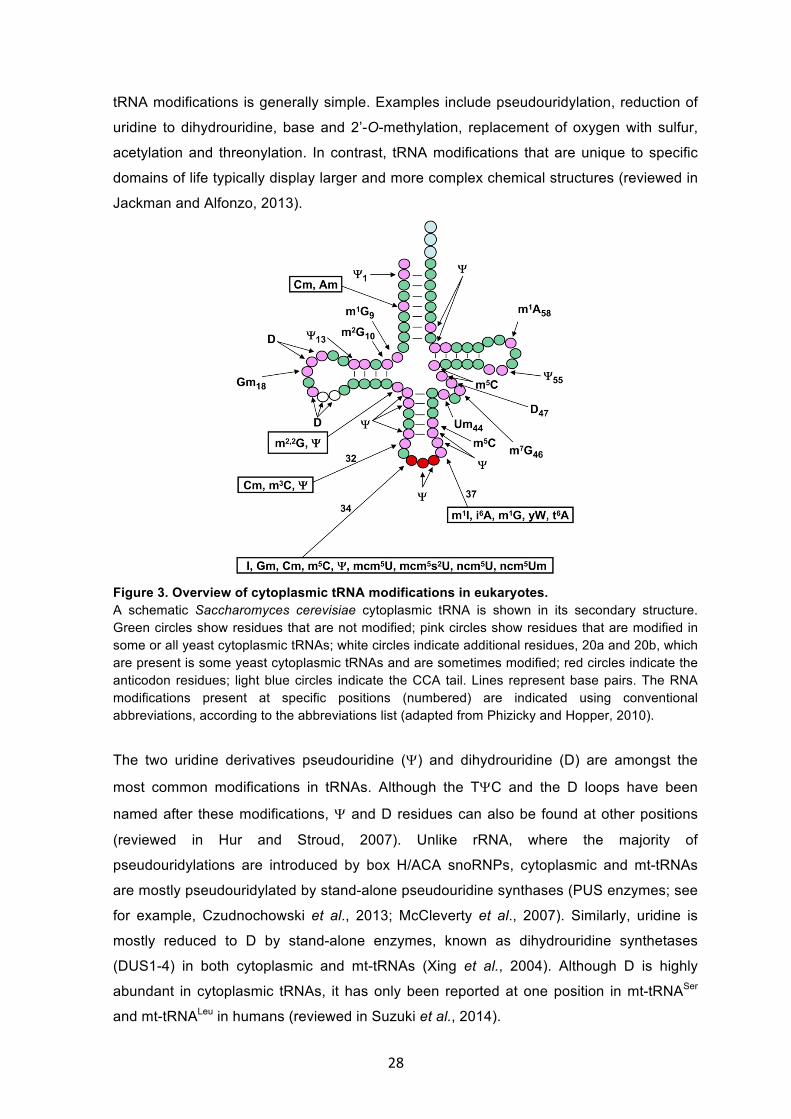
1.3.2 Diversity of tRNA modifications A striking feature of tRNAs from all living organisms is that they carry the most numerous
and chemically diverse post-transcriptional modifications. On average, each tRNA carries
14 modifications, and the type and extent of these modifications vary between different
tRNAs and in some cases between similar isoacceptors (Fig. 3; reviewed in Nachtergaele
and He, 2017). While some tRNA modifications at specific positions are conserved among
the three phylogenetic domains of life, other modifications vary between tRNAs from
different organisms within the same domain of life (reviewed in Phizicky and Alfonzo,
2010; Sprinzl and Vassilenko, 2005). The chemical nature of the evolutionarily conserved
28
tRNA modifications is generally simple. Examples include pseudouridylation, reduction of
uridine to dihydrouridine, base and 2’-O-methylation, replacement of oxygen with sulfur,
acetylation and threonylation. In contrast, tRNA modifications that are unique to specific
domains of life typically display larger and more complex chemical structures (reviewed in
Jackman and Alfonzo, 2013).
Figure 3. Overview of cytoplasmic tRNA modifications in eukaryotes. A schematic Saccharomyces cerevisiae cytoplasmic tRNA is shown in its secondary structure. Green circles show residues that are not modified; pink circles show residues that are modified in some or all yeast cytoplasmic tRNAs; white circles indicate additional residues, 20a and 20b, which are present is some yeast cytoplasmic tRNAs and are sometimes modified; red circles indicate the anticodon residues; light blue circles indicate the CCA tail. Lines represent base pairs. The RNA modifications present at specific positions (numbered) are indicated using conventional abbreviations, according to the abbreviations list (adapted from Phizicky and Hopper, 2010).
The two uridine derivatives pseudouridine (Y) and dihydrouridine (D) are amongst the
most common modifications in tRNAs. Although the TYC and the D loops have been
named after these modifications, Y and D residues can also be found at other positions
(reviewed in Hur and Stroud, 2007). Unlike rRNA, where the majority of
pseudouridylations are introduced by box H/ACA snoRNPs, cytoplasmic and mt-tRNAs
are mostly pseudouridylated by stand-alone pseudouridine synthases (PUS enzymes; see
for example, Czudnochowski et al., 2013; McCleverty et al., 2007). Similarly, uridine is
mostly reduced to D by stand-alone enzymes, known as dihydrouridine synthetases
(DUS1-4) in both cytoplasmic and mt-tRNAs (Xing et al., 2004). Although D is highly
abundant in cytoplasmic tRNAs, it has only been reported at one position in mt-tRNASer
and mt-tRNALeu in humans (reviewed in Suzuki et al., 2014).
29
Additionally, tRNAs also undergo base methylation at multiple positions. A classic
example is the conserved N1-methyladenosine (m1A) at position 58 of many cytoplasmic
and mitochondrial tRNAs (Anderson et al., 1998). In humans, these modifications are
installed by the TRMT61A/TRMT6 complex on cytoplasmic tRNAs and by TRMT61B on
mt-tRNAs (Anderson et al., 2000; Ozanick et al., 2005). Furthermore, cytosines can be
methylated at nitrogen 3 and carbon 5, generating N3-methylcytosine (m3C) and C5-
methylcytosine (m5C) modifications on tRNAs respectively. m3C modifications can be
installed on tRNAThr(UGU) and tRNA(CCU) at position 32 by methyltransferase-like (METTL)2
and on tRNASer(AGA) and tRNASer(GCU) at the same position by METTL6 (Xu et al., 2017).
m5C modifications can be installed on tRNA, as well as other classes of RNA, in humans
by members of the Nol1/Nop2/SUN domain (NSUN) family of putative methyltransferases
(NSUN1-7) and by DNA methyltransferase 2 (DNMT2; Tuorto et al., 2012). For example,
NSUN2 and DNMT2 introduce m5C modifications at position 34 of tRNALeu and at position
38 of tRNAAsp respectively (Brzezicha et al., 2006; Goll et al., 2006). The enzymes
installing m5C modifications on mt-tRNA are not well characterized but the known and
putative m5C methyltransferases NSUN2, NSUN3 and NSUN4 have all been detected in
human mitochondria, making them candidates for catalysing such reactions. Other
examples of conserved base methylations that occur in specific cytoplasmic or
mitochondrial tRNAs, include N1 or N2 or N7-methylguanosine (m1G, m2G or m7G) and N3
or C5-methyluridine (m3U or m5U). Dimethylation of tRNA residues has also been reported
and examples include N2,N2-dimethylguanosine (m2,2G) and N6,N6-dimethyladenosine
(m2,2A or m62A; reviewed in Jackman and Alfonzo, 2013).
The diverse and abundant nature of tRNA modifications, and the evolutionary
conservation of many individual modifications implies that they play important roles in
ensuring proper structure and function of tRNAs.
1.3.3 Functions of tRNA modifications It is generally thought that modifications in the core of tRNAs influence folding and
stability, whereas modifications in or around the anticodon affect efficiency or accuracy of
translation (reviewed in Phizicky and Hopper, 2010). For example, the lack of m5U at
position 54 in tRNAfMet and tRNAPhe lowers the melting temperature of the tRNA by 2-6°C
in vitro and the lack of m1A at position 9 of mt-tRNALys causes misfolding of the tRNA
(Sengupta et al., 2000; Helm et al., 1999). Interestingly, many tRNAs contain both y,
which increases RNA rigidity, and D, which promotes RNA flexibility, and it has been
suggested that the balance between the opposite effects caused by these two
modifications is important for maintaining the optimal structure of some tRNAs (reviewed
in Zagryadskaya et al., 2004; Giege et al., 2012). The lack of specific modifications can
30
also lead to degradation of the tRNA. For example, the lack of m1A modifications at
position 58 in trm6 temperature-sensitive mutants increases the turnover of the initiator
tRNAMet (Anderson et al., 1998). Additionally, tRNA modifications at various positions
serve as identity elements; for example, the 2’-O-ribosyl phosphate modification at
position 64 in tRNAMet allows discrimination between the initiator and elongator tRNAsMet
in yeast (Astrom et al., 1994).
In the anticodon loop, two positions are modified in almost every tRNA, position 37 and
the wobble nucleotide at position 34, and the modifications present in these positions
show the largest chemical diversity. In most cytoplasmic and mt-tRNAs, position 37 is a
modified purine (reviewed in Jackman and Alfonzo, 2013). Modifications at this position
typically reduce its Watson-Crick base pairing potential, thereby preventing unspecific
interactions with nearby tRNA or mRNA nucleotides and also helping maintain an open
structure of the anticodon loop. Consistent with this, the lack of m1G37 formation in
tRNAArg or tRNALeu results in increased frameshifting by the ribosome and consequently
affects translation fidelity and cellular growth (Bjork et al., 2001; Urbonavicius et al., 2001).
Notably, m1G37 is also present in mt-tRNAs and mutations in the enzyme installing the
modification (TRMT5) have been linked to multiple mitochondrial respiratory chain
deficiencies highlighting the importance of the modification at this position (Powell et al.,
2015). In some cases, m1G37 is further modified into wybutosine (yW), which prevents -1
frameshifting by allowing base stacking interactions that stabilize codon-anticodon base
pairing in the A site of the ribosome (Waas et al., 2007; de Crecy-Lagard et al., 2010).
When position 37 is an adenosine, it can be further modified in a number of different ways.
In both cytoplasmic and mt-tRNAs, threonylcarbamoyl adenosine (t6A) at position 37
maintains an open conformation of the anticodon loop and promotes codon-anticodon
interactions, which maintain the speed and accuracy of translation (Morin et al., 1998;
Thiaville et al., 2016). Alternatively, adenosine at position 37 can be isopentenylated
forming a bulky modification known as isopentenyl adenosine (i6A), which is found in a
subset cytoplasmic and mitochondrial tRNAs. In yeast, i6A has been shown to enhance
translation efficiency of specific codons (Lamichhane et al., 2011). Coupled with these
modifications at position 37, various modifications at the wobble position (34) ensure
efficient, accurate and flexible decoding. One of the best-understood examples is the
thiolation of U34 forming 2-thiouridine (s2U), which enhances anticodon rigidity, and,
combined with other modifications at carbon 5, serves as a translation efficiency and
fidelity determinant (Johansson et al., 2008). Another prevalent modification at the wobble
position in various cytoplasmic and mt-tRNAs is the replacement of guanosine with the 7-
deaza guanosine derivative, queuosine (Q; Katze et al., 1984). It has been suggested that
Q regulates the strength of specific codon-anticodon interactions, however, the exact
31
function of the modification is still unknown (Morris et al., 1999). tRNA modifications at the
wobble position are particularly important for decoding the non-universal mitochondrial
genetic code in mammals, where they allow non-canonical base pairing with the third
nucleotide of the mRNA codon (reviewed in Bohnsack and Sloan, in press). For example,
the 5-formylcytosine modification (f5C34) in the mt-tRNAMet has been proposed to increase
the decoding capacity of the tRNA by influencing the thermodynamic and structural
features of the anticodon (Bilbille et al., 2011). Such effects could possibly contribute to
the ability of the mt-tRNAMet to decode AUG, AUU and AUA codons during translation
initiation and AUG and AUU codons during translation elongation.
Through regulation of codon usage, modifications in the anticodon loop have also been
suggested to influence translation of specific mRNAs (reviewed in Gustilo et al., 2008).
For example, the 5-methylcarbonylmethyluridine modifications (mcm5U) at the wobble
position of tRNAARG(UCU) and tRNAGLU(UUC) increase the translation of mRNAs enriched in
these codons upon DNA damage (Begley et al., 2007). Strikingly, the mcm5U34
modification enhances the translation of two mRNAs highly enriched in these codons,
RNR1 and RNR3, which encode key proteins involved in the DNA damage response,
suggesting a link between tRNA modifications and cellular responses. Similarly, m5C
modifications installed in tRNAs by NSUN2 and DNMT2 have been implicated in stress
responses, as the lack of these modifications leads to endonucleolytic cleavage of the
tRNAs and accumulation of tRNA fragments, which downregulate protein translation and
promote apoptosis (Blanco et al., 2014). However, the mechanisms by which tRNA
modifications are regulated in response to different cellular conditions remain not fully
understood. One exciting means by which tRNA modifications have been shown to be
dynamically regulated is through the presence of demethylases (so called “eraser”
proteins), which selectively remove the modification in certain conditions. Indeed, the
alpha-ketoglutarate and Fe(II)-dependent dioxygenase ALKBH1 (ABH1) has been recently
identified as a tRNA demethylase that catalyses the selective removal of m1A
modifications at position 58 of selected cytoplasmic and mitochondrial tRNA, thereby
regulating the utility of these tRNAs in translation (Liu et al., 2016).
Although defects in tRNA structure and translation due to lack of individual modifications
have been observed, it has also been suggested that tRNA modifications act in concert.
This model is supported by the findings that the combinatory depletion of m7G and m5C
modifications at positions 46 and 49 respectively in trm8 trm4 yeast mutants causes
specific degradation of tRNAVal(ACC) (Alexandrov et al., 2006) and that the combinatory
depletion of non-essential genes encoding tRNA modification enzymes cause growth
defects in yeast (Chernyakov et al., 2008). Furthermore, the installation of tRNA
modifications at different positions have been shown in some cases to be coordinated. For
32
example, the introduction of an m3C modification at position 32 of cytoplasmic tRNAsSer is
dependent on the presence of an i6A modification at position 37, suggesting that the
functions of these tRNA modifications may be interconnected (Arimbasseri et al., 2016).
In short, this network of diverse, and often complex, chemical modifications plays
significant roles in maintaining the structure, stability and function of cytoplasmic and mt-
tRNAs, which modulate gene expression and regulate cellular processes. Therefore, it has
been proposed that defects in tRNA modifications may play crucial roles in human
diseases.
1.3.4 tRNA modifications and disease Growing evidence links defects in tRNA modifications to complex human pathologies,
such as neurological disorders, metabolic diseases, cancer and mitochondrial-linked
disorders (reviewed in Sarin and Leidel, 2015). Such defects can result from alterations in
genes encoding tRNA modification enzymes, or mutations in tRNA sequences that
prevent the installation of modifications. For example, mutations in the human IKBKAP
gene, which encodes IKAP, a subunit of the human Elongator complex, have been linked
to a complex genetic neuropathy affecting the autonomic nervous system, called familial
dysautonomia (FD; reviewed in Slaugenhaupt and Gusella, 2002). The Elongator complex
has been shown to play a role in the formation of the mcm5U and 5-
methoxycarbonylmethyl-2-thiouridine (mcm5s2U) modifications in U34 of several tRNAs
(Huang et al., 2005). The most prevalent mutation identified in FD patients causes tissue-
specific exon skipping and reduced IKAP levels, leading to reduced levels of mcm5U and
mcm5s2U modifications (Anderson et al. 2001; Slaugenhaupt et al., 2001). Another tRNA
modification enzyme that is associated with a neurological condition is FTSJ1, a homolog
of the yeast Trm7, which is required for the 2’-O-methylation at several positions of
different tRNAs (Pintard et al., 2002). Mutations in FTSJ1 are implicated in non-syndromic
X-linked mental retardation (Freude et al., 2004). Furthermore, mutations in NSUN2,
which installs an m5C modification at the wobble position of tRNALeu(CCA), have been linked
to autosomal recessive intellectual disability (Abbasi-Moheb et al., 2012; Khan et al.,
2012).
Lack of specific tRNA modifications have also been associated with metabolic disorders
such as type 2 diabetes, which is of particular interest because it affects health and
economies on a global scale (reviewed in Zimmet et al., 2001). Mutations in CDKAL1,
which encodes the methylthiotransferase that is required for the formation of 2-methylthio-
N6-threonylcarbamoyladenosine (ms2t6A) at position 37 of tRNALys(UUU), leads to reduced
modification levels, perturbed proinsulin processing and reduced insulin secretion in b
33
cells (Ohara-Imaizumi et al., 2010; Wei et al., 2011). Notably, CDKAL1 has also been
identified as a risk factor for Psoriasis and Crohn’s disease (Quaranta et al., 2009).
It has been long known that cancer cells often contain more methylated tRNA nucleotides
than tRNAs derived from non-tumor cells and elevated tRNA methyltransferase activity
has been observed during tumorigenesis, however subsequent studies have shown that
also hypomodification of tRNAs is associated with cancer (Tsutsui et al., 1966; Dirheimer
et al., 1995). For example, NSUN2 is highly expressed in a range of human and mice
tumor types including breast cancer, colorectal cancer and squamous cell carcinoma,
although it is expressed at low levels in non-tumor cells and in benign papillomas (Frye
and Watt, 2006). Similarly, TRM12, which is the human homolog of the yeast enzyme that
catalyses the formation of wybutosine at position 37 of tRNAPhe, is overexpressed in
multiple breast cancer tumors (Rodriguez et al., 2007). In contrast, reduced levels of Q34
modifications in tRNAs have been observed in leukaemia, lymphoma and other types of
tumors (Shindo-Okada et al., 1981; Huang et al., 1992). It has been suggested that Q
deficiency is due to reduced activity of the enzyme that installs the modification, tRNA-
guanine transglycosylase (TGTase), in tumor cells (Morris et al., 1999; Costa et al., 2004).
In a similar way, expression of TRMT5, which catalyses the m7G modification at position
37 of several tRNAs, is downregulated in colorectal cancers (reviewed in Sarin and Leidel,
2015).
Besides alterations in the tRNA modification enzymes, mutations in the genes encoding
tRNAs can also prevent the installation of specific modifications and lead to serious
pathologies. This is particularly important in mitochondria, where modifications in the
anticodon of mt-tRNAs influence their decoding capacity and enable the use of the non-
conventional mitochondrial genetic code. For example, mutations in mt-tRNALeu(UAA) and
mt-tRNALys(UUU) lead to mitochondrial encephalomyopathy with lactic acidosis and stroke-
like episode (MELAS) and myoclonus epilepsy associated with ragged-red fibers
(MERRF) syndromes respectively (Fig. 4; Yasukawa et al., 2000a, 2000b). In both
syndromes, the mutations disrupt the tertiary structure of tRNAs and prevent their
recognition by the taurine transferases, which are suggested to be MTO1 and GTPBP3
(Tischner et al., 2015; Chen et al., 2016), leading to 5-taurinomethyluridine (tm5U)
hypomodification of U34 in MELAS patients and 5-taurinomethyl-2-thiouridine (tm5s2U)
hypomodification of U34 in MERRF patients (Kirino et al., 2005). The lack of tm5U
impedes mt-tRNALeu(UAA) decoding UUG codons, but it does not affect reading of UUA
codons. In MELAS patients, this leads to a specific reduction in ND6, which is a
component of the respiratory chain complex I that is translated from a transcript enriched
in UUG codons. However, the absence of tm5S2U prevents mt-tRNALys(UUU) from reading
34
both of its cognate codons (AAG and AAA), leading to reduced overall mitochondrial
translation in MERRF patients (reviewed in Suzuki et al., 2011).
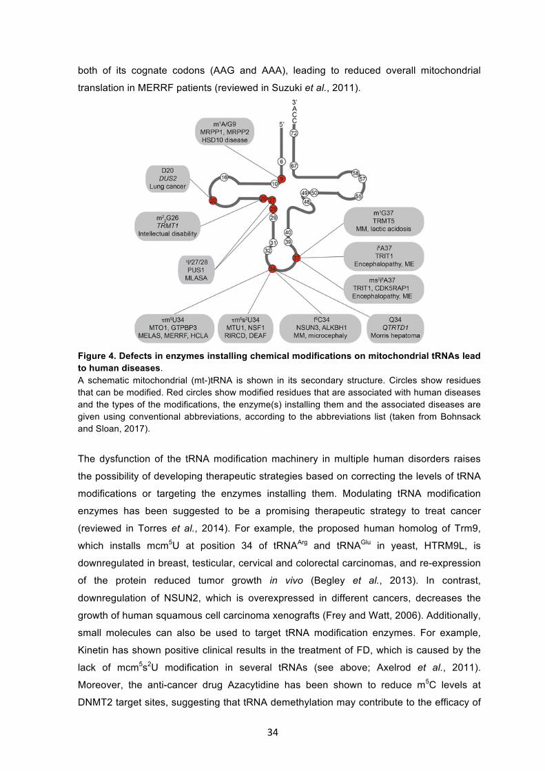
Figure 4. Defects in enzymes installing chemical modifications on mitochondrial tRNAs lead to human diseases. A schematic mitochondrial (mt-)tRNA is shown in its secondary structure. Circles show residues that can be modified. Red circles show modified residues that are associated with human diseases and the types of the modifications, the enzyme(s) installing them and the associated diseases are given using conventional abbreviations, according to the abbreviations list (taken from Bohnsack and Sloan, 2017).
The dysfunction of the tRNA modification machinery in multiple human disorders raises
the possibility of developing therapeutic strategies based on correcting the levels of tRNA
modifications or targeting the enzymes installing them. Modulating tRNA modification
enzymes has been suggested to be a promising therapeutic strategy to treat cancer
(reviewed in Torres et al., 2014). For example, the proposed human homolog of Trm9,
which installs mcm5U at position 34 of tRNAArg and tRNAGlu in yeast, HTRM9L, is
downregulated in breast, testicular, cervical and colorectal carcinomas, and re-expression
of the protein reduced tumor growth in vivo (Begley et al., 2013). In contrast,
downregulation of NSUN2, which is overexpressed in different cancers, decreases the
growth of human squamous cell carcinoma xenografts (Frey and Watt, 2006). Additionally,
small molecules can also be used to target tRNA modification enzymes. For example,
Kinetin has shown positive clinical results in the treatment of FD, which is caused by the
lack of mcm5s2U modification in several tRNAs (see above; Axelrod et al., 2011).
Moreover, the anti-cancer drug Azacytidine has been shown to reduce m5C levels at
DNMT2 target sites, suggesting that tRNA demethylation may contribute to the efficacy of
35
the drug (Schaefer et al., 2009). Besides their potential as therapeutic tools, changes in
tRNA modifications could also be used as diagnostic markers for certain diseases. For
example, the human homolog of Trm10, which installs m1G modifications at position 9 in
several tRNAs in yeast, HRG9MTD2, is a good candidate to diagnose early onset and late
onset colorectal cancer patients (Berg et al., 2010).
Despite the increasing number of reports linking tRNA modification to disease, we still lack
understanding of the mechanisms by which many tRNA modifications are installed or
regulated, and their precise biological roles.
1.4 Modifications in other RNA species 1.4.1 Transcriptome-wide mapping approaches Although RNA modifications have been recognized from the early days of molecular
biology (examples include Cohn and Volkin, 1951; Davis and Allen, 1957), the majority of
early research was limited to modifications in highly abundant RNAs, such as tRNAs,
rRNAs and snRNAs (reviewed in Schwartz, 2016). This is because detection of
modifications in RNAs expressed at low levels, such as mRNAs, is technically challenging
for classical biochemical methods. However, recently, transcriptome-wide studies of RNA
modifications using next generation sequencing (NGS) technologies allowed the
identification and mapping of several modification types in all classes of RNA, including
mRNAs and long non-coding RNAs (reviewed in Roundtree et al., 2017). NGS
technologies involve the reverse transcription of RNAs into cDNAs, which are ligated to
adaptors for sequencing, and mapping of the obtained sequence reads to the genome
and read-end mapping enables the sites of the modifications to be determined in a
transcriptome-wide manner. Such RNA modification mapping techniques require a means
by which nucleotides carrying specific modification can be differentiated from non-modified
nucleotides. One approach by which this can be achieved is based on distinctive reverse
transcription signatures (RT signature) resulting from nucleotides misincorporation or
abortive cDNA products caused by large RNA modifications or chemical adducts on the
Watson-Crick face of the modified residues (Ebhardt et al., 2009; Findeiss et al., 2011).
This approach has been used in transcriptome-wide mapping of m1A modifications after
antibody-mediated enrichment (Li et al., 2016; Dominissini et al., 2016). Interestingly, RNA
modifications, such as pseudouridine (Y), which do not themselves cause reverse
transcriptase stalling, can also be analysed by such approaches as, chemical treatments
of RNA can enhance the RT signature of specific RNA modifications. For example, N-
cyclohexyl-N’-(2-morpholinoethyl)carbodiimide methyl-p-toluenesulfonate (CMCT)
treatment of RNAs leads to formation of RT-arresting adducts at Y residues, allowing their
detection and the generation of transcriptome-wide maps of the modification (Carlile et al.,
36
2014; Li et al., 2015). Furthermore, the different reactivities of modified and unmodified
nucleotides to specific chemical treatments can be exploited to enable detection of
modification sites in cellular RNAs. This is the basis of the well-known bisulfite sequencing
approach, which can be applied to both DNA and RNA. Here, bisulfite treatment
deaminates unmodified cytidines to uridines but does not affect 5-methylcytidines, and this
conversion can be detected by sequencing after reverse transcription (Schaefer et al.,
2009).
An alternative approach for the detection of modified sites in RNAs is through the use of
antibodies that specifically recognise modified nucleotides to enrich modification-
containing RNA fragments, which are then converted into a cDNA library for sequencing.
This approach is used for transcriptome-wide mapping of several RNA modifications,
including N6-methyladenosine (m6A; Dominissini et al., 2012; Meyer et al., 2012). m6A
mapping combined with RNAi-mediated knockdowns of METTL3 and METTL14 allowed
identification of the METTL3-METTL14 heterodimer as a methyltransferase complex that
installs m6A modifications on multiple RNAs (Liu et al., 2014). Moreover, the antibody-
mediated immunoprecipitation of 5-hydroxymethylcytosine (hm5C)-containing RNA
fragments uncovered the transcriptome-wide distribution of the modification in Drosophila
melanogaster cells (Delatte et al., 2016). However, these antibody-based techniques
enable mapping of the modifications to RNA regions of approximately 100 nucleotides but
do not provide their exact nucleotide positions. For m6A mapping, a significant
improvement in the resolution of the mapping approaches was obtained by crosslinking
the antibodies to the modified sites in RNA, which induced unique signature mutations
upon reverse transcription, allowing the generation of the first single-nucleotide resolution
map of m6A (Linder et al., 2015). In short, this rapidly expanding spectrum of techniques
for the detection of RNA modifications in cellular RNAs has led to a resurgence in the field
of ‘epitranscriptomics’ through generating transcriptome-wide maps of different
modifications on all classes of RNA. These methods allow studying the dynamic
occurrence of RNA modifications under different conditions and their potential roles in
regulation of gene expression and cellular behaviour (reviewed in Helm and Motorin,
2017).
1.4.2 Modifications in other non-coding RNAs Besides tRNAs and rRNAs, modifications on other classes of non-coding RNAs that play
crucial roles in cellular homeostasis have been identified (reviewed in Esteller and
Pandolfi, 2017). For example, the long non-coding RNA (lncRNA) XIST, which mediates
transcriptional repression of multiple genes on the X chromosome during female
mammalian development, has been recently shown to be highly methylated and the
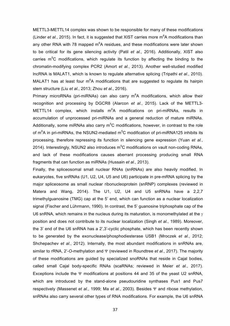
37
METTL3-METTL14 complex was shown to be responsible for many of these modifications
(Linder et al., 2015). In fact, it is suggested that XIST carries more m6A modifications than
any other RNA with 78 mapped m6A residues, and these modifications were later shown
to be critical for its gene silencing activity (Patil et al., 2016). Additionally, XIST also
carries m5C modifications, which regulate its function by affecting the binding to the
chromatin-modifying complex PCR2 (Amort et al., 2013). Another well-studied modified
lncRNA is MALAT1, which is known to regulate alternative splicing (Tripathi et al., 2010).
MALAT1 has at least four m6A modifications that are suggested to regulate its hairpin
stem structure (Liu et al., 2013; Zhou et al., 2016).
Primary microRNAs (pri-miRNAs) can also carry m6A modifications, which allow their
recognition and processing by DGCR8 (Alarcon et al., 2015). Lack of the METTL3-
METTL14 complex, which installs m6A modifications on pri-miRNAs, results in
accumulation of unprocessed pri-miRNAs and a general reduction of mature miRNAs.
Additionally, some miRNAs also carry m5C modifications, however, in contrast to the role
of m6A in pri-miRNAs, the NSUN2-mediated m5C modification of pri-miRNA125 inhibits its
processing, therefore repressing its function in silencing gene expression (Yuan et al.,
2014). Interestingly, NSUN2 also introduces m5C modifications on vault non-coding RNAs,
and lack of these modifications causes aberrant processing producing small RNA
fragments that can function as miRNAs (Hussain et al., 2013).
Finally, the spliceosomal small nuclear RNAs (snRNAs) are also heavily modified. In
eukaryotes, five snRNAs (U1, U2, U4, U5 and U6) participate in pre-mRNA splicing by the
major spliceosome as small nuclear ribonucleoprotein (snRNP) complexes (reviewed in
Matera and Wang, 2014). The U1, U2, U4 and U5 snRNAs have a 2,2,7
trimethylguanosine (TMG) cap at the 5’ end, which can function as a nuclear localization
signal (Fischer and Lührmann, 1990). In contrast, the 5’ guanosine triphosphate cap of the
U6 snRNA, which remains in the nucleus during its maturation, is monomethylated at the g
position and does not contribute to its nuclear localization (Singh et al., 1989). Moreover,
the 3’ end of the U6 snRNA has a 2’,3’-cyclic phosphate, which has been recently shown
to be generated by the exonuclease/phosphodiesterase USB1 (Mroczek et al., 2012;
Shchepachev et al., 2012). Internally, the most abundant modifications in snRNAs are,
similar to rRNA, 2’-O-methylation and Y (reviewed in Roundtree et al., 2017). The majority
of these modifications are guided by specialized snoRNAs that reside in Cajal bodies,
called small Cajal body-specific RNAs (scaRNAs; reviewed in Meier et al., 2017).
Exceptions include the Y modifications at positions 44 and 35 of the yeast U2 snRNA,
which are introduced by the stand-alone pseudouridine synthases Pus1 and Pus7
respectively (Massenet et al., 1999; Ma et al., 2003). Besides Y and ribose methylation,
snRNAs also carry several other types of RNA modifications. For example, the U6 snRNA
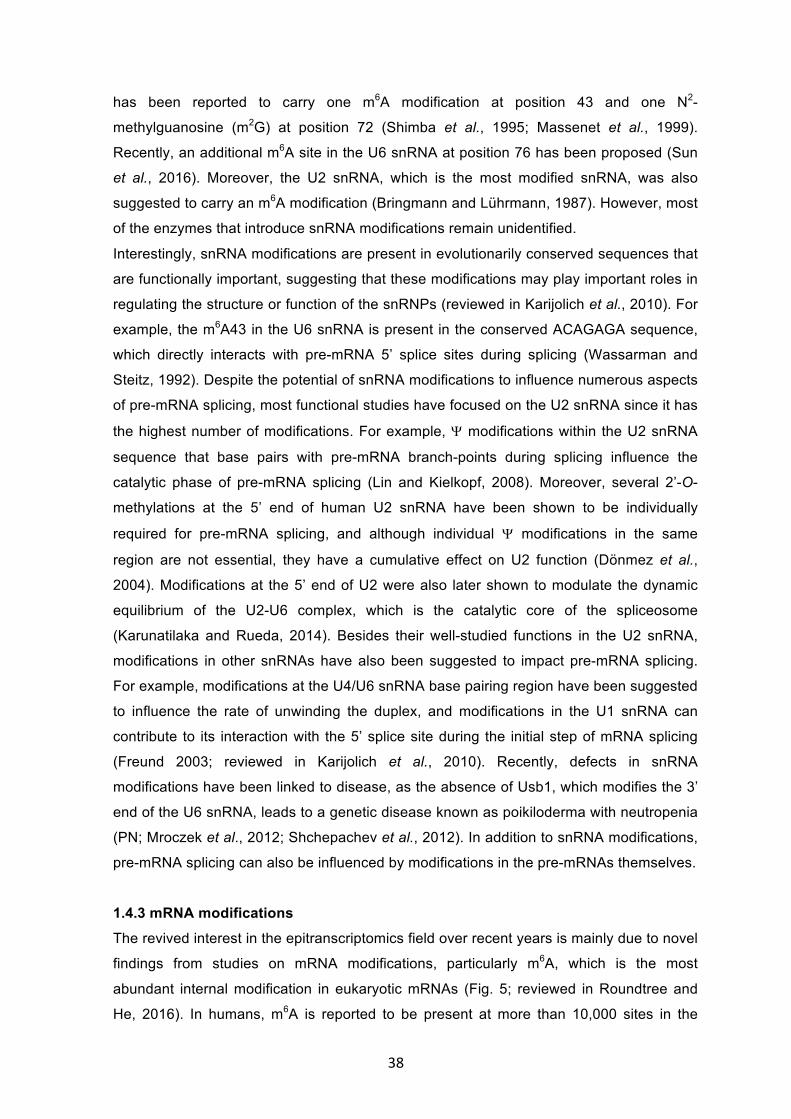
38
has been reported to carry one m6A modification at position 43 and one N2-
methylguanosine (m2G) at position 72 (Shimba et al., 1995; Massenet et al., 1999).
Recently, an additional m6A site in the U6 snRNA at position 76 has been proposed (Sun
et al., 2016). Moreover, the U2 snRNA, which is the most modified snRNA, was also
suggested to carry an m6A modification (Bringmann and Lührmann, 1987). However, most
of the enzymes that introduce snRNA modifications remain unidentified.
Interestingly, snRNA modifications are present in evolutionarily conserved sequences that
are functionally important, suggesting that these modifications may play important roles in
regulating the structure or function of the snRNPs (reviewed in Karijolich et al., 2010). For
example, the m6A43 in the U6 snRNA is present in the conserved ACAGAGA sequence,
which directly interacts with pre-mRNA 5’ splice sites during splicing (Wassarman and
Steitz, 1992). Despite the potential of snRNA modifications to influence numerous aspects
of pre-mRNA splicing, most functional studies have focused on the U2 snRNA since it has
the highest number of modifications. For example, Y modifications within the U2 snRNA
sequence that base pairs with pre-mRNA branch-points during splicing influence the
catalytic phase of pre-mRNA splicing (Lin and Kielkopf, 2008). Moreover, several 2’-O-
methylations at the 5’ end of human U2 snRNA have been shown to be individually
required for pre-mRNA splicing, and although individual Y modifications in the same
region are not essential, they have a cumulative effect on U2 function (Dönmez et al.,
2004). Modifications at the 5’ end of U2 were also later shown to modulate the dynamic
equilibrium of the U2-U6 complex, which is the catalytic core of the spliceosome
(Karunatilaka and Rueda, 2014). Besides their well-studied functions in the U2 snRNA,
modifications in other snRNAs have also been suggested to impact pre-mRNA splicing.
For example, modifications at the U4/U6 snRNA base pairing region have been suggested
to influence the rate of unwinding the duplex, and modifications in the U1 snRNA can
contribute to its interaction with the 5’ splice site during the initial step of mRNA splicing
(Freund 2003; reviewed in Karijolich et al., 2010). Recently, defects in snRNA
modifications have been linked to disease, as the absence of Usb1, which modifies the 3’
end of the U6 snRNA, leads to a genetic disease known as poikiloderma with neutropenia
(PN; Mroczek et al., 2012; Shchepachev et al., 2012). In addition to snRNA modifications,
pre-mRNA splicing can also be influenced by modifications in the pre-mRNAs themselves.
1.4.3 mRNA modifications The revived interest in the epitranscriptomics field over recent years is mainly due to novel
findings from studies on mRNA modifications, particularly m6A, which is the most
abundant internal modification in eukaryotic mRNAs (Fig. 5; reviewed in Roundtree and
He, 2016). In humans, m6A is reported to be present at more than 10,000 sites in the
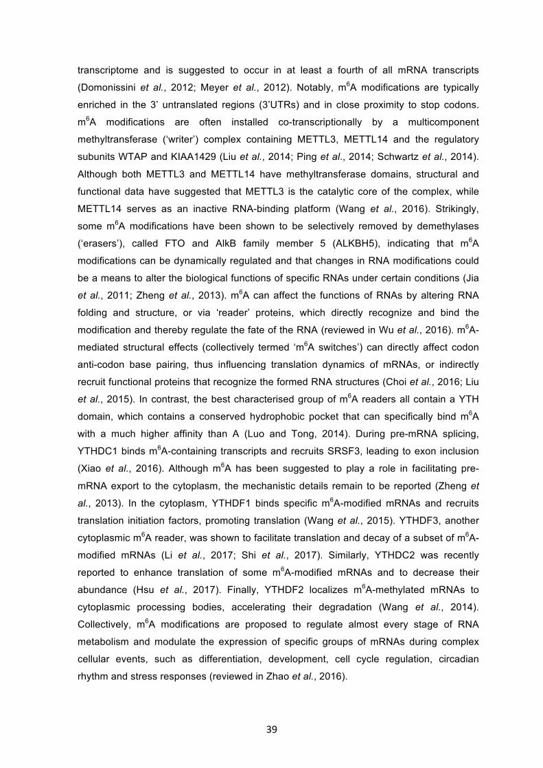
39
transcriptome and is suggested to occur in at least a fourth of all mRNA transcripts
(Domonissini et al., 2012; Meyer et al., 2012). Notably, m6A modifications are typically
enriched in the 3’ untranslated regions (3’UTRs) and in close proximity to stop codons.
m6A modifications are often installed co-transcriptionally by a multicomponent
methyltransferase (‘writer’) complex containing METTL3, METTL14 and the regulatory
subunits WTAP and KIAA1429 (Liu et al., 2014; Ping et al., 2014; Schwartz et al., 2014).
Although both METTL3 and METTL14 have methyltransferase domains, structural and
functional data have suggested that METTL3 is the catalytic core of the complex, while
METTL14 serves as an inactive RNA-binding platform (Wang et al., 2016). Strikingly,
some m6A modifications have been shown to be selectively removed by demethylases
(‘erasers’), called FTO and AlkB family member 5 (ALKBH5), indicating that m6A
modifications can be dynamically regulated and that changes in RNA modifications could
be a means to alter the biological functions of specific RNAs under certain conditions (Jia
et al., 2011; Zheng et al., 2013). m6A can affect the functions of RNAs by altering RNA
folding and structure, or via ‘reader’ proteins, which directly recognize and bind the
modification and thereby regulate the fate of the RNA (reviewed in Wu et al., 2016). m6A-
mediated structural effects (collectively termed ‘m6A switches’) can directly affect codon
anti-codon base pairing, thus influencing translation dynamics of mRNAs, or indirectly
recruit functional proteins that recognize the formed RNA structures (Choi et al., 2016; Liu
et al., 2015). In contrast, the best characterised group of m6A readers all contain a YTH
domain, which contains a conserved hydrophobic pocket that can specifically bind m6A
with a much higher affinity than A (Luo and Tong, 2014). During pre-mRNA splicing,
YTHDC1 binds m6A-containing transcripts and recruits SRSF3, leading to exon inclusion
(Xiao et al., 2016). Although m6A has been suggested to play a role in facilitating pre-
mRNA export to the cytoplasm, the mechanistic details remain to be reported (Zheng et
al., 2013). In the cytoplasm, YTHDF1 binds specific m6A-modified mRNAs and recruits
translation initiation factors, promoting translation (Wang et al., 2015). YTHDF3, another
cytoplasmic m6A reader, was shown to facilitate translation and decay of a subset of m6A-
modified mRNAs (Li et al., 2017; Shi et al., 2017). Similarly, YTHDC2 was recently
reported to enhance translation of some m6A-modified mRNAs and to decrease their
abundance (Hsu et al., 2017). Finally, YTHDF2 localizes m6A-methylated mRNAs to
cytoplasmic processing bodies, accelerating their degradation (Wang et al., 2014).
Collectively, m6A modifications are proposed to regulate almost every stage of RNA
metabolism and modulate the expression of specific groups of mRNAs during complex
cellular events, such as differentiation, development, cell cycle regulation, circadian
rhythm and stress responses (reviewed in Zhao et al., 2016).
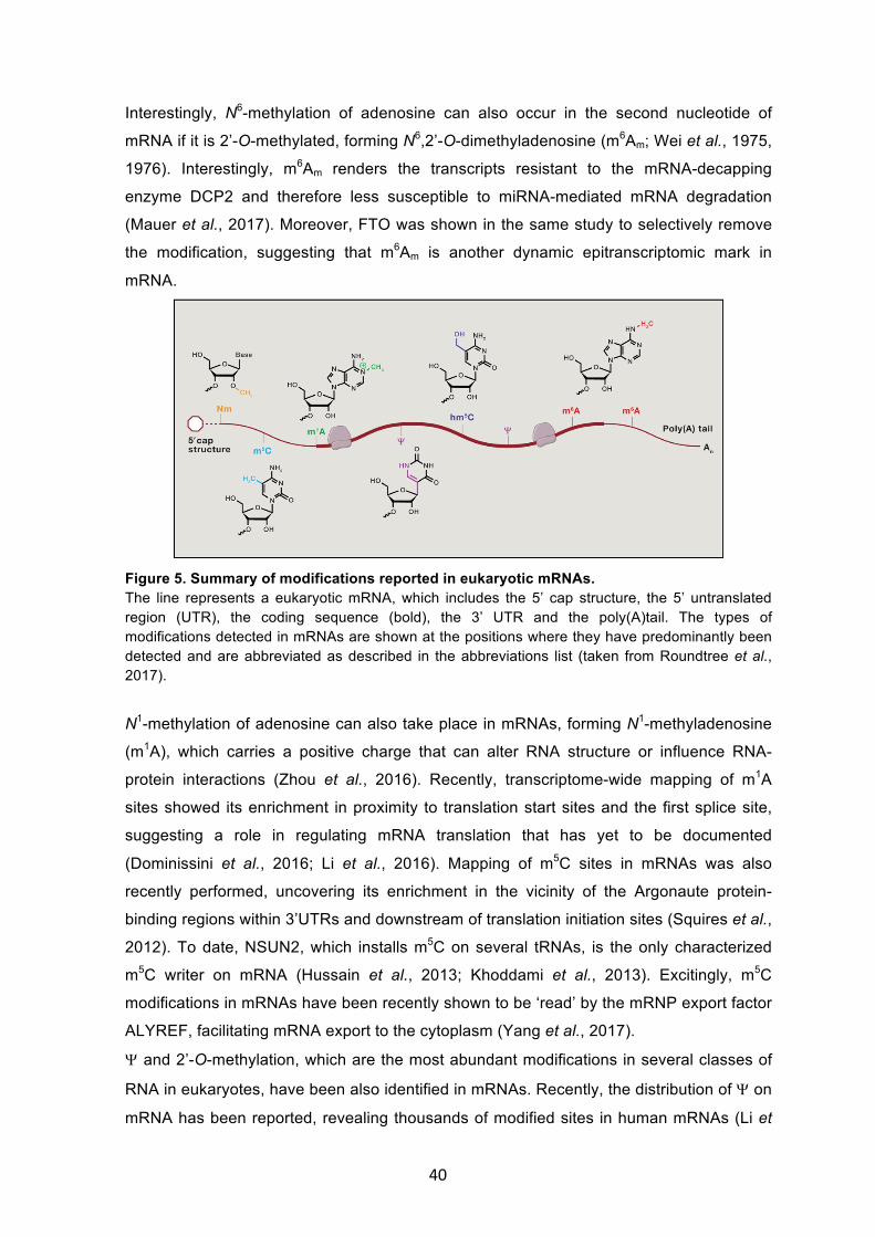
40
Interestingly, N6-methylation of adenosine can also occur in the second nucleotide of
mRNA if it is 2’-O-methylated, forming N6,2’-O-dimethyladenosine (m6Am; Wei et al., 1975,
1976). Interestingly, m6Am renders the transcripts resistant to the mRNA-decapping
enzyme DCP2 and therefore less susceptible to miRNA-mediated mRNA degradation
(Mauer et al., 2017). Moreover, FTO was shown in the same study to selectively remove
the modification, suggesting that m6Am is another dynamic epitranscriptomic mark in
mRNA.
Figure 5. Summary of modifications reported in eukaryotic mRNAs. The line represents a eukaryotic mRNA, which includes the 5’ cap structure, the 5’ untranslated region (UTR), the coding sequence (bold), the 3’ UTR and the poly(A)tail. The types of modifications detected in mRNAs are shown at the positions where they have predominantly been detected and are abbreviated as described in the abbreviations list (taken from Roundtree et al., 2017).
N1-methylation of adenosine can also take place in mRNAs, forming N1-methyladenosine
(m1A), which carries a positive charge that can alter RNA structure or influence RNA-
protein interactions (Zhou et al., 2016). Recently, transcriptome-wide mapping of m1A
sites showed its enrichment in proximity to translation start sites and the first splice site,
suggesting a role in regulating mRNA translation that has yet to be documented
(Dominissini et al., 2016; Li et al., 2016). Mapping of m5C sites in mRNAs was also
recently performed, uncovering its enrichment in the vicinity of the Argonaute protein-
binding regions within 3’UTRs and downstream of translation initiation sites (Squires et al.,
2012). To date, NSUN2, which installs m5C on several tRNAs, is the only characterized
m5C writer on mRNA (Hussain et al., 2013; Khoddami et al., 2013). Excitingly, m5C
modifications in mRNAs have been recently shown to be ‘read’ by the mRNP export factor
ALYREF, facilitating mRNA export to the cytoplasm (Yang et al., 2017).
Y and 2’-O-methylation, which are the most abundant modifications in several classes of
RNA in eukaryotes, have been also identified in mRNAs. Recently, the distribution of Y on
mRNA has been reported, revealing thousands of modified sites in human mRNAs (Li et
41
al., 2015; Carlile et al., 2014; Lovejoy et al., 2014). Members of the pseudouridine
synthetase (PUS) family have been shown to mediate pseudouridylation of mRNAs
(Karijolich et al., 2011). Although the precise roles of Y in mRNA have not been shown in
detail yet, in E. coli, it has been suggested that Y modification of the first nucleotide of
stop codons affects base pairing during decoding, leading to increased read-through
(Fernandez et al., 2013). The distribution of 2’-O-methylation sites on mRNAs was
recently reported, uncovering the presence of more than 7000 2’-O-methylation sites (Dai
et al., 2017). It has been suggested that certain snoRNAs bind to mRNAs where it is
possible that they install 2’-O-methylations, however, experimental evidence of these
interactions is lacking and the exact mechanism of installing the modification on mRNA is
still not clear (Bachellerie et al., 2002; Cavaille et al., 2000). Similarly, the exact functions
of non-cap 2’-O-methylations in mRNAs are not fully understood, but in vitro studies have
suggested that it can inhibit A to I RNA editing (Beal et al., 2007).
Taken together, the last decade has witnessed great progress in the field of RNA
modifications, unravelling transcriptome-wide maps of different modifications on different
RNA species and their dynamic occurrence. However, we are still "scratching the surface"
of the field, since we still need to understand the mechanisms by which different RNA
modifications are installed as well as their contribution to signalling pathways, human
physiology and disease.
42
Chapter Two: Aims of this work Recent studies have shown that chemical modifications are widespread in almost all
classes of RNA and these modifications are emerging as key players in the regulation of
gene expression. Modifications expand the features of RNA residues, thereby influencing
RNA secondary structures, base pairing properties and/ or RNA-protein interactions.
These functions enable RNA modifications to regulate the fates of the modified RNAs and
affect cellular processes that involve ribonucleoprotein (RNP) complexes, such as
ribosome biogenesis, mRNA splicing and translation. Interestingly, some RNA
modifications were recently shown to be selectively removed by ‘eraser’ proteins,
suggesting that they represent a dynamic layer of regulation of gene expression.
Moreover, defects in RNA-modifiying enzymes have been linked to human diseases, such
as cancer and metabolic disorders.
The enzymes that install many RNA modifications and the biological functions of these
modifications remain uncharacterized and knowledge of the molecular mechanisms of
most diseases caused by defects in RNA modification enzymes is often lacking. Here, we
aimed to characterise several putative RNA methyltransferases for which no functions
were known. We set out to determine the RNA substrates of the putative m6A and m5C
methyltransferases METTL16, NSUN6 and NSUN3 in a transcriptome-wide manner using
the crosslinking and analysis of cDNA (CRAC) approach. In addition, we aimed to
characterize the modification activity of these enzymes using in vivo and in vitro
methylation assays and to identify recognition elements that are required for recruitment
of the RNA-modifying enzymes to their specific substrate RNAs. Furthermore, to gain
insight into the cellular functions of the modifications installed by these enzymes, we
analysed the effects of lack of these modifications/modification enzymes on the target
pathways identified by CRAC. Another important aspect of this work was to better
understand how defects in RNA modification enzymes can cause disease, and we studied
Bowen-Conradi syndrome (BCS), which is caused by a point mutation in the gene
encoding the rRNA methyltransferase EMG1, to elucidate the molecular basis of this
disease.
Collectively, our studies aimed to characterize RNA-modifying enzymes by providing
insights into their modification substrates, cellular functions and their involvement in
human diseases.
43
Manuscript 1
Warda AS*, Kretschmer J*, Hackert P, Lenz C, Urlaub H, Höbartner C, Sloan KE,
Bohnsack MT (2017) METTL16 is a N6-methyladenosine (m6A) methyltransferase that
targets pre-mRNAs and various non-coding RNAs. EMBO Rep, in press.
* These authors contributed equally to this work.
URL: http://embor.embopress.org/content/18/11/2004.long
doi: 10.15252/embr.201744940
Author contributions: Figure 1: Generation of HEK293 cell line expressing METTL16-His-Prc-Flag (AW); UV-
and PAR-CRAC experiments for METTL16 (PH); bioinformatics analysis of METTL16
CRAC data (JK, KS, MB); RNA-IP after crosslinking and northern blotting for snRNAs
(AW).
Figure 2: RNAi against METTL16 (AW); Bioinformatics analysis of METTL16 CRAC data
(JK, KS, MB); anti-m6A RNA-immunoprecipitation and northern blotting for snRNAs (AW).
Figure 3: Native RNA-immunoprecipitation and northern blotting for snRNAs (AW);
immunoprecipitation and western blotting for La, LARP7 and MEPCE (AW, PH); mass
spectrometry of immunoprecipitation eluates (CL, HU).
Figure 4: Bioinformatics analysis of METTL16 CRAC data (JK, MB); immunoprecipitation
of METTL16-associated RNAs (AW); quantitative RT-PCR analysis of METTL16-
associated RNAs (JK).
Figure EV1: Generation of HEK293 cell line expressing METTL16-GFP and
immunofluorescence analysis of METTL16 (AW).
Figure EV2: Bioinformatics analysis of METTL16 CRAC data (JK, KS, MB).
Non-experimental contributions: Markus Bohnsack, Claudia Höbartner and Katherine
Sloan conceived the study and supervised the work. Katherine Sloan, Markus Bohnsack
and Ahmed Warda wrote the manuscript and prepared the figures. All authors analysed
data and contributed to preparation of the manuscript.

44
Manuscript 2
Haag S, Warda AS, Kretschmer J, Günnigmann MA, Höbartner C, Bohnsack MT (2015)
NSUN6 is a human RNA methyltransferase that catalyzes formation of m5C72 in specific
tRNAs. RNA 21: 1532-1543.
URL: http://rnajournal.cshlp.org/content/21/9/1532.long
doi: 10.1261/rna.051524.115
Author contributions: Figure 1: Generation of HEK293 cell line expressing NSUN6-His-Prc-Flag (SH); UV- and
5-azaC-CRAC experiments for NSUN6 (SH); bioinformatics analysis of NSUN6 CRAC
data (JK, SH).
Figure 2: Bioinformatics analysis of NSUN6 CRAC data (JK, SH); RNA-IP after
crosslinking and northern blotting for tRNAs (SH).
Figure 3: RNAi against NSUN6 (SH); purification of recombinant NSUN6 (SH);
preparation of RNA substrates by in vitro transcription and RNA synthesis (SH, CH); in
vitro methylation assays using recombinant NSUN6 (SH, MG).
Figure 4: Cell fractionation and western blotting (SH, AW); generation of cell line
expressing NSUN6-GFP (SH); immunofluorescence of NSUN6, GOPC, GM130; PCM1
and tubulin (AW).
Non-experimental contributions: Markus Bohnsack and Sara Haag conceived the study
and wrote the manuscript. Markus Bohnsack supervised the work. All authors analysed
data and contributed to preparation of the manuscript.
45
Manuscript 3
Haag S*, Sloan KE*, Ranjan N*, Warda AS*, Kretschmer J, Blessing C, Hübner B,
Seikowski J, Dennerlein S, Rehling P, Rodnina MV, Höbartner C, Bohnsack MT (2016)
NSUN3 and ABH1 modify the wobble position of mt-tRNAMet to expand codon recognition
in mitochondrial translation. EMBO J 35: 2104-2119.
* These authors contributed equally to this work.
URL: http://emboj.embopress.org/content/35/19/2104.long
doi: 10.15252/embj.201694885
Author contributions: Figure 1: Generation of HEK293 cell line expressing NSUN3-GFP (SH); fluorescence
microscopy of NSUN3-GFP and a mitotracker (AW); purification of mitochondria,
mitochondrial fractionation and western blotting (AW, SD).
Figure 2: Generation of HEK293 cell line expressing NSUN3-His-Prc-Flag (SH);
bioinformatics analysis of NSUN3 CRAC data (JK, MB, SH); RNA-IP after crosslinking
and northern blotting for mt-tRNAs (SH, KS).
Figure 3: Preparation of RNA substrates by in vitro transcription and RNA synthesis (SH,
CH); in vitro methylation assay (SH).
Figure 4: Preparation of RNA substrates and in vitro methylation assays (SH, AW).
Figure 5: Immunofluorescence against ABH1 (AW); RNA-IP after crosslinking and
northern blotting for mt-tRNAs (SH, KS, BH).
Figure 6: Recombinant expression of ABH1, ABH1R338A, ABH1D233A and FTO (BH, SH); in
vitro methylation/oxidation assays (BH, KS, CH, SH); preparation and RP-HPLC analysis
of m5C-, hm5C- and f5C-containing ASL (CH, KS).
Figure 7: Purification of recombinant human mitochondrial translation factors (KS, CB,
MB); preparation of m5C-, hm5C- and f5C-containg mt-tRNAMet (CH, KS); ribosome-binding
studies using bacterial in vitro translation system (NR).
Figure 8: RNAi against NSUN3 and ABH1 (SH, KS, BH, AW); TMI-labelling and primer
extension analysis of f5C34 in mt-tRNAMet (KS, CH); Bisulfite sequencing of mt-tRNAMet
after RNAi (SH, CB, JK, MB).
Figure 9: 35S-labelling of RNAi-treated cells and analysis of mitochondrial protein levels
(AW, SD). Figure EV1: Bioinformatics analysis of NSUN3 CRAC data (JK, SH); in vitro methylation
assays (SH); RNA-IP after crosslinking and northern blotting for mt-tRNAs (KS, SH).
Figure EV2: Western blotting for sub-mitochondrial localisation of ABH1 (AW, SD).
46
Figure EV3: Analysis of cell viability of RNAi against NSUN3 or ABH1 (KS)
Non-experimental contributions: Markus Bohnsack, Claudia Höbartner, Sara Haag and
Katherine Sloan conceived the study. Peter Rehling, Marina Rodina, Markus Bohnsack,
Claudia Höbartner, Katherine Sloan and Sara Haag supervised the research. Markus
Bohnsack, Katherine Sloan and Claudia Höbartner wrote the manuscript.
47
Manuscript 4
Warda AS, Freytag B, Haag S, Sloan KE, Görlich D, Bohnsack MT (2016) Effects of the
Bowen-Conradi syndrome mutation in EMG1 on its nuclear import, stability and nucleolar
recruitment. Hum Mol Genet 25: 5353-5364.
URL: https://academic.oup.com/hmg/article/25/24/5353/2593613/Effects-of-the-Bowen-
Conradi-syndrome-mutation-in
doi: 10.1093/hmg/ddw351.
Author contributions: Figure 1: Generation of HEK293 cell line expressing EMG1 and EMG1D86G (AW); RNAi-
mediated depletion of EMG1 (AW); Northern blot analysis of pre-rRNA levels (AW).
Figure 2: Immunofluorescence analysis of EMG1 and EMG1D86G localisation (AW).
Figure 3: Purification of recombinant EMG1 (AW, SH); Binding assays between EMG1
and import receptors (AW); Immunoprecipitation of EMG1-containing complexes for mass
spectrometry analysis (SH); RNAi against NOP14, NOC4L and UTP14 (AW); northern blot
and pulse-chase labelling analysis of pre-rRNA levels (AW, KS).
Figure 4: Immunofluorescence analysis of EMG1, NOP14, NOC4L and UTP14 after RNAi
(AW).
Figure 5: Nuclear import assays in permeabilised cells using recombinant EMG1 and
importins (AW, BF).
Figure 6: In vitro chaperone assays using recombinant EMG1 and importins (AW, BF).
Figure S1: Proteasome inhibitor treatment and western blotting for EMG1 (AW).
Figure S2: Immunoprecipitation assays and western blotting (AW); in vitro methylation
assay using recombinant EMG1 and EMG1D86G (AW).
Figure S3: In vitro chaperone assays using recombinant EMG1 and importins (AW, BF).
Non-experimental contributions: Markus Bohnsack conceived the study, and Katherine
Sloan, Markus Bohnsack and Ahmed Warda wrote the manuscript and prepared the
figures. Markus Bohnsack, Dirk Görlich, Katherine Sloan and Sara Haag supervised the
work. All authors analysed data and contributed to preparation of the manuscript.
48
Chapter Four: Discussion More than 150 distinct chemical modifications have been reported in RNAs from diverse
organisms. In recent years, our knowledge of RNA modifications has substantially
advanced with transcriptome-wide studies showing that the landscape of different RNA
modifications represents an important dynamic layer of regulation of gene expression.
However, many enzymes responsible for installing particular RNA modifications, their
biological roles in cellular processes and their contributions to human diseases remain to
be uncovered.
4.1 Characterization of the novel m6A writer METTL16 N6-methyladenosine (m6A) is the most abundant internal modification in mRNAs and
lncRNAs (reviewed in Roignant and Soller, 2017). Mapping of m6A has revealed its
enrichment within a RRACH sequence motif (R=A or G; H=A or U or C) around stop
codons in mRNAs (Dominissini et al., 2012; Meyer et al., 2012). Recent studies showed
that m6A modifications can regulate almost all stages of RNA metabolism, including (pre-)
mRNA splicing, processing, nuclear export, translation and degradation, and therefore,
m6A influences multiple cellular processes, such as differentiation and development
(reviewed in Roundtree et al., 2017). Initial studies showed that m6A can be reversed by
the dioxygenases FTO and ALKBH5, indicating that the modification is highly dynamic and
can be regulated in different cellular conditions (Jia et al., 2011; Zheng et al., 2013).
However, recently, m6A was shown to be not the preferred target of FTO in vivo and the
dynamic regulation of m6A was proposed to occur on nascent pre-mRNAs at the level of
the m6A writers, and it was suggested that m6As are relatively stable after the release of
pre-mRNAs into the nucleoplasm (Mauer et al., 2017; Ke et al., 2017). Mis-regulation of
the installation or removal of m6A modifications can lead to human diseases, such as
cancers and metabolic disorders (reviewed in Batista, 2017).
m6A can be installed by a ‘writer’ methyltransferase complex containing METTL3 and
METTL14, as well as the regulatory subunit WTAP (Liu et al., 2014; Ping et al., 2014).
Although the binding sites of the METTL3-METTL14 complex show a significant overlap
with previously mapped m6A sites and the RRACH sequence motif, numerous m6A
modifications do not map within the METTL3-METTL14 binding sites and lie within
different sequence contexts, raising the exciting possibility that other m6A writers may
exist in human cells. Based on its homology to the Escherichia coli protein YbiN, which
introduces an m6A modification at position 1618 in the 23S rRNA (Sergiev et al., 2008),
we hypothesised that METTL16 could possess such activity in human cells.
49
Using crosslinking and analysis of cDNA (CRAC; Bohnsack et al., 2012), we identified the
RNA binding sites of METTL16 in a transcriptome-wide manner. Our CRAC analysis
identified numerous lncRNAs, pre-mRNAs and the U6 snRNA as direct interaction
partners of METTL16 in vivo. Consistent with a previous report, our data confirms the
association of METTL16 with a triple helix close to the 3’-end of the MALAT1 lncRNA,
which is required for its nuclear expression (Brown et al., 2016). Moreover, we observed
the association of METTL16 with the MAT2A mRNA, which encodes the S-
adenosylmethionine (SAM) synthetase. The binding of METTL16 to MAT2A mRNA was
recently reported to promote MAT2A splicing under SAM-limiting conditions, therefore
METTL16 increases the synthesis of the SAM synthetase and maintains SAM
homeostasis (Pendleton et al., 2017).
Collectively, our CRAC data identified 355 pre-mRNAs associated with METTL16 in vivo.
Interestingly, and in contrast to the binding of the METTL3-METTL14 complex in proximity
to stop codons (Liu et al., 2014), the majority of METTL16 binding sites in pre-mRNAs lie
within introns, indicating that METTL16 installs a different subset of m6A modifications that
likely have different functions. Together with the results of a recent study, where m6A
modifications were suggested to be installed on chromatin-bound pre-mRNAs (Ke et al.,
2017), and the nuclear localization of METTL16, this suggests that METTL16 installs m6A
modifications on nascent pre-mRNAs during their synthesis and processing in the
nucleus. Although the precise functions of intronic m6A modifications installed by
METTL16 are not yet clear, in Drosophila melanogaster, intronic m6A modifications have
been shown to be required for female-specific alternative splicing of Sxl (sex-lethal gene)
(Haussmann et al., 2016). However, our analysis showed that the majority of the pre-
mRNA introns bound by METTL16 are constitutively spliced, suggesting that the m6A
modifications installed by METTL16 in pre-mRNA introns are likely involved in other
functions than regulating alternative splicing of these introns. It is possible that intronic
m6A modifications affect the RNA secondary structure or long-distance interactions.
Structural changes at conserved sequences, such as the splice sites or the branch point,
could affect the binding of splicing factor proteins or influence base pairing with
spliceosomal snRNAs, thereby affecting splicing efficiency (reviewed in Buratti and
Baralle, 2004). Changes in RNA secondary structures of intronic regions other than the
conserved splicing sequences can vary the relative distance between these conserved
elements, which could similarly affect splicing efficiency (Deshler and Rossi, 1991).
Furthermore, intronic m6A modifications in pre-mRNAs could be bound by ‘reader’
proteins that influence pre-mRNA splicing or processing, for example, by the recruitment
of splicing factors in a similar way to the m6A reader YTHDC1, which recruits SRSF3 and
promotes exon inclusion (Xiao et al., 2016).
50
The direct binding of various lncRNAs, pre-mRNAs and the U6 snRNA by METTL16
suggested that they may be modification targets. We tested this hypothesis on the U6
snRNA, as it has been previously shown to carry an m6A modification at position 43,
installed by an unknown enzyme (Shimba et al., 1995). Interestingly, the amount of the
m6A-modified U6 snRNA was reduced to around 50% upon the knockdown of METTL16,
compared to the amount of the m6A-modified U2 snRNA, demonstrating that METTL16 is
an m6A writer that installs m6A43 in human U6 snRNA. The finding that the modification
level falls only to 50% may be explained by the high stability of the U6 snRNA (Sauterer et
al., 1988), which could lead to the detection of U6 snRNAs that are modified prior to the
depletion of METTL16, or by the detection of the recently proposed m6A at position 76 of
the U6 snRNA (Sun et al., 2016). Importantly, m6A76 lies in a binding site of the METTL3-
METTL14 complex and within a RRACH sequence, suggesting that it is introduced by
METTL3-METTL14 rather than METTL16 (Liu et al., 2014).
Although the precise function of the METTL16-mediated m6A43 in the U6 snRNA remains
unknown, the location of the modification within the conserved ACm6AGAGA sequence,
which base pairs with the 5’ splice site of pre-mRNAs during splicing, suggests an
important role in the regulation of pre-mRNA splicing. This is supported by the finding that
mutating this sequence is lethal in yeast (Peebles et al., 1995; Keating et al., 2010).
Based on recent structural studies of human pre-spliceosomal particles, it is most likely
that m6A43 exerts an effect on the RNA structure or base pairing properties rather than
functioning through the recruitment of a reader protein (Agofonov et al., 2016; Bertram et
al., 2017). The U6 snRNA undergoes conformational changes during its assembly into
spliceosomal complexes (Fortner et al., 1994) and m6A43 may influence local RNA
secondary structures that are required to accommodate these conformational changes.
Furthermore, m6A43 may affect U6-pre-mRNA interactions, which are important for 5’
splice site recognition and the splicing reaction (McPheeters, 1996). Interestingly, during
splicing, the ACm6AGAGA sequence is contacted by the U5 snRNP protein PRP8, which
plays crucial roles in the formation of the catalytic core of the spliceosome (Galej et al.,
2013). It is possible that m6A43 influences the interaction of PRP8 with the U6 RNA
sequence and consequently affects the function of PRP8 in splicing. This model is
supported by the observation that the expression levels of METTL16 and PRP8 are co-
regulated in human cells (Kohn et al., 2014). Recently, frameshift mutations in the gene
encoding METTL16 have been suggested to play a role in the tumorigenesis of colorectal
cancer, but whether this phenotype is related to its methyltransferase activity or not
remains unclear (Yeon et al., 2017).
The U6 snRNA is present in cells as part of three distinct snRNP complexes: a U6 mono-
snRNP, the U4/U6 di-snRNP or the U4/U6.U5 tri-snRNP (reviewed in Patel et al., 2008).
51
Our finding that METTL16 associates with the U6 snRNA, but not with the U4 or the U5
snRNAs, in native immunoprecipitation experiments indicates that the m6A43 modification
is installed in the context of the U6 mono-snRNP, during early maturation stages of the U6
snRNA. Maturation steps of the U6 snRNA include synthesis by RNA polymerase III in the
nucleus, addition of a 5’ monomethylated guanosine triphosphate cap structure and
formation of a 3’ uridine tract, which is initially bound by the chaperone-like protein La that
plays a role in stabilizing the transcript (reviewed in Mroczek and Dziembowski, 2013).
Further maturation of the 3’ end of the U6 snRNA leads to the replacement of the La
protein with the LSm2-8 proteins, which also stabilize the transcript (Mayes et al., 1999).
The LSm-chaperoned U6 snRNP enters the nucleoli where it undergoes snoRNP-guided
2’-O-methylation and pseudouridylation, before it is returned to the nucleoplasm where
additional proteins associate to form the mono-U6 snRNP (reviewed in Meier et al., 2017).
In order to gain further insights into the timing of m6A43 installation, we identified protein
interaction partners of METTL16. Immunoprecipitation experiments followed by mass
spectrometry revealed the La protein, the La-associated protein LARP7 and the
guanosine triphosphate capping enzyme MEPCE as interaction partners of METTL16. Our
finding that these interactions are RNA-dependent suggests that they are likely mediated
by the U6 snRNA, however, we cannot exclude that the interactions are mediated by other
RNAs. For example, the 7SK snRNA, which we also found to interact with METTL16,
similarly binds La, LARP7 and MEPCE (Uchikawa et al., 2015) suggesting that these
interactions may also be formed in the context of the 7SK RNP. The interaction of
METTL16 with the U6 snRNA and MEPCE and LARP7 suggests that METTL16 binds 5’-
capped U6 snRNA, and since La and LARP7 favour binding to oligo-U sequences, it was
anticipated that METTL16 binds 3’-oligouridylated U6 snRNA (Pannone et al., 1998;
Markert et al., 2008). Further analysis of our CRAC data revealed the association of
METTL16 with U6 containing non-genomically-encoded U’s, further supporting this
hypothesis. Together with the prominent nuclear localization of METTL16, our data
indicate that the METTL16-mediated m6A43 modification is one of the earliest events
during the biogenesis of the U6 snRNP, more specifically, METTL16 binds to nuclear 5’-
capped oligouridylated U6 snRNA while it is chaperoned by the La protein, before the
installation of 2’-O-methylation and pseudouridylation modifications in the nucleolus. It is
possible that the presence of m6A43 is a pre-requisite for the installation of the snoRNP-
guided modifications on the U6 snRNA. While this has not yet been demonstrated, the
close proximity of m6A43 to snoRNA-guided modifications, together with the extensive
base pairing interactions snoRNAs form with their substrates and the known role of m6A in
regulating RNA-RNA base pairing interactions, makes this a strong possibility. Such
52
coordination of installing different modifications has been previously reported in tRNASer,
where i6A37 is required for the installation of m3C32 (Arimbasseri et al., 2016).
Besides the U6 snRNA, it is possible that METTL16 installs m6A modifications on other in
vivo RNA targets identified in our CRAC results. Sequencing of m6A-containing RNA
fragments, with or without the knockdown of METTL16, has been recently performed, and
showed reduced m6A modification levels on multiple RNAs upon METTL16 knockdown
(Pendleton et al., 2017). However, since the lack of METTL16 causes a reduction of SAM
levels, it is difficult to differentiate RNAs that are directly bound and modified by METTL16
from RNAs that have reduced m6A modification as a secondary effect caused by the lack
of SAM. Indeed, Pendleton and colleagues showed that the GNPTG and GMIP mRNAs,
which contain m6As that are highly sensitive to depletion of METTL16, are not bound by
METTL16. However, comparison of the transcriptome-wide mapping of the binding sites of
METTL16 by our CRAC experiments with this m6A-seq dataset allowed the identification
of METTL16-dependent m6A modification sites that are directly bound by METTL16,
strongly suggesting that these modifications are introduced by METTL16. Together, the
identification of METTL16 as an m6A methyltransferase that targets a range of RNA
substrates provides important insights into the installation of the m6A landscape in
different classes of RNA. In the future, characterization of the METTL16-mediated m6A
modifications will lead to understanding their functional roles and how different their
modes of actions are from the m6A modifications installed by the METTL3-METTL14
complex.
The identification of METTL16 as a second active m6A methyltransferase in human cells
raises the question of why multiple m6A writer proteins are required. The presence of
more than one m6A writer may allow recognition of different modification substrates. In
contrast to the METTL3-METTL14 complex, which recognizes the RRACH sequence motif
in its substrate RNAs, the sequence and/or structural elements that allow METTL16 to
recognize its targets remain unknown. Interestingly, the UACAGAGAA sequence is
methylated by METTL16 in both the U6 snRNA and the MAT2A pre-mRNA (Pendleton et
al., 2017). However, this sequence is absent from other METTL16 targets, showing that it
is not the only recognition element for METTL16. Therefore, it is more likely that METTL16
recognizes secondary structures, which is in line with the binding of METTL16 to hairpin 1
in the MAT2A pre-mRNA and to the highly structured U6 snRNA as well as other
structured (l)ncRNAs, e.g. MALAT1, vault RNAs, Y-RNAs, etc. Crystal structures of
METTL16 with different RNA binding partners may provide insights into the required
elements for substrate recognition. Another possible explanation for the need for multiple
m6A writers is that m6A modifications that are installed by one writer on specific RNAs
need to be co-regulated during a certain cellular response and this is achieved by
53
modulating the activity of the corresponding writer protein. For example, the METTL3-
METTL14 complex installs m6A modifications on pluripotency-promoting transcripts, such
as mRNAs encoding NANOG and other Yamanaka factors, reducing their stability during
differentiation of embryonic stem cells (Geula et al., 2015). Interestingly, the presence of
known m6As that do not appear to be installed by either the METTL3-METTL14 complex
or METTL16, such as in the 28S rRNA (Linder et al., 2015), raises the possibility that
more m6A writers remain to be identified in human cells. The speculation that more m6A
writers exist in human cells is also supported by the detection of m6A modifications in
mitochondrial RNAs (Sun et al., 2016), where neither of the known m6A writers has so far
been reported to localise.
4.2 Characterization of the putative m5C methyltransferases NSUN6 and NSUN3 Similar to m6A, 5-methylcytosine (m5C) is an evolutionarily conserved modification that is
present in RNAs from the three domains of life (reviewed in Motorin et al., 2010).
Recently, transcriptome-wide studies have suggested that m5C modifications are
widespread in mRNAs as well as several classes of non-coding RNA, including tRNAs
and rRNAs (Squires et al., 2012; Edelheit et al., 2013). In these RNAs, m5C has been
shown to play important roles, including modulating RNA structural stability, translation,
RNA processing and degradation (see for example, Blanco et al., 2016; Alexandrov et al.,
2006; Tuorto et al., 2012). Excitingly, m5C modifications in mRNA have been recently
shown to be specifically recognised by a reader protein, ALYREF, which can specifically
bind the modification and influence export of m5C-modified mRNAs to the cytoplasm
(Yang et al., 2017).
m5C modifications in RNA can be installed by DNMT2 or by one of the members of the
Nol1/Nop2/SUN domain (NSUN) family (Goll et al., 2006). The human genome encodes
seven NSUN proteins (NSUN1-7), which can target distinct classes of RNA in different
cellular compartments (Chi and Delgado-Olguin, 2013). Examples include modification of
mRNA (NSUN2; Hussain et al., 2013), tRNA (NSUN2; Schaefer et al., 2010) or rRNA in
the cytoplasm (NSUN1, NSUN5; Sloan et al., 2013; Schrosser et al., 2015) or in the
mitochondria (NSUN4; Metodiev et al., 2014). Furthermore, NSUN7, which is highly
expressed in testes, has been suggested to install m5C modifications in enhancer RNAs
and affect their stability (Chi and Delgado-Olguin, 2013; Aguilo et al., 2016). Here, we
identified the RNA substrates of NSUN6 and NSUN3, providing insights into their cellular
functions.
Transcriptome-wide mapping of NSUN6 binding sites in vivo using UV CRAC revealed
binding to tRNACys and tRNAThr. Interestingly, CRAC using 5-azacytidine as a crosslinking
reagent (5-aza CRAC), which traps m5C methyltransferases on their RNA substrates in a
54
covalent intermediate that is formed during the methylation reaction (Khoddami and
Carins, 2013), showed binding of NSUN6 to the same tRNAs, demonstrating that NSUN6
is an m5C tRNA methyltransferase that modifies these tRNA. This finding was confirmed
in vitro using methylation assays (Haag et al., 2017), and these assays further revealed
that NSUN6 methylates cytosine C72 at the 3’ end of the acceptor stems of tRNACys and
tRNAThr. This was in line with the CRAC data, where most of the sequence reads in the
NSUN6 sample map to the 3’ ends of these two tRNAs, and a transcriptome-wide
mapping study that showed the presence of an m5C modification at this nucleotide in
tRNAThr in vivo (Squires et al., 2012). Although the precise functions of the NSUN6-
mediated m5C modifications remain unknown, based on their presence at the 3’ acceptor
stems of the tRNAs, it is tempting to speculate that the m5C72 modifications may affect
the aminoacylation of the tRNAs and therefore their function in translation.
Modifications can be introduced at various stages during the maturation of tRNAs. For
example, the nucleolar localization of NSUN2 suggests that it methylates the substrate
tRNAs at early stages in their maturation, likely co-transcriptionally (Frye and Watt, 2006).
In contrast, the cytoplasmic localization of NSUN6 suggests that it modifies its substrate
tRNAs at a late step in their biogenesis. Interestingly, we observed that NSUN6, unlike
most other tRNA-modifying enzymes, co-localizes with the pericentriolar matrix (PCM) and
the cis-golgi. Although there is no evidence that tRNAs function at the Golgi apparatus
and the association with the Golgi apparatus is not a typical step of tRNA biogenesis, such
tethering of proteins to cytoplasmic structures might play a role in preventing them from
entering the nucleus by diffusion or while the nuclear envelope is dis-/re-assembled during
mitosis (Wang et al., 2000). The cytoplasmic localization of NSUN6 likely ensures that
NSUN6 only modifies its tRNA substrates after their nuclear maturation but since NSUN6
can methylate unmodified tRNACys and tRNAThr transcripts in vitro, it is unlikely that other
nucleotide modifications installed on these tRNAs during the early stages of their
maturation are a pre-requisite for NSUN6-mediated m5C72 modification.
In contrast to NSUN6, which methylates cytoplasmic tRNAs, NSUN3 CRAC results
showed an association of NSUN3 with mitochondrial tRNAs, which is in line with the
localization of NSUN3 in the mitochondrial matrix. Detailed analysis of the CRAC data
showed the association of NSUN3 with the mitochondrial (mt-)tRNAMet. Furthermore, we
demonstrated in vivo and in vitro that NSUN3 installs an m5C at cytosine 34, the wobble
position, of mt-tRNAMet. However, this position was previously reported to contain a 5-
formylcytosine (f5C; Moriya et al., 1994), therefore we hypothesized that m5C34 installed
by NSUN3 can be further oxidized by a specific dioxygenase to form f5C34. We focused
on the AlkB-like Fe(II)/ alpha-ketoglutarate-dependent dioxygenases (ALKBH) as
candidates for catalysing such a reaction, since members of this protein family have been

55
previously reported to perform oxidative reactions on RNA modifications (reviewed in
Shen et al., 2014). Interestingly, we found that ALKBH1/ABH1, similar to NSUN3, is
localized to the mitochondrial matrix and binds mt-tRNAMet, suggesting that it could
perform such activity. Indeed, oxidation assays have revealed that ABH1 oxidises m5C34,
generating f5C34 in mt-tRNAMet. Using bisulfite sequencing, we showed that C34 in mt-
tRNAMet is almost fully modified in vivo, and although the majority of the modification is
f5C, in line with previous reports of f5C34 in mt-tRNAMet (Moriya et al., 1994), a portion of
cytosines appear to carry m5C modifications introduced by NSUN3. This is consistent with
results of a parallel study that reported the presence of an m5C modification at position 34
of mt-tRNAMet in vivo (Van Haute et al., 2016). However, we cannot exclude that the
detection of m5C34 may result from partial reduction of f5C34 during the bisulfite
sequencing protocol. Moreover, it remains unclear if m5C34 is just an intermediate during
the formation of f5C34 or it has a cellular function itself. However, our finding that m5C34-
containing mt-tRNAMet binds AUG and AUA codons in the P site stronger than mt-tRNAsMet
containing different modification states of C34, suggests a potential role for m5C34 in
codon recognition by mt-tRNAMet. In DNA, oxidation of m5C by TET enzymes can form 5-
hydroxymethylcytosine (hm5C) as an intermediate product (Ito et al., 2011). Recently, it
was found that ABH1 first hydroxylates m5C34 to form hm5C34, and then oxidizes hm5C34
generating f5C34 in mt-tRNAMet (Kawarada et al., 2017). However, in our in vitro assays,
the conversion of m5C34 to f5C34 was highly efficient and the presence of hm5C34 as an
intermediate was not significantly detected. Together with our finding that hm5C34 does
not show a significant effect in the codon recognition experiments, it seems unlikely that
hm5C34 plays an important function on mt-tRNAMet.
Human mitochondria contain their own genome, which encodes 13 mRNAs that are
translated by mitochondrial ribosomes to form the essential subunits of the oxidative
phosphorylation complex (OXPHOS; reviewed in Powell et al., 2015). The mitochondrial
protein synthesis machinery uses a minimalistic set of 22 mt-tRNAs, which are
responsible for decoding 60 sense codons. Therefore, mitochondria use a non-
conventional genetic code, where all mt-tRNAs recognise at least two different codons
(reviewed in Suzuki & Suzuki, 2014). The expanded decoding capacity of several mt-
tRNAs has been attributed to chemical modifications in the wobble position (reviewed in
Bohnsack and Sloan, 2017). For example, the 5-taurinomethyluridine at the wobble
position of mt-tRNATrp enables UGA codons, which are normally read as stop codons
during cytoplasmic translation, to encode Tryptophan (Suzuki et al., 2002). Similarly,
f5C34, which is installed by the consecutive action of NSUN3 and ABH1, has been
suggested to expand the codon recognition capacity of mt-tRNAMet (Moriya et al., 1994). In
contrast to cytoplasmic translation, where the initiator tRNAiMet decodes methionine during
56
translation initiation and the elongator tRNAeMet decodes methionine during translation
elongation, mitochondria contain only one mt-tRNAMet that functions during both initiation
and elongation (Takeuchi et al., 2001). In addition to decoding the standard AUG codon
for the incorporation of methionine, mt-tRNAMet can decode AUA codons during translation
initiation and elongation and AUU codons during the initiation of NADH dehydrogenase 2
(ND2) translation (Tekamoto et al., 1995). f5C34 was shown to allow mt-tRNAMet to decode
AUA codons, as well as AUG codons, as it is able to pair with both A and G at the third
position of the codon (Bilbille et al., 2011; Cantara et al., 2013). Consistent with this, our in
vitro codon recognition experiments with mitochondrial translation factors demonstrate
that the different modification states of C34 in mt-tRNAMet can influence the ability of the
tRNA to decode multiple codons during translation initiation and elongation, suggesting
that the modification plays a role in mitochondrial translation. Indeed, we found that the
depletion of NSUN3 or ABH1 leads to a general reduction in translation of all
mitochondrial proteins, in line with the presence of AUA codons encoding methionine in all
of the mitochondrial mRNAs. This is further supported by the findings that oxidative
phosphorylation and respiratory coupling of mitochondria are impaired in NSUN3 knockout
cells (Nakano et al., 2016), and that depletion of NSUN3 or ABH1 reduces cellular growth.
Together, our data describe the biosynthetic pathway of installing f5C34 in the wobble
position of mt-tRNAMet by NSUN3 and ABH1, and show the importance of the modification
in expanding the decoding capacity of the tRNA, which is essential for the mitochondrial
translation machinery.
Taken together with the previously described functions of NSUN1, NSUN2, NSUN4 and
NSUN5 in modification of cytoplasmic and mitochondrial rRNAs and cytoplasmic tRNAs,
these data show that members of the NSUN protein family of m5C methyltransferases
target diverse cellular RNAs. Therefore, an interesting question is how these different
proteins recognize their specific substrates in the complex cellular environment. NSUN
proteins can localize in different subcellular compartments via localization signals in their
sequences or via recruitment into a complex. For example, NSUN1 contains a nuclear
targeting signal that was shown to be required for its nuclear and nucleolar localization
(Valdez et al., 1994) and NSUN4 was shown to form a complex with MTERF4, which is
required for its recruitment to the mitochondrial large ribosomal subunit (Spahr et al.,
2012). The recruitment of NSUN proteins to their localization sites may allow their
presence in the vicinity of their substrates or regulate their sequence of action in
conjunction with other proteins. For example, the tethering of NSUN6 to cytoplasmic
structures may facilitate the modification of tRNAs after their export to the cytoplasm.
Based on its sequence and prediction of its domain structure, NSUN6 contains an N-
terminal pseudouridine synthase and archaeosine transglycosylase (PUA) domain and C-
57
terminal methyltransferase domain that includes an RNA recognition motif (RRM) and the
catalytic core. In our study, we showed that NSUN6 methylates C72 at the 3’ end of the
acceptor stems of tRNACys and tRNAThr and is exclusively recruited to tRNAs that contain
a cytosine at position 72. Consistent with this, a recently published structure of NSUN6
revealed extensive interactions between C72 and the catalytic core of NSUN6 (Liu et al.,
2017). Interestingly, we found that the CCA tail of the substrate tRNA is essential for
recognition by NSUN6, as deletion or exchange of one of the three nucleotides abolishes
the methylation reaction. This is consistent with the finding that the surface of the PUA
domain of NSUN6 precisely recognizes the CCA tail of the substrate tRNAs (Liu et al.,
2017). Binding of NSUN6 to CCA-tailed tRNA substrates might represent a quality control
mechanism ensuring that the m5C72 modification is installed on tRNA substrates that
contain an appropriate 3’ end and therefore will be functional. However, the observation
that NSUN6 only methylates a subset of CCA-containing tRNAs indicates that the CCA-
tail and C72 are not sufficient for NSUN6 to recognize its substrate tRNAs. Indeed,
mutational analysis has revealed more recognition elements including U73, which acts as
a discriminator base that directly binds the RRM motif, and more distant nucleotides in the
D stem that form extensive electrostatic interactions with the PUA domain (Long et al.,
2016; Liu et al., 2017). Consistent with this, we found that NSUN6 does not methylate the
C72-containing tRNAArg, which lacks the discriminator base U73. Taken together, NSUN6
requires a delicate network for recognition of tRNA substrates, involving both primary
sequence elements and tertiary structural features. Recently, NSUN6 was found to form a
complex with LLGL2 and the lncRNA MAYA for the methylation of Hippo/MST at lysine 59
(Li et al., 2017). Based on the structure of NSUN6, it is likely that these interactions are
mediated by the RNA-binding PUA domain (Liu et al., 2017).
In our study of NSUN3, we gain insight into how the protein recognizes tRNAMet in order to
methylate C34 in the anticodon loop. We observed that mutation of the neighbouring
cytosines C33 and C32 reduces the extent of methylation of C34 by NSUN3 in vitro,
suggesting that these residues might contribute to the binding or methylation by NSUN3.
Furthermore, mutation of C39, which base pairs with G31 at the base of the anticodon
stem, abolished NSUN3-mediate methylation of C34, suggesting that NSUN3 might
recognise the anticodon step loop (ASL) of mt-tRNAMet. Interestingly, swapping the
nucleotides at positions 31 and 39 of the anticodon stem, which results in a stable stem
structure but a different sequence, did not affect C34 methylation, further supporting that
NSUN3 requires the ASL for substrate recognition. Furthermore, NSUN3 can methylate a
chemically synthesized ASL, indicating that the ASL is sufficient for recognition and
methylation by NSUN3. This is in contrast to NSUN6, which requires a correctly folded,
full-length tRNA as a substrate (Long et al., 2016).
58
The NSUN3-mediated methylation of C34 of mt-tRNAMet is a pre-requisite for subsequent
oxidation by ABH1. Interestingly, ABH1 was previously reported to recognize different
substrates other than mt-tRNAMet. For example, ABH1 can demethylate 3-methylcytosine
(m3C) in single-stranded RNA and DNA in vitro (Westbye et al., 2008), and is suggested
to act as a histone demethylase and a DNA lyase at abasic sites (Müller et al., 2010;
Ougland et al., 2012). Moreover, ABH1 was recently suggested to demethylate m1A at
position 58 in a subset of cytoplasmic tRNAs (Liu et al., 2017). Although structural
information on how ABH1 recognizes its substrates is lacking, it is tempting to speculate
that ABH1 might recognize structural elements that are shared between its diverse
substrates. It is possible that different protein co-factors recruit ABH1 to its target sites, in
a similar way to the rRNA/tRNA acetyltransferase NAT10, which requires the adaptor
protein THUMPD1 for its role in tRNA modification (Sharma et al., 2015) but is targeted to
its modification sites in the 18S rRNA by snoRNAs (Sharma et al., 2017).
Aberrant expression or mutations of several NSUN proteins have been linked to human
diseases. NSUN5 is among the completely deleted genes in Williams-Beuren syndrome
(Merla et al., 2002). Moreover, mutations in NSUN2 cause intellectual disability (Khan et
al., 2012; Abbasi-Moheb et al., 2012) and mutations in NSUN7 are associated with
reduced sperm motility and infertility (Harris et al., 2007; Khosronezhad et al., 2015).
Recently, most of the disease-related mutations in NSUN proteins were suggested to
impair m5C methylation of RNA, implying the direct association between defects of m5C
modifications and these diseases (Liu et al., 2017). In contrast, NSUN6 was reported to
play a role in bone metastasis through the methylation of the Hippo/MST1 protein complex
and the activation of YAP in tumour cells, showing that NSUN proteins could also play a
role in human diseases through other functions than m5C modification of RNA (Li et al.,
2017).
In mitochondria, point mutations in mt-tRNAs can interfere with the installation of
modifications and cause diseases (see for example, Brule et al., 1999; Yasukawa et al.,
2005; Yarham et al., 2014). To date, eight pathogenic mutations have been reported in
mt-tRNAMet and suggested to lead to mitochondrial disorders that manifest with a broad
range of symptoms (Lott et al., 2013). Interestingly, we found that one of these pathogenic
mutations in mt-tRNAMet (C39U), which destabilizes the ASL structure, abolishes the
methylation at C34 due to disruption of the ASL. Moreover, another pathogenic mutation
in mt-tRNAMet, A37G, was shown to abolish the methylation by NSUN3, further supporting
that defects in modification at C34 and consequently, mitochondrial translation and
function are the underlying cause of these diseases (Nakano et al., 2016). Consistent with
this, a patient with mitochondrial disease symptoms was recently shown to lack f5C
modification of C34 of mt-tRNAMet (Van Haute et al., 2016). Furthermore, exome
59
sequencing of DNA derived from patient cells identified loss-of-function variants of the
NSUN3 gene and that lead to a lack of functional NSUN3 protein. Taken together, lack of
the modifications installed by the NSUN proteins can lead to diseases, and our data
provide insights into the basic mechanistic aspects of these modifications, which might
help in explaining the molecular mechanisms of these diseases.
4.3 Understanding the molecular basis of Bowen-Conradi syndrome A growing number of human disorders have been linked to ribosome dysfunction. Such
disorders, which are known as ‘ribosomopathies’, are generally characterized by
developmental defects, haematological dysfunction, craniofacial anomalies, and increased
incidence of cancer (reviewed in Narla and Erbert, 2010). Although many of the mutations
in ribosomal proteins or biogenesis factors that cause these diseases have been
identified, the precise functions of these factors in human ribosome assembly often remain
uncharacterized and moreover, the molecular basis of most of these diseases are not
thoroughly understood. Bowen-Conradi syndrome (BCS) is a severe autosomal recessive
disorder that is characterized by bone marrow failure, craniofacial abnormalities and early
infant death (Lowry et al., 2003). BCS is caused by a point mutation in the gene encoding
the rRNA methyltransferase EMG1, leading to an aspartate to glycine exchange at
position 86 (D86G; Armistead et al., 2009). In yeast, Emg1 is an essential nucleolar
protein that plays a role in the maturation of the small ribosomal subunit, where it
participates in the unique hypermodification of uridine 1191 of the 18S rRNA (Meyer et al.,
2011). The hypermodification begins with the isomerization of uridine 1191 to
pseudouridine (Y) by the action of the H/ACA box snoRNP snR35, which allows the
subsequent N1 methylation by Emg1 (Wurm et al., 2010). Recently, the cytoplasmic
enzyme Tsr3 was shown to catalyse the addition of a 3-amino-3-carboxypropyl (acp)
moiety, generating the N1-methyl-N3-aminocarboxypropylpseudouridine (m1acp3Y)
modification (Meyer et al., 2016). Although the presence of Emg1 within its binding site in
pre-ribosomal complexes is required for small ribosomal subunit biogenesis, the
methyltransferase activity was shown to be not essential (Meyer et al., 2011).
While the functions of Emg1 in biogenesis of the small ribosomal subunit are relatively
well understood, characterizing the effects of the BCS mutation in EMG1 on its function
was necessary in order to understand the molecular basis of the syndrome and thereby,
potentially enable the development of therapeutic agents to treat this disorder in the
future. We discovered that the BCS mutation destabilizes the EMG1 protein, which is
consistent with the previous finding that EMG1 protein levels are dramatically reduced in
BCS patient fibroblasts (Armistead et al., 2009). Structural analysis of EMG1 suggests
that the destabilization of EMG1D86G may be explained by the high conformational
60
flexibility of glycine compared to aspartate, which likely mitigates the stabilizing effect of
proline 85 in EMG1, or that the BCS mutation disrupts a stabilizing salt-bridge between
aspartate 86 and arginine 84 (Leulliot et al., 2008; Armistead et al., 2009). Interestingly,
the reduced levels of EMG1D86G could be rescued by inhibiting the proteasome, implying
that EMG1D86G is largely degraded in BCS cells.
In line with a conserved role for Emg1 in the biogenesis of the small ribosomal subunit (Liu
and Thiele, 2001), the reduced expression of EMG1D86G compared to EMG1 lead to
defects in maturation of the 18S rRNA. This is consistent with the finding that the rate of
the 18S rRNA processing was reduced in BCS patient cells (Armistead et al., 2014).
Interestingly, overexpression of EMG1D86G to protein levels that were comparable to the
endogenous levels of wildtype EMG1 could rescue the pre-rRNA processing defects,
implying that the BCS mutation in EMG1 does not directly impair the function of EMG1 in
ribosome biogenesis, but the reduced EMG1D86G protein levels cause the small ribosomal
subunit biogenesis defects. Consistent with this, our in vitro methylation assays
demonstrated that EMG1D86G still has methylation activity, implying that the lack of the
m1acpY1248 modification is not the main reason for the molecular defect observed in
BCS.
In order to obtain further insights into the effects of the BCS mutation, we monitored the
localization of EMG1D86G. In contrast to the nucleolar localization of wildtype EMG1, only a
portion of EMG1D86G is recruited to the nucleolus, whereas the majority of the protein
forms foci in the nucleoplasm. Together with the previously observed increased insolubility
and dimerization of EMG1D86G (Armistead et al., 2009; Meyer et al., 2011), these nuclear
foci likely present protein aggregates. The mislocalization of EMG1D86G to the nucleoplasm
suggested that the mutation prevents its specific recruitment to the nucleolus, leading to
destabilization. Our finding that a sub-complex containing NOP14, NOC4L and UTP14A
recruits EMG1 to the nucleolus allowed us to address whether the nucleolar fraction of
EMG1D86G is specifically recruited to the nucleolus or formed by non-specific accumulation
of the protein. In line with the observations that EMG1D86G can perform its function in
ribosome biogenesis and methylate its substrate residue, our data showed that the
nucleolar fraction EMG1D86G is recruited to the nucleolus by its interaction partners,
implying that it the BCS mutation does not directly affect nucleolar recruitment of EMG1.
The presence of EMG1D86G in foci in the nucleoplasm raised the question of how EMG1 is
imported to the nucleus after its translation in the cytoplasm and why it does not
aggregate in the cytoplasm or during import. The import of proteins to the nucleus occurs
via nuclear pore complexes (NPCs), which contain, in their central channels, numerous
nucleoporins possessing phenylalanine-glycine-rich repeats (FG-repeats) that constitute a
permeability barrier (Frey et al., 2006; Frey and Görlich, 2007). In the cytoplasm, where
61
the concentration of RanGTP is low, cargo proteins form complexes with import receptors
(importins), which bridge interactions between cargo proteins and the FG-repeat
meshwork to facilitate the complex to be translocated across the NPC (reviewed in
Schmidt and Görlich, 2016). In the nucleus, the high levels of RanGTP disassemble the
nuclear import complex and release the cargo proteins (reviewed in Görlich, 1998; Görlich
and Kutay, 1999). We found that the nuclear import of EMG1 can be mediated by Importin
(IMP)a/b or by the IMPb/7 heterodimer. Besides the primary function of import receptors in
mediating nuclear import of proteins, they can also chaperone basic proteins, such as
RNA binding proteins or histones, by shielding their basic regions and preventing their
multivalent interactions with polyanions in the cytoplasm that could otherwise lead to
aggregation (Jäkel et al., 2002; Bäuerle et al., 2002). The presence of a basic patch on
the surface of Emg1, within which the BCS mutation lies, lead us to hypothesize that
IMPa/b and/or the IMPb/7 heterodimer might chaperone EMG1 and EMG1D86G in the
cytoplasm (Leulliot et al., 2008; Taylor et al., 2008; Thomas et al., 2010). Indeed, we
found that the IMPb/7 heterodimer chaperones EMG1 and EMG1D86G efficiently,
suggesting that it represents the physiological nuclear import receptor that chaperones
EMG1 and EMG1D86G in the cytoplasm. This is consistent with previous reports showing
that IMPb/7 heterodimer can chaperone highly basic proteins, such as the histone H1 and
the ribosomal proteins RPL4 (uL4) and RPL6 (eL6) (Jäkel et al., 2002; Bäuerle et al.,
2002). Interestingly, it has been suggested that the nuclear disassembly of the cargo-
IMPb/7 complex takes place in two successive steps: (i) binding of RanGTP to IMPb and
release of the cargo-IMP7 complex (ii) transfer of the cargo from the cargo-IMP7 complex
to its binding site (Jäkel et al., 1998, 1999). This suggests that IMP7 remains associated
with the basic proteins in the nucleus, chaperoning them until they form specific
interactions with their binding partners.
In addition to importins, a group of proteins (called ‘’dedicated chaperones’’) were recently
shown to prevent ribosomal proteins from aggregation and degradation (reviewed in Pillet
et al., 2016). Since ribosomal proteins and ribosome biogenesis factors possess highly
basic regions that make them prone to aggregation, it is likely that they are shielded
already during their synthesis. Indeed, several ribosomal proteins, Rps3, Rpl3, Rpl4, Rpl5
and Rpl10 were shown to be bound by their dedicated chaperones co-translationally
(Pausch et al., 2015; Pillet et al., 2015). Although the co-translational binding of importins
to their substrates remains to be documented, it is tempting to speculate that EMG1 is
likely chaperoned by the IMPb/7 heterodimer while it is being translated. Alternatively, it is
possible that an additional cytoplasmic chaperone protects EMG1 from aggregation on the
ribosome before it is captured by the Impb/7 heterodimer. While importins chaperone
basic proteins in the cytoplasm and dissociate from the import complex in the nucleus,
62
most dedicated chaperones accompany ribosomal proteins to their assembly sites on
nucleolar pre-ribosomes (reviewed in Pillet et al., 2016). It is likely that these basic
proteins require to be shielded until they reach their final incorporation sites. Interestingly,
importins can hand over the substrate proteins to dedicated chaperons in the nucleus. For
example, Rps26 is chaperoned in the cytoplasm by its transport receptors (Kap123,
Kap121/Pse1 and Kap104) and after it is imported to the nucleus, Rps26 is bound by the
dedicated chaperone Tsr2, which displaces the protein from its transport receptors and
facilitates its nucleolar recruitment and integration into pre-ribosomal complexes (Schütz
et al., 2014). It is possible that EMG1 is chaperoned in the nucleoplasm by its interaction
partners (NOP14, NOC4L and UTP14A), which may form a complex with EMG1 that is
then incorporated into the nucleolus. Alternatively, EMG1 can be chaperoned in the
nucleus by IMP7, which may remain associated with EMG1 after the release of IMPb by
RanGTP. In BCS, it is possible that the residence of EMG1D86G with its import receptors in
the nucleus is not sufficient to enable the protein to bind its specific interaction partners,
which is in line with our observation that EMGD86G binds less than wildtype EMG1 to its
interactions partners and is not fully recruited to the nucleolus. Collectively, our data
suggest that EMG1D86G is chaperoned in the cytoplasm by the IMPb/7 heterodimer, but
after its import to the nucleus, the disassembly of the EMG1D86G-IMPb/7 complex and the
release of EMG1D86G leads to its aggregation and degradation. The reduced levels of
EMG1D86G lead to defects in maturation of the small ribosomal subunit.
Impaired ribosome biogenesis results in the inhibition of the E3 ubiquitin ligase HDM2 by
the 5S RNP, causing increased levels of the tumor suppressor p53 (Sloan et al., 2013).
Together with the observation that the knockdown of EMG1 in U2OS cells leads to
increased expression of p53 (unpublished data, the Bohnsack lab), it is possible that p53
levels are elevated in BCS patients, leading to cell cycle arrest and apoptosis. This is
supported by the previous finding that BCS patient fibroblasts accumulate in the G2/M
transition of the cell cycle, resulting in reduced proliferation rates (Armistead et al., 2014).
Similar to other ribosomopathies (reviewed in Narla and Ebert, 2010), symptoms of BCS
include bone marrow failure and bone abnormalities. These common symptoms may
result from p53-mediated apoptosis of rapidly growing cells, such as erythroblasts and
bone-forming cells, which are sensitive to impaired ribosome biogenesis. However, each
ribosomopathy is clinically distinct. For example, some ribosomopathies such as X-linked
Dyskeratosis Congenita (X-DC) lead to increased incidence of cancer, which is surprising
considering the elevated p53 levels due to impaired ribosome biogenesis. One possible
explanation is that defects in ribosome biogenesis may lead to the production of impaired
ribosomes that have differential translation activities that promote tumorigenesis. For
example, in X-DC, the produced ribosomes have defects in translating a subset of
63
mRNAs, including the mRNA encoding the tumor suppressor p27, leading to malignant
transformation (Yoon et al., 2006).
To date, bone marrow transplantation is the only definitive treatment for the severe
haematological dysfunction in most ribosomopathies (Vlachos et al., 2001). Based on our
data, screening for a drug that prevents the degradation of EMG1D86G, for example, by
targeting one of the ubiquitin ligases that specifically target EMG1D86G for proteasome-
dependent degradation, may be one step towards developing a treatment for BCS.
However, our data showed that EMG1D86G is prone to aggregation, therefore such a drug
might lead to secondary pathogenic effects due to increased aggregated protein levels.
Alternatively, developing a drug that prevents the misfolding and aggregation of EMG1D86G
may be a better approach. This could be achieved, for example, by fluorescence
microscopy-based screening for a compound that prevents the accumulation of EMG1D86G
in nuclear foci. Taken together, our data provide insights into the molecular basis of BCS
and raise the possibility of developing therapeutics for treatment of the syndrome.
4.4 RNA modifications and RNA modification enzymes Interestingly, some modifications are widely spread in cellular RNAs while others are rare.
For example, m6A and m5C modifications are found in different classes of RNA, whereas
so far f5C has been only detected in the wobble position (f5C34) of mt-tRNAMet
(Dominissini et al., 2012; Squires et al., 2012; Moriya et al., 1994). This is likely due to the
diverse roles that individual modifications have on RNAs. For example, m6A modifications
influence RNA secondary structure by destabilizing RNA duplexes or by promoting base
stacking and the stability of single-stranded RNAs (Roost et al., 2015). These RNA-
stabilizing effects are likely required in different contexts for the functions of RNAs and
RNA-protein complexes. Similarly, the topological effects of m5C modifications, such as
base stacking and stability of RNA structures, are likely required in multiple cellular RNAs
(Motorin et al., 2010). In contrast, f5C was shown to allow base pairing of cytosine to
adenine and guanine, which is a specific feature that enables mt-tRNAMet to decode AUG
and AUA codons, therefore f5C appears to be a rare modification in cellular RNAs (Bilbille
et al., 2011). Similarly, although the exact function of the m1acp3Y modification is not
known, it is likely that the particular chemical effect from this chemically complex
modification is only required in limited contexts.
In a similar way, some RNA modification enzymes modify only a limited number of targets
while other enzymes have multiple substrates. For example, only one site (U1248 of the
18S rRNA) is suggested to be modified by EMG1 (Meyer et al., 2011), whereas our data
indicated that METTL16 introduces m6A modifications in numerous RNA substrates.
Furthermore, the METTL3-METTL14 complex was shown to install m6A modifications in
64
even more RNA substrates than METTL16 (Liu et al., 2014). This may reflect co-
regulation between m6A modifications on different substrates. Grouping of certain mRNA
transcripts, for example, for a certain process such as translation or decay can be
important for regulation of cellular events including the cell cycle, the circadian rhythm,
differentiation and development (reviewed in Zhao et al., 2016). Furthermore, some RNA
modification enzymes can act on more than one type of modification substrate.
Interestingly, although the f5C modification generated on mt-tRNAMet by ABH1 is rare,
ABH1 was also reported to demethylate m1A modifications in several cytoplasmic tRNAs
and to demethylate m3C modifications in single-stranded RNA and DNA, and to act as a
DNA lyase at abasic sites (Liu et al., 2016; Westbye et al., 2008; Müller et al., 2010;
Ougland et al., 2012). Moreover, our data indicate that NSUN6 methylates cytoplasmic
tRNAs and recently, NSUN6 was shown to be involved in methylation of Hippo/ MST (Li et
al., 2017). The ability of RNA modification enzymes to modify more than one type of
substrate may represent a mechanism for cross-regulation of these substrates and their
action in certain cellular pathways.
Besides their modification functions, some enzymes can also perform additional functions
in the cell. For example, Emg1 was shown to have an essential assembly role in the
biogenesis of the small ribosomal subunit, in addition to its function in the
hypermodification of the 18S rRNA (Meyer et al., 2016). Interestingly, METTL16 was
shown to promote splicing of MAT2A pre-mRNA, but in a methylation independent
manner (Pendleton et al., 2017). Together with our model that the METTL16-mediated
m6A43 in the U6 snRNA has a role in splicing, this suggests that METTL16 may regulate
pre-mRNA splicing in methylation dependent and independent ways. The ability of RNA
modification enzymes to perform additional functions may represent a means to co-
ordinate RNA modification with other aspects of RNA/RNP biogenesis or function. In the
case where RNA modifications are essential for the function of RNAs, this could be a
means of quality control to ensure that only correctly modified, mature RNAs are
produced.
An emerging concept in the RNA modifications field is the presence of residues that are
partially/ substoichiometrically modified (reviewed in Sloan et al., 2017; Roundtree et al.,
2017). In the context of rRNA, partially modified residues were proposed to fine-tune
translation, whereas constitutively modified residues, which are present in functionally
important regions, are essential for rRNA folding and the assembly of ribosomes (Krogh et
al., 2016). The m1acp3Y modification, where EMG1 functions, was found to be fully
modified in yeast (Taoka et al., 2016), suggesting that the modification has an important
function on the ribosome. This is in line with the finding that depletion of snR35, which
installs the first step of the modification, leads to ribosome biogenesis defects (Liang et al.,
65
2009). Similarly, our data indicated that C34 of mt-tRNAMet is almost fully modified by
NSUN3 and ABH1 in vivo. This is supported by our finding that NSUN3 and ABH1 are
required for mitochondrial translation by expanding codon recognition by mt-tRNAMet.
Similarly, and consistent with our model that METTL16 installs m6A43 in an essential
conserved sequence in the U6 snRNA, it is likely that m6A43 is not dynamically regulated.
In contrast, it is possible that the intronic m6A modifications installed by METTL16 on pre-
mRNA introns are dynamically regulated, in line with previous findings that many
modifications, such as m6A, m1A and y, occur at substoichiometric levels on mRNAs
(Zheng et al., 2013; Dominissini et al., 2016; Carlile et al., 2014).
RNA modifications can be regulated according to environmental signals such as stress
signals and the metabolic status of the cell. For example, in yeast, oxidative stress was
shown to cause a change in the levels of m5C, 2’-O-methylcytosine, N2,N2-
dimethylguanosine and t6A modifications in tRNAs (Chan et al., 2010). Consistent with
this, m5C methylation of tRNALeu(CAA) was later shown to facilitate the translation of a TTG-
rich mRNA that encodes RPL22A, which is suggested to be involved in oxidative stress
response (Chan et al., 2012). Recently, mRNA modifications were suggested to be
dynamically regulated by demethylases in response to cellular stresses. For example, UV-
induced DNA damage was shown to regulate m6A levels on mRNAs by the METTL3-
METTL14 methyltransferase complex and the demethylase ALKBH5 (Xiang et al., 2017).
However, response to environmental stresses may be only one side of RNA modifications
dynamics. Many RNA modification enzymes require cofactors or utilise chemical moieties
that are interconnected with, or produced during metabolic pathways, suggesting that
RNA modifications may be adjusted according to the availability of metabolites and play
roles in maintaining cell homeostasis (reviewed in Helm and Alfonzo, 2004). For example,
the biosynthesis of i6A, which is present at position 37 of several tRNAs, requires DMAPP
(dimethylallylpyrophosphate). DMAPP is derived from acetyl-CoA, which may be derived
for example from glycolysis via pyruvate, suggesting a link between the metabolic status
and the function of t6A in promoting translation efficiency (Benko et al., 2000; Thiaville et
al., 2016). We have shown that the oxidation of m5C34 to f5C34 in mt-tRNAMet is catalysed
by ABH1, which requires alpha (a)-ketoglutarate as a cofactor, that itself is an
intermediate of the citric acid cycle (CAC; reviewed in Akram et al., 2014). a-ketoglutarate
may signal that the CAC is active and that enough NADH and succinate are produced and
can be utilized in oxidative phosphorylation (OXPHOS). The a-ketoglutarate-mediated
activation of ABH1 is required for efficient translation of mitochondrial proteins, which form
the OXPHOS complexes that utilize NADH and succinate for the production of the majority
of cellular energy (reviewed in Dennerlein et al., 2017).
66
Taken together, RNA modifications are integral for the functionality of core aspects of the
gene expression machinery, as they can be specifically regulated in different cellular
conditions and can play major roles in regulation of gene expression for the maintenance
of cell homeostasis.
67
Chapter Five: Bibliography
Abbasi-Moheb, L., Mertel, S., Gonsior, M., Nouri-Vahid, L., Kahrizi, K., Cirak, S.,
Wieczorek, D., Motazacker, M.M., Esmaeeli-Nieh, S., Cremer, K., et al. (2012). Mutations
in NSUN2 cause autosomal-recessive intellectual disability. American journal of human
genetics 90, 847-855.
Agafonov, D.E., Kastner, B., Dybkov, O., Hofele, R.V., Liu, W.T., Urlaub, H., Luhrmann,
R., and Stark, H. (2016). Molecular architecture of the human U4/U6.U5 tri-snRNP.
Science 351, 1416-1420.
Aguilo, F., Li, S., Balasubramaniyan, N., Sancho, A., Benko, S., Zhang, F., Vashisht, A.,
Rengasamy, M., Andino, B., Chen, C.H., et al. (2016). Deposition of 5-Methylcytosine on
Enhancer RNAs Enables the Coactivator Function of PGC-1alpha. Cell reports 14, 479-
492.
Akram, M. (2014). Citric acid cycle and role of its intermediates in metabolism. Cell
biochemistry and biophysics 68, 475-478.
Alarcon, C.R., Lee, H., Goodarzi, H., Halberg, N., and Tavazoie, S.F. (2015). N6-
methyladenosine marks primary microRNAs for processing. Nature 519, 482-485.
Albanese, V., Reissmann, S., and Frydman, J. (2010). A ribosome-anchored chaperone
network that facilitates eukaryotic ribosome biogenesis. The Journal of cell biology 189,
69-81.
Albanese, V., Yam, A.Y., Baughman, J., Parnot, C., and Frydman, J. (2006). Systems
analyses reveal two chaperone networks with distinct functions in eukaryotic cells. Cell
124, 75-88.
Alexandrov, A., Chernyakov, I., Gu, W., Hiley, S.L., Hughes, T.R., Grayhack, E.J., and
Phizicky, E.M. (2006). Rapid tRNA decay can result from lack of nonessential
modifications. Molecular cell 21, 87-96.
Amort, T., Souliere, M.F., Wille, A., Jia, X.Y., Fiegl, H., Worle, H., Micura, R., and Lusser,
A. (2013). Long non-coding RNAs as targets for cytosine methylation. RNA biology 10,
1003-1008.
68
Anderson, J., Phan, L., Cuesta, R., Carlson, B.A., Pak, M., Asano, K., Bjork, G.R.,
Tamame, M., and Hinnebusch, A.G. (1998). The essential Gcd10p-Gcd14p nuclear
complex is required for 1-methyladenosine modification and maturation of initiator
methionyl-tRNA. Genes & development 12, 3650-3662.
Anderson, J., Phan, L., and Hinnebusch, A.G. (2000). The Gcd10p/Gcd14p complex is the
essential two-subunit tRNA(1-methyladenosine) methyltransferase of Saccharomyces
cerevisiae. Proceedings of the National Academy of Sciences of the United States of
America 97, 5173-5178.
Anderson, S., Bankier, A.T., Barrell, B.G., de Bruijn, M.H., Coulson, A.R., Drouin, J.,
Eperon, I.C., Nierlich, D.P., Roe, B.A., Sanger, F., et al. (1981). Sequence and
organization of the human mitochondrial genome. Nature 290, 457-465.
Anderson, S.L., Coli, R., Daly, I.W., Kichula, E.A., Rork, M.J., Volpi, S.A., Ekstein, J., and
Rubin, B.Y. (2001). Familial dysautonomia is caused by mutations of the IKAP gene.
American journal of human genetics 68, 753-758.
Anger, A.M., Armache, J.P., Berninghausen, O., Habeck, M., Subklewe, M., Wilson, D.N.,
and Beckmann, R. (2013). Structures of the human and Drosophila 80S ribosome. Nature
497, 80-85.
Arimbasseri, A.G., Iben, J., Wei, F.Y., Rijal, K., Tomizawa, K., Hafner, M., and Maraia,
R.J. (2016). Evolving specificity of tRNA 3-methyl-cytidine-32 (m3C32) modification: a
subset of tRNAsSer requires N6-isopentenylation of A37. RNA 22, 1400-1410.
Armistead, J., Hemming, R., Patel, N., and Triggs-Raine, B. (2014). Mutation of EMG1
causing Bowen-Conradi syndrome results in reduced cell proliferation rates concomitant
with G2/M arrest and 18S rRNA processing delay. BBA clinical 1, 33-43.
Armistead, J., Khatkar, S., Meyer, B., Mark, B.L., Patel, N., Coghlan, G., Lamont, R.E.,
Liu, S., Wiechert, J., Cattini, P.A., et al. (2009). Mutation of a gene essential for ribosome
biogenesis, EMG1, causes Bowen-Conradi syndrome. American journal of human
genetics 84, 728-739.
Astrom, S.U., and Bystrom, A.S. (1994). Rit1, a tRNA backbone-modifying enzyme that
mediates initiator and elongator tRNA discrimination. Cell 79, 535-546.
69
Axelrod, F.B., Liebes, L., Gold-Von Simson, G., Mendoza, S., Mull, J., Leyne, M.,
Norcliffe-Kaufmann, L., Kaufmann, H., and Slaugenhaupt, S.A. (2011). Kinetin improves
IKBKAP mRNA splicing in patients with familial dysautonomia. Pediatric research 70, 480-
483.
Bachellerie, J.P., Cavaille, J., and Huttenhofer, A. (2002). The expanding snoRNA world.
Biochimie 84, 775-790.
Badertscher, L., Wild, T., Montellese, C., Alexander, L.T., Bammert, L., Sarazova, M.,
Stebler, M., Csucs, G., Mayer, T.U., Zamboni, N., et al. (2015). Genome-wide RNAi
Screening Identifies Protein Modules Required for 40S Subunit Synthesis in Human Cells.
Cell reports 13, 2879-2891.
Bai, B., Moore, H.M., and Laiho, M. (2013). CRM1 and its ribosome export adaptor NMD3
localize to the nucleolus and affect rRNA synthesis. Nucleus 4, 315-325.
Barrio-Garcia, C., Thoms, M., Flemming, D., Kater, L., Berninghausen, O., Bassler, J.,
Beckmann, R., and Hurt, E. (2016). Architecture of the Rix1-Rea1 checkpoint machinery
during pre-60S-ribosome remodeling. Nature structural & molecular biology 23, 37-44.
Batista, P.J. (2017). The RNA Modification N6-methyladenosine and Its Implications in
Human Disease. Genomics, proteomics & bioinformatics 15, 154-163.
Baudin-Baillieu, A., Fabret, C., Liang, X.H., Piekna-Przybylska, D., Fournier, M.J., and
Rousset, J.P. (2009). Nucleotide modifications in three functionally important regions of
the Saccharomyces cerevisiae ribosome affect translation accuracy. Nucleic acids
research 37, 7665-7677.
Bauerle, M., Doenecke, D., and Albig, W. (2002). The requirement of H1 histones for a
heterodimeric nuclear import receptor. The Journal of biological chemistry 277, 32480-
32489.
Baxter-Roshek, J.L., Petrov, A.N., and Dinman, J.D. (2007). Optimization of ribosome
structure and function by rRNA base modification. PloS one 2, e174.
Beal, P.A., Maydanovych, O., and Pokharel, S. (2007). The chemistry and biology of RNA
editing by adenosine deaminases. Nucleic acids symposium series, 83-84.
70
Begley, U., Dyavaiah, M., Patil, A., Rooney, J.P., DiRenzo, D., Young, C.M., Conklin,
D.S., Zitomer, R.S., and Begley, T.J. (2007). Trm9-catalyzed tRNA modifications link
translation to the DNA damage response. Molecular cell 28, 860-870.
Begley, U., Sosa, M.S., Avivar-Valderas, A., Patil, A., Endres, L., Estrada, Y., Chan, C.T.,
Su, D., Dedon, P.C., Aguirre-Ghiso, J.A., et al. (2013). A human tRNA methyltransferase
9-like protein prevents tumour growth by regulating LIN9 and HIF1-alpha. EMBO
molecular medicine 5, 366-383.
Bellodi, C., Krasnykh, O., Haynes, N., Theodoropoulou, M., Peng, G., Montanaro, L., and
Ruggero, D. (2010). Loss of function of the tumor suppressor DKC1 perturbs p27
translation control and contributes to pituitary tumorigenesis. Cancer research 70, 6026-
6035.
Ben-Shem, A., Garreau de Loubresse, N., Melnikov, S., Jenner, L., Yusupova, G., and
Yusupov, M. (2011). The structure of the eukaryotic ribosome at 3.0 A resolution. Science
334, 1524-1529.
Benko, A.L., Vaduva, G., Martin, N.C., and Hopper, A.K. (2000). Competition between a
sterol biosynthetic enzyme and tRNA modification in addition to changes in the protein
synthesis machinery causes altered nonsense suppression. Proceedings of the National
Academy of Sciences of the United States of America 97, 61-66.
Berg, M., Agesen, T.H., Thiis-Evensen, E., group, I.N.-s., Merok, M.A., Teixeira, M.R.,
Vatn, M.H., Nesbakken, A., Skotheim, R.I., and Lothe, R.A. (2010). Distinct high resolution
genome profiles of early onset and late onset colorectal cancer integrated with gene
expression data identify candidate susceptibility loci. Molecular cancer 9, 100.
Bertram, K., Agafonov, D.E., Liu, W.T., Dybkov, O., Will, C.L., Hartmuth, K., Urlaub, H.,
Kastner, B., Stark, H., and Luhrmann, R. (2017). Cryo-EM structure of a human
spliceosome activated for step 2 of splicing. Nature 542, 318-323.
Bilbille, Y., Gustilo, E.M., Harris, K.A., Jones, C.N., Lusic, H., Kaiser, R.J., Delaney, M.O.,
Spremulli, L.L., Deiters, A., and Agris, P.F. (2011). The human mitochondrial tRNAMet:
structure/function relationship of a unique modification in the decoding of unconventional
codons. Journal of molecular biology 406, 257-274.
71
Birkedal, U., Christensen-Dalsgaard, M., Krogh, N., Sabarinathan, R., Gorodkin, J., and
Nielsen, H. (2015). Profiling of ribose methylations in RNA by high-throughput
sequencing. Angewandte Chemie 54, 451-455.
Bjork, G.R., Jacobsson, K., Nilsson, K., Johansson, M.J., Bystrom, A.S., and Persson,
O.P. (2001). A primordial tRNA modification required for the evolution of life? The EMBO
journal 20, 231-239.
Blanco, S., Bandiera, R., Popis, M., Hussain, S., Lombard, P., Aleksic, J., Sajini, A.,
Tanna, H., Cortes-Garrido, R., Gkatza, N., et al. (2016). Stem cell function and stress
response are controlled by protein synthesis. Nature 534, 335-340.
Blanco, S., Dietmann, S., Flores, J.V., Hussain, S., Kutter, C., Humphreys, P., Lukk, M.,
Lombard, P., Treps, L., Popis, M., et al. (2014). Aberrant methylation of tRNAs links
cellular stress to neuro-developmental disorders. The EMBO journal 33, 2020-2039.
Bohnsack, M.T., Regener, K., Schwappach, B., Saffrich, R., Paraskeva, E., Hartmann, E.,
and Görlich, D. (2002). Exp5 exports eEF1A via tRNA from nuclei and synergizes with
other transport pathways to confine translation to the cytoplasm. The EMBO journal 21,
6205-6215.
Bohnsack, M.T., and Sloan, K.E. (2017). The mitochondrial epitranscriptome: the roles of
RNA modifications in mitochondrial translation and human disease. Cellular and molecular
life sciences : CMLS.
Bohnsack, M.T., Tollervey, D., and Granneman, S. (2012). Identification of RNA helicase
target sites by UV cross-linking and analysis of cDNA. Methods in enzymology 511, 275-
288.
Bolze, A., Mahlaoui, N., Byun, M., Turner, B., Trede, N., Ellis, S.R., Abhyankar, A., Itan,
Y., Patin, E., Brebner, S., et al. (2013). Ribosomal protein SA haploinsufficiency in
humans with isolated congenital asplenia. Science 340, 976-978.
Boocock, G.R., Morrison, J.A., Popovic, M., Richards, N., Ellis, L., Durie, P.R., and
Rommens, J.M. (2003). Mutations in SBDS are associated with Shwachman-Diamond
syndrome. Nature genetics 33, 97-101.
72
Boultwood, J., Pellagatti, A., and Wainscoat, J.S. (2012). Haploinsufficiency of ribosomal
proteins and p53 activation in anemia: Diamond-Blackfan anemia and the 5q- syndrome.
Advances in biological regulation 52, 196-203.
Bourgeois, G., Ney, M., Gaspar, I., Aigueperse, C., Schaefer, M., Kellner, S., Helm, M.,
and Motorin, Y. (2015). Eukaryotic rRNA Modification by Yeast 5-Methylcytosine-
Methyltransferases and Human Proliferation-Associated Antigen p120. PloS one 10,
e0133321.
Brand, R.C., Klootwijk, J., Planta, R.J., and Maden, B.E. (1978). Biosynthesis of a
hypermodified nucleotide in Saccharomyces carlsbergensis 17S and HeLa-cell 18S
ribosomal ribonucleic acid. The Biochemical journal 169, 71-77.
Bringmann, P., and Luhrmann, R. (1987). Antibodies specific for N6-methyladenosine
react with intact snRNPs U2 and U4/U6. FEBS letters 213, 309-315.
Brown, J.A., Kinzig, C.G., DeGregorio, S.J., and Steitz, J.A. (2016). Methyltransferase-like
protein 16 binds the 3'-terminal triple helix of MALAT1 long noncoding RNA. Proceedings
of the National Academy of Sciences of the United States of America 113, 14013-14018.
Brule, H., Holmes, W.M., Keith, G., Giege, R., and Florentz, C. (1998). Effect of a
mutation in the anticodon of human mitochondrial tRNAPro on its post-transcriptional
modification pattern. Nucleic acids research 26, 537-543.
Brzezicha, B., Schmidt, M., Makalowska, I., Jarmolowski, A., Pienkowska, J., and
Szweykowska-Kulinska, Z. (2006). Identification of human tRNA:m5C methyltransferase
catalysing intron-dependent m5C formation in the first position of the anticodon of the pre-
tRNA Leu (CAA). Nucleic acids research 34, 6034-6043.
Brzezniak, L.K., Bijata, M., Szczesny, R.J., and Stepien, P.P. (2011). Involvement of
human ELAC2 gene product in 3' end processing of mitochondrial tRNAs. RNA biology 8,
616-626.
Buchhaupt, M., Meyer, B., Kotter, P., and Entian, K.D. (2006). Genetic evidence for 18S
rRNA binding and an Rps19p assembly function of yeast nucleolar protein Nep1p.
Molecular genetics and genomics : MGG 276, 273-284.
73
Buchhaupt, M., Sharma, S., Kellner, S., Oswald, S., Paetzold, M., Peifer, C., Watzinger,
P., Schrader, J., Helm, M., and Entian, K.D. (2014). Partial methylation at Am100 in 18S
rRNA of baker's yeast reveals ribosome heterogeneity on the level of eukaryotic rRNA
modification. PloS one 9, e89640.
Buratti, E., and Baralle, F.E. (2004). Influence of RNA secondary structure on the pre-
mRNA splicing process. Molecular and cellular biology 24, 10505-10514.
Calado, A., Treichel, N., Muller, E.C., Otto, A., and Kutay, U. (2002). Exportin-5-mediated
nuclear export of eukaryotic elongation factor 1A and tRNA. The EMBO journal 21, 6216-
6224.
Calvino, F.R., Kharde, S., Ori, A., Hendricks, A., Wild, K., Kressler, D., Bange, G., Hurt,
E., Beck, M., and Sinning, I. (2015). Symportin 1 chaperones 5S RNP assembly during
ribosome biogenesis by occupying an essential rRNA-binding site. Nature
communications 6, 6510.
Cantara, W.A., Crain, P.F., Rozenski, J., McCloskey, J.A., Harris, K.A., Zhang, X.,
Vendeix, F.A., Fabris, D., and Agris, P.F. (2011). The RNA Modification Database,
RNAMDB: 2011 update. Nucleic acids research 39, D195-201.
Carlile, T.M., Rojas-Duran, M.F., Zinshteyn, B., Shin, H., Bartoli, K.M., and Gilbert, W.V.
(2014). Pseudouridine profiling reveals regulated mRNA pseudouridylation in yeast and
human cells. Nature 515, 143-146.
Cavaille, J., Seitz, H., Paulsen, M., Ferguson-Smith, A.C., and Bachellerie, J.P. (2002).
Identification of tandemly-repeated C/D snoRNA genes at the imprinted human 14q32
domain reminiscent of those at the Prader-Willi/Angelman syndrome region. Human
molecular genetics 11, 1527-1538.
Chagnon, P., Michaud, J., Mitchell, G., Mercier, J., Marion, J.F., Drouin, E., Rasquin-
Weber, A., Hudson, T.J., and Richter, A. (2002). A missense mutation (R565W) in cirhin
(FLJ14728) in North American Indian childhood cirrhosis. American journal of human
genetics 71, 1443-1449.
Chan, C.T., Dyavaiah, M., DeMott, M.S., Taghizadeh, K., Dedon, P.C., and Begley, T.J.
(2010). A quantitative systems approach reveals dynamic control of tRNA modifications
74
during cellular stress. PLoS genetics 6, e1001247.
Chan, C.T., Pang, Y.L., Deng, W., Babu, I.R., Dyavaiah, M., Begley, T.J., and Dedon,
P.C. (2012). Reprogramming of tRNA modifications controls the oxidative stress response
by codon-biased translation of proteins. Nature communications 3, 937.
Charette, M., and Gray, M.W. (2000). Pseudouridine in RNA: what, where, how, and why.
IUBMB life 49, 341-351.
Chen, D., Li, F., Yang, Q., Tian, M., Zhang, Z., Zhang, Q., Chen, Y., and Guan, M.X.
(2016). The defective expression of gtpbp3 related to tRNA modification alters the
mitochondrial function and development of zebrafish. The international journal of
biochemistry & cell biology 77, 1-9.
Chernyakov, I., Whipple, J.M., Kotelawala, L., Grayhack, E.J., and Phizicky, E.M. (2008).
Degradation of several hypomodified mature tRNA species in Saccharomyces cerevisiae
is mediated by Met22 and the 5'-3' exonucleases Rat1 and Xrn1. Genes & development
22, 1369-1380.
Chi, L., and Delgado-Olguin, P. (2013). Expression of NOL1/NOP2/sun domain (Nsun)
RNA methyltransferase family genes in early mouse embryogenesis. Gene expression
patterns : GEP 13, 319-327.
Choi, J., Ieong, K.W., Demirci, H., Chen, J., Petrov, A., Prabhakar, A., O'Leary, S.E.,
Dominissini, D., Rechavi, G., Soltis, S.M., et al. (2016). N(6)-methyladenosine in mRNA
disrupts tRNA selection and translation-elongation dynamics. Nature structural &
molecular biology 23, 110-115.
Ciganda, M., and Williams, N. (2011). Eukaryotic 5S rRNA biogenesis. Wiley
interdisciplinary reviews RNA 2, 523-533.
Cmejla, R., Cmejlova, J., Handrkova, H., Petrak, J., and Pospisilova, D. (2007).
Ribosomal protein S17 gene (RPS17) is mutated in Diamond-Blackfan anemia. Human
mutation 28, 1178-1182.
Cohn W.E., and Volkin E. (1951). Nucleoside-5’-Phosphates from Ribonucleic Acid.
Nature 167, 483-484.
75
Cook, A.G., Fukuhara, N., Jinek, M., and Conti, E. (2009). Structures of the tRNA export
factor in the nuclear and cytosolic states. Nature 461, 60-65.
Costa, A., Pais de Barros, J.P., Keith, G., Baranowski, W., and Desgres, J. (2004).
Determination of queuosine derivatives by reverse-phase liquid chromatography for the
hypomodification study of Q-bearing tRNAs from various mammal liver cells. Journal of
chromatography B, Analytical technologies in the biomedical and life sciences 801, 237-
247.
Cowling, V.H., Turner, S.A., and Cole, M.D. (2014). Burkitt's lymphoma-associated c-Myc
mutations converge on a dramatically altered target gene response and implicate
Nol5a/Nop56 in oncogenesis. Oncogene 33, 3519-3527.
Czudnochowski, N., Wang, A.L., Finer-Moore, J., and Stroud, R.M. (2013). In human
pseudouridine synthase 1 (hPus1), a C-terminal helical insert blocks tRNA from binding in
the same orientation as in the Pus1 bacterial homologue TruA, consistent with their
different target selectivities. Journal of molecular biology 425, 3875-3887.
Dai, Q., Moshitch-Moshkovitz, S., Han, D., Kol, N., Amariglio, N., Rechavi, G.,
Dominissini, D., and He, C. (2017). Nm-seq maps 2'-O-methylation sites in human mRNA
with base precision. Nature methods 14, 695-698.
Davis, F.F., and Allen, F.W. (1957). Ribonucleic acids from yeast which contain a fifth
nucleotide. The Journal of biological chemistry 227, 907-915.
de Crecy-Lagard, V., Brochier-Armanet, C., Urbonavicius, J., Fernandez, B., Phillips, G.,
Lyons, B., Noma, A., Alvarez, S., Droogmans, L., Armengaud, J., et al. (2010).
Biosynthesis of wyosine derivatives in tRNA: an ancient and highly diverse pathway in
Archaea. Molecular biology and evolution 27, 2062-2077.
Decatur, W.A., and Fournier, M.J. (2002). rRNA modifications and ribosome function.
Trends in biochemical sciences 27, 344-351.
Decatur, W.A., and Schnare, M.N. (2008). Different mechanisms for pseudouridine
formation in yeast 5S and 5.8S rRNAs. Molecular and cellular biology 28, 3089-3100.
Delatte, B., Wang, F., Ngoc, L.V., Collignon, E., Bonvin, E., Deplus, R., Calonne, E.,
76
Hassabi, B., Putmans, P., Awe, S., et al. (2016). RNA biochemistry. Transcriptome-wide
distribution and function of RNA hydroxymethylcytosine. Science 351, 282-285.
Dennerlein, S., Wang, C., and Rehling, P. (2017). Plasticity of Mitochondrial Translation.
Trends in cell biology.
Deshler, J.O., and Rossi, J.J. (1991). Unexpected point mutations activate cryptic 3' splice
sites by perturbing a natural secondary structure within a yeast intron. Genes &
development 5, 1252-1263.
Dirheimer, G., Baranowski, W., and Keith, G. (1995). Variations in tRNA modifications,
particularly of their queuine content in higher eukaryotes. Its relation to malignancy
grading. Biochimie 77, 99-103.
Doll, A., and Grzeschik, K.H. (2001). Characterization of two novel genes, WBSCR20 and
WBSCR22, deleted in Williams-Beuren syndrome. Cytogenetics and cell genetics 95, 20-
27.
Dominissini, D., Moshitch-Moshkovitz, S., Schwartz, S., Salmon-Divon, M., Ungar, L.,
Osenberg, S., Cesarkas, K., Jacob-Hirsch, J., Amariglio, N., Kupiec, M., et al. (2012).
Topology of the human and mouse m6A RNA methylomes revealed by m6A-seq. Nature
485, 201-206.
Dominissini, D., Nachtergaele, S., Moshitch-Moshkovitz, S., Peer, E., Kol, N., Ben-Haim,
M.S., Dai, Q., Di Segni, A., Salmon-Divon, M., Clark, W.C., et al. (2016). The dynamic
N(1)-methyladenosine methylome in eukaryotic messenger RNA. Nature 530, 441-446.
Donati, G., Peddigari, S., Mercer, C.A., and Thomas, G. (2013). 5S ribosomal RNA is an
essential component of a nascent ribosomal precursor complex that regulates the Hdm2-
p53 checkpoint. Cell reports 4, 87-98.
Donmez, G., Hartmuth, K., and Luhrmann, R. (2004). Modified nucleotides at the 5' end of
human U2 snRNA are required for spliceosomal E-complex formation. RNA 10, 1925-
1933.
Draptchinskaia, N., Gustavsson, P., Andersson, B., Pettersson, M., Willig, T.N., Dianzani,
I., Ball, S., Tchernia, G., Klar, J., Matsson, H., et al. (1999). The gene encoding ribosomal
77
protein S19 is mutated in Diamond-Blackfan anaemia. Nature genetics 21, 169-175.
Ebert, B.L., Pretz, J., Bosco, J., Chang, C.Y., Tamayo, P., Galili, N., Raza, A., Root, D.E.,
Attar, E., Ellis, S.R., et al. (2008). Identification of RPS14 as a 5q- syndrome gene by RNA
interference screen. Nature 451, 335-339.
Ebhardt, H.A., Tsang, H.H., Dai, D.C., Liu, Y., Bostan, B., and Fahlman, R.P. (2009).
Meta-analysis of small RNA-sequencing errors reveals ubiquitous post-transcriptional
RNA modifications. Nucleic acids research 37, 2461-2470.
Edelheit, S., Schwartz, S., Mumbach, M.R., Wurtzel, O., and Sorek, R. (2013).
Transcriptome-wide mapping of 5-methylcytidine RNA modifications in bacteria, archaea,
and yeast reveals m5C within archaeal mRNAs. PLoS genetics 9, e1003602.
Enright, C.A., Maxwell, E.S., Eliceiri, G.L., and Sollner-Webb, B. (1996). 5'ETS rRNA
processing facilitated by four small RNAs: U14, E3, U17, and U3. RNA 2, 1094-1099.
Esguerra, J., Warringer, J., and Blomberg, A. (2008). Functional importance of individual
rRNA 2'-O-ribose methylations revealed by high-resolution phenotyping. RNA 14, 649-
656.
Esteller, M., and Pandolfi, P.P. (2017). The Epitranscriptome of Noncoding RNAs in
Cancer. Cancer discovery 7, 359-368.
Faza, M.B., Chang, Y., Occhipinti, L., Kemmler, S., and Panse, V.G. (2012). Role of
Mex67-Mtr2 in the nuclear export of 40S pre-ribosomes. PLoS genetics 8, e1002915.
Fernandez, I.S., Ng, C.L., Kelley, A.C., Wu, G., Yu, Y.T., and Ramakrishnan, V. (2013).
Unusual base pairing during the decoding of a stop codon by the ribosome. Nature 500,
107-110.
Figaro, S., Wacheul, L., Schillewaert, S., Graille, M., Huvelle, E., Mongeard, R., Zorbas,
C., Lafontaine, D.L., and Heurgue-Hamard, V. (2012). Trm112 is required for Bud23-
mediated methylation of the 18S rRNA at position G1575. Molecular and cellular biology
32, 2254-2267.
Findeiss, S., Langenberger, D., Stadler, P.F., and Hoffmann, S. (2011). Traces of post-
78
transcriptional RNA modifications in deep sequencing data. Biological chemistry 392, 305-
313.
Fischer, U., and Luhrmann, R. (1990). An essential signaling role for the m3G cap in the
transport of U1 snRNP to the nucleus. Science 249, 786-790.
Fortner, D.M., Troy, R.G., and Brow, D.A. (1994). A stem/loop in U6 RNA defines a
conformational switch required for pre-mRNA splicing. Genes & development 8, 221-233.
Freude, K., Hoffmann, K., Jensen, L.R., Delatycki, M.B., des Portes, V., Moser, B., Hamel,
B., van Bokhoven, H., Moraine, C., Fryns, J.P., et al. (2004). Mutations in the FTSJ1 gene
coding for a novel S-adenosylmethionine-binding protein cause nonsyndromic X-linked
mental retardation. American journal of human genetics 75, 305-309.
Freund, M., Asang, C., Kammler, S., Konermann, C., Krummheuer, J., Hipp, M., Meyer, I.,
Gierling, W., Theiss, S., Preuss, T., et al. (2003). A novel approach to describe a U1
snRNA binding site. Nucleic acids research 31, 6963-6975.
Frey, S., and Görlich, D. (2007). A saturated FG-repeat hydrogel can reproduce the
permeability properties of nuclear pore complexes. Cell 130, 512-523.
Frey, S., Richter, R.P., and Görlich, D. (2006). FG-rich repeats of nuclear pore proteins
form a three-dimensional meshwork with hydrogel-like properties. Science 314, 815-817.
Frye, M., and Watt, F.M. (2006). The RNA methyltransferase Misu (NSun2) mediates
Myc-induced proliferation and is upregulated in tumors. Current biology : CB 16, 971-981.
Fu, L., Guerrero, C.R., Zhong, N., Amato, N.J., Liu, Y., Liu, S., Cai, Q., Ji, D., Jin, S.G.,
Niedernhofer, L.J., et al. (2014). Tet-mediated formation of 5-hydroxymethylcytosine in
RNA. Journal of the American Chemical Society 136, 11582-11585.
Fujishima, K., and Kanai, A. (2014). tRNA gene diversity in the three domains of life.
Frontiers in genetics 5, 142.
Fumagalli, S., Di Cara, A., Neb-Gulati, A., Natt, F., Schwemberger, S., Hall, J., Babcock,
G.F., Bernardi, R., Pandolfi, P.P., and Thomas, G. (2009). Absence of nucleolar disruption
after impairment of 40S ribosome biogenesis reveals an rpL11-translation-dependent
79
mechanism of p53 induction. Nature cell biology 11, 501-508.
Ganot, P., Bortolin, M.L., and Kiss, T. (1997). Site-specific pseudouridine formation in
preribosomal RNA is guided by small nucleolar RNAs. Cell 89, 799-809.
Gazda, H.T., Grabowska, A., Merida-Long, L.B., Latawiec, E., Schneider, H.E., Lipton,
J.M., Vlachos, A., Atsidaftos, E., Ball, S.E., Orfali, K.A., et al. (2006). Ribosomal protein
S24 gene is mutated in Diamond-Blackfan anemia. American journal of human genetics
79, 1110-1118.
Geula, S., Moshitch-Moshkovitz, S., Dominissini, D., Mansour, A.A., Kol, N., Salmon-
Divon, M., Hershkovitz, V., Peer, E., Mor, N., Manor, Y.S., et al. (2015). Stem cells. m6A
mRNA methylation facilitates resolution of naive pluripotency toward differentiation.
Science 347, 1002-1006.
Ghalei, H., Schaub, F.X., Doherty, J.R., Noguchi, Y., Roush, W.R., Cleveland, J.L.,
Stroupe, M.E., and Karbstein, K. (2015). Hrr25/CK1delta-directed release of Ltv1 from
pre-40S ribosomes is necessary for ribosome assembly and cell growth. The Journal of
cell biology 208, 745-759.
Giege, R., Juhling, F., Putz, J., Stadler, P., Sauter, C., and Florentz, C. (2012). Structure
of transfer RNAs: similarity and variability. Wiley interdisciplinary reviews RNA 3, 37-61.
Gigova, A., Duggimpudi, S., Pollex, T., Schaefer, M., and Kos, M. (2014). A cluster of
methylations in the domain IV of 25S rRNA is required for ribosome stability. RNA 20,
1632-1644.
Goll, M.G., Kirpekar, F., Maggert, K.A., Yoder, J.A., Hsieh, C.L., Zhang, X., Golic, K.G.,
Jacobsen, S.E., and Bestor, T.H. (2006). Methylation of tRNAAsp by the DNA
methyltransferase homolog Dnmt2. Science 311, 395-398.
Görlich, D. (1998). Transport into and out of the cell nucleus. The EMBO journal 17, 2721-
2727.
Görlich, D., and Kutay, U. (1999). Transport between the cell nucleus and the cytoplasm.
Annual review of cell and developmental biology 15, 607-660.

80
Gustilo, E.M., Vendeix, F.A., and Agris, P.F. (2008). tRNA's modifications bring order to
gene expression. Current opinion in microbiology 11, 134-140.
Haag, S., Kretschmer, J., and Bohnsack, M.T. (2015). WBSCR22/Merm1 is required for
late nuclear pre-ribosomal RNA processing and mediates N7-methylation of G1639 in
human 18S rRNA. RNA 21, 180-187.
Haag, S., Sloan, K.E., Hobartner, C., and Bohnsack, M.T. (2017). In Vitro Assays for RNA
Methyltransferase Activity. Methods in molecular biology 1562, 259-268.
Haag, S., Sloan, K.E., Ranjan, N., Warda, A.S., Kretschmer, J., Blessing, C., Hubner, B.,
Seikowski, J., Dennerlein, S., Rehling, P., et al. (2016). NSUN3 and ABH1 modify the
wobble position of mt-tRNAMet to expand codon recognition in mitochondrial translation.
The EMBO journal 35, 2104-2119.
Haag, S., Warda, A.S., Kretschmer, J., Gunnigmann, M.A., Hobartner, C., and Bohnsack,
M.T. (2015). NSUN6 is a human RNA methyltransferase that catalyzes formation of
m5C72 in specific tRNAs. RNA 21, 1532-1543.
Harris, T., Marquez, B., Suarez, S., and Schimenti, J. (2007). Sperm motility defects and
infertility in male mice with a mutation in Nsun7, a member of the Sun domain-containing
family of putative RNA methyltransferases. Biology of reproduction 77, 376-382.
Haussmann, I.U., Bodi, Z., Sanchez-Moran, E., Mongan, N.P., Archer, N., Fray, R.G., and
Soller, M. (2016). m6A potentiates Sxl alternative pre-mRNA splicing for robust Drosophila
sex determination. Nature 540, 301-304.
Hayrapetyan, A., Grosjean, H., and Helm, M. (2009). Effect of a quaternary pentamine on
RNA stabilization and enzymatic methylation. Biological chemistry 390, 851-861.
Hedges, J., West, M., and Johnson, A.W. (2005). Release of the export adapter, Nmd3p,
from the 60S ribosomal subunit requires Rpl10p and the cytoplasmic GTPase Lsg1p. The
EMBO journal 24, 567-579.
Helm, M. (2006). Post-transcriptional nucleotide modification and alternative folding of
RNA. Nucleic acids research 34, 721-733.
81
Helm, M., and Alfonzo, J.D. (2014). Posttranscriptional RNA Modifications: playing
metabolic games in a cell's chemical Legoland. Chemistry & biology 21, 174-185.
Helm, M., Giege, R., and Florentz, C. (1999). A Watson-Crick base-pair-disrupting methyl
group (m1A9) is sufficient for cloverleaf folding of human mitochondrial tRNALys.
Biochemistry 38, 13338-13346.
Helm, M., and Motorin, Y. (2017). Detecting RNA modifications in the epitranscriptome:
predict and validate. Nature reviews Genetics 18, 275-291.
Henras, A.K., Plisson-Chastang, C., O'Donohue, M.F., Chakraborty, A., and Gleizes, P.E.
(2015). An overview of pre-ribosomal RNA processing in eukaryotes. Wiley
interdisciplinary reviews RNA 6, 225-242.
Henras, A.K., Soudet, J., Gerus, M., Lebaron, S., Caizergues-Ferrer, M., Mougin, A., and
Henry, Y. (2008). The post-transcriptional steps of eukaryotic ribosome biogenesis.
Cellular and molecular life sciences : CMLS 65, 2334-2359.
Holzmann, J., Frank, P., Loffler, E., Bennett, K.L., Gerner, C., and Rossmanith, W. (2008).
RNase P without RNA: identification and functional reconstitution of the human
mitochondrial tRNA processing enzyme. Cell 135, 462-474.
Hopper, A.K., Pai, D.A., and Engelke, D.R. (2010). Cellular dynamics of tRNAs and their
genes. FEBS letters 584, 310-317.
Hopper, A.K., and Phizicky, E.M. (2003). tRNA transfers to the limelight. Genes &
development 17, 162-180.
Hsu, P.J., Zhu, Y., Ma, H., Guo, Y., Shi, X., Liu, Y., Qi, M., Lu, Z., Shi, H., Wang, J., et al.
(2017). Ythdc2 is an N6-methyladenosine binding protein that regulates mammalian
spermatogenesis. Cell research 27, 1115-1127.
Huang, B., Johansson, M.J., and Bystrom, A.S. (2005). An early step in wobble uridine
tRNA modification requires the Elongator complex. RNA 11, 424-436.
Huang, B.S., Wu, R.T., and Chien, K.Y. (1992). Relationship of the queuine content of
transfer ribonucleic acids to histopathological grading and survival in human lung cancer.
82
Cancer research 52, 4696-4700.
Hunziker, M., Barandun, J., Petfalski, E., Tan, D., Delan-Forino, C., Molloy, K.R., Kim,
K.H., Dunn-Davies, H., Shi, Y., Chaker-Margot, M., et al. (2016). UtpA and UtpB
chaperone nascent pre-ribosomal RNA and U3 snoRNA to initiate eukaryotic ribosome
assembly. Nature communications 7, 12090.
Hur, S., and Stroud, R.M. (2007). How U38, 39, and 40 of many tRNAs become the
targets for pseudouridylation by TruA. Molecular cell 26, 189-203.
Hurt, E., Hannus, S., Schmelzl, B., Lau, D., Tollervey, D., and Simos, G. (1999). A novel
in vivo assay reveals inhibition of ribosomal nuclear export in ran-cycle and nucleoporin
mutants. The Journal of cell biology 144, 389-401.
Hussain, S., Aleksic, J., Blanco, S., Dietmann, S., and Frye, M. (2013). Characterizing 5-
methylcytosine in the mammalian epitranscriptome. Genome biology 14, 215.
Hussain, S., Sajini, A.A., Blanco, S., Dietmann, S., Lombard, P., Sugimoto, Y., Paramor,
M., Gleeson, J.G., Odom, D.T., Ule, J., et al. (2013). NSun2-mediated cytosine-5
methylation of vault noncoding RNA determines its processing into regulatory small RNAs.
Cell reports 4, 255-261.
Ito, S., Akamatsu, Y., Noma, A., Kimura, S., Miyauchi, K., Ikeuchi, Y., Suzuki, T., and
Suzuki, T. (2014). A single acetylation of 18 S rRNA is essential for biogenesis of the
small ribosomal subunit in Saccharomyces cerevisiae. The Journal of biological chemistry
289, 26201-26212.
Ito, S., Shen, L., Dai, Q., Wu, S.C., Collins, L.B., Swenberg, J.A., He, C., and Zhang, Y.
(2011). Tet proteins can convert 5-methylcytosine to 5-formylcytosine and 5-
carboxylcytosine. Science 333, 1300-1303.
Jackman, J.E., and Alfonzo, J.D. (2013). Transfer RNA modifications: nature's
combinatorial chemistry playground. Wiley interdisciplinary reviews RNA 4, 35-48.
Jady, B.E., and Kiss, T. (2001). A small nucleolar guide RNA functions both in 2'-O-ribose
methylation and pseudouridylation of the U5 spliceosomal RNA. The EMBO journal 20,
541-551.
83
Jäkel, S., Albig, W., Kutay, U., Bischoff, F.R., Schwamborn, K., Doenecke, D., and
Görlich, D. (1999). The importin beta/importin 7 heterodimer is a functional nuclear import
receptor for histone H1. The EMBO journal 18, 2411-2423.
Jäkel, S., and Görlich, D. (1998). Importin beta, transportin, RanBP5 and RanBP7
mediate nuclear import of ribosomal proteins in mammalian cells. The EMBO journal 17,
4491-4502.
Jäkel, S., Mingot, J.M., Schwarzmaier, P., Hartmann, E., and Görlich, D. (2002). Importins
fulfil a dual function as nuclear import receptors and cytoplasmic chaperones for exposed
basic domains. The EMBO journal 21, 377-386.
Jia, G., Fu, Y., Zhao, X., Dai, Q., Zheng, G., Yang, Y., Yi, C., Lindahl, T., Pan, T., Yang,
Y.G., et al. (2011). N6-methyladenosine in nuclear RNA is a major substrate of the
obesity-associated FTO. Nature chemical biology 7, 885-887.
Johansson, M.J., Esberg, A., Huang, B., Bjork, G.R., and Bystrom, A.S. (2008).
Eukaryotic wobble uridine modifications promote a functionally redundant decoding
system. Molecular and cellular biology 28, 3301-3312.
Jones, N.C., Lynn, M.L., Gaudenz, K., Sakai, D., Aoto, K., Rey, J.P., Glynn, E.F.,
Ellington, L., Du, C., Dixon, J., et al. (2008). Prevention of the neurocristopathy Treacher
Collins syndrome through inhibition of p53 function. Nature medicine 14, 125-133.
Kappel, L., Loibl, M., Zisser, G., Klein, I., Fruhmann, G., Gruber, C., Unterweger, S.,
Rechberger, G., Pertschy, B., and Bergler, H. (2012). Rlp24 activates the AAA-ATPase
Drg1 to initiate cytoplasmic pre-60S maturation. The Journal of cell biology 199, 771-782.
Karijolich, J., and Yu, Y.T. (2010). Spliceosomal snRNA modifications and their function.
RNA biology 7, 192-204.
Karijolich, J., and Yu, Y.T. (2011). Converting nonsense codons into sense codons by
targeted pseudouridylation. Nature 474, 395-398.
Karunatilaka, K.S., and Rueda, D. (2014). Post-transcriptional modifications modulate
conformational dynamics in human U2-U6 snRNA complex. RNA 20, 16-23.
Katze, J.R., Gunduz, U., Smith, D.L., Cheng, C.S., and McCloskey, J.A. (1984). Evidence
84
that the nucleic acid base queuine is incorporated intact into tRNA by animal cells.
Biochemistry 23, 1171-1176.
Kawarada, L., Suzuki, T., Ohira, T., Hirata, S., Miyauchi, K., and Suzuki, T. (2017).
ALKBH1 is an RNA dioxygenase responsible for cytoplasmic and mitochondrial tRNA
modifications. Nucleic acids research 45, 7401-7415.
Ke, S., Pandya-Jones, A., Saito, Y., Fak, J.J., Vagbo, C.B., Geula, S., Hanna, J.H., Black,
D.L., Darnell, J.E., Jr., and Darnell, R.B. (2017). m6A mRNA modifications are deposited
in nascent pre-mRNA and are not required for splicing but do specify cytoplasmic
turnover. Genes & development 31, 990-1006.
Keating, K.S., Toor, N., Perlman, P.S., and Pyle, A.M. (2010). A structural analysis of the
group II intron active site and implications for the spliceosome. RNA 16, 1-9.
Khan, M.A., Rafiq, M.A., Noor, A., Hussain, S., Flores, J.V., Rupp, V., Vincent, A.K., Malli,
R., Ali, G., Khan, F.S., et al. (2012). Mutation in NSUN2, which encodes an RNA
methyltransferase, causes autosomal-recessive intellectual disability. American journal of
human genetics 90, 856-863.
Khanna-Gupta, A. (2013). Bone Marrow Failure Syndromes: The Ribosomopathies.
Journal of bone marrow research 1.
Kharde, S., Calvino, F.R., Gumiero, A., Wild, K., and Sinning, I. (2015). The structure of
Rpf2-Rrs1 explains its role in ribosome biogenesis. Nucleic acids research 43, 7083-7095.
Khoddami, V., and Cairns, B.R. (2013). Identification of direct targets and modified bases
of RNA cytosine methyltransferases. Nature biotechnology 31, 458-464.
Khosronezhad, N., Hosseinzadeh Colagar, A., and Mortazavi, S.M. (2015). The Nsun7
(A11337)-deletion mutation, causes reduction of its protein rate and associated with
sperm motility defect in infertile men. Journal of assisted reproduction and genetics 32,
807-815.
Kim, S.H., Quigley, G.J., Suddath, F.L., McPherson, A., Sneden, D., Kim, J.J., Weinzierl,
J., and Rich, A. (1973). Three-dimensional structure of yeast phenylalanine transfer RNA:
folding of the polynucleotide chain. Science 179, 285-288.
85
King, T.H., Liu, B., McCully, R.R., and Fournier, M.J. (2003). Ribosome structure and
activity are altered in cells lacking snoRNPs that form pseudouridines in the peptidyl
transferase center. Molecular cell 11, 425-435.
Kirino, Y., and Suzuki, T. (2005). Human mitochondrial diseases associated with tRNA
wobble modification deficiency. RNA biology 2, 41-44.
Kiss-Laszlo, Z., Henry, Y., Bachellerie, J.P., Caizergues-Ferrer, M., and Kiss, T. (1996).
Site-specific ribose methylation of preribosomal RNA: a novel function for small nucleolar
RNAs. Cell 85, 1077-1088.
Kohn, K.W., Zeeberg, B.M., Reinhold, W.C., and Pommier, Y. (2014). Gene expression
correlations in human cancer cell lines define molecular interaction networks for epithelial
phenotype. PloS one 9, e99269.
Kondrashov, N., Pusic, A., Stumpf, C.R., Shimizu, K., Hsieh, A.C., Ishijima, J., Shiroishi,
T., and Barna, M. (2011). Ribosome-mediated specificity in Hox mRNA translation and
vertebrate tissue patterning. Cell 145, 383-397.
Kornprobst, M., Turk, M., Kellner, N., Cheng, J., Flemming, D., Kos-Braun, I., Kos, M.,
Thoms, M., Berninghausen, O., Beckmann, R., et al. (2016). Architecture of the 90S Pre-
ribosome: A Structural View on the Birth of the Eukaryotic Ribosome. Cell 166, 380-393.
Kos, M., and Tollervey, D. (2010). Yeast pre-rRNA processing and modification occur
cotranscriptionally. Molecular cell 37, 809-820.
Kressler, D., Hurt, E., and Bassler, J. (2010). Driving ribosome assembly. Biochimica et
biophysica acta 1803, 673-683.
Kressler, D., Hurt, E., and Bassler, J. (2017). A Puzzle of Life: Crafting Ribosomal
Subunits. Trends in biochemical sciences 42, 640-654.
Krogh, N., Jansson, M.D., Hafner, S.J., Tehler, D., Birkedal, U., Christensen-Dalsgaard,
M., Lund, A.H., and Nielsen, H. (2016). Profiling of 2'-O-Me in human rRNA reveals a
subset of fractionally modified positions and provides evidence for ribosome
heterogeneity. Nucleic acids research 44, 7884-7895.
86
Kuhn, H., Hierlmeier, T., Merl, J., Jakob, S., Aguissa-Toure, A.H., Milkereit, P., and
Tschochner, H. (2009). The Noc-domain containing C-terminus of Noc4p mediates both
formation of the Noc4p-Nop14p submodule and its incorporation into the SSU
processome. PloS one 4, e8370.
Kutay, U., Lipowsky, G., Izaurralde, E., Bischoff, F.R., Schwarzmaier, P., Hartmann, E.,
and Görlich, D. (1998). Identification of a tRNA-specific nuclear export receptor. Molecular
cell 1, 359-369.
Lafontaine, D.L., Bousquet-Antonelli, C., Henry, Y., Caizergues-Ferrer, M., and Tollervey,
D. (1998). The box H + ACA snoRNAs carry Cbf5p, the putative rRNA pseudouridine
synthase. Genes & development 12, 527-537.
Lafontaine, D.L., Preiss, T., and Tollervey, D. (1998). Yeast 18S rRNA dimethylase
Dim1p: a quality control mechanism in ribosome synthesis? Molecular and cellular biology
18, 2360-2370.
Lamichhane, T.N., Blewett, N.H., and Maraia, R.J. (2011). Plasticity and diversity of tRNA
anticodon determinants of substrate recognition by eukaryotic A37
isopentenyltransferases. RNA 17, 1846-1857.
Lapeyre, B., and Purushothaman, S.K. (2004). Spb1p-directed formation of Gm2922 in
the ribosome catalytic center occurs at a late processing stage. Molecular cell 16, 663-
669.
Larrieu, D., Britton, S., Demir, M., Rodriguez, R., and Jackson, S.P. (2014). Chemical
inhibition of NAT10 corrects defects of laminopathic cells. Science 344, 527-532.
Lebaron, S., Schneider, C., van Nues, R.W., Swiatkowska, A., Walsh, D., Bottcher, B.,
Granneman, S., Watkins, N.J., and Tollervey, D. (2012). Proofreading of pre-40S
ribosome maturation by a translation initiation factor and 60S subunits. Nature structural &
molecular biology 19, 744-753.
Lee, A.S., Burdeinick-Kerr, R., and Whelan, S.P. (2013). A ribosome-specialized
translation initiation pathway is required for cap-dependent translation of vesicular
stomatitis virus mRNAs. Proceedings of the National Academy of Sciences of the United
States of America 110, 324-329.
87
Leidig, C., Thoms, M., Holdermann, I., Bradatsch, B., Berninghausen, O., Bange, G.,
Sinning, I., Hurt, E., and Beckmann, R. (2014). 60S ribosome biogenesis requires rotation
of the 5S ribonucleoprotein particle. Nature communications 5, 3491.
Leisegang, M.S., Martin, R., Ramirez, A.S., and Bohnsack, M.T. (2012). Exportin t and
Exportin 5: tRNA and miRNA biogenesis - and beyond. Biological chemistry 393, 599-604.
Lestrade, L., and Weber, M.J. (2006). snoRNA-LBME-db, a comprehensive database of
human H/ACA and C/D box snoRNAs. Nucleic acids research 34, D158-162.
Leulliot, N., Bohnsack, M.T., Graille, M., Tollervey, D., and Van Tilbeurgh, H. (2008). The
yeast ribosome synthesis factor Emg1 is a novel member of the superfamily of alpha/beta
knot fold methyltransferases. Nucleic acids research 36, 629-639.
Li, A., Chen, Y.S., Ping, X.L., Yang, X., Xiao, W., Yang, Y., Sun, H.Y., Zhu, Q., Baidya, P.,
Wang, X., et al. (2017). Cytoplasmic m6A reader YTHDF3 promotes mRNA translation.
Cell research 27, 444-447.
Li, C., Wang, S., Xing, Z., Lin, A., Liang, K., Song, J., Hu, Q., Yao, J., Chen, Z., Park,
P.K., et al. (2017). A ROR1-HER3-lncRNA signalling axis modulates the Hippo-YAP
pathway to regulate bone metastasis. Nature cell biology 19, 106-119.
Li, X., Xiong, X., Wang, K., Wang, L., Shu, X., Ma, S., and Yi, C. (2016). Transcriptome-
wide mapping reveals reversible and dynamic N(1)-methyladenosine methylome. Nature
chemical biology 12, 311-316.
Li, X., Zhu, P., Ma, S., Song, J., Bai, J., Sun, F., and Yi, C. (2015). Chemical pulldown
reveals dynamic pseudouridylation of the mammalian transcriptome. Nature chemical
biology 11, 592-597.
Liang, X.H., Liu, Q., and Fournier, M.J. (2009). Loss of rRNA modifications in the
decoding center of the ribosome impairs translation and strongly delays pre-rRNA
processing. RNA 15, 1716-1728.
Liao, J., Yu, L., Mei, Y., Guarnera, M., Shen, J., Li, R., Liu, Z., and Jiang, F. (2010). Small
nucleolar RNA signatures as biomarkers for non-small-cell lung cancer. Molecular cancer
9, 198.
88
Lin, Y., and Kielkopf, C.L. (2008). X-ray structures of U2 snRNA-branchpoint duplexes
containing conserved pseudouridines. Biochemistry 47, 5503-5514.
Linder, B., Grozhik, A.V., Olarerin-George, A.O., Meydan, C., Mason, C.E., and Jaffrey,
S.R. (2015). Single-nucleotide-resolution mapping of m6A and m6Am throughout the
transcriptome. Nature methods 12, 767-772.
Lipowsky, G., Bischoff, F.R., Izaurralde, E., Kutay, U., Schafer, S., Gross, H.J., Beier, H.,
and Görlich, D. (1999). Coordination of tRNA nuclear export with processing of tRNA. Rna
5, 539-549.
Liu, F., Clark, W., Luo, G., Wang, X., Fu, Y., Wei, J., Wang, X., Hao, Z., Dai, Q., Zheng,
G., et al. (2016). ALKBH1-Mediated tRNA Demethylation Regulates Translation. Cell 167,
816-828 e816.
Liu, J., Yue, Y., Han, D., Wang, X., Fu, Y., Zhang, L., Jia, G., Yu, M., Lu, Z., Deng, X., et
al. (2014). A METTL3-METTL14 complex mediates mammalian nuclear RNA N6-
adenosine methylation. Nature chemical biology 10, 93-95.
Liu, N., Dai, Q., Zheng, G., He, C., Parisien, M., and Pan, T. (2015). N(6)-
methyladenosine-dependent RNA structural switches regulate RNA-protein interactions.
Nature 518, 560-564.
Liu, N., Parisien, M., Dai, Q., Zheng, G., He, C., and Pan, T. (2013). Probing N6-
methyladenosine RNA modification status at single nucleotide resolution in mRNA and
long noncoding RNA. RNA 19, 1848-1856.
Liu, P.C., and Thiele, D.J. (2001). Novel stress-responsive genes EMG1 and NOP14
encode conserved, interacting proteins required for 40S ribosome biogenesis. Molecular
biology of the cell 12, 3644-3657.
Liu, R.J., Long, T., Li, J., Li, H., and Wang, E.D. (2017). Structural basis for substrate
binding and catalytic mechanism of a human RNA:m5C methyltransferase NSun6. Nucleic
acids research 45, 6684-6697.
Long, T., Li, J., Li, H., Zhou, M., Zhou, X.L., Liu, R.J., and Wang, E.D. (2016). Sequence-
specific and Shape-selective RNA Recognition by the Human RNA 5-Methylcytosine

89
Methyltransferase NSun6. The Journal of biological chemistry 291, 24293-24303.
Lott, M.T., Leipzig, J.N., Derbeneva, O., Xie, H.M., Chalkia, D., Sarmady, M., Procaccio,
V., and Wallace, D.C. (2013). mtDNA Variation and Analysis Using Mitomap and
Mitomaster. Current protocols in bioinformatics 44, 1 23 21-26.
Lovejoy, A.F., Riordan, D.P., and Brown, P.O. (2014). Transcriptome-wide mapping of
pseudouridines: pseudouridine synthases modify specific mRNAs in S. cerevisiae. PloS
one 9, e110799.
Lowe, T.M., and Eddy, S.R. (1999). A computational screen for methylation guide
snoRNAs in yeast. Science 283, 1168-1171.
Lowry, R.B., Innes, A.M., Bernier, F.P., McLeod, D.R., Greenberg, C.R., Chudley, A.E.,
Chodirker, B., Marles, S.L., Crumley, M.J., Loredo-Osti, J.C., et al. (2003). Bowen-Conradi
syndrome: a clinical and genetic study. American journal of medical genetics Part A 120A,
423-428.
Luo, S., and Tong, L. (2014). Molecular basis for the recognition of methylated adenines
in RNA by the eukaryotic YTH domain. Proceedings of the National Academy of Sciences
of the United States of America 111, 13834-13839.
Ma, C., Yan, K., Tan, D., Li, N., Zhang, Y., Yuan, Y., Li, Z., Dong, M.Q., Lei, J., and Gao,
N. (2016). Structural dynamics of the yeast Shwachman-Diamond syndrome protein
(Sdo1) on the ribosome and its implication in the 60S subunit maturation. Protein & cell 7,
187-200.
Ma, X., Zhao, X., and Yu, Y.T. (2003). Pseudouridylation (Psi) of U2 snRNA in S.
cerevisiae is catalyzed by an RNA-independent mechanism. The EMBO journal 22, 1889-
1897.
Machnicka, M.A., Milanowska, K., Osman Oglou, O., Purta, E., Kurkowska, M., Olchowik,
A., Januszewski, W., Kalinowski, S., Dunin-Horkawicz, S., Rother, K.M., et al. (2013).
MODOMICS: a database of RNA modification pathways--2013 update. Nucleic acids
research 41, D262-267.
Marcel, V., Catez, F., and Diaz, J.J. (2015). Ribosome heterogeneity in tumorigenesis: the
90
rRNA point of view. Molecular & cellular oncology 2, e983755.
Marcel, V., Ghayad, S.E., Belin, S., Therizols, G., Morel, A.P., Solano-Gonzalez, E.,
Vendrell, J.A., Hacot, S., Mertani, H.C., Albaret, M.A., et al. (2013). p53 acts as a
safeguard of translational control by regulating fibrillarin and rRNA methylation in cancer.
Cancer cell 24, 318-330.
Markert, A., Grimm, M., Martinez, J., Wiesner, J., Meyerhans, A., Meyuhas, O., Sickmann,
A., and Fischer, U. (2008). The La-related protein LARP7 is a component of the 7SK
ribonucleoprotein and affects transcription of cellular and viral polymerase II genes.
EMBO reports 9, 569-575.
Martin, R., Hackert, P., Ruprecht, M., Simm, S., Bruning, L., Mirus, O., Sloan, K.E., Kudla,
G., Schleiff, E., and Bohnsack, M.T. (2014). A pre-ribosomal RNA interaction network
involving snoRNAs and the Rok1 helicase. RNA 20, 1173-1182.
Martin, R., Straub, A.U., Doebele, C., and Bohnsack, M.T. (2013). DExD/H-box RNA
helicases in ribosome biogenesis. RNA biology 10, 4-18.
Massenet, S., Motorin, Y., Lafontaine, D.L., Hurt, E.C., Grosjean, H., and Branlant, C.
(1999). Pseudouridine mapping in the Saccharomyces cerevisiae spliceosomal U small
nuclear RNAs (snRNAs) reveals that pseudouridine synthase pus1p exhibits a dual
substrate specificity for U2 snRNA and tRNA. Molecular and cellular biology 19, 2142-
2154.
Matera, A.G., and Wang, Z. (2014). A day in the life of the spliceosome. Nature reviews
Molecular cell biology 15, 108-121.
Matsuo, Y., Granneman, S., Thoms, M., Manikas, R.G., Tollervey, D., and Hurt, E. (2014).
Coupled GTPase and remodelling ATPase activities form a checkpoint for ribosome
export. Nature 505, 112-116.
Mauer, J., Luo, X., Blanjoie, A., Jiao, X., Grozhik, A.V., Patil, D.P., Linder, B., Pickering,
B.F., Vasseur, J.J., Chen, Q., et al. (2017). Reversible methylation of m6Am in the 5' cap
controls mRNA stability. Nature 541, 371-375.
Mayes, A.E., Verdone, L., Legrain, P., and Beggs, J.D. (1999). Characterization of Sm-like
91
proteins in yeast and their association with U6 snRNA. The EMBO journal 18, 4321-4331.
McCann, K.L., and Baserga, S.J. (2013). Genetics. Mysterious ribosomopathies. Science
341, 849-850.
McCleverty, C.J., Hornsby, M., Spraggon, G., and Kreusch, A. (2007). Crystal structure of
human Pus10, a novel pseudouridine synthase. Journal of molecular biology 373, 1243-
1254.
McGowan, K.A., Li, J.Z., Park, C.Y., Beaudry, V., Tabor, H.K., Sabnis, A.J., Zhang, W.,
Fuchs, H., de Angelis, M.H., Myers, R.M., et al. (2008). Ribosomal mutations cause p53-
mediated dark skin and pleiotropic effects. Nature genetics 40, 963-970.
McPheeters, D.S. (1996). Interactions of the yeast U6 RNA with the pre-mRNA branch
site. RNA 2, 1110-1123.
Meier, U.T. (2017). RNA modification in Cajal bodies. RNA biology 14, 693-700.
Melnikov, S., Ben-Shem, A., Garreau de Loubresse, N., Jenner, L., Yusupova, G., and
Yusupov, M. (2012). One core, two shells: bacterial and eukaryotic ribosomes. Nature
structural & molecular biology 19, 560-567.
Merla, G., Ucla, C., Guipponi, M., and Reymond, A. (2002). Identification of additional
transcripts in the Williams-Beuren syndrome critical region. Human genetics 110, 429-
438.
Metodiev, M.D., Spahr, H., Loguercio Polosa, P., Meharg, C., Becker, C., Altmueller, J.,
Habermann, B., Larsson, N.G., and Ruzzenente, B. (2014). NSUN4 is a dual function
mitochondrial protein required for both methylation of 12S rRNA and coordination of
mitoribosomal assembly. PLoS genetics 10, e1004110.
Meyer, B., Wurm, J.P., Kotter, P., Leisegang, M.S., Schilling, V., Buchhaupt, M., Held, M.,
Bahr, U., Karas, M., Heckel, A., et al. (2011). The Bowen-Conradi syndrome protein Nep1
(Emg1) has a dual role in eukaryotic ribosome biogenesis, as an essential assembly
factor and in the methylation of Psi1191 in yeast 18S rRNA. Nucleic acids research 39,
1526-1537.
92
Meyer, B., Wurm, J.P., Sharma, S., Immer, C., Pogoryelov, D., Kotter, P., Lafontaine,
D.L., Wohnert, J., and Entian, K.D. (2016). Ribosome biogenesis factor Tsr3 is the
aminocarboxypropyl transferase responsible for 18S rRNA hypermodification in yeast and
humans. Nucleic acids research 44, 4304-4316.
Meyer, K.D., and Jaffrey, S.R. (2017). Rethinking m6A Readers, Writers, and Erasers.
Annual review of cell and developmental biology.
Meyer, K.D., Saletore, Y., Zumbo, P., Elemento, O., Mason, C.E., and Jaffrey, S.R.
(2012). Comprehensive analysis of mRNA methylation reveals enrichment in 3' UTRs and
near stop codons. Cell 149, 1635-1646.
Mitterer, V., Murat, G., Rety, S., Blaud, M., Delbos, L., Stanborough, T., Bergler, H.,
Leulliot, N., Kressler, D., and Pertschy, B. (2016). Sequential domain assembly of
ribosomal protein S3 drives 40S subunit maturation. Nature communications 7, 10336.
Morin, A., Auxilien, S., Senger, B., Tewari, R., and Grosjean, H. (1998). Structural
requirements for enzymatic formation of threonylcarbamoyladenosine (t6A) in tRNA: an in
vivo study with Xenopus laevis oocytes. RNA 4, 24-37.
Moriya, J., Yokogawa, T., Wakita, K., Ueda, T., Nishikawa, K., Crain, P.F., Hashizume, T.,
Pomerantz, S.C., McCloskey, J.A., Kawai, G., et al. (1994). A novel modified nucleoside
found at the first position of the anticodon of methionine tRNA from bovine liver
mitochondria. Biochemistry 33, 2234-2239.
Morris, R.C., Brown, K.G., and Elliott, M.S. (1999). The effect of queuosine on tRNA
structure and function. Journal of biomolecular structure & dynamics 16, 757-774.
Motorin, Y., and Helm, M. (2011). RNA nucleotide methylation. Wiley interdisciplinary
reviews RNA 2, 611-631.
Motorin, Y., Lyko, F., and Helm, M. (2010). 5-methylcytosine in RNA: detection, enzymatic
formation and biological functions. Nucleic acids research 38, 1415-1430.
Mroczek, S., and Dziembowski, A. (2013). U6 RNA biogenesis and disease association.
Wiley interdisciplinary reviews RNA 4, 581-592.
93
Mroczek, S., Krwawicz, J., Kutner, J., Lazniewski, M., Kucinski, I., Ginalski, K., and
Dziembowski, A. (2012). C16orf57, a gene mutated in poikiloderma with neutropenia,
encodes a putative phosphodiesterase responsible for the U6 snRNA 3' end modification.
Genes & development 26, 1911-1925.
Muller, T.A., Meek, K., and Hausinger, R.P. (2010). Human AlkB homologue 1 (ABH1)
exhibits DNA lyase activity at abasic sites. DNA repair 9, 58-65.
Mullineux, S.T., and Lafontaine, D.L. (2012). Mapping the cleavage sites on mammalian
pre-rRNAs: where do we stand? Biochimie 94, 1521-1532.
Nachtergaele, S., and He, C. (2017). The emerging biology of RNA post-transcriptional
modifications. RNA biology 14, 156-163.
Nagaike, T., Suzuki, T., Tomari, Y., Takemoto-Hori, C., Negayama, F., Watanabe, K., and
Ueda, T. (2001). Identification and characterization of mammalian mitochondrial tRNA
nucleotidyltransferases. The Journal of biological chemistry 276, 40041-40049.
Nakamoto, K., Ito, A., Watabe, K., Koma, Y., Asada, H., Yoshikawa, K., Shinomura, Y.,
Matsuzawa, Y., Nojima, H., and Kitamura, Y. (2001). Increased expression of a nucleolar
Nop5/Sik family member in metastatic melanoma cells: evidence for its role in nucleolar
sizing and function. The American journal of pathology 159, 1363-1374.
Nakano, S., Suzuki, T., Kawarada, L., Iwata, H., Asano, K., and Suzuki, T. (2016). NSUN3
methylase initiates 5-formylcytidine biogenesis in human mitochondrial tRNA(Met). Nature
chemical biology 12, 546-551.
Nakazawa, Y., Arai, H., and Fujita, N. (2011). The novel metastasis promoter
Merm1/Wbscr22 enhances tumor cell survival in the vasculature by suppressing
Zac1/p53-dependent apoptosis. Cancer research 71, 1146-1155.
Narla, A., and Ebert, B.L. (2010). Ribosomopathies: human disorders of ribosome
dysfunction. Blood 115, 3196-3205.
Ohara-Imaizumi, M., Yoshida, M., Aoyagi, K., Saito, T., Okamura, T., Takenaka, H.,
Akimoto, Y., Nakamichi, Y., Takanashi-Yanobu, R., Nishiwaki, C., et al. (2010). Deletion of
CDKAL1 affects mitochondrial ATP generation and first-phase insulin exocytosis. PloS
94
one 5, e15553.
Oie, S., Matsuzaki, K., Yokoyama, W., Tokunaga, S., Waku, T., Han, S.I., Iwasaki, N.,
Mikogai, A., Yasuzawa-Tanaka, K., Kishimoto, H., et al. (2014). Hepatic rRNA
transcription regulates high-fat-diet-induced obesity. Cell reports 7, 807-820.
Ott, M., Amunts, A., and Brown, A. (2016). Organization and Regulation of Mitochondrial
Protein Synthesis. Annual review of biochemistry 85, 77-101.
Ougland, R., Lando, D., Jonson, I., Dahl, J.A., Moen, M.N., Nordstrand, L.M., Rognes, T.,
Lee, J.T., Klungland, A., Kouzarides, T., et al. (2012). ALKBH1 is a histone H2A
dioxygenase involved in neural differentiation. Stem cells 30, 2672-2682.
Ozanick, S., Krecic, A., Andersland, J., and Anderson, J.T. (2005). The bipartite structure
of the tRNA m1A58 methyltransferase from S. cerevisiae is conserved in humans. RNA
11, 1281-1290.
Pannone, B.K., Xue, D., and Wolin, S.L. (1998). A role for the yeast La protein in U6
snRNP assembly: evidence that the La protein is a molecular chaperone for RNA
polymerase III transcripts. The EMBO journal 17, 7442-7453.
Patel, S.B., and Bellini, M. (2008). The assembly of a spliceosomal small nuclear
ribonucleoprotein particle. Nucleic acids research 36, 6482-6493.
Patil, D.P., Chen, C.K., Pickering, B.F., Chow, A., Jackson, C., Guttman, M., and Jaffrey,
S.R. (2016). m(6)A RNA methylation promotes XIST-mediated transcriptional repression.
Nature 537, 369-373.
Pausch, P., Singh, U., Ahmed, Y.L., Pillet, B., Murat, G., Altegoer, F., Stier, G., Thoms,
M., Hurt, E., Sinning, I., et al. (2015). Co-translational capturing of nascent ribosomal
proteins by their dedicated chaperones. Nature communications 6, 7494.
Paushkin, S.V., Patel, M., Furia, B.S., Peltz, S.W., and Trotta, C.R. (2004). Identification
of a human endonuclease complex reveals a link between tRNA splicing and pre-mRNA 3'
end formation. Cell 117, 311-321.
Peebles, C.L., Zhang, M., Perlman, P.S., and Franzen, J.S. (1995). Catalytically critical
95
nucleotide in domain 5 of a group II intron. Proceedings of the National Academy of
Sciences of the United States of America 92, 4422-4426.
Pelava, A., Schneider, C., and Watkins, N.J. (2016). The importance of ribosome
production, and the 5S RNP-MDM2 pathway, in health and disease. Biochemical Society
transactions 44, 1086-1090.
Pellagatti, A., Hellstrom-Lindberg, E., Giagounidis, A., Perry, J., Malcovati, L., Della Porta,
M.G., Jadersten, M., Killick, S., Fidler, C., Cazzola, M., et al. (2008). Haploinsufficiency of
RPS14 in 5q- syndrome is associated with deregulation of ribosomal- and translation-
related genes. British journal of haematology 142, 57-64.
Pendleton, K.E., Chen, B., Liu, K., Hunter, O.V., Xie, Y., Tu, B.P., and Conrad, N.K.
(2017). The U6 snRNA m6A Methyltransferase METTL16 Regulates SAM Synthetase
Intron Retention. Cell 169, 824-835 e814.
Petrov, A.S., Bernier, C.R., Hsiao, C., Norris, A.M., Kovacs, N.A., Waterbury, C.C.,
Stepanov, V.G., Harvey, S.C., Fox, G.E., Wartell, R.M., et al. (2014). Evolution of the
ribosome at atomic resolution. Proceedings of the National Academy of Sciences of the
United States of America 111, 10251-10256.
Phizicky, E.M., and Alfonzo, J.D. (2010). Do all modifications benefit all tRNAs? FEBS
letters 584, 265-271.
Phizicky, E.M., and Hopper, A.K. (2010). tRNA biology charges to the front. Genes &
development 24, 1832-1860.
Piekna-Przybylska, D., Decatur, W.A., and Fournier, M.J. (2008). The 3D rRNA
modification maps database: with interactive tools for ribosome analysis. Nucleic acids
research 36, D178-183.
Pillet, B., Garcia-Gomez, J.J., Pausch, P., Falquet, L., Bange, G., de la Cruz, J., and
Kressler, D. (2015). The Dedicated Chaperone Acl4 Escorts Ribosomal Protein Rpl4 to Its
Nuclear Pre-60S Assembly Site. PLoS genetics 11, e1005565.
Pillet, B., Mitterer, V., Kressler, D., and Pertschy, B. (2017). Hold on to your friends:
Dedicated chaperones of ribosomal proteins: Dedicated chaperones mediate the safe
96
transfer of ribosomal proteins to their site of pre-ribosome incorporation. BioEssays : news
and reviews in molecular, cellular and developmental biology 39, 1-12.
Ping, X.L., Sun, B.F., Wang, L., Xiao, W., Yang, X., Wang, W.J., Adhikari, S., Shi, Y., Lv,
Y., Chen, Y.S., et al. (2014). Mammalian WTAP is a regulatory subunit of the RNA N6-
methyladenosine methyltransferase. Cell research 24, 177-189.
Pintard, L., Lecointe, F., Bujnicki, J.M., Bonnerot, C., Grosjean, H., and Lapeyre, B.
(2002). Trm7p catalyses the formation of two 2'-O-methylriboses in yeast tRNA anticodon
loop. The EMBO journal 21, 1811-1820.
Popow, J., Englert, M., Weitzer, S., Schleiffer, A., Mierzwa, B., Mechtler, K., Trowitzsch,
S., Will, C.L., Luhrmann, R., Soll, D., et al. (2011). HSPC117 is the essential subunit of a
human tRNA splicing ligase complex. Science 331, 760-764.
Powell, C.A., Kopajtich, R., D'Souza, A.R., Rorbach, J., Kremer, L.S., Husain, R.A.,
Dallabona, C., Donnini, C., Alston, C.L., Griffin, H., et al. (2015). TRMT5 Mutations Cause
a Defect in Post-transcriptional Modification of Mitochondrial tRNA Associated with
Multiple Respiratory-Chain Deficiencies. American journal of human genetics 97, 319-328.
Powell, C.A., Nicholls, T.J., and Minczuk, M. (2015). Nuclear-encoded factors involved in
post-transcriptional processing and modification of mitochondrial tRNAs in human
disease. Frontiers in genetics 6, 79.
Prusiner, P., Yathindra, N., and Sundaralingam, M. (1974). Effect of ribose O(2')-
methylation on the conformation of nucleosides and nucleotides. Biochimica et biophysica
acta 366, 115-123.
Quaranta, M., Burden, A.D., Griffiths, C.E., Worthington, J., Barker, J.N., Trembath, R.C.,
and Capon, F. (2009). Differential contribution of CDKAL1 variants to psoriasis, Crohn's
disease and type II diabetes. Genes and immunity 10, 654-658.
Reichert, A.S., Thurlow, D.L., and Morl, M. (2001). A eubacterial origin for the human
tRNA nucleotidyltransferase? Biological chemistry 382, 1431-1438.
Ribbeck, K., and Görlich, D. (2001). Kinetic analysis of translocation through nuclear pore
complexes. The EMBO journal 20, 1320-1330.
97
Robertus, J.D., Ladner, J.E., Finch, J.T., Rhodes, D., Brown, R.S., Clark, B.F., and Klug,
A. (1974). Structure of yeast phenylalanine tRNA at 3 A resolution. Nature 250, 546-551.
Rodriguez, V., Chen, Y., Elkahloun, A., Dutra, A., Pak, E., and Chandrasekharappa, S.
(2007). Chromosome 8 BAC array comparative genomic hybridization and expression
analysis identify amplification and overexpression of TRMT12 in breast cancer. Genes,
chromosomes & cancer 46, 694-707.
Roignant, J.Y., and Soller, M. (2017). m6A in mRNA: An Ancient Mechanism for Fine-
Tuning Gene Expression. Trends in genetics : TIG 33, 380-390.
Roost, C., Lynch, S.R., Batista, P.J., Qu, K., Chang, H.Y., and Kool, E.T. (2015). Structure
and thermodynamics of N6-methyladenosine in RNA: a spring-loaded base modification.
Journal of the American Chemical Society 137, 2107-2115.
Rossmanith, W. (2011). Localization of human RNase Z isoforms: dual
nuclear/mitochondrial targeting of the ELAC2 gene product by alternative translation
initiation. PloS one 6, e19152.
Roundtree, I.A., Evans, M.E., Pan, T., and He, C. (2017). Dynamic RNA Modifications in
Gene Expression Regulation. Cell 169, 1187-1200.
Roundtree, I.A., and He, C. (2016). RNA epigenetics--chemical messages for
posttranscriptional gene regulation. Current opinion in chemical biology 30, 46-51.
Samarsky, D.A., Balakin, A.G., and Fournier, M.J. (1995). Characterization of three new
snRNAs from Saccharomyces cerevisiae: snR34, snR35 and snR36. Nucleic acids
research 23, 2548-2554.
Sardana, R., Zhu, J., Gill, M., and Johnson, A.W. (2014). Physical and functional
interaction between the methyltransferase Bud23 and the essential DEAH-box RNA
helicase Ecm16. Molecular and cellular biology 34, 2208-2220.
Sarin, L.P., and Leidel, S.A. (2014). Modify or die?--RNA modification defects in
metazoans. RNA biology 11, 1555-1567.
Sato, G., Saijo, Y., Uchiyama, B., Kumano, N., Sugawara, S., Fujimura, S., Sato, M.,

98
Sagawa, M., Ohkuda, K., Koike, K., et al. (1999). Prognostic value of nucleolar protein
p120 in patients with resected lung adenocarcinoma. Journal of clinical oncology : official
journal of the American Society of Clinical Oncology 17, 2721-2727.
Sauterer, R.A., Feeney, R.J., and Zieve, G.W. (1988). Cytoplasmic assembly of snRNP
particles from stored proteins and newly transcribed snRNA's in L929 mouse fibroblasts.
Experimental cell research 176, 344-359.
Schaefer, M., Hagemann, S., Hanna, K., and Lyko, F. (2009). Azacytidine inhibits RNA
methylation at DNMT2 target sites in human cancer cell lines. Cancer research 69, 8127-
8132.
Schaefer, M., Pollex, T., Hanna, K., and Lyko, F. (2009). RNA cytosine methylation
analysis by bisulfite sequencing. Nucleic acids research 37, e12.
Schafer, T., Strauss, D., Petfalski, E., Tollervey, D., and Hurt, E. (2003). The path from
nucleolar 90S to cytoplasmic 40S pre-ribosomes. The EMBO journal 22, 1370-1380.
Schmidt, H.B., and Görlich, D. (2016). Transport Selectivity of Nuclear Pores, Phase
Separation, and Membraneless Organelles. Trends in biochemical sciences 41, 46-61.
Schosserer, M., Minois, N., Angerer, T.B., Amring, M., Dellago, H., Harreither, E., Calle-
Perez, A., Pircher, A., Gerstl, M.P., Pfeifenberger, S., et al. (2015). Methylation of
ribosomal RNA by NSUN5 is a conserved mechanism modulating organismal lifespan.
Nature communications 6, 6158.
Schwartz, S. (2016). Cracking the epitranscriptome. RNA 22, 169-174.
Schwartz, S., Bernstein, D.A., Mumbach, M.R., Jovanovic, M., Herbst, R.H., Leon-
Ricardo, B.X., Engreitz, J.M., Guttman, M., Satija, R., Lander, E.S., et al. (2014).
Transcriptome-wide mapping reveals widespread dynamic-regulated pseudouridylation of
ncRNA and mRNA. Cell 159, 148-162.
Schwartz, S., Mumbach, M.R., Jovanovic, M., Wang, T., Maciag, K., Bushkin, G.G.,
Mertins, P., Ter-Ovanesyan, D., Habib, N., Cacchiarelli, D., et al. (2014). Perturbation of
m6A writers reveals two distinct classes of mRNA methylation at internal and 5' sites. Cell
reports 8, 284-296.
99
Sengupta, R., Vainauskas, S., Yarian, C., Sochacka, E., Malkiewicz, A., Guenther, R.H.,
Koshlap, K.M., and Agris, P.F. (2000). Modified constructs of the tRNA TPsiC domain to
probe substrate conformational requirements of m(1)A(58) and m(5)U(54) tRNA
methyltransferases. Nucleic acids research 28, 1374-1380.
Sergiev, P.V., Serebryakova, M.V., Bogdanov, A.A., and Dontsova, O.A. (2008). The ybiN
gene of Escherichia coli encodes adenine-N6 methyltransferase specific for modification
of A1618 of 23 S ribosomal RNA, a methylated residue located close to the ribosomal exit
tunnel. Journal of molecular biology 375, 291-300.
Shaheen, H.H., Horetsky, R.L., Kimball, S.R., Murthi, A., Jefferson, L.S., and Hopper, A.K.
(2007). Retrograde nuclear accumulation of cytoplasmic tRNA in rat hepatoma cells in
response to amino acid deprivation. Proceedings of the National Academy of Sciences of
the United States of America 104, 8845-8850.
Sharma, S., and Lafontaine, D.L. (2015). 'View From A Bridge': A New Perspective on
Eukaryotic rRNA Base Modification. Trends in biochemical sciences 40, 560-575.
Sharma, S., Langhendries, J.L., Watzinger, P., Kotter, P., Entian, K.D., and Lafontaine,
D.L. (2015). Yeast Kre33 and human NAT10 are conserved 18S rRNA cytosine
acetyltransferases that modify tRNAs assisted by the adaptor Tan1/THUMPD1. Nucleic
acids research 43, 2242-2258.
Sharma, S., Watzinger, P., Kotter, P., and Entian, K.D. (2013). Identification of a novel
methyltransferase, Bmt2, responsible for the N-1-methyl-adenosine base modification of
25S rRNA in Saccharomyces cerevisiae. Nucleic acids research 41, 5428-5443.
Sharma, S., Yang, J., Duttmann, S., Watzinger, P., Kotter, P., and Entian, K.D. (2014).
Identification of novel methyltransferases, Bmt5 and Bmt6, responsible for the m3U
methylations of 25S rRNA in Saccharomyces cerevisiae. Nucleic acids research 42, 3246-
3260.
Sharma, S., Yang, J., van Nues, R., Watzinger, P., Kotter, P., Lafontaine, D.L.J.,
Granneman, S., and Entian, K.D. (2017). Specialized box C/D snoRNPs act as antisense
guides to target RNA base acetylation. PLoS genetics 13, e1006804.
Shchepachev, V., Wischnewski, H., Missiaglia, E., Soneson, C., and Azzalin, C.M. (2012).
100
Mpn1, mutated in poikiloderma with neutropenia protein 1, is a conserved 3'-to-5' RNA
exonuclease processing U6 small nuclear RNA. Cell reports 2, 855-865.
Shen, L., Song, C.X., He, C., and Zhang, Y. (2014). Mechanism and function of oxidative
reversal of DNA and RNA methylation. Annual review of biochemistry 83, 585-614.
Shi, H., Wang, X., Lu, Z., Zhao, B.S., Ma, H., Hsu, P.J., Liu, C., and He, C. (2017).
YTHDF3 facilitates translation and decay of N6-methyladenosine-modified RNA. Cell
research 27, 315-328.
Shimba, S., Bokar, J.A., Rottman, F., and Reddy, R. (1995). Accurate and efficient N-6-
adenosine methylation in spliceosomal U6 small nuclear RNA by HeLa cell extract in vitro.
Nucleic acids research 23, 2421-2426.
Shindo-Okada, N., Terada, M., and Nishimura, S. (1981). Changes in amount of hypo-
modified tRNA having guanine in place of queuine during erythroid differentiation of
murine erythroleukemia cells. European journal of biochemistry 115, 423-428.
Singh, R., and Reddy, R. (1989). Gamma-monomethyl phosphate: a cap structure in
spliceosomal U6 small nuclear RNA. Proceedings of the National Academy of Sciences of
the United States of America 86, 8280-8283.
Slaugenhaupt, S.A., Blumenfeld, A., Gill, S.P., Leyne, M., Mull, J., Cuajungco, M.P.,
Liebert, C.B., Chadwick, B., Idelson, M., Reznik, L., et al. (2001). Tissue-specific
expression of a splicing mutation in the IKBKAP gene causes familial dysautonomia.
American journal of human genetics 68, 598-605.
Slaugenhaupt, S.A., and Gusella, J.F. (2002). Familial dysautonomia. Current opinion in
genetics & development 12, 307-311.
Sloan, K.E., Bohnsack, M.T., and Watkins, N.J. (2013). The 5S RNP couples p53
homeostasis to ribosome biogenesis and nucleolar stress. Cell reports 5, 237-247.
Sloan, K.E., Gleizes, P.E., and Bohnsack, M.T. (2016). Nucleocytoplasmic Transport of
RNAs and RNA-Protein Complexes. Journal of molecular biology 428, 2040-2059.
101
Sloan, K.E., Leisegang, M.S., Doebele, C., Ramirez, A.S., Simm, S., Safferthal, C.,
Kretschmer, J., Schorge, T., Markoutsa, S., Haag, S., et al. (2015). The association of
late-acting snoRNPs with human pre-ribosomal complexes requires the RNA helicase
DDX21. Nucleic acids research 43, 553-564.
Sloan, K.E., Mattijssen, S., Lebaron, S., Tollervey, D., Pruijn, G.J., and Watkins, N.J.
(2013). Both endonucleolytic and exonucleolytic cleavage mediate ITS1 removal during
human ribosomal RNA processing. The Journal of cell biology 200, 577-588.
Sloan, K.E., Warda, A.S., Sharma, S., Entian, K.D., Lafontaine, D.L.J., and Bohnsack,
M.T. (2017). Tuning the ribosome: The influence of rRNA modification on eukaryotic
ribosome biogenesis and function. RNA biology, in press.
Spahr, H., Habermann, B., Gustafsson, C.M., Larsson, N.G., and Hallberg, B.M. (2012).
Structure of the human MTERF4-NSUN4 protein complex that regulates mitochondrial
ribosome biogenesis. Proceedings of the National Academy of Sciences of the United
States of America 109, 15253-15258.
Sprinzl, M., and Vassilenko, K.S. (2005). Compilation of tRNA sequences and sequences
of tRNA genes. Nucleic acids research 33, D139-140.
Squires, J.E., Patel, H.R., Nousch, M., Sibbritt, T., Humphreys, D.T., Parker, B.J., Suter,
C.M., and Preiss, T. (2012). Widespread occurrence of 5-methylcytosine in human coding
and non-coding RNA. Nucleic acids research 40, 5023-5033.
Stage-Zimmermann, T., Schmidt, U., and Silver, P.A. (2000). Factors affecting nuclear
export of the 60S ribosomal subunit in vivo. Molecular biology of the cell 11, 3777-3789.
Strunk, B.S., Novak, M.N., Young, C.L., and Karbstein, K. (2012). A translation-like cycle
is a quality control checkpoint for maturing 40S ribosome subunits. Cell 150, 111-121.
Su, H., Xu, T., Ganapathy, S., Shadfan, M., Long, M., Huang, T.H., Thompson, I., and
Yuan, Z.M. (2014). Elevated snoRNA biogenesis is essential in breast cancer. Oncogene
33, 1348-1358.
Sun, W.J., Li, J.H., Liu, S., Wu, J., Zhou, H., Qu, L.H., and Yang, J.H. (2016). RMBase: a
resource for decoding the landscape of RNA modifications from high-throughput
102
sequencing data. Nucleic acids research 44, D259-265.
Sung, M.K., Porras-Yakushi, T.R., Reitsma, J.M., Huber, F.M., Sweredoski, M.J., Hoelz,
A., Hess, S., and Deshaies, R.J. (2016). A conserved quality-control pathway that
mediates degradation of unassembled ribosomal proteins. eLife 5.
Suzuki, T., Nagao, A., and Suzuki, T. (2011). Human mitochondrial tRNAs: biogenesis,
function, structural aspects, and diseases. Annual review of genetics 45, 299-329.
Suzuki, T., and Suzuki, T. (2014). A complete landscape of post-transcriptional
modifications in mammalian mitochondrial tRNAs. Nucleic acids research 42, 7346-7357.
Suzuki, T., Suzuki, T., Wada, T., Saigo, K., and Watanabe, K. (2002). Taurine as a
constituent of mitochondrial tRNAs: new insights into the functions of taurine and human
mitochondrial diseases. The EMBO journal 21, 6581-6589.
Tafforeau, L., Zorbas, C., Langhendries, J.L., Mullineux, S.T., Stamatopoulou, V., Mullier,
R., Wacheul, L., and Lafontaine, D.L. (2013). The complexity of human ribosome
biogenesis revealed by systematic nucleolar screening of Pre-rRNA processing factors.
Molecular cell 51, 539-551.
Takaku, H., Minagawa, A., Takagi, M., and Nashimoto, M. (2003). A candidate prostate
cancer susceptibility gene encodes tRNA 3' processing endoribonuclease. Nucleic acids
research 31, 2272-2278.
Takemoto, C., Koike, T., Yokogawa, T., Benkowski, L., Spremulli, L.L., Ueda, T.A.,
Nishikawa, K., and Watanabe, K. (1995). The ability of bovine mitochondrial transfer
RNAMet to decode AUG and AUA codons. Biochimie 77, 104-108.
Takeuchi, N., Vial, L., Panvert, M., Schmitt, E., Watanabe, K., Mechulam, Y., and
Blanquet, S. (2001). Recognition of tRNAs by Methionyl-tRNA transformylase from
mammalian mitochondria. The Journal of biological chemistry 276, 20064-20068.
Taoka, M., Nobe, Y., Hori, M., Takeuchi, A., Masaki, S., Yamauchi, Y., Nakayama, H.,
Takahashi, N., and Isobe, T. (2015). A mass spectrometry-based method for
comprehensive quantitative determination of post-transcriptional RNA modifications: the
complete chemical structure of Schizosaccharomyces pombe ribosomal RNAs. Nucleic
103
acids research 43, e115.
Taoka, M., Nobe, Y., Yamaki, Y., Yamauchi, Y., Ishikawa, H., Takahashi, N., Nakayama,
H., and Isobe, T. (2016). The complete chemical structure of Saccharomyces cerevisiae
rRNA: partial pseudouridylation of U2345 in 25S rRNA by snoRNA snR9. Nucleic acids
research 44, 8951-8961.
Taylor, A.B., Meyer, B., Leal, B.Z., Kotter, P., Schirf, V., Demeler, B., Hart, P.J., Entian,
K.D., and Wohnert, J. (2008). The crystal structure of Nep1 reveals an extended SPOUT-
class methyltransferase fold and a pre-organized SAM-binding site. Nucleic acids
research 36, 1542-1554.
Teng, T., Thomas, G., and Mercer, C.A. (2013). Growth control and ribosomopathies.
Current opinion in genetics & development 23, 63-71.
The Treacher Collins Syndrome Collaborative Group (1996). Positional cloning of a gene
involved in the pathogenesis of Treacher Collins syndrome. Nature genetics 12, 130-136.
Thiaville, P.C., Legendre, R., Rojas-Benitez, D., Baudin-Baillieu, A., Hatin, I., Chalancon,
G., Glavic, A., Namy, O., and de Crecy-Lagard, V. (2016). Global translational impacts of
the loss of the tRNA modification t6A in yeast. Microbial cell 3, 29-45.
Thomas, F., and Kutay, U. (2003). Biogenesis and nuclear export of ribosomal subunits in
higher eukaryotes depend on the CRM1 export pathway. Journal of cell science 116,
2409-2419.
Thomas, S.R., Keller, C.A., Szyk, A., Cannon, J.R., and Laronde-Leblanc, N.A. (2011).
Structural insight into the functional mechanism of Nep1/Emg1 N1-specific pseudouridine
methyltransferase in ribosome biogenesis. Nucleic acids research 39, 2445-2457.
Tischner, C., Hofer, A., Wulff, V., Stepek, J., Dumitru, I., Becker, L., Haack, T., Kremer, L.,
Datta, A.N., Sperl, W., et al. (2015). MTO1 mediates tissue specificity of OXPHOS defects
via tRNA modification and translation optimization, which can be bypassed by dietary
intervention. Human molecular genetics 24, 2247-2266.
Tollervey, D., Lehtonen, H., Jansen, R., Kern, H., and Hurt, E.C. (1993). Temperature-
sensitive mutations demonstrate roles for yeast fibrillarin in pre-rRNA processing, pre-
104
rRNA methylation, and ribosome assembly. Cell 72, 443-457.
Tomecki, R., Sikorski, P.J., and Zakrzewska-Placzek, M. (2017). Comparison of
preribosomal RNA processing pathways in yeast, plant and human cells - focus on
coordinated action of endo- and exoribonucleases. FEBS letters 591, 1801-1850.
Torres, A.G., Batlle, E., and Ribas de Pouplana, L. (2014). Role of tRNA modifications in
human diseases. Trends in molecular medicine 20, 306-314.
Tripathi, V., Ellis, J.D., Shen, Z., Song, D.Y., Pan, Q., Watt, A.T., Freier, S.M., Bennett,
C.F., Sharma, A., Bubulya, P.A., et al. (2010). The nuclear-retained noncoding RNA
MALAT1 regulates alternative splicing by modulating SR splicing factor phosphorylation.
Molecular cell 39, 925-938.
Tsutsui, E., Srinivasan, P.R., and Borek, E. (1966). TRNA methylases in tumors of animal
and human origin. Proceedings of the National Academy of Sciences of the United States
of America 56, 1003-1009.
Tuorto, F., Liebers, R., Musch, T., Schaefer, M., Hofmann, S., Kellner, S., Frye, M., Helm,
M., Stoecklin, G., and Lyko, F. (2012). RNA cytosine methylation by Dnmt2 and NSun2
promotes tRNA stability and protein synthesis. Nature structural & molecular biology 19,
900-905.
Turowski, T.W., Lebaron, S., Zhang, E., Peil, L., Dudnakova, T., Petfalski, E., Granneman,
S., Rappsilber, J., and Tollervey, D. (2014). Rio1 mediates ATP-dependent final
maturation of 40S ribosomal subunits. Nucleic acids research 42, 12189-12199.
Tycowski, K.T., You, Z.H., Graham, P.J., and Steitz, J.A. (1998). Modification of U6
spliceosomal RNA is guided by other small RNAs. Molecular cell 2, 629-638.
Urbonavicius, J., Qian, Q., Durand, J.M., Hagervall, T.G., and Bjork, G.R. (2001).
Improvement of reading frame maintenance is a common function for several tRNA
modifications. The EMBO journal 20, 4863-4873.
Valdez, B.C., Perlaky, L., Henning, D., Saijo, Y., Chan, P.K., and Busch, H. (1994).
Identification of the nuclear and nucleolar localization signals of the protein p120.
Interaction with translocation protein B23. The Journal of biological chemistry 269, 23776-
105
23783.
Van Haute, L., Dietmann, S., Kremer, L., Hussain, S., Pearce, S.F., Powell, C.A.,
Rorbach, J., Lantaff, R., Blanco, S., Sauer, S., et al. (2016). Deficient methylation and
formylation of mt-tRNA(Met) wobble cytosine in a patient carrying mutations in NSUN3.
Nature communications 7, 12039.
van Nues, R.W., Granneman, S., Kudla, G., Sloan, K.E., Chicken, M., Tollervey, D., and
Watkins, N.J. (2011). Box C/D snoRNP catalysed methylation is aided by additional pre-
rRNA base-pairing. The EMBO journal 30, 2420-2430.
Vlachos, A., Federman, N., Reyes-Haley, C., Abramson, J., and Lipton, J.M. (2001).
Hematopoietic stem cell transplantation for Diamond Blackfan anemia: a report from the
Diamond Blackfan Anemia Registry. Bone marrow transplantation 27, 381-386.
Waas, W.F., Druzina, Z., Hanan, M., and Schimmel, P. (2007). Role of a tRNA base
modification and its precursors in frameshifting in eukaryotes. The Journal of biological
chemistry 282, 26026-26034.
Waku, T., Nakajima, Y., Yokoyama, W., Nomura, N., Kako, K., Kobayashi, A., Shimizu, T.,
and Fukamizu, A. (2016). NML-mediated rRNA base methylation links ribosomal subunit
formation to cell proliferation in a p53-dependent manner. Journal of cell science 129,
2382-2393.
Walker, S.C., and Engelke, D.R. (2006). Ribonuclease P: the evolution of an ancient RNA
enzyme. Critical reviews in biochemistry and molecular biology 41, 77-102.
Wang, G., Amanai, K., Wang, B., and Jiang, J. (2000). Interactions with Costal2 and
suppressor of fused regulate nuclear translocation and activity of cubitus interruptus.
Genes & development 14, 2893-2905.
Wang, X., Feng, J., Xue, Y., Guan, Z., Zhang, D., Liu, Z., Gong, Z., Wang, Q., Huang, J.,
Tang, C., et al. (2016). Structural basis of N(6)-adenosine methylation by the METTL3-
METTL14 complex. Nature 534, 575-578.
Wang, X., Lu, Z., Gomez, A., Hon, G.C., Yue, Y., Han, D., Fu, Y., Parisien, M., Dai, Q.,
Jia, G., et al. (2014). N6-methyladenosine-dependent regulation of messenger RNA
106
stability. Nature 505, 117-120.
Wang, X., Zhao, B.S., Roundtree, I.A., Lu, Z., Han, D., Ma, H., Weng, X., Chen, K., Shi,
H., and He, C. (2015). N(6)-methyladenosine Modulates Messenger RNA Translation
Efficiency. Cell 161, 1388-1399.
Warda, A.S., Freytag, B., Haag, S., Sloan, K.E., Görlich, D., and Bohnsack, M.T. (2016).
Effects of the Bowen-Conradi syndrome mutation in EMG1 on its nuclear import, stability
and nucleolar recruitment. Human molecular genetics 25, 5353-5364.
Warner, J.R. (1999). The economics of ribosome biosynthesis in yeast. Trends in
biochemical sciences 24, 437-440.
Wassarman, D.A., and Steitz, J.A. (1992). Interactions of small nuclear RNA's with
precursor messenger RNA during in vitro splicing. Science 257, 1918-1925.
Watkins, N.J., and Bohnsack, M.T. (2012). The box C/D and H/ACA snoRNPs: key
players in the modification, processing and the dynamic folding of ribosomal RNA. Wiley
interdisciplinary reviews RNA 3, 397-414.
Wei, C., Gershowitz, A., and Moss, B. (1975). N6, O2'-dimethyladenosine a novel
methylated ribonucleoside next to the 5' terminal of animal cell and virus mRNAs. Nature
257, 251-253.
Wei, C.M., Gershowitz, A., and Moss, B. (1976). 5'-Terminal and internal methylated
nucleotide sequences in HeLa cell mRNA. Biochemistry 15, 397-401.
Wei, F.Y., Suzuki, T., Watanabe, S., Kimura, S., Kaitsuka, T., Fujimura, A., Matsui, H.,
Atta, M., Michiue, H., Fontecave, M., et al. (2011). Deficit of tRNA(Lys) modification by
Cdkal1 causes the development of type 2 diabetes in mice. The Journal of clinical
investigation 121, 3598-3608.
Weis, F., Giudice, E., Churcher, M., Jin, L., Hilcenko, C., Wong, C.C., Traynor, D., Kay,
R.R., and Warren, A.J. (2015). Mechanism of eIF6 release from the nascent 60S
ribosomal subunit. Nature structural & molecular biology 22, 914-919.
Westbye, M.P., Feyzi, E., Aas, P.A., Vagbo, C.B., Talstad, V.A., Kavli, B., Hagen, L.,
107
Sundheim, O., Akbari, M., Liabakk, N.B., et al. (2008). Human AlkB homolog 1 is a
mitochondrial protein that demethylates 3-methylcytosine in DNA and RNA. The Journal
of biological chemistry 283, 25046-25056.
Whitney, M.L., Hurto, R.L., Shaheen, H.H., and Hopper, A.K. (2007). Rapid and reversible
nuclear accumulation of cytoplasmic tRNA in response to nutrient availability. Molecular
biology of the cell 18, 2678-2686.
Wild, T., Horvath, P., Wyler, E., Widmann, B., Badertscher, L., Zemp, I., Kozak, K., Csucs,
G., Lund, E., and Kutay, U. (2010). A protein inventory of human ribosome biogenesis
reveals an essential function of exportin 5 in 60S subunit export. PLoS biology 8,
e1000522.
Woolford, J.L., Jr., and Baserga, S.J. (2013). Ribosome biogenesis in the yeast
Saccharomyces cerevisiae. Genetics 195, 643-681.
Wu, R., Jiang, D., Wang, Y., and Wang, X. (2016). N (6)-Methyladenosine (m(6)A)
Methylation in mRNA with A Dynamic and Reversible Epigenetic Modification. Molecular
biotechnology 58, 450-459.
Wu, S., Tutuncuoglu, B., Yan, K., Brown, H., Zhang, Y., Tan, D., Gamalinda, M., Yuan, Y.,
Li, Z., Jakovljevic, J., et al. (2016). Diverse roles of assembly factors revealed by
structures of late nuclear pre-60S ribosomes. Nature 534, 133-137.
Wurm, J.P., Meyer, B., Bahr, U., Held, M., Frolow, O., Kotter, P., Engels, J.W., Heckel, A.,
Karas, M., Entian, K.D., et al. (2010). The ribosome assembly factor Nep1 responsible for
Bowen-Conradi syndrome is a pseudouridine-N1-specific methyltransferase. Nucleic acids
research 38, 2387-2398.
Xiang, Y., Laurent, B., Hsu, C.H., Nachtergaele, S., Lu, Z., Sheng, W., Xu, C., Chen, H.,
Ouyang, J., Wang, S., et al. (2017). RNA m6A methylation regulates the ultraviolet-
induced DNA damage response. Nature 543, 573-576.
Xiao, W., Adhikari, S., Dahal, U., Chen, Y.S., Hao, Y.J., Sun, B.F., Sun, H.Y., Li, A., Ping,
X.L., Lai, W.Y., et al. (2016). Nuclear m(6)A Reader YTHDC1 Regulates mRNA Splicing.
Molecular cell 61, 507-519.
108
Xing, F., Hiley, S.L., Hughes, T.R., and Phizicky, E.M. (2004). The specificities of four
yeast dihydrouridine synthases for cytoplasmic tRNAs. The Journal of biological chemistry
279, 17850-17860.
Xu, L., Liu, X., Sheng, N., Oo, K.S., Liang, J., Chionh, Y.H., Xu, J., Ye, F., Gao, Y.G.,
Dedon, P.C., et al. (2017). Three distinct 3-methylcytidine (m3C) methyltransferases
modify tRNA and mRNA in mice and humans. The Journal of biological chemistry 292,
14695-14703.
Yang, X., Yang, Y., Sun, B.F., Chen, Y.S., Xu, J.W., Lai, W.Y., Li, A., Wang, X., Bhattarai,
D.P., Xiao, W., et al. (2017). 5-methylcytosine promotes mRNA export - NSUN2 as the
methyltransferase and ALYREF as an m5C reader. Cell research 27, 606-625.
Yao, W., Roser, D., Kohler, A., Bradatsch, B., Bassler, J., and Hurt, E. (2007). Nuclear
export of ribosomal 60S subunits by the general mRNA export receptor Mex67-Mtr2.
Molecular cell 26, 51-62.
Yarham, J.W., Lamichhane, T.N., Pyle, A., Mattijssen, S., Baruffini, E., Bruni, F., Donnini,
C., Vassilev, A., He, L., Blakely, E.L., et al. (2014). Defective i6A37 modification of
mitochondrial and cytosolic tRNAs results from pathogenic mutations in TRIT1 and its
substrate tRNA. PLoS genetics 10, e1004424.
Yasukawa, T., Kirino, Y., Ishii, N., Holt, I.J., Jacobs, H.T., Makifuchi, T., Fukuhara, N.,
Ohta, S., Suzuki, T., and Watanabe, K. (2005). Wobble modification deficiency in mutant
tRNAs in patients with mitochondrial diseases. FEBS letters 579, 2948-2952.
Yasukawa, T., Suzuki, T., Ishii, N., Ueda, T., Ohta, S., and Watanabe, K. (2000). Defect in
modification at the anticodon wobble nucleotide of mitochondrial tRNA(Lys) with the
MERRF encephalomyopathy pathogenic mutation. FEBS letters 467, 175-178.
Yasukawa, T., Suzuki, T., Ueda, T., Ohta, S., and Watanabe, K. (2000). Modification
defect at anticodon wobble nucleotide of mitochondrial tRNAs(Leu)(UUR) with pathogenic
mutations of mitochondrial myopathy, encephalopathy, lactic acidosis, and stroke-like
episodes. The Journal of biological chemistry 275, 4251-4257.
Yeon, S.Y., Jo, Y.S., Choi, E.J., Kim, M.S., Yoo, N.J., and Lee, S.H. (2017). Frameshift
Mutations in Repeat Sequences of ANK3, HACD4, TCP10L, TP53BP1, MFN1, LCMT2,

109
RNMT, TRMT6, METTL8 and METTL16 Genes in Colon Cancers. Pathology oncology
research : POR.
Yoon, A., Peng, G., Brandenburger, Y., Zollo, O., Xu, W., Rego, E., and Ruggero, D.
(2006). Impaired control of IRES-mediated translation in X-linked dyskeratosis congenita.
Science 312, 902-906.
Yoshihisa, T., Ohshima, C., Yunoki-Esaki, K., and Endo, T. (2007). Cytoplasmic splicing
of tRNA in Saccharomyces cerevisiae. Genes to cells : devoted to molecular & cellular
mechanisms 12, 285-297.
Yoshihisa, T., Yunoki-Esaki, K., Ohshima, C., Tanaka, N., and Endo, T. (2003). Possibility
of cytoplasmic pre-tRNA splicing: the yeast tRNA splicing endonuclease mainly localizes
on the mitochondria. Molecular biology of the cell 14, 3266-3279.
Yuan, S., Tang, H., Xing, J., Fan, X., Cai, X., Li, Q., Han, P., Luo, Y., Zhang, Z., Jiang, B.,
et al. (2014). Methylation by NSun2 represses the levels and function of microRNA 125b.
Molecular and cellular biology 34, 3630-3641.
Zagryadskaya, E.I., Kotlova, N., and Steinberg, S.V. (2004). Key elements in maintenance
of the tRNA L-shape. Journal of molecular biology 340, 435-444.
Zemp, I., Wild, T., O'Donohue, M.F., Wandrey, F., Widmann, B., Gleizes, P.E., and Kutay,
U. (2009). Distinct cytoplasmic maturation steps of 40S ribosomal subunit precursors
require hRio2. The Journal of cell biology 185, 1167-1180.
Zhang, J., Harnpicharnchai, P., Jakovljevic, J., Tang, L., Guo, Y., Oeffinger, M., Rout,
M.P., Hiley, S.L., Hughes, T., and Woolford, J.L., Jr. (2007). Assembly factors Rpf2 and
Rrs1 recruit 5S rRNA and ribosomal proteins rpL5 and rpL11 into nascent ribosomes.
Genes & development 21, 2580-2592.
Zhao, B.S., Roundtree, I.A., and He, C. (2017). Post-transcriptional gene regulation by
mRNA modifications. Nature reviews Molecular cell biology 18, 31-42.
Zheng, G., Dahl, J.A., Niu, Y., Fedorcsak, P., Huang, C.M., Li, C.J., Vagbo, C.B., Shi, Y.,
Wang, W.L., Song, S.H., et al. (2013). ALKBH5 is a mammalian RNA demethylase that
impacts RNA metabolism and mouse fertility. Molecular cell 49, 18-29.
110
Zhou, H., Kimsey, I.J., Nikolova, E.N., Sathyamoorthy, B., Grazioli, G., McSally, J., Bai, T.,
Wunderlich, C.H., Kreutz, C., Andricioaei, I., et al. (2016). m(1)A and m(1)G disrupt A-
RNA structure through the intrinsic instability of Hoogsteen base pairs. Nature structural &
molecular biology 23, 803-810.
Zhou, K.I., Parisien, M., Dai, Q., Liu, N., Diatchenko, L., Sachleben, J.R., and Pan, T.
(2016). N(6)-Methyladenosine Modification in a Long Noncoding RNA Hairpin
Predisposes Its Conformation to Protein Binding. Journal of molecular biology 428, 822-
833.
Zimmet, P., Alberti, K.G., and Shaw, J. (2001). Global and societal implications of the
diabetes epidemic. Nature 414, 782-787.
Zorbas, C., Nicolas, E., Wacheul, L., Huvelle, E., Heurgue-Hamard, V., and Lafontaine,
D.L. (2015). The human 18S rRNA base methyltransferases DIMT1L and WBSCR22-
TRMT112 but not rRNA modification are required for ribosome biogenesis. Molecular
biology of the cell 26, 2080-2095.


![Co-Regulation of Histone-Modifying Enzymes in Cancer · Co-Regulation of Histone-Modifying Enzymes in Cancer ... specific HMT EZH2 [4,7,8,9,10]. ... Co-Regulation of Histone-Modifying](https://static.fdocuments.in/doc/165x107/5acc7b777f8b9a875a8ca304/co-regulation-of-histone-modifying-enzymes-in-cancer-of-histone-modifying-enzymes.jpg)