Characterization of Phospholipid Molecular...
38
27 Characterization of Phospholipid Molecular Species by Means of HPLC-Tandem Mass Spectrometry Natale G. Frega, Deborah Pacetti and Emanuele Boselli Department of Agricultural, Food and Environmental Sciences, Marche Polytechnic University Ancona, Italy 1. Introduction Phospholipids are the main constituents of the permeability barrier of cells and subcellular organelles. The phospholipid bilayer is the environment in which isolated proteins or dynamic nanoassemblies of sterols, sphingolipids, and proteins, called lipid rafts, act their vital functions, such as energy transduction, signal transduction, solute transport, DNA replication, protein targeting and trafficking, cell-cell recognition, secretion and many others (Lingwood et al., 2010). Christie et al. (2011) reported a detailed description of the general structure of phospholipids. According to Sud et al., 2007, phoshopholipids represent the category of lipids with the highest variety of structures (7775), followed in descending order, by polyketides (6713), fatty acyls (3942), sphingolipids (3936), glycerolipids (3044), sterol lipids (2196) and others. The positive effects of dietary phospholipids (PL) on hepatic lipid metabolism, atherosclerosis, obesity-related disorders and cardiovascular disease is a consistent experimental evidence (Shirouchi et al., 2007). The daily intake of PL can vary from 2 to 8 g per day and represents 1-10% of total daily fat intake (Cohn et al., 2008). The main natural phospholipid is phosphatidylcholine (lecithin) which is completely absorbed in humans. Natural or synthetic PLs are extracted from eggs and soybean and used as drug delivery systems since decades (Papahadjopoulos, 1978). During brain development, one of the most efficient forms of supplying 3-fatty acids such as DHA (docosahexaenoic acid) are phospholipids (Bourre & Dumont, 2002). In animal studies, it was reported that krill oil (rich in 3-containing PLs) had stronger effects with respect to fish oil (rich in 3-containing triacylglycerols) in increasing the DHA level in rat brain (Di Marzo et al., 2010). Although 3 are predominantly linked in the position Sn-1,3 of triacylglycerols (TG) in seal blubber oil, they are esterified in the Sn-2 position of the TGs and PLs of eggs obtained after feeding laying hens with enriched diets. Moreover, more 3 fatty acids are incorporated in phosphatidylethanolamine (PE) and phosphatidylcholine (PC) than in TGs (Pacetti et al., 2005). Apoptotic events and age-related diseases are strictly related to oxidized phospholipids. Much research remains to be done for the PL characterization by Liquid chromatography (LC) – tandem mass spectrometry (MS) aimed to understand their biological and www.intechopen.com
Transcript of Characterization of Phospholipid Molecular...
27
Characterization of Phospholipid Molecular Species by Means of HPLC-Tandem
Mass Spectrometry
Natale G. Frega, Deborah Pacetti and Emanuele Boselli Department of Agricultural, Food and Environmental Sciences,
Marche Polytechnic University Ancona,
Italy
1. Introduction
Phospholipids are the main constituents of the permeability barrier of cells and subcellular organelles. The phospholipid bilayer is the environment in which isolated proteins or dynamic nanoassemblies of sterols, sphingolipids, and proteins, called lipid rafts, act their vital functions, such as energy transduction, signal transduction, solute transport, DNA replication, protein targeting and trafficking, cell-cell recognition, secretion and many others (Lingwood et al., 2010). Christie et al. (2011) reported a detailed description of the general structure of phospholipids. According to Sud et al., 2007, phoshopholipids represent the category of lipids with the highest variety of structures (7775), followed in descending order, by polyketides (6713), fatty acyls (3942), sphingolipids (3936), glycerolipids (3044), sterol lipids (2196) and others. The positive effects of dietary phospholipids (PL) on hepatic lipid metabolism, atherosclerosis, obesity-related disorders and cardiovascular disease is a consistent experimental evidence (Shirouchi et al., 2007). The daily intake of PL can vary from 2 to 8 g per day and represents 1-10% of total daily fat intake (Cohn et al., 2008). The main natural phospholipid is phosphatidylcholine (lecithin) which is completely absorbed in humans. Natural or synthetic PLs are extracted from eggs and soybean and used as drug delivery systems since decades (Papahadjopoulos, 1978).
During brain development, one of the most efficient forms of supplying 3-fatty acids such as DHA (docosahexaenoic acid) are phospholipids (Bourre & Dumont, 2002). In animal studies, it
was reported that krill oil (rich in 3-containing PLs) had stronger effects with respect to fish
oil (rich in 3-containing triacylglycerols) in increasing the DHA level in rat brain (Di Marzo et
al., 2010). Although 3 are predominantly linked in the position Sn-1,3 of triacylglycerols (TG) in seal blubber oil, they are esterified in the Sn-2 position of the TGs and PLs of eggs obtained
after feeding laying hens with enriched diets. Moreover, more 3 fatty acids are incorporated in phosphatidylethanolamine (PE) and phosphatidylcholine (PC) than in TGs (Pacetti et al., 2005). Apoptotic events and age-related diseases are strictly related to oxidized phospholipids. Much research remains to be done for the PL characterization by Liquid chromatography (LC) – tandem mass spectrometry (MS) aimed to understand their biological and
www.intechopen.com
Tandem Mass Spectrometry – Applications and Principles
638
physiopathological activity (Domingues et al., 2008). In the food industry, phospholipids are considered among the best emulsifying agents. Commercial preparations derived from soybean and corn oils (commercial lecithins) are used extensively in manufactured foods such as bakery items, frostings, non-dairy creamers, confectionery products and ice creams. Staling and off-flavors are often related to the deterioration of the functional lipids in foods (Weihrauch et al., 1983). PLs extracts from eggs and/or fish products can play a pivotal role as an innovative food ingredient (Dumay et al., 2009; Commission Decision, 2000). Recently, a
lipid extract rich of 3-containing PLs from krill (Euphausia superba) has been authorised by the EC as novel food/food ingredient (Commission Decision, 2009). Species, geographical origin, and production method of fish (i.e. wild/farmed) are the information which must be labelled in fishery and aquaculture products according to EU labelling regulations (Commission Regulation (EC) No 2065/2001). TGs are considered as markers for wild and farmed fish since their profile reflects the diet lipids (Standal et al., 2010), whereas the phospholipid profile is less affected by the diet and can be related to other variables, such as the species and stock (Joensen et al., 2000).
2. Classes of the main phospholipids and their abundance
The typical range of composition of a mammalian nucleated cell was reported by Vance & Steenbergen (2005). According to these authors, phosphatidylcholine and phosphatidylethanolamine, the quantitatively major phospholipids, range 45–55 % and 15–25 % of total lipids, respectively. Other quantitatively minor phospholipids are phosphatidylinositol (10–15%), phosphatidylserine (2–10%), phosphatidic acid (1–2%), sphingomyelin (5–10%), cardiolipin (2–5%) and glycosphingolipids (2–5). Cardiolipin (CL) can be found predominantly in the mitochondrial inner membrane (Xiao et al., 2011). CL shows different physiological roles, including the maintainance of the structure of membrane-embedded proteins and proton trapping during oxidative phosphorylation (Houtkooper & Vaz, 2008). Abnormal cardiolipin metabolism was related to a life-threatening inherited disease, the Barth syndrome, as well as other important diseases, such as ischemia/reperfusion injury, heart failure, neurodegeneration, and diabetes (Chicco & Sparagna, 2007). Anticardiolipin (aCL) antibody of IgG and/or IgM isotype in serum or plasma, present in medium or high titer, is one of the classification criteria for the antiphospholipid syndrome (APS). APS is associated with thrombosis, heart valve lesions, renal small-artery vasculopathy, chronic renal ischemia and thrombocytopenia (Miyakis et al., 2006). Phosphatidic acid (PA) has recently been reported as a lipid messenger involved in various cellular functions in plants, animals, and microorganisms. PA is linked to different regulatory processes, such as signaling pathways in cell growth, proliferation and reproduction, as well as in responses to hormones and biotic and abiotic stresses (Wang et al., 2006). Phosphatidylcholine (PC) and phosphatidylethanolamine (PE) are the most abundant phospholipids in eukaryotic cells and thus have major roles in the formation and maintenance of vesicular membranes (Henneberry et al., 2001). In mammalian cell membranes, PC constitutes 40–50% of total phospholipids and can be synthesized by either of two pathways, the methylation pathway or the CDP-choline pathway. PE is the second most abundant mammalian membrane phospholipid and constitutes about 45% of total phospholipids in the brain, but only about 20%of total phospholipids in the liver (Morita et
www.intechopen.com
Characterization of Phospholipid Molecular Species by Means of HPLC-Tandem Mass Spectrometry
639
al., 2010). Both PC and PE are considered a source of arachidonic acid (together with phosphatidylinositol), which is liberated by phospholipase A2. Arachidonic acid is converted via the cyclo-oxygenase and lipoxygenase pathways to eicosanoids, including prostaglandins, thromboxanes, prostacyclins, leukotrienes and lipoxins (Billah & Anthes, 1990). Although many prokaryotes lack it, PC can be found in significant amounts in membranes of rather diverse bacteria. It is assumed that more than 10% of all bacteria possess PC (Sohlenkamp et al., 2003). In prokaryotic cells, in which PE is the most abundant membrane phospholipid, all of the PE is derived from PS decarboxylation (Vance & Steenbergen, 2005). Lysophospholipids such as Lysophosphatidylcholine (LPC) are released by phospholipase A2 together with the fatty acid. According to Qiao et al., 2006, LPC is proinflammatory and atherogenic. In the vascular endothelium, LPC increased permeability and expression of proinflammatory mediators such as adhesion molecules and cytokines. However, saturated LysoPC in high concentrations reduced melanoma cell adhesion in vitro (Jantscheff et al., 2011). Certain lysophospholipids such as ether-linked lysophosphatidylcholine are acetylated by a specific acetyltransferase to form platelet activating factors (Hanahan, 1986). Ether-linked PE includes plasmanylethanolamine and plasmenylethanolamine which are characterized by an ether bond (not an ester) at the sn-1 position of the glycerol moiety. Plasmenylethanolamine is also called phosphatidylethanolamine plasmalogen (pPE) and displays a cis double bond on the alkyl chain, adjacent to the ether bond, forming a vinyl-ether linkage. Plasmalogens of PE and PC range approx. 18% of phospholipids in humans, but the plasmalogen content of individual tissues or cell types varies widely. It was demonstrated that liver cells export plasmenylethanolamine with lipoproteins. In erythrocytes, kidney, lung, testes and skeletal muscle, the plasmenyl forms account for 20-40% of the total amount of ethanolamine phosholipids. These values were found to be higher in brain, heart, lymphocytes, spleen, macrophages and polymorphonuclear leukocytes. Phosphatidylinositol (PI) is the precursor of phosphatidylinositol phosphates upon the activation of PI-kinases, which are implicated in thrombin-mediated signaling processes in platelets (Holinstat et al., 2009). Phosphatidylinositol transfer proteins (PITPs) bind and facilitate the transport of phosphatidylinositol (PI) and phosphatidylcholine between membrane compartments; they are able to sequester PI in their hydrophobic pocket. Their knockdown affects signal transduction, membrane trafficking, stem cell viability, neurite outgrowth and cytokinesis (Cockroft et al., 2011). Phosphatidylserine (PS) is a quantitatively minor membrane phospholipid. It is produced both by prokaryotic and eukaryotic cells. PS is not simmetrically exposed across the cell membrane. It is generally exposed on the outside surface of cell membranes and is widely believed to play a key role in the removal of apoptotic cells and in initiation of the blood clotting cascade. Upon decarboxylation, PS is also converted into phosphatidylethanolamine in bacteria, yeast and mammalian cells. PS is present in brain in higher amounts with respect to other mammalian tissues such as the liver, particularly in molecular species containing docosahexaenoic acid, which accounts for 30-40% of the total fatty acid content of the human grey matter. Moreover, PS is a cofactor of important enzymes, such as some protein kinases (Vance et al., 2005). Sphingomyelin (Sph) is one of the main phospholipid component of the lipid rafts (Simons et al., 1997) which affect the membrane fluidity and membrane protein trafficking. Sphingolipid-cholesterol nanoassemblies are essential to the activity of some membrane
www.intechopen.com

Tandem Mass Spectrometry – Applications and Principles
640
proteins because they regulate the subcompartmentalization propensity of the phospholipid bilayer at little energetic cost (Lingwood and Simons, 2010).
3. Methods of analysis of phospholipids
The characterization of the phospholipid profile is a strategic tool in order to evaluate the nutritional properties of several food products, food ingredients, nutraceuticals, functional foods, cosmetics and pharmaceutical products containing polar lipids (Cohn et al., 2008) or liposomes (Henna Lu et al., 2010). Moreover, the increased commercial use of phospholipids as ingredients for functional food, baby-food and pet food, has lead to the development of specific analytical methods in order to separate and identify phospholipids (Le Grandois et al., 2009; Standal et al., 2010; Winther et al., 2010). For these reasons, the terms ‘phospholipidomics’ and ‘phospholipidomic analysis’ have been recently introduced to define the study of the phospholipid classes in their biological and natural environment in order to detect characteristic fingerprints of metabolic processes (Viola et al., 2007). FTIR was used for the determination of the total content of phospholipids in biceps femoris of pigs (Villé et al., 1995). However, the fingerprint of the phospholipid classes and possibly of the molecular species of PL is often the necessary premise for biological, medical and food research studies.
3.1 Extraction
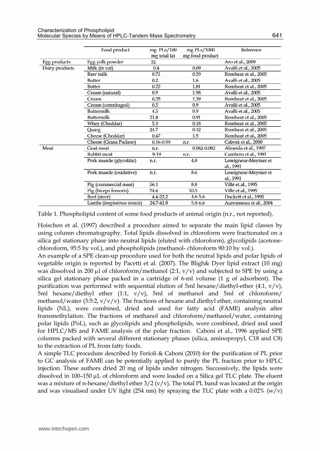
The isolation of phospholipids at an analytical scale is usually accomplished upon solvent/solvent extraction of the total lipids of a sample and further purification of the PL fraction. In food products, the main fraction of total lipids is usually formed by neutral lipids, i.e., triacylglycerols. Phospholipids are the minority of the total lipids, ranging from less than 0.1% in some plant oils, to about 5-25% in some dairy products, 20-30% of the total lipids in egg derived products and more in lean meats. In soy, lecithin is bound to proteins. A detailed survey of the phospholipid content of several kinds of food was reviewed by Weihrauch & Son (1983). Table 1 reports the total phospholipid content of egg, dairy and meat products as reported by more recent literature. For analytical purposes, the extraction of the total lipid fraction according to the procedures firstly described by Folch et al. (1957) and Bligh & Dyer (1959) and their successive modifications more than 50 years ago are still extensively used, because the extraction yield is practically quantitative. Thus, these methods are preferred to the extraction with petroleum ether or Soxhlet extraction. Novel methods of extraction of total lipids are based on pressurized liquid extraction (PLE) (Boselli et al., 2001) and even neat supercritical CO2 (Boselli et al., 2000).
3.2 Isolation and purification
The clean-up of the PL fraction is needed prior to HPLC analysis in order to increase the sensitivity of the determination when the PL content is relatively low compared to the neutral lipids. Usually, membrane lipids (lean meat or fish or egg products and cell membranes) are supposed to contain a PL content (Table 1) which is suitable for direct HPLC analysis. This is also the case of commercial lecithins (AOCS, 2007; ISO, 2009). However, if a better sensitivity is needed, the main ancillary techniques used for PL cleanup prior to HPLC analysis are column chromatography, solid phase extraction (SPE) and thin layer chromatography (TLC) with a normal phase approach.
www.intechopen.com
Characterization of Phospholipid Molecular Species by Means of HPLC-Tandem Mass Spectrometry
641
Table 1. Phospholipid content of some food products of animal origin (n.r., not reported).
Hoischen et al. (1997) described a procedure aimed to separate the main lipid classes by
using column chromatography. Total lipids dissolved in chloroform were fractionated on a
silica gel stationary phase into neutral lipids (eluted with chloroform), glycolipids (acetone-
chloroform, 95:5 by vol.), and phospholipids (methanol- chloroform 90:10 by vol.).
An example of a SPE clean-up procedure used for both the neutral lipids and polar lipids of vegetable origin is reported by Pacetti et al. (2007). The Bligh& Dyer lipid extract (10 mg)
was dissolved in 200 l of chloroform/methanol (2:1, v/v) and subjected to SPE by using a silica gel stationary phase packed in a cartridge of 6-ml volume (1 g of adsorbent). The purification was performed with sequential elution of 5ml hexane/diethyl-ether (4:1, v/v), 5ml hexane/diethyl ether (1:1, v/v), 5ml of methanol and 5ml of chloroform/ methanol/water (3:5:2, v/v/v). The fractions of hexane and diethyl ether, containing neutral lipids (NL), were combined, dried and used for fatty acid (FAME) analysis after transmethylation. The fractions of methanol and chloroform/methanol/water, containing polar lipids (PoL), such as glycolipids and phospholipids, were combined, dried and used for HPLC/MS and FAME analysis of the polar fraction. Caboni et al., 1996 applied SPE columns packed with several different stationary phases (silica, aminopropyl, C18 and C8) to the extraction of PL from fatty foods. A simple TLC procedure described by Ferioli & Caboni (2010) for the purification of PL prior to GC analysis of FAME can be potentially applied to purify the PL fraction prior to HPLC injection. These authors dried 20 mg of lipids under nitrogen. Successively, the lipids were
dissolved in 100–150 L of chloroform and were loaded on a Silica gel TLC plate. The eluent was a mixture of n-hexane/diethyl ether 3/2 (v/v). The total PL band was located at the origin and was visualised under UV light (254 nm) by spraying the TLC plate with a 0.02% (w/v)
www.intechopen.com

Tandem Mass Spectrometry – Applications and Principles
642
ethanolic solution of 2,7-dichlorofluorescein (sodium salt) and then was scraped off and collected. Successively, PL were extracted three times with chloroform (1 mL each).
4. HPLC Separation, detection and quantitation
4.1 HPLC separation
The separation of PL into different classes can be accomplished either with normal phase (NP) or reversed phase (RP) high performance liquid chromatography. NP-HPLC is more extensively applied than RP-HPLC to characterize PL from biological tissue and food products. With NP-HPLC, phospholipids are separated essentially on the basis of the polarity of the head group. They elute by order of increasing polarity: phosphatidylglycerol (PG), phosphatidylinositol (PI), phosphatidylethanolamine (PE), phosphatidylserine (PS), phosphatidylcholine (PC), sphingomyelin (Sph). The lyso forms elute after their corresponding parent phospholipid. Almost all the published methods use a porous silica stationary phase, usually 100 or 250 mm in length, with an internal diameter ranging from 2.0 to 4.6 mm. Recently, monolithic silica gel columns have been used increasingly more often for the separation of lipids, and several applications have been published (Jakab & Forgács, 2002; Merlin et al, 2006). Graeve and Janseen (2009) improved the HPLC–evaporative light scattering (ELSD) method, which permits the separation and quantification of lipid classes ranging from squalene (with a very low polarity) to highly polar lysophosphatidylcholine, using a monolithic silica stationary phase (100mm×4.6mm I.D. column). Other methods use silica gels that are chemically modified with diol (Silversand & Haux, 1997; Sas et al, 1999), cyanopropyl or amminopropyl moieties to alter the polarity. However, the acidic phospholipids will not be recovered using an amminopropyl column with the same efficiency as can be achieved by using an unmodified silica column. Yoon and Kim (2002) investigated the HPLC separation of phosphatidylcholine originated from egg yolk. Column temperature, mobile phase composition (isopropanol/hexane/methanol; methanol/water; methanol) and kinds of stationary phase (silica, bonded phases like C1, NH2, CN and diol) were varied to understand the effectiveness of PC separation. Pure methanol as mobile phase, silica as a stationary phase and temperature of 45°C represented the best HPLC operating condition. When the NH2-bonded and diol column are used, the peaks of PC and Sph overlapped. Anyway, Neron at al. (2004) successfully applied the diol column in order to separate and quantitate the wheat flour phospholipids during dough mixing in the presence of phospholipase. They showed that the diol column allowed an efficient separation in less than 16 min of all PL classes of wheat flour (N-acyllysophosphatidylethanolamine, N-acylphosphatidylethanolamine, PE, PC, PG, Lyso-PC and PI). Pang et al (2009) achieved the optimal separation and quantification of seven major phospholipid classes in human blood (PS, PE, PG, PI, PC, Lyso-PC and Sph) by using elution with mobile phase hexane (A) and 2-propanol with water, formic acid and ammonia as modifiers (B) and using a HPLC diol
column (250mm×3.0mm, i.d., 5.0 m, particle size). An isocratic elution method (A, 30%; B, 70%) was used for better repeatability and no balance time. The choice of mobile phase is strictly dependent on the method of detection. The mobile phases containing phosphate or sulfate buffer salts are incompatible with electrospray ionization. The selectivity of the solvents used in the mobile phase can exert a marked effect on the separation of individual phospholipids, and in particular it can change the order of elution of specific components. Specific examples of NP-HPLC separation of PL in biological
www.intechopen.com
Characterization of Phospholipid Molecular Species by Means of HPLC-Tandem Mass Spectrometry
643
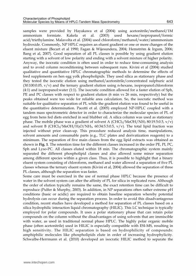
samples were provided by Hayakawa et al (2004) using acetonitrile/methanol/1M ammonium formiate. Kakela et al. (2003) used hexane/isopropanol/formic acid/triethylamine. Malavolta et al. (2004) used chloroform/methanol/water/ammmonium hydroxide. Commonly, NP HPLC requires an eluent gradient or one or more changes of the eluent mixture (Becart et al 1990; Fagan & Wijesundera, 2004; Hemström & Irgum, 2006; Bang et al, 2007). Good separation of all PL classes is possible by using gradient elution starting with a solvent of low polarity and ending with a solvent mixture of higher polarity. Anyway, the isocratic condition is often used in order to reduce time-consuming analysis and to avoid column reconditioning between subsequent runs. Kivini et al (2004) studied qualitative and quantitative HPLC chromatographic methods to determine the effects of feed supplements on hen egg yolk phospholipids. They used silica as stationary phase and they tested the isocratic elution using methanol/acetonitrile/concentrated sulphuric acid (30:100:0.05, v/v) and the ternary gradient elution using n-hexane, isopropanol:chloroform (4:1) and isopropanol:water (1:1). The isocratic condition allowed for a faster elution of Sph, PE and PC classes with respect to gradient elution (6 min vs 26 min, respectively) but the peaks obtained were too wide for reliable area calculation. So, the isocratic method was suitable for qualitative separation of PL while the gradient elution was found to be useful in the quantitative determination. Pacetti et al. (2005) employed NP-HPLC coupled with a tandem mass spectrometric detector in order to characterize the molecular species of PL of egg from hens fed diets enriched in seal blubber oil. A silica column was used as stationary phase. The mobile phase was a gradient of solvent A (CHCl3/MeOH/NH3 80:19.5:0.5, v/v) and solvent B (CHCl3/MeOH/H2O/NH3, 60:34:5.5:0.5, v/v). The total lipid fraction was injected without prior clean-up. This procedure reduced analysis time, manipulations, solvent amounts and consumable parts (e.g., TLC plates and derivatization reagents) to a minimum. The separation of five main classes from the egg lipid extracts was obtained as shown in Fig. 1. The retention time for the different classes increased in the order PE, PI, PC, Sph and Lyso-PC. All classes eluted within 18 min. The chromatographic system mainly separated the different phospholipid classes and also allowed some minor separation among different species within a given class. Thus, it is possible to highlight that a binary eluent system consisting of chloroform, methanol and water allowed a separation of five PL classes whereas the ternary eluent system (Kivini et al, 2004) allowed the separation of three PL classes, although the separation was faster. Some care must be exercised in the use of normal phase HPLC because the presence of water in the solvent system can alter the affinity of PL for silica in replicated runs. Although the order of elution typically remains the same, the exact retention time can be difficult to reproduce (Pulfer & Murphy, 2003). In addition, in NP separations often rather extreme pH conditions (basic or acidic) are required to obtain baseline separation whereby potential hydrolysis can occur during the separation process. In order to avoid this disadvantageous condition, recent studies have developed a method for separation of PL classes based on a silica hydrophilic interaction liquid chromatography (HILIC). This LC technique is typically employed for polar compounds. It uses a polar stationary phase that can retain polar compounds on the column without the disadvantages of using solvents that are immiscible with water, as used in traditional normal phase HPLC. The highly polar organic mobile phase (often acetonitrile) used in HILIC is especially compatible with ESI-MS, resulting in high sensitivity. The HILIC separation is based on hydrophilicity of compounds: amphiphilic molecules like phospholipids elute in order of increasing hydrophylicity. Schwalbe-Herrmann et al. (2010) developed an isocratic HILIC method to separate the
www.intechopen.com
Tandem Mass Spectrometry – Applications and Principles
644
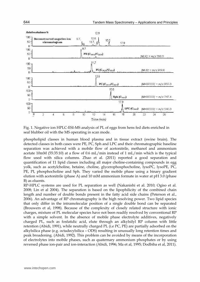
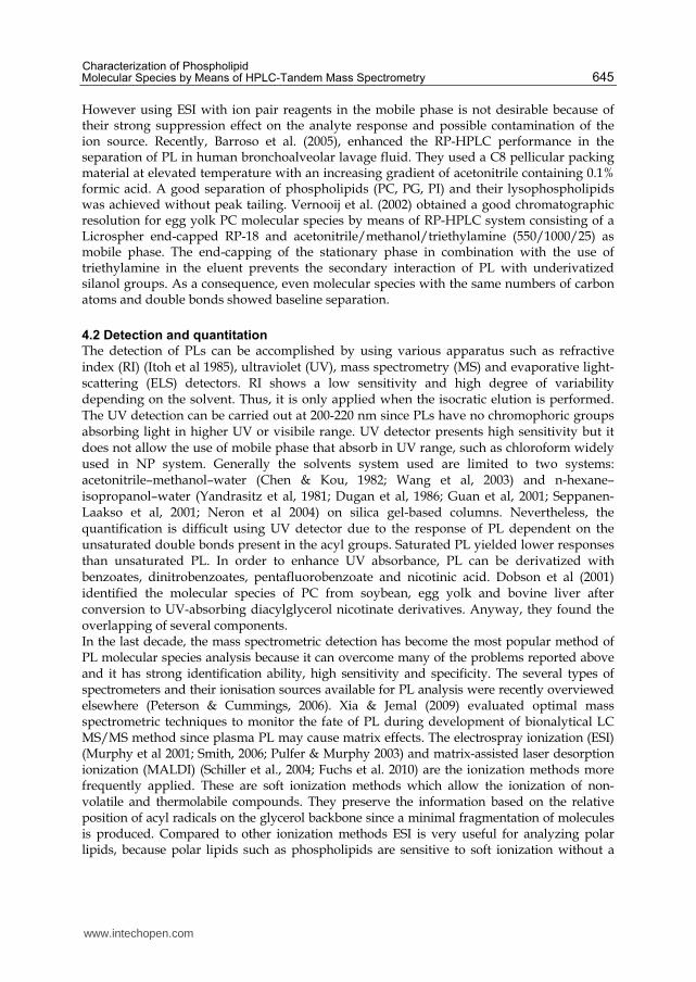
Fig. 1. Negative ion HPLC-ESI-MS analysis of PL of eggs from hens fed diets enriched in seal blubber oil with the MS operating in scan mode.
phospholipid classes in human blood plasma and in tissue extract (swine brain). The detected classes in both cases were PE, PC, Sph and LPC and their chromatographic baseline separation was achieved with a mobile flow of acetonitrile, methanol and ammonium acetate 10mM (55:35:10) at a flow of 0.6 mL/min instead of 1 mL/min which is the typical flow used with silica columns. Zhao et al. (2011) reported a good separation and quantification of 11 lipid classes including all major choline-containing compounds in egg yolk, such as acetylcholine, betaine, choline, glycerophosphocholine, lysoPC, lysoPE, PC, PE, PI, phosphocholine and Sph. They varied the mobile phase using a binary gradient elution with acetonitrile (phase A) and 10 mM ammonium formate in water at pH 3.0 (phase B) as eluents. RP-HPLC systems are used for PL separation as well (Nakanishi et al. 2010; Ogiso et al. 2008; Lin et al 2004). The separation is based on the lipophilicity of the combined chain length and number of double bonds present in the fatty acid side chains (Peterson et al., 2006). An advantage of RP chromatography is the high resolving power. Two lipid species that only differ in the intramolecular position of a single double bond can be separated (Brouwers et al, 1998). Because of the complexity of closely related structure with ionic charges, mixture of PL molecular species have not been readily resolved by conventional RP with a simple solvent. In the absence of mobile phase electrolyte additives, negatively charged PL, such as fosfatidic acid, elute through an alkylsilyl RP column with little retention (Abidi, 1991), while neutrally charged PL (i.e PC, PE) are partially adsorbed on the alkylsilica phase (e.g. octadecylsilica – ODS) resulting in unusually long retention times and peak broadening. (Abidi, 1992). This problem can be avoided by means of the incorporation of electrolytes into mobile phases, such as quaternary ammonium phosphates or by using reversed phase ion-pair and ion-interaction (Abidi, 1996; Ma et al, 1995; Dodbiba et al, 2011).
www.intechopen.com
Characterization of Phospholipid Molecular Species by Means of HPLC-Tandem Mass Spectrometry
645
However using ESI with ion pair reagents in the mobile phase is not desirable because of their strong suppression effect on the analyte response and possible contamination of the ion source. Recently, Barroso et al. (2005), enhanced the RP-HPLC performance in the separation of PL in human bronchoalveolar lavage fluid. They used a C8 pellicular packing material at elevated temperature with an increasing gradient of acetonitrile containing 0.1% formic acid. A good separation of phospholipids (PC, PG, PI) and their lysophospholipids was achieved without peak tailing. Vernooij et al. (2002) obtained a good chromatographic resolution for egg yolk PC molecular species by means of RP-HPLC system consisting of a Licrospher end-capped RP-18 and acetonitrile/methanol/triethylamine (550/1000/25) as mobile phase. The end-capping of the stationary phase in combination with the use of triethylamine in the eluent prevents the secondary interaction of PL with underivatized silanol groups. As a consequence, even molecular species with the same numbers of carbon atoms and double bonds showed baseline separation.
4.2 Detection and quantitation
The detection of PLs can be accomplished by using various apparatus such as refractive index (RI) (Itoh et al 1985), ultraviolet (UV), mass spectrometry (MS) and evaporative light-scattering (ELS) detectors. RI shows a low sensitivity and high degree of variability depending on the solvent. Thus, it is only applied when the isocratic elution is performed. The UV detection can be carried out at 200-220 nm since PLs have no chromophoric groups absorbing light in higher UV or visibile range. UV detector presents high sensitivity but it does not allow the use of mobile phase that absorb in UV range, such as chloroform widely used in NP system. Generally the solvents system used are limited to two systems: acetonitrile–methanol–water (Chen & Kou, 1982; Wang et al, 2003) and n-hexane– isopropanol–water (Yandrasitz et al, 1981; Dugan et al, 1986; Guan et al, 2001; Seppanen-Laakso et al, 2001; Neron et al 2004) on silica gel-based columns. Nevertheless, the quantification is difficult using UV detector due to the response of PL dependent on the unsaturated double bonds present in the acyl groups. Saturated PL yielded lower responses than unsaturated PL. In order to enhance UV absorbance, PL can be derivatized with benzoates, dinitrobenzoates, pentafluorobenzoate and nicotinic acid. Dobson et al (2001) identified the molecular species of PC from soybean, egg yolk and bovine liver after conversion to UV-absorbing diacylglycerol nicotinate derivatives. Anyway, they found the overlapping of several components. In the last decade, the mass spectrometric detection has become the most popular method of PL molecular species analysis because it can overcome many of the problems reported above and it has strong identification ability, high sensitivity and specificity. The several types of spectrometers and their ionisation sources available for PL analysis were recently overviewed elsewhere (Peterson & Cummings, 2006). Xia & Jemal (2009) evaluated optimal mass spectrometric techniques to monitor the fate of PL during development of bionalytical LC MS/MS method since plasma PL may cause matrix effects. The electrospray ionization (ESI) (Murphy et al 2001; Smith, 2006; Pulfer & Murphy 2003) and matrix-assisted laser desorption ionization (MALDI) (Schiller et al., 2004; Fuchs et al. 2010) are the ionization methods more frequently applied. These are soft ionization methods which allow the ionization of non-volatile and thermolabile compounds. They preserve the information based on the relative position of acyl radicals on the glycerol backbone since a minimal fragmentation of molecules is produced. Compared to other ionization methods ESI is very useful for analyzing polar lipids, because polar lipids such as phospholipids are sensitive to soft ionization without a
www.intechopen.com
Tandem Mass Spectrometry – Applications and Principles
646
derivatization procedure. Furthermore, ESI can be coupled with nano-flow HPLC easily (Bang et al, 2006; Isaac et al, 2003; Ito et al, 2008).Two different approaches can be adopted for analysis of PLs by means of ESI. The direct infusion of crude lipids extracts to the MS instrument was proposed by Benfenati & Reginato (1985), whereas the other approach employs liquid chromatography coupled on line with mass detector (LC-ESI-MS). The latest is more frequently applied compared to direct infusion, since the combination of HPLC and ESI MS reduces the ionisation suppression effect of low abundance PL species generated by the presence of polar impurities (Annesley, 2003; Issac et al 2003; Hermansson et al 2005; Houjou et al, 2005). Positive or negative ES ionization will occur at either the phosphate group or on the head group of PL. Normally, positively charged phospholipids such as PC and Sph appear as ions in the positive mode, whereas PE, PI, PG, PS and PA form ions in negative mode. It is possible to detect PG in the positive ion if ammonium adducts are formed. The quantitative analyses performed by using ESI MS are not straightforward because it is not possible to make the assumption that the intensity of any observed ion is proportional to the concentration of PL in the mixture. MS signals of PL are influenced by different effects related to acyl chain length, the degree of acyl chain unsaturation and concentration. Several authors (Berdeaux et al; Ahn et al 2007) have shown that the ESI-MS response increases with increasing degree of insaturation of the fatty acid side chains whereas it decreases with an increase in chain length when the phospholipids concentration range in the samples is higher than picomolar. Only the picomolar range concentration results, in fact, in a linear response. This non-linearity is more severe in negative ionization rather than positive mode (Zacarias et al, 2002). To overcome the dependence of ESI signal to the PL structures, in order to quantify the species, an internal standard chemically similar to the sample of interest can be used. A deuterated standard of each PL molecular species should ideally be used (Koc et al, 2002; Enjalbal et al., 2004). However, deuterated standards are very expensive and not always available for each PL molecular species (several hundreds of different phospholipid molecular species are present in biological samples considering all the subclasses and individual molecular species within a class). Moreover, even if an isotopic internal standard is applied, the scaling response implies a nonlinear relationship between intensity and concentration. The larger the discrepancy in intensity between the internal standard and the analyte of interest is correlated with a greater error in estimating the concentration when a linear approximation is used. Thus, the other possible way is to use a synthetic PL standard that has a low abundance in the sample, since PL species of the same class show similar ESI MS peak intensities. The standard should have a molecular weight outside of the molecular weight envelope that encompasses the naturally occurring molecular species. Pacetti et al. (2004) achieved the quantification of seven PC molecular species from the serum of cystic fibrosis subjects using ESI MS in the negative scan mode with 1,2-diundecanoyl-sn-glycero-phosphocholine as the internal standard. They showed that the use of mass spectrometry as detector allowed a low limit of quantification (12 ppm of PC). Han and Gross (2005) used 15:0-15:0 diacyl PS and 14:1-14:1 PC. When is sufficient to observe differences in phospholipid composition among different samples, a semi-quantitative approach can be also performed. The relative abundance of individual molecular species within a phospholipid class can be calculated from the single ion current responses. This strategy is widely applied to address a specific biochemical issue or to characterize the phospholipid composition in foods. Otherwise to the ESI MS detection, for quantification purposes the use of an Evaporative
Light Scattering Detector (ELSD) resulted more fast and easy. It allows the non-selective
determination of all the non volatile components in the polar lipid extract and a precise
www.intechopen.com

Characterization of Phospholipid Molecular Species by Means of HPLC-Tandem Mass Spectrometry
647
quantification of the PL classes, when a calibration curve is calculated for each phospholipid
class. It is very sensitive and it can quantitate nanomolar amounts of PL. ELSD usually gives
a stable flat baseline. Its response is independent of the number of double bonds in the
molecules. The ELSD baseline signal is scarcely affected by solvent changes (Stith et al, 2000;
Becart et al, 1990) and thus allows the use of gradient elution needed to manage complex
separations. Differently, the ELSD response can be affected by parameters of the
nebulization step and particularly the mobile phase composition that affects the size of the
droplets (Godoy Ramos et al 2008). Fig. 2 shows how the evaporation temperature affects
the ELSD response: the temperature increase is correlated to a change of the baseline profile.
Fig. 2. NP-HPLC- ELSD trace of a standard PL mixture (CL,PE,PI,PS,PC, Sph, LPC). The gas flow (from air compressor) of the ELSD was 7.5 L/min and the vaporization temperature was set: a) at 60 °C b) 65 °C.
The effect of the different mobile phase composition on variation of baseline is reported in figure 3. In the literature, several papers reported the quantification of PL with ELSD (Rodríguez-Alcalá & Fontecha, 2010; Seri et al, 2010; Wang et al, 2009; Rombaut et al 2007). Yan et al 2010 have successfully quantified PL extracted from human, porcine, and bovine erythrocyte ghost membranes using isocratic elution on a silica column coupled to an ELS detector. The detection limits for PS, PE, PC and Sph were 50, 50, 80 and 150 ng, respectively. Caboni et al. (2000) determined the content of phospholipids and lysophospholipids in Grana Cheese samples at different ripening stages. Narváez-Rivas and coworker (2001) developed a HPLC/ELSD method that allowed the quantification of cardiolipin, phosphatidylethanolamine, phosphatidylinositol, phosphatidylserine, phosphatidylcholine, and sphingomyelin in subcutaneous fat from Iberian pig. The limit of quantification were below 0.03 and 0.05 mg kg−1 for each phospholipid class. The performance of charged aerosol detection (CAD) was compared to ELSD for the NP-HPLC analysis of Leishmania membrane PL (phosphatidic acid, PG, cardiolipin, PI, PE, PS, lysoPE, PC, Sph and lysoPC) classes. The accuracy of the methods ranged from 62.8 to 115.8% and from 58.4 to 110.5% for ELSD and CAD, respectively. With HPLC-ELSD the limits of detection (LODs) were between 71 and 1195 ng and the limits of quantification (LOQs) were
www.intechopen.com

Tandem Mass Spectrometry – Applications and Principles
648
Fig. 3. NP-HPLC-ELSD trace of PL mixture obtained using a silica column (3, 150 mm x 4.6 mm) as stationary phase and a) a gradient of solvent A [CHCl3/ CH3OH /NH4OH (30%) 70:25:1, v/v] and solvent B [CHCl3/CH3OH/H2O/NH4OH (30%) 60:34:5.5:0.5, v/v], b) a gradient of A [CHCl3/ CH3OH /NH4OH (30%) 60:40:1, v/v] and solvent B [CHCl3/CH3OH/H2O/NH4OH (30%) 60:34:5.5:0.5, v/v]. The gas flow of the ELSD was 7.5 L/min and the evaporation temperature was 60 °C.
between 215 and 3622 ng. With HPLC-CAD, the LODs were between 15 and 249 ng whereas the limits of quantification (LOQs) were between 45 and 707 ng (Godoy Ramos et al, 2008). Considering that the ELS detector is really useful for qualitative analysis and that ESI MS detector is really indispensable to discriminate the PL molecular species, some authors suggest the use of the strategy where HPLC apparatus is coupled on-line with both detection systems. Boselli and co-workers (2008) proposed an analytical method for the simultaneous quantification of phospholipid classes and identification of phospholipid molecular species within each class in raw and cooked pork meat. The NP-HPLC apparatus was coupled on-line with two detection systems: an ion trap equipped with electrospray ionization for tandem mass spectrometry and an ELSD. Donato et al (2011) performed the characterization and quantification of phospholipid fraction in cow’s and donkey’s milk simultaneously coupling HPLC system with hybrid ion trap-time of flight (IT-TOF) mass analyzer and an ELS detector.
5. ES ionization and tandem mass spectrometry (MS/MS)
The ES ionization generates lipid ions by adding a proton [M+H]+, by adding a cation, such as the sodium [M+Na]+ ions or by removing a proton, [M−H]−. Chloride [M−Cl]− and formiate [M−HCOO]− adduct ions or multiply-charged ions such as [M+2H]2+, are also easily formed. The formation of ion species is strictly affected by the concentration of Na+ and the pH of the solution, with which the ESI events take place. Dimeric molecular species are often formed during ionization if the PL concentration is high. The variable formation of adducts and the multiplicity of PL molecular species present in the samples considerably complicate molecular species assignments from such complex spectra. Thus, often, assigning definite molecular identities to individual ion peaks is not
www.intechopen.com
Characterization of Phospholipid Molecular Species by Means of HPLC-Tandem Mass Spectrometry
649
possible with single MS. This problem becomes especially important for isobaric molecular species of the same phospholipid class, which have identical mass but differ in combination of acyl chains, or for the identification of oxidized PL. In these cases, experiments of tandem mass spectrometry (MS/MS) are needed as additional approach (Cui & Thomas, 2009; Postle et al., 2007). In MS/MS experiments, the precursor ion created in the first MS undergoes a further fragmentation either by collision-activated dissociation (CAD) or spontaneous dissociation. Larsen et al. (2001) compared the product ion achieved using CAD with an ion-trap mass spectrometry (ITMS) with those obtained with a triple quadrupole. In the fragmentation upon CAD in an ion-trap the main product ions were the sn-1 and sn-2 lyso-phospholipids whereas CAD in triple quadrupole gave primarily the sn-1 and sn-2 carboxylate anions. Hsu & Turk (2009) describe the mechanisms underlying the fragmentation processes under low-energy CAD with tandem quadrupole mass spectrometry and with multiple-stage ion-trap mass spectrometry of PL in various ions forms generated by ESI in negative and positive-ion modes ([M−H]−, and [M−2H+Alk]−; [M+H]+, [M+Alk]+, [M−H+2Alk]+; Alk = Na, Li). They noted that the fragment ions leading to structural characterization arising from CAD of the [M+H]+ ions are often of low abundance, and thus are less useful for structural identification. In contrast, fragment ions arising from alkali adduct ions, in particular, the [M+Li]+ ions, as well as from the [M−H]− ions are abundant and are suitable for unambiguous structural identification. Moreover, for many PL CID of [M+H]+ reveals information about the polar headgroup, while collision induced dissociation of the negative [M−H]− provides information about the fatty acyl chain. The discussion of product ions spectra achieved using CAD with ITMS of different PL ions generated by ESI (positive and negative) is reported below.
5.1 Cardiolipin (CL) The approach using multiple-stage ITMS methods for the characterization of CL as its positive and negative ions was widely studied by several authors. Hsu and co-workers described the characterization of CL molecular species as their [M-H]- , [M-2H]2- and [M-2H+Na]- ions generated by ESI in negative ions mode (Hsu et al 2005; Hsu & Turk 2006a) and as their sodiated adducts ([M-2H+3Na]+, [M-H+2Na]+, [M+Na]+) formed by positive ES ionization (Hsu & Turk 2006b). The MS2-spectra of the [M - H]- and of the [M - 2H]2- ions contain two sets of prominent fragment ions that comprise a phosphatidic acid (fatty acid/fatty acid-PA), a dehydrated phosphatidylglycerol, and a anion formed by phosphatidic acid and tricyclic glycerophosphate ester (phosphatidic acid + 136). The substantial differences in the abundance of the two distinct phosphatidic anions observed in the MS2-spectra of the [M - H]- and [M - 2H]2- and ions lead to the assignment of the phosphatidyl moieties attached to the 1’ or 3’ position of central glycerol. The differences in the intensity of carboxylate anions reflecting the fatty acyl substituent shown, exclusively, in the MS2-spectra of the [M - 2H]2- provide information to identify the fatty acyl substituents and their position in the glycerol backbone: the carboxylate anion containing the fatty acyl substituent at sn-1 (or sn-1’) is less abundant than the corresponding ion having that at sn-2 (or sn-2’). Anyway, the abundance of these two carboxylate ions are reversed in MS3-spectra of the [M - H]- ions. The MS2-spectra of all sodiated adducts (positive and negative) showed two prominent fragment ion pairs that consist of the phosphatidyl moieties attached to the 1’- and 3’-position of the central glycerol, respectively, resulting from the differential losses of the diacylglycerol moieties containing A and B glycerol, respectively.
www.intechopen.com

Tandem Mass Spectrometry – Applications and Principles
650
The identities of the fatty acyl substituents and their positions on the glycerol backbones (glycerol A and B) are deduced from further degradation (MS3 experiments) of the above ion pairs that give the fragment ions reflecting the fatty acid substituents at the sn-1 (or sn-1’) and sn-2 (or sn-2’) positions. The ions that arise from losses of the fatty acid substituents at sn-1 and sn-1’, respectively, are prominent. Different MS2 spectra of the monosodiated adducts ([M+Na]+) were obtained by Boselli et al. (2008). They identified the pork meat CL molecular species and the CAD fragmentation of the m/z 1471.6, i.e. [M+Na]+ (C18:1/C18:3)/(C18:2/C18:2)-CL or (C18:2/C18:2)/(C18:2/C18:2)-CL, which resulted the preponderant species in both raw and cooked meat and produced only two complementary fragments due to the cleavage at the phosphatidylglyceridic ester, as reported in Fig. 4. These results point out that only the MS2-spectra of the [M-2H] 2- is sufficient to confirm structural assignment of CL since it contains complementary information. Conversely, the MS2-spectra of the all CL sodiated adducts are not suitable to elucidate the composition of the isobaric species; thus, the experiments of third order (MS3) would be needed.
Fig. 4. Positive MS2-spectra of the [M+Na]+ of CL(C18:1)/(C18:3)/(C18:2)/(C18:2) or (C18:2)/(C18:2)/(C18:2)/(C18:2) at m/z= 1471.6
5.2 Phosphatidic acid (PA)
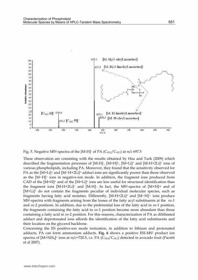
When being subjected to ESI in negative-ion mode, phosphatidic acid yields a [M-H]- ion. Its product ion spectra is really suitable to identify the PA species since it shows fragments arising from the loss of both the fatty acyl substituents. In Fig. 5 is reported a product ion spectrum of m/z= 697.5, i.e PA (C18:1/C18:2) as [M-H]- ion which was detected in the almond of the avocado (Persea americana Mill) fruit. The spectrum is dominated by the fragments at m/z = 415.1 and m/z = 417.3 arising from neutral loss of water followed by loss of oleyl moieties (RC18:1CO) and linoleyl moieties (RC18:2CO), respectively. The fragment ions at m/z = 279.3 and m/z = 281.3, corresponding to the carboxylate anions, are also visible.
www.intechopen.com
Characterization of Phospholipid Molecular Species by Means of HPLC-Tandem Mass Spectrometry
651
Fig. 5. Negative MS2-spectra of the [M-H]- of PA (C18:2/C18:1) at m/z 697.5
These observation are consisting with the results obtained by Hsu and Turk (2009) which described the fragmentation processes of [M-H]-, [M+H]+, [M+Li]+ and [M-H+2Li]+ ions of various phospholipids, including PA. Moreover, they found that the sensitivity observed for PA as the [M+Li]+ and [M−H+2Li]+ adduct ions are significantly poorer than those observed as the [M−H]− ions in negative-ion mode. In addition, the fragment ions produced from CAD of the [M+H]+ and of the [M+Li]+ ions are less useful for structural identification than the fragment ions [M-H+2Li]+ and [M-H]-. In fact, the MS2-spectra of [M+H]+ and of [M+Li]+ do not contain the fragments peculiar of individual molecular species, such as fragments having fatty acid moieties. Differently, [M-H+2Li]+ and [M−H]− ions produce MS2-spectra with fragments arising from the losses of the fatty acyl substituents at the sn-1 and sn-2 positions. In addition, due to the preferential loss of the fatty acid in sn-1 position, the fragments containing the fatty acid in sn-1 position become more abundant than those containing a fatty acid in sn-2 position. For this reasons, characterization of PA as dilithiated adduct and deprotonated ions affords the identification of the fatty acid substituents and their location on the glycerol backbone. Concerning the ES positive-ion mode ionization, in addition to lithium and protonated adducts, PA can form ammonium adducts. Fig. 6 shows a positive ESI-MS2 product ion spectra of [M+NH4]+ ions at m/z=720.5, i.e. PA (C18:0/C18:1) detected in avocado fruit (Pacetti et al 2007).
www.intechopen.com
Tandem Mass Spectrometry – Applications and Principles
652
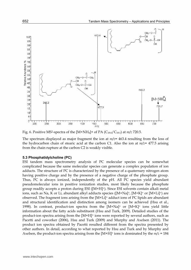
Fig. 6. Positive MS2-spectra of the [M+NH4]+ of PA (C18:0/C18:1) at m/z 720.5.
The spectrum displayed as major fragment the ion at m/z= 463.4 resulting from the loss of the hydrocarbon chain of stearic acid at the carbon C1. Also the ion at m/z= 477.5 arising from the chain rupture at the carbon C2 is weakly visible.
5.3 Phosphatidylcholine (PC)
ESI tandem mass spectrometry analysis of PC molecular species can be somewhat complicated because the same molecular species can generate a complex population of ion adducts. The structure of PC is characterized by the presence of a quaternary nitrogen atom having positive charge and by the presence of a negative charge of the phosphate group. Thus, PC is always ionized, independently of the pH. All PC species yield abundant pseudomolecular ions in positive ionization studies, most likely because the phosphate group readily accepts a proton during ESI ([M+H]+). Since ESI solvents contain alkali metal ions, such as Na, K or Li, abundant alkyl adducts species ([M+Na]+, [M+K]+ or [M+Li]+) are observed. The fragment ions arising from the [M+Li]+ adduct ions of PC lipids are abundant and structural identification and distinction among isomers can be achieved (Hsu et al., 1998). In contrast, product-ion spectra from the [M+Na]+ or [M+K]+ ions yield little information about the fatty acids substituent (Hsu and Turk, 2009). Detailed studies of the product-ion spectra arising from the [M+H]+ ions were reported by several authors, such as Pacetti and coworker (2006), Hsu and Turk (2009) and Murphy and Axelsen (2011). The product ion spectra obtained by Pacetti resulted different from the spectra presented by other authors. In detail, according to what reported by Hsu and Turk and by Murphy and Axelsen, the product-ion spectra arising from the [M+H]+ ions is dominated by the m/z = 184
www.intechopen.com
Characterization of Phospholipid Molecular Species by Means of HPLC-Tandem Mass Spectrometry
653
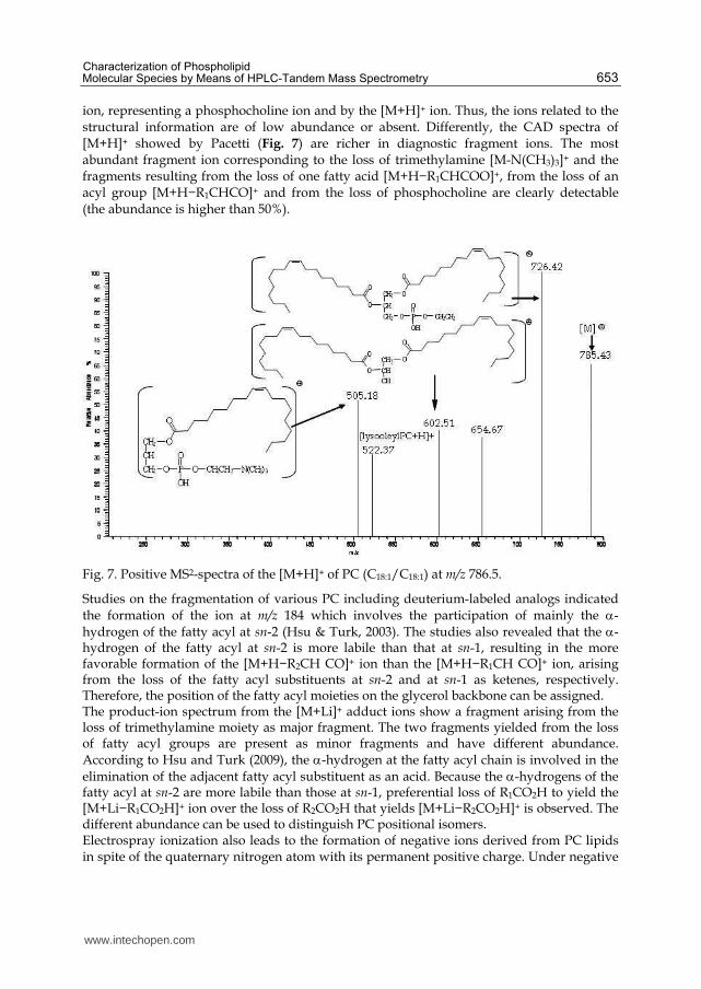
ion, representing a phosphocholine ion and by the [M+H]+ ion. Thus, the ions related to the structural information are of low abundance or absent. Differently, the CAD spectra of [M+H]+ showed by Pacetti (Fig. 7) are richer in diagnostic fragment ions. The most abundant fragment ion corresponding to the loss of trimethylamine [M-N(CH3)3]+ and the fragments resulting from the loss of one fatty acid [M+H−R1CHCOO]+, from the loss of an acyl group [M+H−R1CHCO]+ and from the loss of phosphocholine are clearly detectable (the abundance is higher than 50%).
Fig. 7. Positive MS2-spectra of the [M+H]+ of PC (C18:1/C18:1) at m/z 786.5.
Studies on the fragmentation of various PC including deuterium-labeled analogs indicated the formation of the ion at m/z 184 which involves the participation of mainly the -hydrogen of the fatty acyl at sn-2 (Hsu & Turk, 2003). The studies also revealed that the -hydrogen of the fatty acyl at sn-2 is more labile than that at sn-1, resulting in the more favorable formation of the [M+H−R2CH CO]+ ion than the [M+H−R1CH CO]+ ion, arising from the loss of the fatty acyl substituents at sn-2 and at sn-1 as ketenes, respectively. Therefore, the position of the fatty acyl moieties on the glycerol backbone can be assigned. The product-ion spectrum from the [M+Li]+ adduct ions show a fragment arising from the loss of trimethylamine moiety as major fragment. The two fragments yielded from the loss of fatty acyl groups are present as minor fragments and have different abundance. According to Hsu and Turk (2009), the -hydrogen at the fatty acyl chain is involved in the
elimination of the adjacent fatty acyl substituent as an acid. Because the -hydrogens of the fatty acyl at sn-2 are more labile than those at sn-1, preferential loss of R1CO2H to yield the [M+Li−R1CO2H]+ ion over the loss of R2CO2H that yields [M+Li−R2CO2H]+ is observed. The different abundance can be used to distinguish PC positional isomers. Electrospray ionization also leads to the formation of negative ions derived from PC lipids in spite of the quaternary nitrogen atom with its permanent positive charge. Under negative
www.intechopen.com

Tandem Mass Spectrometry – Applications and Principles
654
ESI condition, PC generate [M-15]- ions, resulting from the demethylation of the choline moiety and the negative ions by forming adducts with anions, such as chloride [M+35]-, formiate [M+45]- and acetate [M+59]-. When ESI is performed using a spectrometer with high orifice potentials, the [M-15]- ions becomes the most important since the high potential leads also to the demethylation of the anion adducts. The MS2 spectra of [M-15]- and [M+59]- ions show anion fragments which are more suitable for identification of the fatty acid substituents and location of their position on the glycerol backbone compared to the product ion spectra obtained from [M+35]- and from [M+45]- ions. In detail, the product-ion spectra obtained from [M-15]- ion are characterized by the presence of both carboxylate anions [R1COO]- and [R2COO]−. The latest fragment results more abundant than [R1COO]-. Anyway, this discrepancy of abundance is not so visible when the PC molecular species contain a long-chain fatty acid linked to the sn-2 position (Berdeaux et al 2010). Kerwin, Tuininga, & Ericsson (1994) described the formation of an ion cluster with acetate [M+59]- and the subsequent decomposition of that ion species during collisional activation to yield the [M-15]- ion that corresponds to the loss of methyl acetate, as well as ions characteristic of the fatty acyl group esterified at the sn-1 and sn-2 positions. Pacetti et al. 2005 observed that the product ion spectra obtained from [M+35]- and from [M+45]- ions contain only the [M−CH3−HCOO]− anion fragment; thus, the negative MS2-spectrum of the [M+NH4]+ of PC (C16:0/C18:1) at m/z 804.4 contains the fragment at 744.4 m/z.
5.4 Lysophosphatidylcholine (LPC)
The behavior of LPC molecular species under ESI is equal to those shown by PC. LPC
molecular species can be inferred from their protonated molecules [M + H]+ when positive
ESI is performed. Negative ESI yields the demethylated ion ([M–CH3]-). The presence of
metal or anions in the ESI system generates the adduct ions, such as ([M + Na]+, [M + Li]+,
[M+HCOO]-, ([M+ Cl]-). As displayed for PC, the product ion mass spectra related to the
[M+H]+ ion result more informative than all the other ions.
An example of MS2 spectra yielded by the [M+H]+ ion at m/z= 568.3, i.e sn-lyso-
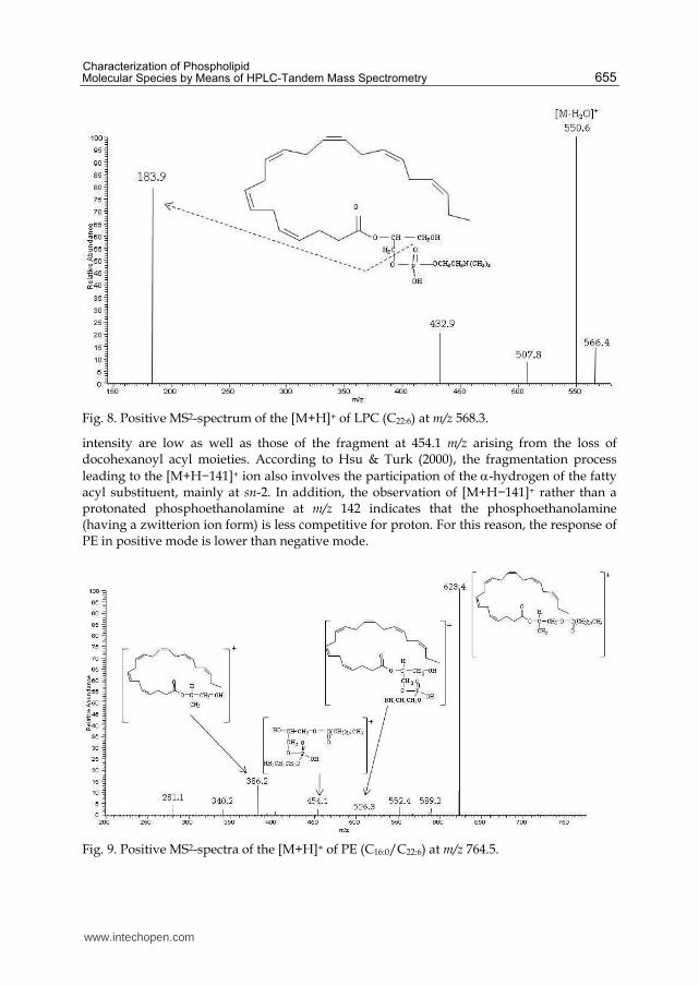
docosahexaenoic-phosphatidylcholine [LPC (C22:6)], is reported in Fig. 8. The spectrum
displays two major fragments. One at m/z= 550.2 given by the loss of one water molecule
[M-H2O]+ and the other at m/z = 183.9 corresponding to the polar headgroup.
Differently, the product ion spectrum from the ion at m/z = 612.3, corresponding to LPC C22:6
as a [M+HCOO]- ion, displays the unique fragment arising from demethylation of choline
[M-CH3]+) with 552.1 m/z.
5.5 Phosphatidylethanolamine (PE)
PE can form positive and negative ions. In the ESI mass spectra, [M+H]+ ions are usually detected when working in positive mode, and [M−H]− ions are displayed in negative mode. The product ion spectra arising from the [M+H]+ are often simple and therefore less applicable, although achievable, for structure characterization. They displayed a most abundant fragment resulting from the loss of the polar headgroup ([M-NH3(CH2)2OPO3H]+
= [M+H−141]+) and a low intensity fragment resulting from the loss of one acyl group ([M-R1CO]+). For example, the product-ion spectrum of the [M+H]+ ion of PE (C16:0/C22:6) at m/z 764.5 (Fig. 9) is dominated by the m/z 623.4 ([M+H−141]+) ion. The fragments at m/z 526.3 and at m/z 386.2, corresponding to the loss of palmitoyl acyl moieties and the simultaneous loss of both polar head group and palmitoyl acyl group, respectively, are also visible. Their
www.intechopen.com
Characterization of Phospholipid Molecular Species by Means of HPLC-Tandem Mass Spectrometry
655
Fig. 8. Positive MS2-spectrum of the [M+H]+ of LPC (C22:6) at m/z 568.3.
intensity are low as well as those of the fragment at 454.1 m/z arising from the loss of docohexanoyl acyl moieties. According to Hsu & Turk (2000), the fragmentation process
leading to the [M+H−141]+ ion also involves the participation of the -hydrogen of the fatty acyl substituent, mainly at sn-2. In addition, the observation of [M+H−141]+ rather than a protonated phosphoethanolamine at m/z 142 indicates that the phosphoethanolamine (having a zwitterion ion form) is less competitive for proton. For this reason, the response of PE in positive mode is lower than negative mode.
Fig. 9. Positive MS2-spectra of the [M+H]+ of PE (C16:0/C22:6) at m/z 764.5.
www.intechopen.com
Tandem Mass Spectrometry – Applications and Principles
656
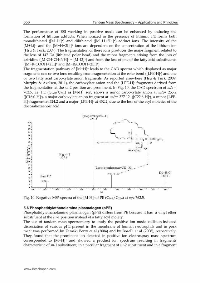
The performance of ESI working in positive mode can be enhanced by inducing the formation of lithium adducts. When ionized in the presence of lithium, PE forms both monolithiated ([M+Li]+) and dilithiated ([M−H+2Li]+) adduct ions. The intensity of the [M+Li]+ and the [M−H+2Li]+ ions are dependent on the concentration of the lithium ion (Hsu & Turk, 2009). The fragmentation of these ions produces the major fragment related to the loss of 147 Da (lithiated polar head) and the minor fragments arising from the loss of aziridine ([M-CH2CH2NH]+ = [M-43]+) and from the loss of one of the fatty acid substituents ([M−R1COOH+2Li]+ and [M−R2COOH+2Li]+). The fragmentation pathway of [M−H]− leads to the CAD spectra which displayed as major fragments one or two ions resulting from fragmentation at the ester bond ([LPE-H]-) and one or two fatty acid carboxylate anion fragments. As reported elsewhere (Hsu & Turk, 2009; Murphy & Axelsen, 2011), the carboxylate anion and the [LPE-H]- fragments derived from the fragmentation at the sn-2 position are prominent. In Fig. 10, the CAD spectrum of m/z = 762.5, i.e. PE (C16:0/C22:6) as [M-H]- ion, shows a minor carboxylate anion at m/z= 255.2 ([C16:0-H]-), a major carboxylate anion fragment at m/z= 327.12 ([C22:6-H]-), a minor [LPE-H]- fragment at 524.2 and a major [LPE-H]- at 452.2, due to the loss of the acyl moieties of the docosahexaenoic acid.
Fig. 10. Negative MS2-spectra of the [M-H]- of PE (C16:0/C22:6) at m/z 762.5.
5.6 Phosphatidylethanolamine plasmalogen (pPE)
Phosphatidylethanolamine plasmalogen (pPE) differs from PE because it has a vinyl ether substituent at the sn-1 position instead of a fatty acyl moiety. The use of tandem mass spectrometry to study the positive ion mode collision-induced dissociation of various pPE present in the membrane of human neutrophils and in pork meat was performed by Zemski Berry et al (2004) and by Boselli et al (2008), respectively. They found that the prominent ion detected in positive ion electrospray mass spectrum corresponded to [M+H]+ and showed a product ion spectrum resulting in fragments characteristic of sn-1 substituent, in a peculiar fragment of sn-2 substituent and in a fragment
www.intechopen.com

Characterization of Phospholipid Molecular Species by Means of HPLC-Tandem Mass Spectrometry
657
ion deriving from the loss of the polar head group ([M-NH3(CH2)2OPO3H]+). As an example, the CAD of m/z= 700.4, i.e pPE [M+H]+(p16:0/C18:2) (Fig. 11) resulted in a major fragment at m/z 364.1, containing the phosphatidylethanolamine head group linked with alky-1’-enyl moieties of sn-1 substituent, in a minor fragment at m/z 559.5, resulting from the loss of the polar headgroup ([M-NH3(CH2)2OPO3H]+ and in a fragment at m/z 337.1, corresponding to the loss of both polar head and 1-O-alky-1’-enyl groups. In contrast, as seen for MS2 mass spectra of all PE [M+H]+ ions, the fragment resulting from the loss of the polar headgroup is present as minor fragment in the product ion spectrum of pPE [M+H]+. For this reason, the quantification of pPE in a lipid mixture by using constant neutral loss of 141 Da is not suitable as it is for quantification of PE.
Fig. 11. Positive MS2-spectra of the [M+H]+ pPE (p16:0/C20:4) at m/z 700.4.
The mechanism of formation of the predominant fragment ion at m/z 364.1 was proposed by Zemski Berry & Murphy (2004). It involves the oxygen atom at the sn-1 position attacking the phosphorous atom, which results in the formation of a new O-P bond and concomitant abstraction of the hydrogen from C-2 of the glycerol backbone to form a double bond between C-1 and C-2 of the glycerol structure. Since this fragment is characteristic of the hexadecyl vinyl ether group linked in sn-1 position, it is possible to detect all the pPE molecular species containing this substituent by using a precursor ion scan at m/z= 364 or all pPE species with an octadecyl vinyl ether by a precursor ion scan of m/z = 392. The characterization of pPE molecular species by means of ESI working in negative ion-mode is reported by Malavolta et al (2004) and by Hsu & Turk 2009. Both groups observed the [M-H]- ions in negative mode but the product ion spectra obtained by the two authors are different. In detail, Hsu and Turk reported that the CAD spectra of pPE is dominated by the carboxylate anion identifying the fatty acyl moiety of the molecule. Differently, Malavolta et al. showed that the tandem mass spectrum was similar to that obtained with PE. The spectrum has an anion generated from the loss of the fatty acyl moiety ([M-RCO-H]-) as predominat fragment and a carboxylate anion fragment ([RCOO]-) with lower
www.intechopen.com
Tandem Mass Spectrometry – Applications and Principles
658
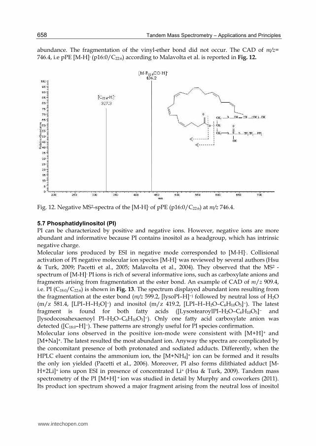
abundance. The fragmentation of the vinyl-ether bond did not occur. The CAD of m/z= 746.4, i.e pPE [M-H]- (p16:0/C22:6) according to Malavolta et al. is reported in Fig. 12.
Fig. 12. Negative MS2-spectra of the [M-H]- of pPE (p16:0/C22:6) at m/z 746.4.
5.7 Phosphatidylinositol (PI)
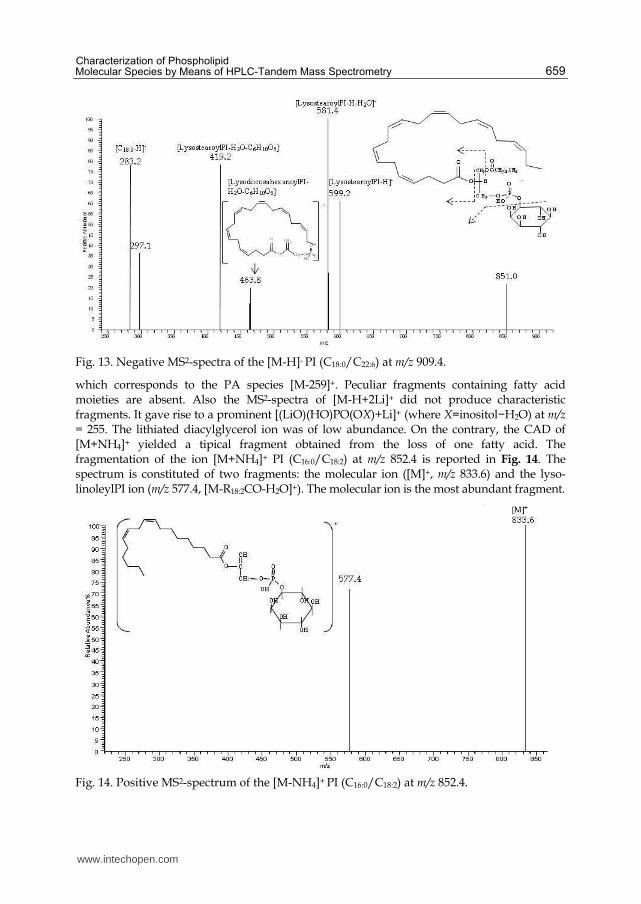
PI can be characterized by positive and negative ions. However, negative ions are more abundant and informative because PI contains inositol as a headgroup, which has intrinsic negative charge. Molecular ions produced by ESI in negative mode corresponded to [M-H]-. Collisional activation of PI negative molecular ion species [M-H]- was reviewed by several authors (Hsu & Turk, 2009; Pacetti et al., 2005; Malavolta et al., 2004). They observed that the MS2 -spectrum of [M-H]- PI ions is rich of several informative ions, such as carboxylate anions and fragments arising from fragmentation at the ester bond. An example of CAD of m/z 909.4, i.e. PI (C18:0/C22:6) is shown in Fig. 13. The spectrum displayed abundant ions resulting from the fragmentation at the ester bond (m/z 599.2, [lysoPI–H]−) followed by neutral loss of H2O (m/z 581.4, [LPI–H–H2O]−) and inositol (m/z 419.2, [LPI–H–H2O–C6H10O5]−). The latest fragment is found for both fatty acids ([LysostearoylPI–H2O–C6H10O5]− and [lysodocosahexaenoyl PI–H2O–C6H10O5]−). Only one fatty acid carboxylate anion was detected ([C18:0–H]−). These patterns are strongly useful for PI species confirmation. Molecular ions observed in the positive ion-mode were consistent with [M+H]+ and
[M+Na]+. The latest resulted the most abundant ion. Anyway the spectra are complicated by
the concomitant presence of both protonated and sodiated adducts. Differently, when the
HPLC eluent contains the ammonium ion, the [M+NH4]+ ion can be formed and it results
the only ion yielded (Pacetti et al., 2006). Moreover, PI also forms dilithiated adduct [M-
H+2Li]+ ions upon ESI in presence of concentrated Li+ (Hsu & Turk, 2009). Tandem mass
spectrometry of the PI [M+H] + ion was studied in detail by Murphy and coworkers (2011).
Its product ion spectrum showed a major fragment arising from the neutral loss of inositol
www.intechopen.com
Characterization of Phospholipid Molecular Species by Means of HPLC-Tandem Mass Spectrometry
659
Fig. 13. Negative MS2-spectra of the [M-H]- PI (C18:0/C22:6) at m/z 909.4.
which corresponds to the PA species [M-259]+. Peculiar fragments containing fatty acid moieties are absent. Also the MS2-spectra of [M-H+2Li]+ did not produce characteristic fragments. It gave rise to a prominent [(LiO)(HO)PO(OX)+Li]+ (where X=inositol−H2O) at m/z = 255. The lithiated diacylglycerol ion was of low abundance. On the contrary, the CAD of [M+NH4]+ yielded a tipical fragment obtained from the loss of one fatty acid. The fragmentation of the ion [M+NH4]+ PI (C16:0/C18:2) at m/z 852.4 is reported in Fig. 14. The spectrum is constituted of two fragments: the molecular ion ([M]+, m/z 833.6) and the lyso-linoleylPI ion (m/z 577.4, [M-R18:2CO-H2O]+). The molecular ion is the most abundant fragment.
Fig. 14. Positive MS2-spectrum of the [M-NH4]+ PI (C16:0/C18:2) at m/z 852.4.
www.intechopen.com

Tandem Mass Spectrometry – Applications and Principles
660
5.8 Phosphatidylserine (PS)
The characterization of PS molecular species by means of ESI ion trap MS yields a positive-
ion in form [M+H]+ and negative ions with [M−H]−.
The tandem mass spectrum of the [M-H]- ion contains complete structural information and
offers the utmost sensitivity for structural determination. In contrast, product-ion spectra
from the [M+H]+ species are rather simple and are less useful for structural characterization.
This statement can be confirmed by comparing the product MS2 spectra of [M-H]– ion of PS
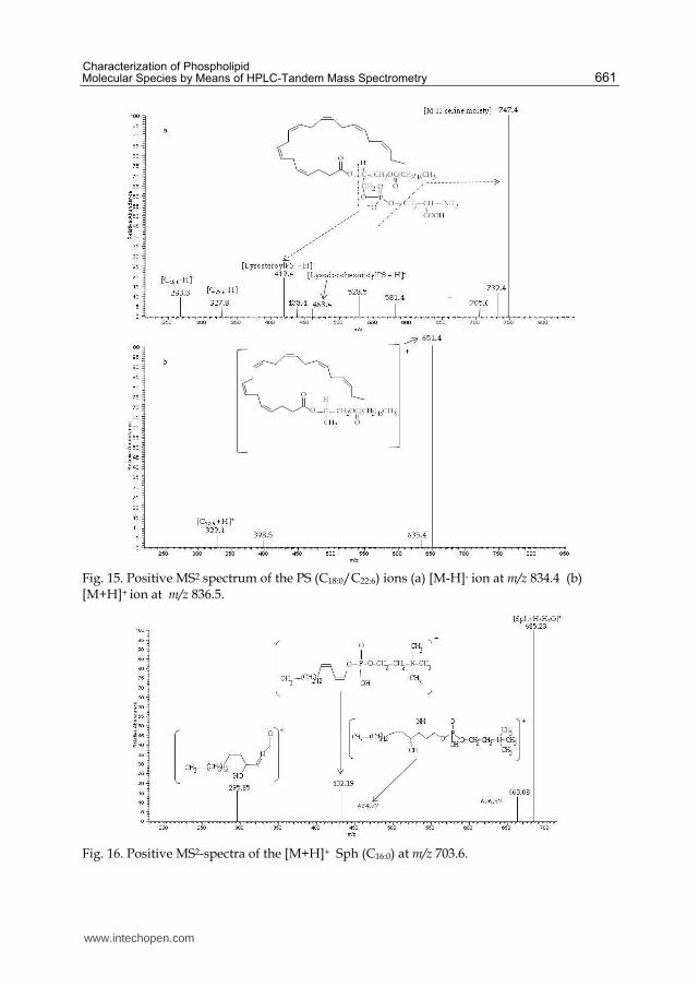
(C18:0/C22:6) at m/z = 834.4 (Fig 15a) with MS2 spectra of [M +H]+ of the same PS species at
m/z 836.5 (Fig 15b) detected in the lipid fraction of marine fish. The CAD of [M-H]– produces
an abundant ion resulting from the loss of the head group (m/z 747.4, [PA-H]-), two ions
resulting from the fragmentation at the ester bond followed by the loss of the serine group
[LPA-H]- and small carboxylate ion fragments. Thus, the two carboxylate anion fragments at
m/z 283.5 and m/z 327.3 and the two [LPA-H]- ions at 419.4 and at 463.1 identify PS
(C18:0/C22:6) as the parent ion. Differently, the CAD of [M +H]+ at m/z = 836.5 is dominated
by m/z 651.4 ([M+H−(HO)2P(O)O-serine]+) arising from loss of the phosphoserine moiety.
Only one of the acylium ions at m/z = 329.1 reflecting the fatty acyl sostituents (C22:6) is
present; anyway, its signal is low.
In the presence of alkali ions, PS molecular species in the form of [M+Alk]+ and
[M−H+2Alk]+ (Alk = Li, Na) can also be observed, attributable to the fact that PS possesses
two anionic charge sites of which one can attach to an Alk+ (Hsu & Turk, 2005). Ions
informative for structural characterization of PS are of low abundance in the MS2 spectra of
alkali adduct ions. The MS2-spectrum of the [M+Alk]+ ion contains a unique ion
corresponding to the internal loss of a phosphate group probably via the fragmentation
processes involving rearrangement steps. The [M−H+2Alk]+ ion of PS yields a major [M-
H+2Alk- 87]+ ion, which is equivalent to an alkali adduct ion of a monoalkali salt of PA and
gives rise to a greater abundance of [M-H+2Alk-87-R1CO2H]+ than [M-H+2Alk-87-
R2CO2H]+.
5.9 Sphingomyelin (Sph)
ESI of sphingomyelin follows closely that of PC and LPC. The presence of the quaternary
nitrogen atom, with permanent positive charge, dominates the ESI behaviour of this
molecule. Thus, the Sph generates more abundant ions in the positive ion mode, than the
negative ion mode. Moreover, in negative ion mode, Sph can be analyzed only if the
addition of an anionic reagent (acetate or formiate) in the spray solvent is performed.
In detail, (+)-ESI yields a protonated ion [M+H]+ and a lithium adduct [M+Li]+, whereas, in
(-)-ESI, Sph is detected as chlorine [M+Cl]-, formiate [M+HCOO]- and acetate [M+CH3COO]-
adducts.
As reported by Murphy and Axelsen (2011) lithiated-sphingomyelin ions produce [M-
59+Li]+ ions representing the neutral loss of (CH3)3N and [M-183+Li]+ ions representing the
neutral loss of phosphocholine. Differently, CAD of [M+H]+ yields the only fragment at m/z
184 (phosphocholine group). This evidence contrasts with the results obtained by Boselli et
al (2008) where the CAD of [M+H]+ at m/z 703.6, i.e. Sph (C16:0), yielded fragments deriving
from the loss of water (m/z 685.2, [M+H-H2O]+), the loss of the acyl group (m/z 464.8), the
loss of the amidic moiety (m/z 432.2) and a fragment deriving from sphingosin (m/z 295.9)
(Fig. 16).
www.intechopen.com
Characterization of Phospholipid Molecular Species by Means of HPLC-Tandem Mass Spectrometry
661
Fig. 15. Positive MS2 spectrum of the PS (C18:0/C22:6) ions (a) [M-H]- ion at m/z 834.4 (b) [M+H]+ ion at m/z 836.5.
Fig. 16. Positive MS2-spectra of the [M+H]+ Sph (C16:0) at m/z 703.6.
www.intechopen.com
Tandem Mass Spectrometry – Applications and Principles
662
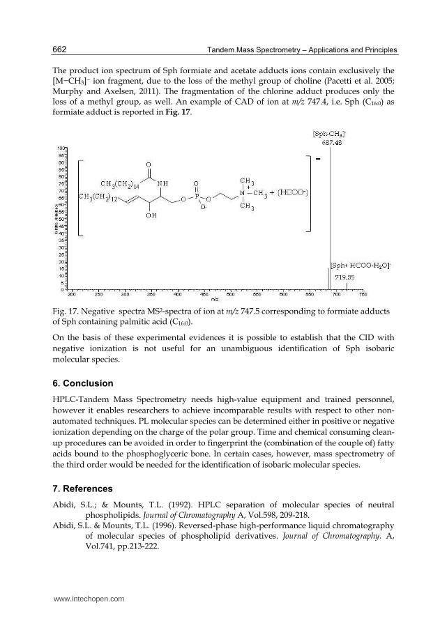
The product ion spectrum of Sph formiate and acetate adducts ions contain exclusively the [M−CH3]− ion fragment, due to the loss of the methyl group of choline (Pacetti et al. 2005; Murphy and Axelsen, 2011). The fragmentation of the chlorine adduct produces only the loss of a methyl group, as well. An example of CAD of ion at m/z 747.4, i.e. Sph (C16:0) as formiate adduct is reported in Fig. 17.
Fig. 17. Negative spectra MS2-spectra of ion at m/z 747.5 corresponding to formiate adducts of Sph containing palmitic acid (C16:0).
On the basis of these experimental evidences it is possible to establish that the CID with
negative ionization is not useful for an unambiguous identification of Sph isobaric
molecular species.
6. Conclusion
HPLC-Tandem Mass Spectrometry needs high-value equipment and trained personnel,
however it enables researchers to achieve incomparable results with respect to other non-
automated techniques. PL molecular species can be determined either in positive or negative
ionization depending on the charge of the polar group. Time and chemical consuming clean-
up procedures can be avoided in order to fingerprint the (combination of the couple of) fatty
acids bound to the phosphoglyceric bone. In certain cases, however, mass spectrometry of
the third order would be needed for the identification of isobaric molecular species.
7. References
Abidi, S.L.; & Mounts, T.L. (1992). HPLC separation of molecular species of neutral phospholipids. Journal of Chromatography A, Vol.598, 209-218.
Abidi, S.L. & Mounts, T.L. (1996). Reversed-phase high-performance liquid chromatography of molecular species of phospholipid derivatives. Journal of Chromatography. A, Vol.741, pp.213-222.
www.intechopen.com

Characterization of Phospholipid Molecular Species by Means of HPLC-Tandem Mass Spectrometry
663
Abidi, S.L.; (1991). HPLC of phosphatidic acids and related polar lipids. Journal of Chromatography A, Vol.587, pp. 193-203.
Ahn, E.J.; Kim, H.; Chung, B.C. & Moon, M.H. (2007). Quantitative analysis of phosphatidylcholine in rat liver tissue by nanoflow liquid chromatography/tandem mass spectrometry. Journal of Separation Science, Vol.30, No.16, (August 2007), pp. 2598-2604.
Almeida, M.M.M.; Zapata, J.F.F.; Martins, C.B.; & Maia, G.A. (1997) Cholesterol and Phospholipid Levels in Goat Meat As Affected By Dietary Calcium, Pesquisa Agropecuaria Brasileira, 32, pp. 555-558.
Annesley, T.M. (2003). Ion Suppression in Mass Spectrometry. Clinical Chemistry, Vol.49, pp.1041-1044.
AOCS Official Method Ja 7c-07 Revised (2007) Determination of Lecithin Phospholipids by HPLC-LSD.
Avalli, A. & G. Contarini (2005) Determination of phospholipids in dairy products by SPE/HPLC/ELSD, Journal of Chromatography A, 1071 pp. 185–190.
Bang, D.Y.; Ahn, E.J. & Moon, M.H. (2007). Shotgun analysis of phospholipids from mouse liver and brain by nanoflow liquid chromatography/tandem mass spectrometry. Journal of Chromatography B, Vol.852, No.1-2, (June 2007), pp. 268–277.
Bang, D.Y.; Kang, D. & Moon, M.H. (2006). Nanoflow liquid chromatography-tandem mass spectrometry for the characterization of intact phosphatidylcholines from soybean, bovine brain, and liver. Journal of Chromatography A. Vol. 1104, pp 222–229.
Barroso, B. & Bischoff, R. (2005). LC–MS analysis of phospholipids and lysophospholipids in human bronchoalveolar lavage fluid. Journal of Chromatography B, Vol.814, No.1, (January 2005), pp. 21-28.
Becart, J.; Chevalier, C. & Biesse, J.P. (1990). Quantitative analysis of phospholipids by HPLC with light scattering detector: application to raw materials for cosmetic use. Journal of High Resolution Chromatography, Vol.13, No.2, February 1990, pp 126-129.
Benfenati, E. & Reginato, R. (1985). A comparison of three methods of soft ionization mass spectrometry of crude phospholipid extracts. Biological Mass Spectrometry, Vol.12, No.11, pp. 643–651.
Berdeaux, O.; Juaneda, P.; Martine, L.; Cabaret, S.; Bretillon, L. & Acar, N. (2010) Identification and quantification of phosphatidylcholines containing very-long-chain polyunsaturated fatty acid in bovine and human retina using liquid chromatography/tandem mass spectrometry. Journal of Chromatography A, Vol.1217, pp. 7738–7748.
Billah, M.M. & Anthes, J. C. (1990) The regulation and cellular functions of phosphatidylcholine hydrolysis, Biochem. J. 269, pp.281-291.
Bligh, E.G. & Dyer, W.J. (1959) A Rapid Method Of Total Lipid Extraction And Purification, Can. J. Biochem. Physiol. 37 pp. 911-917.
Boselli, E. & Caboni, M.F. (2000) Supercritical carbon dioxide extraction of phospholipids from dried egg yolk without organic modifier. Journal of Supercritical Fluids Vol. 19 pp. 45–50.
Boselli, E.; Pacetti, D.; Curzi, F. & Frega, N.G. (2008). Determination of Phospholipid Molecular Species in Pork Meat by High Performance Liquid Chromatography-Tandem Mass Spectrometry and Evaporative Light Scattering Detection. Meat Science, Vol.78, No.3, (March 2008), pp. 305–313.
www.intechopen.com

Tandem Mass Spectrometry – Applications and Principles
664
Boselli, E.; Velazco, V.; Caboni, M.F. & Lercker, G. (2001) Pressurized liquid extraction of lipids for the determination of oxysterols in egg-containing food. Journal of Chromatography A, Vol. 917 pp. 239–244.
Bourre J.M. & Dumont O. (2002) The administration of pig brain phospholipids versus soybean phospholipids in the diet during the period of brain development in the rat results in greater increments of brain docosahexaenoic acid, Neuroscience Letter 335 pp. 129-133.
Brouwers, J.F.; Versluis, C.; Van Golde, L.M. & Tielens A.G. (1998). 5-Octadecenoic acid: evidence for a novel type of fatty acid modification in schistosomes. Biochemical Journal, Vol.334, No.2, (September 1998), pp. 315-319.
Caboni, M. A., Menotta, S., Lercker, G. (1996).Separation and analysis of phospholipids in different foods with a light-scattering detector. J. Am. Oil Chem. Soc.73,1561-1566.
Caboni, M. F.; Boselli, E. & Menotta, S. (2000) HPLC-ELSD determination of Phospholipids and Lysophospholipids in Grana Cheese samples at different ripening stages. Scienza e Tecnica Lattiero-casearia, Vol.51, No.1, (February 2000), pp. 7-24.
Cambero, M.I.; de la Hoz, L.; Sanz, B. & Ordonez, J.A. (1991) Lipid and fatty acid composition of rabbit meat: Part 2.--Phospholipids Meat Science, 29, pp. 167-176.
Chen, S.S.H. & Kou, A.Y. (1982). Improved procedure for the separation of phospholipids by high-performance liquid chromatography. Journal of Chromatography B, Vol.227, No.1, (January 1982), pp. 25–31.
Chicco AJ, & Sparagna GC (2007) Role of cardiolipin alterations in mitochondrial dysfunction and disease. Am J Physiol Cell Physiol Vol. 292, pp.33–44.
Christie, W.W. (2011) Complex Glycerolipids, in The lipid library, The American Oil Chemists’ Society, (http://lipidlibrary.aocs.org/).
Cockcroft, S. & Garner, K. (2011) Function of the phosphatidylinositol transfer protein gene family: is phosphatidylinositol transfer the mechanism of action? Critical Reviews in Biochemistry 46, pp. 89-117.
Cohn, J.S., Wat; E., Kamili, A. & Tandy, S. (2008) Dietary phospholipids, hepatic lipid metabolism and cardiovascular disease, Current Opinion in Lipidology, 19, pp. 257-262.
Commission Decision 2000/195/EC authorising phospholipides (85% and 100%) to be placed on the market in the Community, Official Journal L 061, 08/03/2000, p. 12.
Commission Decision 2009/752/EC authorising the placing on the market of a lipid extract from Antarctic Krill (Euphausia superba) as a novel food ingredient under Regulation (EC) No 258/97 of the European Parliament and of the Council, Official Journal L268 of 13 October 2009, p.33.
Commission Regulation (EC) No 2065/2001 of 22 October 2001 laying down detailed rules for the application of Council Regulation (EC) No 104/2000 as regards informing consumers about fishery and aquaculture products (1) EN Official Journal of the European Communities 23.10.2001L 278/6.
Cui, Z. & Thomas, M.J. (2009). Phospholipid profiling by tandem mass spectrometry. Journal of Chromatography B, Vol.877, No.26, (September 2009), pp. 2709–2715.
Di Marzo, V.; Griinari M.; Carta G.; Murru, E.; Ligresti A.; Cordeddu L.; Giordano E.; Bisogno, T.; Collu M.; Batetta B.; Sanna F.; Uda S.; Berge K. & Banni S. (2010) Dietary krill oil increases docosahexaenoic acid and reduces 2-arachidonoylglycerol
www.intechopen.com
Characterization of Phospholipid Molecular Species by Means of HPLC-Tandem Mass Spectrometry
665
but not N-acylethanolamine levels in the brain of obese Zucker rats. Int Dairy J 20, pp. 231-235.
Dobson, G & Deighton N. (2001). Analysis of phospholipid molecular species by liquid chromatography-atmospheric pressure chemical ionisation mass spectrometry of diacylglicerols nicotinates. Chemistry and Physics of Lipids, Vol.111, No.1, pp. 1-17.
Dodbiba, E.; Xu, C.; Payagala, T.; Wanigasekara, E.; Moon, M.H. & Armstrong, D.W. (2011). Use of ion pairing reagents for sensitive detection and separation of phospholipids in the positive ion mode LC-ESI-MS. Analyst, Vol.136, No.8, pp. 1586-1593.
Domingues, M. R. M.; Reis A. & Domingues, P. (2008) Mass spectrometry analysis of oxidized phospholipids, Chemistry and Physics of Lipids, Vol. 156, pp. 1-12.
Donato, P.; Cacciola, F.; Cichello, F.; Russo, M.; Dugo, P. & Mondello, L. (2011). Determination of phospholipids in milk samples by means of hydrophilic interaction liquid chromatography coupled to evaporative light scattering and mass spectrometry detection. Journal of Chromatography A, Vol.1218, No.37, (September 2011), pp. 6476-6482.
Duckett, S. K.; Wagner, D. G. ; Yates, L. D.; Dolezal H. G. & May, S. G. (1993) Effects of time on feed on beef nutrient composition J Anim Sci 71 pp.2079-2088.
Dugan, L.L.; Demediuk, P.; Pendley, C.E. & Horrocks, L.A. (1986). Separation of phospholipids by high-performance liquid chromatography: All major classes, including ethanolamine and choline plasmalogens, and most minor classes, including lysophosphatidylethanolamine. Journal of Chromatography, Vol.378, pp. 317–327.
Dumay, Allery, Donnay-Moreno, Barnathan, Jaouen, Carbonneau & Bergé (2009) Optimization of hydrolysis of sardine (Sardina pilchardus) heads with Protamex: enhancement of lipid and phospholipid extraction Journal of the Science of Food and Agriculture Vol. 89, pp. 1599–1606.
Enjalbal, C.; Roggero, R.; Cerdan, R.; Martinez, J.; Vial, H. & Aubagnac, J.L. (2004). Automated Monitoring of Phosphatidylcholine Biosyntheses in Plasmodium falciparum by Electrospray Ionization Mass Spectrometry through Stable Isotope Labeling Experiments. Analytical Chemistry, Vol.76, No.15, (June 2004), pp. 4515-4521.
Fagan, P. & Wijesundera, C. (2004). Liquid chromatographic analysis of milk phospholipids with on-line pre-concentration. Journal of Chromatography A, Vol. 1054, No.1-2, (October 2004), pp. 241-249.
Ferioli, F. & Caboni, M.F. (2010) Composition of phospholipid fraction in raw chicken meat and pre-cooked chicken patties: influence of feeding fat sources and processing technology Eur Food Res Technol (2010) 231:117–126.
Folch, J.; Lees, M. & Sloane Stanley, G.H. (1957) A Simple Method For The Isolation And Purification Of Total Lipides From Animal Tissues J. Biol. Chem. 226 pp. 497-509.
Fuchs, B.; Süß, R. & Schiller, J. (2010) An update of MALDI-TOF mass spectrometry in lipid research. Progress in Lipid Research, Vol.49, pp. 450–475.
Godoy Ramos, R.; Libong, D.; Rakotomanga, M.; Gaudin, K.; Loiseau, P.M. & Chaminade, P. (2008). Comparison between charged aerosol detection and light scattering detection for the analysis of Leishmania membrane phospholipids. Journal of Chromatography A, Vol. 1209, No.1-2, (October 2008), pp. 88–94.
www.intechopen.com
Tandem Mass Spectrometry – Applications and Principles
666
Graeve, M. & Janssen, D. (2009). Improved separation and quantification of neutral and polar lipid classes by HPLC–ELSD using a monolithic silica phase: Application to exceptional marine lipids. Journal of Chromatography B, Vol.877, pp. 1815–1819.
Guan, Z.; Grünler, J.; Piao, S. & Sindelar, P.J. (2001). Separation and Quantitation of Phospholipids and Their Ether Analogues by High-Performance Liquid Chromatography. Analytical Biochemistry, Vol.297, No.2 (October 2001), pp. 137–143.
Han, X. & Gross, R.W. (2005). Shotgun lipidomics: Electrospray ionization mass spectrometric analysis and quantitation of cellular lipidomes directly from crude extracts of biological samples. Mass Spectrometry Reviews, Vol.24, No.3, pp 367-412.
Hanahan, D. J. (1986) Platelet Activating Factor: A Biologically Active Phosphoglyceride, Annu. Rev. Biochem. 55, pp. 483-509.
Hayakawa, J. & Okabayashi, Y.( 2004). Simultaneous analysis of phospholipid in rabbit bronchoalveolar lavage fluid by liquid chromatography/mass spectrometry. Journal of Pharmaceutical and Biomedical Analysis, Vol.35, No.3, (May 2004), pp. 583-592.
Hemström, P. & Irgum K. (2006). Hydrophilic interaction chromatography. Journal of Separation Science, Vol.29, No.12, (August 2006), pp 1784-1821.
Henna Lu, F.S.; Nielsen, N.S.; Timm-Heinrich, M. & Jacobsen, C. (2010) Oxidative Stability of Marine Phospholipids in the Liposomal Form and Their Applications, Lipids, 46, pp. 3-23.
Henneberry, A.L.; Lagace, T.A.; Ridgway, N.D.; McMaster, C.R. (2001) Phosphatidylcholine Synthesis Influences the Diacylglycerol Homeostasis Required for Sec14p-dependent Golgi Function and Cell Growth, Mol. Biol. Cell Vol. 12, pp. 511-520.
Hermansson, M.; Uphoff, A.; Käkelä, R. & Somerharju, P. (2005). Automated Quantitative Analysis of Complex Lipidomes by Liquid Chromatography/Mass Spectrometry. Analytical Chemistry, Vol.77, No.7, (February 2005), pp. 2166-275.
Hoischen, C.; Gura, K.; Luge, C. & Gumpert, J. (1997) Lipid and Fatty Acid Composition of Cytoplasmic Membranes from Streptomyces hygroscopicus and Its Stable Protoplast-Type L Form. Journal of Bacteriology Vol. 179, No.11, pp. 3430–3436.
Holinstat, M.; Preininger, A.M.; Milne, S.B.; Hudson, W.J.; Brown, H.A. & Hamm, H.E. (2009) Irreversible Platelet Activation Requires Protease-Activated Receptor 1-Mediated Signaling to Phosphatidylinositol Phosphates, Mol Pharmacol Vol. 76 pp. 301–313.
Houjou, T.; Yamatani, K.; Imagawa, M.; Shimizu, T. & Taguchi, R. (2005). A shotgun tandem mass spectrometric analysis of phospholipids with normal-phase and/or reverse-phase liquid chromatography/electrospray ionization mass spectrometry. Rapid Communication in Mass Spectrometry. Vol.19, No.5, (February 2005), pp. 654-666.
Houtkooper R.H. & Vaz F.M. (2008) Cardiolipin, the heart of mitochondrial metabolism. Cell Mol Life Sci Vol. 65 pp. 2493–2506.
Hsu, F.F.; Bohrer, A. & Turk, J. (1998) Formation of Lithiated Adducts of Glycerophosphocholine Lipids Facilitates their Identification by Electrospray Ionization Tandem Mass Spectrometry. Journal of the American Society for Mass Spectrometry Vol. 9, No. 5 pp. 516-526.
Hsu, F.F. & Turk, J. (2000). Charge-remote and charge-driven fragmentation processes in diacyl glycerophosphoethanolamine upon low-energy collisional activation: a
www.intechopen.com
Characterization of Phospholipid Molecular Species by Means of HPLC-Tandem Mass Spectrometry
667
mechanistic proposal. Journal of the American Society for Mass Spectrometry, Vol.11, No.10 (October 2000) pp. 892-899.
Hsu, F.F. & Turk, J. (2003) Electrospray ionization/tandem quadrupole mass spectrometric studies on phosphatidylcholines: the fragmentation processes. Journal of the American Society for Mass Spectrometry Vol. 14, No. 4 pp. 352-363.
Hsu, F.F. & Turk, J. (2006). Characterization of Cardiolipin as the Sodiated Ions by Positive Ion-Electrospray Ionization with Multiple Stage Quadrupole Ion Trap Mass Spectrometry. Journal of the American Society of Mass Spectrometry, Vol.17, No.8, (August 2006), pp. 1146-1157.
Hsu, F.F. & Turk, J. (2006). Structural Characterization of Cardiolipin from Escherichia coli by Electrospray Ionization with Multiple Stage Quadrupole Ion-Trap Mass Spectrometric Analysis of [M-2H+Na]- Ions. Journal of the American Society of Mass Spectrometry. Vol.17, No.3, (March 2006), pp. 420–429.
Hsu, F.F. & Turk, J. (2009). Electrospray ionization with low-energy collisionally activated dissociation tandem mass spectrometry of glycerophospholipids: Mechanisms of fragmentation and structural characterization. Journal of Chromatography B, Vol.877, No.26, (September 2009), pp. 2673-2695.
Hsu, F.F.; Turk, J.; Rhoades, E.R.; Russell, D.G.; Shi, Y. & Groisman, E.A. (2005) Structural Characterization of Cardiolipin by Tandem Quadrupole and Multiple-Stage Quadrupole Ion-Trap Mass Spectrometry with Electrospray Ionization. Journal of the American Society of Mass Spectrometry, Vol.16, No.4, (April 2005), pp. 491–504.
Isaac, G.; Bylund, D.; Mansson, J.E.; Markides, K.E. & Bergquist, J. (2003). Analysis of phosphatidylcholine and sphingomyelin molecular species from brain extracts using capillary liquid chromatography electrospray ionization mass spectrometry. Journal Neuroscience Methods, Vol.128, pp. 111–119.
ISO 11701:2009 (2009) Vegetable fats and oils -- Determination of phospholipids content in lecithins by HPLC using a light-scattering detector.
Ito, S.; Nabetani, T.; Shinoda, Y.; Nagatsuka, Y. & Hirabayashi, Y. (2008). Quantitative analysis of a novel glucosylated phospholipid by liquid chromatography–mass spectrometry. Analytical Biochemistry, Vol.376, pp. 252–257.
Itoh, K.; Suzuki, A.; Kuroki,Y. & Akino, T. (1985). High performance liquid chromatographic separation of diacylglycerol acetates to quantitate disaturated species of lung phosphatidylcholine. Lipids, Vol.20, No.9, (September 1985), pp. 611-616.
Jakab, A. & Forgács, E. (2002). Characterization of plant oils on a monolithic silica column by high-performance liquid chromatography-atmospheric pressure chemical ionization-mass spectrometry. Chromatographia, Vol.56, No.1, pp. S69-S73.
Jantscheff, P.; Schlesinger, M.; Fritzsche, J.; Taylor, L.A.; Graeser, R.; Kirfel, G.; Fürst, D.O.; Massing, U.; Bendas, G. (2011) Lysophosphatidylcholine Pretreatment Reduces VLA-4 and P-Selectin–Mediated B16.F10 Melanoma Cell Adhesion In vitro and Inhibits Metastasis-Like Lung Invasion In vivo Mol. Cancer Ther. Vol. 10 pp. 186-197.
Joensen, H.; Steingrund, P.; Fjallstein, I.; & Grahl-Nielsen, O. (2000) Discrimination between two reared stocks of cod (Gadus morhua) from the Faroe Islands by chemometry of the fatty acid composition in the heart tissue Marine Biology Vol. 136, pp. 573-580.
Kakela, R.; Somerharju, P. & Tyynelä, J. (2003). Analysis of phospholipid molecular species in brains from patients with infantile and juvenile neuronal-ceroid lipofuscinosis
www.intechopen.com
Tandem Mass Spectrometry – Applications and Principles
668
using liquid chromatography-electrospray ionization mass spectrometry. Journal of Neurochemistry, Vol.84, No.3, (March 2003), pp. 1051-1065.
Kerwin, J.L.; Tuininga A.R., Ericsson, L.H. (1994). Identification of molecular species of glycerophospholipids and sphingomyelin using electrospray mass spectrometry. Journal of Lipid Research, Vol.35, pp 1102–1114.
Kivini, H.; Järvenpää, E.P.; Aro, H.; Huopalahti, R. & Ryhänen, E.L. (2004). Qualitative and Quantitative Liquid Chromatographic Analysis Methods for the Determination of the Effects of Feed Supplements on Hen Egg Yolk Phospholipids. Journal of Agricultural and Food Chemistry, Vol.52, No.13, (June 2004), pp. 4289-4295.
Koc, H.; Mar, M.H.; Ranasinghe, A.; Swenberg, J.A. & Zeisel, S.H. (2002). Quantitation of choline and its metabolites in tissues and foods by liquid chromatography/electrospray ionization-isotope dilution mass spectrometry. Analytical Chemistry, Vol.74, No.18, pp. 4734-4740.
Larsen, A.; Uran, S.; Jacobsen, P.B. & Skotland, T. (2001). Collision-induced dissociation of glycero phospholipids using electrospray ion-trap mass spectrometry. Rapid Communication in Mass Spectrometry, Vol.15, No.24, pp. 2393-8.
Le Grandois, J.; Marchioni, E.; Zhao, M.; Giuffrida, F.; Ennahar, S. & Bindler, F. (2009) Investigation of Natural Phosphatidylcholine Sources: Separation and Identification by Liquid Chromatography−Electrospray Ionization−Tandem Mass Spectrometry (LC−ESI−MS2) of Molecular Species. J. Agric. Food Chem., Vol. 57 No.14, pp. 6014–6020.
Leseigneur-Meynier, A., & Gandemer, G., (1991) Lipid composition of pork muscle in relation to the metabolic type of the fibres, Meat Science, Vol. 29, pp. 229-241.
Lin, J.T.; Ikeda M.D. & McKeon A.T. (2004). Comparison of the Incorporation of Oleate and Ricinoleate into Phosphatidylcholines and Acylglycerols in Soybean Microsomes Journal Agriculture and Food Chemistry, Vol.52, No.5, (February 2004), pp. 1152-1156.
Lingwood, D. & Simons, K. (2010) Lipid Rafts As a Membrane-Organizing Principle, Science Vol. 327, pp. 46-50.
Ma, Y.C. & Kim, H.Y.(1995). Development of the on-line high-performance liquid chromatography/thermospray mass spectrometry method for the analysis of phospholipid molecular species in rat brain. Analytical Biochemistry, Vol.226, No.2, (April 1995), pp. 293-301.
Malavolta, M.; Bocci, F.; Boselli, E. & Frega, N.G. (2004). Normal phase liquid chromatography–electrospray ionization tandem mass spectrometry analysis of phospholipid molecular species in blood mononuclear cells: application to cystic fibrosis. Journal of Chromatography B, Vol.810, No.2, (October 2004), pp. 173-186.
Merlin, J.F.; Gresti, J.; Bellenger, S. & Narce, M. (2006). Fast high performance liquid chromatography analysis in lipidomics: Separation of radiolabelled fatty acids and phosphatidylcholine molecular species using a monolithic C18 silica column. Analytica Chimica Acta, Vol.565, No.2, (April 2006), pp. 163-167.
Miyakis, S.; Lockshin, M.D.; Atsumi, T.; Branch, D.W.; Brey, R.L.; Cervera, R.; Derksen, R.H.W.M.; De Groot, P.G.; Koike, T.; Meroni, P.L.; Reber, G.; Shoenfeld, Y.; Tincani, A.; Vlachoyiannopoulos, P.G. & Krilis, S.A. (2006) International consensus statement on an update of the classification criteria for definite antiphospholipid syndrome (APS), Journal of Thrombosis and Haemostasis Vol. 4 pp.295–306.
www.intechopen.com

Characterization of Phospholipid Molecular Species by Means of HPLC-Tandem Mass Spectrometry
669
Morita, S.; Takeuchi, A. & Kitagawa S. (2010) Functional analysis of two isoforms of phosphatidylethanolamine N-methyltransferase, Biochem. J. Vol. 432, pp. 387–398.
Murphy, R.C. & Axelsen , P.H. (2011). Mass spectrometric analysis of long-chain lipids. Mass Spectrometry Reviews, Vol.30, No.4, (July-August 2011), pp. 579-599.
Murphy, R.C.; Fiedler J. & Hevko, J. (2001). Analysis of Nonvolatile Lipids by Mass Spectrometry. Chemical Reviews, Vol.101, pp. 479-526.
Nagan, N. & Zoeller, R. A. (2001) Plasmalogens: biosynthesis and functions. Prog. Lipid Res. Vol. 40, pp. 199–229.
Nakanishi, H.; Iida, Y.; Shimizu, T. & Taguchi, R. (2010). Separation and quantification of sn-1 and sn-2 fatty acid positional isomers in phosphatidylcholine by RPLC-ESIMS/MS. The Journal of Biochemistry, Vol.147, No.2, pp. 245-256.
Narváez-Rivas, M.; Gallardo, E.; Ríos, J.J. & León-Camacho, M. (2011). A new high-performance liquid chromatographic method with evaporative light scattering detector for the analysis of phospholipids. Application to Iberian pig subcutaneous fat. Journal of Chromatography A, Vol.1218, pp 3453–3458.
Neron, S.; El Amrani, F.; Potus, J. & Nicolas, J. (2004). Separation and quantification by high-performance liquid chromatography with light scattering detection of the main wheat flour phospholipids during dough mixing in the presence of phospholipase. Journal of Chromatography A. Vol.1047, No.1, (August 2004), pp. 77-83.
Ogiso, H.; Suzuki, T. & Taguchi, R. (2008). Development of a reverse-phase liquid chromatography electrospray ionization mass spectrometry method for lipidomics, improving detection of phosphatidic acid and phosphatidylserine. Analytical Biochemistry, Vol.375, No.1, (April 2008), pp. 124–131.
Pacetti, D.; Hulan, H.W.; Schreiner, M.; Boselli, E. & Frega, N.G. (2005) Positional analysis of egg triacylglycerols and phospholipids from hens fed diets enriched with refined seal blubber oil. J Sci Food Agric Vol.85 pp. 1703–1714.
Pacetti, D.; Boselli, E.; Hulan, H.W. & Frega, N.G. (2005). High performance liquid chromatography-tandem mass spectrometry of phospholipid molecular species in eggs from hens fed diets enriched in seal blubber oil. Journal of Chromatography A, Vol.1097, No.1-2, (December 2005), pp. 66–73.
Pacetti, D.; Boselli, E.; Lucci, P. & Frega, N.G. (2007). Simultaneous analysis of glycolipids and phospholids molecular species in avocado (Persea americana Mill) fruit. Journal of Chromatography A, Vol.1150, No.1-2, (May 2007), pp. 241-251.
Pacetti, D.; Malavolta, M.; Bocci, F.; Boselli, E. & Frega, N.G. (2004). HPLC-ESI-MS-MS Analysis and quantification of phosphatidylcholine molecular species in the serum of cystic fibrosis subjects supplemented with docosahexaenoic acid. Rapid Communications in Mass Spectrometry, Vol.18, No.20, (October 2004), pp. 2395-2400.
Pang, L.Q.; Liang, Q.L.; Wang, Y.M.; Ping, L. & Luo, G.A. (2008). Simultaneous determination and quantification of seven major phospholipid classes in human blood using normal-phase liquid chromatography coupled with electrospray mass spectrometry and the application in diabetes nephropathy. Journal of Chromatography B, Vol.869, No.1-2, (June 2008), pp. 118–125.
Papahadjopoulos D. (1978) Liposomes and Their Uses in Biology and Medicine, Ann NY Acad. Sci., New York Academy of Science, New York, Vol. 308.
www.intechopen.com

Tandem Mass Spectrometry – Applications and Principles
670
Peterson, B.L. & Cummings, B.S. (2006). A review of chromatographic methods for the assessment of phospholipids in biological samples. Biomedical Chromatography, Vol.20, No.3, (March 2006), pp. 227-43.
Postle, A.D.; Wilton, D.C.; Hunt, A.N. & Attard G.S. (2007). Probing phospholipid dynamics by electrospray ionisation mass spectrometry. Progress in Lipid Research, Vol.46, No.3-4, (May-July 2007), pp. 200–224.
Pulfer, M. & Murphy, R.C. (2003). Electrospray mass spectrometry of phospholipids. Mass Spectrometry Reviews, Vol.22, No.5, (August 2003), pp. 332–364.
Qiao, J.; Huang, F.; Naikawadi, R.P.; Kim, K.S.; Said, T.; Lum, H. (2006) Lysophosphatidylcholine impairs endothelial barrier function through the G protein-coupled receptor GPR4, AJP - Lung Physiol, Vol. 291, pp. L91-L101.
Rodríguez-Alcalá, L.M. & Fontecha, J. (2010). Major lipid classes separation of buttermilk, and cows, goats and ewes milk by high performance liquid chromatography with an evaporative light scattering detector focused on the phospholipid fraction. Journal Chromatography A, Vol.1217, No.18, (April 2010), pp. 3063-3066.
Rombaut R.; Camp, J. V. & Dewettinck, K. (2005) Analysis of Phospho- and Sphingolipids in Dairy Products by a New HPLC Method. J. Dairy Sci. Vol. 88 pp. 482–488.
Rombaut, R.; Dewettinck, K. & Van Camp, J. (2007). Phospho and sphingolipid content of selected dairy products as determined by HPLC coupled to an evaporative light scattering detector (HPLC–ELSD). Journal of Food Composition and Analysis, Vol.20, No.3-4, (May 2007), pp. 308-312.
Sas, B.; Peys, E. & Helsen, M. (1999). Efficient method for (lyso)phospholipid class separation by high-performance liquid chromatography using an evaporative light-scattering detector. Journal of Chromatography A, Vol.864, No.1, pp. 179-182, ISSN 0021-9673
Schiller, J.; Süß, R.; Arnhold, J.; Fuchs, B.; Leßig J.; Müller M.; Petković, M.; Spalteholz, H.; Zschörnig, O. & Arnold, K. (2004). Matrix-assisted laser desorption and ionization time of flight (MALDI-TOF) mass spectrometry in lipid and phospholipid research. Progress in Lipid Research, Vol.43, No.5, (September 2004), pp. 449–488.
Schwalbe Herrmann, M.; Willmann, J. & Leibfritz, D. (2010). Separation of phospholipid classes by hydrophilic interaction chromatography detected by electrospray ionization mass spectrometry. Journal of Chromatography A, Vol.1217, No.32, (August 2010), pp. 5179–5183.
Seppänen Laakso, T.; Laakso, I.; Vanhanen, H.; Kiviranta, K.; Lehtimäki, T. & Hiltunen, R. (2001). Major human plasma lipid classes determined by quantitative high-performance liquid chromatography, their variation and associations with phospholipid fatty acids. Journal of Chromatography B, Vol.754, No.2, (April 2001), pp. 437–445.
Seri, H.I.; Naim, H.Y. & Hassan, T. (2010). A high performance liquid chromatography (HPLC) method with evaporative light scattering detector for quantification of major phospholipids classes of donkey serum. Veterinarski Arhiv, Vol.80, No.3, pp. 365-373.
Shirouchi B.; Nagao, K.; Inoue N.; Ohkubo, T.; Hibino, H. & Yanagita T. (2007) Effect of dietary omega 3 phosphatidylcholine on obesity-related disorders in obese Otsuka Long-Evans Tokushima fatty rats. J Agric Food Chem Vol. 55 pp.7170-7176.
www.intechopen.com

Characterization of Phospholipid Molecular Species by Means of HPLC-Tandem Mass Spectrometry
671
Silversand, C. & Haux, C. (1997). Improved high-performance liquid chromatographic method for the separation and quantification of lipid classes: application to fish lipids. Journal of Chromatography B, Vol. 703, No1-2, (December 1997), pp. 7-14.
Simons K. & Ikonen E. (1997). Functional rafts in cell membranes. Nature 387 pp. 569–72. Smith, R.D. (2006). Future directions for electrospray ionization for biological analysis using
mass spectrometry. Biotechniques, Vol.41, pp. 147–148. Sohlenkamp, C.; Lopez-Lara, I.M. & Geiger, O. (2003) Biosynthesis of phosphatidylcholine
in bacteria Progress in Lipid Research Vol. 42, pp. 115-162. Standal, I.B.; Axelson, D.E. & Aursand, M. (2010) 13C NMR as a tool for authentication of
different gadoid fish species with emphasis on phospholipid profiles. Food Chemistry Vol. 121, pp.608-615.
Stith, B.J.; Hall, J.; Ayres, P.; Waggoner, L.; Moore, J.D. & Shaw, W.A. (2000). Quantification of major classes of Xenopus phospholipids by high performance liquid chromatography with evaporative light scattering detection. Journal of Lipid Research, Vol.41,(September 2000), pp. 1448–1454.
Sud, M.; Fahy, E.; Cotter, D.; Brown, A.; Dennis, E.A.; Glass, C.K.; Merrill, A.H. Jr.; Murphy, R.C.; Raetz, C.R.; Russell, D.W.; & Subramaniam, S. (2007) LMSD: LIPID MAPS structure database. Nucleic Acids Research Vol. 35 pp. D527-32.
Vance, J.E.; Steenbergen, R. (2005). Metabolism and functions of phosphatidylserine. Progress in Lipid Research Vol. 44 pp. 207–234.
Vernooij, E.A.; Kettenes van den Bosch, J.; Underberg, W.J. & Crommelin, J.A. (2002). Chemical hydrolysis of DOTAP and DOPE in a liposomal environment. Journal of Controlled Release, Vol.79, No.1-3, (February 2002), pp. 299-303.
Villé, H.; Maes G.; De Schrijver, R.; Spincemaille, G.; Rombouts G. & Geers, R. Determination of Phospholipid Content of Intramuscular Fat by Fourier Transform Infrared Spectroscopy. Meat Science, Vol. 41, No. 3, pp. 283-291.
Viola, A.; Saywell, V.; Villard, L.; Cozzone, P.J. & Lutz, N.W. (2007) Metabolic Fingerprints of Altered Brain Growth, Osmoregulation and Neurotransmission in a Rett Syndrome Model, PLoS ONE, Vol.1, pp. 1-9.
Wang, D.; Xu, W.; Zhou, G.; Zhu, Y.; Li, C. & Yang, M. (2009). Determination of intramuscular phospholipid classes and molecular species in Gaoyou duck. Food Chemistry, Vol.112, No.1, (January 2009), pp. 150-155.
Wang, F.; Zhao, Y. & Wang, P. (2003). Separation and Determination of Phospholipids in Biological Samples by High-Performance Liquid Chromatography. Journal of Chromatographic Science, Vol.41, No.3, (March 2003), pp. 142–144.
Wang, X.; Pattada Devaiah, S.; Zhang, W. & Welti R. (2006) Signaling functions of phosphatidic acid. Progress in Lipid Research Vol. 45, pp. 250-278.
Weihrauch, J.L.; Son, Y.S. (1983) The Phospholipid Content of Foods, JAOCS, Vol. 60 pp. 1971-1978.
Winther, B.; Hoem, N.; Berge, K. & Reubsaet, L. (2010). Elucidation of phosphatidylcholine composition in krill oil extracted from Euphausia superba. Lipids Vol. 46, pp. 25-36
Xia, Y.Q. & Jemal M. (2009). Phospholipids in liquid chromatography/mass spectrometry bioanalysis: comparison of three tandem mass spectrometric techniques for monitoring plasma phospholipids, the effect of mobile phase composition on phospholipids elution and the association of phospholipids with matrix effects. Rapid Communication Mass Spectrometry, Vol.23, pp. 2125–2138.
www.intechopen.com
Tandem Mass Spectrometry – Applications and Principles
672
Xiao, J.; Engel, J.L.; Zhanga, J.; Chen, M.J.; Manning, G. & Dixon, J.E. (2011) Structural and functional analysis of PTPMT1, a phosphatase required for cardiolipin synthesis, PNAS Vol.29 pp. 11860-11865.
Yan, K.P.; Zhu, H.L.; Dan, N. & Chen, C. (2010). An Improved Method for the Separation and Quantification of Major Phospholipid Classes by LC-ELSD. Chromatographia, Vol.72, No9-10. pp 815-819.
Yandrasitz, J.R.; Berry, G. & Segal, S. (1981). High-performance liquid chromatography of phospholipids with UV detection: optimization of separations on silica. Journal of Chromatography B, Vol.225, No.2, (October 1981), pp. 319–328.
Yoon, T.H. & Kim, I.H. (2002). Phosphatidylcholine isolation from egg yolk phospholipids by high-performance liquid chromatography. Journal of Chromatography A, Vol.949, No.1-2, (March 2002), pp. 209-216.
Zacarias, A.; Bolanowski, D. & Bhatnagar, A. ( 2002). Comparative measurements of multicomponent phospholipid mixtures by electrospray mass spectroscopy: relating ion intensity to concentration. Analytical Biochemistry, Vol.308, No.1, (September 2002), pp. 152-159.
Zemski Berry, K.A., & Murphy, R.C. (2004). Electrospray Ionization Tandem Mass Spectrometry of Glycerophosphoethanolamine Plasmalogen Phospholipids. Journal of the American Society of Mass Spectrometry, Vol.15, No.10, pp. 1499-1508.
Zhao, Y.Y.; Xiong, Y. & Curtis, J.M. (2011). Measurement of phospholipids by hydrophilic interaction liquid chromatography coupled to tandem mass spectrometry: The determination of choline containing compounds in foods. Journal of Chromatography A, Vol.1218, No.32, (August 2011), pp. 5470– 5479.
www.intechopen.com
Tandem Mass Spectrometry - Applications and PrinciplesEdited by Dr Jeevan Prasain
ISBN 978-953-51-0141-3Hard cover, 794 pagesPublisher InTechPublished online 29, February, 2012Published in print edition February, 2012
InTech EuropeUniversity Campus STeP Ri Slavka Krautzeka 83/A 51000 Rijeka, Croatia Phone: +385 (51) 770 447 Fax: +385 (51) 686 166www.intechopen.com
InTech ChinaUnit 405, Office Block, Hotel Equatorial Shanghai No.65, Yan An Road (West), Shanghai, 200040, China
Phone: +86-21-62489820 Fax: +86-21-62489821
Tandem Mass Spectrometry - Applications and Principles presents comprehensive coverage of theory,instrumentation and major applications of tandem mass spectrometry. The areas covered range from theanalysis of drug metabolites, proteins and complex lipids to clinical diagnosis. This book serves multiple groupsof audiences; professional (academic and industry), graduate students and general readers interested in theuse of modern mass spectrometry in solving critical questions of chemical and biological sciences.
How to referenceIn order to correctly reference this scholarly work, feel free to copy and paste the following:
Natale G. Frega, Deborah Pacetti and Emanuele Boselli (2012). Characterization of Phospholipid MolecularSpecies by Means of HPLC-Tandem Mass Spectrometry, Tandem Mass Spectrometry - Applications andPrinciples, Dr Jeevan Prasain (Ed.), ISBN: 978-953-51-0141-3, InTech, Available from:http://www.intechopen.com/books/tandem-mass-spectrometry-applications-and-principles/characterization-of-phospholipid-molecular-species-by-means-of-hplc-tandem-mass-spectrometry

© 2012 The Author(s). Licensee IntechOpen. This is an open access articledistributed under the terms of the Creative Commons Attribution 3.0License, which permits unrestricted use, distribution, and reproduction inany medium, provided the original work is properly cited.