Characterization of submicron aerosols during a month of ...

Characterization of NLRP12 during the Development ofAllergic Airway Disease in MiceIrving C. Allen1,2, John D. Lich1¤, Janelle C. Arthur2, Corey M. Jania3, Reid A. Roberts2, Justin B.
Callaway2, Stephen L. Tilley3, Jenny P.-Y. Ting1,2*
1 Lineberger Comprehensive Cancer Center, Institute of Inflammatory Diseases and Center for Translational Immunology, The University of North Carolina at Chapel Hill,
Chapel Hill, North Carolina, United States of America, 2 Department of Microbiology and Immunology, The University of North Carolina at Chapel Hill, Chapel Hill, North
Carolina, United States of America, 3 Division of Pulmonary and Critical Care Medicine, Department of Medicine, The University of North Carolina at Chapel Hill, Chapel Hill,
North Carolina, United States of America
Abstract
Among the 22 members of the nucleotide binding-domain, leucine rich repeat-containing (NLR) family, less than half havebeen functionally characterized. Of those that have been well studied, most form caspase-1 activating inflammasomes.NLRP12 is a unique NLR that has been shown to attenuate inflammatory pathways in biochemical assays and mediate thelymph node homing of activated skin dendritic cells in contact hypersensitivity responses. Since the mechanism betweenthese two important observations remains elusive, we further evaluated the contribution of NLRP12 to organ specificadaptive immune responses by focusing on the lung, which, like skin, is exposed to both exogenous and endogenousinflammatory agents. In models of allergic airway inflammation induced by either acute ovalbumin (OVA) exposure orchronic house dust mite (HDM) antigen exposure, Nlrp122/2 mice displayed subtle differences in eosinophil and monocyteinfiltration into the airways. However, the overall development of allergic airway disease and airway function was notsignificantly altered by NLRP12 deficiency. Together, the combined data suggest that NLRP12 does not play a vital role inregulating Th2 driven airway inflammation using common model systems that are physiologically relevant to humandisease. Thus, the allergic airway inflammation models described here should be appropriate for subsequent studies thatseek to decipher the contribution of NLRP12 in mediating the host response to agents associated with asthma exacerbation.
Citation: Allen IC, Lich JD, Arthur JC, Jania CM, Roberts RA, et al. (2012) Characterization of NLRP12 during the Development of Allergic Airway Disease inMice. PLoS ONE 7(1): e30612. doi:10.1371/journal.pone.0030612
Editor: Hong Wei Chu, National Jewish Health, United States of America
Received September 5, 2011; Accepted December 22, 2011; Published January 23, 2012
Copyright: � 2012 Allen et al. This is an open-access article distributed under the terms of the Creative Commons Attribution License, which permitsunrestricted use, distribution, and reproduction in any medium, provided the original author and source are credited.
Funding: This work was supported by the National Institutes of Health A1063031, U19-AI067798, U19-AI077437, DK38108, DE016326 and HL068141 (J.P.Y. Ting)and F32-AI-082895-01; T32-AR007416 and T32-CA009156 (I.C. Allen). J.P.Y.T. is a Sandler Program Award Recipient. This work was also supported by a NationalInstitutes of Health National Research Service Trainee Award (J.C.A.) and the American Cancer Society (J.D.L. and I.C.A.). The funders had no role in study design,data collection and analysis, decision to publish, or preparation of the manuscript.
Competing Interests: The authors have declared that no competing interests exist.
* E-mail: [email protected]
¤ Current address: Pattern Recognition Receptor Discovery Performance Unit, Immuno-Inflammation CEDD, GlaxoSmithKline, Collegeville, Pennsylvania, UnitedStates of America
Introduction
NLRP12 (also MONARCH-1/PYPAF7) is a member of the
nucleotide binding domain, leucine rich repeats-containing family
(NLR) of proteins, which sense pathogens and pathogen products
in the cell cytoplasm [1]. The NLR family of proteins has been
increasingly associated with various aspects of innate and adaptive
immune system regulation, inflammation, and autoimmunity.
Several prototypic NLR family members, including CIITA,
NLRP3 and NOD2, have emerged as major contributing factors
in a variety of human diseases [2]. To date, the majority of NLR
studies have focused on a subgroup of NLR family members that
are capable of forming a multiprotein complex, termed the
inflammasome, with the NLR adaptor protein PYCARD (ASC)
and Caspase-1. The inflammasome functions to cleave pro-IL-1band pro-IL-18 into their active cytokines. NLRs that are associated
with this subgroup are inherently proinflammatory and include
NLRP3 and NLRC4 (IPAF). In addition to the inflammasome
forming NLRs, recent studies have also revealed a second
subgroup of NLRs that have anti-inflammatory functions, which
dampen overzealous immune responses. The members of this
subgroup include NLRP12, NLRX1, NLRC3 and NLRC5
[3,4,5,6,7]. While the overwhelming majority of studies have
focused on the role of the NLRs in mediating the host innate
immune response, several recent studies have suggested that select
NLRs may also participate in the initiation of the adaptive
immune response.
NLRP12 was originally suggested to form an inflammasome
with PYCARD [1]. However, more recently, NLRP12 has been
characterized as a negative regulator of both canonical and non-
canonical NF-kB signaling [5,6]. NLRP12 was shown to interact
with and inhibit the accumulation of hyperphosphorylated
IRAK1, downstream of TLR signaling, to attenuate canonical
NF-kB signaling [6]. Similarly, NLRP12 associates with NIK in
the non-canonical NF-kB pathway, which results in the rapid
proteosomal degradation of the kinase [5]. NF-kB regulates a
variety of inflammatory pathways that may directly contribute to
asthma pathogenesis. However, because of its central role in innate
immunity, NF-kB and modulators of NF-kB signaling are more
typically associated with modulating the host immune response to
PLoS ONE | www.plosone.org 1 January 2012 | Volume 7 | Issue 1 | e30612

agents associated with asthma exacerbation. Prior to this research,
only one additional publication has explored the in vivo role of
NLRP12. In this previous work, NLRP12 was found to attenuate
the development of contact hypersensitivity [8]. The underlying
mechanism was found to be associated with altered dendritic cell
and granulocyte migration in response to chemokine signaling [8].
This finding was consistent with results from human association
studies that reported identifying an association between specific
mutations in NLRP12 and a subgroup of atopic dermatitis patients
[9]. Because contact hypersensitivity and asthma share many of
the same immunopathological features, we sought to characterize
the in vivo contribution of NLRP12 in common mouse models of
allergic airway inflammation.
Anti-inflammatory biopharmaceuticals are considered to be a
key element in the clinical regulation of innate and allergic airway
inflammation in several human lung diseases. Thus, proteins that
function as negative regulators of inflammation are of immense
clinical and scientific value. NLRP12 has been shown to be a
robust inhibitor of various inflammatory pathways and influence
the development of contact hypersensitivity. Therefore, we
hypothesized that we would observe attenuated allergic lung
disease in acute OVA or chronic house dust mite (HDM) antigen
exposure in Nlrp122/2 mice.
Results
NLRP12 does not affect OVA mediated allergic airwayinflammation
NLRP12 has been shown, in vitro, to attenuate signaling
pathways that are essential mediators of the innate immune
response to pathogens [5,6]. However, recent data has demon-
strated an in vivo role for NLRP12 in models of contact
hypersensitivity [8]. Nlrp122/2 mice were found to have defective
dendritic cell homing to the lymph node, which resulted in
attenuated contact hypersensitivity to cutaneously applied
allergens [8]. Therefore, we initially assessed the development
of acute allergic airway inflammation in Nlrp122/2 mice. We
utilized an ovalbumin (OVA) model, with aluminum hydroxide
(alum) as an adjuvant during the sensitization stage, to induce
airway inflammation (Figure 1A). OVA-mediated airway
inflammation has long been utilized as a surrogate model for
human asthma and has been useful in dissecting out major
mediators of allergic airway inflammation and airway hyper-
responsiveness. OVA sensitization followed by airway challenges
with the antigen resulted in a significant increase in BALF
cellularity in both Nlrp122/2 and wild type animals (Figure 1B).
Morphological assessments of cells obtained from BALF revealed
that the increased cellularity was associated with a significant
increase in airway eosinophils and to a lesser extent increased
macrophages and lymphocytes in both the Nlrp122/2 and wild
type mice (Figure 1C). BALF from Nlrp122/2 show a trend
towards enhanced eosinophils relative to wild type controls, but
the difference was not statistically significant. These findings
suggest that NLRP12 does not play a role in leukocyte migration
into the airways in this model of allergic airway disease.
The interleukin IL-13 is a critical Th2 cytokine that drives the
induction of airway inflammation, mucus production and airway
hypersensitivity in the OVA model. Thus, we evaluated the role
of NLRP12 in local and systemic production of IL-13 following
the completion of the allergic airway disease model. As shown in
Figure 1D, OVA sensitization and challenge resulted in a
significant increase in BALF and serum levels of IL-13. However,
no significant differences were observed between the Nlrp122/2
mice and wild type animals (Figure 1D). A previous study
indicated that NLRP12 deficiency dramatically alters dendritic
cell activity [8], which suggests that NLRP12 may influence
antigen sensitization. Thus, in addition to IL-13, we also
evaluated serum levels of immunoglobulin E (IgE). Following
OVA immunization and challenge, we observed a significant
increase in IgE levels. However, the levels of OVA specific IgE
were not significantly altered between Nlrp122/2 and wild type
mice (Figure 1E). Together, these data suggest that NLRP12
does not influence the generation of IL-13 or the production of
antigen specific IgE during OVA mediated allergic airway
disease.
Previous studies have noted an occasional disassociation
between inflammation and airway hyperresponsiveness (AHR)
in both human asthma and in mouse models. Thus, we also
evaluated airway reactivity in the Nlrp122/2 mice. Following
exposure to the bronchoconstricting agent methacholine (MCh),
changes in central airway resistance (Rn) and tissue damping (G)
were evaluated utilizing a small animal ventilator (flexiVent;
Scireq). At the highest concentrations (40 mg/ml), MCh ex-
posure generated a significant increase in both Rn and G in the
OVA challenged mice (Figure 1F). However, no significant
differences were observed between the wild type and Nlrp122/2
animals. Thus, NLRP12 deficiency does not alter the develop-
ment of airway reactivity following OVA mediated airway
inflammation.
The OVA mediated allergic airway disease model is character-
ized by acute eosinophilic peribroncholar and perivascular
inflammation. To further evaluate the role of NLRP12 in the
resulting histopathology associated with this model, histological
and morphometric assessments of lung sections were conducted.
The following characteristics were evaluated in H&E stained lung
sections: overall inflammation; perivascular and peribroncholar
cuffing in the lung parenchyma and airway; and airway epithelial
cell morphology (Figure 2A). As expected, lung inflammation was
increased in all of the OVA sensitized and challenged mice
(Figure 2A). However, as shown by the semi-quantitative
histology score, no significant differences were detected in lung
inflammation between the Nlrp122/2 mice and wild type animals
(Figure 2B). Together, these data suggest that NLRP12 does not
influence the induction of allergic airway inflammation in the
acute OVA model.
NLRP12 was previously shown to influence the development of
contact hypersensitivity [8]. Thus, we next considered the
possibility that the lack of phenotype in the Nlrp122/2 mice could
be associated with altered Nlrp12 expression between the lung and
the ear/epidermis. To evaluate this hypothesis, we assessed Nlrp12
expression in relevant cells and tissues utilizing a publically
accessible microarray meta-analysis search engine (http://www.
nextbio.com/b/search/ba.nb). Nlrp12 was expressed in cells
isolated from BALF, lung, lymph node, spleen, ear and epidermis
at comparable levels (Figure 2C). Thus, differences in Nlrp12
expression are likely not associated with the differences in
phenotype observed between the OVA mediated airway inflam-
mation and contact hypersensitivity models.
In addition to airway inflammation, mucus hyperproduction
and goblet cell hyperplasia and metaplasia is also a common
finding in human asthmatics. Thus, we next sought to examine the
contribution of NLRP12 in the production of airway mucus
during the acute OVA model. Upon sensitization and challenge
with OVA, both Nlrp122/2 and wild type mice displayed a
significant increase in the number of goblet cells (Figure 3A).
However, mucus quantification (Vs) using AB/PAS scoring did
not reveal any significant differences in airway mucus hypersecre-
tion between Nlrp122/2 and wild type mice (Figure 3A–B). This
NLRP12 and Lung Inflammation
PLoS ONE | www.plosone.org 2 January 2012 | Volume 7 | Issue 1 | e30612

Figure 1. NLRP12 deficiency does not influence the development of acute allergic airway inflammation. A) Schematic illustrating OVAmediated acute allergic airway inflammation with an alum adjuvant. B) Mice immunized with OVA/alum and challenged with OVA (OVA/OVA)demonstrated a significant increase in BALF cellularity compared to wild type and Nlrp122/2 mice immunized with OVA/alum but challenged withsaline (OVA/Saline). C) Differential staining of the BALF cellularity revealed that mice immunized and challenged with OVA demonstrated a significantincrease in airway leukocyte populations, which were predominately eosinophilic. D) A significant increase in local (BALF) and systemic (serum) IL-13
NLRP12 and Lung Inflammation
PLoS ONE | www.plosone.org 3 January 2012 | Volume 7 | Issue 1 | e30612
suggests that NLRP12 does not influence the production of mucus
in the acute OVA model.
Subepithelial collagen deposition is a commonly observed
feature of asthma and is typically associated with airway
remodeling and hyper-responsiveness. The deposition of fibrin,
collagen and collagen precursors is facilitated by a host of genes
and many are regulated, either directly or indirectly by NF-kB.
However, the airway remodeling that is typically observed in
human asthma is not recapitulated in short term acute OVA
mouse models. Because NLRP12 has been shown to function as a
negative regulator of NF-kB, it is possible that loss of NLRP12
could result in accelerated airway remodeling. To evaluate this
possibility, we utilized Masson’s Trichrome staining of lung
sections to assess collagen and collagen precursor deposition in
the lungs. Consistent with the characteristics of this model, we did
not observe an increase in Trichrome positive staining following
the short term acute OVA airway challenge in any of the mice
tested (Figure 3C). Thus, the deletion of Nlrp12 did not appear to
have an effect on collagen deposition in the OVA mediated
allergic airway disease model.
NLRP12 does not play a role in chronic, Dust MiteAllergen induced, allergic airway inflammation
The OVA/alum model is ideal for studying broad mechanisms
associated with allergic airway disease. However, this model
induces robust airway inflammation, which tends to obscure subtle
phenotypes. Likewise, short term acute models such as the OVA/
alum model utilized here do not accurately recapitulate many of
the cardinal features associated with human asthma [10].
Therefore, we sought to assess the contribution of NLRP12 in a
chronic model of allergic airway disease induced by house dust
mite antigen (DMA) from Dermatophagoides pteronyssinus and
Dermatophagoides farinae (Figure 4A). DMA is ubiquitous in the
environment and has been shown to induce persistent Th2 driven
airway inflammation and airway remodeling in mice. The clinical
features associated with the DMA mouse model are more
characteristic of human asthma than the OVA based acute airway
disease models [11]. As seen in Figure 4B, chronic DMA
administration induced a robust increase in BALF cellularity in
both Nlrp122/2 and wild type mice. Further morphometric
analysis revealed that the BALF cellularity was predominately
was observed in all OVA immunized and challenged animals regardless of genotype. OVA/Saline, n = 6; Nlrp122/2, n = 12; wild type, n = 15. E) Asignificant increase in OVA specific IgE was detected in serum collected from both wild type and Nlrp122/2 mice following OVA immunization andchallenge. F) Central airway resistance (Rn) and tissue damping (G) in response to methacholine (MCh) was evaluated in wild type and Nlrp122/2
mice following OVA challenge. Mock (PBS), n = 4; Wild Type, n = 8; Nlrp122/2, n = 5.doi:10.1371/journal.pone.0030612.g001
Figure 2. NLRP12 deficiency does not alter lung histopathology following the acute allergic airway inflammation model. Whole lungswere harvested 24 hours after the last airway challenge with either saline or OVA, and the left lobe was assessed and scored at specific locationsalong the main bronchi. Representative histology sections from the apical region along the main bronchi are shown. A) Lung inflammation wasassessed in H&E stained sections (106original magnification). B) Histology images were evaluated by blinded reviewers for a variety of inflammatoryparameters and scored on a scale of 0 (absent) to 3 (severe). The scores for each parameter were averaged to generate the histology score as shown.A significant increase in inflammation was observed in mice immunized with OVA/alum and challenged with saline (OVA/Saline) compared to miceimmunized with OVA/Alum but challenged with OVA (OVA/OVA). No significant differences in inflammation were observed between the Nlrp122/2
and wild type mice. OVA/Saline, n = 6; Nlrp122/2, n = 12; wild type, n = 15. C) Nlrp12 expression in mouse cells and tissues was compiled using apublically accessible microarray meta-analysis search engine (http://www.nextbio.com/b/search/ba.nb).doi:10.1371/journal.pone.0030612.g002
NLRP12 and Lung Inflammation
PLoS ONE | www.plosone.org 4 January 2012 | Volume 7 | Issue 1 | e30612

composed of monocytes (Figure 4C). This is in sharp contrast to
the predominant airway eosinophilia that is typically observed in
the acute OVA based models. Eosinophils were significantly
elevated in DMA treated animals (Figure 4C); however, this
increase was minimal compared to the levels observed in the OVA
treated animals (Figure 1C). No significant difference in total
BALF cellularity or composition was detected between Nlrp122/2
and wild type mice.
Figure 3. NLRP12 deficiency did not influence mucus production or collagen deposition following OVA sensitization and challenge.A) Mucus production was assessed following treatment with the Alcian blue/periodic acid-Schiffs reaction (AB/PAS) (206original magnification). B)The volume of AB/PAS-stained mucosubstance per square millimeter of basal lamina (Vs) was determined. A significant increase in mucus wasobserved in the OVA/OVA mice; however, no significant differences were observed between the Nlrp122/2 and wild type mice. C) Masson’s trichromestaining was used to assess collagen deposition in the lungs. No significant differences were observed for collagen deposition in the acute allergicairway inflammation model, regardless of genotype or treatment. OVA/Saline, n = 6; Nlrp122/2, n = 12; wild type, n = 15.doi:10.1371/journal.pone.0030612.g003
Figure 4. NLRP12 deficiency does not influence the development of Dust Mite Antigen induced chronic airway inflammation. A)Schematic illustrating the DMA mediated chronic airway inflammation model. B) Mice challenged with DMA demonstrated a significant increase inBALF cellularity compared to mice challenged with saline regardless of genotype. C) Differential staining of the BALF cellularity revealed that micechallenged with DMA demonstrated a significant increase in airway leukocyte populations, which were predominately monocytic in composition.Saline, n = 6; Nlrp122/2, n = 8; wild type, n = 12.doi:10.1371/journal.pone.0030612.g004
NLRP12 and Lung Inflammation
PLoS ONE | www.plosone.org 5 January 2012 | Volume 7 | Issue 1 | e30612

Similar to the acute OVA model, DMA administration is also
characterized by the induction of a wide array of Th2 cytokines
and chemokines. In addition to IL-13, DMA has been reported to
stimulate increased levels of IL-4 and IL-5, which are both critical
for the production of allergen specific IgE and the recruitment of
eosinophils to the airway. In the OVA models, the IL-4 and IL-5
can be difficult to accurately assess. Indeed, IL-4 and IL-5 were
found to be below the level of detection in all of the samples
assessed in the acute OVA model and in the serum and BALF
from the mice treated with DMA (data not shown). However, as
seen in Figure 5A, we were able to detect low levels of IL-4 and
IL-5, and high levels of IL-13 in the supernatants collected from
whole lung homogenates. DMA induced a significant increase in
these cytokines in the lungs of all treated mice. However, no
significant differences were detected in the cytokine levels between
the Nlrp122/2 and wild type animals (Figure 5A). In addition to
lung homogenates, we also assessed the BALF levels of IL-13, IL-
1b and IFNc. As seen in Figure 5B, we did observe a significant
increase in IL-1b in the DMA treated animals. However, these
levels were at the lower limits of detection for the ELISA kit. We
also observed a significant increase in IFNc in all mice that were
challenged. However, no significant differences in IFNc produc-
tion was detected between the Nlrp122/2 and wild type mice.
Similar to OVA mediated allergic airway disease, chronic DMA
induced allergic airway disease has been shown to induce
significant increases in airway inflammation and mucus hyper-
production. However, unlike the acute nature of the OVA model,
DMA induces a significantly attenuated disease. Likewise, because
of the extended period of DMA exposure and chronic nature of
the model, increased airway remodeling is a commonly identified
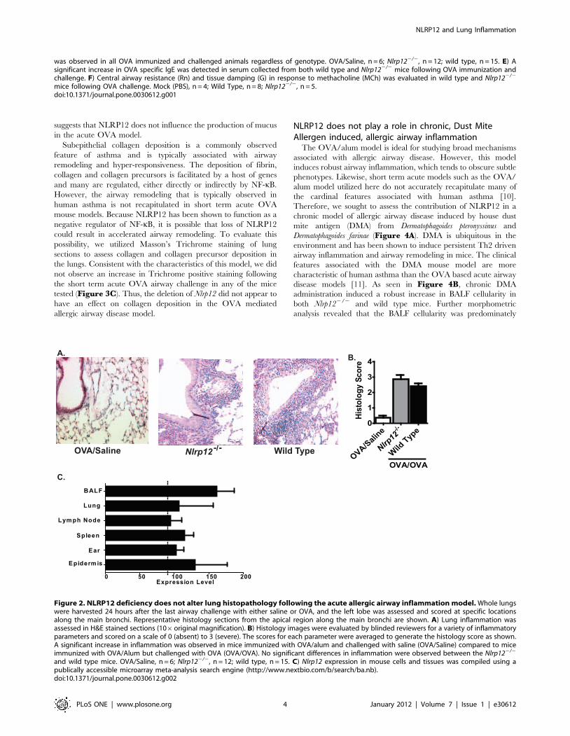
feature. Following 10 weeks of i.n. DMA administration, we
observed a significant increase in airway inflammation
(Figure 6A–B) and airway mucus production (Figure 7A–B).
Unlike the acute OVA model, we also observed an increase in
Massons Trichrome positive regions in the lungs of mice treated
with DMA (Figure 7C). Together, these data confirm previously
reported accounts that the DMA model induces a less acute
disease with clinical features that are more consistent with human
asthma. However, in all cases, no significant differences were
detected between Nlrp122/2 and wild type animals. Together,
these data suggest that NLRP12 does not influence the
development of either acute or chronic allergic airway disease.
Figure 5. NLRP12 does not contribute to the production of Th2 associated cytokines following chronic HDM administration. A–B) Asignificant increase in Th2 associated proinflammatory mediators was observed in the lung (IL-4, IL-5, and IL-13), BALF (IL-13, IFN-c) and serum (totalIgE) from all DMA challenged animals. The Th1 associated cytokine, IL-1b, was also increased in the BALF; however, the data shown were extrapolatedfrom the standard curve, which has 31.3 pg/ml as the lowest level of detection for the ELISA. Saline, n = 6; Nlrp122/2, n = 8; wild type, n = 12.doi:10.1371/journal.pone.0030612.g005
NLRP12 and Lung Inflammation
PLoS ONE | www.plosone.org 6 January 2012 | Volume 7 | Issue 1 | e30612
Discussion
NLRP12 has been shown to influence both canonical and non-
canonical NF-kB signaling and classical and nonclassical MHC
class I gene expression in vitro [5,6,12]. In vivo, Nlrp12 deficiency
significantly influences the development of contact hypersensitivity
[8]. Based on these findings we hypothesized that the Nlrp122/2
mouse would provide a suitable model to study the contribution of
this gene in allergic diseases, including asthma. However, despite
notable trends, we did not detect any significant differences in the
development of either acute OVA or HDM mediated allergic lung
disease in the Nlrp122/2 mice compared to the wild type animals.
A number of explanations are possible that may explain our
inability to observe significant differences in these lung disease
models. The most obvious explanation is that NLRP12 does not
have a discernable effect on the pathogenesis of allergic lung
disease in the mouse or NLRP12 may function in either a
temporal, situational or stimuli specific manner that was not
captured in the data generated by the lung inflammation models
discussed here. NLRP12 activity may be restricted to a specific cell
0
Figure 6. NLRP12 deficiency does not alter lung histopathology following chronic, DMA mediated, allergic airway inflammation.Whole lungs were harvested 24 hours after the last airway challenge with either saline or DMA, and the left lobe was assessed and scored at specificlocations along the main bronchi. Representative histology sections from the apical region along the main bronchi are shown. A) Lung inflammationwas assessed in H&E stained sections (106 original magnification). B) Histology images were evaluated by blinded reviewers for a variety ofinflammatory parameters and scored on a scale of 0 (absent) to 3 (severe). The scores for each parameter were averaged to generate the histologyscore as shown. A significant increase in inflammation was observed in mice challenged with DMA compared to mice challenged with saline alone.No significant differences were observed between the Nlrp122/2 and wild type mice. Saline, n = 6; Nlrp122/2, n = 8; wild type, n = 12.doi:10.1371/journal.pone.0030612.g006
Figure 7. NLRP12 deficiency did not influence mucus production or collagen deposition in the lungs following HDM. A) Mucusproduction was assessed following treatment with the Alcian blue/periodic acid-Schiffs reaction (AB/PAS) (206original magnification). B) The volumeof AB/PAS-stained mucosubstance per square millimeter of basal lamina (Vs) was determined. A significant increase in mucus was observed in theDMA mice; however, no significant differences were observed between the Nlrp122/2 and wild type mice. C) Masson’s trichrome staining was used toassess collagen deposition in the lungs. Histopathology assessments revealed an increase in collagen deposition in the DMA treated animalscompared to the saline treated mice. No significant differences were observed between the Nlrp122/2 and wild type mice. Saline, n = 6; Nlrp122/2,n = 8; wild type, n = 12.doi:10.1371/journal.pone.0030612.g007
NLRP12 and Lung Inflammation
PLoS ONE | www.plosone.org 7 January 2012 | Volume 7 | Issue 1 | e30612
type or tissue that may contribute to the development of contact
hypersensitivity and atopic dermititis, while not significantly
contributing to lung inflammation. This hypothesis is similar to
in vivo data published for TLR2, which has been shown to play a
vital role in the host immune response during contact hypersen-
sitivity. Similar to NLRP12, TLR2 has been shown to be an
essential mediator of the immune response to oxazolone (OX) in
an allergic contact dermatitis model [13]. However, unlike the
findings for OX, Tlr22/2 and wild type mice demonstrated similar
levels of inflammation in models involving epicutaneous sensitiza-
tion with OVA [13]. Other in vivo studies have suggested that
TLR2 functions as a negative regulator of allergic airway
inflammation following either DMA exposure or acute OVA
challenges [14,15,16]. In the context of NLRP12, several studies
have associated functional studies with gene expression data in
humans and rodents. These studies have shown that NLRP12 is
differentially expressed between species and transiently increased
during various models of lung inflammation [6,8,17]. Together,
these data support a scenario where NLRP12 does not influence
the development of allergic airway inflammation. However, as
illustrated by the contact hypersensitivity phenotypes previously
reported, it is likely that NLRP12 has a more dramatic role in
other models of inflammatory diseases through a temporal and
tissue specific mechanism.
A second hypothesis for the failure to observe an in vivo
phenotype in the lung inflammation models in the Nlrp122/2 mice
suggests that the models and analyses we utilized in this study are
too broad to effectively discern mild or moderate phenotypes.
Indeed, the OVA model described here induced a vigorous Th2
mediated immune response. One valid criticism of this model is
that the acute nature and robust inflammation tends to obscure the
contribution of several important mediators to the pathogenesis of
the disease. To avoid this issue, studies have suggested utilizing
chronic models, such as long term OVA exposure (without alum)
or DMA exposure, to assess allergic airway inflammation
[10,11,18]. Thus, in an effort to address this concern, we also
utilized DMA to induce airway inflammation and did not observe
any discernable phenotypic differences in the Nlrp122/2 mice.
However, we cannot rule out the possibility that higher resolution
in vivo models or analysis may reveal a more significant
contribution for NLRP12 in mediating subtle aspects of inflam-
mation in the lung.
While this study reports that NLRP12 does not affect detectable
difference in allergic lung inflammation, it is important to
recognize that additional in vivo assessments in the lung or in
other tissues may reveal important functions for this NLR in other
disease processes. Our data suggests that NLRP12 does not
contribute in a detectable way to the development of allergic
airway disease models tested here. However, it is interesting to
speculate that it may function as either a positive or negative
regulator of innate immune responses to agents associated with
asthma exacerbations. Thus, our data shows that the acute OVA
and chronic HDM models are appropriate for future studies that
aim to decipher the contribution of NLRP12 in asthma
exacerbations, which is an area of intense scientific and clinical
interest.
Materials and Methods
Ethics StatementAll studies were conducted under the approval of the
Institutional Care and Use Committee (IACUC) for The
University of North Carolina at Chapel Hill (IACUC protocol
approval #’s 07-170, 10-146, which are specific for the animal
models utilized in this publication) and in accordance with the
National Institutes of Health Guide for the Care and Use of
Laboratory Animals.
Experimental AnimalsNlrp122/2 animals were kindly provided by Millennium Inc.
and were generated as previously described [8]. All animals were
backcrossed 9 generations onto C57Bl/6 mice (Jackson Labora-
tories). Genotypes were confirmed via PCR of genomic tail DNA
utilizing the following primer sets: Forward-1, 59-CCCA-
CAAAGTGATGTTGGACTG-39, Forward-2, 59-GCAGCG-
CATCGCCTT CTATC-39, Reverse, 59-GAAGCAACCTCC-
GAATCAGAC-39. All mice were maintained under specific
pathogen free conditions and all experiments were performed
with 6–12 week old age- and sex- matched mice.
Induction of allergic airway inflammationAllergic airway inflammation was induced by ovalbumin (OVA)
as previously described [19]. Mice were sensitized by i.p. injection
of 20 mg of OVA (Grade V; Sigma) emulsified in aluminum
hydroxide (Sigma) in a total volume of 200 ml, on days 221 and
27. Airway inflammation was induced via intranasal (i.n.)
administration of OVA (1% in saline) for 5 days (days 1–5).
Control mouse groups received the two OVA immunizations, but
were i.n. challenged with saline. Mice were harvested 24 hours
following the last i.n. administration of OVA.
Allergic airway inflammation (chronic) was induced by house
dust mite antigen (DMA) challenge. Mice were exposed i.n. to
0.05 AU/ml of purified 50:50 DerP and DerF whole body extract
(Greer Laboratories, Lenoir, NC) in 50 ul of saline for 5
consecutive days, followed by 2 days of rest, for 10 consecutive
weeks. Mice were harvested 24 hours following the last i.n.
administration of DMA.
Techniques for Assessing Airway InflammationFollowing the completion of the respective models, mice were
euthanized and serum was collected by cardiac puncture. To
assess the cellularity and cytokine levels in the bronchoalveolar
lavage fluid (BALF), mice were perfused with HBSS and a tracheal
cannula was inserted below the larynx. The lungs were lavaged 3
times with 1 ml of HBSS. The resultant BALF was centrifuged to
separate the cellular components and cell free supernatants.
Protein levels were assessed from serum, cell free lung homoge-
nates and cell free BALF supernatants by ELISA (OptEIA, BD or
R&D Biosystems). Total BALF cellularity was evaluated, following
red blood cell lysis via hypotonic saline treatment, using a
hemacytometer. BALF composition was determined by differential
staining of samples that were cytospun onto slides and stained with
Diff-Quik (Dade Behring). Leukocytes were characterized based
on morphology assessments of $200 cells per BALF sample. Using
morphology based assessments, all monocyte derived cells,
including macrophages, were classified as monocytes.
For histopathology, following BALF collection, the lungs were
fixed by inflation to 20-cm pressure, immersed in 4% parafor-
maldehyde (PFA) and whole inflated lungs were embedded in
paraffin wax. To evaluate airway inflammation, fixed lung slices
(5-mm) were stained with hematoxylin and eosin (H&E). Sections
of the left lung lobe were cut to yield the maximum longitudinal
visualization of the intrapulmonary main axial airway. These
sections were examined and inflammation was scored by an
experienced reviewer who was blinded to genotype and treatment.
Histopathology was evaluated and the following inflammatory
parameters were scored between 0 (absent) and 3 (severe):
mononuclear and polymorphonuclear cell infiltration; airway
NLRP12 and Lung Inflammation
PLoS ONE | www.plosone.org 8 January 2012 | Volume 7 | Issue 1 | e30612

epithelial cell hyperplasia and injury; extravasation; perivascular
and peribroncheolar cuffing; and the percent of the lung involved
with inflammation. This scoring system has been previously
described [19,20,21]. These parameter scores were averaged for a
total histology score. In addition to histopathology, goblet cell
hyperplasia was also assessed in the allergic airway disease models.
Sections of the left lung lobes were sectioned, as described above,
and stained with the Alcian-blue/periodic acid-schiff reaction
(AB/PAS). In an effort to avoid bias for certain regions and to
consistently view the identical region in all slides, a 2-mm length of
airway was marked and digitally imaged. The region that was
evaluated is located midway along the length of the main axial
airway. Using ImageJ software (NIH, National Technical
Information Service, Springfield, VA), the length and area of the
AB/PAS-stained region in the lung sections were measured and
the data is expressed as the mean volume density (Vs = nl/mm2
basal lamina+SEM of AB/PAS-stained material within the
epithelium), as previously described [22]. To evaluate collagen
deposition in the lungs, sections were prepared as described above,
stained with Masson’s Trichrome and assessed by an experienced
reviewer who was blinded to genotype and treatment, as
previously descbribed [23].
Measurement of Lung Function by Small AnimalVentilator
Mice were anesthetized with pentobarbital sodium, tracheosto-
mized and paralyzed with pancuronium bromide. Mice were then
mechanically ventilated with a computer controlled small animal
ventilator (Scireq, Montreal, Canada) at 300 breaths/min, with a
tidal volume of 6 cc/kg and a PEEP of 3–4 cm water. Mice were
exposed via aerosol to challenges with 0, 10, 20 and 40 mg/ml of
methacholine (MCh; Sigma, St. Louis), which was delivered by
ultrasonic nebulizer (Scireq, Montreal, Canada) for 30 seconds.
During the aerosol exposure, the ventilation rate was reduced to
200 breaths/min and 0.15 cc/kg tidal volume. Following the
aerosol challenge, ventilation was resumed at the original rate.
Forced Oscillatory Mechanics (FOM) were utilized to evaluate
airway reactivity every 10 seconds for 3 minutes following each
MCh challenge. Our analysis was focused on the evaluation of
Newtonian Resistance (Rn), which reflects resistance in the central
airways, and the tissue resistance (G).
Statistical AnalysisAll data are presented as the mean +/2 the standard error of
the mean (SEM). For complex data sets, we utilized an Analysis Of
Variance (ANOVA) followed by either Tukey-Kramer HSD or
Newman-Keuls for multiple comparisons. Single data points were
assessed by the Student’s two-tailed t-test. In all cases, a p-value of
less than 0.05 was considered statistically significant.
Acknowledgments
We would like to thank Dr. Fayyaz S. Sutterwala (University of Iowa), Dr.
Beverly H. Koller (UNC) and Dr. John Bertin (GSK) for their technical
support and supplying the mice used in this manuscript. We would also like
to thank Dr. Denis Gris, Dr. Justin Wilson, Monika Schneider, Dr. Kelly E.
Roney, Dr. Brian P. O’Connor and Dr. Chris B. Moore for their technical
assistance.
Author Contributions
Conceived and designed the experiments: ICA JDL JCA SLT JPT.
Performed the experiments: ICA JDL JCA CMJ RAR JBC. Analyzed the
data: ICA CMJ RAR JBC. Contributed reagents/materials/analysis tools:
ICA JDL JCA CMJ RAR JBC. Wrote the paper: ICA JPT.
References
1. Wang L, Manji GA, Grenier JM, Al-Garawi A, Merriam S, et al. (2002)
PYPAF7, a novel PYRIN-containing Apaf1-like protein that regulates activationof NF-kappa B and caspase-1-dependent cytokine processing. J Biol Chem 277:
29874–29880.
2. Ting JP, Davis BK (2005) CATERPILLER: a novel gene family important inimmunity, cell death, and diseases. Annu Rev Immunol 23: 387–414.
3. Conti BJ, Davis BK, Zhang J, O’Connor W, Jr., Williams KL, et al. (2005)CATERPILLER 16.2 (CLR16.2), a novel NBD/LRR family member that
negatively regulates T cell function. J Biol Chem 280: 18375–18385.
4. Cui J, Zhu L, Xia X, Wang HY, Legras X, et al. (2010) NLRC5 negativelyregulates the NF-kappaB and type I interferon signaling pathways. Cell 141:
483–496.5. Lich JD, Williams KL, Moore CB, Arthur JC, Davis BK, et al. (2007) Monarch-
1 suppresses non-canonical NF-kappaB activation and p52-dependent chemo-kine expression in monocytes. J Immunol 178: 1256–1260.
6. Williams KL, Lich JD, Duncan JA, Reed W, Rallabhandi P, et al. (2005) The
CATERPILLER protein monarch-1 is an antagonist of toll-like receptor-, tumornecrosis factor alpha-, and Mycobacterium tuberculosis-induced pro-inflamma-
tory signals. J Biol Chem 280: 39914–39924.7. Allen IC, Moore CB, Schneider M, Lei Y, Davis BK, et al. (2011) NLRX1
protein attenuates inflammatory responses to infection by interfering with the
RIG-I-MAVS and TRAF6-NF-kappaB signaling pathways. Immunity 34:854–865.
8. Arthur JC, Lich JD, Ye Z, Allen IC, Gris D, et al. (2010) Cutting edge: NLRP12controls dendritic and myeloid cell migration to affect contact hypersensitivity.
J Immunol 185: 4515–4519.9. Macaluso F, Nothnagel M, Parwez Q, Petrasch-Parwez E, Bechara FG, et al.
(2007) Polymorphisms in NACHT-LRR (NLR) genes in atopic dermatitis. Exp
Dermatol 16: 692–698.10. Yu M, Tsai M, Tam SY, Jones C, Zehnder J, et al. (2006) Mast cells can
promote the development of multiple features of chronic asthma in mice. J ClinInvest 116: 1633–1641.
11. Stevenson CS, Birrell MA (2010) Moving towards a new generation of animal
models for asthma and COPD with improved clinical relevance. PharmacolTher.
12. Williams KL, Taxman DJ, Linhoff MW, Reed W, Ting JP (2003) Cutting edge:Monarch-1: a pyrin/nucleotide-binding domain/leucine-rich repeat protein that
controls classical and nonclassical MHC class I genes. J Immunol 170:
5354–5358.13. Jin H, Kumar L, Mathias C, Zurakowski D, Oettgen H, et al. (2009) Toll-like
receptor 2 is important for the T(H)1 response to cutaneous sensitization.
J Allergy Clin Immunol 123: 875–882 e871.14. Hisbergues M, Magi M, Rigaux P, Steuve J, Garcia L, et al. (2007) In vivo and
in vitro immunomodulation of Der p 1 allergen-specific response byLactobacillus plantarum bacteria. Clin Exp Allergy 37: 1286–1295.
15. Patel M, Xu D, Kewin P, Choo-Kang B, McSharry C, et al. (2005) TLR2
agonist ameliorates established allergic airway inflammation by promoting Th1response and not via regulatory T cells. J Immunol 174: 7558–7563.
16. Wu Q, Martin RJ, Rino JG, Jeyaseelan S, Breed R, et al. (2007) A deficientTLR2 signaling promotes airway mucin production in Mycoplasma pneumo-
niae-infected allergic mice. Am J Physiol Lung Cell Mol Physiol 292:L1064–1072.
17. Rao KM, Meighan T (2006) Exposure in vivo to silica or lipopolysaccharide
produces transient or sustained upregulation, respectively, of PYPAF7 andMEFV genes in bronchoalveolar lavage cells in rats. J Toxicol Environ Health A
69: 481–490.18. Matute-Bello G, Frevert CW, Martin TR (2008) Animal models of acute lung
injury. Am J Physiol Lung Cell Mol Physiol 295: L379–399.
19. Allen IC, Pace AJ, Jania LA, Ledford JG, Latour AM, et al. (2006) Expressionand function of NPSR1/GPRA in the lung before and after induction of asthma-
like disease. Am J Physiol Lung Cell Mol Physiol 291: L1005–1017.20. Allen IC, Scull MA, Moore CB, Holl EK, McElvania-TeKippe E, et al. (2009)
The NLRP3 inflammasome mediates in vivo innate immunity to influenza Avirus through recognition of viral RNA. Immunity 30: 556–565.
21. Willingham SB, Allen IC, Bergstralh DT, Brickey WJ, Huang MT, et al. (2009)
NLRP3 (NALP3, Cryopyrin) facilitates in vivo caspase-1 activation, necrosis,and HMGB1 release via inflammasome-dependent and -independent pathways.
J Immunol 183: 2008–2015.22. Cressman VL, Hicks EM, Funkhouser WK, Backlund DC, Koller BH (1998)
The relationship of chronic mucin secretion to airway disease in normal and
CFTR-deficient mice. Am J Respir Cell Mol Biol 19: 853–866.23. Lovgren AK, Jania LA, Hartney JM, Parsons KK, Audoly LP, et al. (2006)
COX-2-derived prostacyclin protects against bleomycin-induced pulmonaryfibrosis. Am J Physiol Lung Cell Mol Physiol 291: L144–156.
NLRP12 and Lung Inflammation
PLoS ONE | www.plosone.org 9 January 2012 | Volume 7 | Issue 1 | e30612