Experiments and Simulations of Directionally Annealed ODS MA 754

Characterization of magnetic materials by
Lorentz microscopy and
electron holography

JEOL JEM-2200FS transmission electron microscope @HU-Berlin
Structure and chemical composition (CTEM,STEM,EDX,EELS)
Magnetic properties(Lorentz microscopy, electron holography)
Electron biprism
Objective mini lens
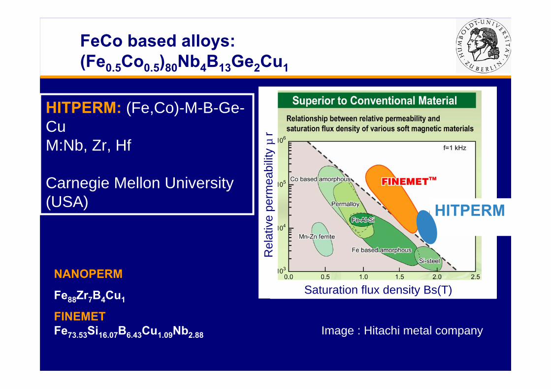
NANOPERM
Fe88Zr7B4Cu1
FINEMETFe73.53Si16.07B6.43Cu1.09Nb2.88
HITPERM: (Fe,Co)-M-B-Ge-CuM:Nb, Zr, Hf
Carnegie Mellon University (USA)
FeCo based alloys: (Fe0.5Co0.5)80Nb4B13Ge2Cu1
Image : Hitachi metal company
HITPERM
Saturation flux density Bs(T)
Rel
ativ
e pe
rmea
bilit
y µ
r

Introduction to softmagneticmaterials
Low coercivity
Easily Magnetized and demagnetized. (Easily moving domain walls).
Applications
•Power Transformers
• Data communication
•Sensors
•Magnetic heads
Soft
Hard

Why we use nanocrystalline materials?
Herzer G. In: Buschow KHJ, editor. Handbook of magnetic materials, vol. 10. Amsterdam:Elsevier Science, 1997. p. 415 [chapter 3].
Relationship between structure and magnetic properties

HAADF images
nanocrystalline
nanocrystalline
Annealed at 550ºC for 1 hour Annealed at 610ºC for 1 hour

Electron diffraction
Halo ring
[111]
110
SAED Nano beam diffraction (NBD)
(Fe0.5Co0.5)80Nb4B13Ge2Cu1Annealed at 550ºC for 1 hour
110200211220222310 bcc structure

Electron diffraction
[111]
SAED NBD
[111]
SAED NBD
550ºC1 hour
610ºC1 hour

HRTEM images
15.3nm
Moiré fringes
Nanocrystalline
2.0Å
2.0Å
(110)
(110)
Amorphous phase
Annealed at 550ºC for 1 hour
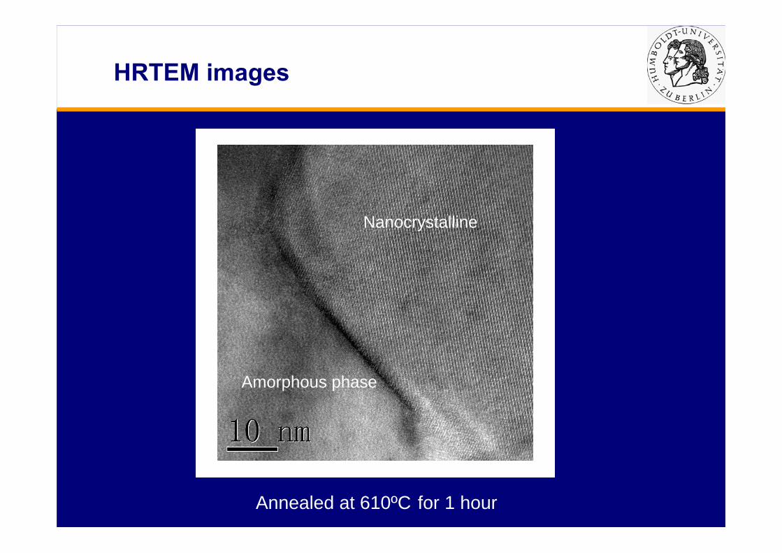
HRTEM images
Amorphous phase
Nanocrystalline
Annealed at 610ºC for 1 hour
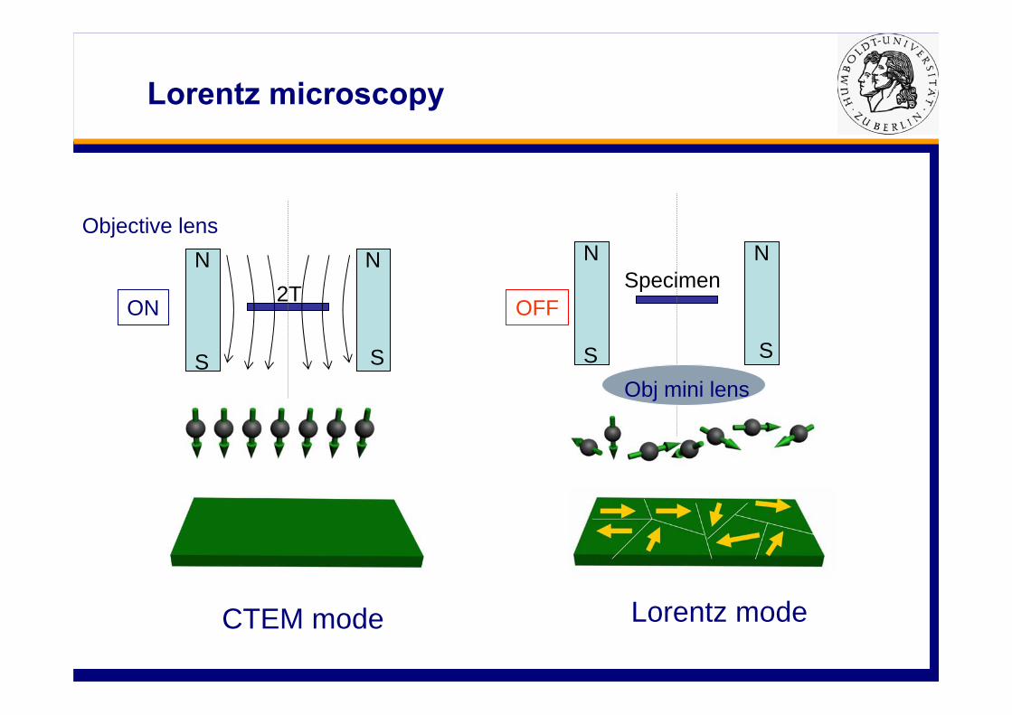
Lorentz microscopy
N N
S S
2T SpecimenN N
S S
Obj mini lens
CTEM mode Lorentz mode
Objective lens
ON OFF

x x x x x x x x· · · ·x x x x · · · · x x x x
Over focused
Under focused
Lorentz microscopy
In focus Under focusedOver focusedAnnealed at 550ºC for 1 hour

Lorentz microscopy
Annealed at 610ºC for 1 hour
In focus Under focusedOver focused
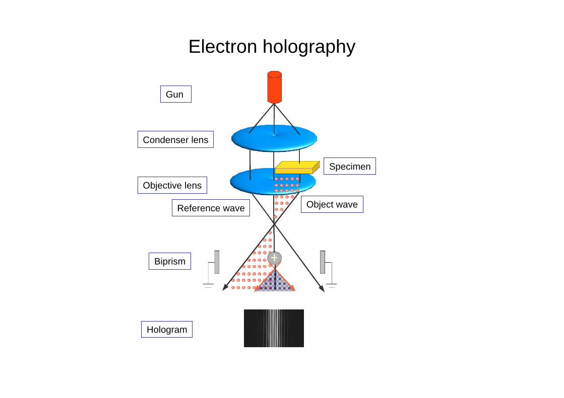
Gun
Biprism
Condenser lens
Objective lens
Specimen
Reference wave
Hologram
Object wave
Electron holography
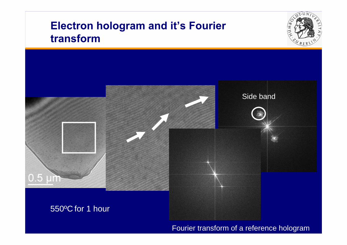
Electron hologram and it’s Fourier transform
Side band
FFT
550ºC for 1 hour
Fourier transform of a reference hologram

Over focusedIn focus Under focused
PhaseAmplitude
Reconstructed phase and amplitude compared to Lorentz mode image
550ºC for 1 hour
Surface plot of phase

Reconstructed phase compared to Lorentz mode image
Annealed at 610ºC for 1 hour
Under focusedIn focus Phase

Objective lens
N N
S S
H
θ
N N
S S
H//
Specimen In-plane magnetic field
H//=H·Sinθ
N N
S S
Objective lens off Objective lens slightly active
Tilt specimen
Specimen
Objective lens
Change of applied field by tilting sample

App
lied
field
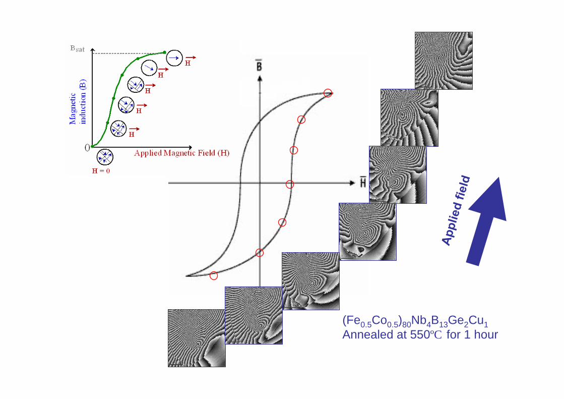
(Fe0.5Co0.5)80Nb4B13Ge2Cu1Annealed at 550ºC for 1 hour
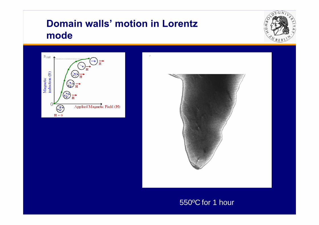
Domain walls’ motion in Lorentz mode
550ºC for 1 hour

Conclusion
Large magnetic domain and few pinning sites in sample annealed at 550℃ for 1 hour were observed by Lorentz microscopy and electron holography. This kind of domain configuration is easy to move in small magnetic field.
Domain configuration becomes more complicated and more pinning sites exist when the annealing temperature was elevated to 610 ℃. This means domain walls in sample need more energy to overcome these barriers. In other words, the coercivity of sample becomes large.