Characterization of Potential Plant Growth Promoting Rhizobacteria ...

Characterization of Diagnostic Tools and Potential Treatments for Alzheimer’s Disease PET ligands and BACE1 inhibitors
Fredrik Jeppsson

ii
iii
Characterization of Diagnostic Tools and Potential Treatments for Alzheimer’s Disease PET ligands and BACE1 inhibitors
Fredrik Jeppsson

iv
Cover picture: “Coronal Brain Slice” Painted by Julian Jeppsson 3 years, 2016. Articles and figures reprinted with permission ©Fredrik Jeppsson, Stockholm University 2016 ISBN 978-91-7649-531-5 Printed in Sweden by Holmbergs, Malmö 2016

v
Till Julian, Nils, Ivar och Johanna

vi

vii
List of Publications
I. Johnson AE, Jeppsson F, Sandell J, Wensbo D, Neelissen JA, Juréus A, Ström P, Norman H, Farde L, Svensson SP. AZD2184: a radioligand for sensitive detection of β-amyloid deposits. J Neurochem. 2009 Mar;108(5):1177-86.
II. Juréus A, Swahn BM, Sandell J, Jeppsson F, Johnson AE, Johnström P, Neelissen JA, Sunnemark D, Farde L, Svensson SP. Characterization of AZD4694, a novel fluorinated Aβ plaque neuroimaging PET radioligand. J Neurochem. 2010 Aug;114(3):784-94.
III. Mattsson A*, Jeppsson F*, Lundkvist J, Svensson A, Nilsson M, Sunnemark D, Nilsson KP, Hammarström P, Holtzman DM, Ferm M, Svensson SP, Juréus A. Altered amyloid PET ligand binding to ApoE-containing plaques in Alzheimer´s disease brain in vitro. Manuscript. (2016)
IV. Jeppsson F, Eketjäll S, Janson J, Karlström S, Gustavsson S, Olsson LL, Radesäter AC, Ploeger B, Cebers G, Kolmodin K, Swahn BM, von Berg S, Bueters T, Fälting J. Discovery of AZD3839, a Potent and Selective BACE1 Inhibitor Clinical Candidate for the Treatment of Alzheimer Disease. J Biol Chem. 2012 Nov 30;287(49):41245-57.
V. Eketjäll S, Janson J, Jeppsson F, Svanhagen A, Kolmodin K, Gustavsson S, Radesäter AC, Eliason K, Briem S, Appelkvist P, Niva C, Berg AL, Karlström S, Swahn BM, Fälting J. AZ-4217: A High Potency BACE Inhibitor Displaying Acute Central Efficacy in Different In vivo Models and Reduced Amyloid Deposition in Tg2576 Mice. J Neurosci. 2013 Jun 12;33(24):10075-84.
*Contributed equally

viii
VI. Eketjäll S, Janson J, Kaspersson K, Bogstedt A, Jeppsson F, Fälting J, Haeberlein SB, Kugler AR, Alexander RC, Cebers G. AZD3293: A Novel, Orally Active BACE1 Inhibitor with High Potency and Permeability and Markedly Slow Off-Rate Kinetics. J Alzheimers Dis. 2016;50(4):1109-23.

ix
Additional publications
• Llona-Minguez S, Höglund A, Jacques SA, Johansson L, Calderón- Montaño JM, Claesson M, Loseva O, Valerie NC, Lundbäck T, Piedrafita J, Maga G, Crespan E, Meijer L, Burgos Morón E, Baranczewski P, Hagbjörk AL, Svensson R, Wiita E, Almlöf I, Visnes T, Jeppsson F, Sigmundsson K, Jensen AJ, Artursson P, Jemth AS, Stenmark P, Warpman Berglund U, Scobie M, Helleday T. Discovery of the First Potent and Selective Inhibitors of Human dCTP Pyrophosphatase 1. J Med Chem. 2016 Feb 11;59(3):1140-8.
• Gad H, Koolmeister T, Jemth AS, Eshtad S, Jacques SA, Ström CE, Svensson LM, Schultz N, Lundbäck T, Einarsdottir BO, Saleh A, Göktürk C, Baranczewski P, Svensson R, Berntsson RP, Gustafsson R, Strömberg K, Sanjiv K, Jacques-Cordonnier MC, Desroses M, Gustavsson AL, Olofsson R, Johansson F, Homan EJ, Loseva O, Bräutigam L, Johansson L, Höglund A, Hagenkort A, Pham T, Altun M, Gaugaz FZ, Vikingsson S, Evers B, Henriksson M, Vallin KS, Wallner OA, Hammarström LG, Wiita E, Almlöf I, Kalderén C, Axelsson H, Djureinovic T, Puigvert JC, Häggblad M, Jeppsson F, Martens U, Lundin C, Lundgren B, Granelli I, Jensen AJ, Artursson P, Nilsson JA, Stenmark P, Scobie M, Berglund UW, Helleday T. MTH1 inhibition eradicates cancer by preventing sanitation of the dNTP pool. Nature. 2014 Apr 10;508(7495):215-21.
• Janson J, Eketjäll S, Tunblad K, Jeppsson F, Von Berg S, Niva C, Radesäter AC, Fälting J, Visser SA. Population PKPD modeling of BACE1 inhibitor-induced reduction in Aβ levels in vivo and correlation to in vitro potency in primary cortical neurons from mouse and guinea pig. Pharm Res. 2014 Mar;31(3):670-83.
x
• Forsberg A, Juréus A, Cselényi Z, Eriksdotter M, Freund-Levi Y, Jeppsson F, Swahn BM, Sandell J, Julin P, Schou M, Andersson J, Johnström P, Varnäs K, Halldin C, Farde L, Svensson S. Low background and high contrast PET imaging of amyloid-β with [11C]AZD2995 and [11C]AZD2184 in Alzheimer's disease patients. Eur J Nucl Med Mol Imaging. 2013 Apr;40(4):580-93.
• Ginman T, Viklund J, Malmström J, Blid J, Emond R, Forsblom R, Johansson A, Kers A, Lake F, Sehgelmeble F, Sterky KJ, Bergh M, Lindgren A, Johansson P, Jeppsson F, Fälting J, Gravenfors Y, Rahm F. Core refinement toward permeable β-secretase (BACE-1) inhibitors with low hERG activity. J Med Chem. 2013 Jun 13;56(11):4181-205.
• Gravenfors Y, Viklund J, Blid J, Ginman T, Karlström S, Kihlström J, Kolmodin K, Lindström J, von Berg S, von Kieseritzky F, Bogar K, Slivo C, Swahn BM, Olsson LL, Johansson P, Eketjäll S, Fälting J, Jeppsson F, Strömberg K, Janson J, Rahm F. New aminoimidazoles as β-secretase (BACE-1) inhibitors showing amyloid-β (Aβ) lowering in brain. J Med Chem. 2012 Nov 8;55(21):9297-311.
• Swahn BM, Kolmodin K, Karlström S, von Berg S, Söderman P, Holenz J, Berg S, Lindström J, Sundström M, Turek D, Kihlström J, Slivo C, Andersson L, Pyring D, Rotticci D, Ohberg L, Kers A, Bogar K, von Kieseritzky F, Bergh M, Olsson LL, Janson J, Eketjäll S, Georgievska B, Jeppsson F, Fälting J. Design and synthesis of β-site amyloid precursor protein cleaving enzyme (BACE1) inhibitors with in vivo brain reduction of β-amyloid peptides. J Med Chem. 2012 Nov 8;55(21):9346-61.
• Swahn BM, Sandell J, Pyring D, Bergh M, Jeppsson F, Juréus A, Neelissen J, Johnström P, Schou M, Svensson S. Synthesis and evaluation of pyridylbenzofuran, pyridylbenzothiazole and pyridylbenzoxazole derivatives as ¹⁸F-PET imaging agents for β-amyloid plaques. Bioorg Med Chem Lett. 2012 Jul 1;22(13):4332-7.

xi
• Mattsson N, Rajendran L, Zetterberg H, Gustavsson M, Andreasson U, Olsson M, Brinkmalm G, Lundkvist J, Jacobson LH, Perrot L, Neumann U, Borghys H, Mercken M, Dhuyvetter D, Jeppsson F, Blennow K, Portelius E. BACE1 inhibition induces a specific cerebrospinal fluid β-amyloid pattern that identifies drug effects in the central nervous system. PLoS One. 2012;7(2):e31084.
• Swahn BM, Wensbo D, Sandell J, Sohn D, Slivo C, Pyring D, Malmström J, Arzel E, Vallin M, Bergh M, Jeppsson F, Johnson AE, Juréus A, Neelissen J, Svensson S. Synthesis and evaluation of 2-pyridylbenzothiazole, 2-pyridylbenzoxazole and 2-pyridylbenzofuran derivatives as 11C-PET imaging agents for beta-amyloid plaques. Bioorg Med Chem Lett. 2010 Mar 15;20(6):1976-80.

xii

xiii
Abstract
Alzheimer’s disease (AD) is a very complex disorder and the most common form of dementia. The two pathological hallmarks of AD are extracellular amyloid-β (Aβ) plaques in cerebral cortex, and intraneuronal neurofibrillary tangles. In the early stages of the disease it can be difficult to accurately diagnose AD, as it is difficult to distinguish from normal signs of aging. There is thus a need for sensitive non-invasive tools, able to detect pathophysiological biomarker changes. One such approach is mo-lecular imaging of Aβ plaque load in brain, using PET (positron emission tomogra-phy) ligands. We have developed and characterized two novel Aβ plaque neuroimaging PET ligands, AZD2184 and AZD4694. The 2-pyridylbenzothiazole derivate AZD2184, is a 11C-labeled PET ligand with a higher signal-to-background ratio compared to the widely used PET ligand PIB, a 11C-labeled phenylbenzothiazole based tool. This makes it possible to detect smaller changes in Aβ plaque deposition load, and there-fore theoretically, also earlier diagnosis. A drawback with 11C-labeled PET ligands is the relatively short half-life. To meet the need for PET ligands with a longer half-life, we developed the pyridylbenzofuran derivate [18F]AZD4694. Although development of fluorinated radioligands is challenging due to the lipophilic nature of aromatic flu-orine, we successfully developed a 18F-labeled PET ligand with a signal-to-back-ground ratio matching PIB, the most widely used 11C-labeled PET ligand in clinical use. 3H-labeled derivates of AZD2184, AZD4694, and PIB, showed lower binding specificity towards Aβ plaques containing ApoE. The ApoE genotype per se did not significantly affect ligand binding, instead, the amount of ApoE incorporated to the Aβ plaques appears to be of importance for the binding characteristics of these amy-loid PET ligands. Beta-secretase 1 (BACE1) mediates the first step in the processing of amyloid pre-cursor protein (APP) to Aβ peptides, making BACE1 inhibition an attractive thera-peutic target in AD. We developed and characterized three novel BACE1 inhibitors, AZD3839, AZ-4217, and AZD3293. AZD3839 and AZ-4217 contains an amidine group which interacts with the catalytic aspartases Asp-32 and Asp-228 of BACE1, effectively inhibiting the enzyme. All three compounds are potent and selective inhib-itors of human BACE1, with in vitro potency demonstrated in several cellular models, including primary cortical neurons. All three compound exhibited dose- and time-de-pendent lowering of plasma, brain, and cerebrospinal fluid Aβ levels in several spe-cies, and two of the compounds (AZD3839 and AZD3293) were progressed into clin-ical trials.
xiv
Contents
1. Introduction .......................................................................................... 19 1.1 The drug discovery and development process ...................................................... 19
1.1.1 Drug discovery screening assays .......................................................... 21 1.1.2 Clinical development ..................................................................................... 23
1.2 Alzheimer’s disease .............................................................................................. 25 1.2.1 Pathological hallmarks .................................................................................. 25 1.2.2 APP processing ............................................................................................ 26 1.2.3 The amyloid cascade hypothesis .................................................................. 28 1.2.4 The role of BACE1 in AD .............................................................................. 29 1.2.5 Genetic risk factors for Alzheimer’s disease ................................................. 31 1.2.6 PET Aβ imaging as biomarker diagnostics of Alzheimer’s disease ............... 32 1.2.7 Therapies for Alzheimer’s disease ................................................................ 34
2. Aims of the thesis................................................................................. 35
3. Methodological considerations ............................................................ 36 3.1 Cells ...................................................................................................................... 36
3.1.1 SH-SY5Y cells .............................................................................................. 36 3.1.2 N2A cells ...................................................................................................... 36 3.1.3 HEK293 cells ................................................................................................ 36 3.1.4 Primary cortical neurons ............................................................................... 37
3.2 Cell treatments ...................................................................................................... 37 3.3 Enzyme inhibition assays ...................................................................................... 37 3.4 Biacore assays ...................................................................................................... 38 3.5 Aβ fibrillation ......................................................................................................... 39 3.6 Aβ binding and competition assay ......................................................................... 39 3.7 Autoradiography .................................................................................................... 40 3.8 Immunohistochemistry .......................................................................................... 41 3.9 In vivo model systems ........................................................................................... 41
3.9.1 C57BL/6 mice ............................................................................................... 41 3.9.2 Transgenic mice ........................................................................................... 42 3.9.3 Guinea Pig .................................................................................................... 42
4. Results and discussion ........................................................................ 43 4.1 Development and preclinical evaluation of PET radioligands for Aβ plaque neuroimaging (paper I, II, and III) ................................................................................ 43
4.1.1 Identification of new putative radioligands (paper I & II) ................................ 43

xv
4.1.2 Characterization of AZD2184 reveals similar affinity, but higher signal to noise ratio compared to PIB (paper I) .............................................................................. 44 4.1.3 Characterization of AZD4694, a PET ligand with longer half-life (paper II) .... 46 4.1.4 Altered binding of PET ligands to plaques in transgenic mice ....................... 47 4.1.5 Altered binding of PET ligands to plaques containing higher levels of ApoE (paper III) ............................................................................................................... 48
4.2 Development and preclinical evaluation of BACE1 inhibitors for potential treatment of Alzheimer’s disease (paper IV, V and VI) ................................................................ 49
4.2.1 Identification of new chemical series of BACE1 inhibitors (paper IV and V) .. 50 4.2.2 In vitro characterization of AZD3839 and AZ-4217 (paper IV & V) ................ 50 4.2.3 In vivo characterization of AZD3839 and AZ-4217 (paper IV & V) ................ 51 4.2.4 Target selectivity of AZD3839 and AZ-4217 (paper IV & V) .......................... 53 4.2.5 Clinical findings from AZD3839, relevant to the in vitro / in vivo characterization .............................................................................................................................. 54 4.2.6 In vitro characterization of AZD3293 (paper VI) ............................................ 55 4.2.7 In vivo characterization of AZD3293 (paper VI) ............................................. 55 4.2.8 Target selectivity of AZD3293 (paper VI) ...................................................... 56 4.2.9 AZD3293 in clinical trials ............................................................................... 57
5. Conclusions ......................................................................................... 58
6. Future perspectives ............................................................................. 60
7. Populärvetenskaplig sammanfattning på svenska .............................. 61
8. Acknowledgements .............................................................................. 63
9. References ........................................................................................... 65

xvi
Abbreviations
Aβ Amyloid Beta AD Alzheimer’s disease ADME Absorption, distribution, metabolism, and excretion AICD APP intracellular domain APH Anterior pharynx defective ApoE Apolipoprotein E APP Amyloid precursor protein BACE Beta-secretase,
β-site amyloid precursor protein cleaving enzyme CDR Clinical dementia rating DH Dunkin-Hartley fAD Familial Alzheimer's disease FDA US Food and Drug Administration FDG Fluorodeoxyglucose FRET Fluorescence resonance energy transfer GGA Golgi-localized, γ-ear containing, ADP-ribosylation factor
binding GPCR G-protein coupled receptor HEK Human embryonic kidney hERG Human ether-a-go-go related gene HFIP Hexafluoroisopropanol HTS High throughput screening LG Lead generation LO Lead optimization MCI Mild cognitive impairment MMSE Mini-mental status examination N2A Neuro-2A NaV Voltage-gated sodium channel NCE New chemical entity NCT Nicastrin NFT Neurofibrillary tangles NRG Neuregulin PD Pharmacodynamics PEN Presenilin enhancer PET Positron emission tomography pFTAA Pentameric formyl thiophene acetic acid

xvii
PIB Pittsburgh compound B PK Pharmacokinetic PoC Proof of concept PoM Proof of mechanism PoP Proof of principle PS1/PSEN1 Presenilin 1 sAD Sporadic Alzheimer’s Disease SAD Single-ascending-dose sAPP Soluble amyloid precursor protein SAR Structure-activity relationship SPR Surface plasmon resonance TdP Torsade de Pointes TR-FRET Time resolved fluorescence resonance energy transfer

xviii

19
1. Introduction
Alzheimer’s disease (AD) was first described by Alois Alzheimer over 100 years ago, but yet today there is no cure for this devastating illness, despite the enormous efforts and resources that have been invested into this. The actual cause of the disease is still debated, but the pathological hallmarks of the dis-ease are extracellular amyloid-β (Aβ) plaques in cerebral cortex and intraneu-ronal neurofibrillary tangles. Many believe that early diagnosis of AD might be a future key in battling the disease, creating a need for sensitive diagnostic tools. The generation and build-up of the Aβ plaques occurs via the amyloid pathway, where the enzyme Beta-secretase 1 (BACE1) mediates the first step, making BACE1 a tractable target for treatment of AD. The focus of this thesis was on characterization of novel Aβ plaque neuroimaging PET (Positron emission tomography) ligands for diagnosis of AD, and inhibitors of human BACE1 for potential treatment of AD.
1.1 The drug discovery and development process Drug discovery is a complex process involving a great number of scientists from various disciplines. The process typically takes 12-15 years and the cost of discovery and development of a new drug or new chemical entity (NCE) is estimated to exceed $1 billion [1-3]. The pharmaceutical industry generally follows a rather generic process:
1) Basic Research: The biological hypothesis or mechanism involved in a disease of interest is established and the biological target (nor-mally proteins or nucleic acids) for the disease mechanism of inter-est is identified and selected.
2) Lead Discovery: A biological high throughput assay is used to iden-tify active compounds against the selected target. The active com-pounds are further refined using medicinal chemistry and the struc-ture activity relationship is established. The pharmacokinetic proper-ties and selectivity profile of the compounds are optimized, and a number of compounds are tested in various animal safety and effect models. In the end a candidate drug is selected for preclinical devel-opment.

20
3) Preclinical Development: Extensive safety and toxicological evalua-tion of the candidate drug to ensure that the compound is safe for testing in humans. The candidate drug is also further characterized in more complex animal models, and if the combined safety and effect data are promising, the company will send in an Investigational New Drug file and apply for the start of clinical trials.
4) Clinical Development: Clinical trials in healthy human volunteers and patients to demonstrate that the compound and its formulation is safe and effective.
5) Regulatory Authorities Filing: Filing the preclinical and clinical data, as well as manufacturing process information, to regulatory au-thorities, to get the drug approved for marketing.
Since there are enormous costs involved in developing an NCE, pharmaceuti-cal companies normally have rather strict guidelines in terms of what a drug discovery and development project must fulfil in order to move forward and get additional resources and funding. An example of a typical drug discovery operating model is illustrated in figure 1 below. Figure 1. Example of a Drug Discovery Operating Model. The drug discovery process takes around 12-15 years and consists of several different preclinical (TS, LG, LO) and clinical (Phase I, II, III) phases. The length of each phase varies depending on project and indication. (PoM: proof of mechanism, PoP: proof of principle, PoC: proof of concept.)
As illustrated in figure 1, the drug discovery and development process nor-mally starts with the identification and validation of a target that is involved in the disease of interest. The target is most often a protein, and the idea is that by modifying the target protein pharmacologically, a desired effect is achieved. These target proteins are generally enzymes, G-protein coupled re-ceptors (GPCRs), ion channels, nuclear hormone receptors, or transporters. It is estimated that currently approved drugs act on only around 300 desired tar-gets [4]. The drug target proteins are normally evaluated and validated using cloning and protein expression, phenotype analysis of cellular systems, and transgenic animal models. In the best of worlds investigations of humans with mutations connected to disease modifications (gain or loss of function) are possible, giving an early clinical validation of the potential drug target. This has for example been the case with the sodium channel NaV1.7 where people

21
with certain mutations are insensitive to pain, thereby clinically validating the protein as a target for pain inhibition [5]. Another case is Alzheimer’s disease (AD) where various mutations in genes coding for proteins in the amyloid pathway give an early onset of the disease, whereas other mutations in the same pathway seems to protect from the disease [6].
1.1.1 Drug discovery screening assays To start testing compounds (or antibodies) for drug target interaction, robust and reliable biological assays and collections of compounds for initial evalu-ation are required. These biological screening assays are the first filters in the lead generation (LG) phase, where potential lead compounds are selected to develop and optimize further. There are different approaches and strategies when conducting the initial screens. One approach is high throughput screen-ing (HTS) campaigns where very large compound collections (up to 1 million compounds) are analysed in an assay [7]. Sometimes more focused screens are done using previously identified compounds hitting specific target classes such as kinases [8]. Another approach is fragment screening, where very small compounds are screened at very high concentrations, which can then be used as building blocks for larger molecules [3]. Many assays used in drug discov-ery programs rely on stable mammalian cell lines overexpressing the target of interest, or on biochemical assays using purified overexpressed recombinant proteins. Assays using intact cells generate a functional read out as a result of compound activity, whereas biochemical assays, which are typically used for GPCRs and enzymes, most often only generate affinity data for the test com-pound and target protein. Simpler biochemical assays, such as radio-ligand competition assays, typically measures the test compound’s ability to displace a radiolabeled high affinity reference compound from the binding site on the protein of interest [9]. Thus, no information is given regarding the agonistic or antagonistic properties of the test compounds, and follow-up assays need to be developed to evaluate the pharmacological properties in detail. Inde-pendent on assay format, a number of factors need to be considered when as-says are developed [3, 10]:
1) Pharmacological relevance: Studies should be performed using ref-erence compounds if available, to determine that the assays pharma-cology is predictive and sensitive enough to pick up compounds with the desired affinity and mode of action.
2) Reproducibility: It should be validated that the assay is reproducible across assay plates, from day to day, and between equipment and op-erators.
3) Costs: Reagents and assay volumes should be optimized to reduce the costs as much as possible.

22
4) Quality: In the pharmaceutical industry, a widely used statistical pa-rameter is the Z´ factor, which provides a measure on the assay win-dow and the variance around the high and low signals in the assay [11].
5) Compound effects: Compound libraries are typically stored in 100% DMSO, and the DMSO effect itself on the assay behaviour needs to be evaluated.
Once a number of hits (up to a few thousand) have been obtained from the initial HTS campaign or more focused screen, these hits are normally reduced and clustered into different series [8]. To achieve this the hits are normally tested in the same assay once again and the verified hits are then grouped based on structural similarities. The idea is to bring forward a number of dif-ferent series to cover a broad spectrum of chemical classes [3, 7]. In the initial screen each compound is normally tested in only one or two concentrations, and the verified hits are then tested in multiple concentrations to generate con-centration-response curves. This is an important step to be able to separate compounds based on target affinity, and to investigate if the compounds be-have in a traditional competitive fashion. A reduced list of hit compounds will then be examined and verified further in a more complex secondary assay for the target [3]. If the initial assay is a simpler radioligand binding assay or en-zymatic assay, the secondary assay is normally a functional assay using cell membranes expressing the target of choice or an assay using whole cells. Dur-ing the process medicinal chemists evaluate the properties of the compounds and once again cluster them into groups, looking for common structure-activ-ity relationships (SAR) covering a number of compounds [3, 8]. Compounds with similar chemical core motifs, but with different additional chemical groups would result in different potencies, thereby adding data to the SAR evaluation. The synthesis of new compounds and generation of new SAR data will further help identify the chemical elements associated with target activity. A selection of representative compounds from each cluster will also be eval-uated on absorption, distribution, metabolism, and excretion (ADME), as well as pharmacokinetic (PK) properties. Selectivity against similar proteins as the intended drug target are also tested at this stage if possible, to add additional data to the evaluation process of the different clusters or series of compounds. Once the hit series have been defined, a rigorous medicinal chemistry and pharmacology effort begins aiming at refining the hit series to produce more potent and selective compounds with desired ADME and PK properties. If structural information about the target protein is known, the SAR investiga-tions can be facilitated using molecular modelling and techniques such as X-ray crystallography or NMR [3, 12, 13]. At this stage the compounds are nor-mally tested in a number of assays in a screening funnel. The primary assay in the funnel is normally simple and often similar to the HTS assay, but with

23
multiple concentrations of each test compound. Compounds meeting a prede-fined cut-off criteria in terms of potency are then moved forward to more com-plex assays, often cellular ones. In parallel, ADME properties are investigated, and selectivity against key targets if possible. In vitro activity against the po-tential test animal versions of target protein is also evaluated to ensure possi-ble effects in in vivo disease models, as well as pharmacokinetic and pharma-codynamics (PK/PD) models and toxicological studies. A selection of com-pounds that meet all or close to all predefined cut-off values in different as-says, will be progressed into in vivo tests, primarily in disease models and PK models at this stage. Physicochemical properties affecting solubility and per-meability are also studied in great detail in order to progress only potentially drug like substances. In the lead optimization (LO) phase of drug discovery, the aim is to maintain the favourable properties of the identified lead compounds and focusing on improving the remaining deficiencies [13]. These deficiencies might for ex-ample be selectivity and permeability problems. When one or two compounds have finally been produced that meet all the predefined criteria in terms of in vitro and in vivo effect profiles, ADME and PK/PD profile, and preclinical safety profile, the compound can be nominated for preclinical candidate se-lection [13]. However, the drug discovery work does normally not end here, as potential back up candidates with a different profile has to be produced, in case the candidate drug fails in further preclinical or clinical evaluation. All information gathered concerning the candidate molecule, together with the chemical manufacturing and control information, will form the basis for a clin-ical trial application to be submitted to regulatory authorities. The process from hit generation to clinical trial application is complex and far from rou-tine, as every project has its own challenges. In the end only around 10% of small molecule projects in the industry manage to deliver a candidate drug ready to enter clinical trials [3].
1.1.2 Clinical development After approval from regulatory authorities and ethics committees, clinical tri-als can start and the safety and efficacy of the candidate drug can be evaluated. Clinical trials for new drugs are sometimes classified into five different phases, but commonly phase I-III receives most attention (see figure 1) [14-16]:
0) Evaluation of PK profile of the drug at sub therapeutic doses in a
few healthy volunteers. Sometimes the microdosing studies in this phase are used to discriminate between a few different drug candi-

24
dates in order to decide which to proceed into further clinical devel-opment. This phase is often skipped and included in the phase I tests.
I) Evaluation of safety profile and dose-finding studies starting at sub therapeutic but ascending doses in up to 100 healthy volunteers. The tests in this phase are designed to evaluate the safety, tolerability, PK and PD of the drug. A proof of mechanism (PoM) is often done in this phase, to demonstrate that the drug reaches the right target in the desired organ. This phase can include single or multiple ascending dose studies where groups of subjects are given a single or multiple low doses of the drug. If these subjects do not show any adverse side effects and PK data are within predefined limits, a new group of sub-jects will be given a new escalated single or multiple dose, up to a predefined dose limit. The food effect on the drug will also be inves-tigated, where subjects will be given identical doses after a meal or while fasted.
II) Drug tested on up to around 300 patients and healthy subjects at therapeutic doses to evaluate the efficacy and safety. This is the clin-ical phase where most drugs fail, since this is the first time the thera-peutic effect of the drug is evaluated in human subjects. The phase is normally divided into IIA where the dose requirements are investi-gated, and IIB where the effect of the drug is investigated. The study can be designed to look at efficacy of the drug, where the effect on a specific set of biomarkers are investigated, or be designed to look at the effectiveness of the drug, where more general outcomes are in-vestigated, such as if the patients feel better or live longer. These studies are often referred to as proof of principle (PoP) and proof of concept (PoC) studies respectively. The phase II trials are most often randomized where some patients receive the drug and others placebo or standard treatment.
III) Drug tested on up to around 3000 patients at therapeutic doses to evaluate the therapeutic effect and safety. This phase is designed to investigate the effectiveness of the drug and are generally carried out as randomized trials at multiple centres around the world over a long period of time. In many cases two successful phase III trials are re-quired to obtain approval from regulatory authorities. Once satisfac-tory phase III trials are completed, the clinical trial results combined with preclinical documentation and chemistry manufacturing control documentation will be sent in as a regulatory submission to authori-ties in order to get an approval to market the drug.
IV) Post marketing surveillance where the now approved drug’s long term effects are monitored in public use for at least two years.

25
1.2 Alzheimer’s disease Alzheimer’s disease (AD) is a devastating neurodegenerative disease that af-fects more than 45 million people worldwide, and the prevalence is increasing due to an aging population and higher diagnosis rates [17, 18]. AD is the cause of around 70% of dementia cases and often begins in people over 65 years of age, although around 5% of cases are early-onset AD which begins before that. At the present time, there are no effective disease-modifying treatments for AD, although treatments that temporarily eases the symptoms are availa-ble. Thus, there is an urgent medical need for treatments that can halt or delay the progress of the disease [19-21]. There is also a great need for reliable bi-omarker based diagnostic methods that can facilitate an early detection of the disease. Today most patients get their diagnosis after neuropsychological, functional, and cognitive tests, which are considered too insensitive since they are only effective when the disease have progressed quite far. Early symptoms of AD are confusion, mood swings and insomnia. Clinically AD is character-ized by progression from episodic memory problems to decline of cognitive functions that at the end-stage of the disease leaves the patients bedridden and totally dependent on care givers. On average, death occurs 9 years after diag-nosis [21], but the cause of death is often other external factors such as infec-tions from pressure wounds or pneumonia [22].
1.2.1 Pathological hallmarks The pathological hallmarks of AD include positive lesions such as extracellu-lar amyloid beta (Aβ) plaques, intracellular neurofibrillary tangles (NFTs), and cerebral amyloid angiopathy. The positive lesions are accompanied by microglial responses. The negative lesions are loss of neurons and synapses [23, 24]. The lesions have their characteristic distribution, with plaques found in the cortical mantle, and tangles initially found in the entorhinal cortex, be-fore spreading to the hippocampus [25]. Inflammation occurs in pathologi-cally vulnerable regions of AD brains, and the deposits of Aβ and NFTs pro-vide a stimuli for inflammation and response from inflammatory mediators such as cytokines, chemokines, and acute and complementary pathways [26]. Extracellular amyloid beta plaques are deposits of Aβ peptides. Aβ is a prod-uct from the sequential cleavage of amyloid precursor protein (APP) by the enzymes β− and γ−secretase (see figure 2 and 3) [27]. Aβ is most commonly occurring as 40 or 42 amino acid peptides, and its physiological role is still under debate, but most likely the peptides are involved in the regulation of synaptic activity [24]. Accumulated Aβ peptides forms soluble intermediate oligomers that are synaptotoxic, and further accumulation and aggregation can lead to the generation of insoluble β-sheets of Aβ fibrils [24]. These β-sheets mainly consists of the more aggregation prone Aβ42 peptides, and are the

26
main constituents of dense core plaques. The Aβ40 peptides primarily makes up the deposits found in cerebral amyloid angiopathy, where amyloid deposits are formed in the blood vessel walls in the central nervous system. The dense core plaques has a compact core and are positive for Thioflavin-S and Congo Red staining, and typically associated with immune responses and synaptic loss. These plaques are part of the pathological diagnosis of AD since their presence is generally associated with cognitive impairment [24]. On the con-trary, diffuse plaques have less defined contours and are negative for Thiofla-vin-S and Congo Red staining. They are not associated with synaptic loss and are commonly found in elderly people with intact cognition [24]. Intracellular neurofibrillary tangles (NFT) are aggregates of hyperphosphory-lated and misfolded tau protein found inside neurons. These tangles later be-come extraneuronal when the neurons die [24]. Normally tau is involved in assembly and stabilization of microtubules, but as it becomes hyperphosphor-ylated it detaches from the microtubules and forms cytotoxic aggregates. This causes the microtubules to disintegrate, eventually destroying the cell´s cyto-skeleton. The spatiotemporal progression of NFTs correlates with the severity of cognitive decline in AD and is used for the pathological diagnosis of the disease [28].
1.2.2 APP processing APP is a large type 1 transmembrane protein expressed in many cell types [29]. Its physiological role is unclear, but as it is found in higher levels in the synaptic areas of neurons, roles in synaptic plasticity and synapse formation have been suggested [30]. In the human brain, APP is processed via two alter-native pathways, the non-amyloidogenic pathway and the amyloidogenic pathway (see figure 2). In the non-amyloidogenic pathway, α-secretase cleaves APP within the Aβ sequence, generating the extracellular fragment soluble APPα (sAPPα) and the membrane bound fragment C83, which is cleaved within its transmembrane domain by γ-secretase to generate the frag-ments P3 and APP intracellular domain (AICD) [27]. In the amyloidogenic pathway, Aβ is formed after sequential cleavage of APP by β-site amyloid precursor protein cleaving enzyme (BACE1, also called Asp2 and memapsin 2) and γ-secretase [27]. BACE1 cleaves APP, thereby generating the N-termi-nus of Aβ, and also producing the membrane bound C-terminal fragment C99. This also generates the extracellular fragment soluble APPβ (sAPPβ). The γ-secretase then cleaves C99 within the transmembrane region to release the Aβ peptide and the intracellular fragment AICD.

27
Figure 2. Amyloidogenic and non-amyloidogenic APP processing pathways. In the amyloidogenic pathway, APP is sequentially cleaved by BACE1, the β-secretase, and the γ-secretase complex, generating Aβ. BACE1 cleavage of APP is a prerequisite for Aβ formation and the rate-limiting step in Aβ generation. Used with permission from [27]. The cleavage position of BACE1 is very precise, occurring at Asp+1 and Glu+11 (see figure 3) [31]. On the other hand, the cleavage position of the γ-secretase is rather imprecise and can occur at various positions, generating Aβ peptides ranging from 30 to 51 residues in length [32] (see figure 3). The most abundant form of Aβ produced via the amyloidogenic pathway is Aβ40, and only around 10% is the more aggregation prone Aβ42. γ-Secretase is a prote-ase complex consisting of four subunits, presenilin 1 or presenilin 2, which are part of the aspartyl protease catalytic domain of γ-secretase. Further, the complex contains nicastrin (NCT), anterior pharynx defective (APH)-1a or -1b, and the presenilin enhancer (PEN)-2, three components of which the bio-logical function is not fully understood [33].

28
Figure 3. APP processing indicating the β, α and γ-secretase cleavage sites. The amino acid sequence of Aβ is shown in bold letters. The β, α, and γ-secretase cleavage sites are shown with arrowheads. Cleavage sites of BACE1 and BACE2 are shown with arrows. The Lys670→Asn/Met671→Leu (KM-NL) Swedish mutation is shown next to the BACE1 cleavage site. Used with permission from [34].
1.2.3 The amyloid cascade hypothesis Aβ42 has received a lot of attention in AD research as it is the major peptide found in the core of senile plaques [35], and a correlation between Aβ42 and cognitive decline has been reported [36]. In healthy human brain there is an equilibrium between the production and clearance of the toxic Aβ42 [37], however under AD conditions the clearance is markedly decreased, leading to buildup and aggregation of Aβ42. This triggers a complex cascade of events such as fibril formation, reactive oxygen species production, neuroinflamma-tion, disruption of synaptic activity, proteasome dysfunction, calcium dysho-meostasis, tau hyperphosphorylation, and eventually neuronal loss and de-mentia [35]. This cascade of events, starting with Aβ production and ending with neuronal loss and dementia, is referred to as the amyloid hypothesis or the amyloid cascade hypothesis [38]. This is one of the main hypothesis re-garding the cause of AD, although not unchallenged [39]. Given the complex-ity of the pathological network of processes leading to AD, it is unlikely that Aβ itself is the only driver of the disease, but more likely acts as the key ini-tiator of AD pathology [39]. One argument against the amyloid hypothesis is the rather poor time and space correlation between Aβ plaque deposition, neu-ronal loss and clinical symptoms in sporadic AD. The Aβ plaque depositions typically first appears in regions such as the frontal lobes, whereas the neu-ronal loss is most extensive in the entorhinal cortex and hippocampus regions, which normally contains rather few Aβ plaques [24, 28]. On the other hand, tau pathology correlates rather well with the neuronal loss, both in time and space [24, 40-42]. The spread of Aβ plaques appear to begin in the cortex and

29
then spread inwards, whereas the tau pathology seems to spread in the oppo-site direction [28]. This discrepancy between Aβ plaque depositions, neurofi-brillary tangles and neuronal death is not yet fully understood but seems to be consistent between familial and sporadic AD. Many clinical findings support Aβ plaque depositions as the initiator of downstream tau pathology and neu-rodegeneration [39]. In young and healthy non AD people, aggregated and phosphorylated tau pathology is present in the brainstem and entorhinal cortex before the onset of any Aβ buildup. At a later age, hippocampal neurofibrillary tau pathology is widely spread and concentrated to the limbic regions in am-yloid free individuals [43]. However, in people with coexisting Aβ plaque depositions, the tau pathology spreads into neocortical regions, and these peo-ple generally develop AD [43]. Tau pathology is also commonly found in hip-pocampus in cognitively normal elderly without any evidence of neurodegen-eration, but substantial neurodegeneration is found in the same region in AD patients carrying Aβ plaque depositions [39]. Many preclinical findings also support the view that Aβ precedes tau toxicity. While cultured tau knockout hippocampal neurons from mice were resistant to Aβ induced toxicity, similar wild type neurons showed degeneration in the presence of Aβ [44]. Further-more, 3D-differentiated human neuronal cells expressing familial AD muta-tions, exhibited high levels of aggregated phosphorylated tau, a pathology that could be blocked by β- or γ-secretase inhibitors [45]. One possible mechanism by which Aβ can trigger tau phosphorylation is via oligomeric forms of Aβ, which do not necessarily correlate with plaques in time or space, but which have been shown to spread via neuritic cell-to-cell transfer in a prion-like propagation mode [46, 47]. Aβ oligomers have been shown to induce tau phosphorylation in vitro and in vivo, possibly by mechanisms involving acti-vation of the Src kinase Fyn, in turn activating specific tau-targeting kinases [39]. Recent clinical findings using the human monoclonal abtibody aducan-umab, that selectively targets aggregated Aβ, supports the amyloid hypothesis. In patients with prodromal or mild AD, one year of monthly treatment with aducanumab reduced brain Aβ in a dose- and time-dependent manner, which was accompanied by a slowing of clinical decline [48].
1.2.4 The role of BACE1 in AD Since BACE1 is the enzyme responsible for initiating Aβ generation, it is a key therapeutic target for inhibition of Aβ production and downstream events in AD. BACE1 is a 501 amino acid long type 1 transmembrane aspartic pro-tease related to pepsin and retroviral aspartic protease families [49]. The en-zyme has a low pH optimum and is predominantly located in acidic intracel-lular vesicles such as endosomes, with the active site of the enzyme located in the acidic lumen side [31]. BACE1 has its highest expression in brain neurons, whereas the close homolog BACE2 (64% amino acid sequence homology) is sparsely expressed in the brain, and shows a lower APP cleaving activity [31].

30
BACE1 is primarily localized to endosomes present in normal presynaptic hippocampal mossy fiber terminals and to dystrophic presynaptic terminals surrounding amyloid plaques. BACE1 containing vesicles have also been identified near active zones, indicating a synaptic role [50]. BACE1 is initially synthesized in the ER as a zymogen, and is glycosylated on four Asn residues [51], and transient acetylation occurs on seven Arg residues [52]. In the Golgi compartment there is further addition of complex carbohydrates and the pro-domain is removed [31]. This maturation of BACE increases the catalytic ac-tivity towards APP at least two times, and the low pH of the late Golgi and Trans Golgi Network and early endosomes enhances the activity of BACE1 [31]. The recycling of BACE1 between the cell surface and the endosomal compartments is regulated by a phosphorylation on a Ser residue and a C-terminal dileucine motif [53, 54]. Dileucine-based sorting motifs have been associated with endosomal and lysosomal targeting from the cell surface, as well as from the trans Golgi network [55]. BACE1 partitioning into lipid rafts is facilitated by S-palmitoylation on four Cys residues located at crossing be-tween the transmembrane and cytosolic regions, although studies have shown that BACE1 processes APP equally efficient if not associated to lipid rafts [56]. The intracellular localization of BACE1 can be regulated by the BACE1 interaction proteins reticulon/Nogo, and an increased expression leads to an increased retention of BACE1 to the ER, which holds a neutral and suboptimal pH for BACE1 activity, and consequently a reduced Aβ generation [57-59]. On the other hand, intracellular trafficking of BACE1 to the endosomes en-hances APP processing and Aβ generation, and is facilitated by GGA (Golgi-localized, γ-ear containing, ADP-ribosylation factor binding) proteins inter-acting with BACE1 C-terminal motifs [60]. The GGA proteins have a key role in sorting and trafficking proteins from the trans Golgi network to the endo-some/lysosome system [61]. Furthermore, studies have shown that levels and activity of BACE1 are increased around twofold in AD brain [62], possibly as a result of various cellular stress, such as impaired glucose metabolism [63], resulting in more efficient BACE1 mRNA translation [64]. Besides APP, BACE1 has a number of other substrates and this might possibly result in unwanted side effects when BACE1 inhibitors are administered. Among these substrates, the voltage-gated sodium channel (NaV1) β2 subunit (NaVβ2) and neuregulin-1 (NRG1), has received a lot of attention [65-67]. Inhibition of BACE1 processing of the membrane-bound NRG1, leads to thin-ner myelin sheets of peripheral sciatic nerves [67], and the NRG1 gene has been linked to schizophrenia [68]. NaVβ2 dysregulation results in changed behavioral phenotypes, as seen in BACE1 knock out mice [69]. NaV1 has a central role in action potential generation and propagation, and its NaVβ2 sub-unit is important for the regulation of NaV1 density and function, and studies of NaVβ2 knockout mice shows a decreased sodium current density [70, 71].

31
Given the multiple highly physiological relevant substrates of BACE1, be-sides APP, careful titration of BACE1 inhibitors in clinical trials will be nec-essary to avoid or monitor mechanism based side effects. Studies have shown an additive Aβ lowering effect when γ-secretase- and BACE1-inhibitors are dosed together [72]. Therefore this might be a way to avoid or lower possible mechanism based side effects of both γ-secretase- and BACE1-inhibitors.
1.2.5 Genetic risk factors for Alzheimer’s disease Human genetics provide strong evidence that Aβ is an AD initiator. Autoso-mal dominant familial AD (fAD) is primarily caused by mutations in three genes, Amyloid precursor protein (APP), Presenilin 1 (PS1 or PSEN1) and more rarely Presenilin 2 (PS2 or PSEN2). These mutations are all connected to APP cleavage and Aβ generation, and have an almost complete penetrance in causing AD [73]. APP encodes the precursor of Aβ, whereas PSEN1 and PSEN2 encodes catalytic subunits of the γ-secretase complex involved in the processing of APP to generate Aβ. fAD mutations results in increased produc-tion of amyloidogenic Aβ42 and accelerated accumulation of Aβ plaques, causing early-onset dementia (<60-65 years) [74, 75]. Down syndrome pa-tients have trisomy 21, i.e. an extra copy of chromosome 21 which harbors the APP gene, and this increased gene dosage causes age-related Aβ plaque dep-ositions and APP dependent dementia [76]. Furthermore, APP mutations in the middle of the Aβ coding region, causing fAD by increased Aβ aggregation or decreased clearance, have been identified. This indicates that Aβ aggrega-tion, rather than increased Aβ production, might cause fAD [77, 78]. In line with the amyloid hypothesis, fAD patients develop AD in the same way as sporadic AD (sAD) patients do, first a buildup of Aβ plaque depositions, fol-lowed by accumulated neurofibrillary tau pathology and later neuronal loss and dementia [79, 80]. Interestingly, fAD mutations clearly affects the age of onset of AD, but once initiated, the disease progression rate is similar as for sAD. This indicates that Aβ initiates the AD pathology, but does not substan-tially contribute to later neurodegeneration [81]. In sAD cases the strongest genetic risk factor is APOE, where the ε4 allele increases the AD risk, whereas the ε2 allele decreases the risk of developing AD, compared to the more com-mon ε3 allele [82]. The ε4 associated risk of developing AD is dose depend-ent, with a 12 times higher risk for homozygous carriers (2% of the popula-tion), compared to around 4 times for heterozygotes (20-25% of the popula-tion) [83]. ApoE has a role in Aβ metabolism, and ApoE4 promotes Aβ ag-gregation and deposition [84]. Interestingly, the differences between the three ApoE isoforms are limited to only two amino acids. This difference affect the structure of ApoE isoforms and their ability to bind lipids, receptors and Aβ [85]. Further human genetic evidence supporting the amyloid hypothesis, is the recent discovery of a protective APP mutation (A673T). This is adjacent

32
to the BACE1 cleavage site, leading to 40% decreased amyloidogenic Aβ pro-duction and subsequently a decreased risk of developing AD [86].
1.2.6 PET Aβ imaging as biomarker diagnostics of Alzheimer’s disease Diagnosis of AD starts with confirming general signs of dementia, such as declines in level of activity and an interference with daily life routines, ac-companied by behavioral and cognitive impairments, using tests such as mini-mental status examination (MMSE) or clinical dementia rating (CDR) [87]. Causes for dementia, other than AD, needs to be excluded. Patients are then classified into either probable AD (typical symptoms with worsening of cog-nition and memory impairment, but no histological confirmation) or possible AD (atypical symptoms but AD most likely diagnosis) [87]. These criteria only allow for diagnostics at later stages of the disease, and to be able to diag-nose earlier the criteria for mild cognitive impairment (MCI) were defined [88]. However, MCI is not specific for AD, but also precedes other types of dementia, which lead to the introduction of the prodromal AD terminology [89]. To diagnose this subtype of MCI, the patients need consistent episodic memory deficits and be positive for at least one supportive biomarker for AD. Biomarkers are variables that can be measured in vivo and provide disease related information on pathological changes. For AD the most widely studied biomarkers are decreased levels of Aβ42 in the CSF (due to retention of the peptide in the brain parenchyma), increased levels of CSF tau, decreased [18F]fluorodeoxyglucose uptake using PET, Aβ imaging using PET, and vol-umetric and structural MRI measuring cerebral atrophy [90, 91]. As shown in figure 4 below, these biomarker changes are believed to precede the clinical symptoms of AD by many years [90]. There are, however, some limitations with these biomarkers; Aβ measurements reflects the deposits of Aβ and not the total amount including oligomeric forms, and elevated tau levels is not exclusive for AD, but exists in other neurodegenerative disorders as well. Still, biomarkers are valuable tools in AD diagnostics. Since the build-up of Aβ is seen at an early stage in the disease, Aβ PET imaging will likely play an im-portant role as the field moves into diagnosing earlier stages of AD where clinical assessments becomes more uncertain [92]. As described in the section Pathological hallmarks, Aβ accumulates prior to the onset of clinical symptoms. Therefore detection of amyloid pathology early in the disease progression, even before a clinical diagnosis of AD can be established, is a key objective of current research in AD diagnosis. Biochem-ical measures of Aβ and tau in CSF have been shown to have utility as poten-tial diagnostic tools, but there is also a need for Aβ plaque specific binding agents that can be used as PET (positron emission tomography) ligands for

33
early diagnosis and monitoring of AD progression in the living human brain. Most patients are, however, not accurately diagnosed until the neuronal dam-age has become extensive, as early cognitive and behavioural symptoms of AD are difficult to distinguish from normal signs of aging. To facilitate early diagnosis there is thus a need for sensitive non-invasive detection of bi-omarkers for the pathophysiology [93]. Furthermore, the preclinical and clin-ical evaluation of amyloid lowering therapies would potentially benefit from sensitive PET ligands allowing for reliable and reproducible detection of small changes in amyloid plaque load [94]. Among the PET ligands evaluated over the years, [11C]PIB (Pittsburgh Com-pound B) (2-[4-(methyl-11C-amino)phenyl]-6-hydroxybenzothiazole), is the most extensively examined [95]. In the first clinical study using the bench-mark PET radiotracer [11C]PIB, AD patients had up to a threefold greater re-tention of the ligand compared with healthy control subjects in brain regions known to contain large amounts of amyloid plaques [96-98]. The accuracy of clinical AD diagnosis (later confirmed by autopsy) in highly special facilities reaches 95% [99]. 96% of these clinically diagnosed AD patients were also PIB positive [95]. However, the observations that [11C]PIB displayed rela-tively high binding in non-amyloid areas may pose a limitation to its ability to measure discrete changes in Aβ load as the disease progresses, or when inter-vened by Aβ lowering drug therapy. [11C]PIB is a widely used and highly ap-preciated tool, but the relatively high levels of white matter retention from [11C]PIB limits signal detection sensitivity. This is of particular interest in pro-dromal phases of AD when plaque levels might be low. In the phase 3 clinical trials of the anti-Aβ antibodies bapineuzumab and solanezumab, amyloid PET imaging suggested that approximately 25% of the mild AD cohort did not have amyloid deposits and thus could not respond to amyloidocentric therapeutic agents [100]. This puts further emphasis on the need for sensitive detection of biomarkers for patient stratification. Preclinical PET imaging studies using [11C]PIB have also revealed limitations when used in mouse models of amy-loidosis, possibly due to a lower number of binding sites compared to AD patients [101, 102]. Moreover 11C-labeled PET tracers can only be used at facilities equipped with a cyclotron due to the short half-life of 20 min. To be able to ship PET tracers and make them available to PET centers further away, current focus is on the development of 18F-labeled PET tracer with a half-life of close to two hours.

34
Figure 4. AD biomarkers through different stages of the disease. Aβ is identified using PET imaging or CSF sampling, Tau is identified by CSF sampling. Neuronal injury and dysfunction is identified by fluorodeoxyglucose (FDG)-PET. Brain structure is identified by structural MRI. Used with permission from [90].
1.2.7 Therapies for Alzheimer’s disease To date, there are no disease modifying therapies available for AD, and current treatments are only symptomatic, offering modest efficacy, and aiming at im-proving cognition [103]. Current approved drugs include acetylcholinesterase inhibitors (Donepezil, Rivastigmine, Galantamine) and an NMDA receptor antagonist (Memantine). Acetylcholinesterase inhibitors are administered to increase the concentration of acetylcholine in the brain, to counteract the loss of cholinergic neurons that occurs in the AD brain, and is the standard treat-ment for mild to moderate AD [104]. However, studies have shown that the cholinergic deficits occur in later stages of the disease, and that an upregula-tion of the cholinergic system can be observed in early stages of the disease [105]. Memantine is administered for moderate to severe AD to counteract the neurodegeneration caused by excitotoxicity, which occur due to overstimula-tion of glutamate receptors [106]. The duration of effect of these treatment options is normally only six to twelve months, before patients cognition and condition gets worse again [107]. Besides these symptomatic relief treatments, much hope and research effort have been put into disease modifying drugs of various kind, for example drugs that target Aβ metabolism or hyperphosphor-ylated tau. Although some of these potentially disease modifying drugs are in ongoing phase 3 clinical trials, others have been terminated due to lack of ef-fect or severe adverse events [49, 108, 109].
35
2. Aims of the thesis
The overall aims of this thesis was to develop and characterize diagnostic tools and potential treatments for Alzheimer’s disease. The specific aims of each paper are presented below. Paper I Develop a 11C-labeled PET radioligand for sensitive detection of beta amyloid plaque load in human brains. Paper II Develop a 18F-labeled PET radioligand with increased half-life, for sensitive detection of beta amyloid plaque load in human brains. Paper III Investigate how in vitro binding of Alzheimer’s diagnostic amyloid PET lig-ands are affected by the presence of ApoE-containing plaques. Paper IV Develop a safe, drug like, specific BACE1 inhibitor to be used as a potential disease modifying treatment against Alzheimer’s disease. Paper V Improve the affinity, efficacy, and safety of the BACE1 inhibitor described in paper IV. Paper VI Develop a novel BACE1 inhibitor with further improved affinity, efficacy, and safety compared to the compounds described in paper IV and V.

36
3. Methodological considerations
The methods and materials used for the work in this thesis are described in each respective paper. Therefore, only general aspects of some key methods will be discussed below.
3.1 Cells In this thesis work different cell types were used. SH-SY5Y cells and primary cortical neurons were used in paper IV, V, and VI. N2A cells were used in paper IV and VI, and HEK293 cells were used in paper VI.
3.1.1 SH-SY5Y cells The human neuroblastoma SH-SY5Y cell line is a third cloned subline origi-nating from SK-N-SH cells, which originally were isolated from the bone mar-row of a four year old girl in the 1970s [110, 111]. SH-SY5Y cells are often used as an in vitro model system of neuronal function and differentiation. The cells are neuroblast-like cells of N-type, normally having short processes and shares many biochemical and functional processes with neuronal cells in vivo [112]. In this thesis SH-SY5Y cells and SH-SY5Y cells overexpressing APP695wt, were used in paper IV, V, and VI as a human in vitro cell model of neuronal cells, to study test compound effects on sAPPβ and Aβ release.
3.1.2 N2A cells The murine neuroblastoma Neuro-2A (N2A) cells are fast growing and possi-ble to differentiate into cells with many neuronal properties. These cells have been used to study for example neurite outgrowth [113] and neurotoxicity [114]. In this thesis N2A cells were used in paper IV and VI as a murine in vitro cell model of neuronal cells, to study test compound effects on Aβ40 release.
3.1.3 HEK293 cells Human embryonic kidney 293 (HEK293) is a cell line derived from a healthy aborted fetus in the early 1970s, and was initiated by the transformation and

37
culture of normal HEK cells with adenovirus 5 DNA. These cells are generally not a good model for typical human cells, but normally work well for trans-fections [115]. In this thesis HEK293 cells overexpressing APP with the Swe-dish mutation (K595N/M596L) were used in paper VI to study the test com-pound effect on Aβ42 release.
3.1.4 Primary cortical neurons Cortical neurons are neuronal cells isolated from the cerebral cortex. Nor-mally, primary cortical neuron cultures are prepared from embryonic animals. This is due to the less extensive axonal and dendritic arbors, and cells at this stage in development are less innervated, making them less susceptible to damage caused by the dissociation of the tissue [116]. In this thesis primary cortical neurons of embryonic origin from C57BL/6 mice and Dunkin-Hartley guinea pigs were used in paper IV, V, and VI, and from Tg2576 mice in paper V and VI.
3.2 Cell treatments Numerous novel BACE1 inhibitors were tested in the different cellular screen-ing assays, and the inhibitors effect on Aβ40/42 and/or sAPPβ secretion from the cells was analyzed using ELISA or SECTOR Imager techniques. Further, the potential cytotoxic effects of the test compounds were evaluated using a cell proliferation/cytotoxicity kit. Typically the test compounds were added in a concentration range from µM to pM to obtain proper concentration response data. The novel BACE1 inhibitors described in this thesis are AZD3839 (Pa-per IV, Fig. 1), AZ-4217 (Paper V, Fig. 1), and AZD3293.
3.3 Enzyme inhibition assays The enzyme inhibition assays used in this thesis (hBACE1, hBACE2, and hCathepsin D, paper IV-VI) are all based on FRET, fluorescence resonance energy transfer, or TR-FRET, time resolved FRET. This technology is com-monly used when studying protein interactions, and relies on non-radiative energy transfer from one fluorophore to another, where the emission wave-length of the donor fluorophore must overlap with the excitation wavelength of the acceptor fluorophore. For this energy transfer to work, the two fluoro-phores must be in close proximity and the distance should normally not exceed 10 nm [117]. Further, the FRET or TR-FRET assays doesn’t normally require any separation step, it is a straight forward mix and measure technique. When FRET is used for the measurement of proteolytic activity on substrates, the

38
fluorophore is separated from a quencher by a short peptide sequence contain-ing the enzyme cleavage site. The enzymatic cleavage of the substrate peptide thus results in fluorescence since the fluorophore and quencher are separated. The advantage of time resolved FRET, is that the donor fluorophore (Euro-pium) has a very long fluorescent lifetime [118]. This makes it possible to measure the fluorescence signal from the donor after any interfering signal from background fluorescence or scattered light has decayed. This is an ad-vantage over other assay methods such as fluorescent polarization [118, 119]. In this thesis, FRET was used to study the test compounds potential inhibition of human Cathepsin D, employing a substrate containing EDANS and DABCYL as the fluorophore and quencher respectively, and an excitation wavelength of 355 nm and an emission wavelength of 460 nm. TR-FRET with an excitation wavelength of 340 nm and an emission wavelength of 615 nm was used for the enzymatic hBACE1 and hBACE2 inhibition assays. The pep-tide substrate sequence was (Europium)CEVNLDAEFK(Qsy7), using Euro-pium as the fluorophore and Qsy7 as the quencher. The final DMSO concen-trations in the reaction mixtures of 20 µl in the hBACE1 and hBACE2 assays were 5% (v/v), and 4% (v/v) in the 25 µl reaction mixture in the Cathepsin D assay, all validated to show negligible effect on enzyme activity. There is a risk that the test compound itself might quench the fluorescent sig-nal from the donor fluorophore, resulting in false information. To evaluate this, the test compounds were also added to reacted mixtures of enzyme and substrate peptide, i.e. to an uninhibited assay mixture that would give a full unquenched signal upon measurement. If the signal remained unquenched af-ter the addition of test compound, the compound did not interfere with the fluorescent signal.
3.4 Biacore assays The Biacore technology is based on surface plasmon resonance (SPR), an op-tical refractometer sensor technology with an ability for real-time monitoring of molecular binding events, often used to measure binding affinities, kinetic rate constants, and thermodynamics [120]. SPR sensors can measure refrac-tive index changes of a medium at the SPR sensing surface, i.e. quantitatively measure the change in mass when something binds to the surface [120]. An advantage of the Biacore system is the use of flow cells to deliver the com-pounds to be analyzed, thereby generating real-time data, making it particu-larly useful for collecting kinetics data. In this thesis, direct binding and dis-placement Biacore assays were used to evaluate the binding mechanisms of AZD3293 and AZD3839 towards the hBACE1 active site in paper VI.

39
3.5 Aβ fibrillation In the scientific literature many different Aβ40 and Aβ42 fibrillation protocols occur. In this thesis, different previously published protocols [121, 122] for Aβ40 fibrillation were used in papers I-III, without any significant difference in results in terms of PET ligand in vitro binding properties. A special protocol for Aβ42 fibril formation was also used, were the peptides were first dissolved in DMSO. This to avoid any non-monomeric formations of peptides before the start of the fibrillation process with this more aggregation prone peptide. In addition to this, another Aβ42 fibrillation protocol was also tested, were the peptides were first dissolved in 100% hexafluoroisopropanol (HFIP) which was then evaporated, before the peptides were dissolved in DMSO. This pro-tocol resulted in similar results in terms of PET ligand in vitro binding prop-erties (data not shown). The formation of fibrils was confirmed with Thiofla-vin-T fluorescence [123] and atomic force microscopy.
3.6 Aβ binding and competition assay Radioligand binding assays are useful tools to study ligand-receptor or other ligand-protein interactions. The radioligands used should have high and spe-cific affinity towards the desired target protein, low non-specific binding, and a high specific activity to be able to detect low densities of target protein [9]. In saturation experiments, a fixed concentration of the target protein of interest is incubated with an increasing concentration of a radiolabeled ligand [9]. Analysis using iterative nonlinear curve-fitting programs, such as GraphPad Prism, calculates the affinity (equilibrium dissociation constant, Kd) of the la-beled ligand for the receptor or other protein. These programs also calculates receptor density (Bmax), and Hill slope (H). The affinity (IC50) of unlabeled ligands can be determined by competition assays, were increasing concentra-tions of the ligands compete for target protein binding sites against a fixed concentration of a pharmacologically characterized radiolabeled ligand [124]. In this thesis (Papers I-III), the assay format was filtration binding assays, where filters are used to collect the target protein with the bound radioligands. The most common analyses of radioligand binding experiments are based on the assumption that the experiments follow the law of mass action, as assumed for the radioligand binding experiments in this thesis. This model assumes that the binding is reversible, and that binding occurs when ligand and target pro-tein collide due to diffusion, and that the binding is remained for a random amount of time [125]. After dissociation the ligand and target protein should remain unchanged, and that equilibrium is reached when the formation rate of new protein-ligand complexes are equal to the dissociation rate of other pro-

40
tein-ligand complexes [126]. When designing and validating radioligand ex-periments, care should be taken to understand nonspecific binding. This bind-ing is normally proportional to the amount of radiolabeled ligand used [9]. The amount of nonspecific binding is determined by measuring the amount of radioligand binding in the presence of a saturating concentration of unlabeled drug with the same binding sites. Alternatively, if the target is a pure protein, the nonspecific binding can be determined as binding to assay solution without the target protein present. Ligand depletion is another factor to consider when setting up radioligand experiments. Ideally, only a small fraction (less than 10%) of the ligand should bind to the target protein or nonspecifically, and therefore one can assume that the free concentration of ligand is equal to the concentration added [127]. Filtration binding assays can often vary from ex-periment to experiment due to the filtration step that normally is carried out by hand. To avoid this and to get more reproducible results, the Aβ competi-tion assay used in this thesis was automated, and the buildup of the assay and the filtration process was carried out by a robotic system.
3.7 Autoradiography Autoradiography is a two dimensional imaging technique to visualize the dis-tribution of a radioactive substance in for example tissue slides [128]. In this thesis the radiation consisted of 3H-labeled substances, i.e. β-rays, and the technique was used in papers I-III. Alternatively other radiation sources such as 125I can be used, given that the molecule of interest is suitable for that kind of labeling. However, the lower energy β-particle of 3H travels a shorter dis-tance from its source, thus exposing photographic emulsion grains in close proximity, producing images with higher resolution than higher energy iso-topes [129]. In vitro autoradiography is a rather quick and reliable method that typically generates imaging data with high enough resolution for most purposes [129]. In this thesis the technique was used on slide-mounted brain sections from APP/PS1 mice and AD patients. The 3H-labeled substances were exposed to phosphorimage plates overnight together with plastic tritium standards. The plates were then processed with a phosphorimager and analyzed with soft-ware. This technique was also used in ex vivo experiments were the 3H-labeled substance of interest was injected into APP/PS1 mice which were then decap-itated. The brains were then frozen, sectioned and analyzed similar to the in vitro experiments. To obtain higher resolution images, slide-mounted brain sections from APP/PS1 mice and AD patients were incubated with 3H-labeled substances, and then dipped into a silver grain liquid emulsion, and left to expose in the

41
dark for up to two weeks. This technique is more time consuming compared to the autoradiography method described above, but normally more detailed and higher resolution images are obtained that can be used for examining the binding identity of ligands in more detail, for example mapping the binding to more specific regions. When setting up autoradiography experiments, care must be taken to ensure that the binding is linear with respect to target protein density, that the stability of the ligand used is stable during the experiment, and that the target protein is intact during the course of the experiment.
3.8 Immunohistochemistry In immunohistochemistry specific antibodies are used to detect and visualize proteins of interest in tissue sections [130]. The antibodies are normally con-jugated to peroxidase or labeled with a fluorophore to allow visualization [131]. The antibody carrying the label could be the antibody that binds to the protein of interest, alternatively it can be a secondary antibody that binds and detects the target binding primary antibody. In this thesis immunohistochem-istry was used in papers I-III, to visualize and map the distribution of Aβ plaque depositions in tissue sections from APP/PS1 mice and AD cortex. This was done primarily to validate that our novel PET ligands were binding the same structures. When setting up immunohistochemistry experiments in gen-eral, it is important to know the specificity and potential background binding of the antibodies that are used.
3.9 In vivo model systems
3.9.1 C57BL/6 mice To study how Aβ levels were reduced after administration of BACE1 inhibi-tors, mice of the C57BL/6 strain were used in papers IV-VI. This strain is a common inbred laboratory mouse strain, and is widely used due to the easy breading, robustness, and the fact that this mammalian species was the second after the human to have its DNA sequenced [132]. This mouse model was used for scaling purposes due to the availability of mouse and human in vitro cell models for translation of potency. The levels of Aβ and exposure were deter-mined in plasma and brain, but not in CSF due to technical difficulties with blood contamination and small volumes.

42
3.9.2 Transgenic mice Tg2576 mice were used in paper V as an in vivo AD like model to study the effect of BACE1 inhibition on amyloid depositions in brain. The Tg2576 model is one of the most well characterized, and widely used, mouse models of cerebral amyloidosis, a feature of AD. It overexpresses the 695-amino acid isoform of human APP carrying the Swedish mutation (KM670/671NL), re-sulting in elevated levels of Aβ and ultimately amyloid plaques [133]. Hemi-zygous mice develop numerous parenchymal Aβ plaques along with some vascular amyloid at the age of 11-13 months. They also show oxidative lipid damage, but no evidence of neurofibrillary tangles or neuronal loss [134]. However, dendritic spine loss has been reported by 4.5 months in the hippo-campus region [135].
3.9.3 Guinea Pig Most mammals have detectable levels of Aβ from birth in several compart-ments including brain, CSF and plasma. In paper IV-VI, male guinea pigs of the Dunkin-Hartley (DH) strain were used to assess Aβ reduction with BACE1 inhibitors. Guinea pigs have high levels of Aβ in CSF and were used as a proof of principle (PoP) translational model, enabling correlation studies of efficacy in brain compared to CSF. The levels of Aβ and exposure were determined in plasma, CSF and brain. Theoretically these studies could have been done in mice, but due to the technical difficulties of taking CSF samples from mice, guinea pigs were chosen instead. Mostly because of previous experiences of working with guinea pigs, and the fact that they have identical protein se-quence homology with humans for Aβ peptides, and are considered to be use-ful translational models [136, 137].

43
4. Results and discussion
4.1 Development and preclinical evaluation of PET radioligands for Aβ plaque neuroimaging (paper I, II, and III) It is of great importance to be able to measure discrete changes in Aβ load as AD progresses or as a result of a drug treatment. However, as discussed in section 1.2.6, there are some limitations when using the current standard PET radiotracers [11C]PIB and flutemetamol ([18F]F-PIB), because of the relative high background signal. This is mainly due to white matter retention of these radioligands, which lowers detection sensitivity of the specific signal. This limited signal detection sensitivity is especially problematic in prodromal phases of AD when plaque levels might be low. One of the main aims of this thesis work was therefore to develop selective, high sensitivity amyloid plaque imaging radioligands, having the positive attributes of the benchmark PET ra-diotracers [11C]PIB and flutemetamol, but with a lower level of background retention, to increase detection sensitivity.
4.1.1 Identification of new putative radioligands (paper I & II) We hypothesized that we could lower the non-specific white matter binding seen with PIB, by decreasing the lipophilicity [138]. A compound with mod-erate lipophilicity (Log D 0–3) has a good balance between solubility and per-meability. The optimum for BBB (blood brain barrier) penetration is at a Log D of around 2 [139]. In an effort to find suitable starting points for further synthesis of mature radioligands, an initial high throughput screening cam-paign was conducted. This screening of a huge number of compounds in a competition binding assay did not lead to any hits that were of high enough potency. Instead we changed the strategy and focused on using existing radi-oligands as starting points to design and synthesize novel ligands with lower lipophilicity and hopefully more favourable signal to noise ratio. The lipo-philicity was measured using a liquid chromatography method to estimate Log D based on retention time. A calibration curve was obtained by running sam-ples with known Log D values using the same method [140]. Furthermore, the compounds were also characterized with regard to binding affinity to Aβ fi-brils, and some compounds were also evaluated based on pharmacokinetic

44
properties and autoradiography. This more focused work on a small number of chemical series, and slowly but steady building an increased understanding of the series structure-activity-relationship towards Aβ fibrils and lipophilic-ity, eventually lead us to the synthesis of our first candidate ligand aimed for 11C-labeling, AZD2184 (2-[6-(methylamino)pyridin-3-yl]-1,3-benzothiazol-6-ol) (Paper I, Fig. 1). This ligand is structurally similar to PIB, with the ex-ception that we introduced an extra nitrogen, resulting in a switch of the phe-nyl ring in PIB to a corresponding pyridine ring in AZD2184. This reduced Log D to 1.8 compared to 2.8 for PIB. In my mind, one mayor drawback with 11C- labeled PET ligands such as PIB and [11C]AZD2184, is the short half-life of these compounds, around 20 min, which limit these compounds to be used at facilities with an on-site cyclotron. After the synthesis and development of [11C]AZD2184 (Paper I), we therefore turned our focus into the development of 18F-labeled amyloid beta PET imag-ing agents (Paper II). These ligands have a longer half-life, around 110 min [141], allowing for a wider distribution of these ligands, which of course makes them more scientifically and commercially attractive. During the work leading up to AZD2184 it became evident that it is essential to keep lipophilic-ity low in order to decrease non-specific binding and increase the wash out rate of non-specific binding. However, the development of fluorinated radi-oligands proved more challenging to us than first anticipated due to the lipo-philic nature of aromatic fluorine. Introducing a random fluorine will thus most likely result in ligands with higher non-specific white matter binding, and it is therefore essential to build knowledge around the structure-activity-relationship regarding the compound series and target protein. Similar to the development of AZD2184, a screening cascade was adopted to a medicinal chemistry effort aiming at minimizing non-specific binding. Non-specific binding is partly a function of Log D. Besides measuring Log D, non-specific binding was approached by cassette dosing experiments in rat where the brain exposure of ligands was compared between 2 and 30 min post-i.v. administra-tion. Again we focused on exploring a small number of compound series. This finally lead us to the design and synthesis of a new benzofuran-derived radi-oligand containing fluorine, AZD4694 (Paper II, Fig. 1), with a Log D of 2.8. In the following sections the findings from the in vitro/in vivo characterization of AZD2184 and AZD4694 will be presented.
4.1.2 Characterization of AZD2184 reveals similar affinity, but higher signal to noise ratio compared to PIB (paper I) When AZD2184 was compared side by side with the benchmark PET radio-tracer PIB in radioligand binding assays, it was found to have similar binding capacity (Bmax), but slightly lower affinity (Kd: 8.4 nM compared to 3.3 nM)

45
(Paper I, Fig. 2). In vitro autoradiography experiments on slices from both AD patients and transgenic mice revealed that while the two ligands were mutually displaceable and showed similar binding distribution patterns, AZD2184 dis-played a lower background binding which results in a higher signal-to-noise ratio compared to PIB (Paper I, Fig. 3 & 4). This improved contrast is because of the lower lipophilicity of AZD2184. Interestingly this is achieved even though AZD2184 has a lower affinity compared to PIB. To prove that AZD2184 binds to amyloid plaques, high resolution autoradiograms were done on slices from adjacent cortical sections from both AD patients and trans-genic mice using both AZD2184 and validated amyloid binding antibodies (Paper I, Fig. 6). This resulted in the labeling of identical structures and con-cludes that AZD2184 binds to amyloid plaques, in accordance with PIB [96, 142, 143]. In vivo experiments in rats using both AZD2184 and PIB showed that the lower lipophilicity of AZD2184 also lead to an increased wash-out rate of the non-specific bound ligand from the brain, which naturally enhances contrast. In transgenic mice, ex vivo autoradiography again showed that tritiated AZD2184 and PIB labels amyloid plaques with similar distribution, and that AZD2184 does so with twice the signal-to-background ratio compared to PIB (Paper I, Fig. 7). Single infusions of AZD2184 followed by decapitation at different time points post injection, demonstrated that the ligand rapidly enters the brain and binds amyloid plaques in a reversible manner in vivo (Paper I, Fig. 7). Taken together, the preclinical in vitro and in vivo data for AZD2184, strongly indicated that the ligand was suitable as a PET radioligand to be used as a diagnostic tool for AD, and that it would be a more sensitive radiotracer than the golden standard at the time. Since the time of publication, our clinical collaborators have validated AZD2184 in healthy volunteers and AD patients in a number of clinical trials [144-149]. AZD2184 was shown to readily pass the blood-brain barrier, with a peak of around 3% of injected dose at 1 min post injection, as predicted by our exposure studies in mice. The maximal contrast (signal to noise) appeared already after about 30 min, and specific binding peaked at about 20 min, indi-cating that the kinetics of [11C]AZD2184 binding are fast and reversible, and allows for multitracer PET measurements. AD patients was reported to have high radioactivity in cortical regions, while controls had uniformly low radio-activity uptake [144]. The same study also concluded that the PET results are consistent with our preclinical findings of low background binding of [3H]AZD2184, and that [11C]AZD2184 is a promising radioligand for detailed mapping of Aβ depositions in AD, due to low non-specific binding, high sig-nal to background ratio and reversible binding as evident from early peak equi-librium. In another clinical study [11C]AZD2184 was reported to detect Aβ

46
with high affinity and specificity and also display a lower degree of nonspe-cific binding than that reported for PIB [145]. In conclusion, it is pleasing to see that the preclinical screening cascade we designed, was robust and predic-tive enough to make it possible to discover and develop a clinically useful PET ligand. All that was predicted using preclinical tests and models proved to be true in real clinical settings in AD patients.
4.1.3 Characterization of AZD4694, a PET ligand with longer half-life (paper II) The fluorinated ligand AZD4694 was benchmarked against the fluorinated version of PIB, F-PIB (flutemetamol), at large using the same approach as during the discovery of AZD2184. The two ligands were found to have similar binding capacity (Bmax) and affinity for Aβ40 fibrils in vitro (Kd: 2.3 nM and 1.6 nM respectively) (Paper II, Fig. 2). Competition studies showed that AZD4694 and F-PIB both fully competed with [3H]AZD2184 binding to Aβ fibrils. These findings indicates a similar binding profile for the three ligands. In vitro autoradiography on adjacent cortical brain sections from confirmed AD cases showed that [3H]AZD4694 labeled Aβ immunoreactive plaques well in correspondence with [3H]PIB and [3H]F-PIB, but with around 40% less nonspecific white matter binding compared to [3H]F-PIB (Paper II, Fig. 3). In our hands, the estimated Log D for AZD4694 and flutemetamol were 2.8 and 3.2, respectively. Again, a better signal-to-noise ratio was achieved due to lower lipophilicity, despite the slightly lower affinity of AZD4694 com-pared to F-PIB. In vivo experiments carried out with AZD4694 showed results similar to our findings with AZD2184. As we hoped and expected, the lower lipophilicity of AZD4694 compared to F-PIB resulted in a lower background binding, and ex vivo autoradiography following in vivo administration of [3H]AZD4694 to Tg2576 mice, showed notably better binding properties compared to [3H]F-PIB, with a more distinct labeling of plaques (Paper II, Fig. 5). To validate that AZD4694 actually labeled Aβ plaques, the binding was confirmed by comparing autoradiography data with congo red labeling and immunohisto-chemistry using validated anti-Aβ antibodies. A time occupancy study in Tg2576 mice revealed that [3H]AZD4694 rapidly enters brain and binds am-yloid plaques in a reversible manner (Paper II, Fig. 6). Collaborators of ours also later validated [18F]AZD4694 clinically in healthy volunteers and AD patients. The radioligand was reported to appear rapidly in the brain as predicted from our preclinical findings [148]. After intravenous injection of [18F]AZD4694 in control subjects, there were no evident differ-ences in radioactivity between brain regions where amyloid plaques normally

47
appear and control regions. However, in AD patients there was conspicuously higher radioactivity in cortical regions than in cerebellum, a region known to be devoid of substantial amounts of fibrillar Aβ [148]. The specific binding was reversible and peaked at around 27 minutes after injection of [18F]AZD4694, which is similar to the pharmacokinetic profile of AZD2184. The same study concluded that [18F]AZD4694 satisfies the requirements for an Aβ radioligand for diagnostic use and evaluation of disease modifying ther-apies in Alzheimer’s disease [148]. In a head-to-head comparison of [18F]AZD4694 to [11C]PIB conducted in 45 individuals (control subjects, mild cognitive impairment subjects, AD patients, and frontal lobe dementia pa-tients) the two radioligands displayed near identical imaging charactheristics, as seen in figure 5 [149]. In the same study it was concluded that the low degree of nonspecific white matter binding of [18F]AZD4694 distinguishes this radioligand from other 18F-labeled radioligands in clinical development phase or already approved for clinical use [18.1]. This means that the fluori-nated ligand we developed behaved in a near identical way as the golden standard 11C ligand on the market, but with a game changing improvement in radioactive half-life. This is, in my opinion, a great achievement, as introduc-ing aromatic fluorine automatically increases lipophilicity and thereby also background binding. With all the work done in the discovery process that eventually lead up to the two ligands AZD2184 and AZD4694, I can conclude that the lipophilicity and the wash-out rate of the ligands are crucial properties for achieving lower background binding. This in combination with a high affinity and adequate brain exposure will pave the road for a successful radioligand.
4.1.4 Altered binding of PET ligands to plaques in transgenic mice During the preclinical characterization of [3H]AZD2184 (Paper I) and [3H]AZD4694 (Paper II) we surprisingly found a discrepancy in binding pat-terns between the cortex from APP/PS1 transgenic mice and cortical sections from AD patients postmortem. In APP/PS1 mice, the core plaques appeared to be more strongly labeled than diffuse deposits, whereas both were labeled in cortical sections from AD patients postmortem (Paper I, Fig. 6, & Paper II, Fig. 4). One explanation for this translational discrepancy is a lower propor-tion of high-affinity binding sites in transgenic mice when compared with hu-man AD brains as has previously been described [101]. It has also been shown that mixed murine/human amyloid fibrils have significantly lower numbers of high affinity binding sites compared with either pure murine or human amy-loid fibrils [150], results that we later experimentally verified ourselves using AZD2184 (unpublished data). Thus interference by small amounts of the en-dogenous mouse Aβ peptide seems to cause structural changes that influence the proportion of high-affinity binding site available for amyloid PET tracers.

48
This is important to consider when evaluating PET ligands in transgenic ani-mal models.
4.1.5 Altered binding of PET ligands to plaques containing higher levels of ApoE (paper III) During the characterization work with AZD2184 and AZD4694 we also made some highly interesting findings when doing autoradiography experiments on brain slices of late stage AD patients. Using 3H-labeled derivatives of these ligands and other clinical amyloid PET-ligands (Florbetapir, Florbetaben and PIB), we found that the PET-ligands showed lower recognition to amyloid deposits positive for ApoE (Paper III, Fig. 3). ApoE-positive Aβ deposits were significantly more abundant in the 4/4 genotype compared to the 3/3 and 3/4 genotypes. We found that the ApoE genotype per se did not significantly af-fect binding of the amyloid PET ligands we tested, instead, the amount of ApoE incorporated to the Aβ plaques appears to be of importance for the bind-ing of the amyloid PET ligands. Therefore, Aβ deposits with the 4/4 genotype which showed strong ApoE labeling, indicative of high amounts of ApoE, were not detected by these ligands. However, many of the PET ligand insen-sitive Aβ/ApoE plaques were labeled with Thioflavine-S, indicating that they do contain fibrillar structures (Paper III, Fig. 4). Our autoradiography findings from brain slices of late stage AD patients, were further investigated in vitro. AZD2184 showed drastically decreased binding to synthetic Aβ fibrils that had been co-fibrillized with either 10% or 50% ApoE4 protein. The formation of fibrils during the presence of ApoE were verified with transmission electron microscopy, as well as another amyloid fluorescence ligand, pFTAA (pentameric formyl thiophene acetic acid) (Paper III, Fig. 5), which has been shown to have a wider sensitivity to fibrillar as-semblies [151, 152]. The data suggests that the protein composition of Aβ plaques and fibrils is important for the binding of amyloid detecting PET lig-ands in Alzheimer’s disease brain. From these findings it is evident that amy-loid tracers used clinically today have different and altered sensitivity to am-yloid plaques dependent on the exact protein compositions of these deposits. Since ApoE4 carriers display an earlier and more severe plaque deposition, they are at risk of being misdiagnosed following an early PET scan, as an effect of the underestimation of their actual plaque load. Our in vitro findings on altered ligand binding are supported by two recent clinical publications, describing lower PET ligand retention in ApoE4 carriers compared to non-carriers [153, 154].

49
Figure 5. [18F]AZD4694 and [11C]PIB PET imaging in 4 subjects. Four examples of sagittal and transaxial [18F]AZD4694 images adjacent to same-slice [11C]PIB images in same subject. Top two subjects were healthy controls, and bottom two were diagnosed with AD. The images with both PET ligands were acquired with same acquisition time frame, processed in the same manner, and are shown on the same scale. This illustrates near-identical appearance of the two PET ligands. Used with permission from [149].
4.2 Development and preclinical evaluation of BACE1 inhibitors for potential treatment of Alzheimer’s disease (paper IV, V and VI) Since β-Site amyloid precursor protein cleaving enzyme1 (BACE1) is one of the key enzymes involved in the generation of Aβ, BACE1 has emerged as a key target for the treatment of AD. Despite huge efforts to develop drug-like BACE1 inhibitors, surprisingly few show in vivo brain efficacy in reducing brain Aβ. One of the main aims of this thesis work was therefore to develop drug like, selective, highly potent, and safe inhibitors of the aspartyl protease BACE1.

50
4.2.1 Identification of new chemical series of BACE1 inhibitors (paper IV and V) We found it very difficult to find drug-like BACE1 inhibitors to use as lead molecules, when using high-throughput screening (HTS). After a number of HTS campaigns other approaches were therefore tested. Through fragment-based lead generation we discovered the lead inhibitor (dihydroisocytosine) that was used as the starting point for scaffold hopping into other chemical compound series such as aminoisoindoles [155]. Despite our extensive efforts, we initially had difficulties in achieving robust in vivo brain efficacy with these aminoisoindole compounds, but were able to pinpoint these shortcom-ings to the properties of the amidine group [155]. We discovered that these properties could be modified by introducing a fluorine substituent ortho to the amidine group, leading to increased BBB permeability. This fluoro-aminoiso-indole series would later include AZD3839 (Paper IV, Fig 1) and AZ-4217 (Paper V, Fig 1). One drawback with the basic compounds in the fluoro-ami-noisoindole series is their affinity for the human ether-a-go-go related gene (hERG)-encoded potassium ion channel. This ion channel is involved in car-diac repolarization and inhibition can lead to QT interval prolongation leading to Torsade de Pointes (TdP) and sudden death [156]. It was therefore of high-est importance to predict and minimize the affinity for the hERG-encoded po-tassium ion channel during the development of the fluoro-aminoisoindole compounds. A similar screening funnel, i.e. in vitro and in vivo assays and tests, were used in the discovery and characterization of AZD3839, AZ-4217, and the other compounds in the fluoro-aminoisoindole series. The results from this characterization are described below.
4.2.2 In vitro characterization of AZD3839 and AZ-4217 (paper IV & V) The initial pharmacological screening filter to rank newly synthesized com-pounds based on affinity for hBACE1 alone, was a biochemical time resolved fluorescence resonance energy transfer (TR-FRET) assay. In this assay AZD3839 showed a Ki of 26.1 nM, and the later discovered AZ-4217 showed an improved Ki of 1.8 nM. Once a good enough potency had been verified the compounds of interest were progressed and tested in various cellular assays, regarding inhibition of sAPPβ or Aβ40 generation. For example, in wild type SH-SY5Y cells, AZD3839 and AZ-4217 showed an IC50 of 16.7 nM and 160 pM respectively, using sAPPβ as readout (Paper IV, Table 2 and Paper V, Table 1). In parallel, compounds of interest were also evaluated regarding DMPK parameters such as CYP inhibition, permeability, metabolism etc. Prior to any potential in vivo effect testing, the compounds were evaluated regarding Aβ40 release in primary cortical cells from foetal C57BL/6 mice or

51
from foetal Dunkin-Hartley guinea pigs. We did this to ensure that the com-pounds had the desired effect in the relevant species, thereby avoiding unnec-essary use of animals, and to some extent modulate the starting doses given in the in vivo tests. The IC50 of AZD3839 and AZ-4217 in the mouse primary cortical cell assay, was 50.9 and 2.7 nM respectively, and in the corresponding guinea pig cell assay 24.8 and 2.0 nM. AZ-4217 was further evaluated in a similar assay using primary cortical cells from foetal Tg2576 mice. These mice overexpress human APP carrying the Swedish mutation (APPSWE). Since BACE1 has a higher affinity for APPSWE compared to wild type APP, this leads to increased APP processing and elevated levels of Aβ. Hence active site BACE1 inhibitors are less potent in this cell system, and consequently AZ-4217 dropped 14-fold in potency. Although a very artificial system, we found the Tg2576 useful, at least in vivo since these mice develop amyloid plaques, whereas wild type mice do not, providing an opportunity to study the treatment effect on plaque burden over time. 25 compounds from the fluoro-aminoisoindole series were also further evaluated in N2A cells, a mouse neu-ronal cell line, showing nearly 1:1 correlation to the primary cortical cells from foetal C57BL/6 mice based on IC50 values [157]. If this drug discovery work were to be continued, the labor intense primary cortical cell assay could hence have been abandoned in favor of the N2A cellular assay, which would also reduce the number of animals used. There was also a linear and robust correlation between IC50 values in the SH-SY5Y cells and primary cortical neurons from both mouse and guinea pig, with a 4.5-fold higher potency in the SH-SY5Y system [157].
4.2.3 In vivo characterization of AZD3839 and AZ-4217 (paper IV & V) A small fraction of all synthesized compounds that were tested in vitro were progressed into in vivo tests, among those AZD3839 and AZ-4217. In our standard first line animal model, C57BL/6 mice, orally dosed AZD3839 and AZ-4217 both lowered brain and plasma Aβ levels in a dose- and time-de-pendent manner (Paper IV, Fig. 3 & Paper V, Fig. 3). Both compounds thereby proved robust in vivo effects, clearly passed the BBB and showed low or mod-erate affinity for efflux transporter systems at the BBB level, something that historically had been a major struggle for us and others in the field of BACE1 inhibitors. AZ-4217 was more potent, showing a 60% reduction of brain Aβ40 (Paper V, Fig. 3) at a dose of 100 µmol/kg, compared to around 50% at 160 µmol/kg for AZD3839 (Paper IV, Fig. 3). At these doses, the effect of AZ3839 lasted for around 8 h, whereas the effect of AZ-4217 was around three times as long before returning to baseline. This higher potency of AZ-4217 was clearly predicted in the in vitro experiments, but was of course also dependent on high enough exposure of compound in the brain. Interestingly, for this

52
chemical series of BACE1 inhibitors, the unbound in vivo IC50 values showed a 1:1 correlation to the in vitro IC50 values from the primary cortical neurons and N2A cells [157], giving a direct translation from in vitro to in vivo. Cor-relations like these increased our confidence in using cell line data to optimize our novel BACE1 inhibitors, and could serve as an inspiring example for oth-ers that you can come a long way with in vitro data, once correlations have been established. The effects of AZ-4217 on insoluble Aβ, which will later deposit into plaques, was further evaluated alongside soluble Aβ in 12 moth old Tg2576 mice, to build confidence for potential future efficacy in a disease setting. In these stud-ies we measured the effects on both the endogenous mouse Aβ pool, as well as the transgenic human Aβ pool. In these studies a higher dose (200 µmol/kg) was used, due to the lower in vitro potency of active site inhibitors for BACE1 when APPSWE is the substrate. The animals were treated acutely, for 7 days or 28 days. Interestingly, reduction (20-30%) of soluble and insoluble human Aβ40 and Aβ42 in the brain were only observed after 28 days of treatment, but acute effects were observed on the production of sAPPβSWE, a direct prod-uct of BACE1 (Paper V, Fig. 5). These effects on sAPPβSWE remained the same regardless of treatment length, proving constant inhibitory effects of brain BACE1 throughout the studies. Further, a bit puzzling to us is that also reduction of endogenous Aβ40/42 in mouse brain was delayed in this model, compared to C57BL/6 mice. A reduction of soluble endogenous Aβ40/42 was first observed after repeated dosing for 7 days, and on insoluble endogenous Aβ40/42 only when treated for 28 days (Paper V, Fig. 6). In the C57BL/6 mouse model the effects on Aβ40/42 reduction were immediate. It seems that the mouse Aβ pool somehow is affected by the presence of transgenic APPSWE. Similar findings have been reported by others [158]. Both compounds were then further evaluated at different doses in guinea pigs since this also allows for CSF sampling besides plasma and brain. Further, guinea pigs are considered valuable translational models since guinea pigs and humans share identical protein sequence homology for Aβ peptides [137]. Both compounds resulted in a reduction of Aβ levels in plasma, brain, and CSF in a dose- and time-dependent manner (Paper IV, Fig. 4 & Paper V, Fig. 4). At a dose of 100 µmol/kg, AZD3839 lowering of Aβ40 CSF levels peaked at a maximum of around 50%, and at the same conditions AZ-4217 induced lowering peaked at just below 70%. At these doses the Aβ40 lowering effects in CSF lasted for up to 8 h for AZD3839 and twice as long for AZ-4217. For both compounds the reduction of both Aβ40 and Aβ42 followed the same pat-tern in plasma, brain, and CSF. Also, the effects in terms of actual Aβ40/42 lowering was similar in CSF and brain, strengthening the idea that these CSF biomarkers are valuable for the prediction of Aβ40/42 changes in the brain.

53
To prepare for potential future clinical trials, we also evaluated AZD3839 fur-ther in an explorative study in cynomolgus monkeys. Since drug transport in and out of the CNS may be more similar between primates and humans, the cynomolgus monkeys may give an indication of drug distribution to the CNS in humans [159]. Despite a large variation in the basal levels of CSF Aβ40/42 and sAPPβ, an intravenous infusion of 20 µmol/kg significantly reduced the levels of these biomarkers in CSF between 3 and 12 h after dose, with the effect being more pronounced on sAPPβ (Paper IV, Fig. 5). A dose of 5.5 µmol/kg demonstrated significant effects on Aβ40/42 after 4.5 h, which is later than expected. Similar effects were seen already after 0.5-1.5 h in guinea pig. The reason for the greater delay in effect compared to guinea pigs is not fully understood, but many factors might play a role, such as the time it takes for the compound to reach target from plasma, Aβ40/42 not being the direct product of BACE1 inhibition, and Aβ40/42 lowering being dependent on its clearance. In this study the exposure of AZD3839 peaked already at the end of the infusion, and had decreased 10-fold after 3 h (Paper IV, Fig. 5).
4.2.4 Target selectivity of AZD3839 and AZ-4217 (paper IV & V) Selectivity for the desired target, in this case hBACE1, is important to mini-mize any potential side effects of the future drug. The affinity for AZD3839 and AZ-4217 were therefore tested against approximately 100 other targets, without any significant hits in the range of the hBACE1 affinity. However, both compound showed significant affinity towards the closely related en-zyme hBACE2. AZD3839 showed a 14-fold selectivity, whereas AZ-4217 was equipotent. BACE2 is expressed in very low levels in neurons compared to BACE1, and the role of BACE2 in APP processing and disease, and if its inhibition is a safety concern, is currently unclear. As described in section 1.2.4, BACE1 is involved in the processing of a number of other substrates besides APP, and inhibition of these could potentially lead to undesired side effects. However, AZD3839 was evaluated in 1-month rodent and dog toxi-cology studies without any indication that this would be the case. Another liability with AZD3839 is the affinity for hERG that we discovered during its preclinical evaluation. In in vitro experiments AZD3839 showed effect in cel-lular models with an IC50 of 4.8 µM for hERG [160]. To further explore this potential liability that could stop the compound from proceeding into clinical trials, we evaluated AZD3839 in a guinea pig monophasic action potential model, and also further in a dog telemetry study [160]. Unfortunately the com-pound showed moderate effects in both these in vivo models resulting in a QT interval prolongation that potentially can lead to torsade de pointes, and fur-ther into ventricular fibrillation and sudden death. Besides the moderate QT interval liability, the overall preclinical profile of AZD3839 was promising,

54
and a calculated risk was taken to progress AZD3839 into a single-ascending-dose (SAD) study in healthy human subjects. Due to liabilities not officially disclosed, the work with AZ-4217 was halted and not progressed into clinical trials.
4.2.5 Clinical findings from AZD3839, relevant to the in vitro / in vivo characterization The hBACE1 inhibitor AZD3839 that we developed was later tested by our clinical colleagues in a SAD study in 72 healthy male human subjects. The study was designed to evaluate the safety profile of the compound with regards to the QT interval prolongation found in the preclinical in vivo studies [160], as well as biomarker effect (plasma Aβ40/42) [161]. The hope was that the desired effect on Aβ40/42 would appear at much lower compound concentra-tion compared to the anticipated QT prolongation effect, i.e. that there would be a large separation between the desired and non-desired effects in terms of compound dose. Starting at extremely low oral doses of AZD3839, ECG re-cordings were performed at the same time points as plasma sampling to eval-uate compound concentration and biomarker effect. In this clinical study, a clear dose-dependent increase in QT interval prolongation was seen, which had been predicted from our preclinical models. It was also concluded that the hERG cellular assay and the guinea pig MAP data served as good predictors of QT interval prolongation in both dogs and humans for AZD3839 [160]. This does not mean that it is true for all compounds, since there are reports of compounds showing high hERG affinity without translating to QT prolonga-tion in man [162]. A clear effect on Aβ40/42 was seen as AZD3839 reduced Aβ40 and Aβ42 in plasma with estimated EC50 values of 46 and 59 nM, re-spectively, and a maximum effect of around 55% [170], which is in excellent agreement with the data we obtained in the preclinical mouse and guinea pig models. Based on the effects of AZD3839 on plasma Aβ40 levels in mouse and guinea pig, the human unbound EC50 values was calculated to be 0.7 and 2.4 nM based on mouse and guinea pig data respectively. These estimates take into account the species differences in potency based on the neuronal cellular assays, which were found to be 9 times for mouse and 5 times for guinea pig. This was in excellent agreement with the data obtained in the healthy subjects, for which an unbound EC50 value of 1.7 nM was found. The maximal inhibi-tory effect (Emax) was also in the same range across species [161]. Going into this SAD study, an anticipated therapeutic effect level of 20% Aβ40/42 reduc-tion in plasma was set as the target to be able to achieve significant central effects on Aβ40/42 reduction to have a clinical effect on AD patients. When using PK/PD modelling on the data obtained in healthy subjects, the safety margin between our desired therapeutic effect level (20% Aβ40/42 reduction
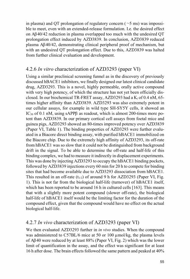
55
in plasma) and QT prolongation of regulatory concern (∼5 ms) was impossi-ble to meet, even with an extended-release formulation. I.e. the desired effect on Aβ40/42 reduction in plasma overlapped too much with the undesired QT prolongation effect induced by AZD3839. In conclusion, AZD3839 reduced plasma Aβ40/42, demonstrating clinical peripheral proof of mechanism, but with an undesired QT prolongation effect. Due to this, AZD3839 was halted from further clinical evaluation and development.
4.2.6 In vitro characterization of AZD3293 (paper VI) Using a similar preclinical screening funnel as in the discovery of previously discussed hBACE1 inhibitors, we finally designed our latest clinical candidate drug, AZD3293. This is a novel, highly permeable, orally active compound with very high potency, of which the structure has not yet been officially dis-closed. In our biochemical TR-FRET assay, AZD3293 had a Ki of 0.4 nM, 65-times higher affinity than AZD3839. AZD3293 was also extremely potent in our cellular assays, for example in wild type SH-SY5Y cells, it showed an IC50 of 0.1 nM, using sAPPβ as readout, which is almost 200-times more po-tent than AZD3839. In our primary cortical cell assays from foetal mice and guinea pigs, AZD3293 showed an 80-times improved potency over AZD3839 (Paper VI, Table 1). The binding properties of AZD3293 were further evalu-ated in a Biacore direct binding assay, with purified hBACE1 immobilized on the Biacore chip. Due to the extremely high affinity of AZD3293, its off-rate from hBACE1 was so slow that it could not be distinguished from background drift in the signal. To be able to determine the off-rate and half-life of this binding complex, we had to measure it indirectly in displacement experiments. This was done by injecting AZD3293 to occupy the hBACE1 binding pockets, followed by AZD3839 injections every 60 min for 20 h to compete for binding sites that had become available due to AZD3293 dissociation from hBACE1. This resulted in an off-rate (t1/2) of around 9 h for AZD3293 (Paper VI, Fig. 1). This is not far from the biological half-life (turnover) of hBACE1 itself, which has been reported to be around 16 h in cultured cells [163]. This means that with a slightly more potent compound (slower off-rate), the biological half-life of hBACE1 itself would be the limiting factor for the duration of the compound effect, given that the compound would have no effect on the actual biological half-life.
4.2.7 In vivo characterization of AZD3293 (paper VI) We then evaluated AZD3293 further in in vivo studies. When the compound was administered to C57BL/6 mice at 50 or 100 µmol/kg, the plasma levels of Aβ40 were reduced by at least 88% (Paper VI, Fig. 2) which was the lower limit of quantification in the assay, and the effect was significant for at least 16 h after dose. The brain effects followed the same pattern and peaked at 40%

56
Aβ40 reduction 3 h after dose, and were back to baseline after 16 h (Paper VI, Fig 2). Repeated dosing of the compound twice-daily for 7 days did not result in any additional effect, meaning that there is no accumulation of compound. In mice we calculated the IC50 for brain Aβ40 reduction to be 0.6 nM based on free AZD3293 concentration in brain. This is in exact agreement with the potency determined in cortical neurons (Paper VI, Table 1). In guinea pigs treated with a single oral dose of AZD3293 at 10 or 30 µmol/kg, the effect on plasma Aβ40 levels peaked at around 75% reduction, regardless of dose. The Aβ42 levels in plasma were reduced by at least 90% which was the lower limit of quantification in the assay. The plasma Aβ lowering effects were profound and significantly reduced up to 48 h after a single dose (Paper VI, Fig. 3). The Aβ40/42 effect in brain peaked at around 70% and 90% reduction at 10 and 30 µmol/kg respectively, and returned towards baseline 24 h after oral dose. The time and dose dependent reduction of Aβ40/42 in guinea pig CSF mir-rored the pattern observed in brain, with a slight time delay in effect (Paper VI, Fig. 4 & 5). This time delay seems rational since the CSF effects are down-stream from the brain effects. In guinea pigs we calculated the IC50 for brain Aβ40 reduction to be 0.9 nM based on free AZD3293 concentration in brain. This is in agreement with the IC50 determined in cortical neurons (0.3 nM). In beagle dogs, repeated daily doses of AZD3293 for 2 weeks resulted in a dose dependent reduction in hippocampal and CSF Aβ40 levels at 24 h after last oral dose. In beagle dogs we calculated the IC50 for brain Aβ40 reduction to be 0.8 nM based on free AZD3293 concentration in brain (Paper VI, Fig. 6). The plasma Aβ40 reduction in dogs was evaluated in a single dose time-re-sponse study at 12, 36, and 60 µmol/kg. It took somewhere between 1 and 2 weeks for the Aβ40 levels in plasma to return to baseline after a single dose, and a maximum reduction was reached at between 24 and 72 h depending on dose. The long lasting in vivo effects is most likely due to the compounds general high gastrointestinal permeability and extremely slow target off-rate. The bioavailability is especially high in dogs, resulting in even longer lasting effects in this particular species.
4.2.8 Target selectivity of AZD3293 (paper VI) AZD3293 is more or less equipotent against hBACE2, and the compound was further evaluated in a panel of more than 350 in vitro assays, resulting in a few hits but none with less than 1000-fold selectivity against hBACE1. In in vitro experiments AZD3293 showed effect against hERG in cellular models with an IC50 of 4.7 µM (Paper VI, Table 1). This is very close to what we had seen with AZD3839, and that eventually lead to its termination in the clinic. Since AZD3293 is a much more potent compound in terms of hBACE1 inhibition, much lower doses will be needed to get desired effects on Aβ reduction. Hence it will most likely be possible to find safe effective doses of AZD3293

57
that will not lead to any hERG mediated side effects in vivo or in potential clinical trials.
4.2.9 AZD3293 in clinical trials AZD3293 has been progressed into clinical trials. In phase I the compound reduced levels of amyloid beta in the CSF of people with AD and in healthy volunteers [164]. The compound was then progressed into a combined phase II/III study with 2200 patients with mild AD, to be enrolled for a two-year treatment period. The study reported encouraging positive safety data during spring 2016, and will now move on to pivotal phase III.

58
5. Conclusions
This thesis describes the characterization of novel Aβ plaque PET ligands for diagnosis of AD, and the characterization of novel BACE1 inhibitors for the potential treatment of AD. The main conclusions of this thesis, based on the findings in the six papers included, are described below.
• We have developed AZD2184 and AZD4694, two novel 11C and
18F-labeled Aβ plaque PET ligands with a higher signal-to-back-ground ratio compared to previously existing PET ligands. This was accomplished by screening for high target affinity, low lipophilicity, fast brain wash-out rate, and adequate brain exposure.
• We found that AZD2184, AZD4694 and other clinical Aβ plaque
PET ligands show lower recognition to amyloid deposits positive for ApoE. The amount of ApoE (regardless of isoform) incorporated to the Aβ plaques appeared to be of importance for the binding. From these findings we conclude that amyloid tracers used clinically today possibly have different and altered sensitivity to amyloid plaques de-pendent on the exact protein compositions of these deposits, thereby possibly underestimating the actual plaque load in some patient pop-ulations.
• We have developed AZD3839, AZ-4217, and AZD3293, three novel BACE1 inhibitors shown to be potent and selective. All three com-pounds exhibited dose- and time-dependent lowering of plasma, brain, and CSF Aβ levels in several species.
• We found a good correlation between the in vitro effects of our BACE1 inhibitors in primary cortical neurons and in vivo brain ef-fects in mouse and guinea pig, making it possible to predict in vivo effects and thereby reduce the number of animal studies.
• For AZD3839, we found an excellent agreement between the effects in our mouse and guinea pig in vivo models and the effects obtained in healthy human subjects, in terms of Aβ lowering in plasma, add-ing translational value to these in vivo models.

59
• We found that AZ-4217 lowered Aβ depositions in 12-month-old Tg2576 mice after 1 month treatment. This strongly supports BACE1 inhibition as concretely impacting amyloid deposition in AD, presenting a potentially important approach for therapeutic in-tervention.
• We observed that the slow off-rate of AZD3293 from the BACE1
active site in vitro, translated into a prolonged Aβ lowering effect in vivo beyond the plasma and CSF exposure of the compound.

60
6. Future perspectives
The major aim of this thesis was to characterize novel amyloid PET ligands that we previously developed for diagnosis of AD, and novel BACE1 inhibi-tors for the potential treatment of AD. The data presented in this thesis indi-cates that some of these novel compounds have a fair chance of clinically ac-complishing what they were originally designed to do. The amyloid PET ligands AZD2184 and AZD4694 were both evaluated in initial clinical studies with satisfying results. Currently, no activities are re-ported for AZD2184, but the 18F-labeled and more commercially attractive compound AZD4694, is currently being evaluated in Phase 2b and Phase 3 registration clinical trials [165]. If these are successful, this novel compound might soon be an available option for AD diagnostic centers around the world. An interesting option to small molecule PET ligands, are the emerging PET ligands based on antibodies. Normally, antibody uptake into the CNS is re-stricted by the BBB, but by conjugating the antibody, researchers recently re-ported they could increase the uptake across the BBB in a mouse model [166]. If this will be a clinical option in future remains to be seen, but it is a very interesting approach. One of the BACE1 inhibitors described in this thesis, AZD3293, is cur-rently in phase III clinical trials, and has been shown to reduce levels of Aβ in the cerebrospinal fluid of people with AD and in healthy volunteers. Recently the US Food and Drug Administration (FDA) announced they will give the development of AZD3293 a Fast Track designation. The FDA’s Fast Track program is designed to expedite the development and review of new therapies to treat serious conditions and tackle key unmet medical needs [167]. If this compound or any other BACE1 inhibitor will be successful in modifying AD and improve patients’ lives remains to be seen, but at least this compound has the potential to seriously challenge the amyloid hypothesis in AD.

61
7. Populärvetenskaplig sammanfattning på svenska
Alzheimers sjukdom är en mycket komplex sjukdom med dödlig utgång, och den vanligaste formen av demens. Sjukdomen kännetecknas av en progressiv försämring av kognition och minne. Med sjukdomen kommer också humör-svängningar och förvirring och med tiden får den drabbade allt svårare att klara sig själv. Idag beräknas över 35 miljoner människor världen över lida av Alzheimers sjukdom, och i dagsläget finns inga behandlingar som botar sjuk-domen, bara läkemedel som kortsiktigt kan lindra symptomen. I hjärnan kän-netecknas sjukdomen dels av ansamlingar, s.k. plack, utanför nervcellerna, bestående av det skadliga proteinet beta-amyloid (Aβ) och dels av nystan av neurofibriller inne i nervcellerna. Tillsammans bidrar detta så småningom till inflammation och massiv nervcellsdöd i hjärnan. Exakt hur dessa faktorer samverkar är inte klarlagt, men forskning visar att ansamlingar av plack kan bidra till förändringar av neurofibrillerna långt senare. Den kliniska diagnosen av Alzheimers sjukdom bygger idag främst på patientens bakgrund, minnes-tester, och uteslutande av andra neurologiska sjukdomar. I de tidiga stadierna av sjukdomen kan det vara svårt att ställa en korrekt diagnos, eftersom det är svårt att skilja sjukdomen från normala tecken på åldrande. Många forskare anser att en tidig och korrekt diagnos av sjukdomen kommer att vara en av nycklarna till att kunna bromsa sjukdomsförloppet i framtiden, förutsatt att mer effektiva behandlingar tas fram. Det finns således ett behov av känsliga diagnostiska verktyg i stånd att detektera tidiga och små förändringar i Alz-heimerpatienternas hjärnor. Ett sådant tillvägagångssätt är molekylär avbild-ning av Aβ-placken i hjärnan med hjälp av radioaktiva s.k. PET (positrone-missionstomografi) -ligander. Ett mål med denna avhandling var att utveckla och karaktärisera två nya PET-ligander (AZD2184 och AZD4694) med hög känslighet. AZD2184 är en 11C-märkt PET-ligand med ett högre signal-till-bakgrund förhållande jämfört med tidigare befintliga verktyg, något som gör det möjligt att upptäcka mindre och tidigare förändringar av plackansamlingar, och därmed teoretiskt också tidigare diagnos. Denna lägre grad av icke-specifik bindning för [11C]AZD2184 jämfört med tidigare existerande verktyg har också utvärderats kliniskt. En begränsning med 11C-märkta PET-ligander är den relativt korta halveringstiden (20 min), vilket innebär att en cyklotron för radioaktiv in-märkning av PET-liganderna måste finnas i omedelbar närhet till lokalen där

62
PET-undersökningen genomförs. För att möta behovet av PET-ligander med en längre halveringstid, utvecklade vi därför [18F]AZD4694, med en halve-ringstid på 110 minuter, vilket möjliggör att substansen kan märkas radioak-tivt på en plats och sedan distribueras regionalt under dagen till närliggande kliniker. Återigen utvecklades därför en PET-ligand med ett högre signal-till-bakgrund förhållande jämfört med tidigare existerande ligander, vilket också utvärderades kliniskt. Ackumuleringen av Aβ-plack i hjärnan, som ett resultat av förändrad pro-duktion eller nedbrytning av Aβ, spelar sannolikt en nyckelroll i sjukdomsför-loppet hos Alzheimerpatienter. Enzymet beta-sekretas (BACE1) svarar för det första steget i genereringen av Aβ, vilket gör BACE1 till ett attraktivt målpro-tein för potentiell behandling av Alzheimers sjukdom. Ett mål med denna av-handling var att utveckla och karaktärisera tre nya BACE1 hämmare, AZD3839, AZ-4217, och AZD3293. Alla tre substanser är potenta och selek-tiva hämmare av humant BACE1 med effekt i flera cellulära modeller, inklu-sive primära kortikala neuroner. Alla tre substanser uppvisade dos- och tids-beroende sänkning av Aβ i plasma, hjärna, och cerebrospinalvätska i flera ar-ter, och två av substanserna (AZD3839 och AZD3293) har tagits vidare till kliniska prövningar.

63
8. Acknowledgements
I would like to start by thanking my supervisor Anna-Lena Ström and my co-supervisor Kerstin Iverfeldt at the Department of Neurochemistry at Stockholm University, for taking on this project, investing a lot of time and effort, and guiding me through the PhD student jungle. I would also like to thank Ülo Langel at the same department for the initial interest in my PhD project and putting me in contact with Anna-Lena and Kerstin. Thank you Marie-Louise Tjörnhammar for helping me with the administrational mat-ters. A huge thank you to my other co-supervisor and long-lasting unofficial mentor Samuel Svensson, you are a true inspiration on so many different lev-els! There are a number of key people at AstraZeneca that deserves some extra attention; Mats Svensson, for believing in me and offering me my first job at Astra, back in 1998, the start of a great journey. Andreas Jaensson for 18 years (and counting) of sporadic lunches in various AstraZeneca canteens. I would also like to thank all my former colleagues at AstraZeneca R&D in Södertälje. Especially the project leader for the BACE project, Johanna Fält-ing, who really made me grow by investing a lot of trust in me, I am forever grateful for that! I would also like to thank my former managers Christin An-dersson and Mervi Vasänge, for your trust and constantly giving me new challenging projects and tasks. A big thank you to some of my former project team mates; Anders Juréus, Johan Sandell, Biljana Georgievska, and Tjerk Bueters, for sharing all your expertise and knowledge, and for being great colleagues and making all the long project meetings such a delight. I would also like to thank my friends at work, for all the lunches, dinners, cof-fees, beers, and making all the long hours in the lab enjoyable; Fredrik Ols-son, Björn Vilson, Kia Strömberg, Santiago Parpal, Tomas Borgegård, Johan Nord, Per-Olof Markgren, and Elisabet Venyike. Thank you Johan Lundkvist for all the ice hockey talk and sharing your AD expertise. I would also like to thank the whole BACE core team, together we made the impos-sible possible! In the world outside science, there are some fantastic people I would like to thank; Fredrik Magnusson for 35 fantastic years of friendship. Anders Stenström and Daniel Lundström for being such great friends for so many

64
years. My sister Annette, always an inspiration. Of course my mom and dad, Birgitta and Lars, for always being there for me and the whole family, and always going that extra mile! Finally, the biggest, warmest and deepest thanks to my wife Johanna and our sons Julian, Nils, and Ivar. Words are just not enough to express my love for you!! You are my heroes!!

65
9. References
1. DiMasi JA, Hansen RW, Grabowski HG. The price of innovation: new estimates of drug development costs. J Health Econ. 2003 Mar;22(2):151-85.
2. Dickson M, Gagnon JP. The cost of new drug discovery and develop-
ment. Discov Med. 2004 Jun;4(22):172-9.
3. Hughes JP, Rees S, Kalindjian SB, Philpott KL. Principles of early drug discovery. Br J Pharmacol. 2011 Mar;162(6):1239-49.
4. Landry Y, Gies JP. Drugs and their molecular targets: an updated over-view. Fundam Clin Pharmacol. 2008 Feb;22(1):1-18.
5. Cox JJ, Reimann F, Nicholas AK, Thornton G, Roberts E, Springell K, Karbani G, Jafri H, Mannan J, Raashid Y, Al-Gazali L, Hamamy H, Valente EM, Gorman S, Williams R, McHale DP, Wood JN, Gribble FM, Woods CG. An SCN9A channelopathy causes congenital inability to ex-perience pain. Nature. 2006 Dec 14;444(7121):894-8.
6. Bertram L, Tanzi RE. Thirty years of Alzheimer’s disease genetics: the implications of systematic meta-analyses. Nat Rev Neurosci. 2008 Oct;9(10):768-78.
7. Walters WP, Namchuk M. Designing screens: how to make your hits a hit. Nat Rev Drug Discov. 2003 Apr;2(4):259-66.
8. Bleicher KH, Böhm HJ, Müller K, Alanine AI. Hit and lead generation: beyond high-throughput screening. Nat Rev Drug Discov. 2003 May;2(5):369-78.
9. Lutz M, Kenakin T. Quantitative Molecular Pharmacology and Informat-ics in Drug Discovery. John Wiley & Sons, Ltd. West Sussex, England. 1999.
10. Pereira DA, Williams JA. Origin and evolution of high throughput screening. Br J Pharmacol. 2007 Sep;152(1):53-61.

66
11. Zhang JH, Chung TD, Oldenburg KR. A Simple Statistical Parameter for Use in Evaluation and Validation of High Throughput Screening Assays. J Biomol Screen. 1999;4(2):67-73.
12. Pellecchia M, Bertini I, Cowburn D, Dalvit C, Giralt E, Jahnke W, James TL, Homans SW, Kessler H, Luchinat C, Meyer B, Oschkinat H, Peng J, Schwalbe H, Siegal G. Perspectives on NMR in drug discovery: a tech-nique comes of age. Nat Rev Drug Discov. 2008 Sep;7(9):738-45.
13. Cheng KC, Korfmacher WA, White RE, Njoroge FG. Lead Optimization in Discovery Drug Metabolism and Pharmacokinetics/Case study: The Hepatitis C Virus (HCV) Protease Inhibitor SCH 503034. Perspect Medicin Chem. 2007 Jun 26;1:1-9.
14. DeMets D, Friedman L, Furberg C. Fundamentals of Clinical Trials (4th ed.). 2010 Springer.
15. www.fda.gov/ForPatients/Approvals/Drugs/ucm405622.htm#Clini-cal_Research_Phase_Studies. (Accessed 2016-11-20)
16. https://www.nlm.nih.gov/services/ctphases.html (Accessed 2016-11-20)
17. Querfurth HW, LaFerla FM. Alzheimer’s Disease. N Engl J Med. 2010 Jan 28;362(4):329-44.
18. Alzheimer’s Association. 2011 Alzheimer’s disease facts and figures. W V Med J. 2011 May-Jun;107(3):82-3.
19. Cummings JL. Alzheimer’s disease. N Engl J Med. 2004 Jul 1;351(1):56-67.
20. Ballard C1, Gauthier S, Corbett A, Brayne C, Aarsland D, Jones E. Alz-heimer’s disease. Lancet. 2011 Mar 19;377(9770):1019-31.
21. Citron M. Alzheimer’s disease: Strategies for disease modification. Nat Rev Drug Discov. 2010 May;9(5):387-98.
22. Förstl H, Kurz A. Clinical Features of Alzheimer's Disease. Eur Arch Psychiatry Clin Neurosci. 1999;249(6):288-90.
23. Selkoe DJ. Alzheimer's disease is a synaptic failure. Science. 2002 Oct 25;298(5594):789-91.
67
24. Serrano-Pozo A, Frosch MP, Masliah E, Hyman BT. Neuropathological alterations in Alzheimer disease. Cold Spring Harb Perspect Med. 2011 Sep;1(1):a006189.
25. Didic M, Barbeau EJ, Felician O, Tramoni E, Guedj E, Poncet M, Cec-caldi M. Which memory system is impaired first in Alzheimer's disease? J Alzheimers Dis. 2011;27(1):11-22.
26. Akiyama H, Barger S, Barnum S, Bradt B, Bauer J, Cole GM, Cooper NR, Eikelenboom P, Emmerling M, Fiebich BL, Finch CE, Frautschy S, Griffin WS, Hampel H, Hull M, Landreth G, Lue L, Mrak R, Mackenzie IR, McGeer PL, O'Banion MK, Pachter J, Pasinetti G, Plata-Salaman C, Rogers J, Rydel R, Shen Y, Streit W, Strohmeyer R, Tooyoma I, Van Muiswinkel FL, Veerhuis R, Walker D, Webster S, Wegrzyniak B, Wenk G, Wyss-Coray T. Inflammation and Alzheimer's disease. Neurobiol Ag-ing. 2000 May-Jun;21(3):383-421.
27. Cole SL, Vassar R. The Alzheimer's disease β-secretase enzyme, BACE1. Mol Neurodegener. 2007 Nov 15;2:22.
28. Braak H, Braak E. Neuropathological stageing of Alzheimer-related changes. Acta Neuropathol. 1991;82(4):239-59.
29. Zheng H, Koo EH. Biology and pathophysiology of the amyloid precur-sor protein. Mol Neurodegener. 2011 Apr 28;6(1):27.
30. Klevanski M, Herrmann U, Weyer SW, Fol R, Cartier N, Wolfer DP, Caldwell JH, Korte M, Müller UC. The APP Intracellular Domain Is Re-quired for Normal Synaptic Morphology, Synaptic Plasticity, and Hippo-campus-Dependent Behavior. J Neurosci. 2015 Dec 9;35(49):16018-33.
31. Vassar R, Kovacs DM, Yan R, Wong PC. The β-Secretase Enzyme BACE in Health and Alzheimer's Disease: Regulation, Cell Biology, Function, and Therapeutic Potential. J Neurosci. 2009 Oct 14;29(41):12787-94
32. Olsson F, Schmidt S, Althoff V, Munter LM, Jin S, Rosqvist S, Lendahl U, Multhaup G, Lundkvist J. Characterization of intermediate steps in amyloid beta (Aβ) production under near-native conditions. J Biol Chem. 2014 Jan 17;289(3):1540-50.
33. Haass C, Kaether C, Thinakaran G, Sisodia S. Trafficking and Proteolytic
Processing of APP. Cold Spring Harb Perspect Med. 2012 May;2(5):a006270.

68
34. Stockley JH, O'Neill C. The proteins BACE1 and BACE2 and beta-secre-tase activity in normal and Alzheimer's disease brain. Biochem Soc Trans. 2007 Jun;35(Pt 3):574-6.
35. LaFerla FM, Green KN, Oddo S. Intracellular amyloid-beta in Alzhei-mer's disease. Nat Rev Neurosci. 2007 Jul;8(7):499-509.
36. Parvathy S1, Davies P, Haroutunian V, Purohit DP, Davis KL, Mohs RC, Park H, Moran TM, Chan JY, Buxbaum JD. Correlation between Abetax-40-, Abetax-42-, and Abetax-43-containing amyloid plaques and cogni-tive decline. Arch Neurol. 2001 Dec;58(12):2025-32.
37. Kurz A, Perneczky R. Amyloid clearance as a treatment target against Alzheimer's disease. J Alzheimers Dis. 2011;24 Suppl 2:61-73.
38. Hardy JA, Higgins GA. Alzheimer's disease: the amyloid cascade hy-pothesis. Science. 1992 Apr 10;256(5054):184-5.
39. Musiek ES, Holtzman DM. Three dimensions of the amyloid hypothesis: time, space and ‘wingmen’. Nat Neurosci. 2015 Jun;18(6):800-6.
40. Gómez-Isla T, Hollister R, West H, Mui S, Growdon JH, Petersen RC, Parisi JE, Hyman BT. Neuronal loss correlates with but exceeds neurofi-brillary tangles in Alzheimer’s disease. Ann Neurol. 1997 Jan;41(1):17-24.
41. Arriagada PV, Marzloff K, Hyman BT. Distribution of Alzheimer-type pathologic changes in nondemented elderly individuals matches the pat-tern in Alzheimer’s disease. Neurology. 1992 Sep;42(9):1681-8.
42. Arriagada PV, Growdon JH, Hedley-Whyte ET, Hyman BT. Neurofibril-lary tangles but not senile plaques parallel duration and severity of Alz-heimer’s disease. Neurology. 1992 Mar;42(3 Pt 1):631-9.
43. Price JL, Morris JC. Tangles and plaques in nondemented aging and “pre-clinical” Alzheimer’s disease. Ann Neurol. 1999 Mar;45(3):358-68.
44. Rapoport M, Dawson HN, Binder LI, Vitek MP, Ferreira A. Tau is es-sential to beta -amyloid-induced neurotoxicity. Proc Natl Acad Sci U S A. 2002 Apr 30;99(9):6364-9.

69
45. Choi SH, Kim YH, Hebisch M, Sliwinski C, Lee S, D'Avanzo C, Chen H, Hooli B, Asselin C, Muffat J, Klee JB, Zhang C, Wainger BJ, Peitz M, Kovacs DM, Woolf CJ, Wagner SL, Tanzi RE, Kim DY. A three-dimensional human neural cell culture model of Alzheimer’s disease. Na-ture. 2014 Nov 13;515(7526):274-8.
46. Nath S, Agholme L, Kurudenkandy FR, Granseth B, Marcusson J, Hall-beck M. Spreading of neurodegenerative pathology via neuron-to-neuron transmission of β-amyloid. J Neurosci. (2012) Jun 27;32(26):8767-77.
47. Domert J, Rao SB, Agholme L, Brorsson AC, Marcusson J, Hallbeck M, Nath S. Spreading of amyloid-β peptides via neuritic cell-to-cell transfer is dependent on insufficient cellular clearance. Neurobiol Dis. 2014 May;65:82-92.
48. Sevigny J, Chiao P, Bussière T, Weinreb PH, Williams L, Maier M, Dunstan R, Salloway S, Chen T, Ling Y, O'Gorman J, Qian F, Arastu M, Li M, Chollate S, Brennan MS, Quintero-Monzon O, Scannevin RH, Ar-nold HM, Engber T, Rhodes K, Ferrero J, Hang Y, Mikulskis A, Grimm J, Hock C, Nitsch RM, Sandrock A. The antibody aducanumab reduces Aβ plaques in Alzheimer's disease. Nature. 2016 Aug 31;537(7618):50-6.
49. Vassar R. BACE1 inhibitor drugs in clinical trials for Alzheimer’s dis-ease. Alzheimers Res Ther. 2014 Dec 24;6(9):89.
50. Kandalepas PC, Sadleir KR, Eimer WA, Zhao J, Nicholson DA, Vassar R. The Alzheimer’s β‑secretase BACE1 localizes to normal presynaptic terminals and to dystrophic presynaptic terminals surrounding amyloid plaques. Acta Neuropathol. 2013 Sep;126(3):329-52.
51. Haniu M, Denis P, Young Y, Mendiaz EA, Fuller J, Hui JO, Bennett BD, Kahn S, Ross S, Burgess T, Katta V, Rogers G, Vassar R, Citron M. Characterization of Alzheimer's beta-secretase protein BACE. A pepsin family member with unusual properties. J Biol Chem. 2000 Jul 14;275(28):21099-106.
52. Costantini C, Ko MH, Jonas MC, Puglielli L. A reversible form of lysine acetylation in the ER and Golgi lumen controls the molecular stabiliza-tion of BACE1. Biochem J. 2007 Nov 1;407(3):383-95.

70
53. Huse JT, Pijak DS, Leslie GJ, Lee VM, Doms RW. Maturation and en-dosomal targeting of beta-site amyloid precursor protein-cleaving en-zyme. The Alzheimer's disease beta-secretase. J Biol Chem. 2000 Oct 27;275(43):33729-37.
54. Walter J, Fluhrer R, Hartung B, Willem M, Kaether C, Capell A, Lam-mich S, Multhaup G, Haass C. Phosphorylation regulates intracellular trafficking of β-secretase. J Biol Chem. 2001 May 4;276(18):14634-41.
55. Sandoval IV, Bakke O. Targeting of membrane proteins to endosomes and lysosomes. Trends Cell Biol. 1994 Aug;4(8):292-7.
56. Vetrivel KS, Meckler X, Chen Y, Nguyen PD, Seidah NG, Vassar R, Wong PC, Fukata M, Kounnas MZ, Thinakaran G. Alzheimer disease Abeta production in the absence of S-palmitoylation-dependent targeting of BACE1 to lipid rafts. J Biol Chem. 2009 Feb 6;284(6):3793-803.
57. He W, Lu Y, Qahwash I, Hu XY, Chang A, Yan R. Reticulon family members modulate BACE1 activity and amyloid-beta peptide genera-tion. Nat Med. 2004 Sep;10(9):959-65.
58. Murayama KS, Kametani F, Saito S, Kume H, Akiyama H, Araki W. Reticulons RTN3 and RTN4-B/C interact with BACE1 and inhibit its ability to produce amyloid beta-protein. Eur J Neurosci. 2006 Sep;24(5):1237-44.
59. Shi Q, Prior M, He W, Tang X, Hu X, Yan R. Reduced amyloid deposi-tion in mice overexpressing RTN3 is adversely affected by preformed dystrophic neurites. J Neurosci. 2009 Jul 22;29(29):9163-73.
60. He X, Chang WP, Koelsch G, Tang J. Memapsin 2 (beta-secretase) cyto-solic domain binds to the VHS domains of GGA1 and GGA2: implica-tions on the endocytosis mechanism of memapsin 2. FEBS Lett. 2002 Jul 31;524(1-3):183-7.
61. Gosh P, Kornfeldt S. The GGA proteins: key players in protein sorting at the trans-Golgi network. Eur J Cell Biol. 2004 Jul;83(6):257-62.
62. Fukumoto H, Cheung BS, Hyman BT, Irizarry MC. Beta-secretase pro-tein and activity are increased in the neocortex in Alzheimer disease. Arch Neurol. 2002 Sep;59(9):1381-9.

71
63. Velliquette RA, O'Connor T, Vassar R. Energy inhibition elevates β-secretase levels and activity and is potentially amyloidogenic in APP transgenic mice: possible early events in Alzheimer's disease pathogene-sis. J Neurosci. 2005 Nov 23;25(47):10874-83.
64. O'Connor T, Sadleir KR, Maus E, Velliquette RA, Zhao J, Cole SL, Eimer WA, Hitt B, Bembinster LA, Lammich S, Lichtenthaler SF, Hebert SS, De Strooper B, Haass C, Bennett DA, Vassar R. Phosphorylation of the translation initiation factor eIF2alpha increases BACE1 levels and promotes amyloidogenesis. Neuron. 2008 Dec 26;60(6):988-1009.
65. Kim DY, Ingano LA, Carey BW, Pettingell WH, Kovacs DM. Prese-nilin/gamma-secretase-mediated cleavage of the voltage-gated sodium channel beta2-subunit regulates cell adhesion and migration. J Biol Chem. 2005 Jun 17;280(24):23251-61.
66. Wong HK, Sakurai T, Oyama F, Kaneko K, Wada K, Miyazaki H, Ku-rosawa M, De Strooper B, Saftig P, Nukina N. beta Subunits of voltage-gated sodium channels are novel substrates of beta-site amyloid precursor protein-cleaving enzyme (BACE1) and gamma-secretase. J Biol Chem. 2005 Jun 17;280(24):23009-17.
67. Willem M, Garratt AN, Novak B, Citron M, Kaufmann S, Rittger A, De-Strooper B, Saftig P, Birchmeier C, Haass C. Control of peripheral nerve myelination by the beta-secretase BACE1. Science. 2006 Oct 27;314(5799):664-6.
68. Stefansson H, Sigurdsson E, Steinthorsdottir V, Bjornsdottir S, Sig-mundsson T, Ghosh S, Brynjolfsson J, Gunnarsdottir S, Ivarsson O, Chou TT, Hjaltason O, Birgisdottir B, Jonsson H, Gudnadottir VG, Gud-mundsdottir E, Bjornsson A, Ingvarsson B, Ingason A, Sigfusson S, Hardardottir H, Harvey RP, Lai D, Zhou M, Brunner D, Mutel V, Gon-zalo A, Lemke G, Sainz J, Johannesson G, Andresson T, Gudbjartsson D, Manolescu A, Frigge ML, Gurney ME, Kong A, Gulcher JR, Peturs-son H, Stefansson K. Neuregulin 1 and susceptibility to schizophrenia. Am J Hum Genet. 2002 Oct;71(4):877-92.
69. Harrison SM, Harper AJ, Hawkins J, Duddy G, Grau E, Pugh PL, Winter PH, Shilliam CS, Hughes ZA, Dawson LA, Gonzalez MI, Upton N, Pan-galos MN, Dingwall C. BACE1 (beta-secretase) transgenic and knockout mice: identification of neurochemical deficits and behavioral changes. Mol Cell Neurosci. 2003 Nov;24(3):646-55.

72
70. Chen C, Bharucha V, Chen Y, Westenbroek RE, Brown A, Malhotra JD, Jones D, Avery C, Gillespie PJ 3rd, Kazen-Gillespie KA, Kazarinova-Noyes K, Shrager P, Saunders TL, Macdonald RL, Ransom BR, Scheuer T, Catterall WA, Isom LL. Reduced sodium channel density, altered volt-age dependence of inactivation, and increased susceptibility to seizures in mice lacking sodium channel beta 2-subunits. Proc Natl Acad Sci U S A. 2002 Dec 24;99(26):17072-7.
71. Lopez-Santiago LF, Pertin M, Morisod X, Chen C, Hong S, Wiley J, De-costerd I, Isom LL. Sodium channel β2 subunits regulate tetrodotoxin-sensitive sodium channels in small dorsal root ganglion neurons and modulate the response to pain. J Neurosci. 2006 Jul 26;26(30):7984-94.
72. Strömberg K, Eketjäll S, Georgievska B, Tunblad K, Eliason K, Olsson F, Radesäter AC, Klintenberg R, Arvidsson PI, von Berg S, Fälting J, Cowburn RF, Dabrowski M. Combining an amyloid-beta (Aβ) cleaving enzyme inhibitor with a γ-secretase modulator results in an additive re-duction of Aβ production. FEBS J. 2015 Jan;282(1):65-73.
73. Levy E, Carman MD, Fernandez-Madrid IJ, Power MD, Lieberburg I, van Duinen SG, Bots GT, Luyendijk W, Frangione B. Mutation of the Alzheimer’s disease amyloid gene in hereditary cerebral hemorrhage, Dutch type. Science. 1990 Jun 1;248(4959):1124-6.
74. Bertram L, Tanzi RE. The genetic epidemiology of neurodegenerative disease. J Clin Invest. 2005 Jun;115(6):1449-57.
75. Citron M, Oltersdorf T, Haass C, McConlogue L, Hung AY, Seubert P, Vigo-Pelfrey C, Lieberburg I, Selkoe DJ. Mutation of the beta-amyloid precursor protein in familial Alzheimer’s disease increases beta-protein production. Nature. 1992 Dec 17;360(6405):672-4.
76. Prasher VP, Farrer MJ, Kessling AM, Fisher EM, West RJ, Barber PC, Butler AC. Molecular mapping of Alzheimer-type dementia in Down’s syndrome. Ann Neurol. 1998 Mar;43(3):380-3.
77. Tsubuki S, Takaki Y, Saido TC. Dutch, Flemish, Italian, and Arctic mu-tations of APP and resistance of Abeta to physiologically relevant prote-olytic degradation. Lancet. 2003 Jun 7;361(9373):1957-8.

73
78. Tomiyama T, Nagata T, Shimada H, Teraoka R, Fukushima A, Kanemitsu H, Takuma H, Kuwano R, Imagawa M, Ataka S, Wada Y, Yoshioka E, Nishizaki T, Watanabe Y, Mori H. A new amyloid beta var-iant favoring oligomerization in Alzheimer’s-type dementia. Ann Neurol. 2008 Mar;63(3):377-87.
79. Shepherd C, McCann H, Halliday GM. Variations in the neuropathology of familial Alzheimer’s disease. Acta Neuropathol. 2009 Jul;118(1):37-52.
80. Bateman RJ, Xiong C, Benzinger TL, Fagan AM, Goate A, Fox NC, Mar-cus DS, Cairns NJ, Xie X, Blazey TM, Holtzman DM, Santacruz A, Buckles V, Oliver A, Moulder K, Aisen PS, Ghetti B, Klunk WE, McDade E, Martins RN, Masters CL, Mayeux R, Ringman JM, Rossor MN, Schofield PR, Sperling RA, Salloway S, Morris JC. Clinical and biomarker changes in dominantly inherited Alzheimer’s disease. N Engl J Med. 2012 Aug 30;367(9):795-804.
81. Ryman DC, Acosta-Baena N, Aisen PS, Bird T, Danek A, Fox NC, Goate A, Frommelt P, Ghetti B, Langbaum JB, Lopera F, Martins R, Masters CL, Mayeux RP, McDade E, Moreno S, Reiman EM, Ringman JM, Sal-loway S, Schofield PR, Sperling R, Tariot PN, Xiong C, Morris JC, Bate-man RJ. Symptom onset in autosomal dominant Alzheimer disease: a systematic review and meta-analysis. Neurology. 2014 Jul 15;83(3):253-60.
82. Corder EH, Saunders AM, Strittmatter WJ, Schmechel DE, Gaskell PC, Small GW, Roses AD, Haines JL, Pericak-Vance MA. Gene dose of apolipoprotein E type 4 allele and the risk of Alzheimer’s disease in late onset families. Science. 1993 Aug 13;261(5123):921-3.
83. Genin E, Hannequin D, Wallon D, Sleegers K, Hiltunen M, Combarros O, Bullido MJ, Engelborghs S, De Deyn P, Berr C, Pasquier F, Dubois B, Tognoni G, Fiévet N, Brouwers N, Bettens K, Arosio B, Coto E, Del Zompo M, Mateo I, Epelbaum J, Frank-Garcia A, Helisalmi S, Porcellini E, Pilotto A, Forti P, Ferri R, Scarpini E, Siciliano G, Solfrizzi V, Sorbi S, Spalletta G, Valdivieso F, Vepsäläinen S, Alvarez V, Bosco P, Man-cuso M, Panza F, Nacmias B, Bossù P, Hanon O, Piccardi P, Annoni G, Seripa D, Galimberti D, Licastro F, Soininen H, Dartigues JF, Kamboh MI, Van Broeckhoven C, Lambert JC, Amouyel P, Campion D. APOE and Alzheimer disease: a major gene with semi-dominant inheritance. Mol Psychiatry. 2011 Sep;16(9):903-7.

74
84. Verghese PB, Castellano JM, Garai K, Wang Y, Jiang H, Shah A, Bu G, Frieden C, Holtzman DM. ApoE influences amyloid-β (Aβ) clearance despite minimal apoE/Aβ association in physiological conditions. Proc Natl Acad Sci U S A. 2013 May 7;110(19):E1807-16.
85. Liu CC, Kanekiyo T, Xu H, Bu G. Apolipoprotein E and Alzheimer dis-ease: risk, mechanisms, and therapy. Nat Rev Neurol. 2013 Feb;9(2):106-18.
86. Jonsson T, Atwal JK, Steinberg S, Snaedal J, Jonsson PV, Bjornsson S, Stefansson H, Sulem P, Gudbjartsson D, Maloney J, Hoyte K, Gustafson A, Liu Y, Lu Y, Bhangale T, Graham RR, Huttenlocher J, Bjornsdottir G, Andreassen OA, Jönsson EG, Palotie A, Behrens TW, Magnusson OT, Kong A, Thorsteinsdottir U, Watts RJ, Stefansson K. A mutation in APP protects against Alzheimer’s disease and age-related cognitive decline. Nature. 2012 Aug 2;488(7409):96-9.
87. McKhann GM, Knopman DS, Chertkow H, Hyman BT, Jack CR Jr, Ka-was CH, Klunk WE, Koroshetz WJ, Manly JJ, Mayeux R, Mohs RC, Morris JC, Rossor MN, Scheltens P, Carrillo MC, Thies B, Weintraub S, Phelps CH. The diagnosis of dementia due to Alzheimer's disease: Rec-ommendations from the National Institute on Aging-Alzheimer's Associ-ation workgroups on diagnostic guidelines for Alzheimer's disease. Alz-heimers Dement. 2011 May;7(3):263-9.
88. Petersen RC, Negash S. Mild cognitive impairment: an overview. CNS Spectr. 2008 Jan;13(1):45-53.
89. Dubois B, Feldman HH, Jacova C, Dekosky ST, Barberger-Gateau P, Cummings J, Delacourte A, Galasko D, Gauthier S, Jicha G, Meguro K, O'brien J, Pasquier F, Robert P, Rossor M, Salloway S, Stern Y, Visser PJ, Scheltens P. Research criteria for the diagnosis of Alzheimer's dis-ease: revising the NINCDS-ADRDA criteria. Lancet Neurol. 2007 Aug;6(8):734-46.
90. Jack CR Jr, Knopman DS, Jagust WJ, Shaw LM, Aisen PS, Weiner MW, Petersen RC, Trojanowski JQ. Hypothetical model of dynamic bi-omarkers of the Alzheimer's pathological cascade. Lancet Neurol. 2010 Jan;9(1):119-28.
91. Blennow K, Hampel H, Weiner M, Zetterberg H. Cerebrospinal fluid and plasma biomarkers in Alzheimer disease. Nat Rev Neurol. 2010 Mar;6(3):131-44.

75
92. Jack CR Jr, Vemuri P, Wiste HJ, Weigand SD, Aisen PS, Trojanowski JQ, Shaw LM, Bernstein MA, Petersen RC, Weiner MW, Knopman DS. Evidence for ordering of Alzheimer disease biomarkers. Arch Neurol. 2011 Dec;68(12):1526-35.
93. Mathis CA, Lopresti BJ, Klunk WE. Impact of amyloid imaging on drug development in Alzheimer's disease. Nucl Med Biol. 2007 Oct;34(7):809-22.
94. Nordberg A. Amyloid imaging in Alzheimer's disease. Curr Opin Neurol. 2007 Aug;20(4):398-402.
95. Klunk WE. Amyloid imaging as a biomarker for cerebral β-amyloidosis and risk prediction for Alzheimer dementia. Neurobiol Aging. 2011 Dec;32 Suppl 1:S20-36.
96. Klunk WE, Engler H, Nordberg A, Wang Y, Blomqvist G, Holt DP, Bergström M, Savitcheva I, Huang GF, Estrada S, Ausén B, Debnath ML, Barletta J, Price JC, Sandell J, Lopresti BJ, Wall A, Koivisto P, An-toni G, Mathis CA, Långström B. Imaging brain amyloid in Alzheimer's disease with Pittsburgh Compound-B. Ann Neurol. 2004 Mar;55(3):306-19.
97. Forsberg A, Engler H, Almkvist O, Blomquist G, Hagman G, Wall A, Ringheim A, Långström B, Nordberg A. PET imaging of amyloid depo-sition in patients with mild cognitive impairment. Neurobiol Aging. 2008 Oct;29(10):1456-65.
98. Pike KE, Savage G, Villemagne VL, Ng S, Moss SA, Maruff P, Mathis CA, Klunk WE, Masters CL, Rowe CC. Beta-amyloid imaging and memory in non-demented individuals: evidence for preclinical Alzhei-mer's disease. Brain. 2007 Nov;130(Pt 11):2837-44.
99. Mayeux R, Saunders AM, Shea S, Mirra S, Evans D, Roses AD, Hyman BT, Crain B, Tang MX, Phelps CH. Utility of the apolipoprotein E gen-otype in the diagnosis of Alzheimer's disease. Alzheimer's Disease Cen-ters Consortium on Apolipoprotein E and Alzheimer's Disease. N Engl J Med. 1998 Feb 19;338(8):506-11.
100. Karran E, Hardy J. A Critique of the Drug Discovery and Phase 3 Clin-ical Programs Targeting the Amyloid Hypothesis for Alzheimer Disease. Ann Neurol. 2014 Aug;76(2):185-205.

76
101. Klunk WE, Lopresti BJ, Ikonomovic MD, Lefterov IM, Koldamova RP, Abrahamson EE, Debnath ML, Holt DP, Huang GF, Shao L, DeKosky ST, Price JC, Mathis CA. Binding of the positron emission tomography tracer Pittsburgh compound-B reflects the amount of amyloid-beta in Alzheimer's disease brain but not in transgenic mouse brain. J Neurosci. 2005 Nov 16;25(46):10598-606.
102. Maeda J, Ji B, Irie T, Tomiyama T, Maruyama M, Okauchi T, Stau-fenbiel M, Iwata N, Ono M, Saido TC, Suzuki K, Mori H, Higuchi M, Suhara T. Longitudinal, quantitative assessment of amyloid, neuroin-flammation, and anti-amyloid treatment in a living mouse model of Alz-heimer's disease enabled by positron emission tomography. J Neurosci. 2007 Oct 10;27(41):10957-68.
103. Hampel H, Prvulovic D, Teipel S, Jessen F, Luckhaus C, Frölich L, Riepe MW, Dodel R, Leyhe T, Bertram L, Hoffmann W, Faltraco F. The future of Alzheimer's disease: the next 10 years. Prog Neurobiol. 2011 Dec;95(4):718-28.
104. Schneider LS, Insel PS, Weiner MW. Treatment with cholinesterase in-hibitors and memantine of patients in the Alzheimer's Disease Neuroim-aging Initiative. Arch Neurol. 2011 Jan;68(1):58-66.
105. Frölich L. The cholinergic pathology in Alzheimer's disease--discrepan-cies between clinical experience and pathophysiological findings. J Neu-ral Transm (Vienna). 2002 Jul;109(7-8):1003-13.
106. Schneider LS, Dagerman KS, Higgins JP, McShane R. Lack of evidence for the efficacy of memantine in mild Alzheimer disease. Arch Neurol. 2011 Aug;68(8):991-8.
107. Feldman HH, Ferris S, Winblad B, Sfikas N, Mancione L, He Y, Tekin S, Burns A, Cummings J, del Ser T, Inzitari D, Orgogozo JM, Sauer H, Scheltens P, Scarpini E, Herrmann N, Farlow M, Potkin S, Charles HC, Fox NC, Lane R. Effect of rivastigmine on delay to diagnosis of Alzhei-mer's disease from mild cognitive impairment: the InDDEx study. Lancet Neurol. 2007 Jun;6(6):501-12.
108. Ji W, Ha I. Drug development for Alzheimer's disease: recent progress. Exp Neurobiol. 2010 Dec;19(3):120-31.
109. Schor NF. What the halted phase III γ-secretase inhibitor trial may (or may not) be telling us. Ann Neurol. 2011 Feb;69(2):237-9.

77
110. Biedler JL, Helson L, Spengler BA Morphology and growth, tumorigen-icity, and cytogenetics of human neuroblastoma cells in continuous cul-ture. Cancer Res. 1973 Nov;33(11):2643-52.
111. Biedler JL, Roffler-Tarlov S, Schachner M, Freedman LS. Multiple neu-rotransmitter synthesis by human neuroblastoma cell lines and clones. Cancer Res. 1978 Nov;38(11 Pt 1):3751-7.
112. Ciccarone V, Spengler BA, Meyers MB, Biedler JL, Ross RA. Pheno-typic diversification in human neuroblastoma cells: expression of distinct neural crest lineages. Cancer Res. 1989 Jan 1;49(1):219-25.
113. Salto R, Vílchez JD, Girón MD, Cabrera E, Campos N, Manzano M, Rueda R, López-Pedrosa JM. β-Hydroxy-β-Methylbutyrate (HMB) Pro-motes Neurite Outgrowth in Neuro2a Cells. PLoS One. 2015 Aug 12;10(8):e0135614.
114. LePage KT, Dickey RW, Gerwick WH, Jester EL, Murray TF. On the use of neuro-2a neuroblastoma cells versus intact neurons in primary cul-ture for neurotoxicity studies. Crit Rev Neurobiol. 2005;17(1):27-50.
115. www.hek293.com (Accessed 2016-09-12)
116. Sciarretta C, Minichiello L. The Preparation of Primary Cortical Neuron Cultures and a Practical Application Using Immunofluorescent Cyto-chemistry. Methods Mol Biol. 2010;633:221-31.
117. Piston DW, Kremers GJ. Fluorescent protein FRET: the good, the bad
and the ugly. Trends Biochem Sci. 2007 Sep;32(9):407-14.
118. Degorce F, Card A, Soh S, Trinquet E, Knapik GP, Xie B. HTRF: A Technology Tailored for Drug Discovery –A Review of Theoretical As-pects and Recent Applications. Curr Chem Genomics. 2009 May 28;3:22-32.
119. https://www.thermofisher.com/uk/en/home/industrial/pharma-bio-pharma/drug-discovery-development/target-and-lead-identification-and-validation/kinasebiology/kinase-activity-assays/lanthascreen-tr-fret-toolbox/lanthascreen-tr-fret-technology-overview.html# (Accessed 2016-11-20)
120. Zeng S, Baillargeat D, Ho HP, Yong KT. Nanomaterials enhanced sur-face plasmon resonance for biological and chemical sensing applications. Chem Soc Rev. 2014 May 21;43(10):3426-52.
78
121. Klunk WE, Wang Y, Huang GF, Debnath ML, Holt DP, Mathis CA. Uncharged thioflavin-T derivates bind to amyloid-beta protein with high affinity and readily enter the brain. Life Sci. 2001 Aug 17;69(13):1471-84.
122. Necula M, Kayed R, Milton S, Glabe CG. Small molecule inhibitors of aggregation indicate that amyloid b oligomerization and fibrillization pathways are independent and distinct. J Biol Chem. 2007 Apr 6;282(14):10311-24.
123. Nilsson MR. Techniques to study amyloid fibril formation in vitro. Methods. 2004 Sep;34(1):151-60.
124. Maguire JJ, Kuc RE, Davenport AP. Radioligand binding assays and their analysis. Methods Mol Biol. 2012;897:31-77.
125. http://www.graphpad.com/guides/prism/6/curvefitting/in-dex.htm?reg_key_concepts_law_of_mass_actio.htm (Accessed 2016-10-12)
126. Motulsky HJ, Mahan LC. The Kinetics of Competitive Radioligand Binding Predicted by the Law of Mass Action. Mol Pharmacol. 1984 Jan;25(1):1-9.
127. Auld DS, Farmen MW, Kahl SD, Kriauciunas A, McKnight KL, Mont-rose C, Weidner JR. Receptor Binding Assays for HTS and Drug Discov-ery. Assay Guidance Manual, Eli Lilly & Company, Indianapolis, IN, 2012
128. Young WS 3rd, Kuhar MJ. A new method for receptor autoradiography: [3H]opioid receptors in rat brain. Brain Res. 1979 Dec 28;179(2):255-70.
129. Solon EG. Autoradiography: high-resolution molecular imaging in pharmaceutical discovery and development. Expert Opin Drug Discov. 2007 Apr;2(4):503-14.
130. Duraiyan J, Govindarajan R, Kaliyappan K, Palanisamy M. Applica-tions of immunohistochemistry. J Pharm Bioallied Sci. 2012 Aug;4(Suppl 2):S307-9.
131. Chen X, Cho DB, Yang PC. Double staining immunohistochemistry. N Am J Med Sci. 2010 May;2(5):241-5.

79
132. Engber D. The Trouble With Black-6: A tiny alcoholic takes over the lab. Slate.com, 2011.
133. Hsiao K, Chapman P, Nilsen S, Eckman C, Harigaya Y, Younkin S, Yang F, Cole G. Correlative memory deficits, Abeta elevation, and am-yloid plaques in transgenic mice. Science. 1996 Oct 4;274(5284):99-102.
134. Irizarry MC, McNamara M, Fedorchak K, Hsiao K, Hyman BT. APPSw transgenic mice develop age-related A beta deposits and neuropil abnor-malities, but no neuronal loss in CA1. J Neuropathol Exp Neurol. 1997 Sep;56(9):965-73.
135. Lanz TA, Carter DB, Merchant KM. Dendritic spine loss in the hippo-campus of young PDAPP and Tg2576 mice and its prevention by the ApoE2 genotype. Neurobiol Dis. 2003 Aug;13(3):246-53.
136. Anderson JJ, Holtz G, Baskin PP, Turner M, Rowe B, Wang B, Kounnas MZ, Lamb BT, Barten D, Felsenstein K, McDonald I, Srinivasan K, Munoz B, Wagner SL. Reductions in beta-amyloid concentrations in vivo by the gamma-secretase inhibitors BMS-289948 and BMS-299897. Bio-chem Pharmacol. 2005 Feb 15;69(4):689-98.
137. Lanz TA, Karmilowicz MJ, Wood KM, Pozdnyakov N, Du P, Pi-otrowski MA, Brown TM, Nolan CE, Richter KE, Finley JE, Fei Q, Eb-binghaus CF, Chen YL, Spracklin DK, Tate B, Geoghegan KF, Lau LF, Auperin DD, Schachter JB. Concentration-dependent modulation of am-yloid-beta in vivo and in vitro using the gamma-secretase inhibitor, LY-450139. J Pharmacol Exp Ther. 2006 Nov;319(2):924-33.
138. Swahn BM, Wensbo D, Sandell J, Sohn D, Slivo C, Pyring D, Malm-ström J, Arzel E, Vallin M, Bergh M, Jeppsson F, Johnson AE, Juréus A, Neelissen J, Svensson S. Synthesis and evaluation of 2-pyridylbenzothi-azole, 2-pyridylbenzoxazole and 2-pyridylbenzofuran derivatives as 11C-PET imaging agents for beta-amyloid plaques. Bioorg Med Chem Lett. 2010 Mar 15;20(6):1976-80.
139. Di L, Kernsy EH. Profiling drug-like properties in discovery research. Curr Opin Chem Biol. 2003 Jun;7(3):402-8.
140. Lombardo F, Shalaeva MY, Tupper KA, Gao F. ElogD(oct): a tool for lipophilicity determination in drug discovery. 2. Basic and neutral com-pounds. J Med Chem. 2001 Jul 19;44(15):2490-7.

80
141. Swahn BM, Sandell J, Pyring D, Bergh M, Jeppsson F, Juréus A, Neel-issen J, Johnström P, Schou M, Svensson S. Synthesis and evaluation of pyridylbenzofuran, pyridylbenzothiazole and pyridylbenzoxazole deriv-atives as ¹⁸F-PET imaging agents for β-amyloid plaques. Bioorg Med Chem Lett. 2012 Jul 1;22(13):4332-7.
142. Lockhart A, Lamb JR, Osredkar T, Sue LI, Joyce JN, Ye L, Libri V, Leppert D, Beach TG. PIB is a non-specific imaging marker of amyloid-beta (Abeta) peptide-related cerebral amyloidosis. Brain. 2007 Oct;130(Pt 10):2607-15.
143. Ikonomovic MD, Klunk WE, Abrahamson EE, Mathis CA, Price JC, Tsopelas ND, Lopresti BJ, Ziolko S, Bi W, Paljug WR, Debnath ML, Hope CE, Isanski BA, Hamilton RL, DeKosky ST. Postmortem corre-lates of in vivo PiB-PET amyloid imaging in a typical case of Alzheimer’s disease. Brain. 2008 Jun;131(Pt 6):1630-45.
144. Nyberg S, Jönhagen ME, Cselényi Z, Halldin C, Julin P, Olsson H, Freund-Levi Y, Andersson J, Varnäs K, Svensson S, Farde L. Detection of amyloid in Alzheimer’s disease with positron emission tomography using [11C]AZD2184. Eur J Nucl Med Mol Imaging. 2009 Nov;36(11):1859-63.
145. Forsberg A, Juréus A, Cselényi Z, Eriksdotter M, Freund-Levi Y, Jepps-son F, Swahn BM, Sandell J, Julin P, Schou M, Andersson J, Johnström P, Varnäs K, Halldin C, Farde L, Svensson S. Low background and high contrast PET imaging of amyloid-β with [11C]AZD2995 and [11C]AZD2184 in Alzheimer’s disease patients. Eur J Nucl Med Mol Im-aging. 2013 Apr;40(4):580-93.
146. Ito H, Shimada H, Shinotoh H, Takano H, Sasaki T, Nogami T, Suzuki M, Nagashima T, Takahata K, Seki C, Kodaka F, Eguchi Y, Fujiwara H, Kimura Y, Hirano S, Ikoma Y, Higuchi M, Kawamura K, Fukumura T, Böö ÉL, Farde L, Suhara T. Quantitative Analysis of Amyloid Deposi-tion in Alzheimer Disease Using PET and the Radiotracer 11C-AZD2184. J Nucl Med. 2014 Jun;55(6):932-8.
147. Mattsson P, Forsberg A, Persson J, Nyberg L, Nilsson LG, Halldin C, Farde L. β-Amyloid binding in elderly subjects with declining or stable episodic memory function measured with PET and [¹¹C]AZD2184. Eur J Nucl Med Mol Imaging. 2015 Sep;42(10):1507-11.

81
148. Cselényi Z1, Jönhagen ME, Forsberg A, Halldin C, Julin P, Schou M, Johnström P, Varnäs K, Svensson S, Farde L. Clinical validation of 18F-AZD4694, an amyloid-β-specific PET radioligand. J Nucl Med. 2012 Mar;53(3):415-24.
149. Rowe CC, Pejoska S, Mulligan RS, Jones G, Chan JG, Svensson S, Cse-lényi Z, Masters CL, Villemagne VL. Head-to-head comparison of 11C-PiB and 18F-AZD4694 (NAV4694) for β-amyloid imaging in aging and dementia. J Nucl Med. 2013 Jun;54(6):880-6.
150. Ye L, Morgenstern JL, Lamb JR, Lockhart A. Characterisation of the binding of amyloid imaging tracers to rodent Abeta fibrils and rodent-human co-polymers. Biochem Biophys Res Commun. 2006 Sep 1;347(3):669-77.
151. Aslund A, Sigurdson CJ, Klingstedt T, Grathwohl S, Bolmont T, Dick-stein DL, Glimsdal E, Prokop S, Lindgren M, Konradsson P, Holtzman DM, Hof PR, Heppner FL, Gandy S, Jucker M, Aguzzi A, Hammarström P, Nilsson KP. Novel pentameric thiophene derivatives for in vitro and in vivo optical imaging of a plethora of protein aggregates in cerebral amyloidoses. ACS Chem Biol. 2009 Aug 21;4(8):673-84.
152. Klingstedt T, Aslund A, Simon RA, Johansson LB, Mason JJ, Nyström S, Hammarström P, Nilsson KP. Synthesis of a library of oligothiophenes and their utilization as fluorescent ligands for spectral assignment of pro-tein aggregates. Org Biomol Chem. 2011 Dec 21;9(24):8356-70.
153. Lehmann M, Ghosh PM, Madison C, Karydas A, Coppola G, O'Neil JP, Huang Y, Miller BL, Jagust WJ, Rabinovici GD. Greater medial temporal hypometabolism and lower cortical amyloid burden in ApoE4-positive AD patients. J Neurol Neurosurg Psychiatry. 2014 Mar;85(3):266-73.
154. Ossenkoppele R, van der Flier WM, Zwan MD, Adriaanse SF, Boellaard R, Windhorst AD, Barkhof F, Lammertsma AA, Scheltens P, van Berckel BN. Differential effect of APOE genotype on amyloid load and glucose metabolism in AD dementia. Neurology. 2013 Jan 22;80(4):359-65.
155. Swahn BM, Kolmodin K, Karlström S, von Berg S, Söderman P, Holenz J, Berg S, Lindström J, Sundström M, Turek D, Kihlström J, Slivo C, Andersson L, Pyring D, Rotticci D, Ohberg L, Kers A, Bogar K, von Kieseritzky F, Bergh M, Olsson LL, Janson J, Eketjäll S, Georgievska B, Jeppsson F, Fälting J. Design and synthesis of β-site amyloid precursor protein cleaving enzyme (BACE1) inhibitors with in vivo brain reduction of β-amyloid peptides. J Med Chem. 2012 Nov 8;55(21):9346-61.
82
156. Roden DM. Drug-induced prolongation of the QT interval. N Engl J Med. 2004 Mar 4;350(10):1013-22.
157. Janson J, Eketjäll S, Tunblad K, Jeppsson F, Von Berg S, Niva C, Rade-säter A, Fälting J, Visser SAG. Population PKPD modeling of BACE1 inhibitor-induced reduction in Aβ levels in vivo and correlation to in vitro potency in primary cortical neurons from mouse and guinea pig. Pharm Res. 2014 Mar;31(3):670-83.
158. Fukumoto H, Takahashi H, Tarui N, Matsui J, Tomita T, Hirode M, Sa-gayama M, Maeda R, Kawamoto M, Hirai K, Terauchi J, Sakura Y, Kaki-hana M, Kato K, Iwatsubo T, Miyamoto M (2010) A noncompetitive BACE1 inhibitor TAK-070 ameliorates Abeta pathology and behavioral deficits in a mouse model of Alzheimer’s Disease. J Neurosci. 2010 Aug 18;30(33):11157-66.
159. Ito K, Uchida Y, Ohtsuki S, Aizawa S, Kawakami H, Katsukura Y, Ka-miie J, Terasaki T. Quantitative membrane protein expression at the blood-brain barrier of adult and younger cynomolgus monkeys. J Pharm Sci. 2011 Sep;100(9):3939-50.
160. Sparve E, Quartino AL, Lüttgen M, Tunblad K, Gårdlund AT, Fälting J, Alexander R, Kågström J, Sjödin L, Bulgak A, Al-Saffar A, Bridgland-Taylor M, Pollard C, Swedberg MD, Vik T, Paulsson B. Prediction and Modeling of Effects on the QTc Interval for Clinical Safety Margin As-sessment, Based on Single-Ascending-Dose Study Data with AZD3839. J Pharmacol Exp Ther. 2014 Aug;350(2):469-78.
161. Quartino A, Huledal G, Sparve E, Lüttgen M, Bueters T, Karlsson P, Olsson T, Paraskos J, Maltby J, Claeson-Bohnstedt K, Lee CM, Alexan-der R, Fälting J, Paulsson B. Population pharmacokinetic and pharmaco-dynamic analysis of plasma Aβ40 and Aβ42 following single oral doses of the BACE1 inhibitor AZD3839 to healthy volunteers. Clin Pharmacol Drug Dev. 2014 Sep;3(5):396-405.
162. Zhang S, Zhou Z, Gong Q, Makielski JC, January CT. Mechanism of block and identification of the verapamil binding domain to HERG po-tassium channels. Circ Res. 1999 May 14;84(9):989-98.
163. Puglielli L, Ellis BC, Saunders AJ, Kovacs DM. Ceramide stabilizes beta-site amyloid precursor protein-cleaving enzyme 1 and promotes am-yloid beta-peptide biogenesis. J Biol Chem. 2003 May 30;278(22):19777-83.

83
164. http://www.alzforum.org/therapeutics/azd3293 (Accessed 2016-10-22)
165. http://www.navidea.com/ (Accessed 2016-10-12)
166. Sehlin D, Fang XT, Cato L, Antoni G, Lannfelt L, Syvänen S. Antibody-
based PET imaging of amyloid beta in mouse models of Alzheimer’s dis-ease. Nat Commun. 2016 Feb 19;7:10759.
167. https://www.astrazeneca.com/media-centre/press-releases/2016/astra-zeneca-and-lilly-receive-fda-fast-track-designation-for-azd3293-as-po-tential-treatment-for-early-alzheimers-disease-22082016.html (Accessed 2016-10-12)