Characterization of a Phenazine and Hexanoyl Homoserine ...
7
J. Microbiol. Biotechnol. (2009), 19(12), 1688–1694 doi: 10.4014/jmb.0904.04022 First published online 26 September 2009 Characterization of a Phenazine and Hexanoyl Homoserine Lactone Producing Pseudomonas aurantiaca Strain PB-St2, Isolated from Sugarcane Stem Mehnaz, Samina 1,2 * , Deeba Noreen Baig 1 , Farrukh Jamil 1 , Brian Weselowski 2 , and George Lazarovits 2 School of Biological Sciences, Quaid-e-Azam Campus, University of the Punjab, Lahore 54590, Pakistan Southern Crop Protection and Food Research Centre, Agriculture and Agri-Food Canada, 1391 Sandford Street, London, Ontario, N5V4T3, Canada Received: April 15, 2009 / Revised: July 17, 2009 / Accepted: July 28, 2009 A novel strain of fluorescent pseudomonad (PB-St2) was isolated from surface-sterilized stems of sugarcane grown in Pakistan. The bacterium was identified as Pseudomonas aurantiaca on the basis of 16S rRNA gene sequence analysis and results from physiological and biochemical characteristics carried out with API50 CH and QTS 24 bacterial identification kits. Assays using substrate-specific media for enzymes revealed lipase and protease activities but cellulase, chitinase, or pectinase were not detected. The bacterium was unable to solubilize phosphate or produce indole acetic acid. However, it did produce HCN, siderophores, and homoserine lactones. In dual culture assays on agar, the bacterium showed antifungal activity against an important pathogen of sugarcane in Pakistan, namely Colletotrichum falcatum, as well as for pathogenic isolates of Fusarium oxysporium and F. lateritium but not against F. solani. The antifungal metabolites were identified using thin-layer chromatography, UV spectra, and MALDI- TOFF spectra and shown to be phenazine-1-carboxylic acid (PCA), 2-hydroxyphenazine (2-OH-PHZ), and N- hexanoyl homoserine lactone (HHL) (assessed using only TLC data). The capacity of this bacterium to produce HCN and 2-OH-PHZ, as well as to inhibit the growth of C. falcatum, has not been previously reported. Keywords: Pseudomonas aurantiaca, phenazine-1-carboxylic acid, 2-hydroxyphenazine, N-hexanoyl homoserine lactone, Colletotrichum falcatum, sugarcane Sugarcane is an important cash crop grown over one million hectares in Pakistan. Although production continues to increase, there has been only a minimal increase in yield per unit area. A major contributor to yield losses is the widely distributed red-rot disease induced by the fungus Colletotrichum falcatum. This disease is responsible for the deterioration of sugarcane stands and is also a problem in other countries such as the U.S.A., Australia, Taiwan, Thailand, India, and Bangladesh. There is no known economically feasible protective measure that one can take to limit the impact of this disease. Most farmers try to use disease-free seed canes for planting, but generally such measures are impractical, as dormant fungal infections are common and not readily diagnosed. The use of biological control agents has been suggested as a potential option for disease control, but as yet there has not been any research undertaken in Pakistan to identify potential biocontrol agents. Fluorescent pseudomonads have received the most prominent attention as candidates for biocontrol agents because of their ability to colonize the surfaces and internal tissues of roots and stems (endo- and exorhizosphere) at high densities. These bacteria can compete successfully with soil microorganisms and have tremendous capacity for production of antifungal secondary metabolites. Over 20 Pseudomonas species are known to synthesize more than 100 aromatic antibiotics and antibiotic-like compounds [4]. Some well-known antibiotics are phenazine-1-carboxylic acid (PCA), phenazine-1-carboxamide (PCN), 2,4-diacetyl phloroglucinol (Phl), pyocyanin, 2-acetamidophenol, pyrolnitrin, pyoluteorin, viscosinamide, and tesin [9]. A number of Pseudomonas-based biocontrol inoculants have now been commercially developed [13]. The objective of the current study was to isolate and screen the most promising bacterial strain for the suppression of Colletotricum falcatum, associated with red-rot disease of sugarcane. For this purpose, we sampled indigenous varieties of sugarcane grown in the vicinity of five cities of Punjab. Bacterial strains were isolated from the roots, *Corresponding author Phone: +00-92-42-9230960; Fax: +00-92-42-9230980; E-mail: [email protected]
Transcript of Characterization of a Phenazine and Hexanoyl Homoserine ...

J. Microbiol. Biotechnol. (2009), 19(12), 1688–1694doi: 10.4014/jmb.0904.04022First published online 26 September 2009
Characterization of a Phenazine and Hexanoyl Homoserine Lactone ProducingPseudomonas aurantiaca Strain PB-St2, Isolated from Sugarcane Stem
Mehnaz, Samina1,2*, Deeba Noreen Baig
1, Farrukh Jamil
1, Brian Weselowski
2, and George Lazarovits
2
1School of Biological Sciences, Quaid-e-Azam Campus, University of the Punjab, Lahore 54590, Pakistan2Southern Crop Protection and Food Research Centre, Agriculture and Agri-Food Canada, 1391 Sandford Street, London, Ontario,N5V4T3, Canada
Received: April 15, 2009 / Revised: July 17, 2009 / Accepted: July 28, 2009
A novel strain of fluorescent pseudomonad (PB-St2) was
isolated from surface-sterilized stems of sugarcane grown
in Pakistan. The bacterium was identified as Pseudomonas
aurantiaca on the basis of 16S rRNA gene sequence
analysis and results from physiological and biochemical
characteristics carried out with API50 CH and QTS 24
bacterial identification kits. Assays using substrate-specific
media for enzymes revealed lipase and protease activities
but cellulase, chitinase, or pectinase were not detected.
The bacterium was unable to solubilize phosphate or
produce indole acetic acid. However, it did produce HCN,
siderophores, and homoserine lactones. In dual culture
assays on agar, the bacterium showed antifungal activity
against an important pathogen of sugarcane in Pakistan,
namely Colletotrichum falcatum, as well as for pathogenic
isolates of Fusarium oxysporium and F. lateritium but not
against F. solani. The antifungal metabolites were identified
using thin-layer chromatography, UV spectra, and MALDI-
TOFF spectra and shown to be phenazine-1-carboxylic
acid (PCA), 2-hydroxyphenazine (2-OH-PHZ), and N-
hexanoyl homoserine lactone (HHL) (assessed using only
TLC data). The capacity of this bacterium to produce
HCN and 2-OH-PHZ, as well as to inhibit the growth of C.
falcatum, has not been previously reported.
Keywords: Pseudomonas aurantiaca, phenazine-1-carboxylic
acid, 2-hydroxyphenazine, N-hexanoyl homoserine lactone,
Colletotrichum falcatum, sugarcane
Sugarcane is an important cash crop grown over one million
hectares in Pakistan. Although production continues to
increase, there has been only a minimal increase in yield
per unit area. A major contributor to yield losses is the
widely distributed red-rot disease induced by the fungus
Colletotrichum falcatum. This disease is responsible for
the deterioration of sugarcane stands and is also a problem
in other countries such as the U.S.A., Australia, Taiwan,
Thailand, India, and Bangladesh. There is no known
economically feasible protective measure that one can take
to limit the impact of this disease. Most farmers try to use
disease-free seed canes for planting, but generally such
measures are impractical, as dormant fungal infections are
common and not readily diagnosed. The use of biological
control agents has been suggested as a potential option for
disease control, but as yet there has not been any research
undertaken in Pakistan to identify potential biocontrol
agents.
Fluorescent pseudomonads have received the most
prominent attention as candidates for biocontrol agents
because of their ability to colonize the surfaces and internal
tissues of roots and stems (endo- and exorhizosphere) at
high densities. These bacteria can compete successfully with
soil microorganisms and have tremendous capacity for
production of antifungal secondary metabolites. Over 20
Pseudomonas species are known to synthesize more than
100 aromatic antibiotics and antibiotic-like compounds [4].
Some well-known antibiotics are phenazine-1-carboxylic
acid (PCA), phenazine-1-carboxamide (PCN), 2,4-diacetyl
phloroglucinol (Phl), pyocyanin, 2-acetamidophenol, pyrolnitrin,
pyoluteorin, viscosinamide, and tesin [9]. A number of
Pseudomonas-based biocontrol inoculants have now been
commercially developed [13].
The objective of the current study was to isolate and
screen the most promising bacterial strain for the suppression
of Colletotricum falcatum, associated with red-rot disease
of sugarcane. For this purpose, we sampled indigenous
varieties of sugarcane grown in the vicinity of five cities of
Punjab. Bacterial strains were isolated from the roots,
*Corresponding authorPhone: +00-92-42-9230960; Fax: +00-92-42-9230980;E-mail: [email protected]
1689 Mehnaz et al.
shoots, and rhizosphere soil of sugarcane. After screening
many isolates for antifungal activity against pathogenic
strains of C. falcatum, we identified one strain with
uniquely potent antifungal activity against this and other
fungal pathogens. Assessment of metabolites such as
antibiotics, siderophores, homoserine lactones, HCN, and
enzymes like protease, cellulase, and lipase, which may be
involved in the mechanisms of biocontrol, was also conducted.
MATERIALS AND METHODS
Isolation and Characterization of PB-St2
Strain PB-St2 was isolated from the stem of an indigenous sugarcane
variety, CPF 240, collected from Pindi Bhattiyan, Punjab. Isolation
was done in semisolid LGI medium [1] and bacterial culture was
maintained on LGI and King’s B media [7]. For identification of
PB-St2, study of cell and colony morphologies, biochemical tests,
and analysis of the gene sequence encoding 16S rRNA were carried
out.
Cell and Colony Morphologies
Colony morphology was observed on LGI and KB plates and cell
morphology was observed using the transmission electron microscope
Zeiss EM902.
Biochemical Tests
Biochemical tests were performed by using API50 CH (BioMerieux,
Marcy l’Etoyle, France) and QTS24 (Desto Laboratories, Karachi,
Pakistan) bacterial identification kits. Catalase was identified by the
MacFadden [10] method using H2O2 and pure culture colonies from
agar plates. Bacterial growth at different temperatures (4-41oC), pH
(5-12), and various NaCl concentrations (0.5-5%) was determined
on LB medium.
PCR Amplification, 16S rDNA Sequence Analysis, and
Phylogenetic Tree
A single colony of PB-St2 was inoculated into LB broth, and after
overnight growth at 30oC, the DNA from the bacterial cells was
isolated by using the QIAGEN blood and cell culture DNA Midi
kit. The DNA was dissolved in 100 µl of TE buffer and used as a
template for PCR amplification of 16S rDNA. The amplification
primers and PCR conditions were the same as previously described
by Mehnaz et al. [15]. Each reaction mixture (50 µl) contained 0.5 µl
of Taq polymerase (5 U/µl), 5 µl of PCR buffer, 2.5 µl of MgCl2
(50 mM), 1 µl of dNTPs (10 mM), 1 µl (10 µM) of each primer
(FGPS4-281 and FGPS1509-153), 37 µl of filter-sterilized milli Q
water, and 2 µl of template DNA. The PCR product was purified by
using a QIAquick PCR purification kit (QIAGEN), cloned in Promega
PGEM T-easy vector, and sequenced on an Applied Biosystems 3730
analyzer. The sequence was deposited in GenBank (Accession No.
EU761590).
An alignment of nucleotide sequences of the isolate PB-St2 and
related Pseudomonas strains was carried out by using Clustal W
(version 1.83). The evolutionary history was inferred using the
Neighbor-Joining method [26]. The percentage of replicate trees in
which the associated taxa clustered together in the bootstrap test
(1,000 replicates) is shown next to the branches [5]. The evolutionary
distances were computed using the Maximum Composite Likelihood
method [28] and are in the units of the number of base substitutions
per site. All positions containing gaps and missing data were
eliminated from the dataset (complete deletion option). There was a
total of 1,423 positions in the final dataset. Phylogenetic analysis
was conducted in MEGA4 software [29].
Antibiotics Resistance Pattern
An antibiotic resistance pattern was identified for ampicillin,
chloramphenicol, gentamicin, kanamycin, rifampicin, spectinomycin,
streptomycin, and tetracycline, from 25 to 100 µg/ml, on LB plates.
Detection of Different Enzymes
Phosphatase and phytase activities were checked on NBRIP [17]
and calcium phytate agar media [24], respectively. Cellulase and
chitinase activities were tested on LB plates, with the respective
substrate added (carboxymethyl-cellulose 1%, chitin 0.5%). Pectinase
and protease activities were checked on the media described by
Kumar et al. [8]. Lipase production was identified on the medium
described by Rashid et al. [23]. Aliquots of bacterial culture (10 µl),
grown overnight in LB broth, were spot-inoculated onto plates.
Plates were incubated for 2-8 days at 30oC and formation of a clear
zone around the bacterial growth spot was considered as a positive
result for enzyme activity.
Indole Acetic Acid (IAA) Production
A bacterial culture was grown for 7 days, in rich medium (g/l; glucose,
2.0; glutamic acid, 1.5; peptone, 1.5; K2HPO4, 0.5; MgSO4·7H2O, 0.5;
yeast extract, 2.0; pH 6) and KB medium. L-Tryptophan (100 mg/l)
was then added to both media as a precursor of IAA biosynthesis.
Cells were pelleted at 10,000 rpm and the supernatants used for
detection and quantification of IAA by using a colorimetric method
[6]. Standard solutions were prepared with pure indole-3-acetic acid
(Sigma) for quantification of IAA produced by a bacterial strain.
Hydrocyanic Acid Production
To qualitatively determine the hydrocyanic acid (HCN) production,
the bacterial strain was streaked onto KB medium, and filter paper
saturated with an alkaline picrate solution (g/l: picric acid, 2.5; Na2CO3,
12.5; pH 13) was placed into the lid of the petri plate containing the
culture [16]. The petri plate was then sealed with parafilm and
incubated for four days. HCN production was assessed by the change
of filter paper from yellow to brown/reddish brown.
Siderophore Production
Siderophore production was detected using the O-CAS method [22].
The pellet obtained from an overnight grown bacterial culture was
resuspended in sterilized water, and 10 µl of this suspension was
then spotted onto LB plates and incubated at 30oC for 48 h. CAS
medium was prepared according to Schwyn and Neilands [27]. Ten
ml of this medium was used as overlay and applied to the LB agar
plate containing 48-h grown bacterial culture. A change in color from
blue to orange (hydroxamate-type siderophore) or purple (catechol-
type siderophore) was considered as a positive reaction. Sterilized
LB and water were used as negative controls.
Detection and Extraction of Acyl Homoserine Lactones (AHL)
C. violaceum CV026 was streaked on LB plate containing 25 µg/ml
kanamycin. After overnight growth at 28oC, cells were transferred to
CHARACTERIZATION OF P. AURANTIACA PB-ST2 AND ITS METABOLITES 1690
sterile water using a sterile loop and the OD was adjusted to 0.4 at
600 nm. Five ml of this bacterial suspension was added to 200 ml of
cooled TY (g/l, tryptone, 5.0; yeast extract, 3.0; CaCl2 1.11) agar
and the agar was poured into plates. A single colony of PB-St2 was
inoculated into 5 ml of LB medium and grown for 24 h. R.
leguminosarum pRL1j1 was individually grown in 5 ml of TY for
24 h. Aliquots 10 µl of PB-St2 and R. leguminosarum pRL1j1 were
spotted on agar plates containing CV026 and air dried. Plates were
incubated for 2-3 days at 28oC. Quorum-sensing was detected by
the appearance of a violet halo around the colony if violacein
production occurred as a result of the activation of the reporter gene
in C. violaceum. R. leguminosarum pRL1j1 was used as positive
control. TY and LB media were used as negative controls. C.
violaceum CV026 and R. leguminosarum pRL1j1 were kindly
provided by Allen Downie, John Innes Centre, Norwich, U.K.
AHL was extracted twice from a cell-free supernatant using
dichloromethane (2.5:1). Residual water in the extract was removed
by addition of anhydrous sodium sulfate. The extract was evaporated
to dryness and the residue was redissolved in ethyl acetate. A 10-µl
sample was used to perform thin-layer chromatography (TLC) on a
silica gel plate. Synthetic N-hexanoyl L-homoserine lactone (HHL;
Sigma) was used as the standard (10.0 ng). The TLC plate was
developed with methanol:water [90:10 (v/v)]. The presence of AHL
was detected by overlaying the TLC plate with an exponentially
grown culture of C. violaceum CV026 [14].
Antifungal Activity
The antifungal activity of PB-St2 was examined using four strains
of fungal pathogens; Colletotrichum falcatum, isolated from local
sugarcane varieties (BF166, C01148, CP77400, SPF234) and three
isolates of Fusarium spp. (F. solani, F. oxysporium, F. lateritium).
C. falcatum strains were kindly provided by Shukur Gunj Research
Institute, Jhang, Pakistan, and Fusarium isolates were purchased
from Fungal Culture Bank, Lahore, Pakistan. Potato dextrose agar
(PDA; Difco, MI, U.S.A.) was used for these assays. A small plug
cut from a growing edge of the mycelium of the target fungus was
seeded at the center of the medium in a petri plate. Aliquots (10 µl)
of bacterial cultures grown in LB medium overnight were spotted
2 cm away from the center (one isolate per plate) and air dried in a
laminar flow cabinet before incubation. Plates were incubated for
10-15 days at 25oC. Antifungal activity was observed by formation
of a zone of inhibition of mycelial growth.
Extraction and Identification of Antifungal Metabolites
A bacterial culture was grown in 200 ml of LB broth for 90 h at
30oC. The culture was harvested at 28,000 rpm for 30 min. The pH
of the cell-free supernatant was adjusted to 9 with 1 N NaOH and
extracted with 30 ml of chloroform. The pH of the supernatant was
then adjusted to 3.0 with 1 N HCl and extracted four times with a
total of 120 ml of chloroform (30 ml chloroform/extraction). The
organic fraction was washed with water, dehydrated with anhydrous
sodium sulfate, and evaporated to dryness. The residue was redissolved
in 500 µl of methanol. A 20 µl sample was applied to a silica gel
plate and chromatography was carried out in chloroform:acetic acid
(49:1; v/v). The silica containing visibly colored yellow and orange
spots was carefully removed from the plate and the compounds it
contained were eluted using 20 µl of methanol. The silica was
removed using centrifugation, and 10 µl of the clear supernatant was
used to check for antifungal activity. The rest of this material was
run on a TLC plate using the same solvent system as previously,
described to check the purity of the compounds. UV spectra of the
repurified compounds were scanned on a UV-2405 spectrophotometer
(Shimadzu, Japan). The mass of the compounds were determined by
a MALDI-TOF (Autoflex III Smartbeam 200 System) vertical mass
spectrometer (Bruker Daltonics, Germany). Samples were analyzed
in α-cyano-4-hydroxycinnamic acid (Matrix A; Sigma), where 5 µl
of purified compounds was individually mixed with 5 µl of Matrix
A (suspended in 0.1% trifluoric acid in water:acetonitrile; 2:1; v/v).
Then, 2 µl of this solution was loaded on a MALDI-TOF plate.
The samples were air dried at room temperature, before analysis.
RESULTS
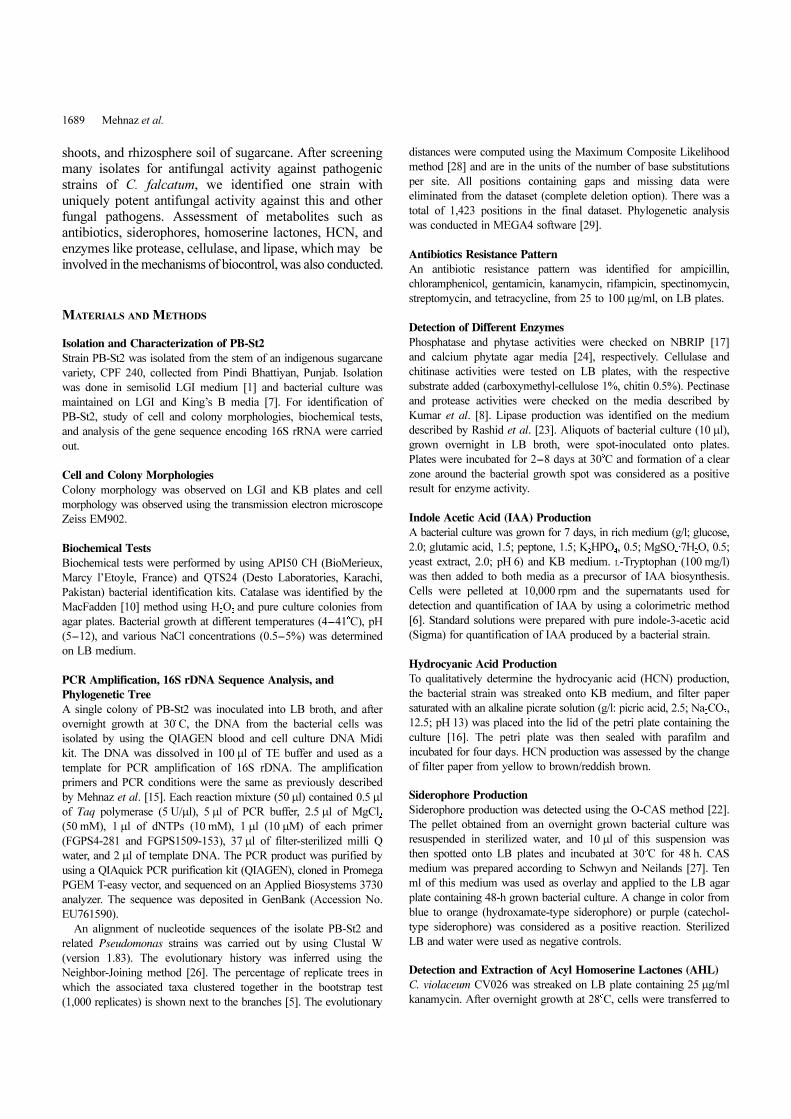
Cell and Colony Morphologies
Isolate PB-St2 formed 1-5 mm diameter, yellow, round,
convex, wax-drop-like colonies, with entire margin on LGI
medium. On King’s B medium, PB-St2 formed 1-3 mm,
round, flat, deep orange colonies that stayed orange while
the medium stained green and fluoresced under UV light.
Cells of the bacterium are rods, 0.8-1.2 µm wide and 1.6-
3.6 µm long, motile, and lophotrichous, with four to six
flagella (Fig. 1).
Biochemical Tests
The isolate utilized glycerol, D-arabinose, L-arabinose, D-
ribose, D-xylose, L-xylose, D-galactose, D-glucose, D-fructose,
D-mannose, D-melibiose, D-mannitol, D-saccharose, D-trehalose,
D-raffinose, D-lyxose, D-fucose, D-arabitol, inositol, gentiobiose,
potassium gluconate, esculin ferric citrate, and N-acetyl
glucosamine, but did not utilize erythritol, dulcitol, D-
mannose, D-adonitol, L-sorbose, D-sorbitol, methyl beta D-
xylopyranoside, methyl alpha D-manno pyranoside, methyl
alpha D-glucopyranoside, amygdalin, arbutin, salicin, D-
cellobiose, D-lactose, inulin, D-melezitose, amidon, glycogen,
xylitol, D-turanose, D-tagatose, L-fucose, D-arabitol, L-arabitol,
potassium 2-ketogluconate, and potassium 5-ketogluconate
as a sole carbon source. The bacterium produced acid from
D-glucose, D-mannitol, and L-arabinose, but not from L-
rhamnose, D-sorbitol, inositol, D-adonitol, D-melibiose, and
Fig. 1. Transmission electron micrograph of sugarcane isolatePB-St2. (Magnification=×12,000)
1691 Mehnaz et al.
D-raffinose. Isolate PB-St2 is positive for arginine dihydrolase,
catalase, cytochrome oxidase, trisodium citrate, gelatin
hydrolysis, and 2-nitrophenyl-β-D-galactopyranoside, and
negative for acetoin, indole production, nitrate reduction,
lysine decarboxylase, 2-nitrophenyl-β-D-galactopyranoside,
ornithine decarboxylase, sodium thiosulfate, tryptophane
deaminase, and urease. Bacterial growth was observed at 4
to 37oC, pH 5 to 10, and up to 3% NaCl concentration.
PCR Amplification and 16S rDNA Sequence Analysis
An almost complete sequence (~1.5 kb) of 16S rDNA was
obtained for isolate PB-St2 and compared with the NCBI
databank through a Blast search. The isolate showed 99.8%
similarity (1,524/1,527 positions) with the sequence of P.
aurantiaca ATCC 33663T (Accession No. DQ682655) and
99.5% similarity (1,521 identities/1,529 positions: 1,485
identities/1,492 positions) with the sequences of P.
aureofaciens ATCC 13985T (Accession No. AY509898)
and P. chlororaphis ATCC 9446T (Accession No. Z76673),
respectively. The phylogenetic tree is shown in Fig. 2.
Antibiotics Resistance Pattern
PB-St2 is resistant to ampicillin, chloramphenicol, and
spectinomycin at 100 µg/ml, streptomycin at 75 µg/ml,
and tetracycline at 25 µg/ml and sensitive to rifampicin,
kanamycin, and gentamicin at 25 µg/ml.
Detection of Different Enzymes, IAA, HCN, and
Siderophore Production
The bacterium showed positive reactions for protease and
lipase and was negative for the production of cellulase,
chitinase, pectinase, phosphatase, and phytase. It did not
produce indole acetic acid in both media, but did produce
HCN and hydroxamate-type siderophores.
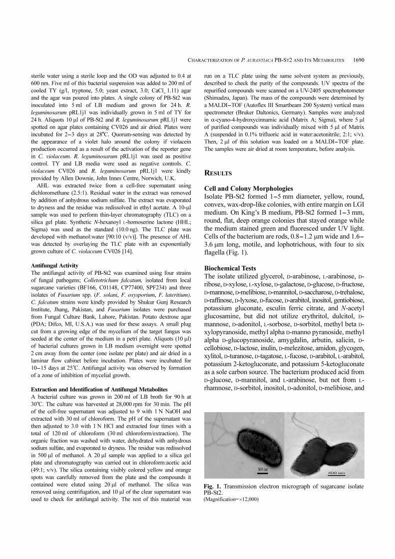
Quorum Sensing and Antifungal Activity
The isolate was positive for quorum-sensing signal production.
On TLC plate, only one purple spot was detected and the
compound was identified as HHL (Fig. 3). Antifungal
activity was observed against all strains of C. falcatum, F.
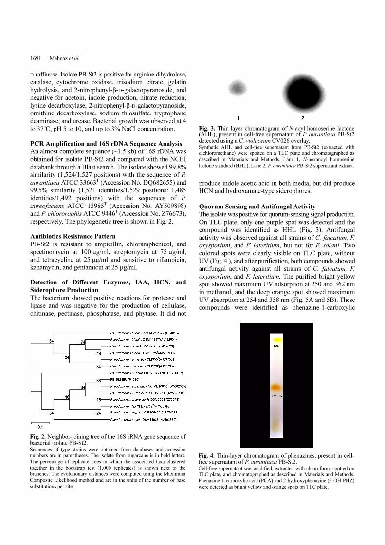
oxysporium, and F. lateritium, but not for F. solani. Two
colored spots were clearly visible on TLC plate, without
UV (Fig. 4.), and after purification, both compounds showed
antifungal activity against all strains of C. falcatum, F.
oxysporium, and F. lateritium. The purified bright yellow
spot showed maximum UV adsorption at 250 and 362 nm
in methanol, and the deep orange spot showed maximum
UV absorption at 254 and 358 nm (Fig. 5A and 5B). These
compounds were identified as phenazine-1-carboxylic
Fig. 2. Neighbor-joining tree of the 16S rRNA gene sequence ofbacterial isolate PB-St2. Sequences of type strains were obtained from databases and accession
numbers are in parentheses. The isolate from sugarcane is in bold letters.
The percentage of replicate trees in which the associated taxa clustered
together in the bootstrap test (1,000 replicates) is shown next to the
branches. The evolutionary distances were computed using the Maximum
Composite Likelihood method and are in the units of the number of base
substitutions per site.
Fig. 3. Thin-layer chromatogram of N-acyl-homoserine lactone(AHL), present in cell-free supernatant of P. aurantiaca PB-St2detected using a C. violaceum CV026 overlay. Synthetic AHL and cell-free supernatant from PB-St2 (extracted with
dichloromethane) were spotted on a TLC plate and chromatographed as
described in Materials and Methods. Lane 1, N-hexanoyl homoserine
lactone standard (HHL); Lane 2, P. aurantiaca PB-St2 supernatant extract.
Fig. 4. Thin-layer chromatogram of phenazines, present in cell-free supernatant of P. aurantiaca PB-St2. Cell-free supernatant was acidified, extracted with chloroform, spotted on
TLC plate, and chromatographed as described in Materials and Methods.
Phenazine-1-carboxylic acid (PCA) and 2-hydroxyphenazine (2-OH-PHZ)
were detected as bright yellow and orange spots on TLC plate.
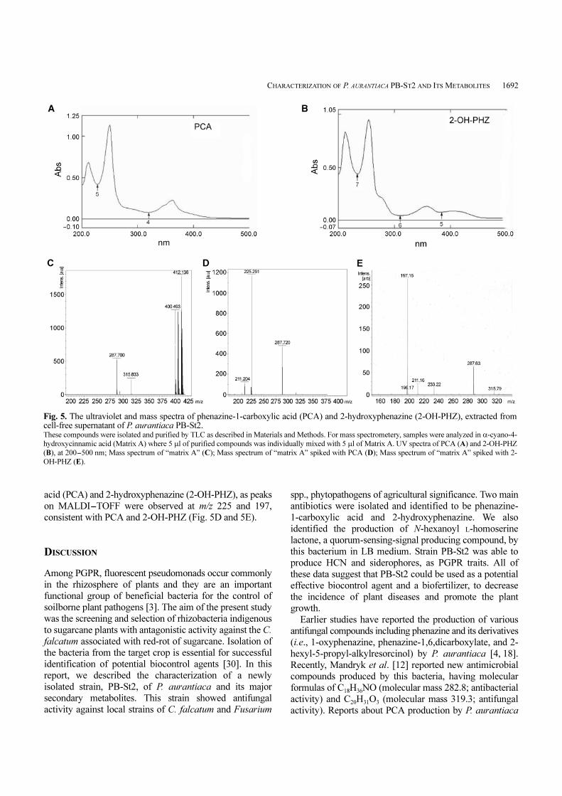
CHARACTERIZATION OF P. AURANTIACA PB-ST2 AND ITS METABOLITES 1692
acid (PCA) and 2-hydroxyphenazine (2-OH-PHZ), as peaks
on MALDI-TOFF were observed at m/z 225 and 197,
consistent with PCA and 2-OH-PHZ (Fig. 5D and 5E).
DISCUSSION
Among PGPR, fluorescent pseudomonads occur commonly
in the rhizosphere of plants and they are an important
functional group of beneficial bacteria for the control of
soilborne plant pathogens [3]. The aim of the present study
was the screening and selection of rhizobacteria indigenous
to sugarcane plants with antagonistic activity against the C.
falcatum associated with red-rot of sugarcane. Isolation of
the bacteria from the target crop is essential for successful
identification of potential biocontrol agents [30]. In this
report, we described the characterization of a newly
isolated strain, PB-St2, of P. aurantiaca and its major
secondary metabolites. This strain showed antifungal
activity against local strains of C. falcatum and Fusarium
spp., phytopathogens of agricultural significance. Two main
antibiotics were isolated and identified to be phenazine-
1-carboxylic acid and 2-hydroxyphenazine. We also
identified the production of N-hexanoyl L-homoserine
lactone, a quorum-sensing-signal producing compound, by
this bacterium in LB medium. Strain PB-St2 was able to
produce HCN and siderophores, as PGPR traits. All of
these data suggest that PB-St2 could be used as a potential
effective biocontrol agent and a biofertilizer, to decrease
the incidence of plant diseases and promote the plant
growth.
Earlier studies have reported the production of various
antifungal compounds including phenazine and its derivatives
(i.e., 1-oxyphenazine, phenazine-1,6,dicarboxylate, and 2-
hexyl-5-propyl-alkylresorcinol) by P. aurantiaca [4, 18].
Recently, Mandryk et al. [12] reported new antimicrobial
compounds produced by this bacteria, having molecular
formulas of C18H36NO (molecular mass 282.8; antibacterial
activity) and C20H31O3 (molecular mass 319.3; antifungal
activity). Reports about PCA production by P. aurantiaca
Fig. 5. The ultraviolet and mass spectra of phenazine-1-carboxylic acid (PCA) and 2-hydroxyphenazine (2-OH-PHZ), extracted fromcell-free supernatant of P. aurantiaca PB-St2. These compounds were isolated and purified by TLC as described in Materials and Methods. For mass spectrometery, samples were analyzed in α-cyano-4-
hydroxycinnamic acid (Matrix A) where 5 µl of purified compounds was individually mixed with 5 µl of Matrix A. UV spectra of PCA (A) and 2-OH-PHZ
(B), at 200-500 nm; Mass spectrum of “matrix A” (C); Mass spectrum of “matrix A” spiked with PCA (D); Mass spectrum of “matrix A” spiked with 2-
OH-PHZ (E).

1693 Mehnaz et al.
could not be found, although Peix et al. [20] used the
production of phenazine-1-carboxylate as a characteristic
feature of P. aureofaciens and P. aurantiaca when they
reclassified these two bacteria as subspecies of P. chlororaphis.
Similarly, 2-OH-PHZ and HCN production by P. aurantiaca
have not been reported. However, siderophore production
in P. aurantiaca is reported by Rovera et al. [25].
A prerequirement for bacterial growth promoting effect,
as a biofungicide or biofertilizer, is the close contact
between the plant and the effective organism. PB-St2
inhibited the growth of C. falcatum strains, which are
isolated from local sugarcane varieties. C. falcatum affects
the plant stem, and the isolation of PB-St2 from the inner
region of the stem supports not only its survival within
tissue but also its potential to be used as a biocontrol
agent. Inhibition of C. falcatum was reported earlier by
Pseudomonas aeruginosa [8] and P. fluorescens [11] but
not by P. aurantiaca.
Quorum sensing is the major mechanism by which
many bacteria regulate production of antifungal factors.
PB-St2 produces N-hexanoyl homoserine lectone (HHL), a
chemical that indicates the presence of a quorum-sensing
mechanism. Feklistova and Maksimova [4] previously
reported the production of HHL by P. aurantiaca B-162.
Pearson et al. [21] and Chin-A-Woeng et al. [2] have
shown that quorum sensing is the most important regulation
mechanism for PCA production in the closely related
species to P. aurantiaca B-162, namely P. aeruginosa, and
for phenazine-1-carboxamide production in P. chlororaphis.
Several Pseudomonas strains have already been marketed
as commercial biocontrol products, such as Cedomon
(BioAgri AB, Upsala, Sweden), a seed treatment based on
a Pseudomonas chlororaphis strain providing protection
against seedborne diseases in barley. Similarly, Mycolytin
is an antifungal biopesticide formed by P. aurantiaca M-
518 [19]. The reports in the literature and the presence of
biocontrol traits (siderophore, HCN, PCA, and 2-OH-PHZ
production) in this strain enhance the potential use of
PB-St2 as an effective biocontrol agent promoting plant
growth with reduced disease incidence. Future studies will
focus on its use as a biocontrol agent in pot and field
experiments.
In this study, for the first time, we report about PB-St2
as a new strain of P. aurantiaca from the sugarcane stem,
with the production of siderophores, HCN, PCA, 2-OH
PHZ, lipase, and protease.
Acknowledgments
We are thankful to Ann Fook Yang, and the EM Unit of
Eastern Cereal and Oil-Seed Research Centre, Agriculture
and Agri-Food Canada, Ottawa, Canada. We are thankful
to Dr. M. Akhter, Director General, School of Biological
Sciences, University of the Punjab, Lahore, Pakistan, for
his guidance in chromatographic analysis.
REFERENCES
1. Cavalcante, V. A. and J. Dobereiner. 1988. A new acid tolerant
nitrogen fixing bacterium associated with sugarcane. Plant Soil
108: 23-31.
2. Chin-A-Woeng, T. F. C., G. V. Bloomberg, and B. J. J.
Lugtenberg. 2003. Phenazines and their role in biocontrol by
Pseudomonas bacteria. New Phytol. 157: 503-523.
3. Ellis, R. J., T. M. Timms-Wilson, and M. J. Bailey. 2000.
Identification of conserved traits in fluorescent pseudomonads
with antifungal activity. Environ. Microbiol. 2: 247-284.
4. Feklistova, I. N. and N. P. Maksimova. 2008. Obtaining
Pseudomonas aurantiaca strains capable of overproduction of
phenazine antibiotics. Microbiology 77: 176-180.
5. Felsenstein, J. 1985. Confidence limits on phylogenies: An
approach using the bootstrap. Evolution 39: 783-791.
6. Gordon, S. A. and R. P. Weber. 1951. Colorimetric estimation
of indole acetic acid. Plant Physiol. 26: 192-195.
7. King, E. O., M. K. Ward, and D. E. Raney. 1954. Two simple
media for the demonstration of pyocyanin and fluorescin. J.
Lab. Clin. Med. 44: 301-307.
8. Kumar, R. S., N. Ayyadurai, P. Pandiaraja, A. V. Reddy, Y.
Venkatesvarlu, O. Prsakash, and N. Sakthivel. 2005. Characterization
of antifungal metabolite produced by a new strain Pseudomonas
aeruginosa PUPa3 that exhibits broad spectrum antifungal
activity and biofertilizing traits. J. Appl. Microbiol. 98: 145-154.
9. Liu, H., Y. He, H. Jiang, H. Peng, X. Huang, X. Zhang, L. S.
Thomashow, and Y. Xu. 2007. Characterization of a phenazine
producing strain Pseudomonas chlororaphis GP72 with broad
spectrum antifungal activity from green pepper rhizosphere.
Curr. Microbiol. 54: 302-306.
10. MacFadden, J. F. 1980. Biochemical Tests for Identification of
Medical Bacteria, pp. 51-54. Williams and Wilkins, Baltimore.
11. Malathi, P., R. Viswanathan, P. Padmanaban, D. Mohanraj, and
A. R. Sundar. 2002. Microbial detoxification of Colletotrichum
falcatum toxin. Curr. Sci. 83: 745-749.
12. Mandryk, M. N., E. Kolomiets, and E. S. Dey. 2007. Characterization
of antimicrobial compounds produced by Pseudomonas aurantiaca
S-1. Pol. J. Microbiol. 56: 245-250.
13. Mark, G. L., J. P. Morrissey, P. Higgins, and F. O’Gara. 2006.
Molecular based strategies to exploit Pseudomonas biocontrol
strains for environmental biotechnology applications. FEMS
Microbiol. Ecol. 56: 167-177.
14. McClean, K. H., M. K. Winson, L. Fish, A. Taylor, S. R. Chhabra,
M. Camara, et al. 1997. Quorum sensing and Chromobacterium
violaceum: Exploitation of violacein production and inhibition
for the detection of N-acyl homoserine lactones. Microbiology
143: 3703-3711.
15. Mehnaz, S., M. S. Mirza, J. Haurat, R. Bally, P. Normand, A.
Bano, and K. A. Malik. 2001. Isolation and 16S rRNA sequence
analysis of beneficial bacteria from the rhizosphere of rice. Can.
J. Microbiol. 47: 110-117.
16. Miller, R. L. and V. J. Higgins. 1970. Association of cyanide
with infection of birdsfoot trefoil by Stemphylium loti.
Phytopathology 60:104-110.
CHARACTERIZATION OF P. AURANTIACA PB-ST2 AND ITS METABOLITES 1694
17. Nautiyal, C. S. 1999. An efficient microbiological growth
medium for screening phosphate solubilizing microorganisms.
FEMS Microbiol. Lett. 170: 265-270.
18. Nowak-Thompson, B., P. E. Hammer, D. S. Hill, J. Stafford, N.
Torkewitz, T. D. Gaffney, S. T. Lam, I. Molnar, and J. M.
Ligon. 2003. 2,5-Dialkylresorcinol biosynthesis in Pseudomonas
aurantiaca: Novel head-to-head condensation of two fatty acid-
derived precursors. J. Bacteriol. 185: 860-869.
19. Omel’yanets, T. G. and G. P. Mel’nik. 1987. Toxicological
evaluation of the microbial preparation mycolytin. Zdravookhranenie
Turkmenistana 6: 8.
20. Peix, A., A. Valverde, R. Rivas, J. M. Igual, M. H. Ramirez-
Bahena, P. F. Mateos, et al. 2007. Reclassification of Pseudomonas
aurantiaca as a synonym of Pseudomonas chlororaphis and
proposal of three subspecies, P. chlororaphis subsp. chlororaphis
subsp. nov., P. chlororaphis subsp. aureofaciens subsp. nov.,
comb. nov., and P. chlororaphis subsp. aurantiaca subsp. nov.,
comb. nov. Int. J. Syst. Evol. Microbiol. 57: 1286-1290.
21. Pearson, J. P., K. M. Gray, L. Passador, K. D. Yucker, A.
Eberhard, B. H. Iglewski, and E. P. Greenberg. 1994. Structure
of the auto-inducer required for expression of Pseudomonas
aeruginosa virulence genes. Proc. Natl. Acad. Sci. U.S.A. 91:
197-201.
22. Perez-Miranda, S., N. Cabirol, R. George-Tellez, L. S. Zamudio-
Rivera, and F. J. Fernandez. 2007. O-CAS, a fast and universal
method for siderophore detection. J. Microbiol. Meth. 70: 127-
131.
23. Rashid, N., Y. Shimada, S. Ezaki, H. A. Tomi, and T. Y.
Imanaka. 2001. Low temperature lipase from psychrotrophic
Pseudomonas sp. strain KB700A. Appl. Environ. Microbiol. 67:
4064-4069.
24. Rosado, A. S., F. S. de Azevedo, D. W. da Cruz, J. D. Van
Elsa, and L. Seldin. 1998. Phenotypic and genetic diversity of
Paenibacillus azotofixans strains isolated from the rhizoplane
soil of different grasses. J. Appl. Microbiol. 84: 216-226.
25. Rovera, M., J. Andres, E. Carlier, C. Pasluosta, and S. Rosas.
2008. Pseudomonas aurantiaca: Plant growth promoting traits,
secondary metabolites and inoculation response, pp. 155-164.
In I. Ahmad, J. Pichtel, and S. Hayat (eds.). Plant-Bacteria
Interactions. Strategies and Techniques to Promote Plant Growth.
Wiley-VCH, Germany.
26. Saitou, N. and M. Nei. 1987. The neighbor-joining method: A
new method for reconstructing phylogenetic trees. Mol. Biol.
Evol. 4: 406-425.
27. Schwyn, B. and J. B. Neilands. 1987. Universal chemical assay
for the detection and determination of siderophores. Anals
Biochem. 160: 46-56.
28. Tamura, K., M. Nei, and S. Kumar. 2004. Prospects for
inferring very large phylogenies by using the neighbor-joining
method. Proc. Natl. Acad. Sci. U.S.A. 101: 11030-11035.
29. Tamura, K., J. Dudley, M. Nei, and S. Kumar. 2007. MEGA4:
Molecular Evolutionary Genetics Analysis (MEGA) software
version 4.0. Mol. Biol. Evol. 24: 1596-1599.
30. William, G. E. and M. J. C. Asher. 1996. Selection of rhizobacteria
for the control of Pythium ultimum and Aphanomyces cochlioides
on sugarbeet seedlings. Crop Protec. 15: 479-486.

![CHAPTER 9 CHEMISTRY Doctoral Thesescrl.du.ac.in/Doc.Bib/2014/Chemistry.pdf · for the synthesis of novel benzo[a]phenazine annulated heterocycles and their photophysical studies.](https://static.fdocuments.in/doc/165x107/605e5323a79a245d50771859/chapter-9-chemistry-doctoral-for-the-synthesis-of-novel-benzoaphenazine-annulated.jpg)