Characterization and Expression of the Arginine Bio ... · genes are scattered (Picard and Dillon,...
9
Characterization and Expression of the Arginine Bio- synthesis Gene Cluster of Streptomyces clavuligerus Received January 1, 2000; revised April 14, 2000; accepted April 14, 2000. *For correspondence. Email [email protected]; Tel. +34-987-291504; Fax. 34-987-2915 06. J. Mol. Microbiol. Biotechnol. (2000) 2(4): 543-550. © 2000 Horizon Scientific Press JMMB Research Article Antonio Rodríguez-García, Álvaro de la Fuente, Rosario Pérez-Redondo, Juan F. Martín, and Paloma Liras* Area of Microbiology, Faculty of Biology, University of León, 24071 León, Spain Abstract A cluster of genes argCJBDRGH containing most of the arginine biosynthesis genes has been found in Streptomyces clavuligerus after sequencing a 8.3 kb DNA region containing overlapping sequences of two DNA fragments known to contain arginine biosynthesis genes. Subcloning, complementation of E. coli arginine auxotrophic strains and enzymatic assays confirmed the identity of each gene. S1 nuclease mapping studies and Northern hybridization analysis revealed the formation of two large transcripts corresponding to argCJBDR and argGH. The amount of each of these mRNAs is 10 to 44 times higher in a S. clavuligerus argR-disrupted mutant than in the wild type confirming the existence of an ArgR-mediated control of arginine biosynthesis gene expression. A low level constitutive monocistronic transcript of argR was observed in S. clavuligerus cells. Most of the argGH transcript initiating at an adenine 29 nt upstream of the argG initiation codon appears to stop at a termination stem and loop structure present downstream of the argG gene. Introduction Arginine biosynthesis genes (arg) and genes for carbamoyl phosphate (required for citrulline) biosynthesis (car) are usually scattered in the genome of most Gram negative bacteria. In E. coli there is an argECBH cluster, in which the argE and argCBH are transcribed divergently (Crabeel et al., 1979). The carAB genes are also clustered, while other genes (argA, argB, argD, argI, argF, argG) map at different positions in the E. coli chromosome. The argECBH cluster is also found in Salmonella typhimurium (Sanderson, 1970) while in Proteus mirabilis and Serratia marcescens includes additionally argG upstream of argH (Prozesky 1968, Matsumoto et al., 1975). In Neisseria gonorrohoeae and Pseudomonas aeruginosa all the arg genes are scattered (Picard and Dillon, 1989; Haas et al., 1977). In Gram positive bacteria clustering of arginine genes appears to be common. In Lactobacillus plantarum a carAargCJBDF cluster has been described (Bringel et al., 1997) with divergent transcription for the car and the arg genes. The same organization for arginine genes ( argCJBDF ) was described in Corynebacterium glutamicum (Sakanyan et al., 1996); in Mycobacterium tuberculosis and Mycobacterium leprae, DNA sequences for a argCJBDFRGH cluster have been deposited in EMBL database (Accession numbers Z85982 and L78811, respectively). Arginine biosynthesis genes in Bacillus subtilis are organized in two operons, one is argCJBD- carAB-argF for early steps in the arginine pathway, including those involved in carbamoyl phosphate biosynthesis (Mountain et al., 1984; O’Reilly and Devine, 1994), and a second operon argGH encodes enzymes for the late steps of the pathway (Piggot and Hoch, 1985). An argE gene has been located by sequencing the B. subtilis genome (Kunst et al., 1997) but its relation with the cyclic pathway of arginine biosynthesis in this bacterium (Figure 1) remains to be elucidated; the regulatory gene ahrC encoding the arginine biosynthesis repressor (North et al., 1989) is located in a separate region. This organization in two separate operons may occur also in other Gram positive bacteria and could explain the lack of information on the location of argG, argH and argR in Lactobacillus and Corynebacteria. Arginine defective mutants of Streptomyces coelicolor have been mapped as scattered at four different locations in the chromosome (Redenbach et al ., 1996). By complementation of E. coli arginine defective mutants a cluster argCJB has been located in S. coelicolor (Hindle et al., 1994) whereas the argG gene is located in an unstable region at the end of the lineal chromosome of this actinomycete (Redenbach et al., 1996). Fragments of S. clavuligerus genome were also able to complement E. coli arginine mutants and the subsequent sequencing of those fragments revealed the presence of two subclusters of arginine genes in this actinomycete: argCJ (Ludovice et al., 1992) and argRGH (Rodríguez-García et al., 1995; 1997). An important question from the regulation point of view is if the two clusters organization occurs in all actinomycetes and how expression of these clusters is controlled by the ArgR repressor. This information is particularly important in S. clavuligerus, since this actinomycete uses arginine as precursor for clavulanic acid biosynthesis (Valentine et al. , 1993). Knowledge of the arginine gene cluster organization and their regulatory mechanisms (Rodríguez- García et al., 1997) is important to understand how the flow of arginine affects clavulanic acid production. In this paper we provide evidence showing that most of the arginine genes in S. clavuligerus are present in a single argCJBDRGH organization, which includes genes for the early and late steps of the arginine pathway as well as the regulatory gene argR. We have also shown that ca- ORF4, in the clavulanic acid cluster, encodes a second functional ornithine N-acetyltransferase similar to ArgJ.
Transcript of Characterization and Expression of the Arginine Bio ... · genes are scattered (Picard and Dillon,...

The Arginine Biosynthesis Cluster of Streptomyces clavuligerus 543
Characterization and Expression of the Arginine Bio-synthesis Gene Cluster of Streptomyces clavuligerus
Received January 1, 2000; revised April 14, 2000; accepted April 14, 2000.*For correspondence. Email [email protected]; Tel. +34-987-291504; Fax.34-987-2915 06.
J. Mol. Microbiol. Biotechnol. (2000) 2(4): 543-550.
© 2000 Horizon Scientific Press
JMMB Research Article
Antonio Rodríguez-García, Álvaro de la Fuente,Rosario Pérez-Redondo, Juan F. Martín,and Paloma Liras*
Area of Microbiology, Faculty of Biology, University of León,24071 León, Spain
Abstract
A cluster of genes argCJBDRGH containing most ofthe arginine biosynthesis genes has been found inStreptomyces clavuligerus after sequencing a 8.3 kbDNA region containing overlapping sequences of twoDNA fragments known to contain arginine biosynthesisgenes. Subcloning, complementation of E. coli arginineauxotrophic strains and enzymatic assays confirmedthe identity of each gene. S1 nuclease mapping studiesand Northern hybridization analysis revealed theformation of two large transcripts corresponding toargCJBDR and argGH. The amount of each of thesemRNAs is 10 to 44 times higher in a S. clavuligerusargR-disrupted mutant than in the wild type confirmingthe existence of an ArgR-mediated control of argininebiosynthesis gene expression. A low level constitutivemonocistronic transcript of argR was observed in S.clavuligerus cells. Most of the argGH transcriptinitiating at an adenine 29 nt upstream of the argGinitiation codon appears to stop at a termination stemand loop structure present downstream of the argGgene.
Introduction
Arginine biosynthesis genes (arg) and genes for carbamoylphosphate (required for citrulline) biosynthesis (car) areusually scattered in the genome of most Gram negativebacteria. In E. coli there is an argECBH cluster, in whichthe argE and argCBH are transcribed divergently (Crabeelet al., 1979). The carAB genes are also clustered, whileother genes (argA, argB, argD, argI, argF, argG) map atdifferent positions in the E. coli chromosome. The argECBHcluster is also found in Salmonella typhimurium(Sanderson, 1970) while in Proteus mirabilis and Serratiamarcescens includes additionally argG upstream of argH(Prozesky 1968, Matsumoto et al., 1975). In Neisseriagonorrohoeae and Pseudomonas aeruginosa all the arggenes are scattered (Picard and Dillon, 1989; Haas et al.,1977).
In Gram positive bacteria clustering of arginine genesappears to be common. In Lactobacillus plantarum acarAargCJBDF cluster has been described (Bringel et al.,
1997) with divergent transcription for the car and the arggenes. The same organization for arginine genes(argCJBDF) was described in Corynebacteriumglutamicum (Sakanyan et al., 1996); in Mycobacteriumtuberculosis and Mycobacterium leprae, DNA sequencesfor a argCJBDFRGH cluster have been deposited in EMBLdatabase (Accession numbers Z85982 and L78811,respectively). Arginine biosynthesis genes in Bacillussubtilis are organized in two operons, one is argCJBD-carAB-argF for early steps in the arginine pathway,including those involved in carbamoyl phosphatebiosynthesis (Mountain et al., 1984; O’Reilly and Devine,1994), and a second operon argGH encodes enzymes forthe late steps of the pathway (Piggot and Hoch, 1985). AnargE gene has been located by sequencing the B. subtilisgenome (Kunst et al., 1997) but its relation with the cyclicpathway of arginine biosynthesis in this bacterium (Figure1) remains to be elucidated; the regulatory gene ahrCencoding the arginine biosynthesis repressor (North et al.,1989) is located in a separate region. This organization intwo separate operons may occur also in other Grampositive bacteria and could explain the lack of informationon the location of argG, argH and argR in Lactobacillusand Corynebacteria.
Arginine defective mutants of Streptomyces coelicolorhave been mapped as scattered at four different locationsin the chromosome (Redenbach et al., 1996). Bycomplementation of E. coli arginine defective mutants acluster argCJB has been located in S. coelicolor (Hindle etal., 1994) whereas the argG gene is located in an unstableregion at the end of the lineal chromosome of thisactinomycete (Redenbach et al., 1996). Fragments of S.clavuligerus genome were also able to complement E. coliarginine mutants and the subsequent sequencing of thosefragments revealed the presence of two subclusters ofarginine genes in this actinomycete: argCJ (Ludovice etal., 1992) and argRGH (Rodríguez-García et al., 1995;1997).
An important question from the regulation point of viewis if the two clusters organization occurs in all actinomycetesand how expression of these clusters is controlled by theArgR repressor. This information is particularly importantin S. clavuligerus, since this actinomycete uses arginineas precursor for clavulanic acid biosynthesis (Valentine etal., 1993). Knowledge of the arginine gene clusterorganization and their regulatory mechanisms (Rodríguez-García et al., 1997) is important to understand how theflow of arginine affects clavulanic acid production.
In this paper we provide evidence showing that mostof the arginine genes in S. clavuligerus are present in asingle argCJBDRGH organization, which includes genesfor the early and late steps of the arginine pathway as wellas the regulatory gene argR. We have also shown that ca-ORF4, in the clavulanic acid cluster, encodes a secondfunctional ornithine N-acetyltransferase similar to ArgJ.
• MALDI-TOF Mass Spectrometry in Microbiology
Edited by: M Kostrzewa, S Schubert (2016) www.caister.com/malditof
• Aspergillus and Penicillium in the Post-genomic Era
Edited by: RP Vries, IB Gelber, MR Andersen (2016) www.caister.com/aspergillus2
• The Bacteriocins: Current Knowledge and Future Prospects
Edited by: RL Dorit, SM Roy, MA Riley (2016) www.caister.com/bacteriocins
• Omics in Plant Disease Resistance
Edited by: V Bhadauria (2016) www.caister.com/opdr
• Acidophiles: Life in Extremely Acidic Environments
Edited by: R Quatrini, DB Johnson (2016) www.caister.com/acidophiles
• Climate Change and Microbial Ecology: Current Research and Future Trends
Edited by: J Marxsen (2016) www.caister.com/climate
• Biofilms in Bioremediation: Current Research and Emerging Technologies
Edited by: G Lear (2016) www.caister.com/biorem
• Microalgae: Current Research and Applications
Edited by: MN Tsaloglou (2016) www.caister.com/microalgae
• Gas Plasma Sterilization in Microbiology: Theory, Applications, Pitfalls and New Perspectives
Edited by: H Shintani, A Sakudo (2016) www.caister.com/gasplasma
• Virus Evolution: Current Research and Future Directions
Edited by: SC Weaver, M Denison, M Roossinck, et al. (2016) www.caister.com/virusevol
• Arboviruses: Molecular Biology, Evolution and Control
Edited by: N Vasilakis, DJ Gubler (2016) www.caister.com/arbo
• Shigella: Molecular and Cellular Biology
Edited by: WD Picking, WL Picking (2016) www.caister.com/shigella
• Aquatic Biofilms: Ecology, Water Quality and Wastewater Treatment
Edited by: AM Romaní, H Guasch, MD Balaguer (2016) www.caister.com/aquaticbiofilms
• Alphaviruses: Current Biology
Edited by: S Mahalingam, L Herrero, B Herring (2016) www.caister.com/alpha
• Thermophilic Microorganisms
Edited by: F Li (2015) www.caister.com/thermophile
• Flow Cytometry in Microbiology: Technology and Applications
Edited by: MG Wilkinson (2015) www.caister.com/flow
• Probiotics and Prebiotics: Current Research and Future Trends
Edited by: K Venema, AP Carmo (2015) www.caister.com/probiotics
• Epigenetics: Current Research and Emerging Trends
Edited by: BP Chadwick (2015) www.caister.com/epigenetics2015
• Corynebacterium glutamicum: From Systems Biology to Biotechnological Applications
Edited by: A Burkovski (2015) www.caister.com/cory2
• Advanced Vaccine Research Methods for the Decade of Vaccines
Edited by: F Bagnoli, R Rappuoli (2015) www.caister.com/vaccines
• Antifungals: From Genomics to Resistance and the Development of Novel Agents
Edited by: AT Coste, P Vandeputte (2015) www.caister.com/antifungals
• Bacteria-Plant Interactions: Advanced Research and Future Trends
Edited by: J Murillo, BA Vinatzer, RW Jackson, et al. (2015) www.caister.com/bacteria-plant
• Aeromonas
Edited by: J Graf (2015) www.caister.com/aeromonas
• Antibiotics: Current Innovations and Future Trends
Edited by: S Sánchez, AL Demain (2015) www.caister.com/antibiotics
• Leishmania: Current Biology and Control
Edited by: S Adak, R Datta (2015) www.caister.com/leish2
• Acanthamoeba: Biology and Pathogenesis (2nd edition)
Author: NA Khan (2015) www.caister.com/acanthamoeba2
• Microarrays: Current Technology, Innovations and Applications
Edited by: Z He (2014) www.caister.com/microarrays2
• Metagenomics of the Microbial Nitrogen Cycle: Theory, Methods and Applications
Edited by: D Marco (2014) www.caister.com/n2
Caister Academic Press is a leading academic publisher of advanced texts in microbiology, molecular biology and medical research. Full details of all our publications at caister.com
Further Reading
Order from caister.com/order

544 Rodríguez-García et al.
Results
Location of the Arginine Gene Cluster of S.clavuligerusThe argC gene was initially located by sequencing a 6.1kb Sau3AI DNA fragment of S. clavuligerus present inplasmid pULML31, which complements the argC deficientmutant E. coli XC33 (Ludovice et al., 1992).
Independently, a 18 kb Sau3AI DNA fragment of S.clavuligerus was found to complement E. coli argG deficientmutants (Rodríguez-García et al., 1995). Further studiesshowed the presence in this DNA fragment of an argRGHcluster (Rodríguez-García et al., 1997).
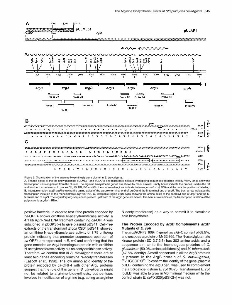
In order to establish the definitive organization of thearginine biosynthesis genes, a 0.5 kb SacI-SphI DNAfragment (Figure 2A) located downstream of argC inpULML31 was mapped and partially sequenced. In parallel,a 2.1 kb SacI-BglII DNA fragment from pULAR1, upstreamof argR (Figure 2A), was also sequenced. The nucleotidesequence of both fragments was found to overlap,indicating that argC and argR (previously though to be intwo different clusters) are closely located in the S.clavuligerus genome. Therefore, a DNA region, covering8.3 kb was sequenced in both orientations.
Analysis of the arg ClusterThe G+C content of the sequenced fragment was 71.3%,with no significant differences of G+C in the intergenicregions. Rare TTA codons for leucine were absent fromthe coding regions, indicating that these genes are notregulated by the bldA-dependent translation control (Leskiwet al., 1991). Analysis of the nucleotide sequence of theentire fragment using the Geneplot Program of DNAstarrevealed the presence of eight ORFs, oriented in the samedirection. Comparison of the amino acid sequencesdeduced from the ORFs with the sequences of argininemetabolic enzymes shows that ORF1 to ORF7, correspondto arginine biosynthesis genes. These genes include, asexpected, argC (nt 123-1148) which correspond to ORF1,argR and argG (nt 4600 to 6604) corresponding to ORF5and ORF6. The proteins encoded by ORF2, ORF3 andORF4 were homologous with ArgJ, ArgB and ArgD of otherorganisms while ORF7 encodes argH. ORF8 was truncatedand its deduced amino acid sequence does not correspondto any enzyme of the arginine biosynthesis pathway.
Putative ribosome binding sites were found upstreamof argJ, argB, argD and argH, but according to theStreptomyces promoter criteria (Strohl, 1992) no clear –10 and –35 boxes were found upstream of the newlydescribed ORFs. Two inverted sequences able to form astem and loop structure with free energy of –37.2 kcal/molwere found downstream of argG (nt 6628-6640/6650-6662). They might act as a transcription terminator of theargG gene. Similar putative terminator sequences werepresent downstream of argH (nt 8162-8172/8182-8192 and8171-8178/8183-8190) giving stem and loop structures withfree energies of –20.2 and –12.6 kcal/mol.
argJ Encodes a Functional Ornithine N-Acetyltransferase: Evidence for a Cyclic ArgininePathwayargJ (ORF2) has 1,152 nt and encodes a protein of 383amino acids with a deduced Mr of 39,733. The amino acidsequence of this protein showed the highest similarity
(74.9% identical residues) with the unpublished sequenceof ArgJ of S. coelicolor (Sanger cosmid SCL24) followedby the ornithine acetyltransferases of M. tuberculosis(47.8% identity) and C. glutamicum (40.7% identity).
E. coli contains N-acetylornithinase but not the argJ-encoded ornithine N-acetyltransferase. Bifunctionalornithine N-acetyltransferases (EC 2.3.1.35) with N-acetylornithinase (EC 3.5.1.16) activity have been reportedin some microorganisms. In order to test whether the argJgene of S. clavuligerus confers acetylornithinase activity a2.1 kb SacI-BglII DNA fragment containing argJ wassubcloned in both orientations in pBSKS(+) (givingplasmids pKS21.1 and pKS21.2) and introduced into E.coli XS1D2, an argE mutant lacking acetylornithinaseactivity. Ornithine N-acetyltransferase activity (0.76 units/mg protein) was found in cell-free extracts of E. coli XS1D[pKS21.1] in which the S. clavuligerus argJ was expressedfrom the lacZ promoter but no activity was found in E. coliXS1D[pKS21.2] in which the gene was subcloned in theopposite orientation. No N-acetylornithinase activity wasfound in any of the transformants. These results confirmthat S. clavuligerus contains a cyclic N-acetyltransferasepathway as reported by Hindle et al. (1994) in S. coelicolor,and not the linear (E. coli type) pathway.
A Second Gene Encoding an N-AcetylornithineAcetyltransferase Occurs in the Clavulanic AcidClusterArgJ shows a 31.1% identity in amino acids over the entireprotein sequence with the protein encoded by ORF4 ofthe clavulanic acid gene cluster (hereafter named ca-ORF4) of S. clavuligerus (Hodgson et al., 1995). The aminoacid sequence of the protein encoded by ca-ORF4 containsmost of the conserved boxes found in ArgJ proteins.However the similarity of the protein encoded by ca-ORF4with the ArgJ protein of S. clavuligerus (31.1%) is lowerthan the similarity between ArgJ proteins of other Gram
Figure 1. Pathways for arginine biosynthesis in different microorganisms.
The Arginine Biosynthesis Cluster of Streptomyces clavuligerus 545
positive bacteria. In order to test if the protein encoded byca-ORF4 shows ornithine N-acetyltransferase activity, a4.1 kb KpnI-NruI DNA fragment containing ca-ORF4 wassubcloned in pBSKS(+) to give plasmid pBS41. Cell-freeextracts of the transformant E.coli XSD1[pBS41] showedan ornithine N-acetyltransferase activity of 1.79 units/mgprotein indicating that promoter sequences upstream ofca-ORF4 are expressed in E. coli and confirming that thegene encodes an ArgJ-homologous protein with ornithineN-acetyltransferase activity but no acetylornithinase activity.Therefore we confirm that in S. clavuligerus there are atleast two genes encoding ornithine N-acetyltransferases(Eascott et al., 1998). The low amino acid identity of theprotein encoded by ca-ORF4 with other ArgJ proteinssuggest that the role of this gene in S. clavuligerus mightnot be related to arginine biosynthesis, but perhapsinvolved in modification of arginine (e.g. acting as arginine
N-acetyltransferase) as a way to commit it to clavulanicacid biosynthesis.
The Protein Encoded by argB Complements argB-
Mutants of E. coliThe argB (ORF3, 909 nt) gene has a G+C content of 68.5%,and encodes a protein of Mr 32,365. The N-acetylglutamatekinase protein (EC 2.7.2.8) has 302 amino acids and asequence similar to the homologous proteins of C.glutamicum (50.0% amino acid identity) and M. tuberculosis(47.8% identity). A motif conserved in all the ArgB proteinsis present in the ArgB protein of S. clavuligerus,66VHGGGPXI73. To confirm the identity of the gene, plasmidpULB, containing the argB gen, was used to complementthe argB deficient strain E. coli XB25. Transformant E. coli[pULB] was able to grow in VB minimal medium while thecontrol strain E. coli XB25[pBSKS+] was not.
Figure 2. Organization of the arginine biosynthesis gene cluster in S. clavuligerus.A. Shaded boxes at the top show plasmids pULML31 and pULAR1 and black bars indicate overlapping sequences detected initially. Wavy lanes show thetranscription units originated from the cluster. The arginine biosynthesis genes are shown by black arrows. Empty boxes indicate the probes used in the S1and Northern experiments. In probes CJ, JB, DR, RG and GH the shadowed regions indicate heterologous (E. coli) DNA and the dots the position of labeling.B. Intergenic region argD-argR showing the amino acids of the carboxyterminal-end of argD and the N-terminal end of argR. The bent arrow indicates thetranscription initiation of the monocistronic argR mRNA. C. Intergenic region argR-argG showing the amino acids of the carboxyl-end of argR and the N-terminal end of argG. The regulatory Arg-sequences present upstream of the argG gene are boxed. The bent arrow indicates the transcription initiation of thepolycistronic argGH mRNA.

546 Rodríguez-García et al.
Characteristics of argDORF4 (1203 nt with a G+C content of 71.3%) encodes a400 amino acid protein with a calculated Mr of 41,972.The argB stop codon overlaps with the initiation codon ofORF3. A search in the Swiss-Prot database with the FASTAProgram revealed that the protein encoded by ORF3 hasa high similarity to acetylornithine aminotransferases(ACOAT) and ornithine aminotransferases (OAT) (54.5%identity with the ArgD of M. tuberculosis and 47.8% withthat of C. glutamicum).
The sequence 214LVLDEVQTGIGRTGHWFAAQAEGVEADVVTLAKGLGGG251 corresponds to thepyridoxal phosphate (PP) binding motif, with K246 as theconserved lysine for covalent PP-binding (Yonaha et al.,1992). Multiple alignment of the ORF4-encoded proteinwith ten known ArgD proteins showed the presence ofdomains conserved in ACOAT’s and OAT’s, but the aminoacid similarity of the ORF4-encoded protein is higher toACOATs of B. subtilis and E. coli than to the correspondingOAT proteins. This observation is consistent with theamination of N-acetylglutamic semialdehyde to N-acetylornithine (which occurs when the intermediate is inthe N-acetylated form).
In order to confirm that the cloned gene correspondsto argD, plasmid pULD was used to transform E. coli CGSC4538, an argD, proA double mutant. The ampicillin resistanttransformants were able to grow in VB mediumsupplemented with proline but also in VB medium withoutsupplementation with either proline or arginine. Since theproA mutation precludes the formation of glutamate-γ-semialdehyde required for proline biosynthesis, thecomplementation of both proA and argD phenotypes bythe argD gene of S. clavuligerus suggests aninterconversion of intermediates of the proline and argininepathways.
Characterization of argHORF7 (1,422 nt), encoding the argH gene(argininosuccinate lyase) was described to be located
downstream of argG by partial sequencing of the gene(Rodríguez-García et al., 1995). It is preceded by aribosome binding motif AGGAG. The protein ArgH has acalculated Mr of 50,915. The amino acid sequence showsthe highest similarity with ArgH of M. tuberculosis (60.4%amino acid identity) followed by the homologous humanand rat proteins (43 and 42% identity). The amino acidsequence 282GSSIMPQKKN291 found in the proteinencoded by ORF7 corresponds to the GSXXMXXKXN motifcharacteristic of all fumarate lyases in which the centralmethionine residue appears to be involved in the activecenter of the enzyme (Woods et al., 1988).
The argH gene present in pULAR11 downstream ofargG was used to transform E. coli CGSC 5359, an argHauxotroph. E. coli 5359[pULAR11] ampicillin resistanttransformants grew in VB minimal medium supplementedwith ampicillin, while the E.coli 5359 strain was unable togrow in VB medium. The growth of E. coli 5359[pULAR11]was slow probably due to low efficiency of transcription ofS. clavuligerus argH gene (see below). This gene isexpressed from the S. clavuligerus argG promoter whichis known to be functional in E. coli (Rodríguez-García etal., 1995).
Location of the Arginine Genes in StreptomycescoelicolorThe S. coelicolor arginine biosynthesis genes have beenreported to be scattered in the genome by classical geneticmethods (Redenbach et al., 1996), but the relation betweenthe locus detected and the proteins encoded has not beenfurther elucidated. To confirm whether the arginine geneswere scattered a blot of an S. coelicolor DNA cosmid libraryprovided by H. Kieser (Norwich, U.K.) was hybridized at68ºC with probes internal to S. clavuligerus argJ, argD,argR, argG and argH genes (probes II, IV, V, VI and VII inFigure 2). Several cosmids that gave faint to stronghybridization (6C12, 8A7, 3H2, D6, 1A4, D40, L10 and L24)and cosmids described as containing arginine-related locus(M1, M2, 4G1) were provided by H. Kieser and throughly
Figure 3. S1 mapping of thetranscripts of the arg cluster usingtotal RNA from 1,3) S. clavuligerusATCC 27064 and 2,4) S. clavuligerusargR::aph. Protection of thecorresponding transcript fragmentwas made with A) probe CJ , B)Probe DR, C) Probe RG, D) ProbeGH. RNA was isolated from cellsgrown in GSPG medium (1,2) or TSBmedium (3,4). Controls: 5) Probe CJ.6) tRNA from S. cerevisiae 7) ProbeDR. 8) Probe GH. The arrows showthe probes used and the protectedbands.

The Arginine Biosynthesis Cluster of Streptomyces clavuligerus 547
analyzed. Each cosmid was digested with NotI andelectrophoresed in 0,8% agarose; the DNA fragments wereblotted to Hybond NX membrane (Amersham) andhybridized with the different probes. Only cosmids L10 andL24 gave strong hybridization signals. A 3.2 kb NotI DNAfragment of cosmid L10 hybridized with probes internal toargJ, argD and argR indicating that these genes arecontiguous in the S. coelicolor genome. A 4.9 kb NotI DNAband of cosmid L24 gave hybridization with the sameprobes suggesting that the genes argJ, D and R areprobably located in the overlapping region between L10and L24. Only the 4.9 kb DNA fragment of cosmid L24gave hybridization with the argH probe. Cosmid 4G1, inwhich the argG gene has been located, did not gavepositive hybridization with the argG probe as expected dueto the major sequence differences between the argG genesof S. clavuligerus and S. coelicolor (Rodríguez-García etal., 1995).
Transcription of arg Genes in the Wild Type S.clavuligerus and in a S. clavuligerus argR-DisruptedMutantIn order to study the transcription of arginine biosynthesisgenes, a S. clavuligerus argR-disrupted mutant wasobtained by transformation with plasmid pHZargR-. About2% of the transformants were mutants disrupted in argRby double recombination and showed the phenotypekan R tsS. The mutation was confirmed by hybridization oftotal DNA of the wild type strain and the disrupted mutantwith probes internal to argR and aphII (kanR) genes.
The transcription of the genes in the arginine clusterwas studied by Northern hybridization and S1 mappingusing total RNA of the wild type S. clavuligerus ATCC 27064and the argR disrupted mutant S. clavuligerus argR::aph.
Transcription of Genes for the Early Steps of the PathwayS1 mapping with the 464 nt CJ probe (Figure 3, lane 5)shows an hybridization band of 428 nt (Figure 3, lanes 2and 4) with S.clavuligerus argR::aph RNA. This band,corresponding to the RNA fragment protected by the CJprobe, was found in cells grown in TSB medium (lane 4)and in GSPG medium (lane 2). S1 mapping experimentswith total RNA from the wild type strain show a band of thesame size but with weaker intensity (Figure 3A, lanes 1, 3and insert). Quantification of the hybridization signalshowed that the amount of RNA specific for argCJ is about30 times higher in S.clavuligerus argR::aph than in the wildtype strain. This result correlates well with the presence ofan ArgR repressible Arg-box sequence upstream of argC(Rodríguez-García et al., 1997).
S1 mapping with the JB probe (286 nt) showed a singleweak band of 272 nt corresponding to the homologousregion in the probe and the protected RNA (not shown).Probe DR (449 nt) protected a RNA fragment of 373 nt(Figure 3B, lanes 1 to 4) which indicates that argD andargR are co-transcribed. Additionally, a weak band ofprotection of 157-158 nt was found in the same S1 mappingexperiment suggesting the presence of an additionalpromoter located upstream of argR. These results suggestthat the most probable transcription initiation site of theargR promoter is at an adenine (nt 4594 in Figure 2) locatedimmediately downstream of the TGA stop codon of argD.
Therefore, it seems that the early genes argCJBDRare transcribed in a single mRNA of about 5 kb; additionallyargR is transcribed from its own promoter. To confirm theseresults Northern analysis of total RNA was made using asprobe a PCR-amplified DNA fragment corresponding tothe whole argR gene (Probe V). A large and diffusehybridization band that might corresponds to the degradedargCJBDR transcript was observed; additionally a stronghybridization band of about 0.6 kb (Figure 4A) was foundcorresponding to the argR monocistronic transcript. Theintensity of the hybridizing bands was higher in RNApreparations from cells grown for 48 and 72 h.
When the signal of the different S1 protection bandswas quantified, the maximal intensity corresponds to thepolycistronic argCJBDR RNA from S. clavuligerusargR::aph grown in GSPG (Figure 3, panel A, lane 2, andpanel B, lane 2). By comparison with the intensity of thesebands (considered as 100% in Table 1 for the derepressedmutant) the intensity of the hybridization band of thepolycistronic RNA from the wild type strain grown in GSPGmedium was 3,5% (using probe CJ) and 2,8% (using probeDR) indicating an increase of about 30 times in mRNAlevels in the derepressed mutant. In cells grown in TSBmedium the intensity of the polycistronic transcript for thewild type strain are 1.5% (probe CJ) and 0.76% (probeDR) (Table 1; Figure 3, panel A, lane 3 and panel B, lane3) with respect to the levels of the derepressed mutant inthe GSPG medium and the data for S. clavuligerusargR::aph amounts to 66.5% (probe CJ) and 30.9% (probeDR) (Table 1). Comparison of the data in GSPG and TSBmedia (Table 1) suggests that the complex TSB mediumprobably contains enough arginine to partially repress thetranscription of arginine biosynthesis genes. The intensityof the signal corresponding to the monocistronic argRtranscript is relatively similar in all the strains and growthconditions used. Therefore it appears that the polycistronicmRNA accounts for most of the ArgR repressor protein
Figure 4. Time course of expression of the arginine genes as shown byNorthern hybridization of total RNA from S. clavuligerus ATCC 27064 grownin GSPG medium for 24, 48, 72 and 96 h. The probes used were V (argR)(left panel) and VI (argG) (right panel). The standards correspond to RNAtype II markers (Boehringer).

548 Rodríguez-García et al.
present in the cells. The results of transcription initiationshowed in Figure 2 for argR and comparison with thehomologous S. coelicolor argR sequence suggest that thetranslation initiation of ArgR is the ATG present at nt 4,594(Figure 2).
Transcription of Genes for the Late Steps of the PathwayUpstream from the coding sequence of argG a putativepromoter region formed by a -35 TTGACG sequenceseparated by 17 nt from the -10 CATACT box was predicted(Rodríguez-García et al., 1995). S1 analysis using theargRG probe showed a 263-264 nt protection band, thatconfirms that argG transcription starts at a A or a C located29 or 30 nt upstream of the GTG translation initiation codon(Figure 2C) in agreement with the initially proposedpromoter. Protection with the argGH probe showed a singlehybridization band of 550 bp. The hybridization bands werestronger in the argR- disrupted mutant (Figure 3A, lanes2, 4 and Figure 3C, lanes 2, 4) than in the wild type strain.Northern hybridization of total RNA from the wild type S.clavuligerus with a 0.83 kb KpnI-NruI DNA fragment (probeVI in Figure 2A) internal to argG showed a strong signal ofabout 1.5 kb which might correspond to a monocistronicargG transcript. Additionally, a degraded transcript of highermolecular weight was observed (Figure 4B). The largetranscript was also observed when a 0.4 kb PvuII probeinternal to argH (probe VII) was used as probe, suggestingthat both genes are co-transcribed.
Discussion
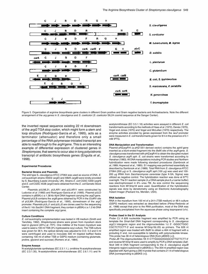
The presence of a cluster of seven contiguous genes forthe biosynthesis of arginine in S. clavuligerus wasunexpected since arg auxotrophic mutants map in fourdifferent loci in the S. coelicolor genome (Redenbach etal., 1996 ).
The hybridization studies of the S. coelicolor DNA withour probes suggest that there are at least two clusters ofarg genes in this model actinomycete. One clustercontaining argC,J,D,R and B while argH is located closebut not linked to the first subcluster. These results agreewith those of Hindle et al. (1994) who found an argCJBcluster in S. coelicolor and with recent data on the geneticmap of S. coelicolor (Sanger Center Sequencing Group).
In addition, the unstable argG of S. coelicolor mapsnear one of the ends of the S. coelicolor linear chromosome(Redenbach et al., 1996) at difference of what occurs in S.
clavuligerus where argG forms part of the arginine clusteras reported in this article. Unstable argG genes are alsopresent in Streptomyces lavendulae, Streptomyceslividans, Streptomyces cattleya, Streptomyces scabies andStreptomyces alboniger. There are two types of argG genesin Streptomyces (Rodríguez-García et al., 1995) and it islikely that the stable argG genes of Streptomyces speciesmight be located in a large arg cluster whereas the unstableargG gene is located in a separate location near the endof the linear genome. This would suggest that argG suffereda translocation in a line of Streptomyces ancestors but notin other group of this large genus.
The absence of an argF gene, encoding ornithinecarbamoyltransferase (forming citrulline) in the arg clusterof S. clavuligerus is surprising; argF is not located in thevicinity, either upstream or downstream of the S.clavuligerus arg cluster although argF is in a central positionin the arg cluster of mycobacteria. Attempts to clone thisgene by complementation of argF mutants of E. coli wereunsuccessful, perhaps due to the lack of expression of itspromoter in E. coli.
Regulation of the expression of the arginine cluster inS. clavuligerus by the arginine level is exerted at thetranscription level. Two canonical 18-nt Arg-boxes with 70to 80% similarity to the consensus Streptomyces Arg-box(Rodríguez-García et al., 1997) occur upstream of the argCand argG genes but only one 18-nt sequence with 50%similarity to Arg-boxes, is present upstream of argR(nucleotides 4581-4598). Indeed formation of themonocistronic argR transcript appears to be weak andconstitutive (Table 1). This is in agreement with the resultsof arginine regulation in E. coli in which the presence oftwo arginine-boxes exerts a cooperative effect resulting ina stronger binding of the regulatory ArgR protein. Bindingof the B. subtilis AhrC arginine repressor to the arg-boxesof argC has been reported previously (Rodríguez-Garcíaet al., 1997). Expression of the arginine biosynthesis genesin the arginine R-disrupted mutant is in the order of 10 to44-fold that of the wild type strain (depending on themedium) (Table I). Arg-boxes are also present upstreamof argC (Hindle et al.,1994), argG (Rodríguez-García etal., 1995) and argH (Sanger Center Sequencing Group) ofS. coelicolor suggesting a similar type of regulation.
Our results are compatible with the co-transcription ofthe argG and argH genes. However the differences inintensity between the 3.3 kb and the 1.5 kb band obtainedby Northern hybridization with the argG probe suggest that
Table 1. Relative Percentage of Expression of the Early and Late arg Genes as Shown by S1 Protection Studies
Strain Transcription Increase* (fold)
S. clavuligerus 27064 S. clavuligerus argR::aph
TSB GSPG TSB GSPG TSB GSPG
Probe CJ mRNA argCJBDR 1.5 3.5 66.5 100.01 44.3 28.5Probe DR mRNA argCJBDR 0.7 2.8 30.9 100.02 44.1 35.7
mRNA argR 0.2 0.5 0.5 0.4 2.5 0.8Probe GH mRNA argGH 3.0 10.5 91.7 100.03 30.5 9.5
1 60,959 net counts2 906,824 net counts3 36,095 net counts* The expression increase is the ratio of mRNA expression of S. clavuligerus argR::aph in relation to the wild type strain
The Arginine Biosynthesis Cluster of Streptomyces clavuligerus 549
FIgure 5. Organization of arginine biosynthesis gene clusters in different Gram positive and Gram negative bacteria and Archaebacteria. Note the differentarrangement of the arg genes in S. clavuligerus and S. coelicolor (S. coelicolor StL24 cosmid sequence at the Sanger Center).
the inverted repeat sequence existing 22 nt downstreamof the argG TGA stop codon, which might form a stem andloop structure (Rodríguez-García et al., 1995), acts as aterminator (attenuator) and therefore only a smallpercentage of the RNA polymerase-iniciated transcript areable to readthrough to the argH gene. This is an interestingexample of differential expression of clustered genes inStreptomyces, that seems to occur also in long polycistronictranscript of antibiotic biosynthesis genes (Enguita et al.,1998).
Experimental Procedures
Bacterial Strains and PlasmidsThe wild type S. clavuligerus ATCC 27064 was used as source of DNA. E.coli auxotroph strains XSlD2 (argE) and XB25 (argB) were kindly providedby S. Baumberg (Leeds University, UK). Strains E. coli CGSC 5359 (argH)and E. coli CGSC 4538 (argD) were obtained from the E. coli Genetic StockCenter.
Plasmids pULML31, pULAR1 and pULAR11 were constructed byLudovice et al. (1992) and Rodríguez-García et al. (1995). Plasmid pULDcontains in pBSK(+) a 1.38 kb KpnI DNA fragment carrying the argD gene.Plasmid pULB contains the argB gene obtained by PCR in the EcoRV siteof pULMA (Rodríguez-García et al., 1995), donwstream of the argCpromoter. Plasmids pULJ1 and pULJ2 are clones used for the sequencingof the 6.1 kb Sau3A1 DNA fragment; they contain in pBSK(+) a 1.2 kb DNAinsert containing the complete argJ gene.
Culture ConditionsE. coli auxotrophy complementation was tested in VB medium (Smith andYanofsky, 1962). Streptomyces strains were grown from mycelium stockkept in glycerol (20%) at -75ºC. One milliliter of the frozen mycelium wasused to seed a 100 ml TSB (3% trypticaseine-soy) culture. The TSB culturewas grown for 36 h, the optical density was adjusted to O.D. 5.0 and 5 mlwere centrifuged and used to inoculate 500 ml trippled baffled flaskscontaining 100 ml of either TSB or GSPG medium (containing glutamate,proline, glycerol and sucrose) (Romero et al., 1984).
Enzyme AssaysN-acetylglutamate synthetase (EC 2.3.1.1.), ornithine N-acetyltransferase(EC 2.3.1.35), N-acetylornithine aminotransferase (EC 2.6.1.11) and N-
acetylornithinase (EC 3.5.1.16) activities were assayed in different E. colitransformants according to the methods of Haas et al. (1972), Denés (1970),Vogel and Jones (1970) and Vogel and McLellan (1970) respectively. Theenzyme activities encoded by genes expressed from the lacZ promoterwere measured in E. coli transformants grown for 8 h in the presence of 0.1mM IPTG.
DNA Manipulation and TransformationPlasmid pHZargR-K (a pHZ1351-derived vector) contains the aphII geneinserted as a blunt-ended fragment into the BstXl site of the argR gene. S.clavuligerus was transformed with pHZargR-K to obtain the disrupted mutantS. clavuligerus argR::aph. E. coli strains were transformed according toHanahan (1983). All DNA manipulations including PCR studies and Northernhybridization were made following standard procedures (Sambrook etal.,1989; Hopwood et al., 1985). S1 mapping was performed essentially asdescribed by Sambrook et al. (1989). Total RNA from S. clavuligerus ATCC27064 (200 µg) or S. clavuligerus argR::aph (100 µg) was used and 100-200 µg tRNA from Saccharomyces cerevisiae (type X-SA, Sigma) wasutilized as negative control. The hybridization reaction was done at 67ºCovernight. The S1 reaction sample (4 µl DNA sample plus 6 µl carrier buffer)was electrophoresed in 6% urea 7M. As size control the sequencingreactions from M13mp18 were used. Quantification of the hybridizationsignals was done by densitometry using an Electronic AutoradiographyInstant Imager (Packard Ins, Meriden, USA).
RNA IsolationRNA in the mycelium from 100 ml of a 24 h (TSB medium) or 48 h culture(GSPG medium) was extracted as described before (Pérez-Redondo etal., 1998) except that prior to the RNA purification, most of the DNA wasspecifically precipitated from the aqueouse phase using isopropanol (v/v).
Probes Used in the S1 AnalysisProbe CJ: A 626 nucleotide fragment was amplified by PCR using astemplate the Smal-SalI DNA fragment corresponding to S. clavuligerusargCJ intergenic region and the oligonucleotides 1J: 5' -GGGCTGGCCGCTCTCCTT-3' and reverse M13mp18(-20) as primers. The 626 ntamplified region was treated with BstXI to obtain a 464 nt fragment with aprotuding 5' end, non-susceptible to labelling with polynucleotide kinase.This probe has 36 nt of heterologous DNA [from pBSKS (+)].Probe JB: The oligonucleotides 1B: 5'-CTTGGGGAGGGCGTTGTTCTT-3'and reverse M13mp18 were used to amplify by PCR a DNA template (SalI-NcoI 349 nt DNA fragment corresponding to the S. clavuligerus argJBintergenic region) subcloned in pBSKS(+). The 354 nt amplified region wasdigested with KpnI giving a 290 nt probe that contains 21 nt of heterologousDNA [corresponding to pBSKS (+)].
550 Rodríguez-García et al.
Probe DR: An oligonucleotide 1R: 5´-ACGCTCAGTCCGTTGTCCG-3'corresponding to the non-coding strand of argR was purified by PAGE and10 pmol were labelled at its 5' end with [γ−32P] ATP (>185 TBq/mmol,Amersham Ltd., England) and 8 units of T4 polynucleotide kinase (MBIFermentas, Lithuania). The labelled oligonucleotide was purified byprecipitation with ammonium acetate using glycogen (20 µg) as carrier.Using this oligonucleotide and M13mp18 (-20) as primers a labelled DNAprobe of 449 nt corresponding to S. clavuligerus argDR intergenic regionwas amplified by PCR. The 76 nt corresponding to the 3'-end of the non-labelled chain correspond to pBSSK(+).Probe RG: The oligonucleotide 1R: 5' -GTTGCTGAATTCGCGGAGCTGCACGACATC-3' , (in which the nt in italics correspond to pBSSK(+) nonhomologue DNA) and 2G: 5'-CTTGATCGCAGGGAGGCAGTA-3' were usedto amplify from plasmid pULAR10 (Rodríguez-García et al., 1995) a 649 ntDNA fragment that was digested with EcoRI.ProbeGH: A 561 nt DNA probe with 11 nt non-homologous nucleotides atthe 5'-end of the non-labelled strand was amplify by PCR using theoligonucleotides 1G: 5'-GTTGCTGAATTCGGCTGGTCGGCATCAAGT-3'and 2H: 5'- CGAACCGT CCGCCCCAGAG-3'.
All the probes were purified from 1.5% agarose gels using the QuiaexGel Extraction Kit (Quiagen, Germany) and denatured (except probe DR),by incubation for 5 min at 65ºC in the presence of NaOH (final concentration0.1 M). The probes were precipitated with sodium acetate pH 5.2:ethanolin the presence of glucogen (10 µg) as carrier, suspended in reaction buffer(Sambrook et al., 1989) and labelled with [γ-32P] ATP and 14 units of T4polynucleotide kinase. After 50 min at 37ºC the enzyme was inactivated byheating and the probes were purified by precipitation with ammoniumacetate:ethanol. Additionally, probes I to VII (Figure 2) were used for lowresolution S1 mapping, Northern analysis or hybridization with S. coelicolorgenome.
Acknowledgements
This work was supported by grants from the CICYT (Madrid) Bio96-0827and by Antibióticos SA, León. We thank H. Kieser (Norwich, U.K.) forproviding the S. coelicolor cosmid library. Álvaro de la Fuente and RosarioPérez-Redondo received fellowships from the PFPI (Madrid) and theUniversity of León, respectively.
References
Bringel, F., Frey, L., Boivin, S., and Hubert, J.C. 1997. Arginine biosynthesisand regulation in Lactobacillus plantarum: the carAB gene and theargCJBDF cluster are divergently transcribed. J. Bacteriol. 179: 2697-2706.
Crabeel, M., Charlier, D., Cunin, R., and Glansdorff, N. 1979. Cloning andendonuclease restriction analysis of argF and of the control region of theargECBH bipolar operon in Escherichia coli. Gene 5: 207-231.
Denés, G. 1970. Ornithine Acetyltransferase. Methods Enzymol. 17A: 273-277.
Eastcott, M., Griffin, A., Barton, B., and Baumberg, S. 1998. Studies on anargJ homologue located within the clavulanic acid gene cluster inStreptomyces clavuligerus. Abstract of the Genetics of IndustrialMicrooganisms (GIM 98) Congress, Jerusalem, Israel, p. 88
Enguita, F.J., Coque, J.J.R., Liras, P., and Martín, J.F. 1998. The nine genesof the Nocardia lactamdurans cephamycin cluster are transcribed intolarge mRNAs from three promoters, two of them located in a bidirectionalpromoter region. J. Bacteriol. 180: 5489-5494.
Haas, D., Holloway, B.W., Schamböck, A., and Leisinger, T. 1977. Thegenetic organization of arginine biosynthesis in Pseudomonas aeruginosa.Mol. Gen. Genet. 154: 7-22.
Hanahan, D. 1983. Studies of transformation of Escherichia coli withplasmids. J. Mol. Biol. 166: 557-580.
Hass, D., Kurer, V., and Leisinger, T. 1972. N-Acetylglutamate synthetaseof Pseudomonas aeruginosa. An assay in vitro and feedback inhibitionby arginine. Eur. J. Biochem. 31: 290-295.
Higgins, D.G., and Sharp, P.M. 1989. Fast and sensitive multiple sequencealignement on a microcomputer. Comp. Appl. Biosci. 5: 151-153.
Hindle, Z., Callis, R., Dowden, S., Rudd, B.A.M., and Baumberg, S. 1994.Cloning and expression in Escherichia coli of a Streptomyces coelicolorA3(2) argCJB gene cluster. Microbiology 140: 311-320.
Hodgson, J.E., Fosberry, A.P., Rawlinson, N.S., Ross, H.N.M., Neal, R.J.,Arnell, J.C., Earl, A.J., and Lawlor, E.J. 1995. Clavulanic acid biosynthesisin Streptomyces clavuligerus: gene cloning and characterization. Gene166: 49-55.
Hopwood, D.A., Bibb, M.J., Chater, K.F., Kieser, T., Bruton, C.J., Kieser,H.M., Lydiate, D.J., Smith, C.P., Ward, J.M., and Schrempf, H. (1985)Genetic manipulation of Streptomyces: a laboratory manual. The JohnInnes Foundation. Norwich, U.K.
Kunst, F., Ogasawara, N., Moszer, I., Albertini, A.M., Alloni, G., Azevedo,
V., Bertero, M.G., Bessieres, P., Bolotin, A., Borchert, S., Borriss, R.,Boursier, L., Brans, A., Braun, M., Brignell, S.C., Bron, S., Brouillet, S.,Bruschi, C.V., Caldwell, B., Capuano, V., Carter, N.M., Choi, S.K., Codani,J.J., Connerton, I.F., Danchin, A., et al. 1997. The complete genomesequence of the gram-positive bacterium Bacillus subtilis. Nature 390:249-256.
Leskiw, B.K., Lawlor, E.J., Fernández-Ábalos, J.M. and Chater, K.F. 1991.TTA codons in some genes prevent their expression in a class ofdevelopmental, antibiotic-negative, Streptomyces mutants. Proc. Natl.Acad. Sci. USA 88: 2461-2465.
Ludovice, M., Martín, J.F., Carrachás, P., and Liras, P. 1992. Characterizationof the Streptomyces clavuligerus argC gene encoding N-acetyl glutamyl-phosphate reductase: Expression in Streptomyces lividans and effect onclavulanic acid production. J. Bacteriol. 174: 4606-4613.
Matsumoto, H., Hosogaya, S., Suzuki, K., and Tazaki, T. 1975. Argininegene cluster of Serratia marcescens. Jpn. J. Microbiol. 19: 35-44.
Mountain, A., Mann, N.H., Munton, R.N., and Baumberg, S. 1984. Cloningof a Bacillus subtilis restriction fragment complementing auxotrophicmutants of eight Escherichia coli genes of arginine biosynthesis. Mol.Gen. Genet. 197: 82-89.
North, A.K., Smith M.C.M., and Baumberg, S. 1989. Nucleotide sequenceof the Bacillus subtilis arginine regulatory gene and homology of itsproducts to the E. coli arginine repressor. Gene 80: 29-38.
O’Reilly, M., and Devine, K.M. 1994. Sequence and analysis of the citrullinebiosynthetic operon argC-F from Bacillus subtilis. Microbiology 140: 1023-1025.
Pérez-Redondo, R., Rodríguez-García, A., Martín J.F., and Liras, P. 1998.The claR gene of Streptomyces clavuligerus, encoding a LysR-typeregulatory protein controlling clavulanic acid biosynthesis, is linked to theclavulanate-9-aldehyde reductase (car) gene. Gene 211: 311-321.
Picard, F.J., and Dillon, J.R. 1989. Cloning and organization of sevenarginine biosynthetic genes from Neisseria gonorrhoeae. J. Bacteriol. 171:1644-1651.
Piggot PJ., and Hoch, J.A. (1985) Revised genetic linkage map of Bacillussubtilis. Microbiol. Rev. 49: 158-179.
Prozesky, O.W. 1968. Transductional analysis of arginine less mutants inProteus mirabilis. J. Gen. Microbiol. 54: 127-143.
Redenbach, M., Kieser, H.M., Denapaite, D., Eichner, A., Cullum, J., Kinashi,H., and Hopwood, D.A. 1996. A set of ordered cosmids and a detailedgenetic and physical map for the 8 Mb Streptomyces coelicolor A3(2)chromosome. Mol. Microbiol. 21: 77-96.
Rodríguez-García, A., Ludovice, M., Martín, J.F., and Liras, P. 1997. Arginineboxes and the argR gene of Streptomyces clavuligerus: evidence for aclear regulation of the arginine pathway. Mol. Microbiol. 25: 219-228.
Rodríguez-García, A., Martín, J.F., and Liras, P. 1995. The argG gene ofStreptomyces clavuligerus has low homology to unstable argG from otheractinomycetes: Effect of amplification on clavulanic acid biosynthesis.Gene 167: 9-15.
Romero, J., Liras, P., and Martín, J.F. 1984. Dissociation of cephamycinand clavulanic acid biosythesis in Streptomyces clavuligerus. Appl.Microbiol. Biotechnol. 20: 318-325.
Sakanyan, V., Petrosyan, P., Lecocq, M., Boyen, A., Legrain, C., Demarez,M., Hallet, J.N., and Glansdorff, N. 1996. Genes and enzymes of theacetyl cycle of arginine biosynthesis in Corynebacterium glutamicum:enzyme evolution in the early steps of the arginine pathway. Microbiology142: 99-108.
Sambrook, J., Fritsch, E.F., and Maniatis, T. 1989. Molecular cloning: alaboratory manual, 2nd ed. Cold Spring Harbor Laboratory, Cold SpringHarbor, N.Y.
Sanderson, K.E. 1970. Current linkage map of Salmonella typhimurium.Bacteriol. Rev. 34: 176-193.
Smith, O.H., and Yanofsky, C. 1962. Biosynthesis of tryptophan. MethodsEnzymol. 5: 794-806.
Strohl, W.R. 1992. Compilation and analysis of DNA sequences associatedwith apparent streptomycete promoters. Nucleic Acid Res. 20: 961-974.
Valentine, B.B., Bailey, C.R., Doherty, A., Morris, J., Elson, S.W., Baggaley,K.H. and Nicholson, N.H. 1993. Evidence that arginine is a later metabolicintermediate than ornithine in the biosynthesis of clavulanic acid byStreptomyces clavuligerus. J. Chem. Soc. Chem. Commun. 1993: 1210-1212.
Vogel, H.J., and Jones, E.E. 1970. Acetylornithine δ-aminotransferase(Escherichia coli). Methods Enzymol. 17A: 260-264.
Vogel, H.J., and McLellan, W.L. 1970. Acetylornithinase. Methods Enzymol.17A: 265-269.
Woods, S.A., Miles J.S., and Guest, J.R. 1988. Sequence homologiesbetween argininosuccinase, aspartase and fumarase: a family ofstructurally-related enzymes. FEMS Microbiol. Lett. 51: 181-186.
Yonaha, K., Nishie, M., and Aibara, S. 1992. The primary structure of α-amino acid:pyruvate aminotransferase. J. Biol. Chem. 267: 12506-12510.


![Arginine...Arginine vasotocin ([8-arginine]-oxytocin) (AVT), the primary antidiuretic principle in submammalian vertebrates, has been reported to be present in mammalian pituitary](https://static.fdocuments.in/doc/165x107/5e81a7e1761a1c6f5832a8ca/arginine-arginine-vasotocin-8-arginine-oxytocin-avt-the-primary-antidiuretic.jpg)