CHARACTERIZATION AND DEVELOPMENT OF EMBELIA RIBES …
22
www.wjpps.com Vol 9, Issue 8, 2020. 2570 Jain et al. World Journal of Pharmacy and Pharmaceutical Sciences CHARACTERIZATION AND DEVELOPMENT OF EMBELIA RIBES PHYTOSOMES Chanchal Jain*, Sonal Gupta and Dr. A. K. Singhai M. Pharm, Department of Pharmaceutics, Lakshmi Narain College of Pharmacy, Bhopal (M.P.). ABSTRACT Embelia ribes, in particular embelin isolated from dried berries of embelia ribes has a wide spectrum of biological activities. Belong to the family Myrsinacae It is commonly known as false black pepper or Vidanga it is widely distributed throughout india.in ayurveda and siddha it is considered widely beneficial in variety of disease. E. Ribes Seeds are rich in alkaloids, flavonoids and phenolic compounds Hence,in present work we have analyzed The DPPH free radical scavenging activity of Hydroalcoholic extract of Embelia ribes were 21.64%, 27.43%, 32.39%, 37.36%, 41.25%, 49.96% at different concentration of 10 μg/ml, 20 μg/ml, 40 μg/ml, 60 μg/ml, 80 μg/ml, 100 μg/ml respectively. The Embelia ribes seed extract has been showed the robust scavenging activity at 100μg/ml, which was 49.96% as compared to Ascorbic acid 84.13%. This activity was lower than ascorbic acid, Embelia ribes hydroalcoholic extract exhibited potent scavenging activity (IC 50 -103.93). Reference standard ascorbic acid showed IC 50 - 17.681. In this study, we prepared the, Embelia ribes -phospholipids complex to improve the lipophilic properties of Embelia ribes. We prepared the complex with different quantity ratios of phospholipids: cholesterol and Embelia ribes such as 1:1:1, 1:2:1, 2:1:1, 2:3:1.The phytosomes formulation having 2:1 ration of phospholipid and cholesterol and 1.0% w/v extract of embelia ribes (F10) showing the greatest entrapment efficiency,(73.32+0.25) with small particle size (313.25) was further evaluated for drug release study. The phytosomal formulation of Embelia ribes (a complex of E.ribes with phosphatidylcholine) has been shown to improve E.ribes bioavailability. WORLD JOURNAL OF PHARMACY AND PHARMACEUTICAL SCIENCES SJIF Impact Factor 7.632 Volume 9, Issue 8, 2570-2591 Research Article ISSN 2278 – 4357 *Corresponding Author Chanchal Jain M. Pharm, Department of Pharmaceutics, Lakshmi Narain College of Pharmacy, Bhopal (M.P.). Article Received on 24 June 2020, Revised on 14 July 2020, Accepted on 03 August 2020 DOI: 10.20959/wjpps20208-16987
Transcript of CHARACTERIZATION AND DEVELOPMENT OF EMBELIA RIBES …
www.wjpps.com Vol 9, Issue 8, 2020.
2570
Jain et al. World Journal of Pharmacy and Pharmaceutical Sciences
CHARACTERIZATION AND DEVELOPMENT OF EMBELIA RIBES
PHYTOSOMES
Chanchal Jain*, Sonal Gupta and Dr. A. K. Singhai
M. Pharm, Department of Pharmaceutics, Lakshmi Narain College of Pharmacy, Bhopal
(M.P.).
ABSTRACT
Embelia ribes, in particular embelin isolated from dried berries of
embelia ribes has a wide spectrum of biological activities. Belong to
the family Myrsinacae It is commonly known as false black pepper or
Vidanga it is widely distributed throughout india.in ayurveda and
siddha it is considered widely beneficial in variety of disease. E. Ribes
Seeds are rich in alkaloids, flavonoids and phenolic compounds
Hence,in present work we have analyzed The DPPH free radical
scavenging activity of Hydroalcoholic extract of Embelia ribes were
21.64%, 27.43%, 32.39%, 37.36%, 41.25%, 49.96% at different
concentration of 10 µg/ml, 20 µg/ml, 40 µg/ml, 60 µg/ml, 80 µg/ml,
100 µg/ml respectively. The Embelia ribes seed extract has been showed the robust
scavenging activity at 100µg/ml, which was 49.96% as compared to Ascorbic acid 84.13%.
This activity was lower than ascorbic acid, Embelia ribes hydroalcoholic extract exhibited
potent scavenging activity (IC50-103.93). Reference standard ascorbic acid showed IC50 -
17.681. In this study, we prepared the, Embelia ribes -phospholipids complex to improve the
lipophilic properties of Embelia ribes. We prepared the complex with different quantity ratios
of phospholipids: cholesterol and Embelia ribes such as 1:1:1, 1:2:1, 2:1:1, 2:3:1.The
phytosomes formulation having 2:1 ration of phospholipid and cholesterol and 1.0% w/v
extract of embelia ribes (F10) showing the greatest entrapment efficiency,(73.32+0.25) with
small particle size (313.25) was further evaluated for drug release study. The phytosomal
formulation of Embelia ribes (a complex of E.ribes with phosphatidylcholine) has been
shown to improve E.ribes bioavailability.
WORLD JOURNAL OF PHARMACY AND PHARMACEUTICAL SCIENCES
SJIF Impact Factor 7.632
Volume 9, Issue 8, 2570-2591 Research Article ISSN 2278 – 4357
*Corresponding Author
Chanchal Jain
M. Pharm, Department of
Pharmaceutics, Lakshmi
Narain College of Pharmacy,
Bhopal (M.P.).
Article Received on
24 June 2020,
Revised on 14 July 2020,
Accepted on 03 August 2020
DOI: 10.20959/wjpps20208-16987
www.wjpps.com Vol 9, Issue 8, 2020.
2571
Jain et al. World Journal of Pharmacy and Pharmaceutical Sciences
KEYWORDS: embelia ribes, phytosomes, antioxidant activity antidiabetic, hydro alcoholic
extraction.
INTRODUCTION
The nano-carrier used in novel drug delivery system of herbal drugs has a potential future,
improving the activity and overcoming the problem associated with herbal constituents. Any
type of medicament could be delivered to its specific site of action with the help of novel
drug delivery system. Researchers are currently underway to develop an ideal drug delivery
system which satisfies the need of targeted site of action. Biological membrane presents a
barrier through which a drug must pass before it gets absorbed or excreted. Lipid solubility
and molecular size of drug molecule pose two major limiting factors to pass the biological
membrane by which drug can be absorbed systematically following oral or topical
administration. During the last century chemical and pharmacological studies have been
performed on a lot of plant extracts in order to know their chemical composition and confirm
the indications of traditional medicine. Preparations of Phyto medicine has been used for
health maintenance since ancient times. The Phytomedicines posses a lot of therapeutic uses.
Phytosome is vesicular drug delivery system in which phytoconstituents of herb extract
surround and bound by lipid (one phyto-constituent molecule linked with at least one
phospholipid molecule). Phytosome protect valuable component of herbal extract from
destruction by digestive secretion and gut bacteria and because of which they shows better
absorption which produces better bioavailability and improved pharmacological and
pharmacokinetic parameters than conventional herbal extract. Phytosomes is the combination
of two words, the term “PHYTO” means plant while “SOME” means cell-like. The
formulation is developed by encapsulating the plant material or plant extract within the
spherical cell like structure, which is an advanced nano-sphere or cell forms of herbal
products that are better absorbed. Phytosomes produces better pharmacokinetic and
pharmacodynamic profile of drug than conventional herbal formulations. It‟s a novel
emerging technique that is applied to phytopharmaceuticals for the enhancement of
bioavailability of natural plant extract for medicinal applications. Phytomedicines, complex
chemical mixture prepared from plants, have been used in India and worldwide from the very
beginning of human civilization and continue to have widespread popular use. Phytosomes
are more bioavailability as compared to herbal extract owing to their enhancedcapacity to
cross the lipid rich biomembranes and finally reaching the blood. Phosphatidylcholine is
www.wjpps.com Vol 9, Issue 8, 2020.
2572
Jain et al. World Journal of Pharmacy and Pharmaceutical Sciences
phospholipids. It is a key component of phytosomes process. Phospholipids are employed as
natural digestive aids and carriers for water soluble and lipid soluble nutrients.
Principle of Phytosome Technology
The phytochemical constituents (flavonoids and terpenoides) of the extracts provide them for
the direct complexation with Phosphatidylcholine. Phytosome results from the reaction of a
stoichiometric amount of the phospholipid with the standardized extract or polyphenolic
constituents in a non-polar solvent. The Phosphatidylcholine is a bi-functional compound
composed of lipophilic phosphatidyl moiety and the hydrophilic choline moiety. The choline
head of phosphatidylcholine molecule binds to phytocomponent while the lipid soluble
phosphatidyl portion comprises the body and tail which then envelops the choline bound
material. Hence, the Phytoconstituents build up a lipid compatible molecular complex with
phospholipid also called as phyto-phospholipid complex.
Biological properties
Phytosomes are novel complex utilized; hence they produce more bioavailability and better
result thanes which are better absorbed and the conventional herbal extract or non-complex
extracts, which has been demonstrated by pharmacokinetic studies or by pharmacodynamic
tests in experimental animals and in human subjects.
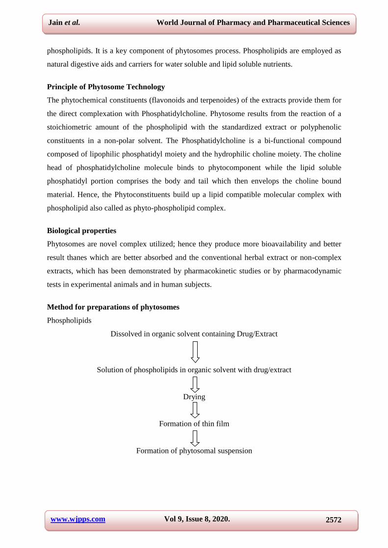
Method for preparations of phytosomes
Phospholipids
Dissolved in organic solvent containing Drug/Extract
Solution of phospholipids in organic solvent with drug/extract
Drying
Formation of thin film
Formation of phytosomal suspension
www.wjpps.com Vol 9, Issue 8, 2020.
2573
Jain et al. World Journal of Pharmacy and Pharmaceutical Sciences
MATERIAL AND METHODS
Collection of Plant material
Seeds of Embelia Ribes were collected from local area of Bhopal in the month of January,
2020 The plant material was identified and authenticated by depositing the herbarium
specimens in Botany Department, Dr.javinder Mehta career college, Bhopal Madhya
Pradesh,india. under the voucher no. Career/Herb/2020/010 Accession no.
1 Defatting of plant material
Seeds of Embelia ribes were shade dried at room temperature. The shade dried plant material
was coarsely powdered and subjected to extraction with petroleum ether by maceration. The
extraction was continued till the defatting of the material had taken place.
2 Extraction by maceration process
100 gm of dried powdered seeds of Embelia ribes has been extracted with hydroalcoholic
solvent (ethanol: water: 70:30) using maceration method for 48 hrs, filtered and dried using
vacuum evaporator at 400C.
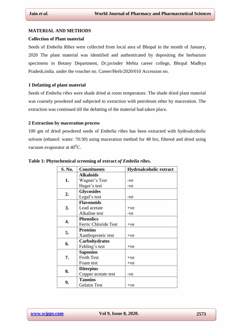
Table 1: Phytochemical screening of extract of Embelia ribes.
S. No. Constituents Hydroalcoholic extract
1.
Alkaloids
Wagner‟s Test
Hager‟s test
-ve
-ve
2. Glycosides
Legal‟s test
-ve
3.
Flavonoids
Lead acetate
Alkaline test
+ve
-ve
4. Phenolics
Ferric Chloride Test
+ve
5. Proteins
Xanthoproteic test
+ve
6. Carbohydrates
Fehling‟s test
+ve
7.
Saponins
Froth Test
Foam test
+ve
+ve
8. Diterpins
Copper acetate test
-ve
9. Tannins
Gelatin Test
+ve
www.wjpps.com Vol 9, Issue 8, 2020.
2574
Jain et al. World Journal of Pharmacy and Pharmaceutical Sciences
Antioxidant activity of extract using DPPH method
DPPH scavenging activity was measured by the spectrophotometer. Stock solution (6 mg in
100ml methanol) was prepared such that 1.5 ml of it in 1.5 ml of methanol gave an initial
absorbance. Decrease in the absorbance in presence of sample extract at different
concentration (10- 100 µg/ml) was noted after 15 minutes. 1.5 ml of DPPH solution was
taken and volume made till 3 ml with methanol, absorbance was taken immediately at 517
nm for control reading. 1.5 ml of DPPH and 1.5 ml of the test sample of different
concentration were put in a series of volumetric flasks and final volume was adjusted to 3 ml
with methanol. Three test samples were taken and each processed similarly. Finally the mean
was taken. Absorbance at zero time was taken for each concentration. Final decrease in
absorbance was noted of DPPH with the sample at different concentration after 15 minutes at
517 nm.
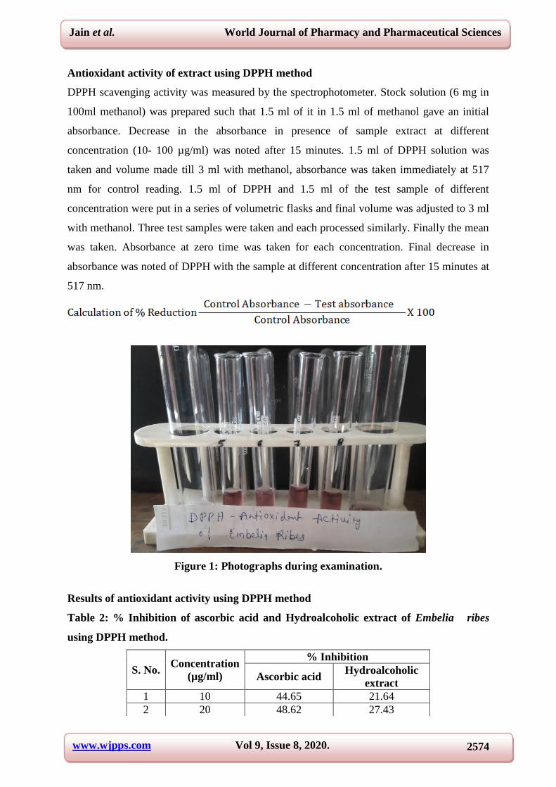
Figure 1: Photographs during examination.
Results of antioxidant activity using DPPH method
Table 2: % Inhibition of ascorbic acid and Hydroalcoholic extract of Embelia ribes
using DPPH method.
S. No. Concentration
(µg/ml)
% Inhibition
Ascorbic acid Hydroalcoholic
extract
1 10 44.65 21.64
2 20 48.62 27.43
www.wjpps.com Vol 9, Issue 8, 2020.
2575
Jain et al. World Journal of Pharmacy and Pharmaceutical Sciences
The outcome of DPPH (1,1-Diphenyl-2-picrylhydrazyl radical) radical scavenging activity of
Hydroalcoholic extract of Embelia ribes was showed and The DPPH free radical scavenging
activity of Hydroalcoholic extract of Embelia ribes were 21.64%, 27.43%, 32.39%, 37.36%,
41.25%, 49.96% at different concentration of 10 µg/ml, 20 µg/ml, 40 µg/ml, 60 µg/ml, 80
µg/ml, 100 µg/ml respectively. The Embelia ribes seed extract has been showed the robust
scavenging activity at 100µg/ml, which was 49.96% as compared to Ascorbic acid 84.13%.
This activity was lower than ascorbic acid, Embelia ribes hydroalcoholic extract exhibited
potent scavenging activity (IC50-103.93). Reference standard ascorbic acid showed IC50 -
17.681.
In vitro anti diabetic activity of Hydroalcoholic extract of Embelia ribes
1 Inhibition of alpha amylase enzyme
Preparation of standard: 10 mg acarbose was dissolved in 10 ml methanol, and various
aliquots of 100- 1000μg/ml were prepared in methanol.
Preparation of sample: 100 mg of dried extract was extracted with 100 ml methanol, filter,
and make up the volume up to 100 ml. 500 µl of this extract was for the estimation of enzyme
inhibition.
METHOD: A total of 500 µl of test samples and standard drug (100-500µg/ml) were added
to 500 µl of 0.20 mM phosphate buffer (pH 6.9) containing α-amylase (0.5mg/ml) solution
and were incubated at 25°C for 10 min. After these, 500 µl of a 1% starch solution in 0.02 M
sodium phosphate buffer (pH 6.9) was added to each tube. The reaction mixtures were then
incubated at 25°C for 10 min. The reaction was stopped with 1.0 ml of 3, 5 dinitrosalicylic
acid colour reagent. The test tubes were then incubated in a boiling water bath for 5 min,
cooled to room temperature.
3 40 65.34 32.39
4 60 69.65 37.36
5 80 77.41 41.25
6 100 84.13 49.96
IC 50 17.681 103.93
www.wjpps.com Vol 9, Issue 8, 2020.
2576
Jain et al. World Journal of Pharmacy and Pharmaceutical Sciences
The reaction mixture was then diluted after adding 10 ml distilled water and absorbance was
measured at 540 nm. Control represent 100% enzyme activity and were conducted in similar
way by replacing extract with vehicle.
A 0 - A t
% Inhibition = ________ X 100
A 0
Where A 0 is the absorbance of the control and A t is the absorbance of the sample.
Results of in vitro anti diabetic activity of Hydroalcoholic extract of Embelia ribes.
Table 4: Absorbances of Hydroalcoholic extract of Embelia ribes.
S. No Acarbose Embelia ribes
Conc. Absorbance Absorbance
1. 100 0.369 0.458
2. 200 0.226 0.321
3. 300 0.145 0.225
4. 400 0.098 0.125
5. 500 0.065 0.098
Control Absorbance = 0.698
Table 4.1: % Inhibition of Hydroalcoholic extract of Embelia ribes.
S. No. Acarbose Embelia ribes
Conc. % Inhibition % Inhibition
1. 100 47.13467 40.974
2. 200 67.62178 50.573
3. 300 79.22636 55.301
4. 400 85.95989 62.034
5. 500 90.68768 65.473
IC 50 value 71.52 221.16
Formulation development of phytosomes
Preparation of phytosomes
The complex was prepared with phospholipids: Cholesterol and Embelia ribes in the ratio of
1:1:1, 1:2:1, 2:1:1, 2:3:1 respectively. Weight amount of extract and phospholipids and
cholesterol were placed in a 100ml round-bottom flask and 25ml of dichloromethane was
added as reaction medium. The mixture was refluxed and the reaction temperature of the
complex was controlled to 50°C for 3 h. The resultant clear mixture was evaporated and 20
ml of n-hexane was added to it with stirring. The precipitated was filtered and dried under
vacuum to remove the traces amount of solvents. The dried residues were gathered and
www.wjpps.com Vol 9, Issue 8, 2020.
2577
Jain et al. World Journal of Pharmacy and Pharmaceutical Sciences
placed in desiccators overnight and stored at room temperature in an amber colored glass
bottle.
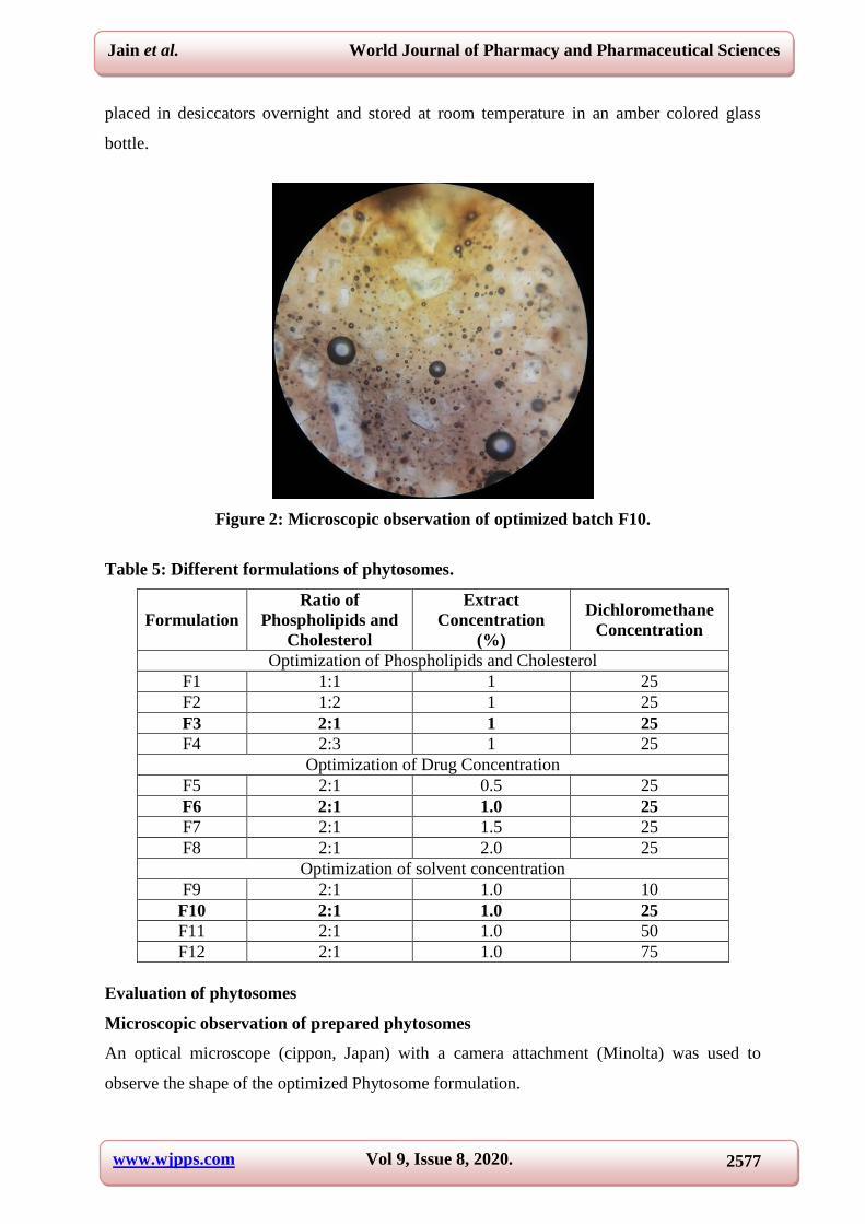
Figure 2: Microscopic observation of optimized batch F10.
Table 5: Different formulations of phytosomes.
Formulation
Ratio of
Phospholipids and
Cholesterol
Extract
Concentration
(%)
Dichloromethane
Concentration
Optimization of Phospholipids and Cholesterol
F1 1:1 1 25
F2 1:2 1 25
F3 2:1 1 25
F4 2:3 1 25
Optimization of Drug Concentration
F5 2:1 0.5 25
F6 2:1 1.0 25
F7 2:1 1.5 25
F8 2:1 2.0 25
Optimization of solvent concentration
F9 2:1 1.0 10
F10 2:1 1.0 25
F11 2:1 1.0 50
F12 2:1 1.0 75
Evaluation of phytosomes
Microscopic observation of prepared phytosomes
An optical microscope (cippon, Japan) with a camera attachment (Minolta) was used to
observe the shape of the optimized Phytosome formulation.
www.wjpps.com Vol 9, Issue 8, 2020.
2578
Jain et al. World Journal of Pharmacy and Pharmaceutical Sciences
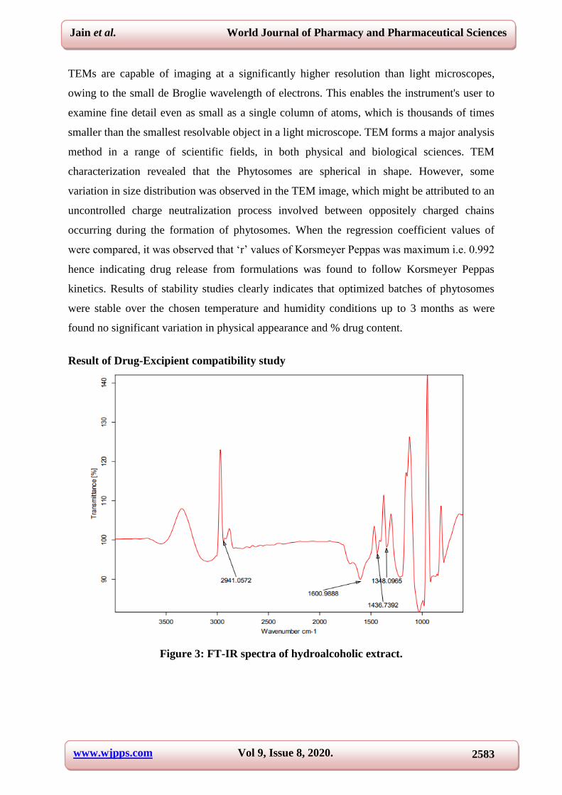
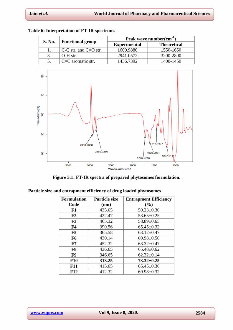
Drug Excipient compatibility study by FT-IR
IR spectra of physical mixture of drug and excipients were recorded by ATR (Attenuated
total reflection) techniques using Fourier transform infrared spectrophotometer. A base line
correction was made and the sample was directely mounted in IR compartment and scanned
at wavelengths 4000 cm-1
to 400 cm-1
.
Entrapment efficiency
Phytosome preparation was taken and subjected to centrifugation using cooling centrifuge
(Remi) at 12000 rpm for an hour at 4.
The clear supernatant was siphoned off carefully to separate the non entrapped flavonoids
and the absorbance of supernatant for non entrapped Embelia ribes was recorded at λmax
420.0 nm using UV/visible spectrophotometer (Labindia 3000+). Sediment was treated with
1ml of 0.1 % Triton x 100 to lyse the vesicles and diluted to 100 ml with 0.1 N HCl and
absorbance taken at 420.0 nm. Amount of quercetin in supernatant and sediment gave a total
amount of Embelia ribes in 1 ml dispersion. The percent entrapment was calculated by
following formula.
Particle size and size distribution
The particle size, size distribution and zeta potential of optimized phytosomes formulation
were determined by dynamic light scattering (DLS) using a computerized inspection system
(Malvern Zetamaster ZEM 5002, Malvern, UK). The electric potential of the phytosomes,
including its Stern layer (zeta potential) was determined by injecting the diluted system into a
zeta potential measurement cell.
Transmission Electron Microscopy: Surface morphology was determined by TEM, for
TEM a drop of the sample was placed on a carbon-coated copper grid and after 15 min it was
negatively stained with 1% aqueous solution of phosphotungustic acid. The grid was allowed
to air dry thoroughly and samples were viewed on a transmission electron microscopy (TEM
Hitachi, H-7500 Tokyo, Japan).
www.wjpps.com Vol 9, Issue 8, 2020.
2579
Jain et al. World Journal of Pharmacy and Pharmaceutical Sciences
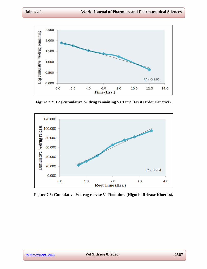
In Vitro dissolution rate studies
In vitro drug release of the sample was carried out using USP- type I dissolution apparatus
(Basket type). The dissolution medium, 900 ml 0.1N HCl was placed into the dissolution
flask maintaining the temperature of 37±0.50C and 75 rpm. 10 mg of prepared phytosomes
was placed in each basket of dissolution apparatus. The apparatus was allowed to run for 8
hours. Sample measuring 3 ml were withdrawn after every interval (30 min, 1 hrs, 2 hrs, 4
hrs, 6 hrs, 8 hrs, and 12 hrs.) up to 12 hours using 10 ml pipette. The fresh dissolution
medium (370C) was replaced every time with the same quantity of the sample and takes the
absorbance at 256.0 nm using spectroscopy.
Mathematical treatment of in-vitro release data: The quantitative analysis of the values
obtained in dissolution/release tests is easier when mathematical formulas that express the
dissolution results as a function of some of the dosage forms characteristics are used.
1. Zero Order kinetics: The pharmaceutical dosage forms following this profile release the
same amount of drug by unit of time and it is the ideal method of drug release in order to
achieve a pharmacological prolonged action. The following relation can, in a simple way,
express this model.
Qt = Qo + Ko t
where Qt is the amount of drug dissolved in time t, Qo is the initial amount of drug in the
solution (most times, Qo=0) and Ko is the zero order release constant58-62
.
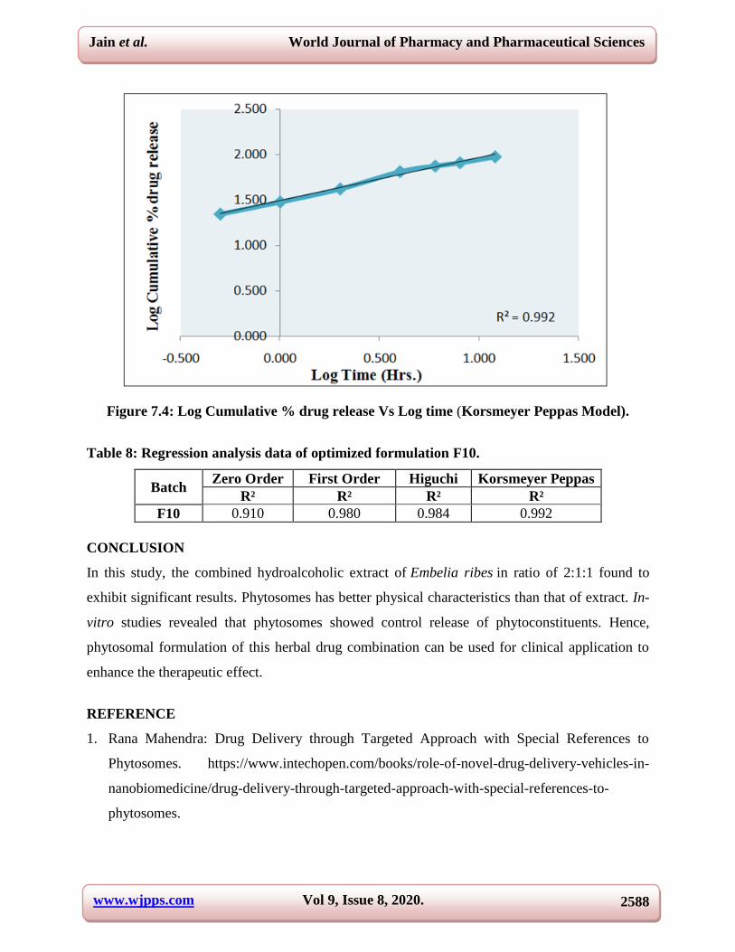
2. First Order kinetics: The following relation expresses this model.
where Qt is the amount of drug dissolved in time t, Qo is the initial amount of drug in the
solution and K1 is the zero order release constant.
In this way a graphic of the decimal logarithm of the released amount of drug versus time
will be linear. The pharmaceutical dosage forms following this dissolution profile, such as
those containing water-soluble drugs in porous matrices, release drug in a way that is
proportional to the amount of drug remaining in its interior, in such way, that the amount of
drug released by unit of time diminish.
www.wjpps.com Vol 9, Issue 8, 2020.
2580
Jain et al. World Journal of Pharmacy and Pharmaceutical Sciences
3. Higuchi Model: Higuchi developed several theoretical models to study the release of
water-soluble and low soluble drugs in semi-solid and/or solid matrixes. Mathematical
expressions were obtained for drug particles dispersed in a uniform matrix behaving as the
diffusion media. The simplified Higuchi model is expressed as.
Where Q is the amount of drug released in time t and KH is the Higuchi dissolution constant.
Higuchi model describes drug release as a diffusion process based in the Fick‟s law, square
root time dependent. This relation can be used to describe the drug dissolution from several
types of modified release pharmaceutical dosage forms such as transdermal systems and
matrix tablets with water-soluble drugs.
4. Korsmeyer Peppas Model: Korsmeyer et al. used a simple empirical equation to describe
general solute release behaviour from controlled release polymer matrices:
where Mt/M is fraction of drug released, a is kinetic constant, t is release time and n is the
diffusional exponent for drug release. ‟n‟ is the slope value of log Mt/M versus log time
curve. Peppas stated that the above equation could adequately describe the release of solutes
from slabs, spheres, cylinders and discs, regardless of the release mechanism.. Peppas used
this n value in order to characterize different release mechanisms, concluding for values for a
slab, of n =0.5 for fickian diffusion and higher values of n, between 0.5 and 1.0, or n =1.0, for
mass transfer following a non-fickian model. In case of a cylinder n =0.45 instead of 0.5, and
0.89 instead of 1.0. This equation can only be used in systems with a drug diffusion
coefficient fairly concentration independent. To the determination of the exponent n the
portion of the release curve where Mt/M < 0.6 should only be used. To use this equation it is
also necessary that release occurs in a one-dimensional way and that the system width-
thickness or length-thickness relation be at least 10. A modified form of this equation was
developed to accommodate the lag time (l) in the beginning of the drug release from the
pharmaceutical dosage form.
When there is the possibility of a burst effect, b, this equation becomes.
www.wjpps.com Vol 9, Issue 8, 2020.
2581
Jain et al. World Journal of Pharmacy and Pharmaceutical Sciences
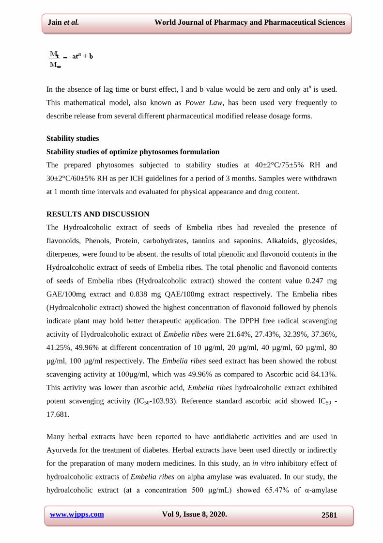
In the absence of lag time or burst effect, l and b value would be zero and only atn
is used.
This mathematical model, also known as Power Law, has been used very frequently to
describe release from several different pharmaceutical modified release dosage forms.
Stability studies
Stability studies of optimize phytosomes formulation
The prepared phytosomes subjected to stability studies at 40±2°C/75±5% RH and
30±2°C/60±5% RH as per ICH guidelines for a period of 3 months. Samples were withdrawn
at 1 month time intervals and evaluated for physical appearance and drug content.
RESULTS AND DISCUSSION
The Hydroalcoholic extract of seeds of Embelia ribes had revealed the presence of
flavonoids, Phenols, Protein, carbohydrates, tannins and saponins. Alkaloids, glycosides,
diterpenes, were found to be absent. the results of total phenolic and flavonoid contents in the
Hydroalcoholic extract of seeds of Embelia ribes. The total phenolic and flavonoid contents
of seeds of Embelia ribes (Hydroalcoholic extract) showed the content value 0.247 mg
GAE/100mg extract and 0.838 mg QAE/100mg extract respectively. The Embelia ribes
(Hydroalcoholic extract) showed the highest concentration of flavonoid followed by phenols
indicate plant may hold better therapeutic application. The DPPH free radical scavenging
activity of Hydroalcoholic extract of Embelia ribes were 21.64%, 27.43%, 32.39%, 37.36%,
41.25%, 49.96% at different concentration of 10 µg/ml, 20 µg/ml, 40 µg/ml, 60 µg/ml, 80
µg/ml, 100 µg/ml respectively. The Embelia ribes seed extract has been showed the robust
scavenging activity at 100µg/ml, which was 49.96% as compared to Ascorbic acid 84.13%.
This activity was lower than ascorbic acid, Embelia ribes hydroalcoholic extract exhibited
potent scavenging activity (IC50-103.93). Reference standard ascorbic acid showed IC50 -
17.681.
Many herbal extracts have been reported to have antidiabetic activities and are used in
Ayurveda for the treatment of diabetes. Herbal extracts have been used directly or indirectly
for the preparation of many modern medicines. In this study, an in vitro inhibitory effect of
hydroalcoholic extracts of Embelia ribes on alpha amylase was evaluated. In our study, the
hydroalcoholic extract (at a concentration 500 μg/mL) showed 65.47% of α-amylase
www.wjpps.com Vol 9, Issue 8, 2020.
2582
Jain et al. World Journal of Pharmacy and Pharmaceutical Sciences
inhibitory activity with IC50 value 221.16μg/mL. At the same time, when compared with
standard acarbose IC50 value was found to be 71.52μg/ml.
Different formulation of Phytosomes were prepared using different amount of phospholipids:
cholesterol and extract and were evaluated for Drug Excipient compatibility study,
Entrapment efficiency and particle size analysis, High performance liquid chromatography,
Transmission Electron Microscopy (TEM), In vitro drug release study of prepared
Phytosomes formulation and Stability study. The appearance or disappearance of peaks
and/or the shift of their positions are often indications of interactions such as hydrogen
bonding. The IR spectra of extract, Fig. 5.7-5.8, shows stretching vibrations at 1600.9880 cm-
1 attributed predominantly to the overlapping stretching vibrations of alkenes (C=C) and
carbonyl (C=O) character. Infrared of extract show stretching vibration at 2941.0572cm-1
due
to O-H groups, C=C aromatic stretching vibration at 1436.7392cm-1
. When the data obtained
from FTIR spectra is compared with the spectra studied it was observed that there are similar
peaks for functional groups in Phytosomes.
From the FTIR data of the physical mixture it is clear that functionalities of drug have
remained unchanged including intensities of the peak. This suggests that during the process
drug, Phospholipids and Cholesterol has not reacted with the drug to give rise to reactant
products. So there is no interaction between them which is in favor to proceed for formulation
of Phytosomes as drug delivery system. The entrapment efficiency of all the prepared
formulations is shown in Table 5.8. The entrapment efficiency of the phytosomes was found
in the range of 50.23±0.36 to 73.32±0.25%.
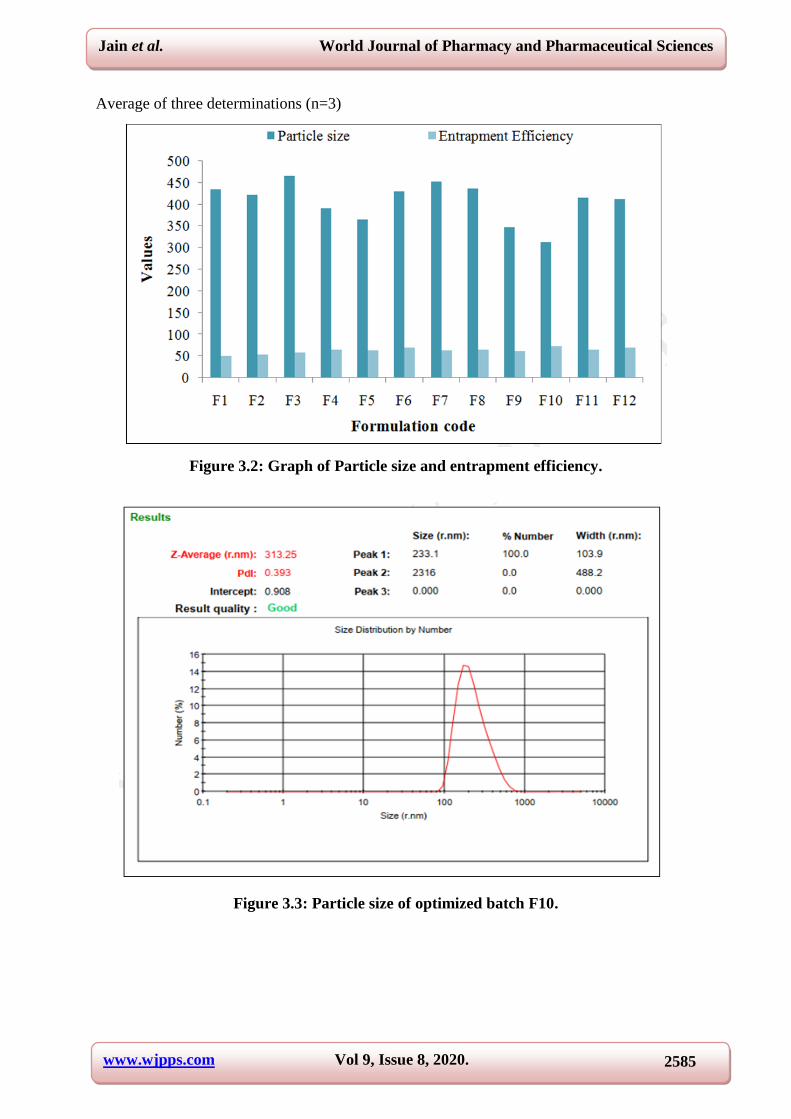
Particle size of all formulations found within range 313.25-465.32nm. Concentration of lipid
has shows significant impact on size of phytosomes. Formulation F10 was found best one
which is further evaluated for drug release study, transmission electron microscopy (TEM),
and stability studies.chromatographic analysis of phytosomes, performed by using
acetonitrile and methanol 50:50 (v/v) solutions as mobile phase and used a flow rate of 1ml
per min and absorbance at 256nm. It gave good separation of quercetin at RT 3.965 min. The
HPLC results of the Hydroalcoholic extract loaded Phytosomes showed major peaks at the
RT 3.962 (min) at a wavelength of 256nm The retention time of peaks was found to similar
retention time of standard 3.965, which confirm the presence of quercetin in Phytosomes. The
Percentage of Quercetin was found in Phytosomes (0.014%).
www.wjpps.com Vol 9, Issue 8, 2020.
2583
Jain et al. World Journal of Pharmacy and Pharmaceutical Sciences
TEMs are capable of imaging at a significantly higher resolution than light microscopes,
owing to the small de Broglie wavelength of electrons. This enables the instrument's user to
examine fine detail even as small as a single column of atoms, which is thousands of times
smaller than the smallest resolvable object in a light microscope. TEM forms a major analysis
method in a range of scientific fields, in both physical and biological sciences. TEM
characterization revealed that the Phytosomes are spherical in shape. However, some
variation in size distribution was observed in the TEM image, which might be attributed to an
uncontrolled charge neutralization process involved between oppositely charged chains
occurring during the formation of phytosomes. When the regression coefficient values of
were compared, it was observed that „r‟ values of Korsmeyer Peppas was maximum i.e. 0.992
hence indicating drug release from formulations was found to follow Korsmeyer Peppas
kinetics. Results of stability studies clearly indicates that optimized batches of phytosomes
were stable over the chosen temperature and humidity conditions up to 3 months as were
found no significant variation in physical appearance and % drug content.
Result of Drug-Excipient compatibility study
Figure 3: FT-IR spectra of hydroalcoholic extract.
www.wjpps.com Vol 9, Issue 8, 2020.
2584
Jain et al. World Journal of Pharmacy and Pharmaceutical Sciences
Table 6: Interpretation of FT-IR spectrum.
S. No. Functional group Peak wave number(cm
-1)
Experimental Theoretical
1. C-C str. and C=O str. 1600.9880 1550-1650
3. O-H str. 2941.0572 3200-2800
5. C=C aromatic str. 1436.7392 1400-1450
Figure 3.1: FT-IR spectra of prepared phytosomes formulation.
Particle size and entrapment efficiency of drug loaded phytosomes
Formulation
Code
Particle size
(nm)
Entrapment Efficiency
(%)
F1 435.65 50.23±0.36
F2 422.47 53.65±0.25
F3 465.32 58.89±0.65
F4 390.56 65.45±0.32
F5 365.58 63.12±0.47
F6 430.14 69.98±0.56
F7 452.32 63.32±0.47
F8 436.65 65.48±0.62
F9 346.65 62.32±0.14
F10 313.25 73.32±0.25
F11 415.65 65.45±0.36
F12 412.32 69.98±0.32
www.wjpps.com Vol 9, Issue 8, 2020.
2585
Jain et al. World Journal of Pharmacy and Pharmaceutical Sciences
Average of three determinations (n=3)
Figure 3.2: Graph of Particle size and entrapment efficiency.
Figure 3.3: Particle size of optimized batch F10.
www.wjpps.com Vol 9, Issue 8, 2020.
2586
Jain et al. World Journal of Pharmacy and Pharmaceutical Sciences
Dissolution rate studies
In vitro drug release study of prepared Phytosomes formulation.
Table 7: In-vitro drug release data for optimized formulation F10.
Time
(h)
Square
Root of
Time(h)1/2
Log
Time Cumulative*%
Drug Release
Log
Cumulative
% Drug
Release
Cumulative
% Drug
Remaining
Log
Cumulative
% Drug
Remaining
0.5 0.707 -0.301 22.32 1.349 77.68 1.890
1 1.000 0.000 30.35 1.482 69.65 1.843
2 1.414 0.301 42.32 1.627 57.68 1.761
4 2.000 0.602 65.56 1.817 34.44 1.537
6 2.449 0.778 75.65 1.879 24.35 1.386
8 2.828 0.903 82.32 1.916 17.68 1.247
12 3.464 1.079 95.65 1.981 4.35 0.638
Figure 7.1: Cumulative % drug released Vs Time (Zero Order Kinetics).
www.wjpps.com Vol 9, Issue 8, 2020.
2587
Jain et al. World Journal of Pharmacy and Pharmaceutical Sciences
Figure 7.2: Log cumulative % drug remaining Vs Time (First Order Kinetics).
Figure 7.3: Cumulative % drug release Vs Root time (Higuchi Release Kinetics).
www.wjpps.com Vol 9, Issue 8, 2020.
2588
Jain et al. World Journal of Pharmacy and Pharmaceutical Sciences
Figure 7.4: Log Cumulative % drug release Vs Log time (Korsmeyer Peppas Model).
Table 8: Regression analysis data of optimized formulation F10.
Batch Zero Order First Order Higuchi Korsmeyer Peppas
R² R² R² R²
F10 0.910 0.980 0.984 0.992
CONCLUSION
In this study, the combined hydroalcoholic extract of Embelia ribes in ratio of 2:1:1 found to
exhibit significant results. Phytosomes has better physical characteristics than that of extract. In-
vitro studies revealed that phytosomes showed control release of phytoconstituents. Hence,
phytosomal formulation of this herbal drug combination can be used for clinical application to
enhance the therapeutic effect.
REFERENCE
1. Rana Mahendra: Drug Delivery through Targeted Approach with Special References to
Phytosomes. https://www.intechopen.com/books/role-of-novel-drug-delivery-vehicles-in-
nanobiomedicine/drug-delivery-through-targeted-approach-with-special-references-to-
phytosomes.
www.wjpps.com Vol 9, Issue 8, 2020.
2589
Jain et al. World Journal of Pharmacy and Pharmaceutical Sciences
2. Pande S. D: Preparation and Evaluation of Phytosomes of Pomegrane Peels. Research
Journal of Pharmacy and Technology, 2015; 8.
3. Sharma D, Bhujbale AA; Phytosomes is a Novel Drug Delivery System based herbal
formulation: An Review; Pharma Tutor, 2018; 6(3): 23-26.
4. Solmaz Rasaie: Nano Phytosomes of Quercetin: A Promising Formulation for Fortification
of Food Products with Antioxidants, Pharmaceutical sciences, 2014; 20: 96-101.
5. American Diabetes Association. Standards of medical care in diabetes. Diabetes Care, 2009;
32(suppl 1): s15.
6. Modak, M., Dixit, P., Londhe, J. Devasagayam. Indian herbs and herbal drugs used for the
treatment of diabetes. J Clin Biochem Nutr, 2007; 40: 163-73.
7. Rathore P. Planterosomes: potential phyto-phospholipid carriers for the bioavailability
enhancement of herbal extracts. International journal of pharmaceutical science and research,
2012; 3: 737-755.
8. Valenzuela A, Aspillaga M, Vial S, Guerra R. Selectivity of silymarin on the increase of the
glutathione content in different tissues of the rat. Planta Med, 1989; 55: 420-422.
9. Saraf S, Kaur CD. Phytoconstituents as photoprotective novel cosmetic formulations.
Pharmacogn Rev, 2010; 4: 1-11.
10. Pandey S. Phytosome: Technical Revolution in Phytomedicine. International Journal of
PharmTech Research, 2010; 2: 627-631.
11. Amin T, Bhat S. A Review on Phytosome Technology as a Novel Approach to Improve the
Bioavailability of Neutraceuticals. International Journal of Advancements in Research and
Technology, 2012; 1: 1-15.
12. Saha S, Sarma A, Saikia P, Chakrabarty T. Phytosome: A Brief Overview. Scholars
Academic Journal of Pharmacy, 2013; 2: 12-20.
13. Battacharya S. Phytosome: Emerging strategy in delivery of herbal drugs and nutraceuticals.
PharmTimes, 2009; 41: 3.
14. Gupta A, Ashawal MS, Saraf S. Phytosome: a novel approach towards functional cosmetics.
J Plant Science, 2007; 644-649.
15. Kareparamban J, Nikam P, Jadhav A, Kadam V. Phytosome: a novel revolution in herbal
drugs. International journal of research in pharmacy and chemistry, 2012; 2: 299-310.
www.wjpps.com Vol 9, Issue 8, 2020.
2590
Jain et al. World Journal of Pharmacy and Pharmaceutical Sciences
16. Tawheed Amin, Suman Vikas Bhat, A Review on Phytosome Technology as a Novel
Approach to Improve The Bioavailability of Nutraceuticals, International Journal of
Advancements in Research & Technology, 2012; 1(3): 1-15.
17. A. Vidyalakshmi, S. Esaki Selvi. Protease Activity of Floral Extracts of Jasminum
grandiflorum L., a Wound Healing Herb. Journal of Medicinal Plants Studies, 2013; 1(4):
11-15.
18. Hossan MS, Fatima A, Rahmatullah M, Khoo TJ, Nissapatorn V, Galochkina AV, Slita AV,
Shtro AA, Nikolaeva Y, Zarubaev VV, Wiart C. Antiviral activity of Embelia ribes Burm. f.
against influenza virus in vitro. Arch Virol, 2018; 163(8): 2121-2131.
19. Durg S, B NK, Vandal R, Dhadde SB, Thippeswamy BS, Veerapur VP, Badami S.
Antipsychotic activity of embelin isolated from Embelia ribes: A preliminary study. Biomed
Pharmacother, 2017; 90: 328-331.
20. Meenu Bist and Shyam Baboo Prasad. Embelia ribes: A valuable medicinal plant. Journal of
Chemical and Pharmaceutical Research, 2016; 8(4): 1229-1233.
21. Sudhakaran MV. Botanical Pharmacognosy of the Fruit of Embelia ribes Burm. F.
Sudhakaran, J Pharmacogn Nat Prod, 2015; 1: 1.
22. K. Souravi and P. E. Rajasekharan. Ethnopharmacological Uses of Embelia ribes Burm. F. -
A Review. IOSR Journal of Pharmacy and Biological Sciences, 2014; 9(3): 23-30.
23. M. Nazam Ansari & U. Bhandari. Antihyperhomocysteinemic Activity of an Ethanol Extract
from Embelia ribes. in Albino Rats. Journal Pharmaceutical Biology, 2008; 46(4): 11-18.
24. Amudha S., Prabal Kumar Manna, Jeganathan N. S. Formulation and evaluation of capsules
of Syzygium Cumini phytosomes. J. Pharm. Sci. Innov, 2018; 7(3): 70-78.
25. Arun Kumar, Bimlesh Kumar, Sachin Kumar Singh, Barinder Kaur, Saurabh Singh, A
review on phytosomes: novel approach for herbal phytochemicals, Asian J Pharm Clin Res,
2017; 10(10): 41-47.
26. Ashwini S Dhase, Shweta S Saboo, Preparation and Evaluation of Phytosomes Containing
Methanolic Extract of Leaves of Aegle Marmelos (Bael), International Journal of PharmTech
Research, 2015; 8(6): 231-240.
27. Solmaz Rasaie, Saeed Ghanbarzadeh, Maryam Mohammadi, Hamed Hamishehkar, Nano
Phytosomes of Quercetin: A Promising Formulation for Fortification of Food Products with
Antioxidants, Pharmaceutical sciences, 2014; 20: 96-101.
www.wjpps.com Vol 9, Issue 8, 2020.
2591
Jain et al. World Journal of Pharmacy and Pharmaceutical Sciences
28. Sandeep Arora, Preparation and Characterization of Phytosomal-Phospholipid Complex of P.
Amarus and its Tablet Formulation, Journal of Pharmaceutical Technology, Research and
Management, Vol. 1 May 2013; 1–18.
29. Tawheed Amin, Suman Vikas Bhat, A Review on Phytosome Technology as a Novel
Approach to Improve The Bioavailability of Nutraceuticals, International Journal of
Advancements in Research & Technology, 2012; 1(3): 1-15.
30. Rajendra Awasthi, Giriraj T Kulkarni, Vivek K Pawar, Phytosomes: an approach to increase
the bioavailability of plant extracts, International Journal of Pharmacy and Pharmaceutical
Sciences, 2011; 3(2): 1-3.
31. Nilesh Jain Brahma P Gupta, Navneet Thakur, Ruchi Jain, Jitendra Banweer, Deepak Kumar
Jain, Surendra Jain, Phytosome: A Novel Drug Delivery System for Herbal Medicine,
International Journal of Pharmaceutical Sciences and Drug Research, 2010; 2(4): 224-228.
32. S Ambati; V Jyothi; A Jyothi, Int. J. Pharm. Tech, 2010; 2: 525-539.
33. B Lal, N Mishra; Int. J. Pharm. Sci. Res, 2013; 4: 3823-3838.
34. R Shelar R, C. Maurya, P. Tekale, K Katkar, V Naik, A Suthar, VS Chauhan; Int. J. Pharm.
Clin. Res, 2009; 1: 146-149.
35. S Asadulla; S Ramandang; Rajasekharan, Int. J. Res. Pharm. Chem, 2011; 1: 2236-2751.
36. MA Khan; A Naidu; M Akbar, Int. J. Pharm. Bio. Arch, 2010; 1: 267-270.
37. D Annapurna; A Srivastava; RT Singh, American J Plant Sci, 2013; 4: 28-35.
38. Vidya V. Kamble, and Nikhil B. Gaikwad. Fluorescence analysis, phytochemical, and
antioxidant activities in leaves and stem of embelia ribes burm. F. Asian Journal of
Pharmaceutical and Clinical Research, 2019; 12(4): 225-9.