
Characterization and Cytotoxic Assessment of Ballistic ... · plates and were caught in the thick...
21
Int. J. Environ. Res. Public Health 2010, 7, 3313-3331; doi:10.3390/ijerph7093313 International Journal of Environmental Research and Public Health ISSN 1660-4601 www.mdpi.com/journal/ijerph Article Characterization and Cytotoxic Assessment of Ballistic Aerosol Particulates for Tungsten Alloy Penetrators into Steel Target Plates Brenda I. Machado 1, *, Lawrence E. Murr 1 , Raquel M. Suro 2 , Sara M. Gaytan 1 , Diana A. Ramirez 1 , Kristine M. Garza 2 and Brian E. Schuster 3 1 Department of Metallurgical and Materials Engineering, The University of Texas at El Paso, El Paso, TX 79968, USA; E-Mails: [email protected] (L.E.M.); [email protected] (S.M.G.); [email protected] (D.A.R.) 2 Department of Biological Sciences, The University of Texas at El Paso, El Paso, TX 79968, USA; E-Mails: [email protected] (R.M.S.); [email protected] (K.M.G.) 3 U.S. Army Research Laboratory, Weapons and Materials Research Directorate (RDRL-WML-H), USA; E-Mail: [email protected] (B.E.S.) * Author to whom correspondence should be addressed; E-Mail: [email protected]; Tel.: +1-915-630-6404; Fax: +1-915-747-8036. Received: 10 July 2010; in revised form: 24 August 2010 / Accepted: 25 August 2010 / Published: 26 August 2010 Abstract: The nature and constituents of ballistic aerosol created by kinetic energy penetrator rods of tungsten heavy alloys (W-Fe-Ni and W-Fe-Co) perforating steel target plates was characterized by scanning and transmission electron microscopy. These aerosol regimes, which can occur in closed, armored military vehicle penetration, are of concern for potential health effects, especially as a consequence of being inhaled. In a controlled volume containing 10 equispaced steel target plates, particulates were systematically collected onto special filters. Filter collections were examined by scanning and transmission electron microscopy (SEM and TEM) which included energy-dispersive (X-ray) spectrometry (EDS). Dark-field TEM identified a significant nanoparticle concentration while EDS in the SEM identified the propensity of mass fraction particulates to consist of Fe and FeO, representing target erosion and formation of an accumulating debris field. Direct exposure of human epithelial cells (A549), a model for lung tissue, to particulates (especially nanoparticulates) collected on individual filters demonstrated OPEN ACCESS
Transcript of Characterization and Cytotoxic Assessment of Ballistic ... · plates and were caught in the thick...
Int. J. Environ. Res. Public Health 2010, 7, 3313-3331; doi:10.3390/ijerph7093313
International Journal of
Environmental Research and Public Health
ISSN 1660-4601 www.mdpi.com/journal/ijerph
Article
Characterization and Cytotoxic Assessment of Ballistic Aerosol Particulates for Tungsten Alloy Penetrators into Steel Target Plates
Brenda I. Machado 1,*, Lawrence E. Murr 1, Raquel M. Suro 2, Sara M. Gaytan 1,
Diana A. Ramirez 1, Kristine M. Garza 2 and Brian E. Schuster 3
1 Department of Metallurgical and Materials Engineering, The University of Texas at El Paso, El
Paso, TX 79968, USA; E-Mails: [email protected] (L.E.M.); [email protected] (S.M.G.);
[email protected] (D.A.R.) 2 Department of Biological Sciences, The University of Texas at El Paso, El Paso, TX 79968, USA;
E-Mails: [email protected] (R.M.S.); [email protected] (K.M.G.) 3 U.S. Army Research Laboratory, Weapons and Materials Research Directorate (RDRL-WML-H),
USA; E-Mail: [email protected] (B.E.S.)
* Author to whom correspondence should be addressed; E-Mail: [email protected];
Tel.: +1-915-630-6404; Fax: +1-915-747-8036.
Received: 10 July 2010; in revised form: 24 August 2010 / Accepted: 25 August 2010 /
Published: 26 August 2010
Abstract: The nature and constituents of ballistic aerosol created by kinetic energy
penetrator rods of tungsten heavy alloys (W-Fe-Ni and W-Fe-Co) perforating steel target
plates was characterized by scanning and transmission electron microscopy. These aerosol
regimes, which can occur in closed, armored military vehicle penetration, are of concern
for potential health effects, especially as a consequence of being inhaled. In a controlled
volume containing 10 equispaced steel target plates, particulates were systematically
collected onto special filters. Filter collections were examined by scanning and
transmission electron microscopy (SEM and TEM) which included energy-dispersive
(X-ray) spectrometry (EDS). Dark-field TEM identified a significant nanoparticle
concentration while EDS in the SEM identified the propensity of mass fraction particulates
to consist of Fe and FeO, representing target erosion and formation of an accumulating
debris field. Direct exposure of human epithelial cells (A549), a model for lung tissue, to
particulates (especially nanoparticulates) collected on individual filters demonstrated
OPEN ACCESS
Report Documentation Page Form ApprovedOMB No. 0704-0188
Public reporting burden for the collection of information is estimated to average 1 hour per response, including the time for reviewing instructions, searching existing data sources, gathering andmaintaining the data needed, and completing and reviewing the collection of information. Send comments regarding this burden estimate or any other aspect of this collection of information,including suggestions for reducing this burden, to Washington Headquarters Services, Directorate for Information Operations and Reports, 1215 Jefferson Davis Highway, Suite 1204, ArlingtonVA 22202-4302. Respondents should be aware that notwithstanding any other provision of law, no person shall be subject to a penalty for failing to comply with a collection of information if itdoes not display a currently valid OMB control number.
1. REPORT DATE 2010 2. REPORT TYPE
3. DATES COVERED 00-00-2010 to 00-00-2010
4. TITLE AND SUBTITLE Characterization And Cytotoxic Assessment Of Ballistic AerosolParticulates For Tungsten Alloy Penetrators Into Steel Target Plates
5a. CONTRACT NUMBER
5b. GRANT NUMBER
5c. PROGRAM ELEMENT NUMBER
6. AUTHOR(S) 5d. PROJECT NUMBER
5e. TASK NUMBER
5f. WORK UNIT NUMBER
7. PERFORMING ORGANIZATION NAME(S) AND ADDRESS(ES) U.S. Army Research Laboratory,Weapons and Materials ResearchDirectorate (RDRL-WML-H),Adelphi,MD, 20783
8. PERFORMING ORGANIZATIONREPORT NUMBER
9. SPONSORING/MONITORING AGENCY NAME(S) AND ADDRESS(ES) 10. SPONSOR/MONITOR’S ACRONYM(S)
11. SPONSOR/MONITOR’S REPORT NUMBER(S)
12. DISTRIBUTION/AVAILABILITY STATEMENT Approved for public release; distribution unlimited
13. SUPPLEMENTARY NOTES Int J Environ. Res. Public Health. 2010 Sep,Government or Federal Purpose Rights License.
14. ABSTRACT The nature and constituents of ballistic aerosol created by kinetic energy penetrator rods of tungsten heavyalloys (W-Fe-Ni and W-Fe-Co) perforating steel target plates was characterized by scanning andtransmission electron microscopy. These aerosol regimes, which can occur in closed, armored militaryvehicle penetration, are of concern for potential health effects, especially as a consequence of being inhaled.In a controlled volume containing 10 equispaced steel target plates, particulates were systematicallycollected onto special filters. Filter collections were examined by scanning and transmission electronmicroscopy (SEM and TEM) which included energy-dispersive (X-ray) spectrometry (EDS). Dark-fieldTEM identified a significant nanoparticle concentration while EDS in the SEM identified the propensity ofmass fraction particulates to consist of Fe and FeO, representing target erosion and formation of anaccumulating debris field. Direct exposure of human epithelial cells (A549), a model for lung tissue, toparticulates (especially nanoparticulates) collected on individual filters demonstrated induction of rapidand global cell death to the extent that production of inflammatory cytokines was entirely inhibited. Theseobservations along with comparisons of a wide range of other nanoparticulate species exhibiting cell deathin A549 culture may suggest severe human toxicity potential for inhaled ballistic aerosol, but thecomplexity of the aerosol (particulate) mix has not yet allowed any particular chemical composition to be identified.
15. SUBJECT TERMS
16. SECURITY CLASSIFICATION OF: 17. LIMITATION OF ABSTRACT Same as
Report (SAR)
18. NUMBEROF PAGES
20
19a. NAME OFRESPONSIBLE PERSON
a. REPORT unclassified
b. ABSTRACT unclassified
c. THIS PAGE unclassified

Standard Form 298 (Rev. 8-98) Prescribed by ANSI Std Z39-18
Int. J. Environ. Res. Public Health 2010, 7
3314
induction of rapid and global cell death to the extent that production of inflammatory
cytokines was entirely inhibited. These observations along with comparisons of a wide
range of other nanoparticulate species exhibiting cell death in A549 culture may suggest
severe human toxicity potential for inhaled ballistic aerosol, but the complexity of the
aerosol (particulate) mix has not yet allowed any particular chemical composition to
be identified.
Keywords: tungsten alloys; nanoparticulates; cytotoxic assays; scanning and transmission
electron microscopy; aerosol; ballistic penetration
1. Introduction
In regards to very small fragments and fragment aerosols, there have been few systematic
observations of associated particulate chemistries, size distributions or cytotoxic responses, especially
pertinent to respiratory inflammatory responses or more serious respiratory health effect indicators;
although recent work by Gold et al. [1] has examined aerosols inside an armored vehicle penetrated by
a kinetic energy tungsten heavy alloy (KE WHA) penetrator. In addition, Guillmette et al. [2] have
also discussed the health risk for depleted uranium (DU) aerosols. It is now well established that
ultra-fine or nanoparticulate materials characteristic of a wide compositional range and particulate
morphologies exhibit respiratory inflammatory and cytotoxic effects for a range of human lung cell
types [3-10].
The present study is concerned with the systematic collection of aerosol particulates associated with
ballistic WHA rod penetration into rolled homogeneous armor (RHA) or related steel armor or armor
plate sequences in a containment vessel. This research is also concerned with the characterization of
these collected particulates using scanning and transmission electron microscopy; including the
analysis of particulate chemistries or elemental compositions utilizing energy dispersive (X-ray)
spectrometry (EDS). Finally, filter-collected aerosol particulates were exposed to human epithelial
(lung) cells in culture to assess their inflammatory and related respiratory health effects.
2. Experimental Procedures
2.1. Materials and Ballistic Testing
Sub-scale WHA penetrators were fired into an array of mild steel plates which were encapsulated in
a steel containment vessel. The penetrators were hemispherical-nose, 65 g rods with a length to
diameter ratio of 20. The WHA rods consisted of either 91% (by weight) W, 5.6% Ni, 1.4% (WA #1)
or 92% W, 6% Ni, 3% Co (WA #2) or and were fabricated using liquid phase sintering [11]. In
Figure 1(a) and (b), we show the typical microstructures of WA #1 and #2 (respectively) consisting of
pure tungsten particles surround by the matrix phase.
The target array consisted of 10 each of 6.25 mm thick mild steel plates spaced 12.5 mm apart
backed by a thick RHA block to capture the residual penetrator. The penetrators were fired from a

Int. J. Environ. Res. Public Health 2010, 7
3315
26 mm smooth bore cannon outfitted with a 37 mm breach. The launch package consisted of a
polypropulux laboratory sabot and obdurator with a steel pusher plate. The penetrators (and entire
launch package) were fired at a velocity of ~1.2 km/s using approximately 170 g of type M2
propellant. The approximate composition of the M2 propellant as reported by Roth and Watchtell is
shown in Table 1 [12]. In all 12 tests, the penetrators completely perforated all of the thin mild steel
plates and were caught in the thick RHA block.
Figure 1. (a) WHA KE penetrator section views. (a) W-Ni-Fe penetrator. (b) W-Ni-Co
penetrator. The spherical or near spherical particles in (a) and (b) are W in the
corresponding alloy matrix.
The containment vessel was an RHA cube with a wall thickness of ~25 mm, with an internal open
volume that was approximately 60 cm on a side. This vessel had a removable top which allowed for
placement of the target array. The vessel had two ports: a ~10 cm diameter port through which the
WHA penetrators were fired and a smaller ~6 mm port to allow for aerosol sampling (discussed
below). This containment vessel is not a closed system as the larger porter is open during the ballistic
Int. J. Environ. Res. Public Health 2010, 7
3316
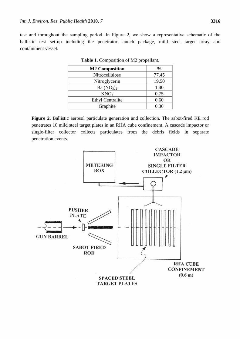
test and throughout the sampling period. In Figure 2, we show a representative schematic of the
ballistic test set-up including the penetrator launch package, mild steel target array and
containment vessel.
Table 1. Composition of M2 propellant.
M2 Composition % Nitrocellulose 77.45 Nitroglycerin 19.50
Ba (NO3)2 1.40 KNO3 0.75
Ethyl Centralite 0.60 Graphite 0.30
Figure 2. Ballistic aerosol particulate generation and collection. The sabot-fired KE rod
penetrates 10 mild steel target plates in an RHA cube confinement. A cascade impactor or
single-filter collector collects particulates from the debris fields in separate
penetration events.
Int. J. Environ. Res. Public Health 2010, 7
3317
2.2. Aerosol Collection and Analysis
Immediately following the ballistic test, aerosol samples were collected using either single-stage
filters or by an eight-stage cascade impactor, under non-sterile conditions. The debris (particulate
aerosol) generated by the rod impact, penetration and perforation as well as the residual propellant
gases were sampled for ~15 minutes through the respective collection media with an airflow rate
of 2 L/min.
The single stage filters were SKC model 225-321A mixed cellulose ester (MCE) filters with a
diameter of 25 mm and a pore-size of 0.8 μm. The eight stage cascade impactor utilized Mylar
substrates with cut-points ranging from 0.5 to 21 μm and with a final backup filter with a 0.25 μm pore
size. The Mylar substrates were sprayed with Dow Corning 316 silicone release lubricant ~16h before
initial weighing to provide an adhesive surface for the airborne particles. The total particulate
concentrations at each cut point were determined from gravimetric analysis. Each substrate was
retained for subsequent analysis in their SKC and Millipore containers and samples for the relative
concentrations of W, Ni, Fe and Co. Analysis was conducted following National Institute for
Occupational Safety and Health (NIOSH) method 7074 which included cellulose ester filter collection,
acid digestion, and analysis by inductively coupled plasma optical emission spectroscopy (ICP-OES).
EPA method 200.7 was used for metal analysis.
2.3. Aerosol Particulate Characterization
Filter collection samples were halved or quartered and utilized for direct particulate collection
observation and analysis by scanning electron microscopy (SEM) and energy-dispersive (X-ray)
spectrometry (EDS). The SEM was a field emission SEM (Hitachi S4800) operated at 20 kV
accelerating potential to assure adequate excitation potential for EDS. Filter sections were also scraped
onto silicon monoxide/formvar-coated 200 mesh, 3 mm copper grids and a second grid placed on top
to form a sandwich for direct observation in the transmission electron microscope (TEM). The single
(MCE) filter collections were more suitable for scraping particles onto TEM grids or onto sticky
carbon supports for SEM. TEM image analysis was performed either in a Hitachi H-8000 electron
microscope operated at 200 kV or a high-resolution TEM (Hitachi H-9500) operating at 300 kV and
fitted with a goniometer-tilt stage. Selected-area electron diffraction (SAED) was utilized to observe
the degree of crystallinity of the collected particulates and to allow for selective dark-field (DF)
imaging utilizing specific diffraction spots. The high-resolution TEM (Hitachi H-9500) operating at
300 kV was also utilized especially for ultra-fine particle (nanoparticle) analysis. It was fitted with an
EDAX-EDS system which allowed for nano-probe (point) analysis and scanned-area analysis.
2.4. In Vitro Cytotoxicity Assays for Particulate Collections
In this study we utilized direct exposure cytotoxicity assays for filter-collected ballistic particulates
as described previously by Soto et al. [8]. Assays were performed for six different assay groups as
summarized in Table 2. Direct exposure assays were performed using particulate specimens collected
using the single-stage filters (SSF) associated with ballistic impacts of WA #1 and WA #2 into the

Int. J. Environ. Res. Public Health 2010, 7
3318
steel target array. There was no measurement of particle weight on the filters. Untreated cells (or
“media” only cells) served as the negative control; cells exposed to a filter on which nothing had been
collected served as the blank control.
Table 2. Direct Contact Cytotoxicity Assay Groups.
Name Description
SSF-WA #1 Single Stage Filter Collections for
WA #1 impacting target array
SSF-WA #2 Single Stage Filter Collections for
WA #2 impacting target array
CI6-WA #1 Cascade Impactor Stage 6
Specimens for WA #1 impacting array
CI6-WA #2 Cascade Impactor Stage 6
Specimens for WA #1 impacting array
Media Negative Control-Untreated cells
Blank Cells exposed to a single stage
filter on which nothing has been collected
These assays measure in vitro cell viability or cell death using colorimetric or optical densitometry
analysis along with cytokine enzyme linked immunosorbant assay (ELISA) studies which measure the
up-regulation and release of interleukins by the cells. The type of ELISA kit used was a BD
Biosciences. In recent ELISA studies, we utilized the immortalized A549 human epithelial lung cell
line which provides an effective in vitro lung cell model and has been widely adopted as a human lung
cell model. The cell line was obtained by the American Tissue Culture Collection (ATCC) from lung
carcinomatous tissue from a 58 year old Caucasian male. The A549 cells were cultured in 12-well
plates at 0.25 × 10⁶ cells per well with F-12 Ham’s media supplemented with 10% Fetal Bovine Serum
(FBS) and 5% penicillin/streptomycin (PS) for several hours to allow the cells to adhere. The cells
were then exposed for 48hs (standard time for acute exposures) with 1/4 of the indicated filter with the
collection side facing towards the monolayer of cells. Following the exposure period, the filters were
removed; the cells were harvested, and were then transferred into a 96-well flat bottom plate to assess
viability via the MTS Assay. This colorimetric assay assesses relative viability as a function of color,
which is directly proportional to the amount of cells available to convert the substrate into a color
product. CellTiter 96 Aqueous One Solution Reagent (Promega), which contains the
tetrazolium compound 3-(4, 5-dimethylthiazol-2-yl)-5-(3-carboxymethoxyphenyl)-2-(4-sulfophenyl)-
2H-tetrazolium, inner salt (MTS), was added to each well. The plate was then incubated for 2 h at 37
°C in a humidified, 5% CO2 atmosphere. Finally, the absorbance was recorded at 490 nm using a 96-
well plate spectrophotometer reader. The plate was a VersaMax Tunable Microplate Reader of
Molecular Devices. Relative viability results were obtained by 1way ANOVA with Bonferroni’s
Multiple Comparison Test.

Int. J. Environ. Res. Public Health 2010, 7
3319
Interleukin (IL)-6 and IL-8 secretion by A549 cells was measured using a commercial human IL-6
and IL-8 enzyme-linked immunosorbent assay (ELISA) kit (Biosource Human IL-6/IL-8 CytoSet).
Supernatants were obtained 48hs after exposure to the different filters and were stored at −20 °C until
subjected to ELISA analysis following the manufacturer’s protocol. The ELISA plates were coated
12 to 18 h at 4 °C with the capture antibody (Anti-Human IL-6 or IL-8). The plates were then blocked
at room temperature with assay buffer for 1 h to prevent non-specific antigen binding. Next, the
standards (Recombinant Human IL-6 or IL-8) and samples were added in duplicate (2 wells per
treatment), immediately followed by the addition of the working detection antibody (Anti-Human IL-6
or IL-8 Biotin) and incubated for 2 h at room temperature. Subsequently, the plates were washed and
the working streptavidin-horseradish peroxidase (HRP) solution was added to each well for 30 min.
The enzyme substrate solution tetramethylbenzidine (TMB) was added for color development and
finally, the enzyme reaction was stopped by the addition of the stop solution containing hydrosulfuric
acid (H2SO4). Absorbance was measured at 450 nm using a 96-well plate spectrophotometer reader.
3. Results and Discussion
3.1. Aerosol Particulate Size Distribution and Composition
In Tables 3 and 4, we should the typical sampling data associated with particulate specimens
captured using the cascade impactors for the WHA rod impact regime. As noted previously, these
specimens were sampled to determine the relative fractions of W, Ni, Fe and Co using ICP-OES while
the total particulate weights (and concentrations) were determined using gravimetric methods. The
“Total Particulate Concentrations” presumably include a large fraction of undetermined particulate
including organic compounds and residual compounds resulting from combustion of the M2 propellant
Table 3. Particulate collection for WA #1 impact experiment.
Stage Cut Point (microns)
Total Particulate
Weight (mg)
Total Particulate
Concentration (mg/m3)
Iron Conc. (mg/m3)
Nickel Conc.
(mg/m3)
Tungsten Conc.
(mg/m3)
1 21 0.432 17 14.8 0.286 1.92 2 15 0.052 2 4.51 0.134 1.36 3 10 0.000 0 3.52 ND 0.217 4 6 0.049 2 7.31 0.118 0.318 5 3.5 0.798 31 21.0 0.312 0.736 6 2 0.953 38 20.9 0.290 0.781 7 0.9 0.677 27 9.97 0.142 0.625 8 0.5 0.204 8 2.25 ND 0.401
Backup Filter
0.25 0.878 35 4.89 ND 2.59
Total 160 89.2 1.28 8.95 Respirable 106 65.0 0.862 3.08
ND—Concentration below instrument detection limit.
Int. J. Environ. Res. Public Health 2010, 7
3320
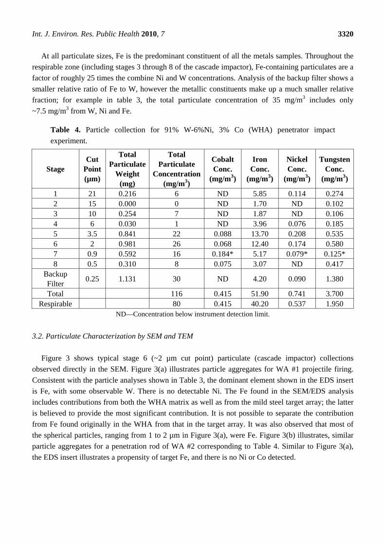
At all particulate sizes, Fe is the predominant constituent of all the metals samples. Throughout the
respirable zone (including stages 3 through 8 of the cascade impactor), Fe-containing particulates are a
factor of roughly 25 times the combine Ni and W concentrations. Analysis of the backup filter shows a
smaller relative ratio of Fe to W, however the metallic constituents make up a much smaller relative
fraction; for example in table 3, the total particulate concentration of 35 mg/m3 includes only
~7.5 mg/m3 from W, Ni and Fe.
Table 4. Particle collection for 91% W-6%Ni, 3% Co (WHA) penetrator impact
experiment.
Stage Cut
Point (µm)
Total Particulate
Weight (mg)
Total Particulate
Concentration (mg/m3)
Cobalt Conc.
(mg/m3)
Iron Conc.
(mg/m3)
Nickel Conc.
(mg/m3)
Tungsten Conc.
(mg/m3)
1 21 0.216 6 ND 5.85 0.114 0.274 2 15 0.000 0 ND 1.70 ND 0.102 3 10 0.254 7 ND 1.87 ND 0.106 4 6 0.030 1 ND 3.96 0.076 0.185 5 3.5 0.841 22 0.088 13.70 0.208 0.535 6 2 0.981 26 0.068 12.40 0.174 0.580 7 0.9 0.592 16 0.184* 5.17 0.079* 0.125* 8 0.5 0.310 8 0.075 3.07 ND 0.417
Backup Filter
0.25 1.131 30 ND 4.20 0.090 1.380
Total 116 0.415 51.90 0.741 3.700 Respirable 80 0.415 40.20 0.537 1.950
ND—Concentration below instrument detection limit.
3.2. Particulate Characterization by SEM and TEM
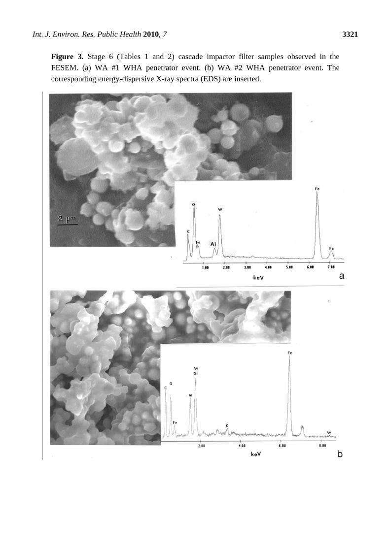
Figure 3 shows typical stage 6 (~2 µm cut point) particulate (cascade impactor) collections
observed directly in the SEM. Figure 3(a) illustrates particle aggregates for WA #1 projectile firing.
Consistent with the particle analyses shown in Table 3, the dominant element shown in the EDS insert
is Fe, with some observable W. There is no detectable Ni. The Fe found in the SEM/EDS analysis
includes contributions from both the WHA matrix as well as from the mild steel target array; the latter
is believed to provide the most significant contribution. It is not possible to separate the contribution
from Fe found originally in the WHA from that in the target array. It was also observed that most of
the spherical particles, ranging from 1 to 2 µm in Figure 3(a), were Fe. Figure 3(b) illustrates, similar
particle aggregates for a penetration rod of WA #2 corresponding to Table 4. Similar to Figure 3(a),
the EDS insert illustrates a propensity of target Fe, and there is no Ni or Co detected.
Int. J. Environ. Res. Public Health 2010, 7
3321
Figure 3. Stage 6 (Tables 1 and 2) cascade impactor filter samples observed in the
FESEM. (a) WA #1 WHA penetrator event. (b) WA #2 WHA penetrator event. The
corresponding energy-dispersive X-ray spectra (EDS) are inserted.
Int. J. Environ. Res. Public Health 2010, 7
3322
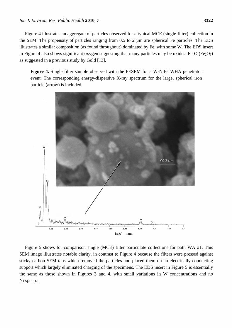
Figure 4 illustrates an aggregate of particles observed for a typical MCE (single-filter) collection in
the SEM. The propensity of particles ranging from 0.5 to 2 µm are spherical Fe particles. The EDS
illustrates a similar composition (as found throughout) dominated by Fe, with some W. The EDS insert
in Figure 4 also shows significant oxygen suggesting that many particles may be oxides: Fe-O (Fe2O3)
as suggested in a previous study by Gold [13].
Figure 4. Single filter sample observed with the FESEM for a W-NiFe WHA penetrator
event. The corresponding energy-dispersive X-ray spectrum for the large, spherical iron
particle (arrow) is included.
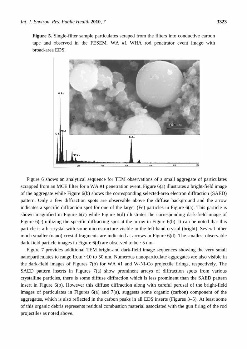
Figure 5 shows for comparison single (MCE) filter particulate collections for both WA #1. This
SEM image illustrates notable clarity, in contrast to Figure 4 because the filters were pressed against
sticky carbon SEM tabs which removed the particles and placed them on an electrically conducting
support which largely eliminated charging of the specimens. The EDS insert in Figure 5 is essentially
the same as those shown in Figures 3 and 4, with small variations in W concentrations and no
Ni spectra.
Int. J. Environ. Res. Public Health 2010, 7
3323
Figure 5. Single-filter sample particulates scraped from the filters into conductive carbon
tape and observed in the FESEM. WA #1 WHA rod penetrator event image with
broad-area EDS.
Figure 6 shows an analytical sequence for TEM observations of a small aggregate of particulates
scrapped from an MCE filter for a WA #1 penetration event. Figure 6(a) illustrates a bright-field image
of the aggregate while Figure 6(b) shows the corresponding selected-area electron diffraction (SAED)
pattern. Only a few diffraction spots are observable above the diffuse background and the arrow
indicates a specific diffraction spot for one of the larger (Fe) particles in Figure 6(a). This particle is
shown magnified in Figure 6(c) while Figure 6(d) illustrates the corresponding dark-field image of
Figure 6(c) utilizing the specific diffracting spot at the arrow in Figure 6(b). It can be noted that this
particle is a bi-crystal with some microstructure visible in the left-hand crystal (bright). Several other
much smaller (nano) crystal fragments are indicated at arrows in Figure 6(d). The smallest observable
dark-field particle images in Figure 6(d) are observed to be ~5 nm.
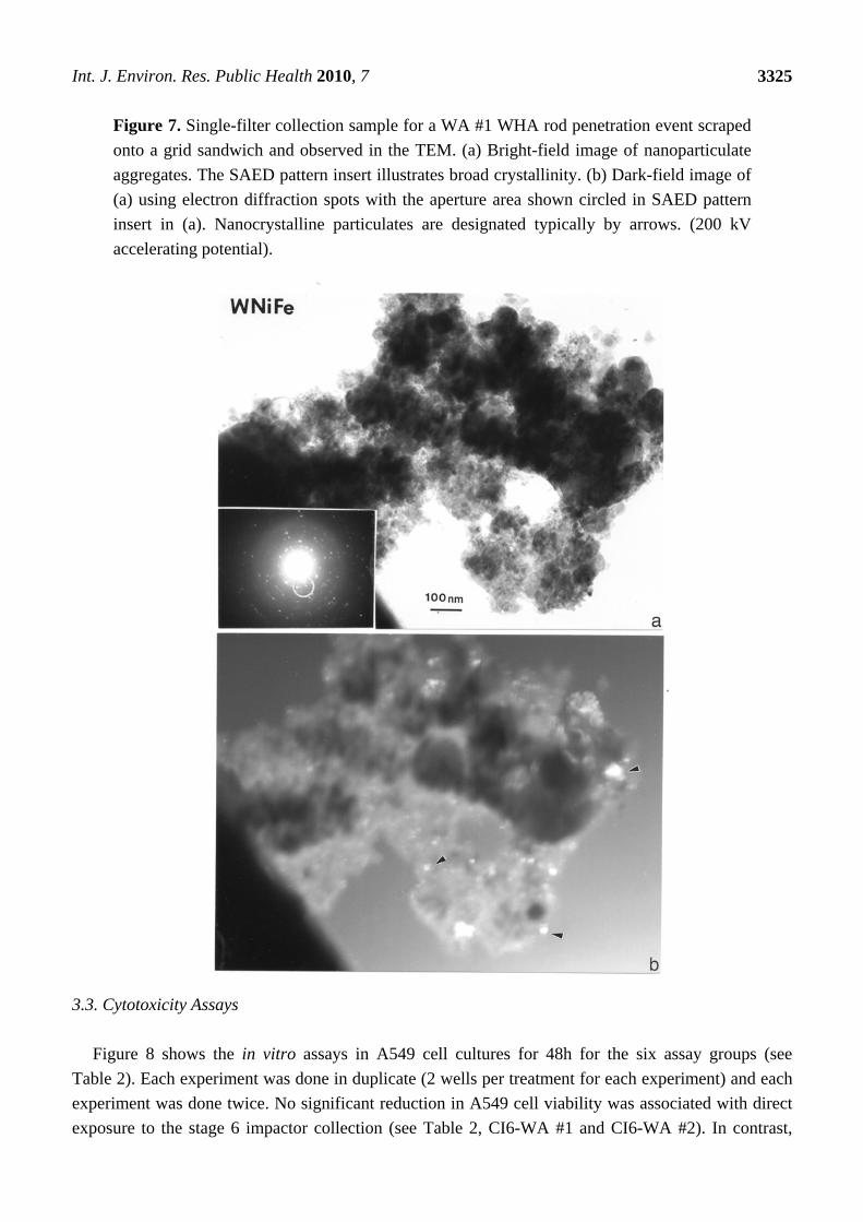
Figure 7 provides additional TEM bright-and dark-field image sequences showing the very small
nanoparticulates to range from ~10 to 50 nm. Numerous nanoparticulate aggregates are also visible in
the dark-field images of Figures 7(b) for WA #1 and W-Ni-Co projectile firings, respectively. The
SAED pattern inserts in Figures 7(a) show prominent arrays of diffraction spots from various
crystalline particles, there is some diffuse diffraction which is less prominent than the SAED pattern
insert in Figure 6(b). However this diffuse diffraction along with careful perusal of the bright-field
images of particulates in Figures 6(a) and 7(a), suggests some organic (carbon) component of the
aggregates, which is also reflected in the carbon peaks in all EDS inserts (Figures 3–5). At least some
of this organic debris represents residual combustion material associated with the gun firing of the rod
projectiles as noted above.

Int. J. Environ. Res. Public Health 2010, 7
3324
Figure 6. WA #1 WHA penetrator event aerosol particulates scraped from a single filter
collector onto a grid sandwich and observed in the TEM. (a) Bright-field image of
particulate aggregates. (b) Selected-area electron diffraction (SAED) pattern for aggregates
in (a). Magnified bright-field image of (a). (d) Dark-field image of (c) using diffraction
spot at arrow in (b). Nanoparticle images are indicated by the arrow. The magnification
markers are designated by the marker in (a). The accelerating potential was 300 kV.
Int. J. Environ. Res. Public Health 2010, 7
3325
Figure 7. Single-filter collection sample for a WA #1 WHA rod penetration event scraped
onto a grid sandwich and observed in the TEM. (a) Bright-field image of nanoparticulate
aggregates. The SAED pattern insert illustrates broad crystallinity. (b) Dark-field image of
(a) using electron diffraction spots with the aperture area shown circled in SAED pattern
insert in (a). Nanocrystalline particulates are designated typically by arrows. (200 kV
accelerating potential).
3.3. Cytotoxicity Assays
Figure 8 shows the in vitro assays in A549 cell cultures for 48h for the six assay groups (see
Table 2). Each experiment was done in duplicate (2 wells per treatment for each experiment) and each
experiment was done twice. No significant reduction in A549 cell viability was associated with direct
exposure to the stage 6 impactor collection (see Table 2, CI6-WA #1 and CI6-WA #2). In contrast,

Int. J. Environ. Res. Public Health 2010, 7
3326
direct exposure of single stage filter samples from impacts of both WA #1 and 2 (SSF-WA #1 and
SSF-WA #2) show very significant or highly cytotoxic responses. Respiratory epithelial cells such as
the A549 lung model cells used in these assays have the ability to synthesize and release inflammatory
cytokines such as interluken (IL)-6 and IL-8, as well as growth factors that modulate differentiation
and inflammatory cells. Interleukin 6 (IL-6) and IL-8 are considered markers for inflammatory
response both in the A549 cell culture models and in bronchial alveolar lavage fluids in
animals [14-19], ideally providing a biomarker link between in vitro and in-vivo studies. Specifically,
IL-6 has been associated with allergic responses involving asthma [16-19] while IL-8 has been linked
with chronic obstructive pulmonary disease (COPD) [5,18-20]. The cells are killed so rapidly that
there is no time to respond, as evident in the complete lack of IL production in Figure 8(d) and (e). The
cells were killed so rapidly that only aggregates of particulates uptaken by the cells remained.
Figure 8. Comparative cytotoxicities (as relative A549 epithelial cell viability in
filter-collected samples) and cytokine production. (a) and (b) Single filter exposure cell
culture assays compared to media or untreated cell control and blank filter control for 48h
exposure. Specific ballistic event numbers are shown for WA #1 and WA #2 penetration
events. (c) Cascade impact stage 6 cell culture assays which show no effect. (d) and (e)
show cytokine (IL 6 and IL 8) assays. Data is presented as the mean ± SEM of duplicate
samples and is one of two representative experiments (*p ≤ 0.01).
***
*** ***
***
***
****
p<0.0001p<0.0001
p<0.0001p<0.05
a
c
b
ed
Int. J. Environ. Res. Public Health 2010, 7
3327
3.4. Comparative Cytoxicity of These and Other Nanoparticulate Species
Specimens collected on the single-stage filters represent the full spectrum aerosol specimens
generated and collected. The stage 6 cascade impactor collections represent a subset of that present in
the single-stage collections. Ideally, the latter collections contained a smaller overall size fraction of
particulates, but both the single filter collections as well as the stage 6 impactor collections contained
nanoparticulates and nanoparticulate aggregates as illustrated in figures 3–5. However, in contrast to
the single filter cytotoxicities illustrated in Figure 8(a) and (b), there was no significant reduction in
A549 cell viability for the stage impactor assays (CI6-WA #1 and #2) (Figure 8(c)). This indicates a
sharp delineation in cytotoxicity with particulate and especially 6 nanoparticulate concentrations since,
as indicated in Tables 1 and 2, the total and respirable fractions are roughly 4 times greater than the
particulate concentrations on the stage 6 impactor filters, even though these filters contained
significantly more particulates (particulate concentrations) than other impactor filters (Tables 1 and 2).
In contrast to other filter exposure cytotoxicity assays and standard in vitro cytotoxicity
comparisons for a wide range of other nanoparticulate materials in culture (A549 cell exposure), the
ballistic aerosol particulates are highly toxic [8,21]. Similar features have been observed for a wide
range of nanoparticulates and nanoparticulate aggregates which also exhibit significant
cytotoxicity [6].
3.5. Respiratory Effects of Nanoparticulates
There is a plethora of evidence over at least the past two decades that metal and metal oxide
particles, especially nanoparticles are toxic, especially in the pulmonary system. Publications by
Cugell et al. [22]; Sullivan et al. [23] and Buzea et al. [24] to name only a few attest to the health
effects of Co, Ni, W, and Fe. These exposures to Co, Ni, and W can cause pulmonary fibrosis asthma,
pulmonary eodema and pneumonia among other effects. Nickel and its compounds in particular have
been demonstrated to cause nasal and lung cancers in the longer term for repeated exposure [23,24]. In
addition, a wide range of metal oxides have also been demonstrated to be toxic or cytotoxic, especially
iron oxide [24,25] as illustrated in the comparative cytotoxicity data in Figure 6.
Inhaled particulates (including coarse nanoparticles <1 µm) initially encounter the mucocillary
clearance by cilia of the bronchial epithelial cells which moves the larger particles (>1 µm) towards the
upper respiratory tract. However the truly nanoparticles (<1 µm) migrate to the alveoli where
phagocytes and other cells with phagocytic abilities work to arrest them. However with phagocytic
impairment, or for nanoparticles <100 nm which are not readily phagocitized, nanoparticles can
accumulate and even aggregate to create oxidative stress and inflammation [26]. This can lead to
various diseases while inflammation plays a major role in coronary heart disease and airway diseases
such as asthma and chronic obstructive pulmonary disease (COPD). Severe inflammation is also
associated with the onset of autoimmune disease. A prominent mechanism responsible for the variety
of nanoparticle toxicities assumes a shift in the redox balance of the cells towards oxidation as a
consequence of the formation of reactive oxygen species (ROS) which can, in the longer term, lead to
DNA damage [24]. In addition to oxidative stress, some nanoparticles can enhance the expression of
specific viral receptors and lead to severe inflammation when exposed to viral infections while other

Int. J. Environ. Res. Public Health 2010, 7
3328
nanoparticles can decrease the expression of certain viral and bacterial receptors which lowers the
resistance to some types of micro-organisms [27]. This phenomenon has been described previously for
the in vitro assays for the collected aerosol particles on filters where IL-8 inhibition prevents the A549
epithelial cells from normal function. This would prevent the lungs from mounting an effective
response to inhaled microbes.
Oberdörster, et al. [26] have demonstrated that aggregated nanoparticles are not as toxic as smaller
concentrations of single particles. However, considerable aggregation observed in this study would
suggest that in lung fluid, many of the aggregated particles, particularly those <100 nm, would
disaggregate. Moreover, Murr et al. [25] have shown that a wide range of aggregated nanoparticulates
are noticeably cytotoxic. Additionally high concentrations of aggregated nanoparticles can produce
lung burden and pulmonary tissue damage depending upon the lung clearance rate [24]. Smaller
nanoparticles (<30 nm) can emulate virus diameters and cross physiological barriers, entering the
circulatory and lymphatic systems where they can translocate to various organs [28]. Inhaled
nanoparticles <100 nm have also been shown to reach the brain via olfactory nerves as well as the
blood-brain barriers [24,30]. Studies have suggested that high concentrations of metals such as Cu, Al,
Fe, and others, together with oxidative stress, may initiate and even promote neurodegenerative
diseases such as Parkinson’s and Alzheimer’s diseases [24].
Current research reviews indicate that with few exceptions, nanoparticles and nanoparticle
aggregates are toxic (if not cytotoxic) to living organisms [24,25]. However, the relationship between
nanoparticle exposure and immune response is not well known. The degree of toxicity for specific
nanoparticles cannot be extrapolated from bulk properties, and materials which are nontoxic in bulk
form may be toxic in nanoparticle or nanoparticle aggregate form [24].
4. Conclusions
As in the case for environmental particulate regimes, including the outdoor environment,
nanoparticulates accounted for the most significant number concentration or particulate abundance of
the ballistic aerosol, with mass abundances exceeding 150 mg/m3 (Table 3). This might be compared
to mass concentrations of ~90 mg/m3 for burning tire soot or natural gas combustion particulate mass
concentrations of ~0.01 mg/m3 as illustrated in the comparative cytotoxicity data in Figure 8(e) [30].
Correspondingly, these small mass abundance values represented the most cytotoxic aerosol category
in contrast to larger concentrations of other soot species. In this work, ballistic aerosol composed
largely of nanoparticulates averaging ~10 nm (Figures 6–7) was shown to be so highly toxic to human
epithelial cells over a short time (48 h) in culture that the cells died (Figure 8(c) and (d)) before they
could produce any responsive cytokines (IL-6 or IL-8) (Figure 13(c) and (d)). Lower ballistic aerosol
concentrations measured to be roughly ¼ of the topic concentrations showed no measurable cell (A549
human epithelial) death after 48 h in culture. While the concentration cut off and exposure time in
culture to affect significant epithelial cell death was not determined, the aerosol characterization and
cytotoxicity assays provide compelling evidence for significant health hazards “for exposure,
inhalation, and respiration of aerosolized metals” as earlier suggested by Gold et al. [1] for military
crew compartment armor steel perforation by a WHA-KE penetrator of the type studied in this work.

Int. J. Environ. Res. Public Health 2010, 7
3329
Although the nanoparticulate species or stoichiometries contributing significantly to epithelial cell
death (Figures 8(a), (b), (c), and (d)) are not fully characterized, considerable quantities
(concentrations) of iron oxides and other metal or metal oxide nanoparticles contribute significantly to
the ballistic aerosol and these have already been demonstrated to be highly cytotoxic as illustrated in
Figure 8(f). It is also not known how these various nanoparticle species contribute synergistically to
the highly toxic behavior of the ballistic aerosol. Despite these shortcomings, this research suggests
that there is the potential for severe or chronic respiratory health effects associated with even s
hort-term exposure to ballistic aerosol at high concentrations associated with KE penetrator perforation
of crew compartments in a variety of armored vehicles.
Acknowledgements
This research was supported by the U.S. Army Research Laboratory (ARL), Aberdeen Proving
Ground, MD (under Contract No. W9119X-08-D0001, Amend. #0002); the NIH-funded RCMI Grant
#2G12RR008124. R.M.S. is supported by a RISE Graduate Student Fellowship Grant
#R25GM069621-02. The two cores used for the cell analyses were the following: (a) Cell Culture and
High Throughput Screening (HTS) Core Facility and (b) The Biomolecule Analysis Core Facility.
References and Notes
1. Gold, K.; Cheng, Y.S.; Holmes T.D. A quantitative analysis of aerosols inside an armored vehicle
perforated by a kinetic energy penetrator containing tungsten, nickel, and cobalt. Military Med.
2007, 172, 393-398.
2. Guilmette, R.A.; Parkhurst, M.A.; Miller, G.; Daxon, E.G.; Lodde, G.M.; Roszell, L.E.; Falo,
G.A.; Szrom, F. Human Health Risk Assessment of Capstone Depleted Uranium Aerosols:
Attachment 3 of Depleted Uranium Aerosol Doses and Risk: Summary of U.S. Assessments;
Battelle Press: Columbus, OH, USA, 2006.
3. Donaldson, K.; Stone, V.; Clouter, A.; Renwick, L.; Mac Nee, W. Ultrafine Particles. Occupat.
Eviron. Med. 2001, 58, 211-216.
4. Oberdörster, G. Pulmonary effects of inhaled ultrafine particles. Int. Arch. Occupat. Environ.
Health 2001, 74, 1-8.
5. Pope, C.A., III; Dockery, D.W. Health effects of fine particulate air pollution: lines that connect.
J. Air Waste Manag. Assoc. 2006, 56, 709-742.
6. Soto, K.F.; Carrasco, A.; Powell, T.G.; Garza, K.M.; Murr, L.E. Comparison in vitro cytotoxicity
assessment of some manufactured nanoparticulate materials characterized by transmission
electron microscopy. J. Nanopart. Res. 2005, 7, 145-169.
7. Murr, L.E.; Soto, K.F.; Garza, K.M. Health Hazards of Manufactured, Natural Environmental and
Anthropogenic Atmospheric Nanoparticulate Materials: Past, Present and Future. In Biomaterial
and Biomedical Engineering; Ahmed, W., Ali, N., Öchsner, A., Eds.; Trans Tech Publishers:
Zurich, Switzerland, 2008; pp. 1-54.
8. Soto, K.F.; Garza, K.M.; Shi, Y.; Murr, L.E. Direct contact cytotoxicity arrays for filter-collected,
carbonaceous (dust) nanoparticulate material and observations of lung cell response. Atmos.
Environ. 2008, 42, 1970-1982.

Int. J. Environ. Res. Public Health 2010, 7
3330
9. Murr, L.E. Microstructures and Nanostructures for Environmental Carbon Nanotubes and
Nanoparticulate Soots. Int. J. Environ. Res. Public Health. 2008, 5, 321-336.
10. Murr, L.E.; Garza, K.M. Natural and Anthropogenic Environmental Nanoparticulates: Their
Microstructural Characterization and Respiratory Health Implications. Atmos. Environ. 2009, 43,
2683-2692.
11. German, R.M. Liquid Phase Sintering; Plenum Press: New York, NY, USA, 1985; p. 251.
12. Roth, J.F.; Watchell, G.P. Heat transfer and chemical kinetics in the ignition of solid propellants.
Ind. Eng. Chem. Fundamen. 1962, 1, 62-67.
13. Gold, K. Analysis of Aerosols Produced During Tests of Tungsten Alloy Kinetic Energy
Penetrators; Technical Report ARWEC-TR-97014, US Army Armament Research; Development
and Engineering Center: Picatinny Arsenal, NJ, USA, 1997.
14. Hetland, R.B.; Refsnes, M.; Myran, T.; Johansen, B.V.; Uthus, N.; Schwarts, P.E. Mineral and/or
metal content as critical determinants of particle-induced release of IL-6 and IL-8 from A549
cells. J. Toxicol. Environ. Health A 2000, 60, 47-65.
15. Hetland, R.B.; Casser, F.R.; Refsnes, M.; Schwarts, P.E.; Lag, M.; Boere, A.F.G.; Dybing, E.
Release of inflammatory cytokines, cell toxicity and apoptosis in epithelial lung cells after
exposure to ambient air particles of different size fractions. Toxicol. In Vitro 2004, 18, 203-212.
16. Nelson, S.I.; Martin, T.R. Cytokines in Pulmonary Disease: Infection and Inflammation Lung
Biology in Health and Disease; Marcel Dekker Inc.: New York, NY, USA, 2000.
17. Van Eeden, S.F.; Tan, W.C.; Suwa, T.; Fujji, T. Cytokines involved in systemic inflammatory
response induced by exposure to particulate matter air pollutants (PM10). Amer. J. Respir. Crit.
Care Med. 2001, 164, 826-830.
18. Chung, K.F. Cytokines in chronic obstructive pulmonary disease. Environ. Respir. J. 2001, 18,
505-595.
19. Inflammatory Mechanisms in Asthma; Holgate, S.T., Busse, W.W., Eds.; Marcel Dekker Inc.:
New York, NY, USA, 1998.
20. Asthma and COPD; Barnes, P.; Drazen, J.; Rennard, S.; Thomson, N., Eds.; Academic Press:
New York, NY, USA, 2008.
21. Soto, K.F.; Garza, K.M.; Murr, L.E. Cytotoxic effects of aggregated nanomaterials. Acta
Biomater. 2007, 3, 351-358.
22. Cugell, D.W.; Morgan, W.K.C.; Perkins, A. The respiratory Effects of Cobalt. Arch. Internal
Med. 1990, 150, 177-183.
23. Clinical Environmental Health and Toxic Exposure, 2nd ed.; Sullivan, S.B., Kviezar, C.R., Eds.;
Lippincott Williams & Wilkins: Philadelphia, PA, USA, 2001.
24. Buzea C.; Pacheco, J.; Robbie, K. Nanomaterials and nanoparticles: sources and toxicity.
Biointerphases 2007, 2, 17-71.
25. Murr, L.E.; Soto, K.F.; Garza, K.M. Health Hazards of Manufactured, Natural Environmental and
other Anthropogenic Atmospheric Nanoparticulate Materials: Past, Present and Future. In
Biomaterials and Biomedical Engineering; Ahmed, W., Ali, N., Öchsner, A., Eds.; Trans. Tech
Publishing: Zurich, Switzerland, 2008; pp. 1-55.
26. Oberdörster, G.; Ferin, J.; Lehnert, B.E.; Correlation between particle size, in vivo particle
persistence, and lung injury. Environ Health Perspect. 1994, 102, 173-179.

Int. J. Environ. Res. Public Health 2010, 7
3331
27. Dobravolvkala, M.A.; McNeil, S.E. Immunological properties of engineered nanomaterials. Nat.
Nanotechnol. 2007, 2, 469-478.
28. Sonavane, G.; Tomoda, K.; Makino. Biodistribution of Colloidal Gold Nanoparticles after
Intravenous Administration: Effect of Particle Size. Colloid Surface B 2008, 66, 274-280.
29. Borm, P.J.; Robbins, D.; Haubols, S. The potential risks of nanomaterials: A review carried out
for ECETOC. Part. Fibre Toxicol. 2006, 3, 11-20.
30. Shi, Y.; Murr, L.E.; Soto, K.F.; Lee, W-Y; Guerrero, P.A.; Ramirez, D.A. Characterization and
comparison of speciated atmospheric carbonaceous particulates and their polycyclic aromatic
hydrocarbon contents in the context of the Paso del Norte airshed along the U.S.-Mexico Border.
Polycyc. Aromatic Comp. 2007, 27, 361-400.
© 2010 by the authors; licensee MDPI, Basel, Switzerland. This article is an open access article
distributed under the terms and conditions of the Creative Commons Attribution license
(http://creativecommons.org/licenses/by/3.0/).