Study of plastic scintillators for fast neutron measurements

eDirect
Nu c l e a r E n g i n e e r i n g a n d T e c h n o l o g y x x x ( 2 0 1 6 ) 1e6
Available online at Scienc
Nuclear Engineering and Technology
journal homepage: www.elsevier .com/locate/net
Original Article
Characteristics of Plastic Scintillators Fabricated bya Polymerization Reaction
Cheol Ho Lee, Jaebum Son, Tae-Hoon Kim, and Yong Kyun Kim*
Department of Nuclear Engineering, Hanyang University, 222 Wangsimni-ro, Seongdong-gu, Seoul 04763, South
Korea
a r t i c l e i n f o
Article history:
Received 27 April 2016
Received in revised form
9 September 2016
Accepted 3 October 2016
Available online xxx
Keywords:
Compton Edge
Emission Wavelength
Light Output
Plastic Scintillator
Polymerization
* Corresponding author.E-mail address: [email protected] (Y
Please cite this article in press as: C.H. LeeNuclear Engineering and Technology (20
http://dx.doi.org/10.1016/j.net.2016.10.0011738-5733/Copyright © 2016, Published by Elthe CC BY-NC-ND license (http://creativecom
a b s t r a c t
Three plastic scintillators of 4.5 cm diameter and 2.5-cm length were fabricated for com-
parison with commercial plastic scintillators using polymerization of the styrene monomer
2.5-diphenyloxazole (PPO) and 1,4-bis benzene (POPOP). Their maximum emission wave-
lengths were determined at 426.06 nm, 426.06 nm, and 425.00 nm with a standard error of
0.2% using a Varian spectrophotometer (Agilent, Santa Clara, CA, USA). Compton edge
spectra were measured using three gamma ray sources [i.e., cesium 137 (137Cs), sodium 22
(22Na), and cobalt 60 (60Co)]. Energy was calibrated by analyzing the Compton edge spectra.
The fabricated scintillators possessed more than 99.7% energy linearity. Light output was
comparable to that of the BC-408 scintillator (Saint-Gobain, Paris, France). The fabricated
scintillators showed a light output of approximately 59e64% of that of the BC-408
scintillator.
Copyright © 2016, Published by Elsevier Korea LLC on behalf of Korean Nuclear Society. This
is an open access article under the CC BY-NC-ND license (http://creativecommons.org/
licenses/by-nc-nd/4.0/).
1. Introduction
A wide range of scintillation materials are used in various
fields of medicine and security and for scientific purposes in
research institutions. Examples of such purposes are medical
imaging, ionizing radiation detection, and spectroscopy.
Scintillators can be composed of organic or inorganic mate-
rials in combinationwith solvents. Gaseousmaterials can also
be used for scintillation counting [1]; the most common
example is helium 3 (3He) counters used for neutron detection
[2]. Scintillation materials are typically liquid, plastic, or
crystal. Plastic scintillators are more durable than liquid
scintillators and can bemachined into nearly any shape. They
.K. Kim).
et al., Characteristics o16), http://dx.doi.org/10.
sevier Korea LLC on behamons.org/licenses/by-nc
havemany advantages such as fast rise and decay times, high
optical transmission, ease of manufacturing, low cost, and
large available size. Because of these characteristics, there has
been an increased interest in developing plastic scintillators
and an interest in their many applications in nuclear physics
and radiation detection, and particle identification [3]. The
most common preparation method for plastic scintillators is
thermal polymerization of a solution containing a liquid
monomer. The polymerization techniques vary with the
composition and size of the desired sample. The polymeri-
zation is initiated slowly at a low temperature and then
completed at a high temperature. In this study, three plastic
scintillators 4.5 cm in diameter and 2.5 cm in length were
f Plastic Scintillators Fabricated by a Polymerization Reaction,1016/j.net.2016.10.001
lf of Korean Nuclear Society. This is an open access article under-nd/4.0/).
Nu c l e a r E n g i n e e r i n g a n d T e c h n o l o g y x x x ( 2 0 1 6 ) 1e62
fabricated by the polymerization of the styrene monomer 2.5-
diphenyloxazole (PPO) and 1,4-bis benzene (POPOP). Gamma
ray spectra were measured using standard gamma ray sour-
ces such as cesium 137 (137Cs), sodium 22 (22Na), and cobalt 60
(60Co). Energy was calibrated by analyzing the pulse spectra.
The purpose of the energy calibration was to convert the
channels in the pulse spectra into gamma ray energy. Relative
light output was estimated to compare the fabricated scintil-
lators with a commercial scintillator (BC-408 scintillator;
Saint-Gobain, Paris, France).
2. Materials and methods
2.1. The plastic scintillator preparation process
Three plastic scintillators were fabricated through polymeri-
zation to compare properties such as emission wavelength
and scintillation efficiency with those of commercial plastic
scintillators. The recipe used in this study requires three
components. The first component is a liquid monomer, which
is the transparent liquid. A commercially available styrene
monomer with 99.5% purity was the solvent. The second
component is commercially sold as 2.5-diphenyloxazole (i.e.,.
PPO) in the form of a white powder and is a scintillating
chemical whose peak emission wavelength is 303 nm, which
lies within the UV spectrum. The third component is POPOP
{i.e., 1,4-bis[2-(phenyloxazolyl)]-benzene} which is a light yel-
low secondary scintillating material. The POPOP component
acts as a scintillator and as awavelength shifter, whichmeans
that it converts the shorter wavelengths emitted from the PPO
into longer wavelengths. Its wavelength peak is at 410 nm,
which is a visible violet light. The styrene monomer was
mixed with PPO and POPOP. Table 1 shows the masses of the
components used for the preparation of plastic scintillators,
as measured by an electronic scale. With a density of 0.906 g/
mL, 100 g of styrene are equivalent to 110.375 mL. Approxi-
mately 80 mL of the styrene monomer are needed to create a
plastic scintillator of 2.5 cm in length. The reference denotes
the masses of the additives (i.e., PPO and POPOP) for 80 mL of
styrene. The mixed solution was poured into 100-mL beakers
to create plastic scintillators of 4.5 cm diameter and 2.5 cm
length, as shown Fig. 1A. The solution was stirred with a
stirrer for 6 hours, and was then stirred inside a 60�C water
bath. The solution was afterwards placed in a high tempera-
ture heater to induce the polymerization reaction. For
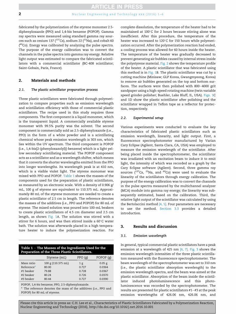
Table 1 e The Masses of the Ingredients Used for thePreparation of the Three Plastic Scintillators.
Styrene (mL) PPO (g) POPOP (g)
Mass ratio 100 g (110.375 mL) 1 g 0.05 g
Referencea 80.00 0.727 0.0364
#1 beaker 79.88 0.728 0.0367
#2 beaker 80.24 0.726 0.0370
#3 beaker 80.44 0.727 0.0390
POPOP, 1,4-bis benzene; PPO, 2.5-diphenyloxazole.a The reference denotes the mass of the additives (i.e., PPO and
POPOP) for 80 mL of styrene.
Please cite this article in press as: C.H. Lee et al., Characteristics oNuclear Engineering and Technology (2016), http://dx.doi.org/10.
complete dissolution, the temperature of the heater had to be
maintained at 100�C for 2 hours because stirring alone was
insufficient. After this procedure, the temperature of the
heater was increased to 120�C for 150 hours while polymeri-
zation occurred. After the polymerization reaction had ended,
a cooling process was allowed for 60 hours inside the heater.
The temperature of the heater was gradually decreased to
prevent generating air bubbles caused by internal stress inside
the polystyrene material. Fig. 2 shows the temperature profile
of the heater. A plastic scintillator that was fabricated using
this method is in Fig. 1B. The plastic scintillator was cut by a
cutting machine (Minisaw; GLP Korea, Gwangmyeong, Korea)
to remove air bubbles generated on the top and bottom sur-
faces. The surfaces were then polished with 800e4000 grit
sandpaper using a high-speed rotatingmachine (twin variable
speed grinder-polisher; Buehler, Lake Bluff, IL, USA). Figs. 1C
and 1D show the plastic scintillator after polishing and the
scintillator wrapped in Teflon tape as a reflector for protec-
tion, respectively.
2.2. Experimental setup
Various experiments were conducted to evaluate the key
characteristics of fabricated plastic scintillators such as
emission wavelength, linearity, and light output. First, a
fluorescence spectrophotometer manufactured by Varian
Cary Eclipse (Agilent, Santa Clara, CA, USA) was employed to
measure the emission wavelength of the scintillator. After
being placed inside the spectrophotometer, the scintillator
was irradiated with an excitation beam to induce it to emit
light, the intensity of which was recorded as a graph by the
Cary Eclipse software (Agilent). Second, three gamma ray
sources (137Cs, 22Na, and 60Co) were used to evaluate the
linearity of the scintillators through energy calibration. The
purpose of the energy calibration was to convert the channels
in the pulse spectra measured by the multichannel analyzer
(MCA) module into gamma ray energy; the linearity was sub-
sequently estimated, based on the calibration. Third, the
relative light output of the scintillator was calculated by using
the Bertolaccini method [4, 5]. Four parameters are necessary
to use the method. Section 3.3 provides a detailed
introduction.
3. Results and discussion
3.1. Emission wavelength
In general, typical commercial plastic scintillators have a peak
emission at a wavelength of 425 nm [6, 7]. Fig. 3 shows the
emission wavelength intensities of the three plastic scintilla-
tors measured with the fluorescence spectrophotometer. The
beamwavelength of the spectrophotometer was set to 310 nm
(i.e., the plastic scintillator absorption wavelength) to the
emission wavelength spectra, and the beam was aimed at the
plastic scintillator. Absorption of the beam inside the scintil-
lator induced photoluminescence and this photo-
luminescence was recorded by the spectrophotometer. The
results are presented for plastic scintillators #1e#3 at the peak
emission wavelengths of 426.06 nm, 426.06 nm, and
f Plastic Scintillators Fabricated by a Polymerization Reaction,1016/j.net.2016.10.001

Fig. 1 e The plastic scintillator preparation process. Step-by-step images show (A) the status after stirring, (B) the scintillator
completed by polymerization reaction, and (C) the finished plastic scintillator. (D) is the scintillator wrapped in Teflon tape
as a reflector for protection.
Nu c l e a r E n g i n e e r i n g a n d T e c h n o l o g y x x x ( 2 0 1 6 ) 1e6 3
425.00 nm, respectively, with 0.2% standard error. When
measuring the emission wavelength of the plastic scintillator
with the spectrophotometer, it is impossible to fully control
the angle of the incident beam with respect to the scintilla-
tor’s surface. This factor may explain the slight difference in
intensity. In addition, an intensity difference can occur
because the solution was not prepared according to the cor-
rect mass ratio (100:1:0.05). However, the fabricated plastic
scintillators have the same properties as commercial
Fig. 2 e Temperature profile of the high temperature heater
over the course of the preparation process.
Please cite this article in press as: C.H. Lee et al., Characteristics oNuclear Engineering and Technology (2016), http://dx.doi.org/10.
scintillators [e.g., BC-408 scintillator (Saint-Gobain)] because
they share the same peak emission wavelength.
3.2. Energy calibration of the scintillator detector
A standard gamma source such as 137Cs, 22Na, and 60Co is
usually used for the energy calibration of a general radiation
Fig. 3 e Emission wavelength intensities of the three
plastic scintillators. Each scintillator shows a peak at a
wavelength of approximately 425 nm.
f Plastic Scintillators Fabricated by a Polymerization Reaction,1016/j.net.2016.10.001

Fig. 4 e Schematic of the experimental setup for measuring
the Compton edge spectra using the fabricated plastic
scintillators.
Nu c l e a r E n g i n e e r i n g a n d T e c h n o l o g y x x x ( 2 0 1 6 ) 1e64
detector. Measurements of the Compton edge spectra using
gamma sources were possible in this study because a plastic
scintillator does not exhibit phosphorescence. In Compton
scattering, a gamma ray is scatted by a free electron. The
energy of the electron is transferred to the detector while the
scattered gamma ray escapes from the detector. The recoil
electron has a well-defined maximum energydthe afore-
mentioned Compton edge, which corresponds to the
maximum transferred energy from the gamma ray to the
electron. This edge can be used for the energy calibration of
the detector and for comparing the relative light output from
each scintillator. The Compton edge corresponds to the
maximum energy transfer from the scattered gamma ray to
the electron. Therefore, the Compton edges of backscattered
gamma rays can be calculated by using the kinematic equa-
tion of energy conservation and by taking into account that
the maximum amount of energy is transferred during the
backscattering of the gamma rays. Table 2 shows the calcu-
lated Compton edge energy values of the gamma ray sources.
In this work, three radioactive sources were used for en-
ergy calibration: 22Na, 60Co, and 137Cs. The Compton edge
energy for each source was calculated, based on the gamma
ray energies taken from Table of Isotopes [8]. For the 60Co
source, an average energy was selected for the Compton edge
calculations because it emits two gamma rays of 1.17MeV and
1.33 MeV. Fig. 4 shows a schematic of the experimental setup
formeasuring the Compton edge spectra. The specifications of
themodules used tomeasure the Compton edge spectra are as
follows. (1) Photonmultiplier tube (PMT): Hamamatsu H6614-
70 (Hamamatsu City, Japan; operating voltage, 1500 V). (2)
Amplifier: Ortec 572A (Ortec, Inc., Tennessee, OR, USA;
shaping time, 0.5 ms). (3) High voltage power supply: Ortec 556
(Ortec, Inc.). (4) Multichannel analyzer: Ortec 919E (Ortec, Inc.).
Fig. 5 shows set up of the modules, based on Fig. 4. The
plastic scintillator was attached to the window of the PMT
using optical grease. Lead bricks (5 cm thick) provided radia-
tion shielding and black tape blocked external light. Maestro
software (Ortec, Inc., Tennessee, OR, USA), which was applied
to the MCA, was used to record the spectra over a measure-
ment time of 600 seconds. Fig. 6 shows the gamma ray pulse
spectra of 137Cs, 22Na, and 60Co obtained with the three plastic
scintillators. To identify the channel, which corresponds to
the Compton edge energy in these spectra, the middle point
where the slope drops to one-half of the value of the Compton
peak was selected. The energy calibration results of each
scintillator are as follows: (1) Scintillator #1 / Channel ¼-32.29554þ 0.79962� energy (R2 ¼ 99.944); (2) Scintillator #2/
Channel ¼ -16.22904 þ 0.70749 � energy (R2 ¼ 99.951); and (3)
Scintillator #3/ Channel¼ -41.55461þ 0.80273� energy (R2¼99.786).
Table 2 e The Calculated Compton Edge Values of theThree Gamma Ray Sources.
Source Activity(mCi)
Gamma energy(keV)
Compton edgeenergy (keV)
22Na 1.231 511.003 340.11060Co 3.314 1173.228/1332.492 Avg. 1040.79137Cs 8.071 661.657 477.334
60Co, cobalt 60; 137Cs, cesium 137; 22Na, sodium 22.
Please cite this article in press as: C.H. Lee et al., Characteristics oNuclear Engineering and Technology (2016), http://dx.doi.org/10.
The energy linearity of each scintillator, obtained through
the energy calibration, was determined as 99.944, 99.951, and
99.786; thus, confirming that the fabricated plastic scintilla-
tors possess excellent linearity. Because of the energy cali-
bration, the pulse spectra for 137Cs, 22Na, and 60Co can be
shown at their correct energy scales (Figs. 6 and 7). The x axes
of the pulse spectra that were measured by the MCA module
for the energy calibration were converted into gamma ray
energy values. The result was in agreement with the general
observation that in the low energy region the response of
scintillators is proportional to the incoming energy.
3.3. Light output
The light yield was determined by measuring the number of
photoelectrons per energy unit (Nphe). This can be achieved
through a comparison of the peak position of a single photo-
electron spectrum (PP1phe) with a characteristic point of any
energy spectrum [4, 5]. In the current measurements, the
Compton edge (477.334 keV, 137Cs) for the 661.657 keV full
energy peak of the 137Cs gamma source was used. Peak posi-
tions were recorded with different gains of the spectroscopy
amplifier K because of the large amplitude differences be-
tween single photoelectron signals and the 137Cs energy
spectrum. The number of photoelectrons per energy unit is
given by the following equation [4, 5]:
Fig. 5 e Experimental setup, based on Fig. 4. The
photonmultiplier tube (PMT) is connected to a high voltage
power supply module (Ortec 556). The distance between
the scintillator and source is 5 cm.
f Plastic Scintillators Fabricated by a Polymerization Reaction,1016/j.net.2016.10.001

Fig. 6 e The gamma ray pulse spectra for three different
sources obtained with each fabricated plastic scintillator.
Fig. 7 e Energy calibration via linear fit for the data points
corresponding to the Compton edges in Fig. 6. The linearity
of scintillators #1e#3 is 99.944, 99.951, and 99.786,
respectively.
Nu c l e a r E n g i n e e r i n g a n d T e c h n o l o g y x x x ( 2 0 1 6 ) 1e6 5
Nphe ¼ (PPE/KE)/(PP1phe/K1phe)/0.477334 (phe/MeV) (1)
in which PPE is the Compton edge peak position of the 137Cs
gamma source, KE is gain of the spectroscopy amplifier in the
experiment. Table 3 shows the light output measured by a
commercial plastic scintillator (BC-408, Saint-Gobain) and by
the fabricated plastic scintillators. Parameters such as
amplifier gain and peak position were obtained through pulse
spectra analysis. The quantumefficiency of the PMT should be
Please cite this article in press as: C.H. Lee et al., Characteristics oNuclear Engineering and Technology (2016), http://dx.doi.org/10.
considered when calculating the light output because there is
a difference in the quantum efficiency for different wave-
lengths. The quantum efficiency is the number of photoelec-
trons emitted from the photocathode divided by the number
of incident photons. The quantum efficiency of the PMT is
given by the following equation [9]:
QE ¼ (S � 1240)/l � 100 (2)
in which QE is the quantum efficiency, S is the radiant
sensitivity in amperes per watt at the given wavelength,
f Plastic Scintillators Fabricated by a Polymerization Reaction,1016/j.net.2016.10.001

Table 3 e Light Output Measured with the BC-408 Scintillator and the Fabricated Plastic Scintillators.
Parameter #1 Scintillator #2 Scintillator #3 Scintillator BC-408
Gain (KE) 10 10 10 10
Gain (K1phe) 1000 1000 1000 1000
Peak position (PPe) 344 317 331 644
Peak position (PP1phe) 119 108 121 140
Light yield (phe/MeV) 605.60 614.91 573.09 963.69
Quantum efficiency (%) 20.95 20.95 21.01 21.01
Light output (ph/MeV) a 2890.05 ± 14.45 2934.47 ± 14.68 2728.07± 13.64 4587.44± 22.94
KE, gain of spectroscopy amplifier; MeV, megaelectron volt; ph, photon; phe, photoelectron; PP1phe, peak position of a single photoelectron
spectrum; PPe, Compton edge peak position of a 137Cs.a The values in this row are {Explanation}.
Nu c l e a r E n g i n e e r i n g a n d T e c h n o l o g y x x x ( 2 0 1 6 ) 1e66
and l is the wavelength in nanometers. The quantum effi-
ciency was calculated as approximately 21% and the light
output was estimated by dividing the quantum efficiency by
the light yield.
The fabricated plastic scintillators exhibited a light output
of approximately 59e64% of the output of the BC-408 scintil-
lator (Saint-Gobain). To increase this value, naphthalene, an
organic compound that is often used to improve solubility,
could be added during the preparation process. In addition,
other liquid monomers such as SR9035 and SR9036 could be
used instead of styrene. Adding the aforementionedmaterials
may improve scintillator properties.
4. Conclusion
Using a polymerization reaction in a high temperature heater,
three plastic scintillators were fabricated to compare their
properties with those of a commercial plastic scintillator.
Styrene, PPO, and POPOP as the scintillator materials were
mixed inside a 100-mL beaker and placed into a high tem-
perature heater for approximately 250 hours. Surface
machining operations such as cutting and polishing were
performed and plastic scintillators of 4.5 cm diameter and
2.5 cm length were fabricated. Three standard gamma ray
sources were used to evaluate the characteristics of the
fabricated scintillators. Pulse spectra were measured via PMT,
amplifier, high voltage power supply, andMCA using different
gamma ray sources. Linearity and light outputwere calculated
through pulse spectra analysis. Based on energy calibrations,
the fabricated scintillators possessed more than 99.7% energy
linearity. The scintillators showed a light output of approxi-
mately 59e64% of that of a BC-408 scintillator (Saint-Gobain).
In the future, to improve the light output, othermaterials such
as naphthalene and the new liquid monomers will be added
and are expected to improve the properties of the scintillator.
Studies related to fast neutron and charged particle detection
will also be conducted to apply the fabricated scintillator in
the field of radiation security.
Please cite this article in press as: C.H. Lee et al., Characteristics oNuclear Engineering and Technology (2016), http://dx.doi.org/10.
Conflicts of interest
All authors have no conflicts of interest to declare.
Acknowledgments
This work was supported by a research grant of Hanyang
University (Seoul, Korea; grant number, HY-2009).
r e f e r e n c e s
[1] H. Penttila, Characterization of a New Plastic ScintillationMaterial and Comparison with Liquid BCe501A Scintillator(Saint-Gobain, Paris, France), Oleksii Poleshchuk, Jyv€askyl€a,Finland, 2015.
[2] D. Reilly, N. Ensslin, H. Smith Jr., S. Kreiner, Passivenondestructive assay of nuclear materials, in: Doug Reilly,Norbert Ensslin, Hastings Smith Jr. (Eds.), Los Alamos NationalLab, Los Alamos, NM, USA, 1991.
[3] Z. Li, W. Chong, H. Yuekun, Z. Xiaojian, S. Feng, Z. Sun,W. Jinjie, A. Henghua, Z. Yuda, Z. Ziping, W. Yifang, Propertiesof plastic scintillators after irradiation, Nucl. Instrum.Methods A 552 (2005) 449e455.
[4] M. Bertolaccini, C. Bussolati, S. Cova, I. De Lotto, E. Gatti,Optimum processing for amplitude distribution evaluation ofa sequence of randomly spaced pulses, Nucl. Instrum.Methods Phys. Res. A61 (1968) 84e88.
[5] M. Moszynski, M. Kapusta, M. Mayhugh, D. Wolski, S.O. Flyckt,Absolute light output of scintillators, IEEE Trans. Nucl. Sci. 44(1997) 1052e1061.
[6] Organic Scintillation Materials, Saint-Gobain Crystals, Paris,France.
[7] G.H. Kim, C.H. Park, C.H. Jung, K.W. Lee, B.K. Seo,Development of the ZnS(Ag)/BC-408 phoswich detector formonitoring radioactive contamination inside pipes, J. KoreanAssoc. Radiat. Prot. 31 (2006) 123e128.
[8] B. Richard, Firestone Table of Isotopes, eigth ed., Wiley, NewYork, 1999.
[9] Photomultiplier Tubes and Related Products, HamamatsuPhotonics Co., Hamamatsu City, Japan, 2010.
f Plastic Scintillators Fabricated by a Polymerization Reaction,1016/j.net.2016.10.001