Characteristics and outcomes of anti-infective de ...
12
RESEARCH Open Access Characteristics and outcomes of anti-infective de-escalation during health care-associated intra-abdominal infections Philippe Montravers 1,2* , Pascal Augustin 1 , Nathalie Grall 2,3,4 , Mathieu Desmard 1,5 , Nicolas Allou 1 , Jean-Pierre Marmuse 2,6 and Jean Guglielminotti 1,2,3 Abstract Background: De-escalation is strongly recommended for antibiotic stewardship. No studies have addressed this issue in the context of health care-associated intra-abdominal infections (HCIAI). We analyzed the factors that could interfere with this process and their clinical consequences in intensive care unit (ICU) patients with HCIAI. Methods: All consecutive patients admitted for the management of HCIAI who survived more than 3 days following their diagnosis, who remained in the ICU for more than 3 days, and who did not undergo early reoperation during the first 3 days were analyzed prospectively in an observational, single-center study in a tertiary care university hospital. Results: Overall, 311 patients with HCIAI were admitted to the ICU. De-escalation was applied in 110 patients (53 %), and no de-escalation was reported in 96 patients (47 %) (escalation in 65 [32 %] and unchanged regimen in 31 [15 %]). Lower proportions of Enterococcus faecium, nonfermenting Gram-negative bacilli (NFGNB), and multidrug- resistant (MDR) strains were cultured in the de-escalation group. No clinical difference was observed at day 7 between patients who were de-escalated and those who were not. Determinants of de-escalation in multivariate analysis were adequate empiric therapy (OR 9.60, 95 % CI 4.02–22.97) and empiric use of vancomycin (OR 3.39, 95 % CI 1.46–7.87), carbapenems (OR 2.64, 95 % CI 1.01–6.91), and aminoglycosides (OR 2.31 95 % CI 1.08–4.94). The presence of NFGNB (OR 0.28, 95 % CI 0.09–0.89) and the presence of MDR bacteria (OR 0.21, 95 % CI 0.09–0.52) were risk factors for non-de-escalation. De-escalation did not change the overall duration of therapy. The risk factors for death at day 28 were presence of fungi (HR 2.64, 95 % CI 1.34–5.17), Sequential Organ Failure Assessment score on admission (HR 1.29, 95 % CI 1.16–1.42), and age (HR 1.03, 95 % CI 1.01–1.05). The survival rate expressed by a Kaplan-Meier curve was similar between groups (log-rank test p value 0.176). Conclusions: De-escalation is a feasible option in patients with polymicrobial infections such as HCIAI, but MDR organisms and NFGNB limit its implementation. Keywords: De-escalation, Health care-associated intra-abdominal infections, Peritonitis, Multidrug-resistant bacteria, Antibiotic therapy * Correspondence: [email protected] 1 Département d’Anesthésie Réanimation, APHP, CHU Bichat-Claude Bernard, Paris, France 2 Université Denis Diderot, PRESS Sorbonne Cité, Paris, France Full list of author information is available at the end of the article © 2016 Montravers et al. Open Access This article is distributed under the terms of the Creative Commons Attribution 4.0 International License (http://creativecommons.org/licenses/by/4.0/), which permits unrestricted use, distribution, and reproduction in any medium, provided you give appropriate credit to the original author(s) and the source, provide a link to the Creative Commons license, and indicate if changes were made. The Creative Commons Public Domain Dedication waiver (http://creativecommons.org/publicdomain/zero/1.0/) applies to the data made available in this article, unless otherwise stated. Montravers et al. Critical Care (2016) 20:83 DOI 10.1186/s13054-016-1267-8
Transcript of Characteristics and outcomes of anti-infective de ...

RESEARCH Open Access
Characteristics and outcomes ofanti-infective de-escalation during healthcare-associated intra-abdominal infectionsPhilippe Montravers1,2*, Pascal Augustin1, Nathalie Grall2,3,4, Mathieu Desmard1,5, Nicolas Allou1,Jean-Pierre Marmuse2,6 and Jean Guglielminotti1,2,3
Abstract
Background: De-escalation is strongly recommended for antibiotic stewardship. No studies have addressed thisissue in the context of health care-associated intra-abdominal infections (HCIAI). We analyzed the factors that couldinterfere with this process and their clinical consequences in intensive care unit (ICU) patients with HCIAI.
Methods: All consecutive patients admitted for the management of HCIAI who survived more than 3 daysfollowing their diagnosis, who remained in the ICU for more than 3 days, and who did not undergo earlyreoperation during the first 3 days were analyzed prospectively in an observational, single-center study in a tertiarycare university hospital.
Results: Overall, 311 patients with HCIAI were admitted to the ICU. De-escalation was applied in 110 patients(53 %), and no de-escalation was reported in 96 patients (47 %) (escalation in 65 [32 %] and unchanged regimen in 31[15 %]). Lower proportions of Enterococcus faecium, nonfermenting Gram-negative bacilli (NFGNB), and multidrug-resistant (MDR) strains were cultured in the de-escalation group. No clinical difference was observed at day 7 betweenpatients who were de-escalated and those who were not. Determinants of de-escalation in multivariate analysis wereadequate empiric therapy (OR 9.60, 95 % CI 4.02–22.97) and empiric use of vancomycin (OR 3.39, 95 % CI 1.46–7.87),carbapenems (OR 2.64, 95 % CI 1.01–6.91), and aminoglycosides (OR 2.31 95 % CI 1.08–4.94). The presence of NFGNB(OR 0.28, 95 % CI 0.09–0.89) and the presence of MDR bacteria(OR 0.21, 95 % CI 0.09–0.52) were risk factors for non-de-escalation. De-escalation did not change the overall duration oftherapy. The risk factors for death at day 28 were presence of fungi (HR 2.64, 95 % CI 1.34–5.17), Sequential OrganFailure Assessment score on admission (HR 1.29, 95 % CI 1.16–1.42), and age (HR 1.03, 95 % CI 1.01–1.05). The survivalrate expressed by a Kaplan-Meier curve was similar between groups(log-rank test p value 0.176).
Conclusions: De-escalation is a feasible option in patients with polymicrobial infections such as HCIAI, but MDRorganisms and NFGNB limit its implementation.
Keywords: De-escalation, Health care-associated intra-abdominal infections, Peritonitis, Multidrug-resistant bacteria,Antibiotic therapy
* Correspondence: [email protected]épartement d’Anesthésie Réanimation, APHP, CHU Bichat-Claude Bernard,Paris, France2Université Denis Diderot, PRESS Sorbonne Cité, Paris, FranceFull list of author information is available at the end of the article
© 2016 Montravers et al. Open Access This article is distributed under the terms of the Creative Commons Attribution 4.0International License (http://creativecommons.org/licenses/by/4.0/), which permits unrestricted use, distribution, andreproduction in any medium, provided you give appropriate credit to the original author(s) and the source, provide a link tothe Creative Commons license, and indicate if changes were made. The Creative Commons Public Domain Dedication waiver(http://creativecommons.org/publicdomain/zero/1.0/) applies to the data made available in this article, unless otherwise stated.
Montravers et al. Critical Care (2016) 20:83 DOI 10.1186/s13054-016-1267-8
BackgroundThe modern concept of reducing both the spectrum ofantimicrobial therapy and its potential to promote resist-ance [1], usually called de-escalation, is strongly recom-mended in all of the recently published guidelines forantibiotic stewardship [2, 3]. Several definitions havebeen used to describe this process [1, 4–7]. Despite thelimited evidence, de-escalation has been recommendedto decrease the likelihood of emergence of resistant or-ganisms [8], to optimize activity, and to reduce toxicityand costs [3].Two types of critically ill patients have been investi-
gated: cohorts with a specific disease—mainly ventilator-associated pneumonia (VAP) [5, 9–13]—and large mixedpopulations with severe sepsis or septic shock or pa-tients who require emergency empiric antibiotic therapy[4, 14–19]. In a recent systematic review, Tabah et al.identified isolation of multiresistant pathogens, polymicro-bial infections, and intra-abdominal infections as factorsnegatively associated with de-escalation [20]. Only a fewstudies have addressed this issue in the context of intra-abdominal infections [4, 14, 18, 21, 22]. The high fre-quency of polymicrobial infection [23, 24] and multidrug-resistant (MDR) organisms [23, 24] as well as the presenceof fungi [17, 25] in this setting might raise specific con-cerns, especially in health care-associated intra-abdominalinfections (HCIAI).The purpose of the present study was to analyze the
frequency of de-escalation, the factors that could inter-fere with this process, and their clinical consequencesin a cohort of intensive care unit (ICU) patients withHCIAI.
MethodsStudy populationFrom January 1999 through December 2011, all con-secutive patients admitted to our ICU for the manage-ment of HCIAI were prospectively included in adatabase and their medical charts were retrospectivelyreviewed. The study was approved by the local institutionalreview board (CEERB CHU Bichat Paris VII University,APHP, Paris, France), which waived the need for signedinformed consent.
Selection of cases and inclusion criteriaPostoperative peritonitis was defined as the first macro-scopic findings of intra-abdominal infection combinedwith positive fluid culture yielding at least one micro-organism (bacteria or fungi) at the time of reoperation(day 0) following a first abdominal surgery [24]. Severalpatients had to be excluded from the analysis due toearly change in their clinical status before de-escalationcould be instituted: patients who died during the first3 days following surgery (microbiologic results not yet
obtained), those who were discharged during the first3 days (incapacity to adequately follow clinical outcomeand antibiotic therapy), and those who underwent earlyreoperation during the first 3 days (high proportion ofpersistent infection and prolonged antibiotic therapy).Similarly, we excluded patients with negative microbio-logic samples, since the concept of de-escalation is ques-tionable and the interpretation of the results is difficult.In these patients, empiric anti-infective therapy wasdiscontinued. Drainage of abscesses, debridement of in-fected and necrotic tissues, abdominal cavity cleansing,irrigation, and definitive control of the source of con-tamination were performed according to the surgicalprinciples used for the management of abdominal sepsis[26]. Ostomy was preferred to primary anastomosis. Noopen-wound management was performed, and the abdo-men was not irrigated after surgery.
Microbiologic dataPeritoneal fluid samples were systematically collectedduring surgery and were immediately sent to the bacteri-ology laboratory [24]. Cultures were performed with identi-fication and susceptibility testing for Gram-positive andGram-negative aerobe bacteria, anaerobes, and fungi. Anti-biotic susceptibility was determined for each organism bythe disk diffusion method, according to the criteria of theAntibiogram Committee of the French Society for Micro-biology [27]. MDR bacteria were defined as those resistantto three or more antimicrobial classes [28]: methicillin-resistant Staphylococcus aureus and methicillin-resistantcoagulase-negative staphylococci; ampicillin-resistantenterococci; Enterobacteriaceae producing an extended-spectrum β-lactamase or producing a derepressed cepha-losporinase; and/or nonfermenting Gram-negative bacilli(NFGNB) resistant to piperacillin-tazobactam, ceftazi-dime, or imipenem-cilastatin, or producing an extended-spectrum β-lactamase.
Management of antibiotic therapyEmpiric anti-infective therapy, systematically started atday 0, took into account the severity of the case andusually combined piperacillin-tazobactam or imipenem-cilastatin with amikacin and vancomycin [23], possiblyassociated with antifungal therapy (mainly fluconazole)based on presumed risk factors [25, 29]. Definitive anti-infective therapy was adapted on the basis of the resultsof identification and antibiotic susceptibility testing(≥48 h). In both situations, therapy was considered ap-propriate when all cultured organisms (bacteria andfungi) were targeted. Anti-infective therapy was pre-scribed by the senior ICU physicians following discus-sion with the consultant microbiologist on a daily basis.The following changes were considered to constitute
de-escalation [1]: withdrawal of one agent (β-lactam,
Montravers et al. Critical Care (2016) 20:83 Page 2 of 12

aminoglycoside, fluoroquinolone, vancomycin, antifungalagent) or narrowing spectrum of activity (β-lactamagents) and/or switch from combination to monother-apy. Discontinuation of unduly administered agentswas also recorded. Changes among cefepime, ceftazidime,piperacillin-tazobactam, and ticarcillin-clavulanate werenot considered to be significant changes of the spectrumof coverage [1].In patients without de-escalation, two situations were
identified according to previous definitions [4]. Main-tained empiric treatment without modification was calledunchanged therapy [4]. Escalation was defined as additionor switch to a new broad-spectrum anti-infective agent(carbapenems, glycopeptides, fluoroquinolones) [4] or up-grade to broader-spectrum β-lactams [1]. When changescombined escalation and de-escalation, the patient wasassigned to the escalation group [4]. In summary, a patientreceiving empiric therapy with piperacillin-tazobactamplus amikacin who was subsequently switched to pipera-cillin and vancomycin was classified as having withdrawalof one agent and escalation.
Data collectionAll patients’ charts were reviewed. Demographic dataand severity scores (Simplified Acute Physiology Score IIscore [30] and Sequential Organ Failure Assessment[SOFA] score [31]) were recorded on admission to theICU. The severity of the underlying medical conditionand the presence of chronic diseases [32] were recorded.The characteristics of initial surgery were recorded.The following clinical and severity characteristics were
assessed at day 0, day 3, and day 7 after surgery for pa-tients still in the ICU [24, 33, 34]: temperature, whiteblood cell count (WBC), serum creatinine, and SOFAscore. Patients meeting the following three criteria atday 3 were arbitrarily defined as improving: (1) a SOFAscore that decreased more than 2 points at day 3 versusday 0 or a SOFA score of 0 points, (2) a WBC that de-creased more than 5000/mm3 between day 0 and day 3or WBC less than 12,500/mm3, and (3) a temperaturedecrease greater than 0.5 °C between day 0 and day 3 ortemperature greater than or equal to 36.5 °C and lessthan 38.1 °C. Similar analyses were used at day 7 tocompare changes in these criteria between days 3 and 7.Medical and surgical complications, additional reopera-tions for persistence of the initial infection or superin-fections (including MDR organisms), death betweendays 3 and 28 following surgery, and discharge from thehospital were assessed.
Statistical analysisResults are expressed as median and interquartile range(IQR) or number and proportion. Statistical significancewas defined as p < 0.05. For statistical analysis, we used
R version 2.14.1 software (R Foundation for StatisticalComputing, Vienna, Austria). For comparisons betweenantibiotic strategy groups (de-escalation, no change, orescalation), we used the χ2 test and Fisher’s exact test fordiscrete variables and unpaired Wilcoxon tests for quan-titative variables. The effect of antibiotic strategy on day28 mortality was assessed with a Kaplan-Meier survivalcurve and tested with a log-rank test.Three multivariable models were developed (1) to
identify risk factors for de-escalation and (2) to assessthe association between antibiotic strategy (de-escal-ation, no change, or escalation) and day 28 or in-hospital mortality. In univariate analysis for these threemodels, we used Fisher’s exact tests and Wilcoxon tests.Unadjusted ORs or HRs were calculated. Variables witha p value less than 0.2 in univariate analysis were enteredinto a multivariate logistic regression model or a Coxproportional hazards model with backward selection.For day 28 and in-hospital mortality, the antibiotic strat-egy was forced until the end of the selection process.Logistic models were evaluated for discrimination withthe c-statistic and for calibration with the Hosmer-Lemeshow test.
ResultsEpidemiologic and clinical characteristicsDuring the study period, 311 ICU patients were admit-ted for the management of HCIAI. Figure 1 displays aflowchart of patients through the study. Overall, 105 pa-tients were excluded, resulting in 206 patients for whomthe de-escalation process was analyzed. De-escalationwas performed in 110 patients (53 % of the analyzedpopulation), and no de-escalation was observed in 96 pa-tients (47 %) (escalation in 65 patients [32 %] and un-changed regimen in 31 patients [15 %]). De-escalationwas never performed after discharge from the ICU. Thefrequency of de-escalation remained stable over thestudy period, ranging between 47 % and 63 % of the ana-lyzed population (not significant; data not shown). Clin-ical characteristics were similar at day 0 in both groups(Table 1). In the non-de-escalation group, a significantlyincreased severity was observed in the patients with anunchanged regimen versus escalation (Table 1).
Microbiologic analysisOverall, 618 microorganisms from peritoneal sampleswere cultured (311 in patients without de-escalation, in-cluding 101 organisms in the unchanged group and 210in the escalation group). Similar microbiologic resultswere observed between de-escalation and no de-escalation groups (data not shown), except for lowerproportions of Enterococcus faecium (9 [3 %] versus 18[7 %] without de-escalation, respectively; p < 0.01) andnon-fermenting Gram-negative bacilli (9 [3 %] versus 22
Montravers et al. Critical Care (2016) 20:83 Page 3 of 12

[8 %], respectively; p < 0.01). Among patients withoutde-escalation, increased proportions of Gram-negativebacteria and Enterobacteriaceae were observed in pa-tients with unchanged regimen compared with thosewhose regimen was escalated (47 [49 %] versus 69[37 %], respectively [p < 0.05]; and 38 [40 %] versus 52[28 %] [p < 0.05]). In the de-escalation group, 23 (21 %)of 110 patients harbored MDR strains compared with54 (56 %) of 96 patients in the non-de-escalationgroup (p < 0.01) (Table 2).
Anti-infective therapyDe-escalation was performed on a microbiologic basis ina median delay of 3 days (IQR 2–4) after surgery. Em-piric treatments and the procedures applied for de-escalation at day 3 are described in Table 3. Empiric useof combination therapy, carbapenems, glycopeptides,and antifungal agents were significantly more frequentin the de-escalation group. Among antifungal agents,echinocandins were minimally prescribed for empirictherapy (six patients in the de-escalation group with dis-continuation in all but one case and one switch toazoles, one patient in the non-de-escalation group).
When taking into account the criteria for de-escalation,we found that 33 patients met three criteria (withdrawing,narrowing, and switching) in the de-escalation group butnone of those who did not de-escalate. However, two cri-teria (withdrawing and narrowing) were reported in 17 pa-tients in the non-de-escalation group. No clinical changebetween days 0 and 3 allowed patients who were subse-quently de-escalated to be differentiated from those whodid not (Table 3).Determinants of de-escalation in multivariate analysis
were adequate empiric therapy (OR 9.60, 95 % CI 4.02–22.97, p < 0.001), empiric use of vancomycin (OR 3.39,95 % CI 1.46–7.87, p = 0.004), carbapenems (OR 2.64,95 % CI 1.01–6.91, p = 0.04), and aminoglycosides (OR2.31, 95 % CI 1.08–4.94, p = 0.03), while presence ofNFGNB (OR 0.28, 95 % CI 0.09–0.89, p = 0.03) andpresence of MDR bacteria (OR 0.21, 95 % CI 0.09–0.52,p < 0.001) were the risk factors for non-de-escalation(c-index 0.880, 95 % CI 0.832–0.928, Hosmer-Lemeshowtest p = 0.14) (Table 4).At day 3, no difference was observed between patients
with an unchanged regimen and those who underwentescalation, except for the higher proportions of
105 patients excluded from the analysisNegative microbiologic samples (n=7)Death during the first three days (n=22)Patients discharged alive during the first three days (n=10)Patients who underwent early reoperation (n=66)
311 patients admitted in ICU for postoperative peritonitis
De-escalation in110 (53%) patients
No de-escalation in96 (47%) patients
De-escalation process analyzed in206 patients
Escalation in65 (32%) patients
Unchanged regimen in31 (15%) patients
Fig. 1 Flowchart of the 206 patients studied. ICU intensive care unit
Montravers et al. Critical Care (2016) 20:83 Page 4 of 12

aminoglycosides and antifungal therapy and the verylow proportion adequate empiric therapy (Table 3).
Clinical evaluation following de-escalationWhen comparing the patients who were de-escalatedand those who did not de-escalate, we found no significantdifference between days 3 and 7 (Table 5). In addition, nosignificant difference in morbidity or mortality criteria wasobserved between these two groups.No significant differences in morbidity or mortality
criteria were observed between patients who underwentescalation and those with an unchanged regimen, exceptfor significantly increased proportions of surgical com-plications and reoperations in patients with an un-changed regimen. The risk factors for death at day 28following surgery in a Cox model are presented inTable 6. The survival rate expressed by a Kaplan-Meiercurve was similar between groups (log-rank test p value0.176) (Fig. 2).The risk factors of in-hospital mortality on multivari-
ate analysis were the emergency initial surgery (OR 2.81,
95 % CI 1.30–6.05, p = 0.008) and SOFA score on ad-mission (OR 1.42, 95 % CI 1.24–1.62, p < 0.001), whilea decreased SOFA score at day 3 had a protective value(OR 0.14, 95 % CI 0.06–0.31, p < 0.001) (c-index 0.852,95 % CI 0.794–0.910, Hosmer-Lemeshow test p = 0.28).
DiscussionIn this single-center observational study, de-escalationwas performed in 53 % of patients treated for HCIAI.De-escalation concerned both antibacterial and antifun-gal therapies. The de-escalation procedure did not mod-ify outcome. No initial clinical characteristic allowedidentification of patients who were subsequently de-escalated. The presence of MDR bacteria and NFGNB aswell as initial monotherapy were the most relevant fac-tors limiting de-escalation. No emergence of resistantorganisms was observed following de-escalation in thepatients who underwent subsequent reoperation.In the absence of data in the literature, we consider
that our results provide an encouraging perspective forantibiotic de-escalation in ICU patients with abdominal
Table 1 Demographic and clinical characteristics of the 206 patients with or without subsequent antibiotic de-escalation
Characteristic De-escalation (n = 110) No de-escalation (n = 96) Escalation (n = 65) No change (n = 31)
Male sex, n (%) 61 (55) 56 (58) 35 (54) 21 (68)
Age, years, median (IQR) 61 (47–72) 66 (51–75)a 63 (47–75) 70 (58–77)
Comorbidities
Fatal underlying disease 30 (27) 32 (33) 21 (32) 11 (35)
Cancer, n (%) 37 (34) 36 (38) 23 (35) 13 (42)
Diabetes, n (%) 17 (15) 15 (16) 9 (14) 6 (19)
Time since initial surgery, days, median (IQR) 7 (5–12) 7 (4–10) 6 (3–9) 8 (5–10)
Antibiotic therapy before reoperation, n (%) 73 (66) 68 (71) 47 (72) 21 (68)
Broad-spectrum interim antibiotic, n (%) 34 (31) 37 (39) 27 (42) 10 (32)
Intraoperative diagnosis
Anastomotic leakage, n (%) 45 (41) 27 (28) 19 (29) 8 (26)
Perforation or ischemia, n (%) 33 (30) 36 (38) 20 (31) 16 (52)a#
Purulent collection, n (%) 19 (17) 17 (18) 13 (20) 4 (13)
No cause, n (%) 19 (17) 20 (21) 15 (23) 5 (16)
Contamination below transverse mesocolon, n (%) 82 (75) 74 (77) 50 (77) 24 (77)
Characteristics at the time of ICU admission
Bacteremia, n (%) 26 (24) 17 (18) 14 (22) 3 (10)
SAPS II score, median (IQR) 45 (34–54) 47 (35–57) 44 (34–56) 51 (42–61)a#
SOFA score, median (IQR) 7 (4–9) 8 (4–10) 7 (4–9) 9 (6–10)
Hemodynamic failureb, n (%) 65 (59) 65 (68) 41 (63) 24 (77)
Respiratory failureb, n (%) 54 (49) 40 (42) 26 (40) 14 (45)
Renal failureb, n (%) 21 (19) 18 (19) 13 (20) 5 (16)
IQR interquartile range, SAPS II Simplified Acute Physiology Score II, SOFA Sequential Organ Failure AssessmentPatients without de-escalation were also analyzed in terms of subsequent antibiotic escalation or no change. Results are expressed as number and proportions ormedian (IQR)ap < 0.05 versus de-escalationbSOFA score of 3 or 4 for each organ#p < 0.05 versus escalation
Montravers et al. Critical Care (2016) 20:83 Page 5 of 12

sepsis. However, some limitations should be considered.Only 110 (35 %) of 311 patients treated for HCIAI werede-escalated. This highly selected population and the in-clusion and exclusion criteria could be considered toconstitute a weakness, but they allow selection of casesin which de-escalation is possible. Our local policy forempiric and definitive antibiotic use and the local char-acteristics of microbial flora must be considered cau-tiously and cannot be generalized. The long studyduration may also have led to changes in the case mix orin antibiotic susceptibility patterns over time, although
our analysis did not confirm this hypothesis. Anothermajor limitation in the interpretation of our results isthe lack of assessment of the quality of source control.The exclusion of patients undergoing early reoperationprobably limited the importance of this issue. The absenceof consensual definitions for de-escalation is another issueto be considered. The quality of de-escalation could beconsidered incomplete in many patients in whom there isroom for improvement, and further reduction of antibioticuse or the use of narrow-spectrum empiric therapy shouldbe considered.
De-escalationNo changeEscalation
100
75
50
25
00 5 10 15 20 25 28
Times (days)
Survival (%)
Log-rank testP-value = 0.176
Fig. 2 Kaplan-Meier survival curves of patients with de-escalation, without any change, and with escalation
Table 2 Multidrug-resistant bacteria cultured from peritoneal fluid of patients with or without subsequent antibiotic de-escalation
Microorganisms De-escalation No de-escalation Escalation No change
Total number of multidrug-resistant bacteria, n (%) 29 (9) 74 (24)a 58 (28) 16 (16)b
Gram-positive bacteria, n (%) 15 (5) 39 (13)a 33 (16) 6 (6)b
Enterococci, n (%) 3 (1) 9 (3) 7 (3) 2 (2)
Staphylococci, n (%) 12 (4) 29 (9) 25 (12) 4 (4)
Staphylococcus aureus, n (%) – 8 (3) 6 (3) 2 (2)
Gram-negative bacteria, n (%) 14 (5) 35 (11)a 25 (12) 10 (10)
Enterobacteriaceae, n (%) 10 (3) 24 (8) 18 (9) 6 (6)
Escherichia coli, n (%) 1 (0) 11 (4) 8 (4) 3 (3)
Enterobacter spp., n (%) 5 (2) 9 (3) 8 (4) 1 (1)
Nonfermenting Gram-negative bacilli, n (%) 3 (1) 11 (4) 7 (3) 4 (4)
Pseudomonas spp., n (%) 2 (1) 6 (2) 4 (2) 2 (2)
Total number of cultured bacteria, n 307 311 210 101
Among the 96 patients without de-escalation, the results were analyzed in terms of subsequent antibiotic escalation or no changeap < 0.01 versus de-escalationbp < 0.05 versus escalation
Montravers et al. Critical Care (2016) 20:83 Page 6 of 12

Observational and retrospective studies have suggestedthat the de-escalation strategy is a safe approach in pa-tients with severe sepsis or septic shock [10–12, 15, 18,19, 22, 35]. In recent European observational trials, thede-escalation rate ranged between 12.8 % of cases in amulticenter study in 41 French ICUs [35] and 64 % in asingle-center analysis focused on patients with severesepsis and septic shock [15]. Some prospective obser-vational studies have suggested that mortality rateswere at least not worse than those observed in patientsnot de-escalated [16, 36–38]. In these reports, the
lengths of ICU and hospital stay were not significantlydifferent [36, 37]. Other authors have reported that de-escalation therapy could even significantly improve theprognosis [4, 13, 39].Prospective randomized trials addressing the issue of
de-escalation are extremely rare. In a cohort of 290 pa-tients treated for VAP, Micek et al. reported a decreasedduration of antibiotic therapy and no significant differ-ences in terms of secondary episodes of VAP and hos-pital mortality [37]. In a group of 116 patients withsevere sepsis either assigned or not to de-escalation,
Table 3 Anti-infective regimens in patients with or without de-escalation and clinical characteristics at day 3
De-escalation (n = 110) No de-escalation (n = 96) Escalation (n = 65) No change (n = 31)
Empiric antibiotic therapy
Monotherapy, n (%) 13 (12) 32 (33)a 20 (31) 12 (29)
Combination of two drugs, n (%) 40 (36) 34 (35) 26 (40) 8 (26)
Combination of three drugs or more, n (%) 57 (49) 30 (33)b 19 (29) 11 (35)
Carbapenem, n (%) 35 (32) 15 (16)a 10 (15) 5 (16)
Piperacillin-tazobactam, n (%) 67 (61) 60 (63) 40 (62) 20 (65)
Vancomycin, n (%) 57 (52) 23 (24)a 15 (23) 8 (26)
Aminoglycosides, n (%) 59 (54) 33 (34)a 27 (42) 6 (19)b
Fluoroquinolones, n (%) 6 (5) 11 (11) 10 (15) 1 (3)
Antifungal therapy, n (%) 47 (43) 23 (24)a 11 (17) 12 (39)b
Azoles, n (%) 41 (37) 20 (21)a 9 (14) 11 (35)b
Adequate empiric therapy, n (%) 100 (91) 37 (39)a 9 (14) 28 (90)c
Reevaluation of antibiotic therapy
Discontinuation of carbapenemsd, n (%) 27/35 (77) 4/15 (27) 4/10 (40) –
Discontinuation of piperacillin-tazobactamd, n (%) 50/67 (75) 25/60 (42) 25/40 (63) –
Discontinuation of vancomycind, n (%) 46/57 (81) 6/23 (26) 6/15 (40) –
Discontinuation of aminoglycosidesd, n (%) 54/59 (92) 21/33 (64) 21/27 (78) –
Discontinuation of fluoroquinolonesd, n (%) 2/6 (33) 6/11 (55) 6/10 (60) –
Discontinuation of antifungal agent d, n (%) 23/47 (49) 4/23 (17) 4/11 (36) –
Withdrawal of at least one agent, n (%) 110 (100) 42 (47)a 42 (65) –
Narrowing spectrum, n (%) 74 (67) 18 (19)a 18 (28) –
Switch to monotherapy, n (%) 54 (49) 7 (7)a 7 (11) –
Interruption of unnecessary agent, n (%) 78 (71) 20 (21)a 20 (31) –
Clinical changes between days 0 and 3
Changes in SOFA score, median (IQR) −2 (−4 to -1) −2 (−4 to 0) −2 (−4 to 0) −2 (−3 to 0)
Decreased SOFA score, n (%) 69 (63) 57 (59) 38 (58) 19 (61)
Decreased temperature, n (%) 69 (63) 64 (67) 41 (63) 23 (74)
Decreased WBC, n (%) 38 (35) 32 (33) 23 (35) 9 (29)
Clinical improvement at day 3, n (%) 17 (15) 18 (19) 14 (22) 4 (13)
IQR interquartile range, SOFA Sequential Organ Failure Assessment, WBC white blood cell countAmong those without de-escalation, the results were analyzed in terms of subsequent antibiotic escalation or no change. Results are expressed as number andproportion of the total number of patientsap < 0.01 versus de-escalationbp < 0.05 versus de-escalationcp < 0.01 versus escalation therapydProportions are expressed as the number of discontinuations of the drug to the total number of patients empirically receiving this class of drug
Montravers et al. Critical Care (2016) 20:83 Page 7 of 12

Leone et al. demonstrated that de-escalation was inferiorto continuation of the initial antibiotic therapy withlength of stay as the primary outcome parameter [40].Furthermore, antibiotic use was higher in the de-escalation group with a higher number of superinfec-tions in the de-escalation group, but mortality wassimilar in the two groups [40].Only four retrospective, single-center, observational
studies have described de-escalation practices for pa-tients with peritonitis [14, 18, 21, 22], including between113 and 229 patients, 10–38 % of whom presented withperitonitis. Although the observed de-escalation rate was23–58 % in this population, none of these studies pro-vided any information on the outcome of de-escalatedpatients. Our analysis is the first to focus on this surgicalpopulation, and our results suggest that de-escalation issafe and does not change the clinical outcome. On thebasis of our results, a prospective multicenter studywould appear to be feasible.Obstacles to de-escalation have been clearly identified
in the literature [20]. The lack of appropriate empirictherapy is the first point to be considered [13, 22]. Ahigh rate of MDR bacteria is an obvious reason for inad-equate empiric therapy and consequently a recognizedfactor for limited de-escalation [20]. However, although
a recent analysis suggested that polymicrobial infectionwas a negative factor for de-escalation [20], we didnot observe this trend in the patients in our presentstudy. Narrow-spectrum empiric therapy is obviouslyanother important determinant limiting the frequencyof de-escalation [22]. However, narrow-spectrum em-piric therapy is not an issue in intra-abdominal infec-tions in which treatment should at least targetanaerobes and Enterobacteriaceae [41]. On the con-trary, monotherapy has been proposed for the treat-ment of peritonitis [41], and this policy could be alimitation on de-escalation [18].Poor or absent clinical improvement is another factor
limiting de-escalation. In the present study, clinical andlaboratory parameters of day 3 were unable to differenti-ate patients in whom de-escalation would be feasibleand those in whom de-escalation could not be per-formed. Interestingly, Garnacho-Montero et al. reportedlower SOFA scores at the time of de-escalation [4], sug-gesting that the criteria for de-escalation may changefrom one population to another. This also means thatprescribers should rely on microbiologic samples andthe laboratory results more than any other criteria. Theconfidence of prescribers in their initial therapy is alsobased on two parameters that have been only minimally
Table 4 Uni- and multivariate analyses of risk factors for de-escalation
Univariate analysis Multivariate analysis
De-escalation(n = 110)
No de-escalation(n = 96)
Unadjustedodds ratio (95 % CI)
p valuea Adjustedodds ratio (95 % CI)
p value
At time of admission
Age, years 61 (47–72) 66 (51–75) 0.98 (0.97–0.99) 0.049 – –
Emergency surgery 37 (34) 44 (46) 0.60 (0.34–1.05) 0.087
Anastomotic leakage 45 (41) 27 (28) 1.76 (0.98–3.17) 0.058 – –
Empiric antibiotic monotherapy 13 (12) 32 (33) 0.26 (0.13–0.54) <0.01 – –
Initial use of carbapenems 35 (32) 15 (16) 2.52 (1.27–4.98) 0.009 2.64 (1.01–6.91) 0.047
Initial use of vancomycin 57 (52) 23 (24) 3.41 (1.87–6.21) <0.0001 3.39 (1.46–7.87) 0.004
Initial use of aminoglycosides 59 (54) 33 (34) 2.20 (1.25–3.88) 0.007 2.31 (1.08–4.94) 0.031
Initial use of fluoroquinolones 6 (5) 11 (11) 0.44 (0.15–1.22) 0.134
Initial use of antifungal agents 47 (43) 23 (24) 2.36 (1.29–4.32) 0.005 – –
At day 3
Presence of Enterococcus faecium 8 (7) 18 (19) 0.33 (0.14–0.82) 0.019 – –
Presence of streptococci 31 (28) 17 (18) 1.82 (0.93–3.55) 0.098 – –
Presence of staphylococci 25 (23) 35 (36) 0.51 (0.27–0.94) 0.032 – –
Presence of NFGNB 8 (7) 22 (23) 0.26 (0.11–0.62) 0.002 0.28 (0.09–0.89) 0.031
Presence of MDR strains 23 (21) 54 (56) 0.20 (0.11– 0.37) <0.0001 0.21 (0.09–0.52) 0.0007
Presence of fungi 30 (27) 41 (43) 0.50 (0.28–0.90) 0.027 – –
Adequate empiric therapy 100 (91) 37 (39) 15.95 (7.38–34.40) <0.0001 9.60 (4.02–22.97) <0.0001
MDR multidrug-resistant, NFGNB nonfermenting Gram-negative bacilliThe c-index of the final model is 0.880 (95 % CI 0.832–0.928), and the Hosmer-Lemeshow test p value is 0.14ap values are derived from Fisher’s exact test or Wilcoxon test
Montravers et al. Critical Care (2016) 20:83 Page 8 of 12

assessed in the literature: adequacy of source controland pharmacokinetics of anti-infective agents.Several reports have indicated early improvement in
patients who underwent de-escalation. Paskovaty et al.,in a cohort of adult patients with cancer admitted to the
ICU for severe sepsis, reported a significantly decreasedSOFA score on day 5 [42]. Two studies of patients withnosocomial pneumonia and ICU-acquired pneumoniareported early decreased SOFA and Acute Physiologyand Chronic Health Evaluation II scores in the de-
Table 5 Clinical presentation at day 7 of empiric therapy and outcome in patients with or without de-escalation
De-escalation (n = 110) No de-escalation (n = 96) Escalation (n = 65) No change (n = 31)
Definitive anti-infective therapy
Monotherapy, n (%) 64 (58) 21 (22) 9 (14) 12(29)a
Combination of two drugs, n (%) 33 (30) 39 (41) 31 (48) 8 (26)b
Combination of three drugs or more, n (%) 13 (12) 36 (38) 25 (38) 11 (35)
Use of carbapenems, n (%) 5 (5) 23 (24)a 18 (28) 5 (16)
Use of piperacillin-tazobactam, n (%) 28 (25) 39 (41)b 19 (29) 20 (65)a
Use of vancomycin, n (%) 11 (10) 41 (43)a 33 (51) 8 (26)b
Use of antifungals, n (%) 29 (26) 43 (45)a 31 (48) 12 (39)
Use of azoles, n (%) 28 (25) 39 (41)b 28 (43) 11 (35)
Use of echinocandins, n (%) 1 (1) 3 (3) 3 (5) –
Duration of anti-infective therapy, days, median (IQR) 10 (10–14) 10 (10–14) 10 (10–14) 10 (10–14)
Clinical changes between days 3 and 7
Number of cases at day 7 91 75 51 24
Changes in SOFA scorec, median (IQR) −1 (−3 to 0) −2 (−4 to 0) −2 (−4 to 0) −2 (−4 to 0)
Decreased SOFA scorec, n (%) 57 (63) 48 (64) 35 (54) 13 (42)
Decreased temperaturec, n (%) 38 (42) 36 (48) 27 (42) 9 (29)
Decreased WBCc, n (%) 31 (34) 17 (23) 12 (18) 5 (16)
Clinical improvement at day 7c, n (%) 16 (18) 9 (12) 9 (14) –
Discharge between days 3 and 7, n (%) 20 (22) 15 (20) 10 (20) 5 (21)
Death between days 3 and 7, n (%) 1 (1) 3 (4) 2 (4) 1 (4)
Medical complications 12 (12) 14 (16) 6 (10) 8 (29)
Surgical complications 26 (25) 21 (24) 10 (17) 11 (39)b
Reoperation, n (%) 38 (35) 35 (36) 19 (29) 16 (51)b
Time to reoperation, days, median (IQR) 6 (5–9) 6 (5–8) 5 (4–8) 7 (6–10)
Superinfection on subsequent reoperationd, n (%) 23 (61) 23 (66) 13 (68) 10 (63)
Emergence of MDR strainsd, n (%) 21 (55) 20 (57) 11 (58) 9 (56)
Emergence of ESBL Enterobacteriaceaed, n (%) 5 (13) 5 (14) 3 (16) 2 (13)
Emergence of MDR NFGNBd, n (%) 9 (24) 5 (14) 2 (11) 3 (19)
Emergence of MRSAd, n (%) 9 (24) 8 (23) 6 (32) 2 (13)
Duration of mechanical ventilatione, days, median (IQR) 7 (3–13) 7 (3–11) 7 (2–10) 7 (3–15)
ICU length of staye, days, median (IQR) 12 (8–20) 12 (8–21) 12 (8–21) 14 (5–23)
Survival at day 28, n (%) 91 (83) 72 (75) 51 (78) 21 (68)
ICU mortality rate, n (%) 23 (21) 32 (33)b 18 (28) 14 (45)
Hospital mortality rate, n (%) 25 (23) 33 (34) 19 (29) 14 (45)
ESBL extended-spectrum β-lactamase, ICU intensive care unit, IQR interquartile range, MDR multidrug-resistant, MRSA methicillin-resistant Staphylococcus aureus,NFGNB nonfermenting Gram-negative bacilli, SOFA Sequential Organ Failure Assessment, WBC white blood cell countAmong those without de-escalation, the results were analyzed in terms of subsequent antibiotic escalation or no changeap < 0.01 versus escalation therapybp < 0.05 versus escalation therapycResults expressed as number of patients at day 7 in the same groupdResults expressed as number of patients who underwent reoperation in the same groupeResults calculated for ICU survivor patients
Montravers et al. Critical Care (2016) 20:83 Page 9 of 12
escalation groups [43, 44]. On the contrary, the inci-dence of organ failure at day 7 was similar in our pa-tients with or without de-escalation. There is no obviousexplanation for this discrepancy, but medical and surgi-cal patients with sepsis may respond in different ways.The rate of antibiotic escalation, although regularly
discussed, is rarely assessed in the literature. In re-cent papers, this rate has ranged between 6.6 % and7.9 % [18, 22]. However, Garnacho et al. reported es-calation in 19 % of patients despite adequate empirictherapy [4]. The mortality rate in this cohort was sig-nificantly increased compared with de-escalation orunchanged therapy (42.9 % versus 27.4 % and 32.6 %,respectively; p = 0.006) [4], while Gonzales et al. re-ported that escalation did not induce any significantchange in prognosis [22]. In these two studies, theheterogeneous case mix resulted in complex analysisof these data. Few studies have reported the fre-quency and prognosis of escalation in peritonitis, butthe effect of escalation appears to be less obvious inthe present cohort.Several beneficial effects of de-escalation have been
hypothesized, including preservation of the patient’secology and decreased emergence of MDR pathogens [3,45]. However, these assumptions have never been clearlydemonstrated. In the present study, we did not observeany significant change in the emergence of resistantpathogens in either the intra-abdominal site or extra-abdominal sites following de-escalation. This is not
surprising, as the detection of emerging MDR organismswas not a specific goal of this study and changes of gutmicrobiota of our patients were not targeted. Similarly,de-escalation does not change the overall duration oftherapy. This point, already mentioned in other studies[22], was also observed in our present study.An abundant literature exists regarding assessment of
antibiotic de-escalation, but few data are available for an-tifungal agents. Several reports in candidemia or invasivecandidiasis suggest that antifungal de-escalation is feasible[46–48], but no study has specifically addressed the issueof intra-abdominal infections. Unwarranted antifungalprescription is frequently reported in ICU patients, whichraises both ecological and financial concerns [49]. De-escalation and/or discontinuation of antifungal treatmentscould be proposed more frequently. Our data suggest thatantifungal de-escalation could be feasible with no specificcomplications.
ConclusionsDe-escalation is a reasonable option, even in patientswith polymicrobial infections such as HCIAI. However,MDR bacteria and NFGNB remain major obstacles toimplementation of de-escalation. The prescriber mustconsider whether the determinants of success havebeen met, especially an adequate empiric therapy.Although our results are reassuring, this strategy needsto be confirmed in a multicenter, randomized, pro-spective trial.
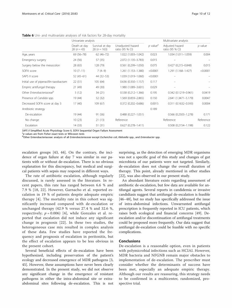
Table 6 Uni- and multivariate analyses of risk factors for 28-day mortality
Univariate analysis Multivariate analysis
Death at day28 (n = 43)
Survival at day28 (n = 163)
Unadjusted hazardratio (95 % CI)
p valuea Adjusted hazardratio (95 % CI)
p value
Age, years 69 (56–78) 62 (46–72) 1.022 (1.003–1.042) 0.023 1.034 (1.011–1.059) 0.004
Emergency surgery 24 (56) 57 (35) 2.072 (1.135–3.783) 0.015 – –
Surgery below the mesocolon 28 (65) 128 (79) 0.561 (0.299–1.050) 0.075 0.427 (0.215–0.848) 0.015
SOFA score 10 (7–11) 7 (4–9) 1.261 (1.153–1.380) <0.0001 1.291 (1.168–1.427) <0.0001
SAPS II score 52 (45–61) 44 (32–53) 1.039 (1.019–1.060) <0.0001 – –
Initial use of piperacillin-tazobactam 22 (51) 105 (64) 0.636 (0.350–1.157) 0.117 – –
Empiric antifungal therapy 21 (49) 49 (30) 1.980 (1.089–3.601) 0.029 – –
Other Enterobacteriaceaeb 5 (12) 34 (21) 0.538 (0.212–1.366) 0.195 0.342 (0.1219–0.961) 0.0419
Presence of Candida spp. 19 (44) 52 (32) 1.569 (0.859–2.865) 0.150 2.641 (1.3471–5.179) 0.0047
Decreased SOFA score at day 3 17 (40) 109 (67) 0.372 (0.202–0.686) 0.0015 0.311 (0.1632–0.593) 0.0004
Antibiotic strategy 0.189
De-escalation 19 (44) 91 (56) 0.488 (0.227–1.051) 0.566 (0.2503–1.278) 0.171
No change 10 (23) 21 (13) Reference Reference Reference
Escalation 14 (33) 51 (31) 0.627 (0.278–1.411) 0.508 (0.2154–1.198) 0.122
SAPS II Simplified Acute Physiology Score II, SOFA Sequential Organ Failure Assessmentap values are from Fisher exact tests or Wilcoxon testsbOther Enterobacteriaceae: analysis of all Enterobacteriaceae except Escherichia coli, Klebsiella spp., and Enterobacter spp.
Montravers et al. Critical Care (2016) 20:83 Page 10 of 12

Key messages
� De-escalation is a reasonable option, even inpolymicrobial infections such as healthcare-associated intra-abdominal infections.
� Multidrug-resistant bacteria and nonfermentingGram-negative bacilli remain a major obstacle inde-escalation.
� The key determinants for de-escalation are susceptiblemicroorganisms and adequate empiric therapy.
AbbreviationsESBL: extended-spectrum β-lactamase; HCIAI: health care–associatedintra-abdominal infection; ICU: intensive care unit; IQR: interquartile range;MDR: multidrug-resistant; MRSA: methicillin-resistant Staphylococcus aureus;NFGNB: nonfermenting Gram-negative bacilli; SAPS II: Simplified AcutePhysiology Score II; SOFA: Sequential Organ Failure Assessment;VAP: ventilator-associated pneumonia; WBC: white blood cell count.
Competing interestsThe authors declare that they have no competing interests.
Authors’ contributionsPM, PA, NG, MD, NA, and JPM carried out the data acquisition, dataprocessing, and data analysis. PM, PA, MD, and JG contributed to statisticalanalysis and participated in drafting the manuscript. All authors madesubstantial contributions to the conception and design of the study. Allauthors read and approved the final manuscript.
Author details1Département d’Anesthésie Réanimation, APHP, CHU Bichat-Claude Bernard,Paris, France. 2Université Denis Diderot, PRESS Sorbonne Cité, Paris, France.3INSERM, UMR 1137, Infection, Antimicrobiens, Modélisation, Evolution, Paris,France. 4Laboratoire de Microbiologie, AP-HP, CHU Bichat-Claude Bernard,Paris, France. 5Service de Réanimation, Centre Hospitalier Sud Francilien,Corbeil-Essonnes, France. 6Service de Chirurgie Générale, APHP, CHUBichat-Claude Bernard, Paris, France.
Received: 12 January 2016 Accepted: 16 March 2016
References1. Weiss E, Zahar JR, Lesprit P, Ruppe E, Leone M, Chastre J, et al. Elaboration
of a consensual definition of de-escalation allowing a ranking of β-lactams.Clin Microbiol Infect. 2015;21:649.e1–10.
2. Dellit TH, Owens RC, McGowan Jr JE, Gerding DN, Weinstein RA, Burke JP, etal. Infectious Diseases Society of America and the Society for HealthcareEpidemiology of America guidelines for developing an institutionalprogram to enhance antimicrobial stewardship. Clin Infect Dis.2007;44:159–77.
3. Dellinger RP, Levy MM, Rhodes A, Annane D, Gerlach H, Opal SM, et al.Surviving Sepsis Campaign: international guidelines for management ofsevere sepsis and septic shock, 2012. Intensive Care Med. 2013;39:165–228.
4. Garnacho-Montero J, Gutiérrez-Pizarraya A, Escoresca-Ortega A, Corcia-Palomo Y,Fernández-Delgado E, Herrera-Melero I, et al. De-escalation of empirical therapy isassociated with lower mortality in patients with severe sepsis and septic shock.Intensive Care Med. 2014;40:32–40.
5. Giantsou E, Liratzopoulos N, Efraimidou E, Panopoulou M, Alepopoulou E,Kartali-Ktenidou S, et al. De-escalation therapy rates are significantly higherby bronchoalveolar lavage than by tracheal aspirate. Intensive Care Med.2007;33:1533–40.
6. Kollef MH. Hospital-acquired pneumonia and de-escalation of antimicrobialtreatment. Crit Care Med. 2001;29:1473–5.
7. Niederman MS. De-escalation therapy in ventilator-associated pneumonia.Curr Opin Crit Care. 2006;12:452–7.
8. Kollef MH. What can be expected from antimicrobial de-escalation in thecritically ill? Intensive Care Med. 2014;40:92–5.
9. Alvarez-Lerma F, Alvarez B, Luque P, Ruiz F, Dominguez-Roldan JM,Quintana E, et al. Empiric broad-spectrum antibiotic therapy of nosocomialpneumonia in the intensive care unit: a prospective observational study. CritCare. 2006;10:R78.
10. Eachempati SR, Hydo LJ, Shou J, Barie PS. Does de-escalation of antibiotictherapy for ventilator-associated pneumonia affect the likelihood ofrecurrent pneumonia or mortality in critically ill surgical patients? J Trauma.2009;66:1343–8.
11. Canadian Critical Care Trials Group. A randomized trial of diagnostic techniquesfor ventilator-associated pneumonia. N Engl J Med. 2006;355:2619–30.
12. Leone M, Garcin F, Bouvenot J, Boyadjev I, Visintini P, Albanese J, et al.Ventilator-associated pneumonia: breaking the vicious circle of antibioticoveruse. Crit Care Med. 2007;35:379–85.
13. Rello J, Vidaur L, Sandiumenge A, Rodriguez A, Gualis B, Boque C, et al.De-escalation therapy in ventilator-associated pneumonia. Crit Care Med.2004;32:2183–90.
14. Heenen S, Jacobs F, Vincent JL. Antibiotic strategies in severe nosocomialsepsis: why do we not de-escalate more often? Crit Care Med.2012;40:1404–9.
15. Leone M, Bourgoin A, Cambon S, Dubuc M, Albanese J, Martin C. Empiricalantimicrobial therapy of septic shock patients: adequacy and impact on theoutcome. Crit Care Med. 2003;31:462–7.
16. Mokart D, Slehofer G, Lambert J, Sannini A, Chow-Chine L, Brun JP, et al.De-escalation of antimicrobial treatment in neutropenic patients withsevere sepsis: results from an observational study. Intensive Care Med.2014;40:41–9.
17. Montravers P, Mira JP, Gangneux JP, Leroy O, Lortholary O. A multicentrestudy of antifungal strategies and outcome of Candida spp. peritonitis inintensive-care units. Clin Microbiol Infect. 2011;17:1061–7.
18. Morel J, Casoetto J, Jospe R, Aubert G, Terrana R, Dumont A, et al.De-escalation as part of a global strategy of empiric antibiotherapymanagement: a retrospective study in a medico-surgical intensive care unit.Crit Care. 2010;14:R225.
19. Warren MM, Gibb AP, Walsh TS. Antibiotic prescription practice in anintensive care unit using twice-weekly collection of screening specimens: aprospective audit in a large UK teaching hospital. J Hosp Infect.2005;59:90–5.
20. Tabah A, Cotta MO, Garnacho-Montero J, Schouten J, Roberts JA, Lipman J,et al. A systematic review of the definitions, determinants, and clinicaloutcomes of antimicrobial de-escalation in the intensive care unit. ClinInfect Dis. 2016;62:1009–17.
21. De Waele JJ, Ravyts M, Depuydt P, Blot SI, Decruyenaere J, Vogelaers D.De-escalation after empirical meropenem treatment in the intensive careunit: fiction or reality? J Crit Care. 2010;25:641–6.
22. Gonzalez L, Cravoisy A, Barraud D, Conrad M, Nace L, Lemarié J, et al.Factors influencing the implementation of antibiotic de-escalation andimpact of this strategy in critically ill patients. Crit Care. 2013;17:R140.
23. Augustin P, Kermarrec N, Muller-Serieys C, Lasocki S, Chosidow D, MarmuseJP, et al. Risk factors for multidrug resistant bacteria and optimization ofempirical antibiotic therapy in postoperative peritonitis. Crit Care.2010;14:R20.
24. Montravers P, Dufour G, Guglieminotti J, Desmard M, Muller C, Houissa H,et al. Dynamic changes of microbial flora and therapeutic consequences inpersistent intra-abdominal sepsis. Crit Care. 2015;19:70.
25. Montravers P, Guglielminotti J, Zappella N, Desmard M, Muller C, Fournier P,et al. Clinical features and outcome of postoperative peritonitis followingbariatric surgery. Obes Surg. 2013;23:1536–44.
26. Marshall JC, Maier RV, Jimenez M, Dellinger EP. Source control in themanagement of severe sepsis and septic shock: an evidence-based review.Crit Care Med. 2004;32(11 Suppl):S513–26.
27. Comité de l’Antibiogramme de la Société Française de Microbiologie(CASFM) 2011. Recommandations 2011. Paris: Société Française deMicrobiologie; 2011. http://www.sfm-microbiologie.org/UserFiles/files/casfm/casfm_2011.pdf. Accessed 4 December 2015.
28. Magiorakos AP, Srinivasan A, Carey RB, Carmeli Y, Falagas ME, Giske CG,et al. Multidrug-resistant, extensively drug-resistant and pandrug-resistantbacteria: an international expert proposal for interim standard definitions foracquired resistance. Clin Microbiol Infect. 2012;18:268–81.
29. Dupont H, Bourichon A, Paugam-Burtz C, Mantz J, Desmonts JM. Can yeastisolation in peritoneal fluid be predicted in intensive care unit patients withperitonitis? Crit Care Med. 2003;31:752–7.
Montravers et al. Critical Care (2016) 20:83 Page 11 of 12

30. Le Gall JR, Lemeshow S, Saulnier F. A new Simplified Acute PhysiologyScore (SAPS II) based on a European/North American multicenter study.JAMA. 2003;270:2957–63.
31. Vincent JL, Moreno R, Takala J, Willatts S, De Mendonça A, Bruining H, et al.The SOFA (Sepsis-related Organ Failure Assessment) score to describe organdysfunction/failure. Intensive Care Med. 1996;22:707–10.
32. McCabe WR, Jackson GG. Gram-negative bacteremia. 1. Etiology andecology. Arch Intern Med. 1962;110:847–55.
33. Montravers P, Gauzit R, Muller C, Marmuse JP, Fichelle A, Desmonts JM.Emergence of antibiotic-resistant bacteria in cases of peritonitis afterintraabdominal surgery affects the efficacy of empirical antimicrobialtherapy. Clin Infect Dis. 1996;23:486–94.
34. Paugam-Burtz C, Dupont H, Marmuse JP, Chosidow D, Malek L, Desmonts JM,et al. Daily organ-system failure for diagnosis of persistent intra-abdominalsepsis after postoperative peritonitis. Intensive Care Med. 2002;28:594–8.
35. Montravers P, Dupont P, Gauzit R, Veber B, Bedos JP, Lepape A, et al.Strategies of initiation and streamlining of antibiotic therapy in 41 Frenchintensive care units. Crit Care. 2010;15:R17.
36. Ibrahim EH, Ward S, Sherman G, Schaiff R, Fraser VJ, Kollef MH. Experiencewith a clinical guideline for the treatment of ventilator-associatedpneumonia. Crit Care Med. 2001;29:1109–15.
37. Micek ST, Ward S, Fraser VJ, Kollef MH. A randomized controlled trial of anantibiotic discontinuation policy for clinically suspected ventilator-associatedpneumonia. Chest. 2004;125:1791–9.
38. Soo Hoo GW, Wen YE, Nguyen TV, Goetz MB. Impact of clinical guidelinesin the management of severe hospital-acquired pneumonia. Chest.2005;128:2778–87.
39. Kollef MH, Morrow LE, Niederman MS, Leeper KV, Anzueto A, Benz-Scott L,et al. Clinical characteristics and treatment patterns among patients withventilator-associated pneumonia. Chest. 2006;129:1210–8.
40. Leone M, Bechis C, Baumstarck K, Lefrant JY, Albanese J, Jaber S, et al.De-escalation versus continuation of empirical antimicrobial treatment insevere sepsis: a multicenter non-blinded randomized noninferiority trial.Intensive Care Med. 2014;40:1399–408.
41. Solomkin JS, Mazuski JE, Bradley JS, Rodvold KA, Goldstein EJ, Baron EJ, et al.Diagnosis and management of complicated intra-abdominal infection inadults and children: guidelines by the Surgical Infection Society and theInfectious Diseases Society of America. Clin Infect Dis. 2010;50:133–64.
42. Paskovaty A, Pastores SM, Gedrimaite Z, Kostelecky N, Riedel ER, Seo SK.Antimicrobial de-escalation in septic cancer patients: is it safe to backdown? Intensive Care Med. 2015;41:2022–3.
43. Knaak E, Cavalieri SJ, Elsasser GN, Preheim LC, Gonitzke A, Destache CJ. Doesantibiotic de-escalation for nosocomial pneumonia impact intensive careunit length of stay? Infect Dis Clin Pract. 2013;21:172–6.
44. Joung MK, Lee JA, Moon SY, Cheong HS, Joo EJ, Ha YE, et al. Impact ofde-escalation therapy on clinical outcomes for intensive care unit-acquiredpneumonia. Crit Care. 2011;15:R79.
45. Timsit JF, Harbarth S, Carlet J. De-escalation as a potential way of reducingantibiotic use and antimicrobial resistance in ICU. Intensive Care Med.2014;40:1580–2.
46. Ruhnke M. Antifungal stewardship in invasive Candida infections. ClinMicrobiol Infect. 2014;20 Suppl 6:11–8.
47. Bal AM, Shankland GS, Scott G, Imtiaz T, Macaulay R, McGill M. Antifungalstep-down therapy based on hospital intravenous to oral switch policy andsusceptibility testing in adult patients with candidaemia: a single centreexperience. Int J Clin Pract. 2014;68:20–7.
48. Bailly S, Leroy O, Montravers P, Constantin JM, Dupont H, Guillemot D, et al.Antifungal de-escalation was not associated with adverse outcome incritically ill patients treated for invasive candidiasis: post hoc analyses of theAmarCAND2 study data. Intensive Care Med. 2015;41:1931–40.
49. Azoulay E, Dupont H, Tabah A, Lortholary O, Stahl JP, Francais A, et al.Systemic antifungal therapy in critically ill patients without invasive fungalinfection. Crit Care Med. 2012;40:813–22.
• We accept pre-submission inquiries
• Our selector tool helps you to find the most relevant journal
• We provide round the clock customer support
• Convenient online submission
• Thorough peer review
• Inclusion in PubMed and all major indexing services
• Maximum visibility for your research
Submit your manuscript atwww.biomedcentral.com/submit
Submit your next manuscript to BioMed Central and we will help you at every step:
Montravers et al. Critical Care (2016) 20:83 Page 12 of 12