Chapter1f5 150328080338-conversion-gate01
89
CHAPTER 1: TRANSPORT
-
Upload
hisham-suhaimi -
Category
Education
-
view
21 -
download
0
Transcript of Chapter1f5 150328080338-conversion-gate01

CHAPTER 1: TRANSPORT
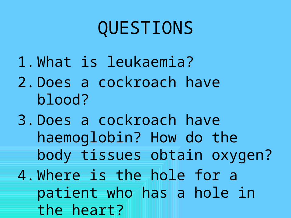
QUESTIONS
1. What is leukaemia?2. Does a cockroach have blood?3. Does a cockroach have
haemoglobin? How do the body tissues obtain oxygen?
4. Where is the hole for a patient who has a hole in the heart?

QUESTIONS
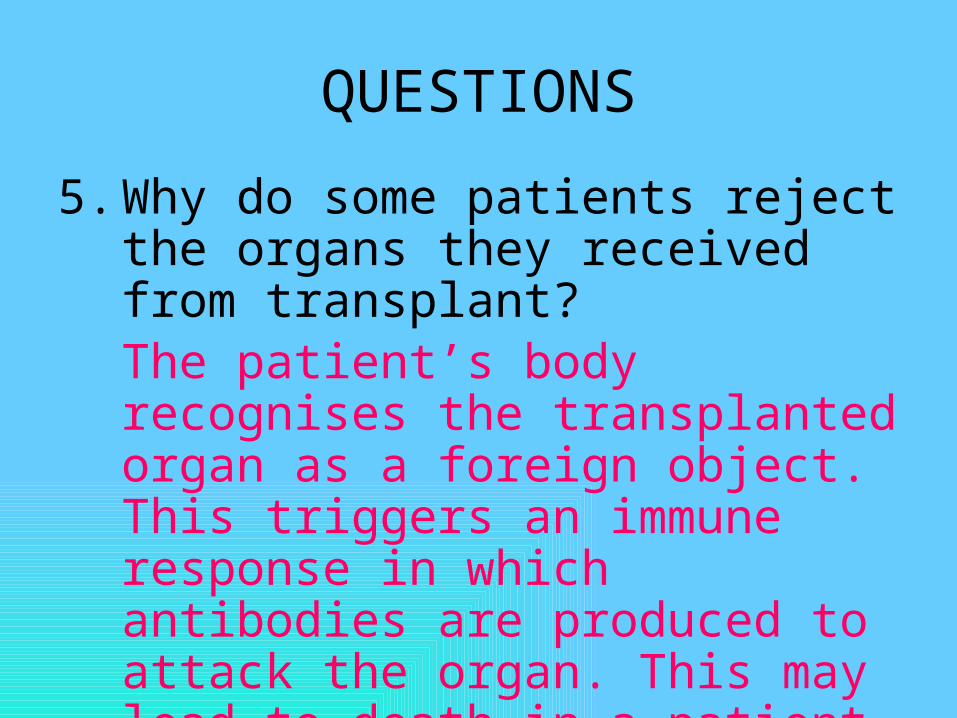
5. Why do some patients reject the organs they received from transplant?
6. Can we be infected with AIDS if we touch a HIV positive patient?
7. Does the blood clot in the blood vessels? Why?
8. What causes the heartbeat?
QUESTIONS
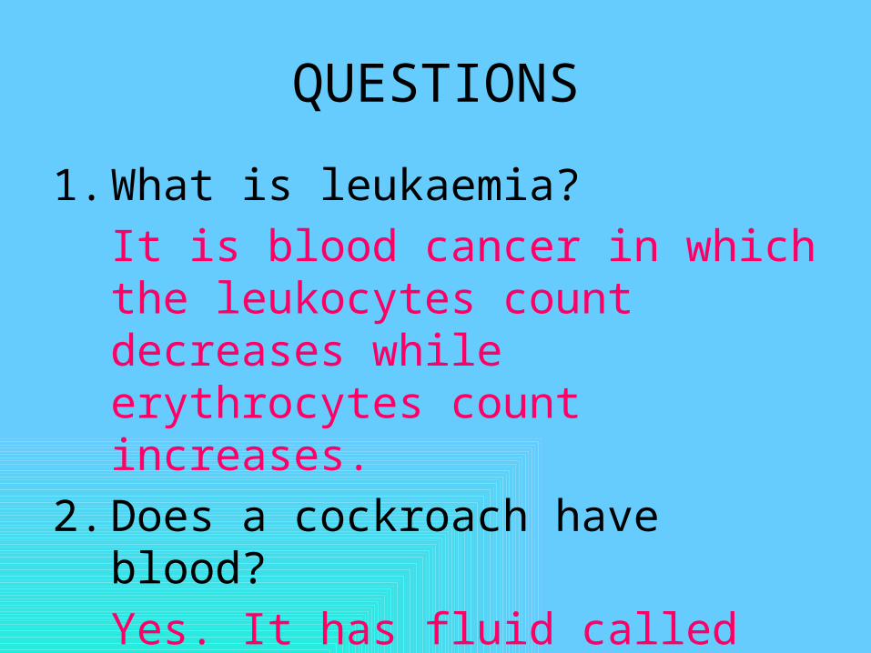
1. What is leukaemia?It is blood cancer in which the leukocytes count decreases while erythrocytes count increases.
2. Does a cockroach have blood?Yes. It has fluid called haemolymph which acts as a medium of transport.

QUESTIONS3. Does a cockroach have
haemoglobin? How do the body tissues obtain oxygen?No. oxygen enters the body directly through the spiracles at the sides of the body. Oxygen is then transported along the tracheal system directly into the body tissues.
4. Where is the hole for a patient who has a hole in the heart?Septum
QUESTIONS
5. Why do some patients reject the organs they received from transplant?The patient’s body recognises the transplanted organ as a foreign object. This triggers an immune response in which antibodies are produced to attack the organ. This may lead to death in a patient.

QUESTIONS
6. Can we be infected with AIDS if we touch a HIV positive patient?We would not be infected if there is no exposed wound at the point of contact between the two persons.

QUESTIONS
7. Does the blood clot in the blood vessels? Why?No. The blood inside the body contains heparin which prevents blood clotting in the blood vessels.
8. What causes the heartbeat?It is the result of the closing of the bicuspid and tricuspid valves as well as the semilunar valves.

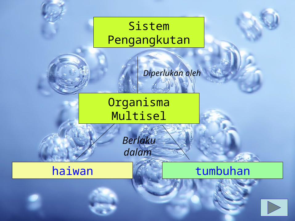
Sistem Pengangkutan
Organisma Multisel
Diperlukan oleh
haiwan tumbuhan
Berlaku dalam

haiwan
Sistem peredaran
Mekanisme pembekuan darah
Sistem limfaLimfa
Salur limfa
Nodus limfa
Sistem pertahan
an
Sistem imun
darah
Salur darah
jantung
Sistem tertutup
Sistem terbuka
Ganda dua
tunggal
Tak lengkap
lengkap
jenis
dibahagikan
perlu
contributes
termasuk
Terdiri drp comprises
Bila musnah
Sel darah, plasma darah
mengandungi

Sistem Peredaran• Membawa nutrien dan oksigen
ke sel• membawa hasil buangan keluar
daripada sel.• melindungi badan daripada
jangkitan• mempunyai tiga komponen
utama;–medium–pam–salur

• medium diperlukan untuk membawa bahan ke seluruh sistem peredaran
• manusia dan haiwan darah• invertebrata (serangga)
hemolimfa• darah adalah sejenis tisu penghubung
yang terdiri daripada plasma, sel- sel darah dan platlet.
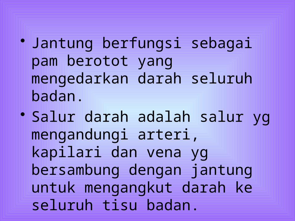
• Jantung berfungsi sebagai pam berotot yang mengedarkan darah seluruh badan.
• Salur darah adalah salur yg mengandungi arteri, kapilari dan vena yg bersambung dengan jantung untuk mengangkut darah ke seluruh tisu badan.


Sel darah merah

Sel darah putih (Granulosit)
Basofil
Eosinofil
Neutrofil

Sel darah putih (agranulosit)
monosit
Limfosit

Fungsi darah
• oksigen daripada peparu ke sel seluruh badan, karbon dioksida daripada sel badan ke peparu.
• Angkut nutrien, hormon dan antibodi ke seluruh badan.
• Angkut hasil buangan keluar daripada sel ke organ perkumuhan.

• Kawal atur– pH darah– Suhu badan– Kandungan air dalam sel
• Melindingi kita– drp kehilangan banyak darah apabila
cedera melalui mekanisma pembekuan darah menyembuh luka.
– drp penyakit dan bantu melawan jangkitan.
Fungsi darah…samb

Fungsi hemolimfa
• Transports water, inorganic salts and organic compounds throughout the haemocoel. Angkut air, garam tak organik dan sebatian organik seluruh hemoselom.
• tidak mengangkut gas respirasi.• Dalam serangga, gas respirasi
diangkut melalui sistem trakea.

Komposisi darah manusia
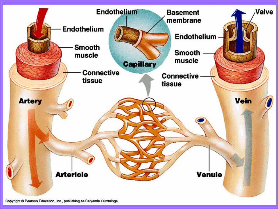

The differences between arteries, capillaries and veins
Characteristic
ArteriesCapillarie
sVeins
WallThick,
muscular, elastic
One-cell thick, no muscle
or elastic tissue
Thin, less muscular
, less elastic
Lumen SmallVery small
Large
Valve No valve No valveHave
valvesBlood
pressureHigh Very low Low

Characteristic
Arteries Capillaries Veins
Direction of
blood flow
From the heart to the
organs
From arteries to
veins
From all parts of the body to the
heart
Blood content
Oxygenated blood
except the pulmonary
artery
Oxygenated blood at the
arteriole ends &
deoxygenated blood at the venule
ends
Deoxygenated blood except the pulmonary
vein
Function
To transport blood
quickly at high
pressure from the
heart to the tissues
Allow rapid gaseous
exchange between the
blood and the body cell by diffusion
Allow blood from the tissues to return to the heart

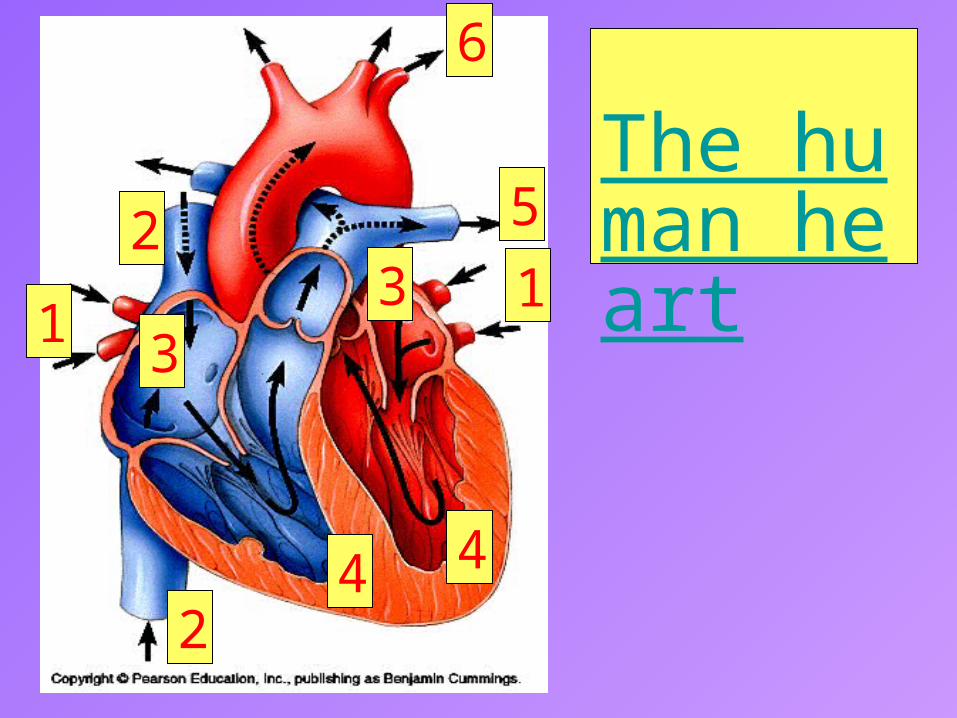
The human heart1
2
3
4
5
6
1
24
3

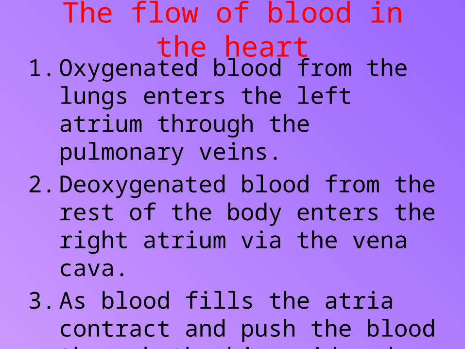
The flow of blood in the heart
1. Oxygenated blood from the lungs enters the left atrium through the pulmonary veins.
2. Deoxygenated blood from the rest of the body enters the right atrium via the vena cava.
3. As blood fills the atria contract and push the blood through the bicuspid and tricuspid valves into the two ventricles.

The flow of blood in the heart… cont
4. When the ventricles contract, the semi-lunar valves are forced open and blood is pushed into the pulmonary arteries and the aorta.
5. Deoxygenated blood is pumped through the pulmonary arteries to the lungs.
6. Oxygenated blood is pump through the aorta to the rest of the body.

Jantung Manusia

The pumping of the heart• Each time the heart contracts, it
acts as a pump which sends blood throughout the body.
• The heart is made up of a strong muscle, called the cardiac muscle.

The pumping of the heart… cont
• The cardiac muscle cells are interconnected
• This interconnection allows electrical impulses to spread rapidly through the heart and, at the same time, stimulates the cardiac muscle cells to contract in a coordinated movement.

The pumping of the heart… cont
• The cardiac muscle is myogenic.
• This means it contracts and relaxes without the need to receive stimulation by nerve impulses to make it contract.
• The contractions of the heart are initiated and coordinated by a pacemaker.

The pumping of the heart… cont
• The pacemaker is a cluster of specialised heart ,muscle cells that set the rate of contraction.
• It is located in the wall of the right atrium.
• The pacemaker generates electrical impulses which spread rapidly over the walls of both atria, causing the atria to contract rhythmically.

The pumping of the heart… cont
• The heart’s primary pacemaker is the sinoatrial (SA) node because it keeps the heartbeats regular.
• From the SA node, the impulses are relayed to the atrioventricular (AV) node, located at the bottom of the right atrium.
• From the AV node, bundle of His fibres, bundle branches and Purkinje fibres send the impulses to the apex of the heart and throughout the walls of the ventricles.


pengecutan otot jantung menghasilkan pengepaman jantung
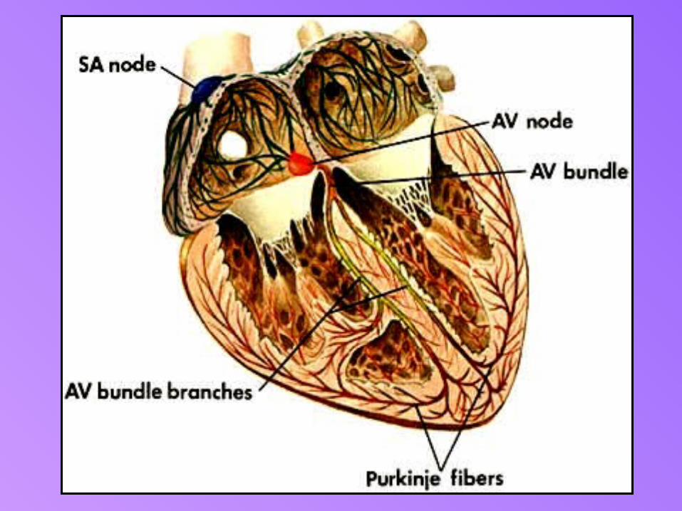

1. The SA node generates electrical impulses.
2. The electrical impulses spread rapidly over the walls of both atria, making the walls contract simultaneously. Contractions of the atria help to pump blood into the ventricles.
3. The electrical signals reach the AV node. The bundle of His fibres, bundle branches and Purkinje fibres send the impulses to the apex of the heart.
4. The electrical impulses spread to the ventricles, causing them to pump and push blood out to the lungs and body

mekanisme pengawalaturan tekanan darah

Mekanisme kawal atur tekanan darah• Tekanan darah adalah daya yang
mengepam darah sepanjang ateri dan kapilari.
• Apabila darah mengalir sepanjang salur, ia memberi tekanan terhadap dinding salur
• Tekanan darah lebih tinggi dalam arteri berbanding dalam vena.
• mengalir daripada kawasan bertekanan tinggi ke bertekanan rendah.
• Semasa pengecutan ventrikel, tekanan darah paling tinggi dalam aorta dan arteri besar apabila darah dipam ke dalam aorta dan arteri pulmonari.
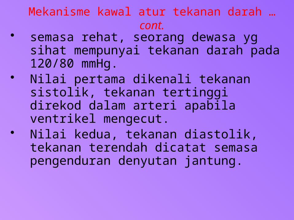
Mekanisme kawal atur tekanan darah … cont.
• semasa rehat, seorang dewasa yg sihat mempunyai tekanan darah pada 120/80 mmHg.
• Nilai pertama dikenali tekanan sistolik, tekanan tertinggi direkod dalam arteri apabila ventrikel mengecut.
• Nilai kedua, tekanan diastolik, tekanan terendah dicatat semasa pengenduran denyutan jantung.

Tekanan darah Elektronik

Perbezaan sistem peredaran manusia, ikan dan amfibia.
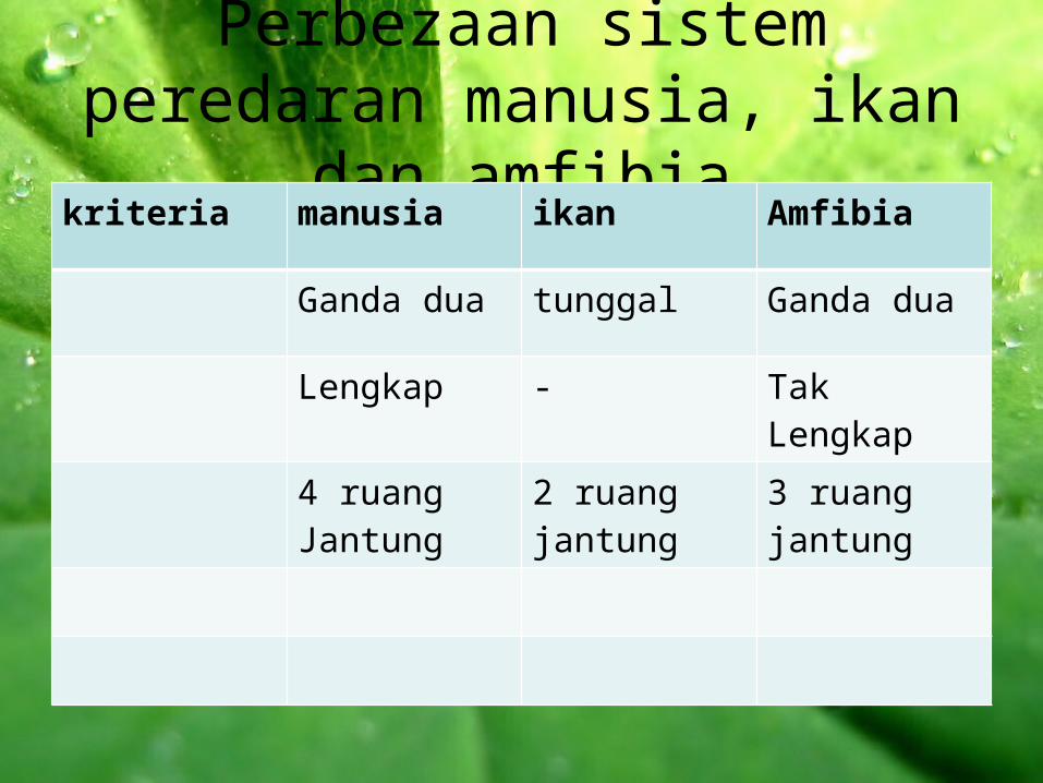
Perbezaan sistem peredaran manusia, ikan dan amfibia
kriteria manusia ikan Amfibia
Ganda dua tunggal Ganda dua
Lengkap - Tak Lengkap
4 ruang Jantung
2 ruang jantung
3 ruang jantung


ProduceTrombokinase
Wound in skin
Platelet gather StickyForms a temporary plug in leaking vessel
Fibrinogen
Fibrin
Forms the threads of the clot
Later
Harden (scab)
Mechanism of blood clotting
Need Vitamin K
Trombokinase
Ion CalciumProthrombinThrombin

Whenever an injury occurs a chain reaction is set off.
Wound in skin

Wound in skin
Platelets gather at a site of the injury and become sticky
Platelet

Forming a temporary plug in the leaking vessel
Wound in skin
Temporary plug

Prothrombin (non-active enzyme) need ion calcium to
convert into thrombin.
Need Vitamin K
Trombokinase
Ion CalciumProthrombinThrombin

Thread of clot
• Thrombin converts soluble fibrinogen (plasma protein formed by the liver) into insoluble fibrin.
• Fibrin forms the threads of the clot.
• A mesh-like network of fibrin traps red blood cells together, forming the blood clot, which later hardens into a scab.

Consequences of an impaired Blood Clotting Mechanism
• Haemophilia is a hereditary disease due to the lack of certain gene for the production of certain clotting factors.
• This is an impaired clotting mechanism which causes serious bleeding particularly in the joints.
• The afflicted person may die as a result of excessive bleeding from even minor cuts and bruises because blood clotting cannot take place.
Haemophilia

Consequences of an impaired Blood Clotting Mechanism
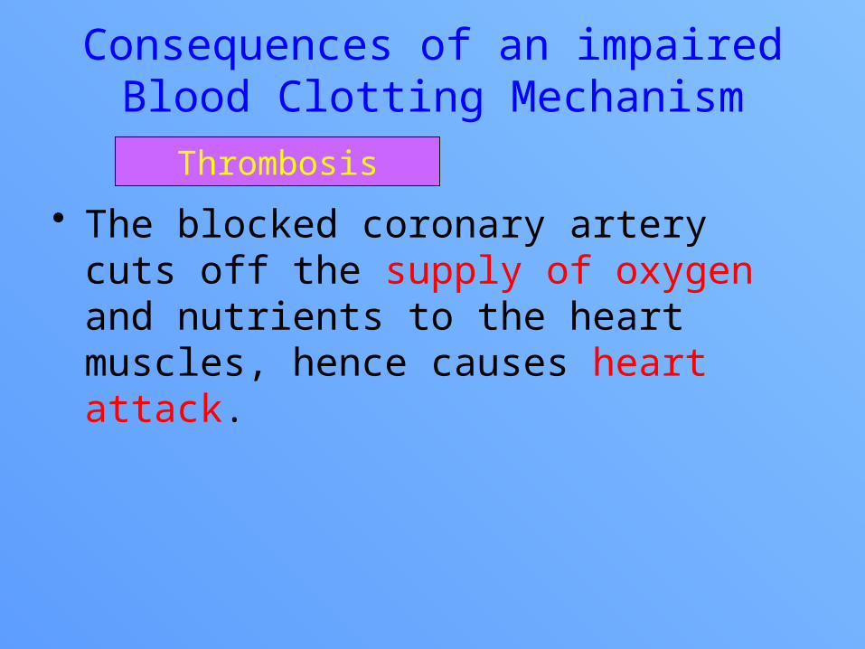
• Sometimes a local blood clot (thrombus) is formed on the damaged rough inner wall of the artery. This may cause blockage of the artery, a condition known as thrombosis.
• When a thrombus dislodges and is carried away by blood circulation, it is known as an embolus. The embolus may be trapped in a small artery where it blocks the blood flow. This condition is called embolism.
Thrombosis
Consequences of an impaired Blood Clotting Mechanism
• The blocked coronary artery cuts off the supply of oxygen and nutrients to the heart muscles, hence causes heart attack.
Thrombosis


Formation of the Interstitial Fluid and Lymph
CO2O2

Formation of the Interstitial Fluid and Lymph
• When the blood flows from arteries into capillaries, there is higher hydrostatic pressure at the arterial end of the capillaries.
• This high pressure forces some fluid out through the capillary walls into the intercellular spaces between the cells.
• Once the fluid leaves the capillary walls, it is called interstitial or tissue fluid. The interstitial fluids fills the spaces between the cells and constantly bathes the cells.

Formation of the Interstitial Fluid and Lymph
• The interstitial fluid that has not been reabsorbed into the bloodstream goes into the lymph capillaries. Once inside the lymph capillaries, the fluid is known as lymph.

Composition of the Interstitial Fluid• The composition of the
interstitial fluid is similar to the blood plasma.–Consists of water, dissolved
nutrients, hormones, waste products, gases, small proteins and leucocytes.
–Has no erythrocytes, platelets and large protein molecules (albumin, globulin and fibrinogen)
Importance of the Interstitial Fluid
• Interstitial fluid is important because :– It forms the internal environment of
the body.– It bathes the cells and supplies
them with oxygen and nutrients which diffuse from the blood through the interstitial fluid into the cells.
–Excretory waste products (carbon dioxide and urea) diffuse out of the cells into the interstitial fluid.

Structure of the Lymphatic System
• The lymphatic system is a one-way system consisting of a network of lymph capillaries, lymphatic vessels and lymph nodes.
• The lymph capillaries are blind-ended tubes located in the spaces between the cells.
• The interstitial fluid that has not been reabsorbed into the bloodstream goes into the lymph capillaries. Once inside the lymph capillaries, the fluid is known as lymph.

Structure of the Lymphatic System
• Lymph is the colourless fluid found in the lymphatic vessels.
• Lymph capillaries converge into larger lymphatic vessels.
• Lymph nodes are located at intervals along the lymphatic vessels. The lymph nodes produce lymphocytes that help to protect the body against infections.

Structure of the Lymphatic System
• Lymph contains a higher number of lymphocytes than blood.
• Within the lymphatic vessels are one-way valves to ensure the continuous flow of the lymph to prevent the backflow of the lymph.

The Relationship between the Lymphatic System and Circulatory System• Lymph is returned to the circulatory
system via the thoracic duct and the right lymphatic duct.
• The vessels from the left side of the body flow into the thoracic duct. The thoracic duct is the largest lymphatic vessel in the body that carries lymph to the left subclavian vein back into the bloodstream.
• The right lymphatic duct transport lymph from the right side of the head and chest into the right subclavian vein.

Role of the Lymphatic System in Transport• Collects the interstitial fluid and
returns it to the circulatory system.• Fats and fat-soluble vitamins are
absorb through lacteals and transported to the blood circulatory system.
• The lymph nodes filter out bacteria and other foreign particles. Phagocytes present in the nodes engulf and destroy foreign particles.
• Lymphocytes produce antibodies which aid in the destruction of pathogens and the neutralization of toxins.

Defence system
specificNon specific
1st line 2nd line3rd line
• Skin
• mucous membrane
Phagocyte
Phagocytosis
lymphocyte
antibody
passive active
naturalartificial
natural artificial
immunisation
immunity
Divided into
Divided into is
through
produce
gives
Divided into
Divided into Divided into
are
Carry out
eg

ROLE OF CIRCULATORY SYSTEM IN THE BODY’S DEFENCE SYSTEM
• Beside transport function, our circulatory system also defends the body against disease abolition of the disease-causing microorganisms or pathogens.
• There are three lines of defence mechanisms in our body:– The first line of defence: prevention of pathogens
entering the body.– The second line of defence: killing the pathogens
that entered our body by action or phagocytic white blood cells.
– The third line of defence: killing the pathogens by means of antibody actions.
Body’s Defence Mechanisms

• Prevention of pathogens entering the body by mean of physical and chemical barriers.
• A non-specific defence, that is never differentiate among various type of pathogens.
i. Skin– As a physical barrier, skin is made up of a
dead keratinised layer, tough enough for pathogens to penetrate.
– If there is a scratch or cut, the blood clots to seal the wound and avoids infections.
– Also acts as chemical barrier as it secretes sweat which contains salt. Sebaceous glands produce sebum which contains acid and oil. All these substances are unfavourable for growth of microorganisms.
– Sweat also contain lysozyme which destroy pathogens.
The First Line of Defence

ii. Tears and Saliva– Contain lysozymes which protect the
eyes and mouth from pathogen invasion.
iii. Gastric juice in stomach– Contain hydrochloric acid which
destroys most pathogens in foods and drinks taken.
iv. Mucous membranes– Secrete mucus in nasal cavity and
trachea to trap the dust particles and spores.
– The cilia in the respiratory track sweep the trapped particles to the pharynx and stimulates sneeze or cough to expel out the pathogens.
The First Line of Defence

• The killing action brought by some of the white blood cells like neutrophil and monocyte. They are called phagocytes and the process is phagocytosis.
• It is also a non-specific defence.• Phagocytosis occur when pathogens get
through the first line defence. Phagocytes move to the infected area due to the stimulation by chemicals released by damaged cells, example cut skin.
• Sometimes the phagocytes are killed by toxins produced by the pathogens.
• Dead bacteria, tissue cells and phagocytes may accumulate to form pus at the site of injury
The Second Line of Defence

• The steps involved in phagocytosis by a phagocyte e.g. Neutrophil
pathogen
pseudopodium
vacuole
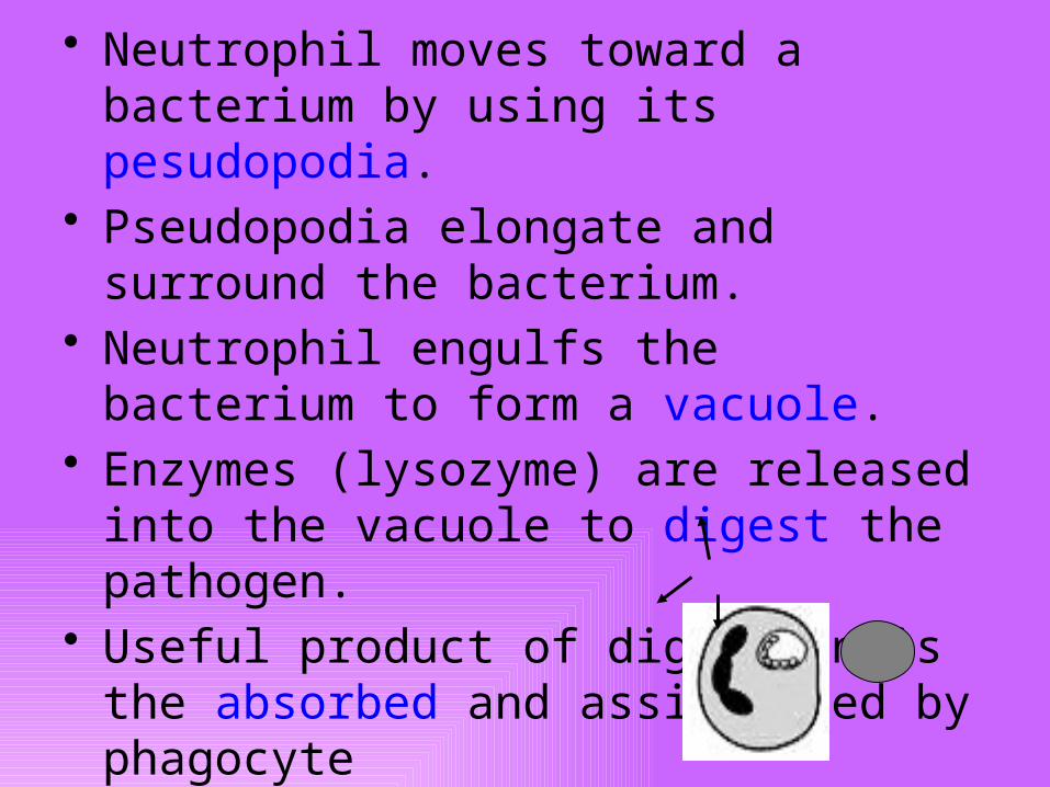
• Neutrophil moves toward a bacterium by using its pesudopodia.
• Pseudopodia elongate and surround the bacterium.
• Neutrophil engulfs the bacterium to form a vacuole.
• Enzymes (lysozyme) are released into the vacuole to digest the pathogen.
• Useful product of digestion is the absorbed and assimilated by phagocyte

• The third line of defence in the body is antibody.
• Antibody is a kind of protein released by lymphocyte in response to the presence of foreign substance, called antigen in our body.
• Lymphocytes are white blood cells found in lymph nodes and in the blood circulatory system. There are two types of lymphocytes, B-lymphocyte that secretes antibodies and T-lymphocyte that helps B-lymphocyte in antibody production.
• An antigen is a substance (usually protein) normally found on the outer surface of pathogen. Different types of pathogen act as different types of antigen.
The Third Line of Defence

• The third line of defence is a specific defence because when a specific antigen invades the body, lymphocyte is stimulated and produces specific antibody to destroy these specific antigens.
• This response is known as immune response because it resists the body from pathogens or diseases.
• After any infection, some lymphocytes remain in the body as memory cells which may last for several months or years. This memory cells help to defend the body against next infection by the same antigen. During this period, someone is sad to be immuned for that particular disease.
• Therefore, the word ‘immunity’ refers to the ability of an organisms to defend itself against infection by pathogens.
The Third Line of Defence

• What is the mechanism used by antibodies to destroy antigen?
– Antibody binds to the specific antigen binding site
– Hence, inactivates antigen by several ways
The Third Line of Defence
Neutralisation
Antibody or antitoxin coats the bacterial toxin or viral binding sites

The Third Line of Defence
Agglutination
Agglutinates bacteria cell and stops their moving and stimulate phagocytosis
Disintegration (lysis)
Breakdown the bacterial cell wall.
The Third Line of Defence
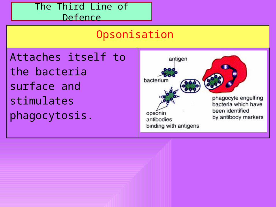
Opsonisation
Attaches itself to the bacteria surface and stimulates phagocytosis.

AIDS
• Acquired Immunodeficiency Syndrome• Caused by HIV – Human
Immunodeficiency Virus• Attacks the central nervous system
and helper T-cells in the body’s immune system.
• Helper T-cells are essential to activate B-cell lymphocyte in antibody production.
• HIV needs 8-10 years of incubation period before the symptom appears.

AIDS
• The immune system of infected person gradually becomes weakened and defenceless against many pathogens.
• Decreases in function of central nervous system followed by body weight loss.
• Eventually death occurs. The patient does not die from AIDS itself but from other secondary infections such as pneumonia and meningitis, tuberculosis, fungal infections or certain forms of cancer like Kaposi’s sarcoma

AIDS – Transmission Methods
• HIV only survive in body fluid such as semen, blood and vaginal fluid.
• Therefore, HIV can be transmitted through :– sexual intercourse– Blood transfusion– Injection with contaminated needle used to
inject drugs• HIV infected mother can pass HIV to her baby
through placenta or breast milk.• HIV cannot be spread by touching, sharing of food
or through the use of public toilets.
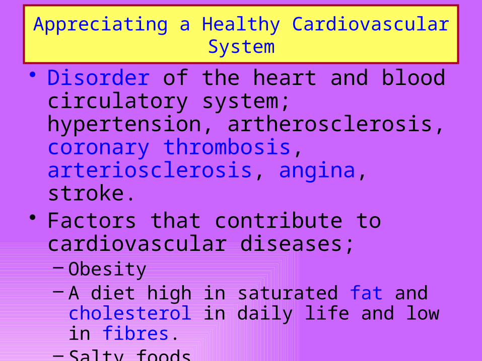
Appreciating a Healthy Cardiovascular System
• Disorder of the heart and blood circulatory system; hypertension, artherosclerosis, coronary thrombosis, arteriosclerosis, angina, stroke.
• Factors that contribute to cardiovascular diseases;– Obesity– A diet high in saturated fat and
cholesterol in daily life and low in fibres.– Salty foods– Lack of exercise– Cigarette smoking– Mental stress
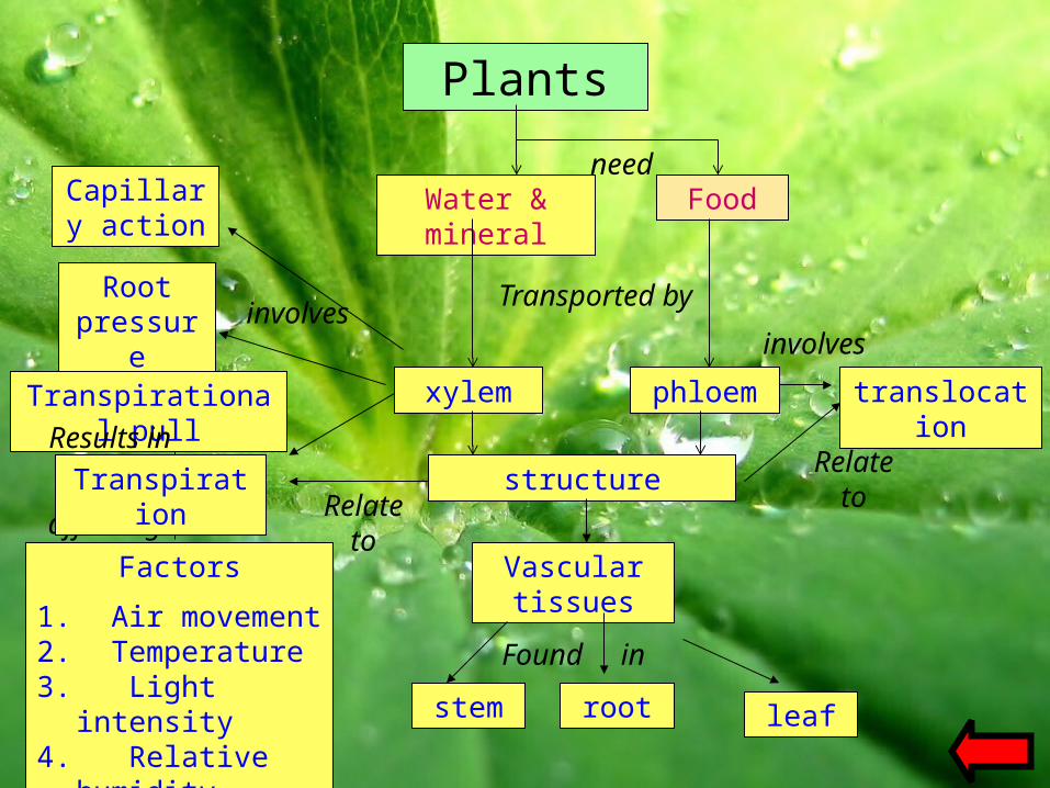
Plants
Water & mineral
Food
xylem phloem
Vascular tissues
involves
stem root leaf
structure
translocation
need
Transported by
Relate toRelate
to
involvesRoot
pressure
Transpirational pull
Factors
1. Air movement2. Temperature3. Light intensity4. Relative
humidity
Capillary action
affecting
Transpiration
Results in
Found in