CHAPTER ONE 1.1 INTRODUCTION - dspace.unza.zm
38
[1] CHAPTER ONE 1.1 INTRODUCTION Intraventricular haemorrhage (IVH) is related to bleeding in the capillary network of the germinal matrix of the developing brain. It is classified into four grades anatomically according to findings on cranial ultrasound depending on whether it is restricted to the subependymal area, extends to the lateral ventricles with or without dilatation and brain parenchyma involvement. 1, 2 . The grading is useful for counselling of the preterm babies’ parents or caregivers about prognosis. 1 IVH occurs mostly in the first three days after birth in preterm neonates born at or before 32 weeks gestation but may also occur beyond the first week. Specific problems that may manifest later in children who had IVH as neonates include cerebral palsy, post- haemorrhagic hydrocephalus, cognitive/intellectual impairment and epilepsy. Very low birth weight/extremely low birth weight (VLBW/ELBW) preterm neonates account for 20% of the total admissions to the neonatal intensive care unit (NICU) at the University Teaching Hospital (UTH) annually (2007 and 2008 NICU ward statistics) with case fatality rates of more than 45%. It is these neonates that are at risk for IVH and its long term sequelae. IVH remains a serious problem and is reported to have an incidence of 50% globally in the VLBW and ELBW infants. 1 In the southern African region, a study in South Africa 3 reported a prevalence of around 53% in VLBW neonates. There were no other known reported studies on prevalence in the sub-region at the time of this study. IVH causes mortality ranging from 27-50% (severe IVH) and about 5% (mild to moderate IVH). 2, 4 This cross sectional study was therefore undertaken to determine the prevalence of IVH in preterm infants with birth weight 1.5kg and less presenting to the NICU at the UTH. Some of the potentially associated risk factors for its occurrence were also studied.
Transcript of CHAPTER ONE 1.1 INTRODUCTION - dspace.unza.zm

[1]
CHAPTER ONE
1.1 INTRODUCTION
Intraventricular haemorrhage (IVH) is related to bleeding in the capillary network of
the germinal matrix of the developing brain. It is classified into four grades
anatomically according to findings on cranial ultrasound depending on whether it is
restricted to the subependymal area, extends to the lateral ventricles with or without
dilatation and brain parenchyma involvement.1, 2
. The grading is useful for
counselling of the preterm babies’ parents or caregivers about prognosis. 1IVH
occurs mostly in the first three days after birth in preterm neonates born at or before
32 weeks gestation but may also occur beyond the first week. Specific problems that
may manifest later in children who had IVH as neonates include cerebral palsy, post-
haemorrhagic hydrocephalus, cognitive/intellectual impairment and epilepsy.
Very low birth weight/extremely low birth weight (VLBW/ELBW) preterm neonates
account for 20% of the total admissions to the neonatal intensive care unit (NICU) at
the University Teaching Hospital (UTH) annually (2007 and 2008 NICU ward
statistics) with case fatality rates of more than 45%. It is these neonates that are at
risk for IVH and its long term sequelae.
IVH remains a serious problem and is reported to have an incidence of 50% globally
in the VLBW and ELBW infants.1 In the southern African region, a study in South
Africa3 reported a prevalence of around 53% in VLBW neonates. There were no
other known reported studies on prevalence in the sub-region at the time of this
study. IVH causes mortality ranging from 27-50% (severe IVH) and about 5% (mild
to moderate IVH).2, 4
This cross sectional study was therefore undertaken to determine the prevalence of
IVH in preterm infants with birth weight 1.5kg and less presenting to the NICU at
the UTH. Some of the potentially associated risk factors for its occurrence were also
studied.

[2]
1.2 STATEMENT OF THE PROBLEM
Prevalence of IVH and associated risk factors is unknown in the VLBW/ELBW
preterm neonates admitted to NICU at UTH as no prevalence studies have been
undertaken before. As such it is a condition that is rarely looked for. Consequently,
there has been no description of the commonly prevalent grades of IVH and its
contribution to the morbidity and mortality in these neonates.
Follow up of the VLBW/ELBW infants on the neonatology unit at the UTH is done
until they attain a weight of 2.5kg and then are discharged. Therefore, any
neurological deficit following IVH will only be noted before or at the time the infant
attains this weight while that manifesting later may be missed.
Cranial ultrasound was not routinely done to screen for IVH in the at-risk
VLBW/ELBW infants1, 2
in the NICU at UTH before this study, in contrast to other
NICUs internationally where it is recommended to be done routinely.42
The cranial
ultrasound is not done routinely because most of the doctors looking after these
neonates on a day to day basis are not trained on it. The radiology department at
UTH does not have a Sonographer specifically assigned to carry out these routine
cranial ultrasounds as well due to staffing challenges. This was revealed by a check
on the NICU before this study was undertaken.
1.3 STUDY JUSTIFICATION
Data from developed countries, where most studies on the subject have been done,
cannot be readily extrapolated due to resource limitations, different antenatal
maternal disease patterns and poor socioeconomic conditions in Zambia generally
and UTH specifically.
This study therefore was undertaken to show the prevalence and severity of, as well
as risk factors associated with IVH in preterm neonates with birth weight 1.5kg and
below admitted to the NICU at the UTH. This information might influence future
management of these infants especially in NICU and follow up upon discharge from
the NICU. Findings of the study may also potentially form a basis for future research
work on the subject matter.

[3]
1.4 LITERATURE REVIEW
The site where intraventricular haemorrhage (IVH) originates is the subependymal
germinal matrix, which is an area of embryonic neurones and glial cells for the
developing brain. “During foetal development this is a site of neuronal proliferation
as neuroblasts divide and migrate into the cerebral parenchyma. By approximately 20
weeks' gestation, neuronal proliferation is completed; however glial cell proliferation
is still on-going. The germinal matrix supports glioblasts division and glial elements
differentiation until approximately 32 weeks' gestation at which time regression is
almost complete.”1
1.4.1 Pathophysiology
Particular features of the germinal matrix that render it susceptible to bleeding
include the following: Metabolically active and mitochondria-rich cells, supplied by
a primitive rete-like capillary network which in turn is fed by the anterior and middle
cerebral arteries, a lack of tight junctions between endothelial cells and also lack of a
strong basement membrane. Being mitochondria-rich the area is quite sensitive to
ischaemia. Another important factor to be considered in the pathophysiology of IVH
is the fact that premature neonates have low and narrower cerebral auto-regulation
capacity to that of term neonates. Loss of cerebral auto-regulation and abrupt
alterations in blood flow and pressure are thought to be responsible for the
occurrence of intraventricular haemorrhage although it is said occasionally to occur
spontaneously in neonatal infants with less than 1kg birth weight.1, 4
Loss of cerebral
auto-regulation leads to a pressure-passive cerebral blood flow pattern leaving
systemic pressure as the determinant of cerebral blood flow. Therefore changes in the
blood flow pattern in the presence of this pressure-passive setup leads to
haemorrhage.
Multiple events can rapidly alter cerebral blood flow and pressure potentially
overwhelming the auto-regulatory mechanisms of the premature neonate. These
include a number of risk factors that have been proposed for the development of IVH
by this mechanism and also by other yet unexplained mechanisms: chorioamnionitis
or intrauterine infection, 5-8
respiratory distress syndrome,5-8
inter-hospital transfer
of the preterm neonate,9-11
breech presentation,12
gender,11-13
premature rupture of

[4]
membranes,6, 7
mode of delivery,7, 12-15
prolonged labour,10,16
postnatal resuscitation
and intubation,10, 11,16
early onset sepsis,17, 18
maternal smoking,19
low birth weight
and gestational age,6, 11, 12, 16, 20-23
repeated endotracheal suctioning10, 16
metabolic
acidosis and rapid bicarbonate infusion10, 13
high frequency ventilation,24
thrombocytopaenia,5 reduced clotting factor levels (specifically factor VII),
25
fertility treatment26
and high sodium intake.27
1.4.2 Clinical aspects
80-90% of IVH occurs between birth and the third day of life with 50% on the first
day while 20-40% will progress in the first week of life. This forms the basis for
screening programmes on seventh day of postnatal life for IVH in preterm neonates.
Delayed bleeding, though rare, may occur after the first week of life and accounts for
10-15%1, 2, 28
IVH has four grades based on radiological appearance. This grading is useful for
prognostic reasons and for counselling parents and caregivers:-
Table 1 IVH grading1
Grade Radiological appearance - Site of haemorrhage
1 Subependymal region and/or germinal matrix (less than 10% ventricular
extension)
2 Subependymal haemorrhage with extension into the lateral ventricles
filling without or with mild ventricular enlargement (10 -50% ventricular
filling)
3 Subependymal haemorrhage with extension into the lateral ventricles with
significant ventricular enlargement (more than 50% ventricular filling)
4 Intraparenchymal haemorrhage
These grades can further be classified as mild (grade 1), moderate (grade 2), and
severe (grades 3 and 4). Ventricular enlargement is defined as mild (0.5–1cm),
moderate (1.0–1.5cm) or severe (>1.5cm).1, 2
IVH remains a significant cause of morbidity & mortality in premature neonates
accounting for 60%-70 % (0.5kg-0.75kg) and 10%-20 %( 1kg-1.5kg) 2 in the USA
and around 50% globally in countries with similar resources1. A study in Brazil,
South America found an incidence of IVH of 53.8% in neonates less than 1kg with

[5]
70% being the frequency for mild IVH29
while another study also in Brazil found an
incidence of periventricular-intraventricular haemorrhage of 42.3% with 63.8%
moderate, 27.7% mild and 8.3% severe IVH.30
Few studies have been done or published in Sub-Saharan Africa on prevalence of
IVH in preterm neonates and the associated risk factors. A study done in South
Africa at Baragwanath hospital found a prevalence of about 53% for infants
weighing less than 1.5kg. In that study 12% had severe IVH3. A study at the central
hospital in Yaoundé, Cameroun, found a prevalence of 58.3% in infants born
between 28-31weeks gestation and 34.8% between 32-34 weeks gestation31
while
one done at a Nigerian university hospital found mild IVH at 22% and 7.5%
moderate to severe IVH.32
Sequelae following intraventricular haemorrhage relate to post-haemorrhagic
hydrocephalus and cerebral parenchyma damage. The last of these is most critical
and careful assessment of periventricular parenchyma in the infant with IVH during
the acute period of illness may be of value in predicting outcome. Further sequelae
are from the shunting procedure in cases of the post haemorrhagic hydrocephalus
that results.
Specific problems that may manifest later in these children include cerebral palsy,
post-haemorrhagic hydrocephalus, cognitive/intellectual impairment and epilepsy.
These are said to occur in higher rates in preterm infants with all IVH grades when
compared to children without IVH.33, 34, 35, 36

[6]
CHAPTER TWO
2.1 MAIN OBJECTIVE
To determine the prevalence of intraventricular haemorrhage and some associated
risk factors in preterm neonates with birth weight 1.5kg and below admitted to the
NICU at the UTH
2.2 SPECIFIC OBJECTIVES
I. To determine the proportion of infants with birth weight 1.5kg and below
who present with IVH in the first seven postnatal days.
II. To determine the most frequent grade of IVH occurring in preterm
neonates with birth weight 1.5kg and below.
[7]
CHAPTER THREE
METHODS
3.1 Study design
This was a cross sectional study undertaken to look at the prevalence and the most
frequent grade of IVH and some associated risk factors in preterm neonates with
birth weight1.5kg and below admitted to the neonatal intensive care unit (NICU) at
the University Teaching Hospital, Lusaka, Zambia from September 15, 2010 to
February 21, 2011.
3.2 Study site
The study was conducted at the NICU at UTH, Lusaka, Zambia. The NICU mostly
admits neonates, both preterm and term, delivered at UTH and the surrounding
outlying areas of Lusaka and Central provinces within a radius of about 50-100
kilometres from the Lusaka urban Central Business District. VLBW (birth weight
1.5kg or less) and ELBW (birth weight 1kg or less) neonates are admitted and treated
following NICU protocols. They are discharged from the NICU when a weight of
1.4kg is reached, are free of any medical complication requiring admission and are
feeding well. They are then reviewed weekly on the ward until a weight of 1.7kg
after which follow-up is in the Out-patient clinic until a weight of 2.5kg is reached.
During the period of the study cranial ultrasound was done routinely on all
ELBW/VLBW admitted to the unit as part of care.
3.3 Sample profile
3.3.1 Subject inclusion criteria
Included in the study were neonates with:
I. Estimated gestational age (EGA) less than or equal to 32 weeks
II. Birth weight 1.5kg and below
III. Post-delivery age less than or equal to seven days on admission to NICU
IV. Written consent to enrol into the study by the infant’s caregiver/parent.

[8]
3.3.2 Subject exclusion criteria
Excluded from the study were neonates whose:
I. caregivers/parents refused consent to enrol into the study
II. age was more than 7 days postnatally
III. EGA was more than 32 weeks
IV. birth weight was more than 1.5kg and
V. Death before the first cranial ultrasound
3.4 Sample size and sampling technique
Neonates meeting the criteria were consecutively enrolled to the study with the help
of two research assistants (nurses) until the sample size of 298 was reached. The
sample size was calculated using the formula N = z²(p)(1-p)/L², assuming 50% IVH
prevalence in the preterm neonates ≤1.5kg birth weight at the 90% confidence level
& non-compliance of 10%, Where N = required sample size, z = 1.64 (Z-alpha,
population constant), p = assumed population IVH prevalence and L = desired width
of confidence interval (0.1). The values for the formula variables were chosen with
the knowledge that the NICU at UTH admits between 500 – 600 preterm neonates
with birth weight ≤ 1.5kg annually and the study was to be conducted over a period
of six months.
3.5 Data collection
On admission to the NICU or shortly thereafter, but usually between 07:30 hours in
the morning and 18:00 hours in the evening of every day of the week during the
study period, VLBW/ELBW were assessed for inclusion into the study. Particular
attention was paid to the birth weight which was taken as the one done on the ward
on admission for those neonates admitted within 48 hours of birth or the one on the
referral form that accompanies all neonates admitted to the ward, for those presenting
later than 48 hours. In the latter case if no birth weight was indicated on the form,
weight done on admission was taken as birth weight. A Secca, model 3341321008,
electronic scale was used for weighing neonates on the ward. The EGA was taken as
the one determined using the mother’s LNMP but or that determined by the new

[9]
expanded Ballard score (Appendix D). The latter was taken as the EGA if it was
greater by more than two weeks that determined by the LNMP. The Ballard score
was done by either the admitting medical officer or the principal investigator within
24-48 hours of admission. If the Ballard score gave an interval such as 32 – 34
weeks, then the lower value, that is 32 weeks, was taken as the EGA.
Presence or absence of RDS due to surfactant deficiency as diagnosed clinically by
the admitting medical officer according to local protocol was noted. Because there is
no laboratory test to diagnose RDS, the diagnosis on the NICU at UTH, as in other
centres, is based on initial clinical symptoms (signs within the first four hours of
birth) and the clinical course (over the next thirty-six to seventy-two hours) and a
chest radiograph consistent with RDS. Response to surfactant treatment is not yet
part of the definition due to non-availability of surfactant in the unit at UTH. Clinical
chorioamnionitis was defined as presence of at least two of the following: maternal
fever (≥38⁰C), PROM (of duration18 hours or more), foetal tachycardia (>160
beats/min), uterine tenderness with a malodourous infant and no other infection
source. Other risk factors and information on the neonate and the maternal antenatal
and intrapartum periods was obtained from the maternal antenatal record and by
direct inquiry from the mother. These were appropriately recorded on the study
questionnaire, (see appendix A).
Informed consent was obtained from the mother/caregiver of the neonate using the
form included in this report (appendix C) after it was read and explained to the
mother/caregiver. The form was available in English and two local languages,
Bemba and Nyanja.
Transfontanelle cranial ultrasound was performed twice: The first within or at 72
hours of life and as soon as possible after enrolment for those presenting to the NICU
older than 72 hours. The second was done on the seventh day of postnatal life. The
ultrasounds were usually done during the day between 06:00 hours in the morning
and 18:00 hours in the evening. The standard saggital and coronal views were done
looking for echodensities (haemorrhage) in the subependymal area, intra- and
periventricular as well as other brain parenchymal areas. Using the information so
gained the IVH grade was determined as indicated in table1. Axial views, where

[10]
indicated, were done for ventricular size determination. Findings were appropriately
recorded on the cranial ultrasound reporting form attached as appendix B. The
cranial ultrasound scanning was performed using the cranial ultrasound machine
(Aloka SSD 900) on the ward which uses a 7.5MHz convex probe ideal for neonatal
cranial ultrasound37
by the principal investigator and initially by two radiographers
from the radiology department at UTH for the first month of the study. The principal
investigator had had prior training in ultrasound image recognition, abdominal,
obstetrical and gynaecological ultrasound. The training was conducted at UTH by
Fontys University of applied sciences (Netherlands) in conjunction with the Zambian
Ministry of Health. The training consisted of both theory and hands-on practical. The
cranial ultrasound images were printed out and attached to both the patient file and
the data collection tools for the particular study subject labelled with the study
number. The study numbers were serial numbers from 1 to 298. Ultrasound images
were only reported by the principal investigator as no other appropriate reporter was
available to the study at the time. Echolucent findings in association with
echodensities on cranial ultrasound in the first three days was not reported as
findings consistent with IVH but more likely periventricular leukomalacia (PVL).
3.6 Data analysis
Data was analysed using Epi info version 3.5.1. The independent variables studied in
addition to birth weight and gestational age were maternal clinical chorioamnionitis,
surfactant deficiency disease, mode of delivery, sex, prolonged rupture of
membranes and place of birth. The dependent variable was any-IVH generated at the
time of analysis. Any-IVH was the highest grade of IVH obtained on any of the time
periods when cranial ultrasound was done (first 3 days or day 7). Multivariate
logistic regression analysis was used in studying the association between
independent and dependent variables at the 95% confidence level comparing those
neonates with and without any-IVH. Logistic regression analysis was also performed
using severe IVH as the outcome variable with the independent variables.

[11]
3.7 Ethical considerations
Ethical clearance was sought and granted from the University of Zambia Biomedical
Research Ethics Committee for the study. All information collected from the
neonate’s parent/caregiver was treated with the utmost confidentiality. Case records
were kept in lockable drawers on the ward. The study procedure and objectives were
communicated to the neonate’s parent/caregiver in the language they best understood
(see appendix C). Findings of the cranial ultrasound and the possible immediate and
long term implications appropriate for the findings were communicated to the
parent/caregiver as soon as they were available. The results were also recorded on
the neonate’s file and promptly made available to the attending doctors for
appropriate management in the immediate period. The management if IVH is present
is supportive (e.g. blood transfusion if anaemia occurred). Repeat cranial ultrasounds
to determine ventricular size and/or daily occipito-frontal circumference were done
so as to detect post-haemorrhagic hydrocephalus. The parent/caregiver to the neonate
did not incur any additional costs as a result of the study.

[12]
CHAPTER FOUR
4.1 RESULTS
A total number of 332 preterm infants with birth weight 1.5kg and below were
admitted to the NICU at UTH between September 2010 and February 2011. 298 of
these met the inclusion criteria and so were recruited into the study and analysis. All
298 patients had at least one cranial ultrasound done. 207 (69.5%) patients had two
cranial ultrasounds done. 85 patients (28.5%) died with 51 (60%) of these without
IVH in the first 72 hours, 2(0.7%) left against medical advice, 1(0.3%) absconded
and 3(1%) were discharged before the second cranial ultrasound could be done,
representing a fallout rate of 30.5%.
Table 2- Subject demographic and clinical characteristics
Estimated gestational age-Mean(SD) 29.3 weeks (±1.93)
Sex - Male – Number (%)
Female – Number (%)
142 (47.7)
156 (52.3)
Birth weight – mean(SD) 1.2kg (±0.22)
Place of birth – UTH- Number (%)
Outside UTH-Number (%)
164 (55)
134 (45)
Any-IVH – Number (%) 102 (34.2)
Any-IVH and IVH by day of Ultrasound Any IVH Day 3 Day 7
Grades –Number(%) 1 56 (54.9) 45 (15.2) 42(20.3)
2 18 (17.7) 15(5.1) 11(5.3)
3 19 (18.6) 17(5.7) 8(3.9)
4 9 (8.8) 7(2.4) 3(1.4)
No IVH- Number (%)
196 (65.8) 212 (71.6) 143(69.1)

[13]
Table 3- Characteristics for the IVH and No IVH groups
Any-IVH No IVH P-Value Odds Ratio (C.I.
at 95%)
Postnatal
Age (hours)
Mean(SD) 26.6(±25.1) 24.8(±28.4) 0.61
Sex Male
Number (%)
48(47.1)
94(48.0)
0.88
0.96(0.60-1.56)
Female
Number (%)
54(52.9)
102(52.0)
0.88
0.96(0.60-1.56)
Place of
birth
UTH
Number (%)
59(57.8)
105(53.6)
0.48
1.19(0.73-1.93)
Outside
UTH
Number (%)
43(42.2)
91(46.4)
0.48
1.19(0.73-1.93)
Birth weight Mean(SD) 1.15(±0.24) 1.26(±0.21) 0.0001
Mode of
Delivery
Number (%)
Vertex 82 (80.4) 154 (78.6) 0.85
Breech 10 (9.8) 18 (9.2)
0.85
C/S 9 (8.8) 23 (11.7) 0.85
Forceps 1 (1) 1 (0.5) 0.85
RDS Number (%) 68 (66.7) 113 (57.7) 0.13 1.49(0.89-2.42)
PROM Number (%) 18 (17.6) 28 (14.3) 0.45 1.29 (0.67-2.46)
Gestational
age
Mean(SD) 28.7(±1.99) 29.6(±1.81) 0.0000
There was no case of clinical chorioamnionitis found as defined in this study among
the 298 neonates’ mothers.RDS was present in the 181(60.7%) while PROM was
present in 46 (15.4%). IVH regardless of the day (any-IVH) was present in 102
(34.2%) with grade 1(mild) IVH being the most frequent in just over half the
neonates at 56 (54.9%). Severe IVH was at 27.5%. A total of 84 (82.4%) neonates
had IVH in the first 3 days and 64 (30.9%) on day 7, with actual breakdown of
grades by day of cranial ultrasound shown in table 2 above.
From table 3 above there was statistically significant difference between neonates
with IVH and no IVH in terms of estimated gestational age and birth weight and no
significant statistical difference in the rest of the risk factors between those with IVH
and no IVH.

[14]
Table 4- multivariate logistic regression model for 7 risk factors and any-IVH
PARAMETER Odds Ratio P-value
Postnatal age 1.00 (1.00-1.02) 0.17
Estimated
gestational age
0.82 (0.69–0.97) 0.02
Place of birth 1.51 (0.88-2.61) 0.13
Mode of delivery Caesarean/breech 0.82 (0.26-2.63) 0.74
Vertex/breech 1.03 (0.43-2.47) 0.94
Forceps/breech 2.30 (0.1-42.75) 0.57
Birth weight 0.25 (0.06-0.98) 0.04
PROM 1.16 (0.59-2.28) 0.68
RDS 1.20 (0.74-2.07) 0.51
The multivariate logistic regression model analysis in table 4 above, showed
statistically significant association between EGA and birth weight and any-IVH. No
significant association between any-IVH and any one of sex, postnatal age, mode of
delivery, place of birth, RDS and PROM was observed.
Further multivariate logistic regression analysis for the risk factors and severe
(grades 3 and 4) IVH showed statistically significant association only between birth
weight and severe IVH (see table 5 below).
Table 5 – multivariate logistic regression model for severe (grades 3 and 4) IVH
PARAMETER Odds Ratio P-value
Postnatal age 1.01 (0.99-1.03) 0.17
Sex 1.40 (0.57-3.42) 0.46
Estimated
gestational age
0.78 (0.59-1.04) 0.09
Place of birth 1.49 (0.55-4.00) 0.43
Mode of delivery Caesarean/breech 10.5 (0.93-119.09) 0.06
Vertex/breech 2.66 (0.31-23.22) 0.38
Forceps/breech 104.15 (2.68-
4040.63)
0.01
Birth weight 0.01 (0.00-0.14) 0.0004
PROM 1.86 (0.65-5.36) 0.25
RDS 1.09 (0.40-2.95) 0.87

[15]
Table 6 IVH progression from the first 3 days to day 7
NUMBER WITH IVH GRADE ON DAY 7 FOR EACH
IVH GRADE INTHE FISRT 3 DAYS
No IVH 1 2 3 4
IVH
GRADE
FIRST 3
DAYS
OF LIFE
(Number)
No IVH 139 16 1 0 0
1(45) 3 24 4 1 1
2(15) 0 1 6 2 0
3(17) 0 0 0 5 1
4(7) 0 0 0 0 1
The above table shows the progression of IVH from the first three days to the
seventh day of postnatal life. The analysis included only those neonates that had
cranial ultrasounds in the first three days of life and then on day 7. The numbers in
the columns of the various IVH grades on day7 are in comparison with each row of
the number with IVH grade in the first three days of life. Of neonates with mild IVH,
4 (2%) showed resolution in the ultrasound findings i.e. three complete with no
echodensities/echolucent images noted and in one features of a lesser grade of IVH
noted by day 7 of postnatal life. Of those who had cranial ultrasound within 72 hours
and then the repeat ultrasound on day 7, no change in sonographic findings for 175
(85.4%) of neonates across all grades of IVH was observed while worsening i.e.
echodensities suggestive of new or higher grade of IVH, was noted in 26 (12.7%%).
Except for the one shown above, all the other six neonates (85.7%) with IVH grade 4
within the first 72 hours of life died before day 7 ultrasound. Not shown is that 57
neonates who had cranial ultrasound and no IVH within the first 72 hours did not
have cranial ultrasound on day 7 for the reasons already given in the first paragraph
of this chapter. These were therefore not included in the analysis table 6 above.

[16]
CHAPTER FIVE
5.1 DISCUSSION
This study investigated the prevalence of IVH in neonates admitted to the NICU at
UTH with birth weight less than or equal to 1.5kg and some of the associated risk
factors. As the only NICU among public institutions in Zambia, it was imperative to
have information on the prevalence of IVH which condition has potential to seriously
compromise the quality of life of affected surviving neonates. The overall prevalence
was found to be 34.2%, with mild (grade 1) being the most frequent at 54.9% while
severe IVH accounted for 27.5%. Similar studies in South Africa3 and Nigeria
32
though with fewer subject numbers, have shown much lower rates in the frequency
of severe IVH but higher overall prevalence of IVH. Over the last two to three
decades the rates for severe IVH which has the worst prognosis both in the short and
long term has remained almost unchanged globally and in some instances even
increased but still much lower than found in this study.38, 39
In developed countries
this may be attributed to the decrease in the death rates of the ELBW/VLBW infants.
In resource-limited settings, one may propose that non-availability of means to
investigate for appropriate risk factors and intervene appropriately or adopt measures
proved to be effective in other settings, may be the reason the rates of severe IVH
have remained unchanged. This may also be true for the UTH in Lusaka where this
study was conducted.
For neonates with IVH, 84 (82.4%) had it in the first three days of life. Overall, only
12.7% of the patients showed worsening in terms of the IVH grade from the first 72
hours to day 7 of life. Of these, 4 (2%) had actually progressed from either mild or
moderate to severe IVH. However, 175 (85.4%) did not show any changes in the
cranial ultrasound findings from the first 72 hours to day7, with over half of these
remaining without IVH(table 6). The foregoing findings were in keeping with what is
known about timing of the occurrence and progression of IVH.2, 28, 40, 41, 42
Of note in
this study is that no neonate without IVH in the first 72 hours of life had severe IVH
by day 7 among those that had cranial ultrasounds on the two time periods. This
would lead one to suggest that the best time for intervention for severe IVH for
neonates with birth weight 1.5kg and less in the NICU at UTH would have to be

[17]
within the first 72 hours of life. In making this suggestion we took into account the
fact that only 51 (17.1%) of the neonates without IVH in the first 72 hours of life
died before the second ultrasound on day 7 and no autopsies were performed on
these as part of the study to see if they had IVH.
Analysis of some of the associated risk factors showed a statistically significant
difference in birth weight and estimated gestational age between neonates with and
those without IVH. These would be useful as a guide to which preterm neonates to
target in terms of prevention of IVH in the NICU at UTH. Recently an observational
study reported a neuroprotective effect of erythropoetin in ELBW infants with
IVH43
. This offers a promising preventative therapeutic option for consideration in
future in the treatment of the high-risk infants identified as above (birth weight less
1.2kg and gestation less than 29 weeks) at the UTH NICU. Other studies looking at
birth weight and gestational age as risk factors for IVH have shown similar
findings.28, 29, 44, 45, 46
Multivariate logistic regression model still showed significant
association between both estimated gestational age and birth weight and IVH but not
mode of delivery, place of birth, postnatal age, surfactant deficiency disease,
prolonged rupture of membranes and sex. It is to be noted that recently studies have
shown that caesarean section may not actually reduce the incidence of IVH or future
neurodevelopmental handicap as was initially thought.38, 44, 47
However further multivariate logistic regression for the risk factors and severe IVH
showed statistically significant association in birth weight only. Only two neonates
(twins) of the 298 were delivered by forceps and one of these had severe IVH. This
number delivered by forceps was too small for any meaningful analysis. It will be
important here to mention that in literature some of the above risk factors are
reported to be significantly associated with IVH: gender with males being more at
risk than females,48, 49, 50
inter-hospital transfer with those being transferred with
increased incidence and severity of IVH than those managed at the hospital or unit
where they are delivered,9, 28
RDS-surfactant deficiency and mode of delivery.9,
51Other studies have not shown a significant association in some of these factors like
this study as indicated above.32, 44, 47
The varied findings may be due to the different
study designs employed.
[18]
An interesting finding was absence of clinical chorioamnionitis among any of the
mothers of neonates included in the study. This could have been due to the clinical
parameters selected for use being less sensitive and less specific for chorioamnionitis
(i.e. maternal fever (≥38⁰C), PROM, foetal tachycardia, uterine tenderness with a
malodourous infant and no other source of infection). For example, few mothers
(about 3) who had fever ≥380C and PROM also had other clearly identifiable
pathologies to explain the fever i.e. pulmonary tuberculosis, malaria and pneumonia
at the same time. Therefore other parameters such as laboratory examination of
amniotic fluid and histopathological examination of the placenta and membranes
would be of great value in documenting chorioamnionitis. This was not possible
under this study.
In a recently published study looking at determinants of survival in VLBW neonates
in a public sector hospital in Johannesburg an argument was made on the need for
locally generated data to guide policy decisions and improve care of these infants:
“There is therefore a lack of current, valid statistics from such units (neonatal units
from developing countries), even though large numbers of patients are treated
annually. It is essential to have this information to guide forward planning for
therapeutic interventions, budgeting and staffing, with the aim of improving
outcome. Local data relevant to a developing country is essential to facilitate this
planning; it is not possible to transpose data from one area to another.” 52
It is hoped
that this study with the findings as discussed above has provided baseline data on the
prevalence of IVH and some associated risk factors in the ELBW/VLBW infants in
the NICU at UTH. This has potential to further improve the care of these neonates.

[19]
CHAPTER SIX
6.1 CONCLUSION
The prevalence of intraventricular haemorrhage in preterm neonates with birth
weight less than or equal to 1.5kg in this study was 34.2% in the first seven days of
postnatal life. The most frequent grade was grade 1 (mild) IVH which accounted for
54.9% of all IVH while severe IVH (grade 3 and 4) was at 27.5%. Though the study
found a similar or even lower overall prevalence to that reported in studies in Africa
and globally, the frequency of severe IVH was relatively very high with a high case
fatality rate (85.7%) in the first seven days of postnatal life in respect of grade 4
IVH.
Risk factors significantly associated with IVH were birth weight and gestational age
while the former was also significantly associated with severe IVH.
6.2 LIMITATIONS
In this study cranial ultrasound on the neonates was not performed beyond the first
week of postnatal life as part of screening for delayed haemorrhage as is
recommended in other units42
. It was also performed only by the principal
investigator beyond the first month of the study after the Sonographer assigned to the
study left thereafter for unforeseen reasons and was not replaced. Future studies at
the UTH NICU can incorporate screening beyond the first week and more than one
reporter for the ultrasound findings.
The study did not include other risk factors that are common reasons for admission to
the NICU at UTH such as neonatal sepsis and asphyxia. Blood pressure and patent
ductus arteriosus were also not included. The unit has serious challenges of
confirming actual septicaemia as well as measuring blood pressure and this, it was
anticipated, would have been a challenge in studying these important modifiable risk
factors. Futures studies are needed to include these and probably investigate further
the finding of a relatively high frequency of severe IVH.
Lastly, autopsies were not performed on those neonates who died before the second
cranial ultrasound could be done to establish the cause of death, if they had no IVH
on ultrasound in the first three days of life. This was not part of the study design.

[20]
However it would be important for future studies to incorporate this especially to
capture those neonates with IVH occurrence beyond the first three days and dying as
a result.
6.3 RECOMMENDATIONS
Routine Cranial ultrasound should be done on all preterm neonates with birth weight
1.5kg or less admitted to the NICU at UTH. This will allow appropriate counselling
of the parent/caregiver about the possible future neurological sequelae if an infant is
found to have IVH. Further studies to look into modifiable risk factors for severe
IVH are needed.

[21]
REFERENCES
1. Annibale D. J, Hill J.: Periventricular-Intraventricular haemorrhage.
emedicine specialties>paediatrics: cardiac diseases and critical care
medicine>neonatology, November 2008.
2. Behrman R. E, Kliegman R. M., Jenson H. B. Nelson textbook of
paediatrics,17th
edition, Philadelphia, WB Saunders, 2004, pp552, 562-564
3. Sandler D. L, Cooper P. A, Bolton K. D, Bental R. Y, Simchowitz I. D:
Periventricular-intraventricular haemorrhage in low-birth-weight infants
at Baragwanath Hospital. South African medical journal 1994;84(1):26-9
4. O’Leary et al Elevated Cerebral Pressure Passivity Is Associated With
Prematurity-Related Intracranial Haemorrhage. Paediatrics 2009;
124(1):302–309
5. Vergani P, Patane` L, Doria P, et al. Risk factors for neonatal
intraventricular haemorrhage in spontaneous prematurity at 32 weeks
gestation or less. Placenta. 2000;21:402–407
6. Verma U, Tejani N, Klein S, et al. Obstetric antecedents of intraventricular
hemorrhage and periventricular leukomalacia in the low-birth weight
neonate. Am J Obstet Gynecol. 1997;176:275–281
7. Hansen A, Leviton A. Labour and delivery characteristics and risks of
cranial ultrasonography abnormalities among very-low-birth-weight
infants. Am J Obstet Gynecol. 1999;181:997–1006
8. Alexander JM, Gilstrap LC, Cox SM, McIntire DM, Leveno KJ. Clinical
chorioamnionitis and the prognosis for very low birth weight infants.
Obstet Gynecol. 1998;91:725–729
9. Mohamed AM, Hany A Transport of premature infants is associated with
increased risk for intraventricular haemorrhage. arch dis child foetal
neonatal ed 2010;95:f403–f407.
10. Volpe JJ. Intraventricular hemorrhage in the premature infant: current
concepts part II. Ann Neurol. 1989;25:109–116

[22]
11. Gleissner M, Jorch G, Avenarius S. Risk factors for intraventricular
haemorrhage in a birth cohort of 3721 premature infants. J Perinat Med.
2000;28:104–110
12. Shankaran S, Bauer CR, Bain R, et al. Prenatal and perinatal risk and
protective factors for neonatal intracranial hemorrhage. Arch Pediatr
Adolesc Med. 1996;150:491–497
13. Synnes AR, Chien LY, Peliowski A, et al. Variations in intraventricular
hemorrhage incidence rates among Canadian neonatal intensive care units.
J Pediatr. 2001;138:525–531
14. Shaver DC, Bada HS, Korones SB, et al. Early and late intraventricular
hemorrhage: the role of obstetric factors. Obstet Gynecol. 1992;80:831–837
15. Ment LR, Oh W, Ehrenkranz RA, et al. Antenatal steroids, delivery mode,
and intraventricular hemorrhage in preterm infants. Am J Obstet Gynecol.
1995;172:795–800
16. Wells JT, Ment LR. Prevention of intraventricular haemorrhage in
preterm infants. Early Hum Dev. 1995;42:209–233
17. Egarter C, Leitich H, Karas H, et al. Antibiotic treatment in preterm
premature rapture of membranes and neonatal morbidity: a metaanalysis.
Am J Obstet Gynecol. 1996;174:589–597
18. Stoll BJ, Gordon T, Korones SB, et al. Early-onset sepsis in very low birth
weight neonates: a report from the National Institute of Child Health and
Human Development Neonatal Research Network. J Pediatr. 1996; 129:72–
80
19. Spinillo A, Ometto A, Stronati M, Piazzi G, Lasci A, Rondini G.
Epidemiologic association between maternal smoking during pregnancy
and intracranial hemorrhage in preterm infants. J Pediatr. 1995;127:472–
478
20. Vohr B, Ment LR. Intraventricular hemorrhage in the preterm infant. Early
Hum Dev. 1996;44:1–16
21. Heuchan AM, Evans N, Henderson Smart DJ, Simpson JM. Perinatal risk
factors for major intraventricular haemorrhage in the Australian and New
Zealand Neonatal Network, 1995–97. Arch Dis Child Fetal Neonatal Ed.
2002;86:86–90

[23]
22. Oh W, Fanaroff AA, Verter J, et al. Neonatal mortality and morbidity in very
low birth weight (VLBW) infants: a seven-year trend analysis of the
Neonatal Research Network data. Pediatr Res. 1996;39:235A
23. Weintraub Z, Solovechick M, Reichman B, et al. Effect of maternal tocolysis
on the incidence of severe periventricular/intraventricular haemorrhage in
very low birth weight infants. Arch Dis Child Fetal Neonatal Ed.
2001;85:F13–F17
24. Cools F, Offringa M. Meta-analysis of elective high frequency ventilation in
preterm infants with respiratory distress syndrome. Arch Dis Fetal
Neonatal Ed. 1999;80:F15–F20
25. Andrzej P, Iwona DW, Marek M, Wojciech F, Wojciech W, Wojciech S,
&Krzysztof MK Coagulation abnormalities and severe intraventricular
hemorrhage in extremely low birth weight infants. The Journal of Maternal-
Foetal and Neonatal Medicine, July 2010; 23(7): 601–606
26. Linder N, Haskin O, Levit O, Klinger G, Prince T, Naor N, Turner P, Karmazyn
B, Sirota L Risk factors for intraventricular haemorrhage in very low birth
weight premature infants: a retrospective case-control study. Paediatrics,
2003 May; 111(5 Pt 1):e590-5.
27. Barnette AR, Myers BJ, Berg CS, Inder TE Sodium intake and
intraventricular haemorrhage in the preterm infant. Ann Neurol. 2010 Jun;
67(6):817-23.
28. Levene M. I., Fawer C. L., Lamont R. F.: Risk factors in the development if
intraventricular haemorrhage in the preterm neonate. Archives of disease in
childhood 1982; 57(6):410-17
29. Mancini MC, Barbosa NE, Banwart D, Silveira S, Guerpelli JL, Leone CR
Intraventricular hemorrhage in very low birth weight infants: associated
risk factors and outcome in the neonatal period. Rev Hosp Clin Fac Med Sao
Paulo. 1999 Sep-Oct;54(5):151
30. Gherpelli JL, Santos Filho AS, Silveira JD, Tani ME, Costa HP Incidence of
peri-intraventricular hemorrhage in preterm newborn infants with birth
weight less than 1500 gms: evaluation of brain ultrasonographic studies
and necropsy. Arq Neuropsiquiatr. 1992 Sep; 50(3):284-8.

[24]
31. Tietche, Koki N, Gonsu F. J, Nko’o A. S, Kago I, Nomo E., Obounou A. D:
Incidence of periventricular-Intraventricular Haemorrhage in a
population of premature neonates at the Yaounde Central Hospital in
Cameroun. Annales de pediatrie 1992;36(6):381-3
32. Ajayi O, Nzeh DA Intraventricular haemorrhage and periventricular
leukomalacia in Nigerian infants of very low birth weight. West Afr J Med.
2003 Jun;22(2):164-6
33. Patra K, Wilson-Costello D, Taylor HG, Mercuri-Minich N, Hack M Grades I-
II intraventricular hemorrhage in extremely low birth weight infants:
effects on neurodevelopment. J Pediatr. 2006 Aug; 149(2):169-73.
34. Falchi M, Palmas G, Pisano T, Meloni M, Gaspa G, Puddu M, De Martinis M,
Fanos V, Cianchetti C, Pruna D Incidence of epilepsy in extremely low-
birthweight infants (<1,000 g): a population study of central and southern
Sardinia. Epilepsia. 2009 Jan; 50 Suppl 1:37-40.
35. Nakamura Y, Harada K, Hashimoto T, Fukuda S, Okudera T, Maruyama H. A
statistical study of autopsied cases of neonatal intraventricular
hemorrhage. Arch Pathol Lab Med. 1990 Sep;114(9):966-9
36. Elise Roze, van Braeckel N. J. A. K, van der Veere C. N, Maathuis C. G. B,
Martijn A, Arend F. B, Functional outcome at school age of preterm infants
with periventricular haemorrhagic infarction. Paediatrics 2009; 123: 1493-
1500
37. Palmer P. E. S, Manual of diagnostic ultrasound. World Health Organization,
1995, p288.
38. Paul DA, Leef KH, Locke RG, Bartoshesky L, Walrath J, Stefano JL.
Increasing illness severity in very low birth weight infants over a 9-year
period. BMC Pediatrics. 2006 Feb 6; 6:2
39. Seneviratne HR, Kroelinger C, Paul DA. Increased caesarean section rate
over time (1994-2006) in Delaware is not associated with improved
outcomes in very low birth weight infants. Del Med J. 2010 May; 82(5):173-
8.

[25]
40. Enzmann D, Murphy-Irwin K, Stevenson D, Ariagno R, Barton J, Sunshine P.
The natural history of subependymal germinal matrix hemorrhage.
American Journal of Perinatology. 1985 April; 2(2):123-33
41. Paneth N et al Incidence and timing of germinal matrix/intraventricular
hemorrhage in low birth weight infants. Am J Epidemiol. 1993 Jun
1;137(11):1167-76.
42. Harris NJ, Palacio D, Ginzel A, Richardson CJ, Swischuk L Are routine
cranial ultrasounds necessary in premature infants greater than 30 weeks
gestation? Am J Perinatol. 2007 Jan; 24(1):17-21.
43. Achim-Peter N, Wolfgang V, Michael W, and Tanja J. Erythropoietin
Improves Neurodevelopmental Outcome of Extremely Preterm Infants Ann
Neurol 2010;67:657–666
44. Vela-Huerta MM, Amador-Licona M, Medina-Ovando N, Aldana-Valenzuela
C. Factors associated with early severe intraventricular haemorrhage in
very low birth weight infants. Neuropediatrics. 2009 Oct; 40(5):224-7.
45. Riskin A, Riskin-Mashiah S, Bader D, Kugelman A, Lerner-Geva L, Boyko V,
Reichman B. Delivery mode and severe intraventricular hemorrhage in
single, very low birth weight, vertex infants. Obstet Gynecol. 2008 Jul;
112(1):21-8
46. Khodapanahandeh F, Khosravi N, Larijani T Risk factors for intraventricular
hemorrhage in very low birth weight infants in Tehran, Iran. Turk J
Pediatr. 2008 May-Jun; 50(3):247-52.
47. Haque KN, Hayes AM, Ahmed Z, Wilde R, Fong CY Caesarean or vaginal
delivery for preterm very-low-birth weight (< or =1,250 g) infant:
experience from a district general hospital in UK. Arch Gynecol Obstet.
2008 Mar; 277(3):207-12.
48. Mohamed MA, Aly H Male gender is associated with intraventricular
haemorrhage. Paediatrics. 2010 Feb; 125(2):e333-9.
[26]
49. Tioseco JA, Aly H, Essers J, Patel K, El-Mohandes AA Male sex and
intraventricular haemorrhage Pediatr Crit Care Med. 2006 Jan;7(1):40-4.
50. Cuestas E, Bas J, Pautasso J Sex differences in intraventricular
haemorrhage rates among very low birth weight newborns. Gend Med.
2009 Jul;6(2):376-82.
51. Morales WJ, Koerten J Obstetric management and intraventricular
hemorrhage in very-low-birth-weight infants. Obstet Gynecol. 1986
Jul;68(1):35-40
52. Ballot E. D, Chirwa F. T and Cooper A. P Determinants of survival in very
low birth weight neonates in a public sector hospital in Johannesburg BMC
Pediatrics 2010, 10:30

[27]
APPENDIX A
QUESTIONNAIRE
Date.........................................................................................
Study Number........................................................................
Hospital number....................................................................
BABY
D.O.B................................
T.O.B....................................
Place of birth…………………………………UTH outside UTH
Postnatal age......................................................
Gestational age by dates (LNMP).....................................
E.G.A (Ballard Score).....................................................................
Sex.........................................................................M .........F
Birth weight...............................Kg
Mode of delivery.....................................................................
Malodorous (foul smelling) infant...........................................Yes No
RDS- as diagnosed by attending doctor...................................Yes No
IVH present…………..Day 3………Yes No Day 7…………Yes
No
Grade of IVH...........................Day 3 Day 7
I I
II II
III III
IV IV
MOTHER
Prolonged Rupture of membranes (>18 hours)...........................Yes No
Presence of fever perinatally......................................................<38⁰ C ≥38⁰C Nil
Presence of foetal tachycardia during labour..............................Yes No
Uterine tenderness during labour................................................Yes No

[28]
APPENDIX B
CRANIAL ULTRASOUND REPORTING FORM
Date.......................................................
Study number.......................................
CRANIAL ULTRASOUND RESULTS Left
Right
Yes No Yes No
ECHODENSISTY SITE xxxxxxx xxxxxx xxxxxxx xxxxxxx
Echodensity present
If yes, mark all that apply XXXXXX XXXXXX XXXXXX XXXXXX
A. Subependymal
B. Intraventricular
If yes, XXXX XXXX XXXX XXXX
i) <10% filled
ii) 10-50% filled
iii) >50% filled
C. Intracerebral
If yes, Mark all that apply XXXX XXXX XXXX XXXX
i) Frontal
ii) Temporal
iii) Parietal
iv) Occipital
v) Thalamus
vi) Posterior fossa
D. Other (Enter below) XXXX XXXX XXXX XXXX
(Adapted from the cranial ultrasound central reader form in the article by Susan R. Hintz et
al, Interobserver reliability and accuracy of cranial ultrasound interpretation in
premature infants. Journal of paediatrics, 2007; 150(6):592 – 596
[29]
APPENDIX C
CONSENT FORM
Invitation
You are invited to participate in this study that is looking at intraventricular bleeding
and some associated risk factors in preterm neonatal infants of gestational age less
than 33 weeks and postnatal age one week or less.
Nature and purpose of the study
This study is being conducted because preterm infants of gestational and postnatal
age indicated above are prone to having intraventricular bleeding as a result of
circumstances at the time of delivery and during admission to neonatal intensive care
units in the period immediately following their delivery. Intraventricular bleeding
further puts them at risk of lifelong neurodevelopmental disability. Identification of
the risk factors that significantly lead to the occurrence of this bleeding will help
with minimisation of the same and hence the occurrence of the bleeding itself and
consequently improve the outlook on future quality of life of the baby.
Procedures of the study
If you accept to participate in the study, information on your antenatal period and the
delivery as well as on your baby will be collected. Thereafter, head ultrasound scan
will be performed on your baby and then you will be informed of the findings.
Possible risks and discomfort
Your baby will not be exposed to any known risks by enrolling into the study.
Ultrasound scanning as is used in the hospital on patients is not known to be
dangerous or harmful to any part of the human body. Your baby will be looked after
on the ward by the attending doctors like any other preterm baby admitted to the
ward.
Possible benefits
Findings on the scan will be used appropriately in the management of your baby by
the attending doctors.
Confidentiality
All information collected in this study is strictly confidential and data or information
that will be collected and reported will not include your names.
[30]
Consent
Your participation in this study is strictly voluntary. You and the baby will not suffer
any consequences if you decide not to participate in the study and you may also
withdraw from the study at any time and for any reason without any consequences to
your baby or yourself.
Thank you for considering your baby's and your participation in this study. If you
have any concerns, clarifications or questions please do not hesitate to contact Dr
Mulindwa or the University of Zambia Biomedical Research Ethics committee on the
following addresses;
Dr Mulindwa Makasa Justin
Department of Paediatrics and child health
UTH
Lusaka
Cell Number: +260977407536
E-mail: [email protected]
The Chairperson
University of Zambia Biomedical Research Ethics Committee
Ridgeway Campus
Box 50110
Lusaka
Zambia
Telephone: +260-211- 256067
Telegrams; UNZA LUSAKA
Telex; UNZALU ZA 44370
Fax: +260-1-250753
E-mail: [email protected]

[31]
I,...........................................................................................hereby confirm that I have
sufficiently been explained to about the nature, conduct, possible benefits and risks
of this clinical study. I have also received and/or read and understood the above
written information about the study and am aware that my personal details and that
of my baby will be anonymously processed into the research report. I have
understood that I may voluntarily, at any point, withdraw my participation and that of
my baby without suffering any consequences. I have been given sufficient time to
ask questions and seek clarifications and of my own free will agree to myself and my
baby's participation in the study.
Participant's signature or thumb print
Date…………/………/…………….
Person obtaining informed consent
Date…………/………/……………

[32]
IPEPALA LYA KUSUMINISHANYAPO (CONSENT FORM)
Ubwite
Mukwai mwaitwa, imwe no mwana wenu, kuku kwatamo lubali mu isambililo ili.
Lilelolesha pa bwafya bwakusuuma umulopa ku bongo elyo na mafya yamo
ayalenga ici ukucitika mu tunya utufyalwa 1.5kg nokubwela panshi elyo ne nshiku
ishefumo imilungu amakumi ya tatu na itatu nokubwelela panshi elyo kabili inshiku
ukufikafye pali cine lubali ukufuma kuku fyalwa.
Umusango we sambililo elyo no mulandu lilecitilwa
Ili sambililo lilecitwa pamulandu wakutila utunya utufyalwa pa nshita isoselwe
pakubala tulakwata ubwafya bwakusuma umulopa ku bongo. Ici cicitika pa mulandu
wa ficitika pa nshita ya kufyalwa na pa nshita ya kutekwa kuno cipatala ca tunya
utwa mweshi umo. Ukusuuma umulopa ku bongobongo kulonaula bongobongo. Ici
cileta amafya yambi mu mweo wa mwana kuntanshi. Isambililo ili lile esha
ukufwailisha ifintu fimo ifisangwa mu tunya utu kwata ubwafya bwa kusuuma kwa
mulopa ku bongo. Ici kuti ca yafwilisha kuku cefyako nangula ukufumyapo ifi fintu
kuntanshi no kwafwilisha aba abaana.
Nga mwasumina uku kwatamo ulubali mwi sambililo ili, twalafwayako ukwishiba
ifintu fimo pa nshita mwali nefumo lya mwana elyo na pa nshita ya kupapa. Ama
pepala yalya ya bomfiwa ilyo muli nefumo kucipimo nayo kuti twafwaya
ukumonapo fimo ifya kwafwilisha kwisambililo line. Pa numa, umwana wenu
akapimwa pa mutwe na mashini intu tubomfya ukumona mukati ka bongo nga cakuti
muli ubwafya bwa kusuuma umulopa. Ici cikacitwa imiku ibili munshiku cine lubali
elyo fyonse ifikasangwa tuka mulondolwela.
Ukukwato lubali mwisambililo ili takwakalenge ubwafya nangula ubusanso ubuli
bonse ku mwana. Mashini tu bomfya ukupiminako ku mutwe ta yaishibwa ukuleta
ubusanso ubuli bonse ku baana. Ilyo umwana wenu ali kuno cipatala
akulaundapwafye bwino kuli ba shing'anga nga baana bonse abatekwa kuno.
Fyonse ifyasangwa panuma ya kupimwa fikabomfiwa muku ndapa umwana ilyo ali
muno cipatala.
Tukasunga inkama ya fyonse fintu tulemwipusha elyo na fyonse tukalemba
pamulandu we sambililo line li tamwakabe amashina yenu no mwana.
Ukusumina ukukwato lubali mwi sambililo ili techakupatikisha iyo, caku ipelafye
mwebene nga mwasumina panuma ya mashiwi yantu twamulondolwela. Nga
mwakana ukukwatamo lubali temulandu iyo. Kabili ngamwasumina ukukwatamo
lubali kuti mwafumamo pa nshita iili yonse ukwabulo mulandu uuli onse
Twamutasha pa nshita intu mwapela elyo napakukwato lubali mwisambililo ili.

[33]
Nga kuli amepusho yambi mwinga kwata panshita iili yonse kuti mwaipusha ba Dr
Mulindwa nangu abakumupando wa kabungwe kaba University of Zambia
Biomedical Research Ethics committee, ngefi:
Dr Mulindwa Makasa Justin
Department of Paediatrics and child health
UTH
Lusaka
Lamya: +260977407536
E-mail: [email protected]
Aba kumupando
University of Zambia Biomedical Research Ethics Committee
Ridgeway Campus
Box 50110
Lusaka
Zambia
Lamya: +260-211- 256067
Telegrams; UNZA LUSAKA
Telex; UNZALU ZA 44370
Fax: +260-1-250753
E-mail: [email protected]
Ine,........................................................................ndesuma nokushininkisha ukuti
nabanondolwela mukulinga umubela kabili nokucitwa kwesambililo ili nafyonse
ifingafwilishako umwana wanda ilyo ali no lubali mwi sambililo ili. Ni
mpokelela/nokubelenga fyonse pe sambililo line li nefyo ishina lyandi ne lya mwana
wandi talyakalumbulwe nangula pamo mukulemba kwe sambililo line. Kabili ni
ng'umfwa no kwishiba ukuti inshita iili yonse kuti nafumamo mwisambililo line no
mwana wanda ukwabula ubwafya nangu bumo. Ni mpelwa nenshita iya linga
ukwipusha amepusho yonse. Elyo kanshi kukufwaya kwandi ne mwine naipela no
mwana wandi ukukwato lubali mwisambililo ili.
Ishina nangu icikakatilo ca bale kwatamo ulubali mwi sambililo
…………………………………………………Ubushiku………/………/……..
Inte pakusuminishanya
…………………………………………………..Ubushiku......../............./.........
[34]
CHIBVOMEKEZO (CONSENT FORM)
Muitanindwa kuzatengako mbali mumaphunziro omwe aona pa za umoyo za ana
akanda amene amatulusa magadzi mumidzipe za mumutu yao ndi zina zache
zoopyeza ku ana awa osakhwanisa mwezi yobadwilamo. Ana obadwa ndi mwezi
yokwana insano ndi umodi ndi sabata imodzi ndipo ubwerera pansi ku ana akhanda
amatsiku okhwanila sabata imodzi.
Cholinga chamaphunziro
Cholinga cha mamphunziro awa ndi kuti ana amagesi omwe amabadwa ndi mwezi
yosankwanila bwino amapezeka uchosa magazi mumizipe za mumitu yao kupyolela
mdzochitika dzomwe zimacitika pa nthawi yobeleka ndiponso pa nthawi yomwe
amakhala mucipatala ca malaiti. Mbvuto iyi yochosa magadzi mumizipe ya mu mutu
ya ana awa. ndikahale ndi choonadi pa mabvuto a umoyo wa ana anu omwe
amapezeka ana awa imayikisa ana mumabvuto yoti samakula bwino ai ndipo
amakhala ndi zilema zosiyana siyana. Udziba chomwe chilengesa kuti ana awa
adzichotsa magadzi chidzathandidzira kucepesta bvuto iyi ndiponso umoyo wa ana
awa udzakhala wabwino.
Mundandanda wamaphunziro
Ngati mudzabvomeredza kuti mwana wanu atengeko mbali mumaphunziro aya
tidzafuntsako mafunso kuti ndizibe zonse zomwe zichitika pomwe munali ndi
pathupi ndi po beleka mwana uyu. Pambuyo pache tidzamukopa chikope cha mu
mutu pa ma tsiku atatu ndi asanu ndi awiri kuyambira tsiku yomwe mwana
adabadwiramo. Zomwe zidzathuluka mu mamphunziro awa tidzamudziwitsani.
Ziopyezo ndi zoipa
Mwana wanu saazagundiwako pa mabvuto aliyontse kamba kohala pa maphunziro
aya ayi. Chipatala chino chimatenga zikope za mutu ya ana ndiponso sitinapedzeko
mbvuto iliyonse.
Angakale malipilo
Chikope cha mu mutu chomwe tizamtenga mwana wana chidzathandidzira kuti ma
dotolo adziwe mo mutsamalira mwana wanu ndipo umoyo wache udzapita
patsongolo.
Chisinsi
Zomwe zidzatulukamo mumaphunziro aya azidzakhala zachisinsi koma ndiza uzako
adotolo anzanga kuti pa modzi tinghathe odziwa motsungira ana awa. Dzina lanu
kapena la mwana wanu sidzapedzekako pa pepala.
Cibvomekezo
Aya maphunziro ndi yozipereka kodzifunira. Ngati mwasankha kusatengako mbali,
inu ndiponso mwana wanu adzatsamalilidwa ngathi ana ena. Muli omasuka kuleka
[35]
maphunziro panthawi ili yonse ndi kusankha kwanu sikudzakhuza chisamalilo chili
chonse chimene mungathe kutenga kuchipatala m’tsogolo.
Zikomo kwambiri pobvomedza mwana wanu kuti atengako mbali mumaphunziro
aya. Ngati muli ndimafunsto khalani omasuka kuti timumatsulileni pomwe
simunabvetsetse. Omwe mungafuntse mungathe kufunsa a Dr. Mulindwa Makasa
Justin Kapena a ku University of Zambia Research and Ethics Committee:
Dr Mulindwa Makasa Justin
Department of Paediatrics and child health
UTH
Lusaka
Lamya: +260977407536
E-mail: [email protected]
The Chairperson
University of Zambia Biomedical Research Ethics Committee
Ridgeway Campus
Box 50110
Lusaka
Zambia
Lamya: +260-211- 256067
Telegrams; UNZA LUSAKA
Telex; UNZALU ZA 44370
Fax: +260-1-250753
E-mail: [email protected]
Ine …………………………………………………..ndasimikidza kuti
andifotokozera mwasatane satane zamamphunziro aya.Andifotokozera Ziopyezo ndi
zoipa ndiponso ndimalipilo amamphunziro aya. Ndalandiratso ndipo ndawerenga
chipepala cha chibvomekezo ndipo ndabvetsetsa mokwanira. Ndidziba kuti zochitika
mumaphunzi aya zizakhala zachitsintsi. Ndidziwa kuti ndiri omasuka kuleka
maphunziro panthawi iliyontse dnipo mwana sazakhala ndi bvuto iliyonse.
Ndinakhala ndimpata ofuntsa mafuntso yonse yazochitika mumaphunziro aya.
Ndavomera kozipereka kuti mwana wanga atengeko mbali mumaphunziro aya.
Ndalandira chikope cha pepala yosainidwa/yosindikizda chala.
…………………………………………….. ….……../…………./………..
Otengako mbali sainani/ sindikidzani chala. Tsiku
……………………………………………….. ………./…………./……….
Dzina la anchito a maphunziro sainani Tsiku
[36]
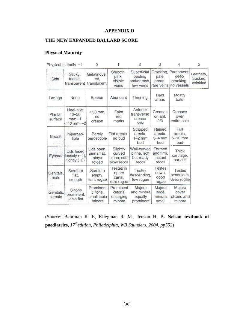
APPENDIX D
THE NEW EXPANDED BALLARD SCORE
Physical Maturity
(Source: Behrman R. E, Kliegman R. M., Jenson H. B. Nelson textbook of
paediatrics, 17th
edition, Philadelphia, WB Saunders, 2004, pp552)

[37]
Neuromuscular maturity
(Source: Behrman R. E, Kliegman R. M., Jenson H. B. Nelson textbook of
paediatrics, 17th
edition, Philadelphia, WB Saunders, 2004, pp552)

[38]
Maturity rating
(Source: Behrman R. E, Kliegman R. M., Jenson H. B. Nelson textbook of
paediatrics, 17th
edition, Philadelphia, WB Saunders, 2004, pp552)