Chapter - aulakinesica.com.ar Inestabilidad... · The CHL, intimate anatomically to the SGHL, also...
16
Transcript of Chapter - aulakinesica.com.ar Inestabilidad... · The CHL, intimate anatomically to the SGHL, also...


Chapter | 17 |
Glenohumeral instabilitySteven C Allen, Russell S VanderWilde, Peter A Huijbregts
CHAPTER CONTENTS
Introduction 234
Anatomy 235
Biomechanics 235
Pathology 236
Diagnosis of glenohumeral instability 239
History 239
Examination 239
Stability tests 240
Apprehension test 240
Relocation test 240
Surprise (release) test 240
Laxity tests 240
NAIOMT SGHL/CHL test 241
NAIOMT MGHL test 241
NAIOMT IGHL test 241
NAIOMT posterior capsule test 241
NAIOMT sulcus stability test for IGHL-AB/PB
and inferior labrum 241
Imaging 242
Prognosis 243
Clinical risk factors 244
Anatomic risk factors 244
Management of glenohumeral instability 244
Conclusion 246
INTRODUCTION
In Chapter 16, glenohumeral instability was discussed inthe context of secondary impingement. In the authors’combined experience, patients presenting with shoulderpain often have underlying instability of the glenohum-eral joint. However, glenohumeral instability presents awide spectrum. On the one end of the instability spectrumis the minor instability (more appropriately classified asatraumatic, involuntary, recurrent, mostly anterior-infe-rior subluxation) with often only history findings indica-tive of its presence that responds well to conservativemanagement. On the other end of the spectrum is thetraumatic dislocation at times with associated fracturesand neurovascular or soft-tissue damage that often posesa surgical indication.
With regard to dislocation, Kr�ner et al (1989) reportedan incidence of 0.17 per 1000 person-years in a generalurban population. Owens et al (2009) reported an inci-dence for the general population of 0.08 versus 1.69 per1000 person-years for military personnel. In about 98%of patients the shoulder dislocates anteriorly, whereas lessthan 2% of dislocations are posterior and only 0.5% infe-rior (Walton et al 2002, Cicak 2004, Camarda et al 2009).
The glenohumeral joint can be unstable in anterior,posterior, or multiple directions. Multi-directional insta-bility (MDI) is symptomatic laxity in two or more direc-tions, one of which is always inferior (Caplan et al2007). It is important to distinguish instability from lax-ity, as the great majority of lax shoulders are not unstable
234© 2011 Elsevier Ltd.
DOI: 10.1016/B978-0-7020-3528-9.00017-0

(McFarland et al 2010). Objectively, laxity describes theextent to which the humeral head can be translated onthe glenoid (Schenk & Brems 1998). In contrast, instabil-ity is an abnormal increase in glenohumeral translationthat causes symptoms related to subluxation or disloca-tion. Shoulder instability becomes a clinically relevantpathology in the presence of: (1) abnormal and usuallyasymmetric laxity, (2) correlating symptoms, and (3) cor-relating pathologic anatomy. When these three elementsare present, an imbalance of the static and dynamic gleno-humeral joint stabilizers occurs and the result is instabil-ity. Likely due to problems with definitive diagnosis,epidemiological data on shoulder instability are notavailable.
Many young athletic patients present to physical ther-apy with shoulder pain due to atraumatic, involuntary,recurrent, mostly anterior-inferior subluxation. However,therapists in a direct access role may also be confrontedwith patients with complaints on the other side of thespectrum and therefore need to also be familiar with thepresentation of frank dislocations, so that they may recog-nize a patient in need of medical-surgical evaluation.
ANATOMY
The anterior shoulder joint capsule has distinct bandsdescribed as superior (SGHL), middle (MGHL) and infe-rior (IGHL) glenohumeral ligaments. The humeral attach-ment of the SGLH lies just superior to the lesser tuberositynear the bicipital groove. The ligament courses anterior tothe biceps tendon to attach to the antero-superior labrum(Levine & Flatow 2000). The MGHL, the most variable(and at times absent) of the glenohumeral ligaments,arises off the humerus at the lesser tuberosity in associa-tion with the subscapularis tendon; its labral attachmentlies just inferior to that of the SGHL. The humeral attach-ment of the inferior capsule or axillary pouch, which con-tains the anterior (AB-IGHL) and posterior (PB-IGHL)band of the IGHL, runs from the 4 to 8 o’clock positionof the humeral head to attach to the inferior labrum(Suglaski et al 2005). The posterior capsule extends fromthe PB-IGHL to the posterior band of the tendon of thelong head of the biceps. It has been subdivided into thesuperior (SC), middle, and posterior capsule. Althoughoften assumed of minor biomechanical importance itshould be noted that the SC has a tissue thickness similarto that of the AB-IGHL (Bey et al 2005).
The coraco-humeral ligament (CHL) arises off the lat-eral aspect of the coracoid process traversing horizontallybeneath the coraco-acromial ligament (CAL). It attachesinto the greater and lesser tuberosities on either side ofthe bicipital groove. In the rotator interval between theinferior margin of the supraspinatus and the superiormargin of the subscapularis, the CHL blends with the
adjacent tendons and the underlying joint capsule. Atthe anterior joint capsule the anterior band of this liga-ment is superficial to and overlies the SGHL.
The coraco-acromial ligament (CAL) spans the superioraspect of the shoulder running from the coracoid processto the anterior and inferior acromion. Lee et al (2001)described a falx or band of tissue that directly connectsthe fibres of the CAL to the conjoint tendon of the rotatorcuff without attaching to the coracoid process (Fig 17.1).In the rotator interval, the CHL is also connected via thisfalx to the CAL, and laxity or damage (also iatrogenic asoccurs during acromioplasty) to the CAL may compro-mise the tension in the CHL.
The intact labrum (discussed in detail in Chapter 18) isfibrous throughout with a fibrocartilaginous transitionzone at its attachment with the glenoid articular cartilage(Abboud & Soslowsky 2002). Firmly attached inferiorlyand found to be looser superiorly and anteriorly, thelabrum increases the depth of the glenohumeral socketby 50% (Cooper et al 1992). As noted above, it serves asthe attachment sites for the glenohumeral ligaments andbiceps tendon.
BIOMECHANICS
All three bands of the glenohumeral joint capsule serve asthe primary passive restraints to external rotation (Turkelet al 1981, O’Connell et al 1990). The SGHL contributesa primary restraint to external rotation in 0� of abduction.The CHL, intimate anatomically to the SGHL, also contri-butes a primary source of passive restraint to external rota-tion in this position (Neer et al 1992, Kuhn et al 2005).The MGHL is believed to be a more important contributorto anterior shoulder stability in 45� of abduction, possiblyimplicating it in midrange shoulder instability (O’Connellet al 1990, Kuhn et al 2005). Together the SGHL and CHL
Coracohumeralligament
Coracoacromialligament
Falx
Fig 17.1 Falx attaching fibres of coraco-acromial ligamentdirectly to conjoint tendon of the rotator cuff.
Chapter | 17 | Glenohumeral instability
235

also limit inferior translation and posterior translation inthe flexed, adducted, and internally rotated shoulder(Levine & Flatow 2000).
The inferior portion of joint capsule acts as a ‘ham-mock’ that checks undue translation of the humeral headon the glenoid. In abduction, this entire complex movesbeneath the humeral head and becomes taut. The AB-IGHL comes under the greatest tension in 90� of abduc-tion, 10� of extension and end-range external rotation.The inferior complex moves anteriorly beneath thehumeral head with external rotation limiting anteriortranslation (Levine & Flatow 2000). In cadaver tests ofthe AB-IGHL complex, the anterior drawer test at 60�
abduction produced high strain at the insertion sites onboth the humerus and glenoid. These two sites corre-spond to the most prevalent failure sites during tensiletesting of the AB-IGHL; specifically the insertion site onthe glenoid is a common site for an anterior labral tear(Bankart lesion). Kuhn et al (2005) reported that theentire IGHL, including the axillary pouch, was the mostimportant restraint for external rotation in both positionsof 15� and 60� abduction.
The PB-IGHL of the inferior recess comes under tensionwith abduction and internal rotation, as the complexmoves posteriorly beneath the humeral head (Levine &Flatow 2000). The PB-IGHL has been implicated in theclinically observed stiff posterior shoulder. After the PB-IGHL, in the flexed and internally rotated shoulder thegreatest tension is found in the posterior shoulder capsuleindicating its role as another posterior stabilizer (Urayama2001).
The CAL is a significant static stabilizer of the gleno-humeral joint at lower elevations (Lee et al 2001).Previously thought to have no functional importanceand surgically released during acromioplasty, compromiseof the CAL allows for increased anterior and inferior trans-lation of the internally and externally rotated shoulder in0 and 30� of abduction indicating the potential for iatro-genic instability after acromioplasty.
The intact labrum contributes to the centring of thehumeral head on the glenoid, and damage to the antero-inferior labrum allows migration of the humeral headtoward the site of lesion. Fehringer et al (2003) concludedthat precise centric position of the glenohumeral joint iswell served by an intact labrum, especially in mid rangeswhere the majority of ligaments are lax. The glenohumerallabrum elevates the glenoid edge contributing to shoulderstability by effectively doubling the depth of the glenoidsocket and serving as a ‘chock block’ to translation (Wal-ton et al 2002) adding as much as 20% to the resistanceto translation forces (Abboud & Soslowsky 2002). In thepreceding chapter the role of the labrum in the concavitycompression mechanism contributing to glenohumeralstability was discussed.
Stability, of course, is not solely provided by passiverestraints. The transverse (subscapularis, infraspinatus,
and teres minor) and frontal plane (supraspinatus anddeltoid) force couples of the concavity compressionmechanism function as local stabilizers (Parsons et al2002). Both the rotator cuff muscles and the primemovers of the shoulder provide muscle force vectors tothe glenohumeral joint that have compressive and shearcomponents (Lee et al 2000). The directions of these forcevectors change substantially from 0� to 90� abduction,although the compressive component provided by therotator cuff is consistently much larger than its shear com-ponent. The shear component can potentially stabilize ordestabilize the joint, depending on its direction. The infra-spinatus and teres minor generate a posterior shear in thelate cocking phase of throwing, thereby contributing toanterior shoulder stabilization, whereas the supraspinatusgenerates a large anterior shear force in end-range thusdestabilizing the joint in the anterior direction (Lee et al2000). The pectoralis major similarly provides an anteriordestabilizing force in the late cocking position (Labriolaet al 2005). The latissimus dorsi and teres major producemore effective inferior shear forces than do infraspinatusand subscapularis; the role of the supraspinatus in thisregard is only minimal (Halder et al 2001a). In Chapter16 the equivocal evidence on the role of the long headof the biceps tendon in stabilization was discussed. Thedeltoid is a significant contributor to anterior stabilityin the position of apprehension, with all three headscontributing equally to stabilization (Kido et al 2003).The lateral deltoid is a key muscle restraint to inferior gle-nohumeral instability (Halder et al 2001b).
PATHOLOGY
Avulsion of the glenoid labrum in the antero-inferiorquadrant called a Bankart or Perthes lesion is the mostcommon pathology seen in anterior shoulder dislocation.It is disruption of the IGHL, and not solely the Bankartlesion that is thought to allow for dislocation (Robinson& Dobson 2004). Bigliani et al (1992) showed that anintra-substance ligament injury occurs before labral avul-sion. An isolated IGHL injury renders the glenohumeraljoint very unstable, even with intact dynamic stabilizers.The typical mechanism for anterior dislocation in theyounger patient is indirect trauma to the abducted,extended, and externally rotated arm most commonly inoverhead sports activities (Bohnsack & Wulker 2002).Primary anterior dislocation of the shoulder occurs com-monly after low-energy falls in the elderly (Robinson &Dobson 2004). An impression fracture of the postero-lateral humeral head called a Hill-Sachs lesion is alsopresent in most patients with anterior instability (Cicak2004, Robinson & Dobson 2004).
At less than 2% of all dislocations, dislocation in a pos-terior direction is uncommon. Posterior dislocation can
Part | 3 | The shoulder region
236

be caused by a fall onto the outstretched arm, a weight‘getting away’ from a weight lifter at terminal extensionof a bench press, a football lineman unable to hold offan opponent with forces axially transmitted through theforward flexed arms, or a hockey player attempting toslow down velocity of a hit into the boards. It may alsoresult from epileptic seizure or electric shock. Posteriordislocation may be associated with fractures of the surgi-cal neck of the humerus or fractures of the tuberosities.Posterior shoulder dislocations with posterior labraldetachment (reverse Bankart lesion) and a humeral ante-romedial impression fracture (reverse Hill-Sachs lesion)need to be considered for surgery. Indications for surgicalrepair include recurrent subluxations or dislocations ormechanical symptoms despite adequate rehabilitation(Seebauer & Keyl 1998, Cicak 2004, Kim et al 2005).
At 0.5% of all dislocations inferior dislocations are evenless common. Mechanisms of injury include direct axialloading through the humerus as might occur when thepatient tries to catch himself overhead when falling froma height. The other mechanism is violent forced abduc-tion of an already abducted shoulder. Impingement ofthe neck or proximal shaft of the humerus against theacromion levers the humeral head inferiorly out of theglenoid. The term ‘luxatio erecta’ refers to the presentationof patient with the arm abducted, elbow flexed, forearmspronated, and hand above the head unable to lower thearm to the side. Associated injuries may include fracturesof the acromion, clavicle, coracoid process, greater tuber-osity, and humeral head. Associated vascular injuries tothe axillary vessels are often serious and require surgerybut are less common than axillary, radial, or ulnar nerveor brachial plexus injuries that mostly recover well indi-cating their neuropraxic nature (Baba et al 2007, Camardaet al 2009). Mallon et al (1990) reviewed 80 cases andreported greater tuberosity fracture or rotator cuff injuriesin 80%, neurological involvement in 60%, and vascularcompromise in 3.3% of cases.
Posterior subluxation is attributed to posterior com-pressive or tensile loading and forced hyperadduction(Kim et al 2005, Robinson & Dobson 2004) with painattributed to excessive translation into the posterior recess.Recurrent posterior shoulder subluxation as a clinicalentity has become increasingly recognized as a less com-mon (2–5%) but important contributor to shoulder insta-bility (Eckenrode et al 2009). A single traumatic event orrepetitive cumulative trauma as may occur in contactsports with high-energy forces directed to the posteriorcapsule may lead to posterior glenohumeral instability.Glenoid retroversion and weakness of the external rota-tors have also been identified as potential contributors(Eckenrode et al 2009).
Excessive anterior translation of the humeral head dur-ing abduction-external rotation leads to plastic deforma-tion of the AB-IGHL and anterior glenohumeral jointsubluxation. This would also indicate that the degree ofexcessive laxity commonly found in the shoulder ofthrowing athletes might be on a progressive track of exces-sive motion and translation leading to symptoms thateventually manifest in labral injury and/or partial-thicknessrotator cuff tears (Kuhn et al 2003). However, laxity andhypermobility is not instability and in fact is a prerequisiteto achieve higher degrees of speed and torque in the throw-ing shoulder (Huijbregts 1998). Considering its frequentassociation with anterior shoulder instability and subluxa-tion, we believe it is important to review the various phasesof overhead throwing and apply clinical reasoning to thekinetic chain to allow for the diagnosis of possible relevantpatho-biomechanical faults.
The overhead throw has five phases: wind-up, earlycocking, late cocking phase, acceleration and follow-through (Fig 17.2). The wind-up phase in the overheadbaseball pitch is a preparatory phase, centred on flexion.A right-handed thrower has a flexion pattern of the leftlower extremity with considerable hip and knee flexion.There also will be a flexion movement of the spine. Both
Wind-up Early cocking Late cocking Acceleration Deceleration Follow-through
Fig 17.2 Five phases of the overhead throwing motion.
Chapter | 17 | Glenohumeral instability
237

hands are in contact with the ball and the shoulders are inan internal rotation-adduction position with bilateralelbow flexion. The pitcher is facing the batter with the leftside of the body. Early cocking starts when the left handloses contact with the ball. The right shoulder moves fromadduction and internal rotation to abduction and externalrotation. The pitcher steps with the previously flexed leftleg in the direction of the batter, and the trunk moves intoextension, right rotation, and left side bending. The late-cocking phase starts when the left foot of the pitcher hitsthe ground. This is the start of a de-rotation movementof the trunk and legs that will contribute to acceleratingthe ball. The right arm and ball still move in the samedirection of horizontal abduction and external rotation.
Acceleration starts with the switch over from shoulderexternal rotation to shoulder internal rotation. This rota-tion is the most important movement of the accelerationphase. In this phase, the shoulder also moves from hori-zontal abduction to horizontal adduction and back inthe direction of horizontal abduction just prior to ballrelease. Ball release by the right hand marks the end ofacceleration. The arm, which has been immensely acceler-ated for the throwing motion, now has to be decelerated.The left lower extremity moves into flexion and trunk intoflexion and left rotation. The right arm is moving intoadduction and internal rotation. The first phase offollow-through (deceleration) is marked by high activityin the muscle complex of the right shoulder with the sec-ond phase of follow-through requiring adequate trunkand lower extremity movement to decrease the forcerequirements about the shoulder and reduce the potentialfor injury (Huijbregts 1998). The overhead throw is anextremely fast activity. Fleisig et al (1995) measured anaverage time of 0.139 � 0.017 (s) from foot contact toball release, a period that corresponds to the late cockingand acceleration phases combined.
Two critical instants are identified in the overheadthrow that place unusually high demands on the shouldercomplex. The first phase identified in late cocking, revealshigh torsional and compressive loads to the shoulder thatmay well exceed the plastic limit of the antero-inferiorcapsuloligamentous complex resulting in pain and insta-bility in this quadrant. The second critical instant occursjust after ball release in the early deceleration phase.Although torsional loading is significantly reduced andshear forces to the anterior restraints are low in this phaseof throwing, the compressive loading to the shoulderjoint is highest at 1100 (N), constituting a more than100% increase from compression during the first criticalinstant. Any unwanted translation of the humeral headin the presence of these compressive forces has the poten-tial to produce damage to the capsule, the closely asso-ciated rotator cuff tendons, and the labrum. Enormousdemands are placed on both the active and passiverestraints of the shoulder with – in addition to these hightorques and forces – an external rotation range of motion
of 140� at the end of late cocking, an internal rotationangular velocity of 7000�/s during acceleration and anangular deceleration of 500,000�/s2 during deceleration(Huijbregts 1998).
In a throwing athlete with glenohumeral instability theclinician should not limit the search for causative or con-tributory dysfunctions to shoulder joint or even the shoul-der girdle. As an example of relevant lower quadrantdysfunctions, a right sacral torsion present at left foot con-tact in the acceleration phase may lead to excessive com-pensation in the shoulder to attain the required armvelocity for competitive throwing. Also maximal lumbarextension and right side bending are required in thisphase of throwing, implying that an unresolved, chronicright postero-lateral disc lesion may also place excessivedemand on the shoulder girdle, as trunk motion in thisquadrant may be limited. This same disc lesion in a moreirritable back problem would compromise the de-rotationand deceleration of the throwing arm where flexion andleft rotation of the trunk are required.
The overhead athlete may also be vulnerable in case oflumbar instability or an increase in the neutral zone of aspinal motion segment (Panjabi 1992). If this instabilityis in a rotational plane to the right in the overhead thrower,the critical zone of acceleration may result in compensationhigher up in the kinetic chain and subsequent injury to theglenohumeral joint. Unilateral weakness of the multifidusmuscles of the lumbar spine may lead to subsequent atro-phy in patients with unilateral back pain (Hides et al1996). Asymmetrical contraction of this group of segmen-tal muscles would result in torsional loading with excessivetranslation of the spinal motion segment and loss of formclosure in the lumbosacral region (Lee 1989) again expos-ing the glenohumeral joint higher up the kinetic chain toexcessive shear or compression. A higher incidence ofosteoarthrosis in the opposite hip from the throwing sidein retired javelin throwers (Schmitt et al 2004) suggeststhe need to tolerate high torsional forces on the left hipin the right-handed overhead athlete. Weakness in the deepleft hip rotators or gluteus medius in the decelerationphase of throwing or limited hip rotation mobility wouldlikely compromise the safety margin for attenuatingforce requirements on the throwing shoulder that thecontribution of the hip muscles in this phase wouldotherwise offer.
With regard to upper quadrant dysfunctions, scapulardynamics require adequate upward rotation, abductionand posterior tilting for optimal shoulder function inthe throwing athlete (Magarey & Jones 1992). Muscleimbalances between the upper trapezius and serratus ante-rior may manifest in scapular winging due to serratusanterior inhibition (Sahrmann 2002) thus compromisingthe well-coordinated movement of the scapula andhumerus and the maintenance of centric position of thehumeral head on the labrum. A hypertonic or stiff levatorscapulae muscle due to C4 facilitation from a cervical
Part | 3 | The shoulder region
238

spine joint dysfunction would tilt the scapula into down-ward rotation and inhibit the ‘chock-block’ mechanismoffered by the labrum (Fowler & Pettman 1992, Waltonet al 2002). This would increase strain in the structureslimiting inferior and anterior translation of the humeralhead. The upper thoracic spine requires adequate rotationand side bending to the right in a right-handed thrower.Hypomobility in this region of the thoracic spine is com-mon and will directly impact the degree of stress placedon the anterior restraints to the glenohumeral joint.
DIAGNOSIS OF GLENOHUMERALINSTABILITY
Although it would be hard to imagine that a clinicianwould not recognize a luxatio erecta, the much more com-mon patient with minor instability is much harder todiagnose and even most posterior dislocations are missedon initial examination (Cicak 2004).
History
Patients with shoulder instability most often present withpain as their primary complaint. Somatic pain describedas deep, aching, and intermittent and located in the ante-rior or posterior shoulder joint is common. Trauma maysuggest dislocation that may have spontaneously reduced,especially when in an anterior direction. Under pathologywe discussed mechanisms of injury that should make theclinician consider anterior, posterior, and inferior disloca-tions. As noted above posterior dislocations are oftenmissed on initial examination with the patient only com-plaining of subjective instability and pain with flexion,adduction, and internal rotation (Cicak 2004).
In the introduction we discussed how in physical ther-apy the commonly used diagnosis of minor instabilitywould be more appropriately classified as atraumatic,involuntary, recurrent, mostly anterior-inferior subluxa-tion. This type of instability is much more common buthard to definitively diagnose. It is most often seen inyoung, overhead throwing athletes or gymnasts. Traumamay play a role but is often more of a cumulative typewith previous minor episodes of injury. Magarey & Jones(1992) have suggested the following (non-validated) his-tory findings as indicative of minor shoulder instability:(1) apprehension with certain movements, (2) sensationof the joint slipping in and out, (3) pain worse with over-head activity, (4) painful catches through range, (5) pain-ful intra-articular clicking or ‘dead arm syndrome’ duringlate cocking, (6) weakness in the late cocking position.Note, however, that at least some of these symptoms canhardly be considered specific. Schenk & Brems (1998)also noted that patients with MDI might present withpain in midrange positions.
Examination
As indicated above, examination of a patient withshoulder instability should not be limited to the shoul-der girdle but include a search for causative or contrib-utory dysfunctions throughout the kinetic chain inupper and lower quadrants. Specific to the shoulder,though, a scanning examination must include assess-ment of 3D posture. Winging of the scapula at restand in 90� abduction can be appreciated with a poste-rior view and indicates the need for specific musclestrength and flexibility tests. A side view may suggestmuscle length restriction in the pectoralis minor evi-denced by the shoulder held in protraction and eleva-tion with scapular inferior border winging. A patientwith an unreduced anterior or inferior dislocation maypresent with the humeral head visible and palpable onthe chest wall out of the glenoid socket. Patients withunreduced posterior dislocation may present with thearm held and fixed in adduction and internal rotation.
Active range of motion in cardinal planes with over-pressure and discreet resistance to muscles while onstretch (Cyriax 1978) as well as combined motionsbegins to examine for provocation and assess integrity.Making them easy to spot, patients with luxatio erectaare not able to lower the arms from the elevated position(Camarda et al 2009). Patients with posterior dislocationmay have the humeral head caught on the posterior gle-noid rim locking the shoulder between 10–60� of inter-nal rotation with no external rotation possible fromthis position (Cicak 2004). Should this initial screennot provoke the patient’s symptoms, the clinician maysuspect a remote source including referred mechanicalpain from the cervical spine or a non-mechanical etiol-ogy. Note that unless there has been recent trauma theremay well be a full or nearly full range of motion (ROM)in the shoulder girdle. A neurovascular examination forcompromise especially in the patient with a possible dis-location will complete the seated examination. Closeinspection of the scapula from a posterior viewpoint dur-ing arm elevation will be helpful for abnormal rhythm,lack of upward rotation, and abduction, or medial bor-der winging as discussed in the chapter on impingement(Chapter 16).
Magarey & Jones (1992) have suggested the following(non-validated) physical examination findings as indica-tive of minor instability: (1) excessive mobility or loss ofnormal end-feel on instability tests with or without appre-hension or intra-articular click, (2) full or excessive ROMwith end-range pain, (3) loose end-feel with less of a liga-mentous character, (4) external rotation at 90� abductionthat is either limited by spasm or shows excessive range,(5) pain free and strong rotator cuff contractions withthe exception of an often weak but pain free infraspinatus.Again note that many of these findings may not be veryspecific.
Chapter | 17 | Glenohumeral instability
239

Stability tests
Special tests for clinical instability of the glenohumeraljoint fall into two categories (Levy et al 1999, Ellenbeckeret al 2002, Tibone et al 2002). Stability tests use provoca-tion, apprehension, and end feel to determine joint integ-rity toward end-range, whereas laxity tests examine themobility of a joint in functional and midranges of motion.Bahk et al (2007) concluded that special tests can add sig-nificantly to our assessment of the unstable shoulder butfindings on clinical laxity examination and relevance toinstability must be placed in perspective. If findings ‘fit’they confirm and solidify the diagnosis. If findings on thesetests do not fit with other history and examination findings,laxity alonemeans nothing except a ‘loose shoulder’, whichis not a diagnosis, but rather a physical exam finding. In theclinic of the second author, throwing-related MDI is oftensuperimposed upon an already genetically loose shouldercapsule that subsequently becomes too loose and ‘decom-pensate’. An important paradox is that the genetic laxity ispartly what initially allows these athletes to succeed at ahigh level in overhead athletics like throwing. Once theydecompensate, the original advantage, however, becomesa detriment.
Apprehension test
The patient is supine on table, glenohumeral joint at theedge of the table but with the scapula supported bythe table. The patient’s shoulder is at 90� of abduction,the elbow is flexed to 90� and the examiner’s knee sup-ports the elbow to prevent extension of the shoulder.The examiner then applies external rotation progressivelyuntil the patient cannot tolerate any further rotation andthe degree of rotation is recorded. In patients with ante-rior instability, a patient report of apprehension and afeeling that the shoulder will come out of joint is consid-ered a positive test. Some authors note that pain in thismaneuver may be indicative of more subtle anterior insta-bility. Sensitivity for this test for the diagnosis of traumaticanterior instability has been established at 52.78% andspecificity at 98.91% (Lo et al 2004).
Relocation test
First described by Jobe et al (1989), the relocation testapplies a posterior force to the humeral head in the posi-tion of apprehension of the above test and is positive if itrelieves the symptoms of apprehension. Although Lo et al(2004) proposed this test to differentiate a subtle instabil-ity in the overhead athlete from rotator cuff impingement,if pain was experienced in the apprehension exam andimproved with the relocation test, the sensitivity and spec-ificity with production and reduction of pain only wereboth low at 40% and 42.65%, respectively. However,when considering solely diminished apprehension a
positive test finding, specificity was 100% for anteriorshoulder instability, although sensitivity stayed low at31.94%. Reduction of symptoms is usually associatedwith an increase in external rotation range.
Surprise (release) test
While holding the final position of the relocation test, theexaminer’s hand is quickly removed from the proximalhumerus and the patient’s response elicited. A positive testis indicated by a sudden return of symptoms noted withthe apprehension test. The surprise test had a sensitivityof 63.89% and specificity of 98.91% for the diagnosis ofanterior instability (Lo et al 2004). Note that the surprisetest finds the shoulder in a more vulnerable position ofgreater external rotation than in the apprehension test.For this reason to perform the surprise test safely andaccurately, we recommend that the apprehension andrelocation tests be performed first. This will give the exam-iner an initial impression of where the patient feels vul-nerable and thus allows careful placement to apply andrelease the posterior directed force within the patient’scomfort level. Performing a surprise test immediatelymay not only startle the patient but can also acutely dislo-cate the shoulder.
Posterior shoulder instability has typically been exam-ined with a load-and-shift test producing posterior transla-tion or by posterior joint line tenderness (Eckenrode et al2009). Patients may also have a positive jerk test. This testis performed with the patient supine, arm to be tested in90� abduction and internal rotation. An axial load is thenapplied as the arm is brought into horizontal adduction.The test is positive if the manoeuvre produces a palpableor audible clunk, as well as pain. Sensitivity and specificitywere reported to be 73% and 98%, respectively, in the diag-nosis of a postero-inferior labral lesion (Kim et al 2005).
Laxity tests
Motion in any simple plane of the glenohumeral joint resultsin coupled motion in two additional planes. A strain gaugeanalysis of the glenohumeral ligaments showed that for eachligament tested, a tension-sharing relationship existed withtransfer of tension among all ligaments (Terry 1991). Still,Fowler & Pettman (1992) in their teaching at the NorthAmerican Institute of Orthopaedic Manual Therapy(NAIOMT) have proposed that the capsuloligamentouscomplex of the glenohumeral joint can be tested in serialfashion to discriminate the predominant site of injury or lossof integrity. Using an electronic digital ruler, Sharp andKisser (unpublished research 2009) in a physical therapydoctoral capstone research project at Andrews University inBerrien Springs, Michigan provided preliminary validationfor the selective tensioning suggested as the rationale forNAIOMT tests for the SGHL, MGHL, and AB-IGHL.
Part | 3 | The shoulder region
240

NAIOMT SGHL/CHL test
With the patient supine, the shoulder to be tested at theedge of the examination table with the scapula supportedon the table, the shoulder is placed in 0� abduction andend-range external rotation followed by an anterior glideof the proximal humerus (Fig 17.3) to test the SGHLand the posterior or lateral band of the CHL. If the armis now allowed to move into 10� extension, the anterioror medial band of the CHL comes more under tension(Fig 17.4).
NAIOMT MGHL test
From the test position of 0� abduction, 10� extension, andend-range external rotation, the examiner now moves thearm to 45� abduction and then applies an anterior medialglide in the plane of the glenohumeral joint surface (Fig17.5) to test the MGHL.
NAIOMT IGHL test
From the MGHL test position, the examiner now movesthe arm to 90� abduction and applies an anterior medial
glide to the proximal humerus to test the AB-IGHL (Fig17.6). The PB-IGHL is tested with the arm maintainedin this position of 90� abduction and 10� extensionbut now with full internal rotation and a posteriorlateral glide applied to the glenohumeral joint (Fig 17.7)(Fowler & Pettman 1992, Levine & Flatow 2000).
NAIOMT posterior capsule test
Bringing the arm to 90� flexion, followed by full internalrotation, then end-range horizontal adduction, and finallyan axial stress applied to the proximal humerus in a pos-terior lateral direction tests the posterior capsule (Fig17.8) (Fowler & Pettman 1992, Urayama 2001).
NAIOMT sulcus stability test for IGHL-AB/PBand inferior labrum
A sulcus sign or dimple created beneath the acromionwhen the subject’s arm is axially tractioned inferiorly,
Fig 17.3 NAIOMT SGHL/posterior CHL test.
Fig 17.4 NAIOMT SGHL/anterior CHL test.
Fig 17.6 NAIOMT AB-IGHL test.
Fig 17.5 NAIOMT MGHL test.
Chapter | 17 | Glenohumeral instability
241

usually in a seated position, was first described as the hall-mark test for MDI if it also produced the patient’s charac-teristic symptoms (Neer & Foster 1980). However, Bahket al (2007) reported the test as provocative for patient’ssymptoms in only a small number of symptomaticpatients. The degree of laxity considered relevant has alsobeen arbitrary and McFarland et al (2003) suggested thatthe sulcus sign might lead to an over-diagnosis of MDI.Pettman (personal communication 2009) has suggesteda modification of the sulcus sign. In this test the examinerstabilizes the scapula by stabilizing with one hand in theaxilla. The other hand rotates the arm into external rota-tion and while holding this position applies an inferiorforce to assess for a sulcus sign suggested as consistentwith a possible labral lesion in the AB-IGHL complex(Fig 17.9). If this sulcus sign is present in the same testwith the arm in full internal rotation, a possible labrallesion in the PB-IGHL complex is suggested (Fig 17.10).
Imaging
From a medical-surgical perspective the diagnosis of insta-bility in the vast majority of patients is based on the his-tory and physical examination in combination withplane radiographs. Plain radiographs are also importantin ruling out fractures often associated with dislocations.Whereas a standard AP view of the uncomplicated unsta-ble shoulder may produce equivocal findings or may behard to interpret, an axillary lateral view is helpful tovisualize the presence and extent of a (reverse) Hill-Sachslesion but may be near impossible to obtain in a patientwhose abduction is severely limited due to pain. The lat-eral scapular plane view is particularly helpful in deter-mining the relationship of the humeral head to theglenoid. In anterior dislocations of the shoulder, thehumeral head lies anterior to the glenoid; in posterior dis-locations it is posterior (Cicak 2004, Workman et al1992).
Fig 17.8 NAIOMT posterior capsule test.
Fig 17.9 NAIOMT sulcus sign in external rotation.
Fig 17.10 NAIOMT sulcus sign in internal rotation.
Fig 17.7 NAIOMT PB-IGHL test.
Part | 3 | The shoulder region
242
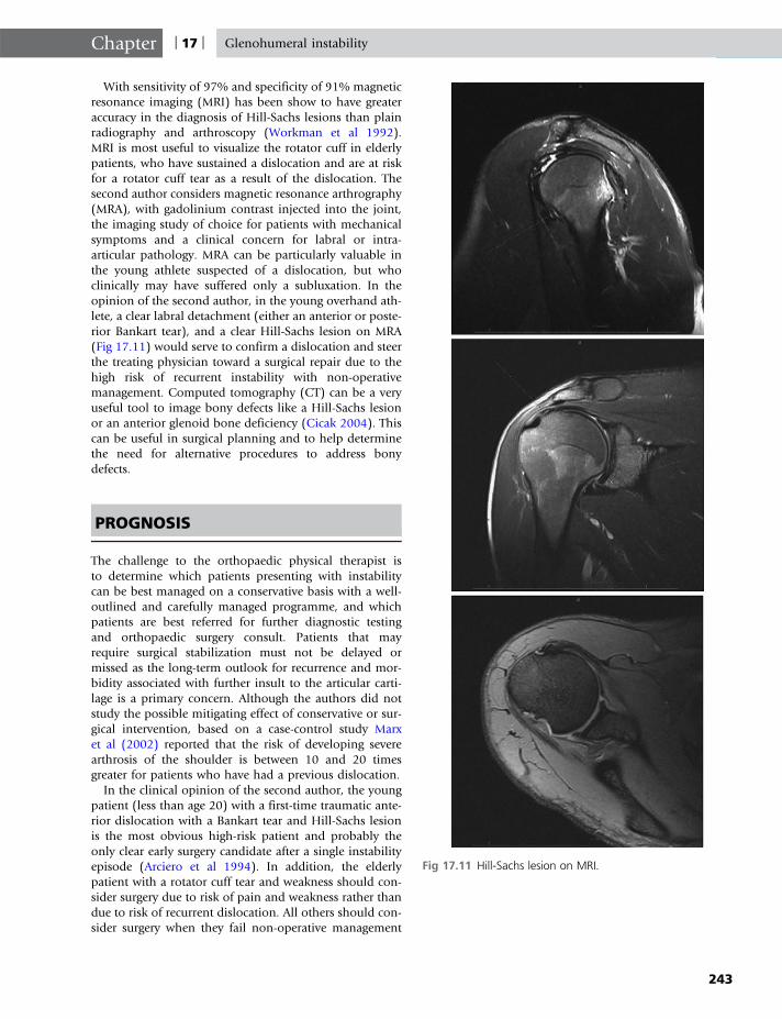
With sensitivity of 97% and specificity of 91% magneticresonance imaging (MRI) has been show to have greateraccuracy in the diagnosis of Hill-Sachs lesions than plainradiography and arthroscopy (Workman et al 1992).MRI is most useful to visualize the rotator cuff in elderlypatients, who have sustained a dislocation and are at riskfor a rotator cuff tear as a result of the dislocation. Thesecond author considers magnetic resonance arthrography(MRA), with gadolinium contrast injected into the joint,the imaging study of choice for patients with mechanicalsymptoms and a clinical concern for labral or intra-articular pathology. MRA can be particularly valuable inthe young athlete suspected of a dislocation, but whoclinically may have suffered only a subluxation. In theopinion of the second author, in the young overhand ath-lete, a clear labral detachment (either an anterior or poste-rior Bankart tear), and a clear Hill-Sachs lesion on MRA(Fig 17.11) would serve to confirm a dislocation and steerthe treating physician toward a surgical repair due to thehigh risk of recurrent instability with non-operativemanagement. Computed tomography (CT) can be a veryuseful tool to image bony defects like a Hill-Sachs lesionor an anterior glenoid bone deficiency (Cicak 2004). Thiscan be useful in surgical planning and to help determinethe need for alternative procedures to address bonydefects.
PROGNOSIS
The challenge to the orthopaedic physical therapist isto determine which patients presenting with instabilitycan be best managed on a conservative basis with a well-outlined and carefully managed programme, and whichpatients are best referred for further diagnostic testingand orthopaedic surgery consult. Patients that mayrequire surgical stabilization must not be delayed ormissed as the long-term outlook for recurrence and mor-bidity associated with further insult to the articular carti-lage is a primary concern. Although the authors did notstudy the possible mitigating effect of conservative or sur-gical intervention, based on a case-control study Marxet al (2002) reported that the risk of developing severearthrosis of the shoulder is between 10 and 20 timesgreater for patients who have had a previous dislocation.
In the clinical opinion of the second author, the youngpatient (less than age 20) with a first-time traumatic ante-rior dislocation with a Bankart tear and Hill-Sachs lesionis the most obvious high-risk patient and probably theonly clear early surgery candidate after a single instabilityepisode (Arciero et al 1994). In addition, the elderlypatient with a rotator cuff tear and weakness should con-sider surgery due to risk of pain and weakness rather thandue to risk of recurrent dislocation. All others should con-sider surgery when they fail non-operative management
Fig 17.11 Hill-Sachs lesion on MRI.
Chapter | 17 | Glenohumeral instability
243

with persisting symptoms or recurrent dislocations or sub-luxation symptoms. Although the exact risk of recurrentinstability is impossible to determine for any individual,risk factors have been identified that help us classify thoseindividuals who are at increased risk of recurrent instabil-ity. Risks of recurrence can be broken down into clinicaland anatomic risk factors.
Clinical risk factors
Age of first dislocation is a very powerful prognosticator.In young patients under age 20, recurrence rates haveranged from 55% to 94%. Te Slaa et al (2004) reported26% recurrence within 4 years. In their study, age wasthe most significant prognostic factor with recurrence in64% of patients less than 20 years of age and in only6% of those older than 40 years. Kralinger et al (2002)also reported age between 21 and 30 as the only factorassociated with recurrence. Note that advancing age ishowever associated with an increased falling risk leadingto recurrent dislocation and an increasing incidence ofrotator cuff tears.
Chronicity refers to the number of times a patient hassuffered a dislocation. Although rare a truly chronicallydislocated shoulder is one that has dislocated and remainsdislocated. An acute dislocation is a shoulder that has justdislocated and is in need of reduction. More commonclinically is the recurrent dislocation. The recurrent dislo-cator is the patient, who reports both prior dislocationsand subsequent reductions. The more frequently a patienthas suffered with an instability episode, the more likely asubsequent recurrence will take place. There is, however,no consensus on the number of instability episodes afterwhich the risk becomes a certainty.
A traumatic aetiology is associatedwith a higher recurrencerisk than either micro-traumatic/overuse or atraumatic insta-bility. Atraumatic instability, especially when associated withgeneralized ligamentous laxity, is in fact a risk factor for surgi-cal failure. Volition is the patient’s ability to reproduce thesymptom or dislocation at will. Some describe their ‘trickshoulder’ and can voluntarily demonstrate the ability to dis-locate or subluxate their joint. These voluntary dislocatorsneed careful screening for emotional and mental healthissues. Although not an absolute surgical contraindication,surgery in this subset of patients needs to be very carefullyconsidered only after failure of non-operative managementand careful counselling of the patient and family.
Although perhaps by some considered an indication oflikely failure with conservative management, patients withMDI may benefit from conservative management. In addi-tion, in a subset of patients with MDI natural history evenwithout conservative intervention seems benign. Kurodaet al (2001) reported spontaneous recovery in 43 of 476shoulders of patients followed for 3 years or longer.Although shoulder kinematics may not be restored withphysical therapy in all patients with MDI but rather may
require surgical intervention and postoperative physicaltherapy (Kiss et al 2009), conservative management hasalso been shown to be effective in an as of yet not clearlyidentified subset of these patients and ultimately may bethe key to a successful outcome. Glenohumeral instabilitycan occur as a complete separation of the humerus fromthe glenoid socket or as a partial separation or subluxa-tion. Dislocations carry a higher risk of associated injuryand recurrence.
Anatomic risk factors
Anatomic risk factors for recurrence include soft tissueand bony defects. Soft tissue defects include rotator cuff,labral, and capsular tears. Bony defects include glenoidrim fractures and (reverse) Hill-Sachs lesions. A Hill-Sachslesion is basically a dent in the humeral head due to animpaction fracture that occurs as the head dislocates andimpacts the anterior or posterior glenoid rim. An engagingHill-Sachs lesion is a dent that hooks over the edgeof the glenoid in a functional range of motion and bylevering causes a high risk for recurrent anterior or poste-rior glenohumeral dislocation (Fig 17.12). A large orengaging Hill-Sachs lesion constitutes a higher risk forrecurrent dislocation.
MANAGEMENT OF GLENOHUMERALINSTABILITY
Inferior and anterior dislocations are generally addressedwith closed reduction with the patient sedated (Babaet al 2007, Camarda et al 2009). Cicak (2004) suggestedthat closed reduction under general anaesthesia of a pos-terior dislocation is most likely to be successful in patientswith a humeral head defect of less than 25% of the artic-ular surface where the dislocation is present less than 3weeks. If closed reduction is unsuccessful the surgeonmay have to progress to an open reduction. Although insome jurisdictions such as most provinces in Canada it
Fig 17.12 Hill-Sachs lesion.
Part | 3 | The shoulder region
244

is within the physical therapy scope of practice to reduceacute dislocations of extremity joints, the risk ofassociated fractures and neurovascular and other soft tis-sue damage, the lack of access to imaging, and the inabil-ity to provide sedation however clearly indicate the needfor medical-surgical referral and management where pos-sible. Surgical management may also include addressingpossibly present anatomic risk factors for recurrenceaddressed above.
In the past, patients post-dislocation were often tempo-rarily immobilized with a sling and the arm in adductionand internal rotation. Bracing in 15–20� of external rota-tion has been suggested as anatomically more beneficialand leading to decreased recurrence (Itoi et al 2001,2003, Funk & Smith 2005) but more recent research haspointed out that the evidence is insufficient to supportuse of one immobilization method over the other or evento support immobilization at all (Kralinger et al 2002,Handoll et al 2006, Smith 2006, Finestone et al 2009).It should be noted that patient compliance with therequired 3 weeks of full-time bracing is often limited irre-spective of the method used.
Although not providing sufficient operational defini-tions of specific interventions, Kralinger et al (2002)reported that whether the patient had participated inphysical therapy did not reduce risk of recurrence. In gen-eral, insufficient evidence is available to guide physicaltherapy management after closed reduction of traumaticanterior dislocation perhaps reflective of insufficientmethodological quality of studies in this area (Handollet al 2006). A recent Cochrane review reported no differ-ences with regard to renewed injury or function betweenopen or arthroscopic stabilizing surgery for anteriorshoulder instability in adults (Pulavarti et al 2009). Schei-bel (2007) discussed the possibility of subscapularisdysfunction after open repairs and considering the impor-tant role of this muscle in the transverse plane stabilizingforce couple this may present another reason to choosearthroscopic over open techniques. Limited evidence sup-ports surgery in young predominantly male adults with afirst acute traumatic dislocation, who are engaged inhighly demanding physical activities for reducing recur-rent dislocation or subluxation but no evidence is avail-able to determine the best treatment for other patientgroups (Handoll & Al-Maiyah 2004).
In the absence of research to guide conservative manage-ment of patients post-dislocation but also those patientswith (minor) instability, therapists have to depend on clin-ical reasoning and extrapolation from basic scienceresearch. Te Slaa et al (2004) did not report sports partici-pation as a risk factor for dislocation, yet Kuroda et al(2001) reported an 8.7-fold increase in incidence of spon-taneous recovery for those patients with atraumatic shoul-der instability who discontinued playing overhead sports.Education with regard to adaptation with regard to high-risk movements in sports seems a logical intervention. In
athletes, this includes modification of the often-ancillaryweight-training activities. Fees et al (1998) suggested a gripwidth during the bench press of less than 1.5 times the bi-acromial distance in patients with anterior shoulder insta-bility. Other suggested modifications included requiredassists when lifting the bar from and onto the rack, decreas-ing the shoulder abduction angle during the bench press,alternating flat and decline bench press to reduce thechance of micro-traumatic injury, and eliminating theincline bench press and the behind-the-neck press andpull-down exercises but also the back squat position wherethe hands stabilize the bar on the shoulders. In patientswith posterior shoulder instability, Fees et al (1998) sug-gested a grip width during the bench press of > 2 timesbi-acromial width, using a shoulder abduction angle >80�, horizontal abduction > 15� at the start and horizontaladduction < 20� at the finish of the bench press motion.They also proposed mandatory assists with (un) rackingthe bar and using only the flat bench press or preferablyno bench press at all.
As discussed in Chapter 16, proprioceptive deficitshave also been identified in patients with anterior gleno-humeral joint instability as well as altered muscleactivation (Myers et al 2004). Decreased rotator cuff co-acti-vation and slower biceps brachii and pectoralismajor activa-tionwere identified and restoration of their normal functionthus should be part of any targeted rehabilitation pro-gramme for anterior instability. A defect in proprioceptionalsomay be a factor in the pathophysiology of MDI (Schenk& Brems 1998). Dynamic exercises incorporating proprio-ceptive neuromuscular facilitation (PNF) have been foundto be effective in the early phases of shoulder rehabilitation(Padua et al 2004). Utilizing well-established principles ofPNF to the upper quadrant such as rhythmic stabilization,eccentric-to-concentric loading, and continuousmovementsassociated with rotation (Knott & Voss 1968), therapists caneasily implement and progress a programme even in earlystages. Closed kinetic chain exercises initially on stableand later on unstable surfaces can increase accuracy ofjoint position sense and enhance stimulation of mechan-oreceptors (Naughton et al 2005, Eckenrode et al 2009).Open-chain oscillatory exercises with the Body Blade canbe used to provide more advanced open-chain dynamicstabilization (Buteau et al 2007). Exercises for muscularendurance and strength need to address both the localstabilizers in an attempt to restore at least the active (ifnot the passive) contribution to the concavity compres-sion mechanism but also the prime movers required forfunctional movement and known to have an effect onglenohumeral stability. A careful progression needs to beadapted to the patient’s functional demands and rehabili-tation goals but also based on a careful and educatedimpression of the therapist with regard to rehabilitationpotential. Chapters 21 and 22 provide examples ofexercises for motor control and other exercises relevantto the shoulder.
Chapter | 17 | Glenohumeral instability
245

CONCLUSION
Shoulder instability can present a challenge to the orthope-dic physical therapist in differential diagnosis and manage-ment. Identification of patients at high risk for recurrenceof shoulder dislocation indicates the need for referral to ashoulder specialist for likely surgical intervention. Furtherresearch on diagnostic and prognostic validity of our clinical
tests such as those outlined above is necessary andwould help drive outcome studies of conservative versussurgical management for possible subgroups of patientsidentified based on our clinical tests. A clear indicationof glenohumeral instability should not limit the therapiststo management solely of the shoulder area. Patients witha history of shoulder instability should be examined forunderlying biomechanical faults in both the upper andlower quadrants that may contribute to the instability inmore dynamic activities.
REFERENCES
Abboud, A.A., Soslowsky, J., 2002.Interplay of the static anddynamic restraints in glenohumeralinstability. Clin. Orthop. Relat. Res.400, 48–57.
Arciero, R.A., Wheeler, J.H., Ryan, J.B.,McBride, J.T., 1994. ArthroscopicBankart repair versus nonoperativetreatment for acute, initial anteriorshoulder dislocations. Am. J. SportsMed. 22, 589–594.
Baba, A.N., Bhat, J.A., Paljor, S.D.,Mir, N.A., Majid, S., 2007. Luxatioerecta: Inferior glenohumeraldislocation–a case report.Int. J. Shoulder Surg. 1, 100–102.
Bahk, M., Keyurapan, E., Tasaki, A., 2007.Laxity testing of the shoulder: A review.Am. J. Sports Med. 35, 131–144.
Bey, M., Hunter, S., Kilambi, N., et al.,2005. The structural and mechanicalproperties of the glenohumeral jointcapsule. J. Shoulder Elbow Surg.14, 201–206.
Bigliani, L.U., Pollock, R.G.,Soslowsky, L.J., Flatow, E.L.,Pawluk, R.J., Mow, V.C., 1992.Tensile properties of the inferiorglenohumeral ligament. J. Orthop.Res. 10, 187–197.
Bohnsack, M., Wulker, N., 2002.Arthroscopic anterior shoulderstabilization: Combined multiplesuture repair and laser-assisted capsularshrinkage. Injury 33, 795–799.
Buteau, J.L., Eriksrud, O., Hasson, S.M.,2007. Rehabilitation of a glenohumeralinstability utilizing the BodyBlade. Physiotherapy Practice23, 333–349.
Camarda, R., Martorana, U., D’Arienzo,M.,2009. A case of bilateral luxatio erecta.Journal of Orthopaedic Traumatology10, 97–99.
Caplan, J., Julien, T.P., Michelson, J.,Neviaser, R.J., 2007. Multidirectionalinstability of the shoulder in elitefemale gymnasts. Am. J. Orthop.36, 660–665.
Cicak, N., 2004. Posterior dislocation ofthe shoulder. J. Bone Joint Surg.86B, 324–332.
Cooper, D.E., Arnoczky, S.P., O’Brien,S.J., Warren, R.F., DiCarlo, E.,Allen, A.A., 1992. Anatomy, histologyand vascularity of the glenoidlabrum: An anatomic study. J. BoneJoint Surg. 74A, 46–52.
Cyriax, J., 1978. Textbook of orthopaedicmedicine, Vol. 1. Cassell, London.
Eckenrode, B.J., Logerstedt, D.S., Sennett, B.J., 2009. Rehabilitation and functionaloutcomes in collegiate wrestlersfollowing a posterior shoulderstabilization procedure. J. Orthop.Sports Phys. Ther. 39, 550–559.
Ellenbecker, T., Bailie, D., Mattalino, A.,et al., 2002. Intrarater and interraterreliability of a manual technique toassess anterior humeral headtranslation of the glenohumeral joint. J.Shoulder Elbow Surg. 11, 470–475.
Fees, M., Decker, T., Snyder-Mackler, L.,Axe, M.J., 1998. Upper extremityweight-training modifications for theinjured athlete. Am. J. Sports Med.26, 732–742.
Fehringer, E.V., Schmidt, G.R.,Boorman, R.S., et al., 2003. Theanteroinferior labrum helps centerthe humeral head on the glenoid. J.Shoulder Elbow Surg. 12, 53–58.
Finestone, A., Milgrom, C., Radeva-Petrova, D.R., et al., 2009. Bracing inexternal rotation for traumaticanterior dislocation of the shoulder.J. Bone Joint Surg. 91,918–921.
Fleisig, G., Andrews, J., Dillman, C.,Escamilla, R., 1995. Kinetics ofbaseball pitching with implicationsabout injury mechanisms. Am. J.Sports Med. 23, 233–239.
Fowler, C., Pettman, E., 1992. NorthAmerican Institute OrthopaedicManual Therapy (NAIOMT) UpperQuadrant Course 600A, Portland.
Funk, L., Smith, M., 2005. Best evidencereport. How to immobilize aftershoulder dislocation? Emerg. Med. J.22, 814–815.
Halder, A.M., Zhao, K.D., O’Driscoll, S.W.,Morrey, B.F., An, K.N., 2001a. Dynamiccontributors to superior shoulderstability. J. Orthop. Res. 19 (2),206–212.
Halder, A.M., Halder, C.G., Zhao, K.D.,O’Driscoll, S.W., Morrey, B.F., An, K.N.,2001b. Dynamic inferior stabilizers ofthe shoulder joint. Clin. Biomech.16, 138–143.
Handoll, H.H.G., Al-Maiyah, M.A., 2004.Surgical versus non-surgicaltreatment for acute anterior shoulderdislocation. Cochrane Database Syst.Rev. (1), Art. No.: CD004325. DOI:10.1002/14651858.CD004325.pub2.
Handoll, H.H.G., Hanchard, N.C.A.,Goodchild, L.M., Feary, J., 2006.Conservative management followingclosed reduction of traumatic anteriordislocation of the shoulder. CochraneDatabase Syst. Rev. (1), Art. No.:CD004962. DOI: 10.1002/14651858.CD004962.pub2.
Hides, J.A., Richardson, C.A., Jull, G.A.,1996. Multifidus muscle recovery isnot automatic after resolution ofacute first episode low back pain.Spine 21, 2763–2769.
Huijbregts, P.A., 1998. Biomechanicsand pathology of the overhead
Part | 3 | The shoulder region
246

throwing motion: A literature review.J. Man. Manip. Ther. 6, 17–23.
Itoi, E., Sashi, R., Minagawa, H.,Shimizu, T., Wakabayashi, I., Sato, K.,2001. Position of immobilizationafter dislocation of the glenohumeraljoint: A study with use of magneticresonance imaging. J. Bone JointSurg. 83B, 661–667.
Itoi, E., Hatakeyama, Y., Kido, T.,Sato, T., Minagawa, H.,Wakabayashi, I., et al., 2003. A newmethod of immobilization aftertraumatic anterior dislocationof the shoulder: A preliminarystudy. J. Shoulder Elbow Surg.12, 413–415.
Jobe, F.W., Kvitne, R.S., Giangarra, C.E.,1989. Shoulder pain in the overheador throwing athlete: The relationshipof anterior instability and rotator cuffimpingement. Orthop. Rev.18, 963–975.
Kido, T., Ito, E., Lee, S.B., Neale, P.G.,An, K.N., 2003. Dynamicstabilizing function of the deltoidmuscle in shoulders with anteriorinstability. Am. J. Sports Med. 31,399–403.
Kim, S.H., Park, J.S., Jeong, W.K., et al.,2005. The Kim Test: A novel test forpostero-inferior labral lesion of theshoulder: a comparison to the jerktest. Am. J. Sports Med.33, 1188–1192.
Kiss, R.M., Illyes, A., Kiss, J., 2009.Physiotherapy versus capsular shiftand physiotherapy inmultidirectional shoulder jointinstability. J. Electromyogr. Kinesiol.(in press).
Knott, M., Voss, D.E., 1968.Proprioceptive neuromuscularfacilitation: Patterns and techniques.Harper and Row, London.
Kralinger, F.S., Golser, K., Wischatta, R.,Wambacher, M., Sperner, G., 2002.Predicting recurrence after primaryanterior shoulder dislocation. Am. J.Sports Med. 30, 116–120.
Kr�ner, K., Lind, T., Jensen, J., 1989. Theepidemiology of shoulderdislocations. Arch. Orthop. TraumaSurg. 108, 288–290.
Kuhn, J.E., Huston, L.J., Soslowsky, L.J.,Shyr, Y., Blasier, R.B., 2005.External rotation of the glenohumeraljoint: Ligament restraints andmuscle effects in the neutral andabducted positions. J. Shoulder
Elbow Surg. 14, (Suppl.):39S–48S.
Kuhn, J.E., Lindholm, S.R., Huston, L.J.,Soslowsky, L.J., Blasier, R.B., 2003.Failure of the biceps superior labralcomplex: A cadaveric biomechanicalinvestigation comparing the latecocking and early decelerationpositions of throwing. Arthroscopy19, 373–379.
Kuroda, S., Sumiyoshi, T., Moriishi, J.,Maruta, K., Ishige, N., 2001. Thenatural course of atraumatic shoulderinstability. J. Shoulder Elbow Surg.10, 100–104.
Labriola, J., Lee, T., Debski, R., et al.,2005. Stability and instability of theglenohumeral joint: the role ofshoulder muscles. J. Shoulder ElbowSurg. 14, 32S–38S.
Lee, D., 1989. The pelvic girdle.Churchill Livingstone, Oxford.
Lee, S.B., Kyu-Jung, K., O’Driscoll, S.,et al., 2000. Dynamic glenohumeralstability provided by the rotator cuffmuscles in the mid-range and end-range of motion. J. Bone Joint Surg.82A, 849–857.
Lee, T., Black, A., Tibone, J., et al., 2001.Release of the coracoacromialligament can lead to glenohumerallaxity: A biomechanical study. J.Shoulder Elbow Surg. 10, 68–72.
Levine,W.M., Flatow, E., 2000. Thepathophysiologyof shoulder instability.Am. J. Sports Med. 28, 910–917.
Levy, A., Linter, S., Kenter, K., Speer, K.,1999. Intra- and interobserverreproducibility of the shoulder laxityexamination. Am. J. Sports Med.27, 460–463.
Lo, I.K., Nonweiler, B., Woolfrey, M.,Litchfield, R., Kirkley, A., 2004. Anevaluation of the apprehension,relocation, and surprise tests foranterior shoulder instability. Am. J.Sports Med. 32, 301–307.
Magarey, M., Jones, M., 1992. Clinicaldiagnosis and management of minorshoulder instability. Aust. J.Physiother. 38, 269–279.
Mallon, W.J., Bassett, F.H., Goldner, R.D.,1990. Luxatio erecta: The inferiorglenohumeral dislocation. J. Orthop.Trauma. 4, 19–24.
Marx, R.G., McCarthy, E.C.,Montemurno, T.D., Altchek, D.W.,Craig, E.V., Warren, R.F., 2002.Development of arthrosis followingdislocation of the shoulder: A case-
control study. J. Shoulder ElbowSurg. 11, 1–5.
McFarland, E.G., Garzon-Muvdi, J.,Jia, X., Desai, P., Petersen, S.A., 2010.Clinical and diagnostic tests forshoulder disorders: a critical review.Br J Sports Med 44, 328–332.
McFarland EG Kim, T.K., Park, H.B.,Neira, C.A., Gutierrez, M.I., 2003.The effect of variation in definitionon the diagnosis of multidirectionalinstability of the shoulder. J. BoneJoint Surg. 85A, 2145–2146.
Myers, J.B., Ju, Y.Y., Hwang, J.H.,McMahon, P.J., Rodosky, M.W.,Lephart, S.M., 2004. Reflexive muscleactivation alterations in shoulders withanterior glenohumeral instability. Am.J. Sports Med. 32, 1013–1021.
Naughton, J., Adams, R., Maher, C.,2005. Upper-body wobble boardtraining effects on thepost-dislocation shoulder. Phys. Ther.Sport 6, 31–37.
Neer, C.S., Foster, C.R., 1980. Inferiorcapsular shift for involuntary inferiorand multidirectional instability of theshoulder: A preliminary report. J.Bone Joint Surg. 62A, 897–908.
Neer, C.S., Satterlee, C.C., Dalsey, R.M.,Flatow, E.L., 1992. The anatomy andpotential effects of contracture of thecoracohumeral ligament. Clin.Orthop. Relat. Res. 280, 182–185.
O’Connell, P.W., Nuber, G.W.,Mileski, R.A., Lautenschlager, E.,1990. The contribution of theglenohumeral ligaments to anteriorstability of the shoulder joint. Am. J.Sports Med. 18, 579–584.
Owens, B.D., Dawson, L., Burks, R.,Cameron, D.L., 2009. Incidence ofshoulder dislocation in the UnitedStates military: Considerations from ahigh-risk population. J. Bone JointSurg. 91A, 791–796.
Padua, D., Guskiewicz, K., Prentice, W.,et al., 2004. The effect of selectshoulder exercises on strength, activeangle reproduction, single-armbalance, and functional importance.Journal of Sports Rehabilitation13, 75–95.
Panjabi, M.M., 1992. The stabilizingsystem of the spine, part II: Neutralzone and instability hypothesis. J.Spinal Disord. 5, 390–396.
Parsons, I.M., Apreleva, M., Fu, F.H.,Woo, S.L., 2002. The effect of rotatorcuff tears on reaction forces at the
Chapter | 17 | Glenohumeral instability
247

glenohumeral joint. J. Orthop. Res.20, 439–446.
Pulavarti, R.S., Symes, T.H., Rangan, A.,2009. Surgical interventions foranterior shoulder instability in adults.Cochrane Database Syst. Rev. (4),Art. No.: CD005077. DOI: 10.1002/14651858.CD005077.pub2.
Robinson, C.M., Dobson, R., 2004.Anterior instability of the shoulderafter trauma. J. Bone Joint Surg.86B, 469–479.
Sahrmann, S., 2002. Diagnosis andtreatment of movement impairmentsyndromes. Mosby, St. Louis.
Scheibel, M., 2007. Subscapularisdysfunction after open instabilityrepair. Int. J. Shoulder Surg. 1, 16–22.
Schenk, T.J., Brems, J.J., 1998.Multidirectional instability of theshoulder: Pathophysiology,diagnosis, and management. J. Am.Acad. Orthop. Surg. 6, 65–72.
Schmitt, H., Brocal, D., Lukoschek, M.,2004. High prevalence of hip arthrosisin former elite javelin throwers and
high jumpers: 41 athletes examinedmore than 10 year after retirementfrom competitive sports. Acta Orthop.Scand. 75, 34–39.
Seebauer, L., Keyl, W., 1998. Posteriorshoulder joint instability.Classification, pathomechanism,diagnosis, conservative and surgicalmanagement. Orthopade27, 542–555.
Smith, T.O., 2006. Immobilizationfollowing traumatic anteriorglenohumeral joint dislocation: Aliterature review. Injury 37, 228–237.
Sugalski, M., Wiater, J.M., Levine, W.,et al., 2005. An anatomic of thehumeral insertion of the inferiorglenohumeral capsule. J. ShoulderElbow Surg. 14, 91–95.
Terry, G.C., 1991. The stabilizingfunction of passive shoulderrestraints. Am. J. Sports Med.19, 26–34.
Te Slaa, R.L., Wijffels, M.P., Brand, R.,Marti, R.K., 2004. The prognosisfollowing acute primary
glenohumeral dislocation. J. BoneJoint Surg. 86B, 58–64.
Tibone, J., Lee, T., Csintalan, R., et al.,2002. Quantitative assessment ofglenohumeral translation. Clin.Orthop. Relat. Res. 400, 93–97.
Turkel, S.J., Panio, M.W., Marshall, J.,Girgis, F., 1981. Stabilizingmechanismspreventing anterior dislocation of theglenohumeral joint. J. Bone Joint Surg.63A, 1208–1217.
Urayama, M., 2001. Function of the 3portions of the inferior glenohumeralligament: a cadaveric study. J.Shoulder Elbow Surg. 10, 589–594.
Walton, J., Tzannes, A., Callanan, M.,et al., 2002. The unstable shoulder inthe adolescent athlete. Am. J. SportsMed. 30, 758–767.
Workman, T.L., Burkhard, T.K., Resnick,D.,et al., 1992. Hill-Sachs lesion:Comparison of detection with MRimaging, radiography, and arthroscopy.Radiology 185, 847–852.
Part | 3 | The shoulder region
248