CHAPTER II MORPHOLOGY AND STRUCTURE OF SHELL · CHAPTER II MORPHOLOGY AND STRUCTURE OF SHELL Page...
32
CHAPTER II MORPHOLOGY AND STRUCTURE OF SHELL Page Appearance and principal axes________________________________________ 16 Dlmenslons_ ___ __ __ _ __ __ __ __ __ __ __ __ _ 20 Shape of shells____ __ __ __ __ __ __ __ __ __ __ __ _ 21 Growth rings and growth radIL______________________________________ 26 Changes In the direction of principal axes of sheIL____________________ 27 Dimensional relationships of shelL _ 29 Shell area__________ __ __ __ __ __ __ __ __ __ __ __ 30 Chalky deposits _ __ __ __ __ __ __ __ __ __ __ __ __ __ 32 Chambering and bllsters__ 35 Structure of shelL _ __ __ __ __ __ __ __ __ 36 Organic material of the sheIL________________________________________ 37 Muscle attachment._ __ __ __ __ __ __ __ __ __ __ 41 Chemical composltlon________________________________________________ 43 Bibliography_ __ __ __ __ __ __ ___ __ __ _ __ __ __ 45 APPEARANCE AND PRINCIPAL AXES The body of the oyster is covered with two calcareous valves joined together by a resilient ligament along the narrow hinge line. The valves are slightly asymmetrical. The left one is larger and deeper than the right one, which acts as a lid. Under normal conditions the oyster rests on the left valve or is cemented by its left valve to the substratum. The difference between the right (flat) and left (cuplike) valve is to a certain degree common to all the species of oysters which have been sufficiently studied. Orton's (1937) state- ment with reference to Ostrea edulis that: "In life the flat or right valve usually rests on the sea bottom and is often referred to as the lower one" is an obvious oversight. In C. virginica the left valve is almost always thicker and heavier than the right one. When oysters of this species are dumped from the deck of a boat and fall through water they come to rest on their left valves. I observed this many times while planting either small oysters not greater than 2 inches in height, or marketable adults of 5 to 6 inches. In the genus Ostrea the difference between the two valves is not great, it is greater in the genus Crassostrea, and extremely pronounced in the oyster of uncertain systematic position from Australia which Saville-Kent (1893) has called "Ostrea mordax var. cornucopiaeformis." 2 2 I am Indebted to H. B. Stenzel for calling my attention to this species and for several suggestions regarding the morphological terminology used In this chapter. 16 The oyster is a nearly bilaterally symmetrical mollusk with the plane of symmetry passing be- tween the two valves parallel to their surfaces. In orienting any bivalve it is customary to hold it vertically with the narrow side uppermost (fig. 15). The narrow end or apex of the shell is called the umbo (plural, umbos or umbones) or beak. A band of horny and elastic material, the ligament (fig. 16) joins the valves at the hinge on which they turn in opening or closing the shell. In many bivalves the hinge carries a series of interlocking teeth, but these structures are absent in the family Ostreidae. The hinge consists of the following parts: a projecting massive structure within the right valve, the buttress, according to Stenzel's terminology, supports the midportion of the ligament and fits the depression on the left valve. The tract made by the buttress during the growth of the shell along the midportion of the ligamental area is the resilifer. On the left valve the resilifer is the tract left on the depression. The central part of the ligament is called resilium. The pointed end of the valve or the beak repre- sents the oldest part of a shell. In old individuals it reaches considerable size (fig. 17). The beaks are usually curved and directed toward the posterior end of the mollusk although in some specimens they may point toward the anterior. In the majority of bivalves other than oysters the beaks usually point forward. The direction and degree of curvature of the beaks of oysters as well as their relative proportions vary greatly as can be seen in figure 18, which represents different shapes found in old shells of C. virginica. Very narrow, straight, or slightly curved beaks of the kind shown in figure 18-1 are usually formed in oysters which grow on soft, muddy bottoms. Extreme development of this type can be seen in the narrow and slender oysters growing under overcrowded conditions on reefs (fig. 19). Other forms of beaks (fig. 18, 2-4) cannot be associat- ed with any particular environment. In fully FISHERY BULLETIN: VOLUME 64, CHAPTER II
Transcript of CHAPTER II MORPHOLOGY AND STRUCTURE OF SHELL · CHAPTER II MORPHOLOGY AND STRUCTURE OF SHELL Page...

CHAPTER II
MORPHOLOGY AND STRUCTURE OF SHELL
PageAppearance and principal axes________________________________________ 16Dlmenslons_ ___ __ __ _ __ __ __ __ __ __ __ __ _ 20Shape of shells____ __ __ __ __ __ __ __ __ __ __ __ _ 21
Growth rings and growth radIL______________________________________ 26Changes In the direction of principal axes of sheIL____________________ 27Dimensional relationships of shelL _ 29Shell area__________ __ __ __ __ __ __ __ __ __ __ __ 30Chalky deposits _ __ __ __ __ __ __ __ __ __ __ __ __ __ 32Chambering and bllsters__ 35Structure of shelL _ __ __ __ __ __ __ __ __ 36
Organic material of the sheIL________________________________________ 37Muscle attachment._ __ __ __ __ __ __ __ __ __ __ 41Chemical composltlon________________________________________________ 43Bibliography___ __ __ __ __ __ ___ __ __ _ __ __ __ 45
APPEARANCE AND PRINCIPAL AXES
The body of the oyster is covered with twocalcareous valves joined together by a resilientligament along the narrow hinge line. The valvesare slightly asymmetrical. The left one is largerand deeper than the right one, which acts as a lid.Under normal conditions the oyster rests on theleft valve or is cemented by its left valve to thesubstratum. The difference between the right(flat) and left (cuplike) valve is to a certain degreecommon to all the species of oysters which havebeen sufficiently studied. Orton's (1937) statement with reference to Ostrea edulis that: "Inlife the flat or right valve usually rests on the seabottom and is often referred to as the lower one"is an obvious oversight.
In C. virginica the left valve is almost alwaysthicker and heavier than the right one. Whenoysters of this species are dumped from the deckof a boat and fall through water they come to reston their left valves. I observed this many timeswhile planting either small oysters not greaterthan 2 inches in height, or marketable adults of 5to 6 inches. In the genus Ostrea the differencebetween the two valves is not great, it is greaterin the genus Crassostrea, and extremely pronouncedin the oyster of uncertain systematic positionfrom Australia which Saville-Kent (1893) hascalled "Ostrea mordax var. cornucopiaeformis." 2
2 I am Indebted to H. B. Stenzel for calling my attention to this species andfor several suggestions regarding the morphological terminology used In thischapter.
16
The oyster is a nearly bilaterally symmetricalmollusk with the plane of symmetry passing between the two valves parallel to their surfaces.In orienting any bivalve it is customary to holdit vertically with the narrow side uppermost (fig.15). The narrow end or apex of the shell is calledthe umbo (plural, umbos or umbones) or beak.A band of horny and elastic material, the ligament(fig. 16) joins the valves at the hinge on whichthey turn in opening or closing the shell.
In many bivalves the hinge carries a series ofinterlocking teeth, but these structures are absentin the family Ostreidae. The hinge consists of thefollowing parts: a projecting massive structurewithin the right valve, the buttress, according toStenzel's terminology, supports the midportion ofthe ligament and fits the depression on the leftvalve. The tract made by the buttress duringthe growth of the shell along the midportion of theligamental area is the resilifer. On the left valvethe resilifer is the tract left on the depression.The central part of the ligament is called resilium.
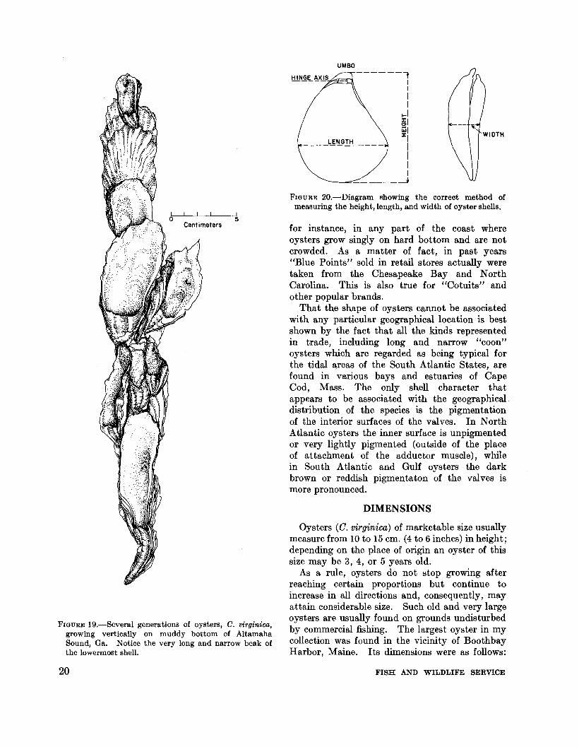
The pointed end of the valve or the beak represents the oldest part of a shell. In old individualsit reaches considerable size (fig. 17). The beaksare usually curved and directed toward theposterior end of the mollusk although in somespecimens they may point toward the anterior.In the majority of bivalves other than oystersthe beaks usually point forward. The directionand degree of curvature of the beaks of oysters aswell as their relative proportions vary greatlyas can be seen in figure 18, which representsdifferent shapes found in old shells of C. virginica.Very narrow, straight, or slightly curved beaks ofthe kind shown in figure 18-1 are usually formedin oysters which grow on soft, muddy bottoms.Extreme development of this type can be seen inthe narrow and slender oysters growing underovercrowded conditions on reefs (fig. 19). Otherforms of beaks (fig. 18, 2-4) cannot be associated with any particular environment. In fully
FISHERY BULLETIN: VOLUME 64, CHAPTER II
oCent imeters
5
FIGURE I5.-Blue Point oyster (C. virginica) from Great South Bay, Long Island, N.Y. The size of this 5-year-oldoyster is about 10 x 6.6 em. (4 x 3 inches). The shell is strong and rounded; its surface is moderately sculptured.Left-outside surface of left valve. Right-inner surface of right valve. Small encircled area under the hinge onthe inner surface of right valve is an imprint of Quenstedt's muscle.
FIGURE I6.-Cross section below the hinge of an adult C.virginica. Left valve at bottom, right valve at top ofthe drawing. The buttress of the right valve fits thedepression on the left valve. The two valves are connected by a ligament (narrow band indicated by verticalstriations) which consists of a central part (resilium)and two outer portions. Slightly magnified. r.v.right valve; bu.-buttress; de.-depression or furrow onleft valve (I.v.); lig.-ligament.
bu.
de.-..........,,-'<-1
6Centimeters
r.v.
f+-#;+-- Ii g.
I.v.
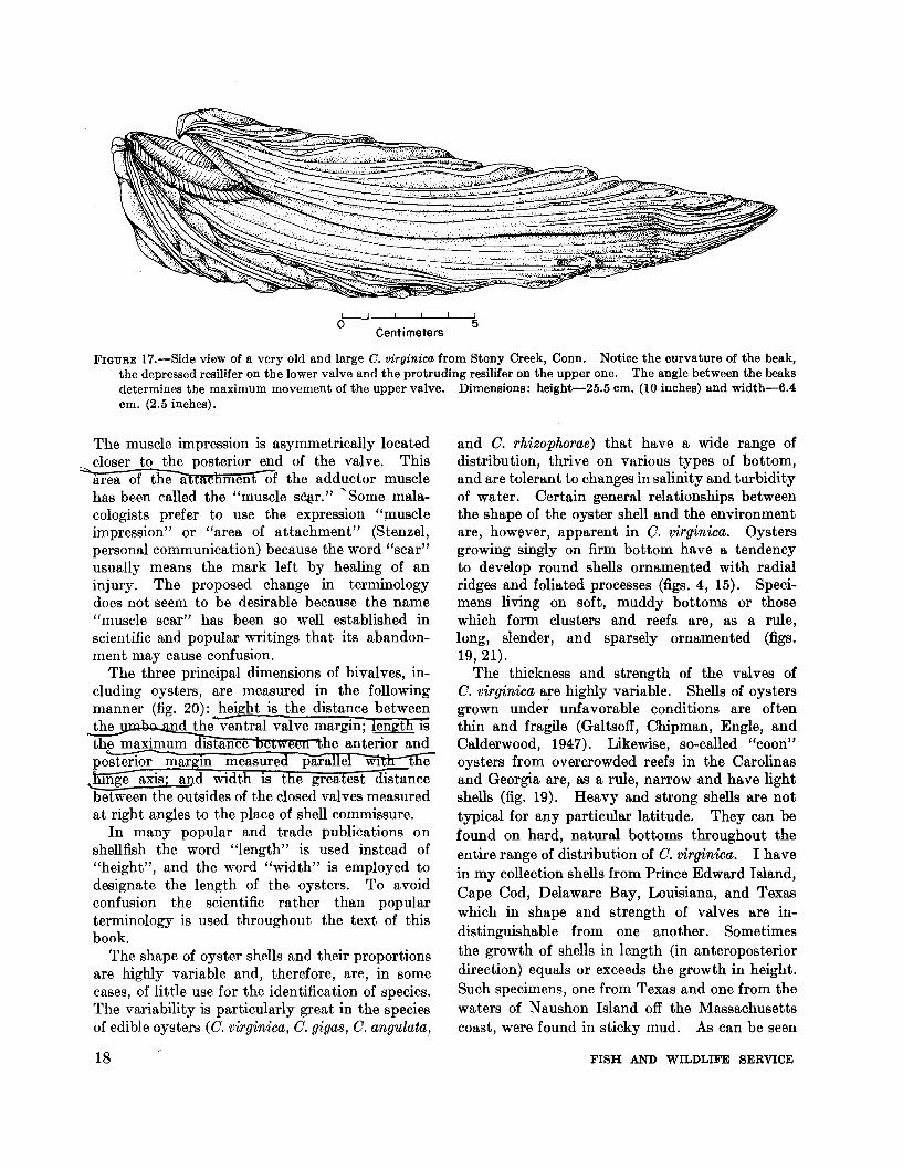
grown O. virgmtca the pointed end of the upper(flat) valve is always shorter than that of itsopposite member (fig. 17). The angle betweenthe two beaks determines the greatest extent towhich the valves can open for feeding or respiration and is, therefore, of significance to the oyster.
If the oyster shell is oriented in such a way tha tboth of its valves are visible and the beaks pointup and toward the observer, the flat valve with ashorter, convex resilifer is the right one and thecuplike valve with the longer concave resiliferis the left one. The dorsal margin of the oysteris the beak or hinge side, the ventra1 fIlargtn tlieopposite. Ii viewed from 'the right (flat) vaivewitn ttie hinge 'end pointing" away from the observer the anterior end of the oyster is at theright side of the valves and the posterior is at theleft.
The posterior and anterior parts of the oystershell may also be identified by the position of themuscle impressIOn, an oval-shaped and highlypigmented area markmg the attachment of theadductor muscle on the inner side of each valve.
MORPHOLOGY AND STRUCTURE OF SHELL 17
oCentimeters
5
FIGURE 17.-Side view of a very old and large C. virginica from Stony Creek, Conn. Notice the curvature of the beak,the depressed resilifer on the lower valve and the protruding resilifer on the upper one. The angle between the beaksdetermines the maximum movement of the upper valve. Dimensions: height-25.5 em. (10 inches) and width-6.4em. (2.5 inches).
The muscle impression is asymmetrically locatedcloser to the posterior end of the valve. This
;;:"'area of theattactlmentOf the adductor musclehas been called the "muscle s~r." ''Some malacologists prefer to use the expression "muscleimpression" or "area of attachment" (Stenzel,personal communication) because the word "scar"usually means the mark left by healing of aninjury. The proposed change in terminologydoes not seem to be desirable because the name"muscle scar" has been so well established inscientific and popular writings that its abandonment may cause confusion.
The three principal dimensions of bivalves, including oysters, are measured in the followingmanner (fig. 20): hei ht is the distance betweenthe d the ventral valve margin;~ IS
the maximum distance he anterior andposterior margm measured para el WI e~axis; and width is the greatest distancebetween the outsides of the closed valves measuredat right angles to the place of shell commissure.
In many popular and trade publications onshellfish the word "length" is used instead of"height", and the word "width" is employed todesignate the length of the oysters. To avoidconfusion the scientific rather than popularterminology is used throughout the text of thisbook.
The shape of oyster shells and their proportionsare highly variable and, therefore, are, in somecases, of little use for the identification of species.The variability is particularly great in the speciesof edible oysters (G. virginica, G. gigas, G. angulata,
18
and G. rhizophorae) that have a wide range ofdistribution, thrive on various types of bottom,and are tolerant to changes in salinity and turbidityof water. Certain general relationships betweenthe shape of the oyster shell and the environmentare, however, apparent in G. virginica. Oystersgrowing singly on firm bottom have a tendencyto develop round shells ornamented with radialridges and foliated processes (figs. 4, 15). Specimens living on soft, muddy bottoms or thosewhich form clusters and reefs are, as a rule,long, slender, and sparsely ornamented (figs.19, 21).
The thickness and strength of the valves ofG. virginica are highly variable. Shells of oystersgrown under unfavorable conditions are oftenthin and fragile (Galtsoff, Chipman, Engle, andCalderwood, 1947). Likewise, so-called "coon"oysters from overcrowded reefs in the Carolinasand Georgia are, as a rule, narrow and have lightshells (fig. 19). Heavy and strong shells are nottypical for any particular latitude. They can befound on hard, natural bottoms throughout theentire range of distribution of G. virginica. I havein my collection shells from Prince Edward Island,Cape Cod, Delaware Bay, Louisiana, and Texaswhich in shape and strength of valves are indistinguishable from one another. Sometimesthe growth of shells in length (in anteroposteriordirection) equals or exceeds the growth in height.Such specimens, one from Texas and one from thewaters of Naushon Island off the Massachusettscoast, were found in sticky mud. As can be seen
FISH AND WILDLIFE SERVICE

2
3
o Centi meters 34
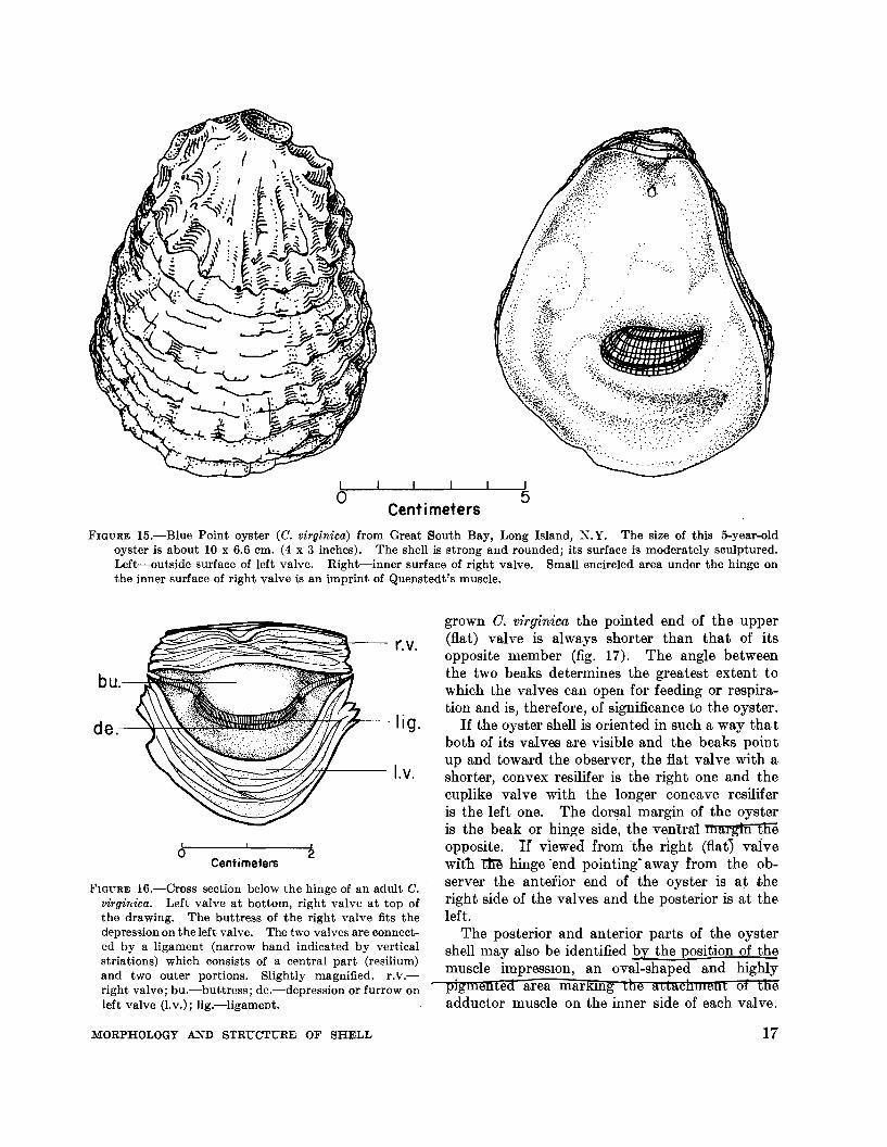
FIGURE 18.-Four shapes of beaks on left valves of old oysters, C. virginica. I-narrow, short and almost straight;2-strongly curved to the posterior; 3--of medium width, pointed forward; 4-very broad and slightly curved to theposterior.
from figure 22, the shells are almost identical inshape and size.
Oysters are frequently marketed under specificbrands or trade names such as Blue Points (fig.15), Cotuits, Chincoteagues, and others which
MORPHOLOGY AND STRUCTURE OF SHELL
imply the existence of local varieties differentin size and shape of shells. There is no evidence,however, to substantiate this claim. So-called"Blue Points" characterized by round shape,strong shell, and medium size may be found,
19
UMBO------'1
II1
1
1-1i§1iii I:1:
1
IIII
---~- --.J
LENGTH------------
FIGURE 20.-Diagram showing the correct method ofmeasuring the height, length, and width of oyster shells.
for instance, in any part of the coast whereoysters grow singly on hard bottom and are notcrowded. As a matter of fact, in past years"Blue Points" sold in retail stores actually weretaken from the Chesapeake Bay and NorthCarolina. This is also true for "Cotuits" andother popular brands.
That the shape of oysters cannot be associatedwith any particular geographical location is bestshown by the fact that all the kinds representedin trade, including long and narrow "coon"oysters which are regarded as being typical forthe tidal /lrreas of the South Atlantic States, arefound in various bays and estuaries of CapeCod, Mass. The only shell character thatappears to be associated with the geographicaldistribution of the species is the pigmentationof the interior surfaces of the valves. In NorthAtlantic oysters the inner surface is unpigmentedor very lightly pigmented (outside of the placeof attachment of the adductor muscle), whilein South Atlantic and Gulf oysters the darkbrown or reddish pigmentaton of the valves ismore pronounced.
DIMENSIONS
Oysters (0. virginica) of marketable size usuallymeasure from 10 to 15 em. (4 to 6 inches) in height;depending on the place of origin an oyster of thissize may be 3, 4, or 5 years old.
As a rule, oysters do not stop growing afterreaching certain proportions but continue toincrease in all directions and, consequently, mayattain considerable size. Such old and very largeoysters are usually found on grounds undisturbedby commercial fishing. The largest oyster in mycollection was found in the vicinity of BoothbayHarbor, Maine. Its dimensions were as follows:
5
FIGURE 19.-Several generations of oysters, C. virginica,growing vertically on muddy bottom of AltamahaSound, Ga. Notice the very long and narrow beak ofthe lowermost shell.
20 FISH AND WILDLIFE SERVICE

Centimeters
FIGURE 21.-Shells of C. giga8 (left) and C. virginica (right) grown on soft, muddy bottom. Note the remarkable similarity in the shape, size, and sculpture of the two species of oysters. The C. giga8 was obtained from the northernpart of Puget Sound and the C. virginica from Georgia. The shells of the two species can be distinguished by theabsence of pigmentation of the muscle impression in C. giga8 and by its lighter shell material.
height-20.6 em. (8.1 inches); height of left andright beak-5.5 em. (2.1 inches) and 4.5 em.(1.75 inches) respectively; length of shell-9.7em. (3.8 inches); maximum width (near thehinge)-6.5 em. (2.6 inches). The total weightwas 1,230 g., the shell weighing 1,175 g., the meat35.8 g., and the balance of 19.2 g., representingthe weight of sea water retained between thevalves. Apparently the largest oyster recordedin American literature is the giant specimen fromthe Damariscotta River, Maine, reproduced innatural size by Ingersoll (1881, pI. 30, p. 32).This shell is 35.5 em. (14.3 inches) in height and11 cm. (about 4.4 inches) in length.
SHAPE OF SHELLS
The shells of many gastropods and bivalves arespiral structures in which the convolutions of thesuccessive whorls follow a definite pattern. Thespiral plan is frequently accentuated by ridges,
MORPHOLOGY AND STRUCTURE OF SHELL
furrows, spines and nodules, or by pigmentedspots which repeat themselves with remarkableregularity. A spiral structure is not restricted tomollusk shells. As a matter of fact, it is very common throughout the animal and plant kingdom aswell as in architecture and art. Examples of agreat variety of spirally built organisms andstructures are given in the beautifully illustratedbooks entitled "Spirals in nature and art" and"Curves of life" (Cook 1903, 1914). As the titleof the second book implies, Cook is inclined toattach some profound significance to the kindof curves found in animal and plant forms. Thisview, inherited from the philosophers of the 18thand 19th centuries, considers the spiral organicstructures as a manifestation of life itself. Theinfluence of this philosophy persisted among somescientists until the thirties of the present century.It can be found, for instance, as late as 1930 in thewritings of a French physiologist, Latrigue (1930)
21

Centi meters
FIGURE 22.-Two left shells of C. virginica grown on sticky mud. On the left side is the oyster from Karankawa Reef inMatagorda Bay, Tex.; on the right is the oyster from Hadley Harbor, Naushon Island, near Woods Hole, Mass.The dimensions of the Texas oyster are 13 by 11.5 cm. (5.1 by 4.5 inches) and for the Hadley Harbor oyster 15.5 by14.5 cm. (6.1 by 5.7 inches).
who in the book, "Biodynamique generale," attributes mysterious and not well-defined meaningto the "stereodynamics of vital vortex." Thesespeculations contributed nothing to the understanding of the processes which underlie the formation of shells and other organic structures.
In the earlier days of science the geometricregularity of shells, particularly that of gastropods,had been a favored object for mathematicalstudies. Properties of curves represented by thecontours of shells, as well as those seen in horns,in flower petals, in the patterns of distribution ofbranches of trees, and in similar objects, werecarefully analyzed. An excellent review of thischapter of the history of science is given in a wellknown book "On growth and form" (Thompson,1942) in which the reader interested in mathematics and its application to the analysis of organicforms will find many stimulating ideas.
Among the array of curves known in mathematics, the kind most frequently encountered inthe shells of mollusks is the logarithmic or equiangular spiral (fig. 23). The latter name refers toone of its fundamental characteristics, describedby Descartes, namely, that the angle between
22
tangent PG (fig. 23) and radius vector OP is constant. Another property of this curve which maybe of interest to biologists is the fact that distancesalong the curve intercepted by any radius vectorare proportional to the length of these radii.D'Arcy Thompson showed that it is possible toapply the mathematical characteristics of curvesto the interpretation of the growth of those shellswhich follow the pattern of a logarithmic spiral.According to his point of view, growth along thespiral contour is considered as a force acting at anypoint P (fig. 23) which may be resolved into twocomponents PF and PK acting in directions perpendicular to each other. If the rates of growthdo not change, the angle the resultant force, i.e.,the tangent PG, makes with the radius vector remains constant. This is the fundamental propertyof the "equiangular" (logarithmic) spiral. Theidea forms the basis of Huxley's (1932) hypothesisof the interaction of two differential growth ratiosin the bivalve shells and also underlies Owen's(1953) concept of the role of the growth components determining the shape of the valves.
Another important characteristic of the growthof bivalves pointed out by Thompson is that
FISH AND WILDLIFE SERVICE

FIGURE 23.-Logarithmic or equiangular spiral. Explanation in text.
increase in size is not accompanied by any changein shape of the shell; the proportions of the latterremain constant, and the shell increases only insize (gnomonic growth). This general rule holdstrue for many free-moving gastropods and bivalves. It is not, however, applicable to sessileforms like oysters, in which the shape of the shellchanges somewhat with size, particularly at theearly stages of growth, and is greatly modifiedby contact with the substratum upon which themollusks rest. The plasticity and variability ofattached forms are probably associated with theirinability to escape the effects of proximateenvironment.
The contour of oyster shell may be either circular(young C. virginica, O. edulis) or elongated andirregular. Spiral curvature may be noticed,however, on a cross section of the lower (concave)valve cut along its height perpendicular to thehinge. The curve can be reproduced by coveringthe cut surface with ink or paint and stampingit on paper. The upper valve is either flat orconvex.
The curvature of bivalve shells is sometimescalled conchoid. The term may be found in
o
FIGURE 24.-Construction of the conchoid curve ofNicomedes. Explanation in text.
general and popular books dealing with bivalveshells, but the author who introduced it in scientific literature could not be traced. The Greekword "conchoid", derived from "conch"--shelland "eidos"-resembling or similar to, impliesthe similarity of the curve to the contour of amolluscan shell.
The curve is symmetric with respect to the 90 0
polar axis (fig. 24). It consists of two branches,one on each side of the fixed horizontal line CDto which the branches approach asymptoticallyas the curve extends to infinity. The curve,known as conchoid of Nicomedes, is constructed bydrawing a line through the series of points P andPI which can be found in the following way: fromthe pole 0 draw a line OP which intersects thefixed line CD at any point Q. Layoff segmentsQP=QP1=b along the radius vector OP. Repeatthe process along the radii originating from the poleo and draw the two branches of the curve byjoining the points. The curve has three distinctforms depending on whether "a" (a distance OQfrom the pole to the point of intersection of thepolar axis with the fixed line CD) is greater, equalto or less than b. The formula of the curve ifb<a, is r=a sec o±b, where r is the locus of theequation and sec 0 is secant of the vectorial angle o.
Sporn (1926) made a detailed mathematicalanalysis of the conchoid curve and consideredthat the curvatures of bivalve shells conform tothis geometrical type. Lison (1942) rejected thisconclusion as not supported by observations andexperimental evidence. He quite correctly statedthat Sporn's work deals exclusively with abstractmathematical analyses of curves which in realityare not those found in molluscan shells. If onecuts a bivalve shell at any angle to the plane ofclosure of the valves, one obtains the curvedlines of the two valves (fig. 25) which only remotelyresemble the conchoid of Nicomedes and touch
GF
MORPHOLOGY AND STRUCTURE OF SHELL 23
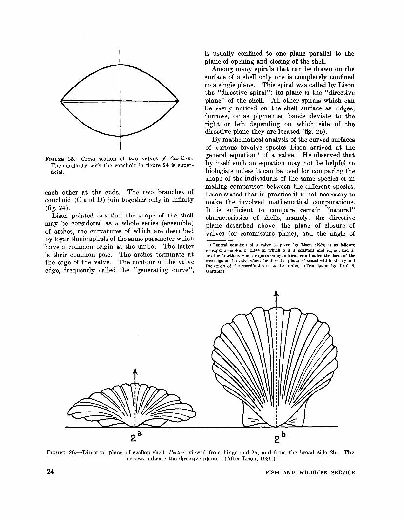
FIGURE 25.-Cross section of two valves of Cardium.The similarity with the conchoid in figure 24 is superficial.
each other at the ends. The two branches ofconchoid (0 and D) join together only in infinity(fig. 24).
Lison pointed out that the shape of the shellmay be considered as a whole series (ensemble)of arches, the curvatures of which are describedby logarithmic spirals of the same parameter whichhave a common origin at the umbo. The latteris their common pole. The arches terminate atthe edge of the valve. The contour of the valveedge, frequently called the "generating curve",
is usually confined to one plane parallel to theplane of opening and closing of the shell.
Among many spirals that can be drawn on thesurface of a shell only one is completely confinedto a single plane. This spiral was called by Lisonthe "directive spiral"; its plane is the "directiveplane" of the shell. All other spirals which canbe easily noticed on the shell surface as ridges,furrows, or as pigmented bands deviate to theright or left depending on which side of thedirective plane they are located (fig. 26).
By mathematical analysis of the curved surfacesof various bivalve species Lison arrived at thegeneral equation 3 of a valve. He observed thatby itself such an equation may not be helpful tobiologists unless it can be used for comparing theshape of the individuals of the same species or inmaking comparison between the different species.Lison stated that in practice it is not necessary tomake the involved mathematical computations.It is sufficient to compare certain "natural"characteristics of shells, namely, the directiveplane described above, the plane of closure ofvalves (or commissure plane), and the angle of
3 General equation of a valve as given by Lison (1939) is as follows:d=O'oPX; oo-=wo+a; z=zoe plto in which p is a constant and UOt "'0, and Zoare the functions which express on cylindrical coordinates the form of thefree edge of the valve when the directive plane is located within the xy andthe origin of the coordinates is at the umbo. (Translation by Paul S.Galtsoff.)
FIGURE 26.-Directive plane of scallop shell, Pecten, viewed from hinge end 2a, and from the broad side 2b. Thearrows indicate the directive plane. (After Lison, 1939.)
24 FISH AND WILDLIFE SERVICE

incidence. The plane of closure of the valvesoriginates at the umbo and passes between theedges of the two opposing valves when they areclosed and touching each other. The angle ofincidence, as defined by Lison, is the angle betweenthe plane of closure and the directive plane. Inround and symmetrical shells of scallops, pearloysters, and other bivalves the directive plane isperpendicular to the plane of closure and theangle of incidence is 90 0 (fig. 26). In the shellsof Cardium orbita, the directive plane forms anacute angle of 81 0 and is much smaller in elongatedshells such as Fimbria jimbriata and Trapeziumoblongum. The comparison between the shellscan easily be made by recording the contours atthe free margins of the valves and determiningthe angle of incidence.
To determine the shape of logarithmic spiral ofthe valve the shell may be sawed along the directive plane (fig. 27) and the section oriented withthe umbo 0 at lower left. If 81 and 82 are respective lengths of the two radii the value of parameter p can be computed by using the fundamentalequation of logarithmic spiral,
p=log. 81-1og. 82
w
(logarithms in this equation are natural, to base e).
In resume, Lison attempted to prove that theform of the shell in which the generating curve isconfined to one plane is determined by threeconditions: (1) the angle of the directive spiral,(2) the angle of incidence, and (3) the outline ofthe generating curve.
Further attention to the problem of the shapeand formation of the bivalve shell was given byOwen (1953). In general he accepted Lison'sconclusions and stated that "the form of the valvesshould be considered with reference to: (a) theoutline of the generative curve, (b) the spiral angleof the normal axis, and (c) the form (i.e., planispiral or turbinate-spiral) of the normal axis."The normal axis is considered by Owen withreference to: (1) the umbo, (2) the margin of themantle edge, and (3) the point at which the greatest transverse diameter of the shell intersects thesurface of the valves. Thus, it can be seen fromthis statement that Owen's "normal axis" doesnot coincide with Lison's directive plane exceptin bilaterally symmetrical valves (fig. 28). According to Owen's view, the direction of growthat any region of the valve margin is the result ofthe combined effect of three different components:(a) a radial component radiating from the umboand acting in the plane of the generating curve,(b) a transverse component acting at right anglesto the plane of the generating curve, and (c) atangential component acting in the plane of thegenerating curve and tangentially to it. Theturbinate-spiral form of some bivalve shells is dueto the presence of the tangential component whichin plani-spiral shells may be absent or inconspicuous. Likewise, the transverse component may begreatly reduced or even absent in the valve.Thus, from this point of view the great variety ofshell forms may be explained as an interaction ofthe three components (fig. 29). Owen's point of
catlte~ior
~directive ~ane
no~maI4)(is --.__ .~""'-
I
~
FIGURE 27.-Valve of a shell sawed along the directive axisdescribes a plane logarithmic spiral. According toLison (1942). OM-radius vector; T-tangent;O-umbo; V-angle between the two radii.
MORPHOLOGY AND STRUCTURE OF SHELL
FIGURE 28.-Comparison of directive plane of Lison withnormal axis (Owen). A-shell not affected by tangential component; B-shell affected by tangential component.
25

be found by experimental and biochemical studieswhich may supply biological meanings to abstractmathematical concepts and equations. Experimental study of the morphogenesis of shellsoffers splendid opportunities for this type ofresearch.
8 1
o
A
8
GROWTH RINGS AND GROWTH RADII
Nearly 250 years ago Reaumur (1709) discoveredthat shells grow by the accretion of materialsecreted at their edges. Since that time thisimportant observation has been confirmed bynumerous subsequent investigations. The ringson the outer surfaces of a bivalve shell, frequentlybut incorrectly described as "concentric", represent the contours of the shell at different ages.Rings are common to all bivalves but are particularly pronounced on the flattened shells ofscallops, clams, and fresh-water mussels. Depending on the shape of the shell, the rings areeither circular or oval with a common point oforigin at the extreme dorsal side near the umbo(figs. 30 and 31). The diagrams clearly showthat the rate of growth along the edge of theshell is not uniform. It is greater along theradius, AD, which corresponds to the directiveaxis of Lison, and gradually decreases on both
lL
5
~~~~r-"ormal axis
norma.l zonep
------~y
y ~R
view is basically similar to Huxley's hypothesis(1932) of differential length growth and widthgrowth of molluscan shells. Owen correctlypoints out an error in Lison's interpretations thatthe lines of equal potential activities involved inthe secreting of shell material at the edges of thevalves are parallel to each other. This is obviously not the case since all lines of growth of thelamellibranch shell radiate from a common nodeof minimum growth near the umbo. For thisreason the comparison of bivalves can be moreconveniently made by using radial coordinates ashas been shown by Yonge (1952a, 1952b).
The mathematical properties of shell surfacesare of interest to the biologist because they mayprovide clues to understanding the quantitativeaspects of the processes of shell formation. It canbe a priori accepted that any organism grows inan orderly fashion following a definite pattern.The origin of this pattern and the nature of theforces responsible for laying out structural materials in accordance with the predeterminedplan are not known. The pattern of shellstructure is determined by the activities at theedge of the shell-forming organ, the mantle. Atthe present state of our knowledge it is impossibleto associate various geometrical terms whichdescribe the shape of the shell with concretephysiological processes and to visualize themorphogenetic and biochemical mechanisms involved in the formation of definite sculptural andcolor patterns. The solution of this problem will
FIGURE 29.-Normal axis and the two growth componentsin the shell of scallop. LS-plane perpendicular to theplane of the generating curve; N-turning point of thellonllave side of the shell shown at right; M and O-auxiliary radii; P-transverse component; R-radial component; UY-normal axis. From Owen (1953).
FIGURE 30.-Diagram of a circular bivalve shell of thekind represented in Pecten, Anomia, and young C.virginica. Radii extending from the umbo to theperiphery of the generating curve are proportional tothe rate of growth at the edge of a circular shellRadius AD corresponds to the directive axis of Lison.
26 FISH AND WILDLIFE SERVICE

B'
FIGURE 31.-Diagram of a shell of adult C. mrgtmca.Radii extend from the umbo to the periphery of thegenerating curve. The principal axis AGF shows thechange in the direction of growth at G. The length ofradii is proportional to the rate of shell growth at theedge.
sides of it along growth radii AC, AB, and ACI ,
AB I .
Circular shells in O. virginica may be found onlyin very young oysters (fig. 32a). Within a fewweeks after setting the shell becomes elliptical,and as elongation (increase in height) continuesthe principal vector of growth shifts to one side(fig. 32b).
A series of curves noticeable on round shells(fig. 32) clearly illustrate the differential rate ofgrowth along the periphery of the valve, whichincreases in size without altering in configuration.Thompson (1942) found an interesting analogybetween this type of growth, radiating from asingle focal point (the umbo), and the theoremof Galileo. Imagine that we have a series ofplanes or gutters originating from a single pointA (fig. 30) and sloping down in a vertical plane
MORPHOLOGY AND STRUCTURE OF SHELL
at various angles along the radii AB, AC, ACI ,
and AB I which end at the periphery of a circle.Balls placed one in each gutter and simultaneouslyreleased will roll down along the vectors B, B I , C,Cr, and D. If there is no friction or other formof resistance, all the balls will reach the peripheryat the same time as the ball dropping verticallyalong AD. The acceleration along any of thevectors, for instance, AB, is found from theformula t2=2/g AD where t IS time and g isacceleration of gravity.
A similar law, involving a more complexformula, applies to cases in which the generatingcurve is nearly elliptical, for instance, in theshells of adult oysters. The rate of growth atdifferent sectors of the periphery of the shellobviously has nothing to do with the accelerationof gravity, but the similarity between the lengthof the radii which represent the rate of growthalong a given direction of the shell and the acceleration along the vectors in the theorem of Galileois striking. It appears reasonable to expect thatthe Galileo formula may be applicable to thephysiological process taking place near the"edgeof the valve. One may assume, for instance, thatthe rate of physiological activities is affected bythe concentration of growth promoting substances or by enzymes involved in the calcificationof the shell and that these factors vary at differentpoints of the mantle edge in conformity withGalileo's formula. Experimental exploration ofthe possibilities suggested by mathematical parallelism may be, therefore, profitable in finding thesolution to the mystery of the formation of shellpatterns.
CHANGES IN THE DIRECTION OFPRINCIPAL AXES OF SHELL
The principal axes of shells of O. virginica arenot as permanent as they are in clams, scallops,and other bivalves in which the shape of thevalves remains fairly constant and is less affectedby environment than in the oyster. The plasticityof oysters of the species Orassostrea is so greatthat their shape cannot be determined geometrically (Lison, 1949). This inability to maintaina definite shape is probably the result of thesedentary living associated with complete lossof the power of locomotion.
In some species of oysters the shells are circularor nearly circular. In such cases the ratio ofthe height of the valve to its length is equal to
27

a
o 0.5Centimeters
b
1.0
FIGURE 32.-Two small C. virginica growing attached to tar paper. Maximum dimension of shell: a-D.85 em.;b-l.D em. At b the principal axis curves to the left.
1.0, as, for instance, in O. rivularis (fig. 8) andO. (Alectryonia) megodon Hanley (fig. 3) (Olsson,1961). Oysters of the latter species from thePacific Coast of Central and South America growsingly, in vertical position, cemented to the rocksby their left valves. The specimens I collectedon Pearl Islands, Gulf of Panama, measured 17to 18 em. in height and 16 to 17 em. in length.The European flat oyster, O. edulis (fig. 9) usuallyforms rounded shells in which the length exceedsthe height. Small, noncommercial species, O.sandwichensis of the Hawaiian Islands and O.mexicana from the Gulf of Panama, are almostcircular with the tendency to extend in lengthrather than in height. Crowded conditions underwhich these species thrive attached to rocks in anarrow tidal zone greatly obscure and distort theshape of their shells.
Small O. virginica growing singly on flat surfaceswithout touching each other are usually round(fig. 32). In a random sample consisting of 100single small oysters (spat about 6 weeks old)varying from 5 to 15 mm. in height and growingon tar paper, the height/length ratio varied from0.6 to 1.2. Nearly half of them (49 percent) wereperfectly round (HjL ratio=l); in 30 percent theratio was less than 1; and in 21 percent the lengthexceeded the height.
28
In small single oysters less than 10 mm. inheight the principal (normal) axis of growth isclearly marked. All other radii symmetricallyoriented on both sides of the principal axis areindicated by the pigmented bands on the surfaceof the shell. The newly deposited shell, discernible at the periphery of the oyster, forms aband which is wider at the ventral edge of theshell and slightly narrows anteriorly and posteriorly (fig. 32a). With the growth of the oysterits principal axis is shifted to the side, curves, andis no longer confined to one plane. The curvatureof the valve becomes a turbinate-spiral. Gradually the oyster becomes slightly oval-shapedand asymmetrical.
The change in the direction of the principalaxis of growth is not associated with the environment since it takes place only in some of theoysters growing under identical conditions. Occasionally oysters are formed in which the pigmentation along the principal axis is so pronouncedthat the dark band which marks its position maybe' mistaken for an artifact (fig. 33) while thesecondary axes are not visible. The shells ofadult O. virginica usually curve slightly to theleft (if the oyster is placed on its left valve andviewed from above). Frequently, however, inverted specimens are found in which the growth
FISH AND WILDLIFE SERVICE

FIGURE 33.-Principal axis of growth of a C. virginicafrom Chatham, Mass., is deeply marked by a pigmentedband.
has shifted into the opposite direction (fig. 34).The "normal" oyster (the right side of the figure)is curved to the left while in the inverted specimen,shown on the left of the figure, the shell curvesto the right. Such "right-handed" oysters areprobably common in all oyster populations sincethey were found in Texas, Chesapeake Bay,Narragansett Bay, and Great Bay, N.H. Inevery other respect the inverted specimens arenormal and had typically cupped left valves withwell-developed grooved beaks. There is no evidence that inversion was caused by mechanicalobstruction or some unusual position on thebottom.
Complete inversion in bivalves was describedby Lamy (1917) for Lucina, Chama, and severalspecies of the subgenus Goodallia (fa,m. Astartidae).It consisted in the appearance of structures, typicalfor the right valve, on the left valve and viceversa. In the case of C. virginica the structuralelements remain unaffected and the inversion islimited to the contours of the valves.
oCent imeters
5
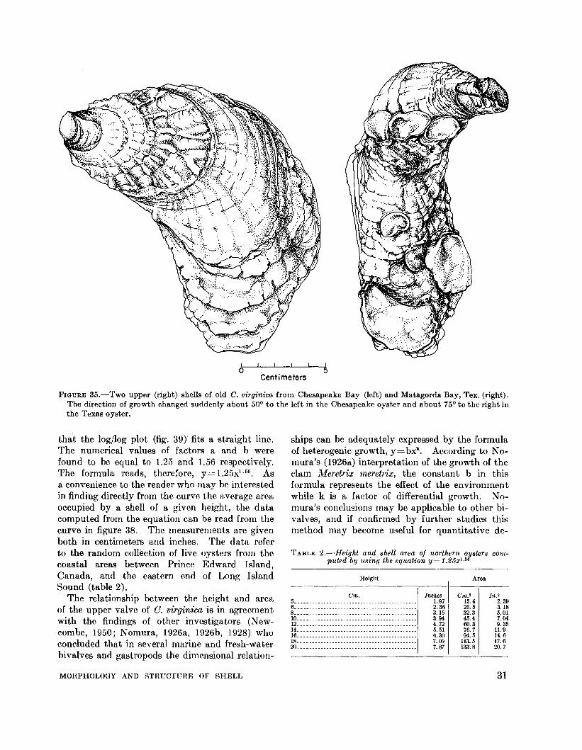
The once established principal axis of growthdoes not always remain unchanged. Occasionallyold oysters are found in which the direction ofgrowth had undergone sudden changes of about90 0
• The change shown in figure 35 took placewhen both oysters were about 6 to 7 years old.
The instability of the principal axis of growthmay be even more pronounced. My collectionhas an oyster (C. virg~nica) found on the banks ofa lagoon near Galveston, Tex., in which the principal axis, clearly indicated by pigmented bands onthe surface of the valves, changed its direction atthe end of each growing period. The resultingzigzag line is clearly visible in the specimen (fig.36).
DIMENSIONAL RELATIONSHIPS OFSHELL
Shape of a bivalve shell is often expressed asa ratio between its height and length or by someother numerical index. Lison (1942) pointed outthat the shape of an oyster shell cannot be expressed in precise geometrical terms, presumablybecause of its great variability. The "index ofshape" determined as a ratio of the sum of heightand width of a shell to its length was used byCrozier (1914) in studying the shells of a clam,Dosinia discus. For the mollusks ranging from 2to 7 cm. in length collected near Beaufort, N. C.this index varied from 1.24 to 1.28 indicating thatthe increase of the species in height and width wasdirectly proportional to the increase in length.Such regularity is not found in the shells of adultC. mrginica taken at random from commerciallyexploited bottoms. For the entire range ofdistribution of this species in the Atlantic andGulf states the index of shape varied from 0.5 to1.3. The histogram (columns in figure 37) showsnearly normal frequency distribution with thepeak of frequencies at O.9. No significant differences were found in the index of shape in thenorthern and southern populations of oystersexamined separately. The boundary between thetwo groups was arbitrarily drawn at the VirginiaNorth Carolina line. The two curves connectingthe frequency points on figure 37 indicate that inthe southern population the index of shape extends from 0.5 to 1.3, while in the northern oystersit varies from 0.6 to 1.2. The difference is probablynot very significant, but it may be due to a greaterpercentage of wild oysters on commercially exploited natural bottoms of the southern states.
MORPHOLOGY AND STRUCTURE OF SHELL733-851 0-64-3
29

· :~..~/ .
~ .....
aCentimeters
FIGURE 34.-Left valves of the two large C. virginica from Narragansett Bay, R.I. On the right is a "normal" oyster;its shell curves to the left. On the left side is an inverted oyster; its shell curves to the right.
Most of the oysters from the North Atlantic andChesapeake states were taken from bottoms onwhich oysters are regularly planted for cultivation.There are no significant differences in the mean,mode, and median of the two groups (table 1).Contrary to the conditions found by Crozier inDosinia discus, the "index of shape" of C. virginicais highly variable.
SHELL AREA
I nformation regarding the approximate areaof an oyster shell of known height may be usefulto oyster growers who want to determine in advance what percentage of the bottom area set
TABLE I.-Index of shape (height+width) of oysters taken bylength
commercial fishery
Locality Mean Standard Mode Mediandeviation
.------------Northern grounds...__ ........... 0.87 0.05 0.94 0.09Southern grounds________________ 0.87 0.02 0.94 0.9
30
aside for planting will be covered by oysters ofknown size. Since the oystermen usually knowthe number of oysters of various sizes needed tomake up a bushel, the information given belowmay be used in determining in advance whetherthe area of the bottom is sufficient to provide spacefor their additional growth.
It is self-evident that the area of the valve increases proportionally to the increase in its lineardimensions. For determining the area a piece ofthin paper was pressed against the inner surfaceof the right (flat) valve and the outlines weredrawn with pencil The area wRS mefiSiired withII; planimeter. The<:'lutljnes of small shells were
~laced over;~h paper and~e number of milli-_~r squares coJ!!!..ted..:..-
. The relationship between the height and shellarea (fig. 38) is represented by an exponentialcurve of a general type y= axb which fits manyempirical data. The y in the formula is the shellarea, and the x is the height. The parabolicnature of the curve is demonstrated by the fact
FISH AND WILDLIFE SERVICE
oCent imeters
5
FIGURE 35.-Two upper (right) shells of old C. virginica from Chesapeake Bay (left) and Matagorda Bay, Tex. (right).The direction of growth changed suddenly about 50° to the left in the Chesapeake oyster and about 75° to the right inthe Texas oyster.
TABLE '2.-Height and shell area oj northern oyster.5 computed by usin(/ the equation y = 1.25x I..l6
ships can be adequately expressed by the formulaof heterogenic growth, y=bxk
• According to Nomura's (1926a) interpretation of the growth of theclam Meretrix meretrix, the constant b in thisformula represents the effect of the environmentwhile k is a factor of differential growth. Nomura's conclusions may be applicable to other bivalves, and if confirmed by further studies thismethod may become useful for quantitative de-
that the log/log plot (fig. 39r fits a straight line.The numerical values of factors a and b werefound to be equal to 1.25 and 1.56 respectively.The formula reads, there;ore, y= 1.25x1. 56 • Asa convenience to the reader who may be interestedin finding directly from the curve the average areaoccupied by a shell of a given height, the datacomputed from the equation can be read from thecurve in figure 38. The measurements are givenboth in centimeters and inches. The data referto the random collection of live oysters from thecoastal areas between Prince Edward Island,Canada, and the eastern end of Long IslandSound (table 2).
The relationship between the height and areaof the upper valve of G. virginica is in agreementwith the findings of other investigators (Newcombe, 1950; Nomura, 1926a, 1926b, 1928) whoconcluded that in several marine and fresh-waterbivalves and gastropods the dimensional relation-
MORPHOLOGY AND RTRUCTURF. OF SHELL
Height
Crn.5 . . .6 • . _8....• _10 _12 _14. . .. _16 _18 . _20 _
Inches1.972.363.153.944.725.516.307.097.87
Area
Cm. 2
15.420.532.345.400.376.794.5
113.5133.8
In.'2.393.185.017.049.35
11.914.617.620.7
31

terminations of the effect of local conditions ongrowth and shape of shells.
CHALKY DEPOSITS
FIGURE 36.-Shell of an adult C. virginica showing periodicchanges in the direction of the principal axis of growth.Note the zigzag line of pigmented bands in the middle ofthe valve. Actual dimensions: 8.5 by 6 em. (3.25 by 2.5inches).
------ S. ATLANTICa GULF
~- -0 N. ATLANTIC50
Ul...J 40...JW:I:Ul
u.300
a::wCD:!:::::> 20z
10
0
FIGURE 37.-Histogram of the distribution of the index ofshape (height+width) of shells of C. virginica from the
lengthAtlantic Coast. Frequency distribution of the index ofNorth Atlantic oysters (open circles) and South Atlanticoysters (points) are shown by two separate curves.
0.5 . 0.6 0.7 0.8 0.9 1.0 1.1 1.2 1.3
INDEX OF SHAPE OF C. virginica
of this explanation was presented by the authorsor by Ranson (1939-41), who fully accepted thetheory without making additional studies andstated positively that chalky deposits are formedwherever there is a local detachment of the mantlefrom the valve.
Considering the possibility that the mantle maybe more easily detached from the valve if theoyster is placed with its lower (cuplike) side uppermost, Korringa (1951) made a simple field experiment. Tn one tray he placed 25 mediumsized O. edulis in their normal position, with theircupped valves undermost; the other tray containedan equal number of oysters resting on their flatvalves. At the end of the growth season he observed no significant differences in the depositionof shell material in the oysters of the two groups.
To determine whether chalky deposits areformed in places of partial detachment of themantle, I performed the following experiment:Small pieces of thin plastic about 1 cm.2 were bentas shallow cups and introduced between themantle and the shell of C. virginica. Tn 10oysters the cups were inserted with the concaveside facing the mantle, in another 10 oysters theposition of the cup was reversed, Le., the concaveside faced the valve. The oysters were kept for
5Cent imeters
o
The glossy, porcelainlike inner surface of anoyster shell is frequently marred by irregularlyshaped white spots which consist of soft andporous material of different appearance and textture than the surrounding shell substance. Theseareas are called "chalky deposits". They arevery common in C. virginica and O. edulis. Sincethe first record of their presence in edible oystersmade by Gray (1833) they have been mentionedfrequently by many biologists. Recent reviewof the literature on the subject is given by Korringa (1951).
The exact location of chalky deposits is ofinterest since some speculations regarding theirrole and orIgin are based on the position they occupy on the shell. Orton and Amirthalingam(1927) assumed that chalky material is formed inthe places where the mantle loses contact withthe shell. No experimental evidence in support
32 FISH AND WILDLIFE SERVICE

2
01;----+2-74-~6"---+8-+'10,--I1\;-2-fAI4-T.16;--'11>8---'2~0:--'0
HEIGHT IN CENTIMETERS
120
100
L5II:
<l 60...J...J....:J:en
40
20
HEIGHT IN
2 3 4
. 18.16..14
1201-:~
<l10 l:!
<l
...J
8 ...J....:J:en....
:. . 6..4
... .
55 days in running sea water in the laboratory.During this time they fed actively and had considerable shell growth along the margin of thevalves. After their removal from the shells thecups were found to be covered with hard calcitedeposits on the sides facing the mantles. Nochalky material was found on cups or on the surface of valves adjacent to the area of insertion.On the other hand, conspicuous chalky areaswere formed along the edge of the shell in placeswhere the opposing valves were in close contactwith each other (fig. 40). It is clear from theseobservations that the detachment of the mantlefrom the inner surface of the shell does not resultin the deposition of chalky material and that suchdeposits may be laid in the narrowest space ofshell cavity where the two valves touch each other.
Suggestions that chalky deposits result fromsecondary solution of calcium salts of the shell(Pelseneer, 1920) or that their formation issomehow related to the abundance of calcareousmaterial in the substratum (Ranson, 1939--41,
FIGURE 40.-Chalky deposits (ch. d.) on the newlyformed shell at the edge of the valve, and near themuscle attachment.
FIGURE 38.-Shell area in cm.2 plotted against height ofshells in em. Inch scales are on top and on the right.
0
0 000
100 . 0 00
90 0°80
0
80 000
7
6
NE 50~
e:t 40l.LJQ:<l:
30
20
d.d.
10
FIGURE 39.-Logarithmic plot of shell area against shellheight.
oCent imeters
5
MORPHOLOGY AND STRUCTURE OF SHELL 33

1943) are not supported by evidence. The innersurface of bivalve shells may become slightlyeroded due to the increased acidity of shell liquorwhen the mollusk remains closed for a long time,but the erosion is, however, not localized; itoccurs over the entire shell surface. As to theeffect of the abundance of lime in the substratumon the formation of chalky deposits, one mustremember that the concentration of calcium saltsdissolved in sea water is fairly uniform and thatcalcium used for building of shells is takendirectly from the solution (see p. 103). Underthese conditions the abundance of calcium carbonates in bottom deposits cannot have anyeffect on the formation of shell.
Chalky areas of shell do not remain unchanged.They become covered by hard substance and inthis way they are incorporated in the thicknessof the valves (fig. 41).
Korringa's theory (1951) that the oysterdeposits chalky material ". . . when growingolder, in its efforts to maintain its efficiency infunctioning" and that "... where possible theoyster always uses soft porous deposits whenquite a lot of shell volume has to be produced . . ."is based on the assumptions: (1) that chalkydeposits most frequently develop in the areaposterior to the muscle attachment, (2) that thelayers of chalky material are more numerous incupped than in flat oysters, (3) that in the areaof the exhalant chamber (in the posteroventralquadrant of the shell) the oyster attempts todecrease the distance between the two valves byrapid deposition of shell material, and (4) thatchalky material is used by the oyster "as a measureof economy, as a cheap padding in smoothing outthe shell's interior." The validity of these
assumptions with reference to C. virg~mca wastested by studing the relative frequency of theoccurrence of chalky deposits on the left andright valves and by estimating the extent ofthese deposits in different parts of the valves.The collection of shells studied for this purposecomprised several hundred adult specimens fromvarious oyster beds along the Atlantic and Gulfcoasts. For determining the distribution ofchalky areas the inner surface of the valves wasarbitrarily divided into four quadrants shown infigure 42 and designated as follows: A-dorsoposterior; B-dorsoanterior; C-ventroposterior;and D-ventroanterior. The following five classescorresponding to the degree of the developmentof chalky deposits in each quadrant wereestablished:
No deposits within the quadrant____________ 01 to 25 percent of the area covered with
deposits_ _______________________________ 1
26 to 50 percent of the area covered withdeposits_ _______________________________ 2
51 to 75 percent of the area covered withdeposits_ ______________ _________________ 3
76 to 100 percent of the area covered withdepo~ts________________________________ 4
With a little practice it was easy to select thecorrect class by visual examination. The firstquestion was whether there is any difference inthe frequency of occurrence and extent of chalkydeposits on right and left valves. For thispurpose the entire surface of the valve was examined and classified. Chalky deposits werefound as often on the right as on the left valveof C. virginica. This is shown in table 3 whichsummarizes the observations made on 472 shellscollected at random at oyster bottoms along the
aCent imeters
5
FIGURE 41.-Left valve of an old C. virginica cut along the principal axis of growth. Chalky areas on both sides of thehypostracum (dark platform for the attachment of the adductor muscle) are enclosed in the thin layers of hardcrystalline material. Hinge on the right. Natural size.
34 FISH AND WILDLIFE SERVICE

TABLE 3.-Percent of valves of C. virginica with chalkydeposits
FIGURE 42.-Four arbitrary quadrants of the inner surfaceof shell used for estimating the distribution and extentof chalky deposits.
Area of valve covered by chalky deposits
ItemClass 1 Class 2 Class 3 Class 4(1-25 (26-50 (51-75 (76-100
percent) percent) percent) percent)---------
Left valve.. __ . __ . ______ . ________ 25.9 13.6 9.8 2.8Right valve.___________________ ._ 24.9 12.1 8.4 1.5
CHAMBERING AND BLISTERS
The French word "chambrage" or chamberinghas been used by European biologists to describeshallow cavities, mostly in the cupped valves ofO. eduli8. The cavities are usually filled with seawater and putrified organic material. In themuseum specimens these spaces are dry and filledwith air. Sometimes only one chamber is found,but occasionally an entire series of cavities maybe present. The chambers may be invaded bytube-forming annelids living in the oyster (Houlbert and Galaine, 1916a, 1916b). The successivelayers of shell material in the chamber are not incontact with each other but surround an emptyspace. This gives the impression that the bodyof the oyster had shrunk or retracted and occupiesonly a small portion of shell space. This view isgenerally accepted by European oyster biologists
than in flat ones and can be found principally inthe area in front of the cloaca, quadrant C according to our terminology. No such differences inthe place of formation or in the type of shellcould be observed in G. virginica.
From the observations on oysters of PrinceEdward Island, Medcof (1944) concluded thatchalky deposits are normal parts of shells andthat they have "functional importance" in preserving "a size relationship between meats andshell cavity" and in regulating "the curvature ofthe inner face of the shell throughout the oyster'slife." There could be no argument about thefirst conclusion that chalky deposits are normalparts of the oyster shell. The fact that theyappear during the first weeks of the oyster's lifeconfirms this statement. The second conclusionthat they preserve the curvature of the shell isimpossible to prove without careful study of alarge number of shells. In comparing the contours of the shells of New England and ChesapeakeBay oysters with and without chalky deposits,I failed to notice any significant difference betweenthe two groups.
Japanese investigators (Tanaka, 1937, 1943)found great variability in the distribution ofchalky deposits in G. giga8 and G. futamien8is.Large porous areas may be found in the shellsof these species near the anus, in front of thelabial palps, or near the gonads. There seemsto be no evidence that they occur primarily inone particular place of the valve. These observations agree with my observations on G. virginica.
':'.B
· '.
c
Atlantic Coast from Long Island Sound to Georgia.Nearly one-half of the total number of valvesexamined (48 percent of left and 53 percent ofright valves) were free of the deposits. (Thepercentage of oysters without chalky depositswas not determined because in many shells of thecollection the valves had separated and couldnot be arranged in pairs.) In about 25 percentof the total number of shells the chalky depositscover less than one-quarter of the valve area.Larger deposits occurred in diminishing numberof shells; those covering more than three-quartersof avaihi,ble space (class 4) comprised less than3 percent of the total number examined.
There was no particular area on the valvesurface where chalky deposits were formed moreoften than in any other place. The differencesin the frequency of their occurrence in differentquadrants of a valve were not significant.
In O. edulis, according to Korringa, chalkydeposits form more often in deep (cupped) shells
MORPHOLOGY AND STRUCTURE OF SHELL 35

(Korringa, 1951; Orton, 1937; Orton and Amirthalingam, 1927; Worsnop and Orton, 1923), whoagree that chambering is caused by the shrinkageof the body, withdrawal of shell-forming organ,and deposition of partitions. Salinity changeswere suggested by Orton as one of the principalcauses of chambering, and shrinkage due tospawning was also considered by Korringa as aprobable factor. These conditions have not beenreported for O. virginica. I did not find anyevidence that chambers or blisters in the Americanoyster are associated with shrinkage or otherbody changes.
It is interesting to add that some taxonomistsof the middle of the past century (Gray, U~33;
Laurent, 1839a, 1839b) were so puzzled by thepresence of chambers that they compared chambered oyster with Nautilus and even suggestedthe possibility of some family relation between thelatter genus and Ostrea!
An interesting shell structl\I'e consisting of aseries of chambers near the hinge end is found inthe Panamanian oyster, O. iridescens. The location of chambers and the regularity at which theyare formed as the shell grows in height can be seenin figure 43 representing a longitudinal section ofthe valve made at a right angle to the hinge.This type of chambering is obviously a part of astructural plan of the shell and is not a result of anaccidental withdrawal of the oyster body or of aninvasion by commensals. Arch-forming septaeof the chambers apparently contribute to thestrength of the hinge and at the same time require
relatively small amounts of building material.What advantage O. iridescens obtains from thistype of structure is of course a matter of speculation.
Chambers found in O. virginica consist ofirregular cavities containing mud or sea water.Such formations are called blisters. Blisters canbe artificially induced by inserting a foreignobject between the mantle and the shell (see p. 105).They are also caused by the invasion of shellcavity by Polydora (see p. 422) or by perforationsof the shell by boring sponges and clams (p. 420).
STRUCTURE 0 F SHELL
For more than a hundred years the structure ofthe molluscan shell was an object of research byzoologists, mineralogists, and geologists. Severalreviews of the voluminous literature (Biedermann,1902a, 1902b; B~ggild, 1930; Cayeux, 1916; Haas,1935; Korringa, 1951; Schenck, 1934; Schlossberger, 1856) deal with the problem from differentpoints of view. Recently these studies have beenextended by the use of X-ray and electron microscope. The methods, especially those of electronmicroscopy, opened entirely new approaches particularly with reference to the structure of theorganic constituents of the shell (Gregoire, 1957;Gregoire, Duchateau, and Florkin, 1950, 1955;Watabe, 1954).
Terminology of molluscan shells is somewhatconfusing depending whether the emphasis isplaced on morphological, crystallographical, ormineralogical properties. The names of different
oCentimeters
5
FIGURE 43.-Shell of O. iridescens cut at right angle to the hinge. Note a series of empty chambers at the hinge area.Specimen from the Gulf of Panama.
36 FISH AND WILDLIFE SERVICE

FIGURE 44.-Prismatic layer at earlier stages of calcification. C. virginica.
layers of shell described in this chapter are thosewhich are found in more recent biological publications (Korringa, 1951; Leenhardt, 1926).
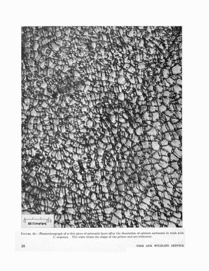
The shell of the oyster consists of four distinctlayers: periostracum, prismatic layer, calciteostracum, and hypostracum. The periostracumis a film of organic material (scleroprotein calledconchiolin), secreted by the cells located near thevery edge of the mantle. The periostracum isvery poorly developed in O. virginica and cannotbe found in old shells. It covers the prismaticlayer which can be best studied by removing fromthe edge of an oyster a small piece of newly formedshell. Microscopic examination reveals that theprismatic layer is made of single units shown infigure 44. Each prism consists of an aggregate ofcalcite crystals (Schmidt, 1931) laid in a matrixof conchiolin which after the dissolution of mineralconstituents in weak hydrocWoric acid retains thegeneral configuration of the prisms (fig. 45). Thedouble refraction of the walls of empty prisms ispronounced and causes slight iridescence noticeable under the microscope. In a well-formed
oMillimeters
0.5
layer the prisms are wedge-shaped and slightlycurved (fig. 46). Conchiolin adhering to theprisms can be destroyed by boiling in potassiumhydroxide solution and the prisms separated(Schmidt, 1931). Their shape and size are veryvariable.
The optical axes of the prism are, in general,perpendicular to the plane of the prismatic layer,but in places they are irregularly inclined towardit.
Calcite-ostracum, called also a subnacreouslayer (Carpenter, 1844, 1847), makes up the majorpart of the shell. The layer consists primarily offoliated sheets of calcite laid between thin membranes of conchiolin. The separate layers areirregularly shaped with their optical axes in accidental position (B~ggild, 1930). In a polished,transverse section of the shell of O. virginica thefolia are laid at various angles to the surface(fig. 47). This layer is frequently interrupted bysoft and porous chalky deposits (upper two layersof fig. 47) which appear to consist of amorphousmaterial. It can be shown, however, that chalkydeposit is formed by minute crystals of calciteoriented at an angle to the foliated lamellae of thehard material.
Hypostracum is a layer of shell material underthe place of the attachment of the adductor muscle.In the shells of O. virginica the layer is pigmentedand consists of aragonite (orthorhombic calciumcarbonate, CaCOs).
For many years oyster shells were considered tobe composed entirely of calcite (B~ggild). Recently Stenzel (1963) has discovered that on eachvalve of an adult O. virginica aragonite is presentas padding of the muscle scar, in the imprint ofQuenstedt's muscle, and in the ligament.
As the oyster grows the adductor muscle increases in size and shifts in the ventral direction.The new areas of attachment become coveredwith aragonite while the older, abandoned partsare overlaid with the calcite. The progress ofthe muscle from hinge toward the ventral side canbe clearly seen on a longitudinal section of theshell where it can be easily distinguished by itsdarker color and greater hardness of the secretedmaterial (fig. 48).
ORGANIC MATERIAL OF THE SHELL
After the removal of mineral salts of the shell byweak acids or by chelating agents, such as sodiumversenate, the insoluble residue appears in the
MORPHOLOGY AND STRUCTURE OF SHELL 37
, I 1
M'II' t 03lime ers
FIGURE 45.-Photomicrograph of a thin picce of prismatic layer after the dissolution of calcium carbonate in weak acid,C. virginica. The walls retain the shape of the prisms and are iridescent.
38 FISH AND WILDLIFE SERVICE

0.5Millimeters
FIGURE 46.-Cross section of a piece of young shell of C.virginica (mounted in bakelite and ground on a glasswheel with carborundum, about 80 x). Periostracum(top line), prismatic layer (middle), and calcite-ostracum(lower).
form of thin, homogenous sheets of organic materialkept together like pages of a book. This substance, discovered in 1855 by Fremy, is known asconchiolin. The name is applied to the organicmaterial insoluble in water, alcohol, ether, coldalkaline hydroxides, and dilute acids. In theliterature it appears also under the names ofconchin, periostracum, epidermis, and epicuticula.Conchiolin is a scleroprotein, the structural formula of which has not yet been determined. Theelementary analysis of conchiolin of O. edulis(Schlossberger, 1856) is as follows: H, 6.5 percent;C, 50.7 percent; N, 16.7 percent. Wetzel (1900)found that conchiolin contains 0.75 percent ofsulfur and Halliburton (quoted from Haas, 1935)assigned to it the following formula: C30 , H 48 , Ng ,
au, which also appears in the third edition of"Hackh's chemical dictionary" (Hackh, 1944).Similarity of conchiolin to chitin' leads manyinvestigators to an error in ascribing chitinouscomposition to structures which were found insoluble in alkaline hydroxides and dilute acids.Thus, the presence of chitin was reported in theshell and ligament of Anodonta, Mya, and Pecten(Wester, 1910). The application of the Schulze's
MORPHOLOGY AND STRUCTURE OF SHELL
test for chitin (intense violet coloration aftertreatment for 24 hours in diaphanol [chlorodioxyacetic acid], followed by a solution of zinc chlorideand iodine), does not confirm these findings (Lison,1953).4
To the naked eye and under the light microscope the conchiolin appears as amorphous, viscousand transparent material which hardens shortlyafter being deposited. Using the electron microscope technique, Gregiore, Duchateau, and Florkin(1955) found that the conchiolin of gastropods andbivalves consists of a fine network with manymeshes of irregular shape and variable dimensions.This is, however, not the case in oyster shells.Conchiolin of the genus Ostrea lacks meshes andunder the electron microscope is of uniform appearance (personal communication by Gregoire).
Cross sections of decalcified shells of C. virginicashow a distinct difference between the stainingproperties of the conchiolin of the prismatic andcalcite-ostracum layers. On the cross sections ofshell shown in figure 49 the two parts can berecognized by the typical foliated appearance ofthe calcite-ostracum and the meshlike structureof the prismatic layer. In the preparation stainedwith Mallory triple dye the organic matter ofthe walls of the prisms are stained reddish-brownwhile the foliae of the calcite-ostracum are bluish.Differential staining indicates the difference inthe chemical composition of the two parts.
The amount of conchiolin in the oyster shell wasstudied by several investigators. As early as1817 Brandes and Bucholz estimated that organicmaterial of the shell constitutes about 0.5 percentof the total weight. Schlossberger (1856) found6.3 percent of organic matter in the prismaticlayer of the oyster but only from 0.8 to 2.2 percentin the calcite-ostracum. According to Douville(1936), the albuminoid content of the oyster shellis 4.8 percent.
According to the determinations made by A.Grijns for Korringa (1951), the conchiolin contentof the prismatic layer of O. edulis varied from 3.4to 4.5 percent against the 0.5 to 0.6 percent in thecalcite-ostracum. The conchiolin content wascalculated from the percentage of N (by Kjeldahlmethod) multiplied by 6.9. The results of mydeterminations of the weight of organic material
• Inasmuch as the same reaction is obtained with cellulose and tunicine,additional tests should be made using Lugol solution and 1 to 2 per centsulphuric acid (H2S0.). With this test chitin Is colored brown, while cellulose and tunlclne are blue.
39

I I I I
o .. 0.3Millimeters
FIGURE 47.-Cross section of the shell of adult C. virginica em.bedded in bakelite and polished on a glass wheel with carborundum.. Two upper layers consist of chalky deposits.
after decalcification of the calcite-ostracum ofC. virginwa shells from Long Island Sound andCape Cod waters are in agreement with thosegiven for O. edulis. The content of conchiolin inmy samples varied from 0.3 to 1.1 percent withthe mode at 0.6 percent. For these analyses 23pieces of shell were taken from 16 adult oystersnot damaged by boring sponge. The samplesvaried in weight from 0.5 to 15 g.
Higher percentage of conchiolin in the prismaticlayer may be expected because this layer represents
40
the new growth of shell which has not yet completely calcified.The role played by conchiolin in the deposition
of calcium salts in the form of calcite or aragonitepresents a very interesting problem which has notyet been solved. Recent electron microscopestudies of pearl oyster shells made by Gregoireshow that the organic material in which aragonitecrystals are laid (Gregoire, Duchliteau, andFlorkin, 1950) is arranged as a series of bricklikestructures. No such arrangement has been de-
FISH AND WILDLIFE SERVICE

oCentimeters
5
FIGURE 48.-Left valve of O. (Alectryonia) megodon cut along the principal axis of growth. Hypostracum (dark striatedlayer) forms a pronounced platform for the attachment of the adductor muscle, and can be traced to its original positionin the young oyster (right). Chalky deposits are regularly arranged between the layers of calcite. Also see fig. 41.
----------------1----- ----
TABLE 4.-Amino acids from the conchiolin of two speciesof oysters
[In parts 01100 parts of protein according to Roche, Ranson, and EyssericLafon (1951)]
Arginine_ 0.45 2_ 90Histidine_________ __ __ __ __ __ __ ____ __ __ __ __ __ ___ __ __ 0.65Lysine____________________________________________ 3.55 4.30Glycine___________________________________________ 15.70 15.70Leucine___________________________________________ 0.51 _Tryptophane______________________________________ 0.48Tyrosine__________________________________________ 3.27 3_ 05Valine_____________________________________________ 0.95 _Cystine_ 0.98Methioninc_ 1.77 1.62
scribed for calcite shells. Present knowledge ofthe chemistry of the organic constituents of theshell is inadequate. It seems reasonable toassume that conchiolin like other proteins is nota single chemical substance common to a largenumber of organisms, but that it differs specifically from animal to animal and may even varyin the different parts of the same shell.
The analysis of amino acids obtained by hydrolysis of conchiolin prepared from decalcifiedshells showed (Roche, Ranson, and EyssericLafon, 1951) that there is a difference in the shellsof the two species of European oysters, O. edulisand C. angulata (table 4).
MUSCLE ATTACHMENT
The place of attachment of the adductor muscleor muscle scar is the most conspicuous area of the
Taking advantage of the fact that both calciteand aragonite are present in the two distinctlayers of shell of the fan oyster (Pinna) and of thepearl oyster (Pinctada) , the French investigators(Roche, Ranson, and Eysseric-Lafon, 1951) attempted to determine whether there is a differencein the chemical composition of the organic materialof the two layers of the shell of the same species.They found that tyrosine and glycine occur inhigher concentrations in the prismatic layer thanin the nacreous part of shells. In the prismaticlayer of calcite portion the content of tyrosinevaries between 11.6 and 17.0 percent and that ofglycine between 25 and 36 percent. In thenacreous part made of aragonite the concentrationof tyrosine was from 2.8 to 6.0 percent and thatof glycine varied between 14.9 and 20.8 percent.The significant differences in the contents of thetwo amino acids in the two parts of the shellmay provide a clue for further studies of the roleof the organic component on the mineral form inwhich the calcium carbonate is deposited by themantle.
Crassos/rea Os/reaangula/a edulis
Amino acids
MORPHOLOGY AND STRUCTURE OF SHELL 41

.: : .. : ,= .. : ~. :.:.:; .. .: =.. : :.: :. '.: '
. .. . " ; ~',,: ,:.; :. :: -. . . '.
,tlt\:l~D;;: :········\.·;.~..;::S.::·i:·;-\:,,,7
figure were obtained in the following manner:the periphery of the impression was circumscribedwith soft pencil; a piece of transparent Scotchadhesive tape was pressed on the impression andthe outline was lifted and mounted on crosssection paper; the area occupied by the impressionwas measured by counting the number of squares.Using this method, I obtained the replicas ofmuscle impressions from 169 shells taken atrandom from various oyster beds of the Atlanticand Gulf Coasts. The impressions are arbitrarilyarranged in four series (A-D) according to theirshape and size. The impression areas of roundand broad shells are shown in the two upper rows,A and B; those of long and narrow shells arearranged in the two lower rows, C and D.
It may be expected that the larger is the shellthe greater is the area of muscle impression.The relationship, as can be seen in fig. 51, isrectilinear although the scatter of plotted datais considerable and the variability increases withthe increase in size. The ratio of muscle impression area to shell surface area varies from 8 to 32with the peRk of frequency distribution at 16 to18 (fig. 52).
A small oval and unpigmented area on the
FIGURE 49.-Cross section of shell of an adult C. virginicaafter decalcification in weak acid, Mallory triple stain.Conchiolin of the prismatic layer is reddish-brown; thatof calcite-ostracum is bluish.
oyster shell. In C. virginica, C. angulata, andmany other species this area is highly pigmented;in O. edulis, C. gigas, pigmentation is eitherabsent or very ligh~.
The muscle scar in C. virginica is located in theposteroventral quadrant of the shell (figs. 15, 21,33). To a certain extent the shape of the scarreflects the shape of the sh~il, being almost roundin broad and round oysters and elongated innarrow and long shells. The area of scar isslightly concave on the side facing the hinge andconvex on the opposite, i.e., ventral side. Curvedgrowth line, parallel to the curvature of theventral edge of the valve, can be seen on thesurface. They are most pronounced in the ventralpart of the muscle impression. Size and shape ofthe scar is variable and often irregular (fig. 50).The outlines of the impressions shown in this
FIGURE 50.-Variations in shape and size of muscle scarson the shells of C. virginica. Rows A and B show thetypes of scars normally found on broad and roundedshells, the length of which is almost equal to or exceedsthe height. Rows C and D are the scars often foundon long and narrow shells in which the height exceedsthe length. Replicas of scars were made from shellscollected at random.
oC7
o
oo
o
Centimetersb
o
c
1.0Millimeters
o
42 FISH AND WILDLIFE SERVICE

g 0
o ..,00 0 0 0 0
o 0 0 0
otI 0 00
0°0 ottP 00
0o§o <) 0, 800 0 0° 00,0 ,
00 ~ 0° 0 0 000000 000 0
o 0 000 0
o 0 0 0 0 0000
o 0 og 00°00'; a000 0 ~o~o O!
o :~{o 0 % ~ooo ~~0000 000 00 ~ 00
eo 0 °00'6 0 0 0
0:eoo og 0OOOQ:lo 08:'
00 0
TABLE 5.-Chemical composition oj oyster shells in percentoj shell weight
[From Hunter and Harrison, 1928]
Sampie 1 Sample 2
0.196
1. 510.28
0.04338.810.00250.090.1890.0090.0730.5800.00090.0035
1. 410.27
0.04538.78
-- -----ii~ii--
0.1830.0090.0750.570
0.0034357.19
Constltuents
1 Loss above 110° C. Ignited.2 Loss to 100° C.3 Average for samples 1 and 2.60.050.020.0 30.0 40.0
SHELL AREA, cm.210.0
5.01-
4.0
"l!<.>
l3' 3.Cll:
'"""....~
~2.0
1.0
00
93.880.480.881.400.320.270.461. 60
PercentChemical
(Analysis supplied by Columbia-Southern Corporation and copied withtheir permission.)
TABLE 6.-Chemical composition oj mud shell.~ received atthe plant oj Columbia-Southern Corporation at CorpusChristi, Tex.
[Percent of constituents in samples dried at 110° C.]
CHEMICAL COMPOSITION
The oyster shell consists primarily of calciumcarbonate, which composes more than 95 percentof the total weight of the shell. The balance ismade up by magnesium carbonate, calcium sulfate, silica, salts of manganese, iron, aluminum,traces of heavy metals, and organic matter.Several analyses of oyster shell found in theliterature are incomplete, particularly with reference to trace elements. Analysis made for theU.S. Bureau of Fisheries by the Bureau ofChemistry of the Department of Agriculture andpublished in 1928 (Hunter and Harrison, 1928) isgiven in table 5.
Dead oyster shells buried in the mud of theinshore waters of Texas and Louisiana are extensively dredged by commercial concerns primarilyfor the manufacture of chicken feed. Analysis ofthese shells as they are received at the plant afterthorough washing in sea ~ater is given in table 6.
The calcium carbonate content of these shells isprobably lower than in live oysters due to theirerosion and dissolution of lime in sea water.The chloride content IS affected by the retention of
2~~
0
8I--
6
2 -0 ~
f--
1---
-
d ~
FIGURE 5l.-The relationship between the area of musclescar and the area of the shell of C. virginica.
o 2 4 6 8 10 12 14 16 18 20 22 24 26 28 30 32
SHELL AREA ':"MUSCLE ATTACHMENT AREA
FIGURE 52.-Frequency distri):mtion of the ratio of musclescar area vs. shell area in the shells of C. virginica ofAtlantic and Gulf States.
3
3
2
2
24
~ 2-J~ 2
~ 18
~ 16
a 14
~ 12
10
8
6
4
2
dorsal half of each valve is the imprint of avestigial muscle in the mantle, discovered in 1867by Quenstedt in the valves of the (larly Jurassicoyster, Gryph,aea arc11:ata Lamark, and found byStenzel (1963) in C. virginica. In my collectionof living C. virginica the imprint is hardly visible(figs. 15, 21, and 22). Slight adhesion of themantle to the valve indicates the location of thisarea which Stenzel calls "imprint of Quenstedt'smuscle."
MORPHOLOGY AND STRUCTURE OF SHELL 43

TABLE 7.-Composition of C. virginica oyster shell dredgedfrom Galveston Bay, accordmg to Smith and Wright(1962)
t~ese salts in the shells after thorough washingwIth sea water of greatly variable salinity. Thepercent of silica, aluminum, and iron, which arealso higher than in the analyses of shells of liveoysters, is at least in part influenced by theefficiency of plant operations in removing mudfrom ~he surface of the shells.
Chemical composition of shells of O. edulis isnot significantly different from that of C. virginica.Table 8 gives the results obtained by Europeanscientists. The data quoted from various sourcesare taken from Vinogradov (1937).
A much more detailed analysis of dead oystershells dredged from the bottom of Galveston Bay8 miles east of San Leon was made recently by theDow Chemical Company (Smith and Wright,1962). The shells were scrubbed in tap waterwith a nylon brush, rinsed in distilled water, driedat 110° C., and ground in a porcelain mortar.With the kind permission of the authors theresults are given in table 7. Additional 19 elements were sought but not found at the followingsensitivity limits:
10 p.p.m.-arsenic, barium.1 p.p.m:-antimony, chromium, cobalt, ger
mamum, gold, lead, lithium, mercury,molybdenum, nickel, vanadium, andzirconium.
0.1 p.p.m.-beryllium, bismuth, cadmium,silver, and tin.
The authors remark that traces of clay entrappedwithin the shell may have influenced the findingsfor titanium, manganese, copper, or zinc; andthat individual variations in silicon, iron, andaluminum were due to contamination not removable by washing. It appears feasible that thesevariations may have been caused by spicules ofboring sponges and algae infesting the shells.
TABLE 8.-Chemical composition of shells of O. edulis (inpercent of ash residue)
gaco.___________________________ 98.60 97.65 96.54 97 00Ma.i!oO.lo-------.---------------- 1. 21 .
g •• -------- -- ----ii:ai2- ---ii:iii2S- -----trace
~r:~f!-----~_~~_~-~::: _::;-~~.~ ::::~: :j:~~~ ----l:
Sample Sample Sample Sample1 2 3 4
Constituent
-----------
According to Creac'h (1957), all shells of O.edulis and C. angulata contain traces of phosphorus. The French biologist found that thephosphorus content is variable. Expressed asP205, it varies in C. angulata from 0.075 to 0.114percent. There is a significant difference in thephosphorus content in various parts of the shell.The amount of phosphorus per unit of volume ofshell material is lower in the chalky deposits thanin the hard por~ion of the shells. Thus, in layinga chalky depOSIt the mollusk utilizes from 2.4 to2.6 times less phosphorus than is needed forsecreting the same volume of harder shellsubstance.
The presence of small quantities of strontium~n calcareous shells of mollusks is of particularlI~terest because. of its apparent relation to aragomte. The marIne organisms containing calciumcarbo~ate as aragonite have relatively higherstrontIUm content than those having calcite shells.The relationship between the two elements isexpressed as strontium-calcium atom ratios(Tho.mpson and Chow, 1955; Trueman, 1944; andAsan, 1950). In C. virginica and C. gigas thestrontium-calcite ratio x 1,000 varies between1.25 and 1.29. Ostrea lurida from California hasa lower strontium content, the ratio being 1.01.!he percentages of Ca, Sr, CO2, and organic matterIn the ~hel~s of t~ee species of oyster and in MyaarenaNa, In whICh the content is the highestamong the bivalves, given by Thompson andChow (1955), are summarized in table 9. The
Concen·tratlon
ConstituentConcen·tration
Constituent
12928
TABLE 9.-The percentage of calcium and strontium in theshells of oysters and soft shell clam[According to Thompson and Chow, 1955]
Species Calcium StrontiumAtom
Carbon Organic ratiodioxide matter Sr/Ca
xl,OOO
O.lurida_. ______ 38.6 0.085 42.5 1.68 1.0C. virginica______ 33.7--37.8 0.92-0.107 41.8-42.4 2.16-2.34 1.25-1.C. gigas_________ 34.6--36.2 0.097-0. 100 32.6-42.5 1. 33-1. 71 1.26-1.M. arenaria _____ 38.6-38.8 0.181-0.246 42.2-42.3 2.22-2.44 2.16-2.91
P.p.m.400340200180116110
54301253210.5
Organic Carbon as CH.__Chlorine (Cll- _Aluminum (All----------Iron (Fel . _Phosphorus (Pl ..-------Manganese (Mnl.-------Fluorine (Fl-- __~~:llS!!lum (~l-----------anlUm (TIl-- .Boron (Bl---_. _Copper (Cul-----------.-Zinc (Znl __Bromine (Brl _Iodine (1)----------------
Percent64.643.50.320.330.160.160.120.58
99.8 %
Calcium (CaO). _Carbon (CO,'._. _Sodium (Na'Ol .• -------Maf.neslum (MgOl.----Sui ur (SO,'---------.--Silicon (SIO,'-----------Strontium (SrOl-.---.--.Moisture (H,Ol----------
Total of major con·stituents.. _.. __
44 FISH AND WILDLIFE SERVICE
salinity and temperature of the wat~r have ~p
parently no influence on Sr/Ca, WhICh rem~ms
fairly constant in calcareous shells. The possIblerole of strontium in the mineralization and formation of shell is discussed in chapter V.
BIBLIOGRAPHYANTHONY, R.
1905. Influence de la fixation pleurothetique sur lamorphologie des mollusques acephales dimyaires.Annales des Sciences Naturelles: Zoology, serie 9,tome 1, pp. 165-396.
ASARI, T AMIYA.1950. Geochemical distribution of strontium. VII.
Strontium contents of shells. Journal of theChemical Society of Japan, vol. 71, pp. 156-158.
BIEDERMANN, W.1902a. Untersuchungen iiber Bau und Entstehung
der Molluskenschalen. Jenaische Zeitschrift fiirNaturwissenschaft, Neue Folge, Band 29, pp. 1-164.
1902b. Ueber die Bedeutung von Krystallisationsprozessen bei der Bildung der Skelette wirbelloserTiere, namentlich der Molluskenschalen. Zeitschrift fUr allgemeine Physiologie, Band 1, pp.154-208.
Bf/lGGILD, O. B.1930. The shell structure of the mollusks. Det
Kongelige Danske Videnskabernes Selskabs Skrifter,raekke 9, bind 2, part 2, pp. 233-326.
BRANDES RUDOLPH, und PROFESSOR BUCHOLZ.1817: Analyse der Austerschalen (O.~trea edulis).
Neues Journal der Pharmacie fUr Aerzte undApotheker, vol. 1, pp. 204-214.
CARPENTER, WILLIAM B.1844, 1847. On the microscopic structure of shells.
Report of the British Association for the Advancement of Science, 14th Meeting, 1844, pp. 1-24;ibid. 17th Meeting, 1847, pp. 93-134.
CAYEUX, LUCIEN.1916. Introduction a l'etude petrographique des
roches sedimentaires. Memoire pour servir al'explication de la carte geologique detaillee de laFrance. Ministere des Travaux Publics, Paris,vol. 1 and 2 (Atlas), 524 pp.
COOK, THEODORE A.1903. Spirals in nature and art. John Murray,
London, 200 pp.1914. Curves of life. Holt Co., New York, 479 pp.
CREAC'H, PAUL V.1957. Variations physiologiques du phosphore total
de la coquille de Gryphaea angulata Lmk. Revuedes Travaux de l'Institut des P~ches Maritimes,tome 21, fascicule 3, pp. 415-430.
CROZIER, WILLIAM J.1914. The growth of the shell in the lamellibranch
Dosinia discus (Reeve). Zoologische Jahrbiicher,Abteilung fiir Anatomie und Ontogenie der Tiere,Band 38, pp. 577-584.
DOUVILLE, HENRI.1936. Le test des lamellibranches: sa formation dans
l'Ostrea edulis. Comptes Rendus Hebdomadaires
MORPHOLOGY AND STRUCTURE OF SHELL788-851 0-64-4
des Seances de l'Academie des Sciences, tome 203,pp. 965- 968.
FREMY, E.1855. Recherches chimiques sur les os. Annales de
Chimie et de Physique, serie 3, tome 43, pp. 47-107.FRIZA, FRANZ.
1932. Zur Kenntnis des Conchiolins der Muschelschalen. Biochemische Zeitschrift, Band 246, pp.29-37.
GALTSOFF, PAUL S.1955. Recent advances in the studies of the structure
and formation of the shell of Crassostrea virginica.Proceedings of the National Shellfisheries Association, vol. 45, August 1954, pp. 116--135.
GALTSOFF, PAUL S., WALTER A. CHIPMAN, Jr., JAMES B.ENGLE, and H. N. CALDERWOOD.
1947. Ecological and physiological studies of theeffect of sulphate pulp mill wastes on oysters in theYork River, Virginia. [U.S.] Fish and WildlifeService, Fishery Bulletin 43, vol. 51, pp. 59-186.
GRAY, JOHN EDWARD.1833. Some observations on the economy of mollus
cous animals, and on the structure of their shells.Philosophical Transactions of the Royal Society ofLondon for the year 1833, part 1, vol. 123, pp.771-819.
GREGOIRE, CHARLES.1957. Topography of the organic components in
mother-of-pearl. Journal of Biophysical and Biochemical Cytology, vol. 3, No.5, pp. 797-808.
GREGOIRE, CHARLES, GH. DUCHATEAU, and M. FLORKIN.1950. Structure, etudiee au microscope electronique,
de nacres decalcifiees de Mollusques (Gasteropodes,Lamellibranches et Cephalopode). Archives Internationales de Physiologie, vol. 58, pp. 117-120.
1955. La trame protidique des nacres et des perles.Annales de l'Institut Oceanographique, nouvelleserie, tome 31, pp. 1-36.
HAAS, F.1935. Bivalvia. Teill. Dr. H. G. Bronns Klassen
und Ordnungen des Tierreichs. Band 3: Mollusca;Abteilung 3: Bivalvia. Akademisehe Verlagsgesellschaft, Leipzig, 984 pp.
HACKH, INGO W. D.1944. Haekh's chemieal dictionary. 3d ed. Re
vised by Julius Grant. Blakiston Co., Philadelphia, Pa., 925 pp.
HOULBERT, C., and C. GALAINE.1916a. Sur Ie chambrage des huttres et sur l'infection
possible des ehambres par Ie fait d'une Annelidetubicole parasite de la coquille. Comptes RendusHebdomadaires des Seances de l'Academie desSciences, tome 162, pp. 54-56.
HOULBERT, C., and C. GALAINE.1916b. Sur les causes du chambrage et sur l'entretien
raisonne des bancs d'huitres naturels. ComptesRendus Hebdomadaires des Seances de l'Academiedes Sciences, tome 162, pp. 301-304.
HUNTER, ALBERT C., and CHANNING W. HARRISON.1928. Bacteriology and chemistry of oysters, with
special reference to regulatory control of produetion, handling, and shipment. U.S. Departmentof Agriculture, Technical Bulletin No. 64, 75 pp.
45

HUXLEY, JULIAN S.1932. Problems of relative growth. Methuen and
Co., London, 276 pp.INGERSOLL, ERNEST.
1881. The oyster-industry. In The history andpresent condition of the fishery industries, 251 pp.Tenth Census of the United States, Departmentof the Interior, Washington, D.C.
KORRINGA, P.1951. On the nature and function of "chalky"
deposits in the shell of Ostrea edulis Linneaus.Proceedings of the California Academy of Sciences,series 4, vol. 27, No.5, pp. 133-158.
LAMY, ED.1917. Coquilles senestres chez les Lamellibranches.
Bulletin du Museum National d'Histoirc Naturelle,Paris, tome 23, pp. 489-493.
LATRIGUE, ALFRED1930. Biodynamique generale fondee sur l'etude du
tourbillon vital d'ether. G. Doin and Co., Inc.,Paris, 60 pp.
LAURENT, J. L. M.1839a. Observations sur la structure de la coquille
de l'huttre commune (Ostreaedulis Linn.). AnnalesFrancaises et :e:trangeres d'Anatomie et de Physiologie, Appliquees a la Medecine et a I'HistoireNaturelle, tome 3, pp. 53-55.
1839b. Resultats d'observations faites sur la coquillede I'huttre commune (Ostrea edulis L.). ComptesRendus Hebdomadaires des Seances de l'Academiedes Sciences, tome 8, pp. 135-136.
LEENHARDT, HENRY.1926. Quelques etudes sur Gryphea angula. (Huttre
du Portugal). Annales de l'Institut Oceanographique, nouvelle serie, tome 3, fascicule 1,pp. 1-90.
LISON, LUCIEN.1939. Forme et mechanique du developpement des
coquilles de Lamellibranches. Annales de laSociete Royale Zoologique de Belgique, tome 70,pp.9-32.
1942. Caracteristiques geometriques naturelles descoquilles de Lamellibranches. Bulletin de laClasse des Sciences, Academie Royale de Belgique,serie 5, tome 28, pp. 377-390.
1949. Recherches sur la forme et la mechanique dedeveloppement des coquilles des Lamellibranches.Memoires, Institut Royale des Sciences Naturellesde Belgique, serie 2, fascicule 34, pp. 1-87.
1953. Histochimie et cytochimie animales. Principes et methodes. 2d ed. Gauthier-Villars, Paris,607 pp.
MEDCOF, J. C.1944. Structure, deposition and quality of oyster
shell (Ostrea virginica Gmelin). Journal of theFisheries Research Board of Canada, vol. 6, No.3,pp. 209-216.
MOYNIER DE VILLEPOIX, R.1892. Recherches sur la formation et l'accroissement
de la coquille des Mollusques. Journal de l'Anatomie et de la Physiologie Normales et Pathologiques de I'Homme et des Animaux, pp. 461-518;ibid., pp. 582-674.
46
NEWCOMBE, CURTIS L.1950. An analysis of certain dimensional relation
ships of the Virginia oyster, Crassostrea virginica(Gmelin). American Naturalist, vol. 84, No. 816,pp. 203-214.
NOMURA, EKITARO.1926a. Further studies on the applicability of a=kb
in expressing the growth relations in molluscanshells. Science Reports of the T6hoku ImperialUniversity, series 4, Biology, vol. 2, No.1, pp. 63-84
1926b. An application of a=kb- in expressing thegrowth relation in the freshwater bivalve, Sphaeriumheterodon Pils. Science Reports of the T6hokuImperial University, series 4, Biology, vol. 2, pp.57-64.
1928. On thc relation between weight and dimensionsin the bivalves Tapes philippinarum and Cythereameretrix. Science Reports of the T6hoku ImperialUniversity, series 4, Biology, vol. 4, no. 3, pp.113-124.
OKEN, LORENZ.1847. Elements of physiophilosophy. Ray Society,
London, 209 pp.OLSSON, AXEL A.
1961. Mollusks of the tropical Eastern Pacificparticularly from the southern half of the PlI.namicPacific faunal province (Panama to Peru). Panarnie-Pacific Pelecypoda. Paleontological ResearchInstitution, Ithaca, N.Y. Norton Printing Co.,Ithaca, N.Y., 574 pp.
ORTON, JAMES H.1937. Oyster biology and oyster-culture, being the
Buckland Lectures for 1935. Edward Arnold andCo., London, 211 pp.
ORTON, JAMES H., and C. AMIRTHALINGAM.1927. Notes on shell-depositions in oysters. Journal
of the Marine Biological Association of the UnitedKingdom, vol. 14, pp. 935-953.
OWEN, G.1953. The shell in the Lamellibranchia. Quarterly
Journal of Microscopical Science, vol. 94, part 1,pp.57-70.
PELSENEER, PAUL.1920. A propos de la formation et de la composition
chimique de la coquille des mollusques. Annalesde la Societe Royale Zoologique et Malacologiquede Belgique, tome 51, pp. 70-74.
QUENSTEDT, F. A.1867. Handbuch der Petrefakenkunde. Laupp,
Tiibingen, 2d ed., p. 598.QUILTER, H. E.
1891. On the molluscan shell and periostracum.Conchologist, vol. 1, pp. 5-8.
RANSON, GILBERT.1939-41. Les hultres et Ie calcaire. Bulletin du
Museum National d'Histoire Naturelle, Paris, serie2, tome 11, pp. 467-472; ibid. tome 12, pp. 426432; ibid. tome 13, pp. 49-66.
1943. La vie des huttres. Histoires Naturelles-l,Collection dirigee par Jean Rostand. Gallimard,Paris, 261 pp.
DEREAUMUR, M.1709. De la formation et de l'accroissement des
coquilles des animaux tant terrestres qu'aquatiques,
FISH AND WILDLIFE SERVICE

soit de mer soit de riviere. Histoire de l'AcademieRoyale des Sciences avec les Memoires de Mathematique et Physique, pp. 475-520. [Reprinted asa separate: Academie Royale des Sciences Paris,Memoires, pp. 364-400.]
DEREAUMUR, M.1716. Eclaircissemens de quelques difficultes sur la
formation et l'accroissement des coquilles. Histoire de l'Academie Royale des Sciences avec lesmemoires de mathematique et physique, pp. 384394. [Reprinted as a separate: Academie Royaledes Sciences Paris, Annee 1716, Memoires, pp.303-311.]
ROCHE, JEAN, GILBERT RANSON, AND MARCELLE EysSERIC-LAFON.
1951. Sur la composition des scIeroproteines descoquilles des Mollusques (conchiolines). ComptesRendus des Seances de la Societe de Biologie et deses Filiales, tome 145, pp. 1474-1477.
SAVILLE-KENT, W.1893. The great barrier reef of Australia: its products
and potentialities. W. H. Allen and Co., Ltd.,London, 387 pp.
SCHENCK, HUBERT G.1934. Literature on the shell structure of pelecypods.
Bulletin du Musee Royal d'Histoire Naturelle deBelgique, tome 10, No. 34, pp. 1-20.
SCHLOSSBERGER, J.1856. Zur naheren Kenntniss der Muschelschalen,
des Byssus und der Chitinfrage. Liebigs Annalender Chemie, Band 98, pp. 99-120.
SCHMIDT, W. J.1931. tiber die Prismenschicht der Schale von
Ostrea edulis L. Zeitschrift fUr Morphologie und(jkologie der Tiere, Band 21, pp. 789-805.
SCHULZE, P.1922. tiber Beziehungen zwischen pflanzlichen und
tierischen Skelettsubstanzen und tiber Chitinreaktionen. Biologisches Zentralblatt, Band 42, pp.388-394.
SMITH, R. A., and E. R. WRIGHT.1962. Elemental composition of oyster shell. Texas
Journal of Science, vol. 14, No.2, pp. 222-224.SPORN, E.
1926. tiber die Gesetzmassigkeiten im Baue derMuschelgehatise. Wilhelm Roux' Archiv fUr Entwicklungsmechanik der Organismen, Band 108, PP.228-242.
STENZEL, HENRY B.1963. Aragonite and calcite as constituents of adult
oyster shells. Science, vol. 142, No. 3589, pp.232-233.
Oysters. In Treatise of invertebrate paleontology.Part N-Mollusca, 6 (Bivalvia). [In press.]
TANAKA, KOJIRo.1937. On the chalky deposits in oysters. Fishery
Investigation, Imperial Fishery Experimental Sta-
MORPHOLOGY AND STRUCTURE OF SHELL
tion, Tokyo, Supplementary Report No. 4, No. 46,pp. 36-44. [English translation by the AtlanticBiological Station, St. Andrews, Canada, 1953.]
TANAKA, KOJIRO.1943. On the effect of the concentration of salt on
the chalky deposits in oysters. Suisan Kenkyushi (Journal of Fisheries), vol. 38, No. 12, pp.222-224. [English translation by the AtlanticBiological Station, St. Andrews, Canada, 1953.]
THOMPSON, D'ARCY W.1942. On growth and form. 2d ed. Cambridge
University Press, Cambridge, England, 1116 pp.THOMPSON, THOMAS G., and TSAIHWA J. CHOW. 1955.
The strontium-calcium atom ratio in carbonatesecreting marine organisms. Deep-Sea Research,vol. 3, suppl., pp. 20-39.
TRUEMAN, E. R.1944. Occurrence of strontium in molluscan shells.
Nature, vol. 153, No 3874, p. 142.VINOGRADOV, A. P.
1937. The elementary chemical composition ofmarine organisms. Composition of shells ofLamellibranchiata. Translation in Memoirs, SearsFoundation for Marine Research, No.2, 1953, pp.303-309.
WATABE, N ORIMITSU.1954. Electron microscopic observations of the ara
gonite crystals on the surface of cultured pearls.I. Report of the Faculty of Fisheries, PrefecturalUniversity of Mie, vol. 1, No.3, pp. 449-454.
WESTER, D. H.1910. tiber die Verbreitung und Lokalisation des
Chitins im Tierreiche. Zoologische Jahrbticher,Abteilung fUr Systematik, Geographie und Biologie der Tiere, Band 28, pp. 531-558.
WETZEL, G.1900. Die organischen Substanzen der Schaalen
von Mytilus und Pinna. Hoppe-Seyler's Zeitschrift fUr physiologische Chemic, Band 29, pp.386-410.
WORSNOP, EDITH, and JAMES H. ORTON.1923. The cause of chambering in oysters and other
lamellibranchs. Nature, vol. III, No. 2775, pp.14-15.
YONGE, C. M.1952a. The monomyarian condition in the Lamelli
branchia. Transactions of the Royal Society ofEdinburgh, vol. 62, part II, pp. 443-478.
1952b. Observations on Siliqua patula Dixon and onevolution within the Solenidac. University ofCalifornia Publications in Zoology, vol. 55, pp. 421438.
ZANDER, ENOCH.1897. Vergleichende und kritische Untersuchungen
zum Verstandnisse der Jodreaktion des Chitins.Pfltigers Archiv fUr die gesammte Physiologie desMcnschen und der Thiere, Band 66, pp. 545-573.
47