Chapter 9 Lecture Outline - Napa Valley College 105/AP E and E... · •Arthrology—science of...
91
1 Chapter 9 Lecture Outline Copyright © McGraw-Hill Education. Permission required for reproduction or display. See separate PowerPoint slides for all figures and tables pre- inserted into PowerPoint without notes.
Transcript of Chapter 9 Lecture Outline - Napa Valley College 105/AP E and E... · •Arthrology—science of...

1
Chapter 9
Lecture Outline
Copyright © McGraw-Hill Education. Permission required for reproduction or display.
See separate PowerPoint slides for all figures and tables pre-
inserted into PowerPoint without notes.

Introduction
• Joints link the bones of the skeletal
system, permit effective movement, and
protect the softer organs
• Joint anatomy and movements will provide
a foundation for the study of muscle
actions
9-2
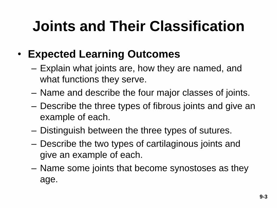
Joints and Their Classification
• Expected Learning Outcomes
– Explain what joints are, how they are named, and
what functions they serve.
– Name and describe the four major classes of joints.
– Describe the three types of fibrous joints and give an
example of each.
– Distinguish between the three types of sutures.
– Describe the two types of cartilaginous joints and
give an example of each.
– Name some joints that become synostoses as they
age.
9-3
9-4
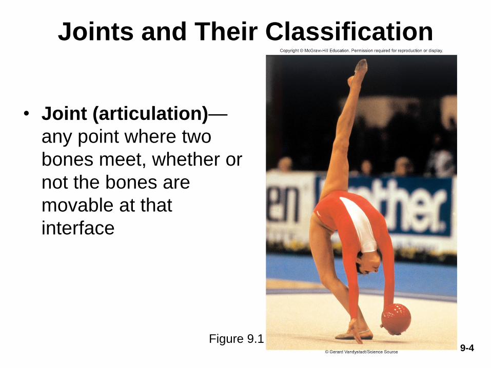
Joints and Their Classification
• Joint (articulation)—
any point where two
bones meet, whether or
not the bones are
movable at that
interface
Figure 9.1

Joints and Their Classification
• Arthrology—science of joint structure, function,
and dysfunction
• Kinesiology—the study of musculoskeletal
movement
– A branch of biomechanics, which deals with a broad
variety of movements and mechanical processes
9-5
9-6
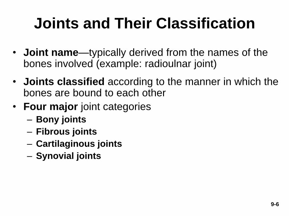
Joints and Their Classification
• Joint name—typically derived from the names of the bones involved (example: radioulnar joint)
• Joints classified according to the manner in which the bones are bound to each other
• Four major joint categories
– Bony joints
– Fibrous joints
– Cartilaginous joints
– Synovial joints

9-7
Bony Joints
• Bony joint, or synostosis—an immobile joint
formed when the gap between two bones
ossifies, and the bones become, in effect, a
single bone
– Examples:
• Left and right mandibular bones in infants
• Cranial sutures in elderly
• Attachment of first rib and sternum with old age
• Can occur in either fibrous or cartilaginous
joint
9-8
Fibrous Joints
• Fibrous joint, synarthrosis, or synarthrodial
joint—adjacent bones are bound by collagen
fibers that emerge from one bone and penetrate
into the other
• Three kinds of fibrous joints
– Sutures
– Gomphoses
– Syndesmoses

9-9
• Sutures—immobile or slightly mobile fibrous joints in which short collagen fibers bind the bones of the skull to each other
• Sutures can be classified as:
– Serrate: interlocking wavy lines
• Coronal, sagittal, and lambdoid sutures
– Lap (squamous): overlapping beveled edges
• Temporal and parietal bones
– Plane (butt): straight, non-overlapping edges
• Palatine processes of the maxillae
Sutures
Figure 9.2a
Copyright © The McGraw-Hill Companies, Inc. Permission required for reproduction or display.
Fibrous connective tissue

SuturesCopyright © The McGraw-Hill Companies, Inc. Permission required for reproduction or display.
Wood
Dovetail joint Miter joint Butt joint
Bone
Serrate suture Lap suture Plane suture
9-10
Figure 9.3

9-11
Gomphoses
• Gomphosis (fibrous joint)—
attachment of a tooth to its
socket
• Held in place by fibrous
periodontal ligament
– Collagen fibers attach
tooth to jawbone
– Allows the tooth to move a little
under the stress of chewing
Copyright © The McGraw-Hill Companies, Inc. Permission required for reproduction or display.
Fibrous connective tissue
(b) Gomphosis
Figure 9.2b
9-12
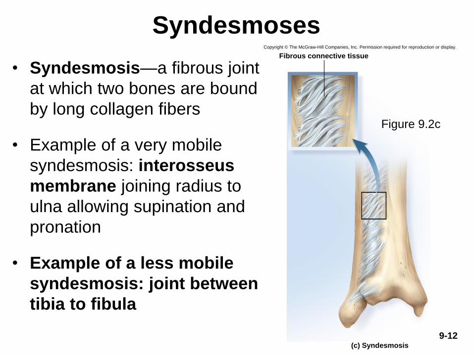
• Syndesmosis—a fibrous joint
at which two bones are bound
by long collagen fibers
• Example of a very mobile
syndesmosis: interosseus
membrane joining radius to
ulna allowing supination and
pronation
• Example of a less mobile
syndesmosis: joint between
tibia to fibula
SyndesmosesCopyright © The McGraw-Hill Companies, Inc. Permission required for reproduction or display.
Fibrous connective tissue
(c) Syndesmosis
Figure 9.2c
9-13
Cartilaginous Joints
• Cartilaginous joint, amphiarthrosis, or
amphiarthrodial joint—two bones are linked
by cartilage
• Two types of cartilaginous joints
– Synchondroses
– Symphyses

9-14
Synchondroses
• Synchrondrosis—bones
joined by hyaline
cartilage
– Temporary joints in the
epiphyseal plates in
children
• Bind epiphysis to diaphysis
– First rib attachment to
sternum
• Other costal cartilages
joined to sternum by
synovial joints
Figure 9.4a,b
Pubic symphysis
Clavicle
Rib 1
(a)
(b)
Sternum
Costal
cartilage
Interpubic disc
(fibrocartilage)
Copyright © The McGraw-Hill Companies, Inc. Permission required for reproduction or display.
9-15
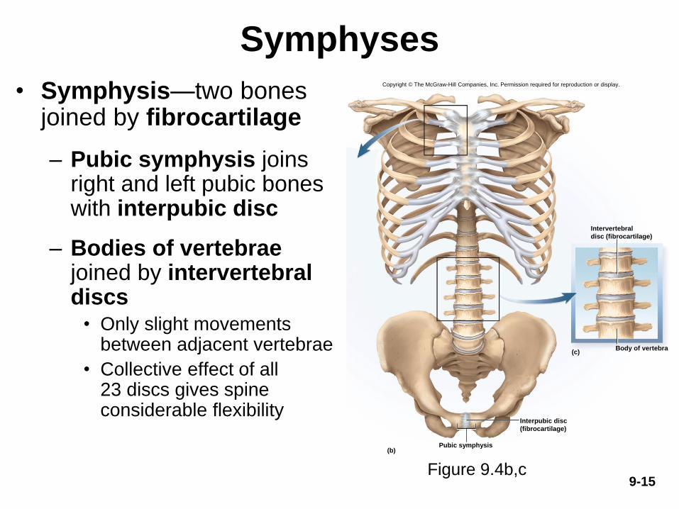
Symphyses
• Symphysis—two bones joined by fibrocartilage
– Pubic symphysis joins right and left pubic bones with interpubic disc
– Bodies of vertebrae joined by intervertebral discs
• Only slight movements between adjacent vertebrae
• Collective effect of all 23 discs gives spine considerable flexibility
Pubic symphysis
Body of vertebra(c)
(b)
Interpubic disc
(fibrocartilage)
Intervertebral
disc (fibrocartilage)
Copyright © The McGraw-Hill Companies, Inc. Permission required for reproduction or display.
Figure 9.4b,c
Synovial Joints
• Expected Learning Outcomes
– Identify the anatomical components of a typical
synovial joint.
– Classify any given joint action as a first-, second-, or
third-class lever.
– Explain how mechanical advantage relates to the
power and speed of joint movement.
– Discuss the factors that determine a joint’s range of
motion.
9-16

Synovial Joints
(Continued)
– Describe the primary axes of rotation that a bone can
have and relate this to a joint’s degrees of freedom.
– Name and describe six classes of synovial joints.
– Use the correct standard terminology for various joint
movements.
9-17

9-18
Synovial Joints
• Synovial joint,
diarthrosis, or
diarthrodial joint—joint in
which two bones are
separated by a joint cavity
• Most familiar type of joint
• Most are freely mobile
• Most structurally
complex type of joint
Copyright © The McGraw-Hill Companies, Inc. Permission required for reproduction or display.
Periosteum
Ligament
Bone
Proximal
phalanx
Joint cavity
containing
synovial fluid
Fibrous
capsule
Articular
cartilages
Joint
capsule
Middle
phalanx
Synovial
membrane
Figure 9.5
9-19
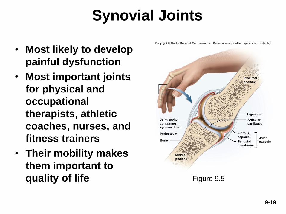
Synovial Joints
• Most likely to develop
painful dysfunction
• Most important joints
for physical and
occupational
therapists, athletic
coaches, nurses, and
fitness trainers
• Their mobility makes
them important to
quality of life
Copyright © The McGraw-Hill Companies, Inc. Permission required for reproduction or display.
Periosteum
Ligament
Bone
Proximal
phalanx
Joint cavity
containing
synovial fluid
Fibrous
capsule
Articular
cartilages
Joint
capsule
Middle
phalanx
Synovial
membrane
Figure 9.5

9-20
General Anatomy of Synovial Joints
• Articular cartilage—layer of hyaline cartilage that
covers the facing surfaces of two bones
– Usually 2 or 3 mm thick
• Joint (articular) cavity—separates articular
surfaces
• Synovial fluid—slippery lubricant in joint cavity
– Rich in albumin and hyaluronic acid
– Gives it a viscous, slippery texture like raw egg whites
– Nourishes articular cartilage and removes waste
– Makes movement of synovial joints almost friction free
9-21
General Anatomy of Synovial Joints
• Joint (articular) capsule—connective tissue that
encloses the cavity and retains the fluid
– Outer fibrous capsule: continuous with periosteum of
adjoining bones
– Inner, cellular, synovial membrane: composed mainly
of fibroblast-like cells that secrete synovial fluid and
macrophages that remove debris from the joint cavity

9-22
General Anatomy of Synovial Joints
• In a few synovial joints, fibrocartilage grows
inward from the joint capsule
– Articular disc forms a pad between articulating
bones that crosses the entire joint capsule
• Example found in temporomandibular joint
– Meniscus: moon-shaped cartilage in knee; in each
knee, menisci extend inward from the left and right
• These cartilages absorb shock and pressure
• Guide bones across each other and improve their fit together
• Stabilize the joints, reducing the chance of dislocation
9-23
General Anatomy of Synovial Joints
• Accessory structures
– Tendon: strip of collagenous tissue attaching muscle to
bone
– Ligament: strip of collagenous tissue attaching one bone
to another
– Bursa: fibrous sac filled with synovial fluid, located
between muscles, where tendons pass over bone, or
between bone and skin
• Cushions muscles, helps tendons slide more easily over
joints, modifies direction of tendon pull
– Tendon sheath: elongated cylindrical bursa wrapped
around a tendon
• In hand and foot
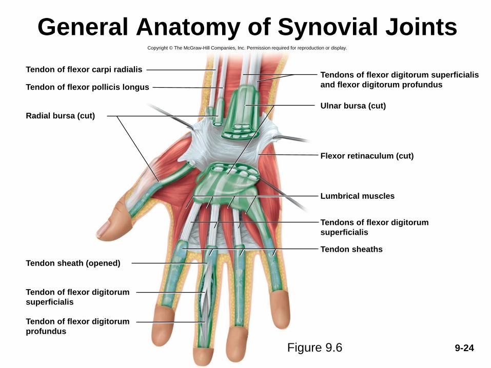
9-24
General Anatomy of Synovial JointsCopyright © The McGraw-Hill Companies, Inc. Permission required for reproduction or display.
Tendon of flexor pollicis longus
Radial bursa (cut)
Flexor retinaculum (cut)
Ulnar bursa (cut)
Lumbrical muscles
Tendon of flexor carpi radialis
Tendon sheaths
Tendon sheath (opened)
Tendon of flexor digitorum
superficialis
Tendon of flexor digitorum
profundus
Tendons of flexor digitorum superficialis
and flexor digitorum profundus
Tendons of flexor digitorum
superficialis
Figure 9.6
Exercise and Articular Cartilage
• Exercise warms synovial fluid
– Becomes less viscous, more easily absorbed by cartilage
• Cartilage then swells and provides a more effective cushion
– Warm-up period before vigorous exercise helps protect cartilage from
undue wear and tear
• Repetitive compression of nonvascular cartilage during
exercise squeezes fluid and metabolic waste out of the
cartilage
• When weight removed, cartilage absorbs synovial fluid like a
sponge taking in oxygen and nutrients to the chondrocytes
• Without exercise, cartilage deteriorates more rapidly from
inadequate nutrition and waste removal
9-25

9-26
Joints and Lever Systems
• Long bones act as levers to enhance the speed or power
of limb movements
• Lever—any elongated, rigid object that rotates around a
fixed point called a fulcrum
• Rotation occurs when an effort applied overcomes
resistance (load) at some other point
– Resistance arm and effort arm are described relative to fulcrumCopyright © The McGraw-Hill Companies, Inc. Permission required for reproduction or display.
Resistance arm
F
RE
Effort arm
Fulcrum
Effort
Resistance
(load)
Figure 9.7

9-27
Mechanical Advantage• Two kinds of advantage conferred by a lever
– Exerting more force against a resisting object than the
force applied to the lever
• Moving a heavy object with help of crowbar
– Moving the resisting object farther or faster than the effort
arm is moved
• Movement of rowing a boat
– A single lever cannot confer both advantages
• As one increases, the other decreases
• Mechanical advantage (MA) of a lever—the ratio of its
output force to its input force
• MA is calculated from length of effort arm divided by
length of resistance arm
9-28
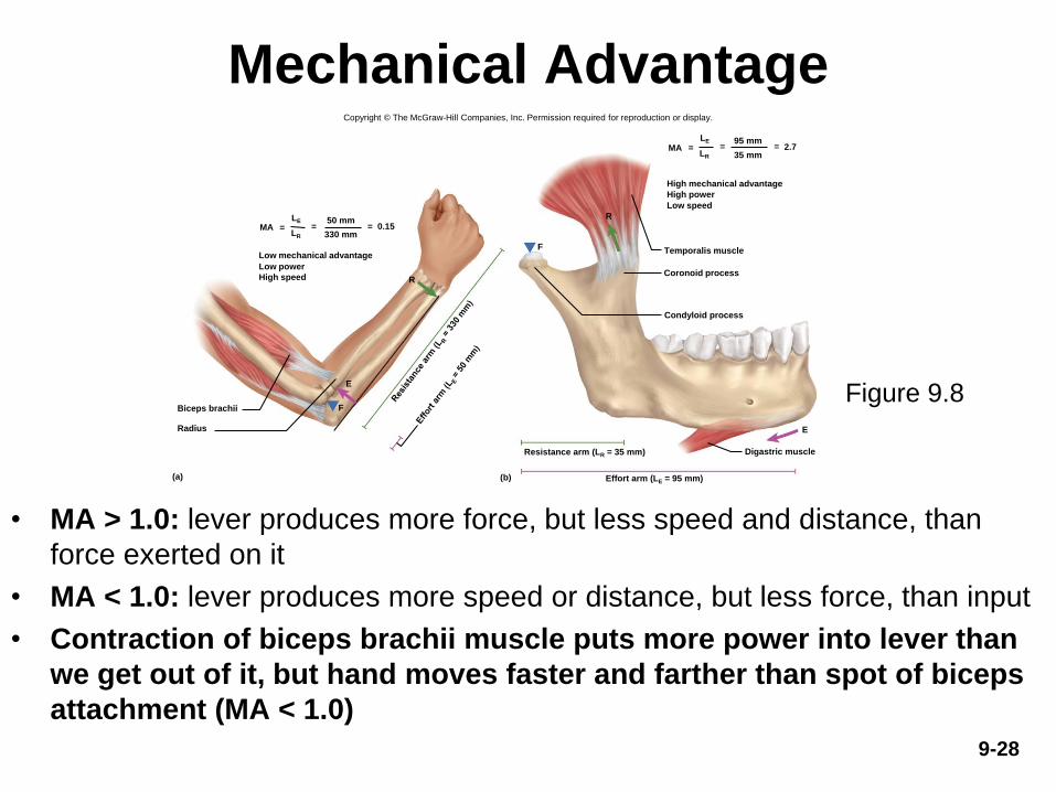
Mechanical Advantage
• MA > 1.0: lever produces more force, but less speed and distance, than
force exerted on it
• MA < 1.0: lever produces more speed or distance, but less force, than input
• Contraction of biceps brachii muscle puts more power into lever than
we get out of it, but hand moves faster and farther than spot of biceps
attachment (MA < 1.0)
Figure 9.8
Copyright © The McGraw-Hill Companies, Inc. Permission required for reproduction or display.
Biceps brachii
50 mm0.15= ==
330 mm
Low mechanical advantage
Low power
High speed
(a) (b)
E
R
F
F
Radius
High mechanical advantage
High power
Low speed
Coronoid process
Condyloid process
Resistance arm (LR = 35 mm)
Effort arm (LE = 95 mm)
E
R
Digastric muscle
MALE
LR
95 mm2.7= ==
35 mmMA
LE
LR
Temporalis muscle
9-29
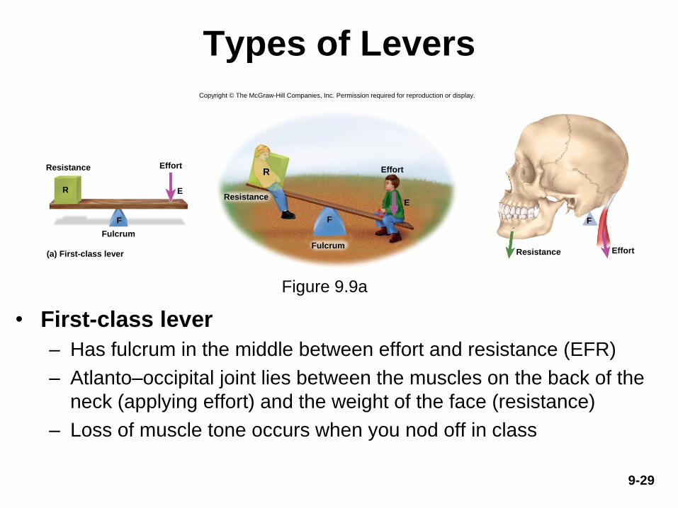
Types of Levers
• First-class lever
– Has fulcrum in the middle between effort and resistance (EFR)
– Atlanto–occipital joint lies between the muscles on the back of the
neck (applying effort) and the weight of the face (resistance)
– Loss of muscle tone occurs when you nod off in class
Copyright © The McGraw-Hill Companies, Inc. Permission required for reproduction or display.
F
E
Fulcrum
Resistance Effort
(a) First-class lever
R
R
F
E
Fulcrum
Effort
Resistance
Resistance Effort
F
Figure 9.9a

9-30
Types of Levers
• Second-class lever
– Resistance between fulcrum and effort (FRE)
– Example: when bouncing a baby on your knee, hip is fulcrum,
baby’s weight is resistance, and effort is applied at the tibia
Figure 9.9b

9-31
Types of Levers
• Third-class lever
– Effort between the resistance and the fulcrum (REF)
– Most joints of the body
– The effort of a biceps curl is applied to the forearm between the
elbow joint (fulcrum) and the weight in the hand (resistance)
Figure 9.9c
Copyright © The McGraw-Hill Companies, Inc. Permission required for reproduction or display.
Effort
Fulcrum
Resistance Effort
(c) Third-class lever
E
FFulcrum
RE
F
RResistance
EffortResistance
F
9-32
Range of Motion
• Range of motion (ROM)—the degrees through which a
joint can move
– Aspect of joint performance
– Physical assessment of a patient’s joint flexibility
• ROM determined by:
– Structure of the articular surfaces
• Elbow—olecranon of ulna fits into olecranon fossa of humerus
– Strength and tautness of ligaments and joint capsules
• Stretching of ligaments increases range of motion
• Double-jointed people have long or slack ligaments
– Action of the muscles and tendons
• Nervous system monitors joint position and muscle tone
• Muscle tone—state of tension maintained in resting muscles
9-33
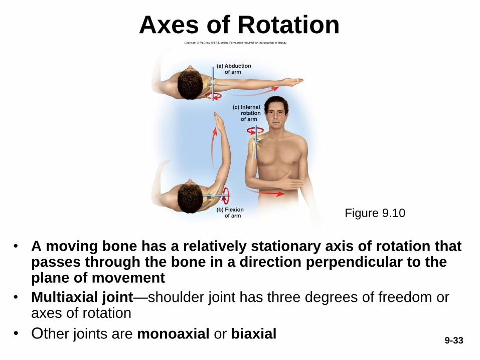
Axes of Rotation
• A moving bone has a relatively stationary axis of rotation that passes through the bone in a direction perpendicular to the plane of movement
• Multiaxial joint—shoulder joint has three degrees of freedom or axes of rotation
• Other joints are monoaxial or biaxial
Figure 9.10

9-34
Classes of Synovial Joints
Head of humerus
Scapula
Carpal bone
Metacarpal bone PhalanxMetacarpalbone
Humerus
Ulna Carpal bones
Radius
Ulna
Ball-and-socket joint
(humeroscapular)
Pivot joint
(radioulnar)
Saddle joint
(trapeziometacarpal)
Hinge joint
(humeroulnar)
Plane joint
(intercarpal)
Condylar joint
(metacarpophalangeal)
Copyright © The McGraw-Hill Companies, Inc. Permission required for reproduction or display.
Figure 9.11
9-35
Classes of Synovial Joints
• Six classes of synovial joints: ball-and-socket,
condylar, saddle, plane, hinge, pivot
• Ball-and-socket joints
– Smooth, hemispherical head fits within cup-like socket
– Only multiaxial joints in body
– Examples: shoulder, hip
• Condylar (ellipsoid) joints
– Oval convex surface of one bone fits into a
complementary-shaped depression on the other
– Biaxial joints—movement in two planes
– Examples: radiocarpal joint, metacarpophalangeal joints
9-36
Classes of Synovial Joints
• Saddle joints
– Both bones have an articular surface that is shaped like a
saddle, one concave, the other convex
– Biaxial joints
– Examples: trapeziometacarpal (opposable thumb),
sternoclavicular joint
• Plane (gliding) joints
– Flat articular surfaces, bones slide over each other
– Usually biaxial joints
– Examples: between carpal bones of wrist; between tarsal
bones of ankle; also between articular processes of
vertebrae

9-37
Classes of Synovial Joints
• Hinge joints
– One bone with convex surface fits into a concave
depression of another bone
– Monoaxial joints—move freely in one plane
– Examples: elbow, knee, joints within fingers, toes
• Pivot joints
– A bone spins on its longitudinal axis
– Monoaxial joints
– Examples: atlantoaxial joint (C1 and C2), radioulnar joint
at the elbow
9-38
Movement of Synovial Joints
• There is a vocabulary for joint movements used in
many medical and scientific fields
– Many terms presented in pairs with opposite or
contrasting meanings
– Need to understand anatomical planes and directional
terms
• Zero position—the position of a joint when a person is
in the standard anatomical position
– Joint movements described as deviating from the zero
position or returning to it

9-39
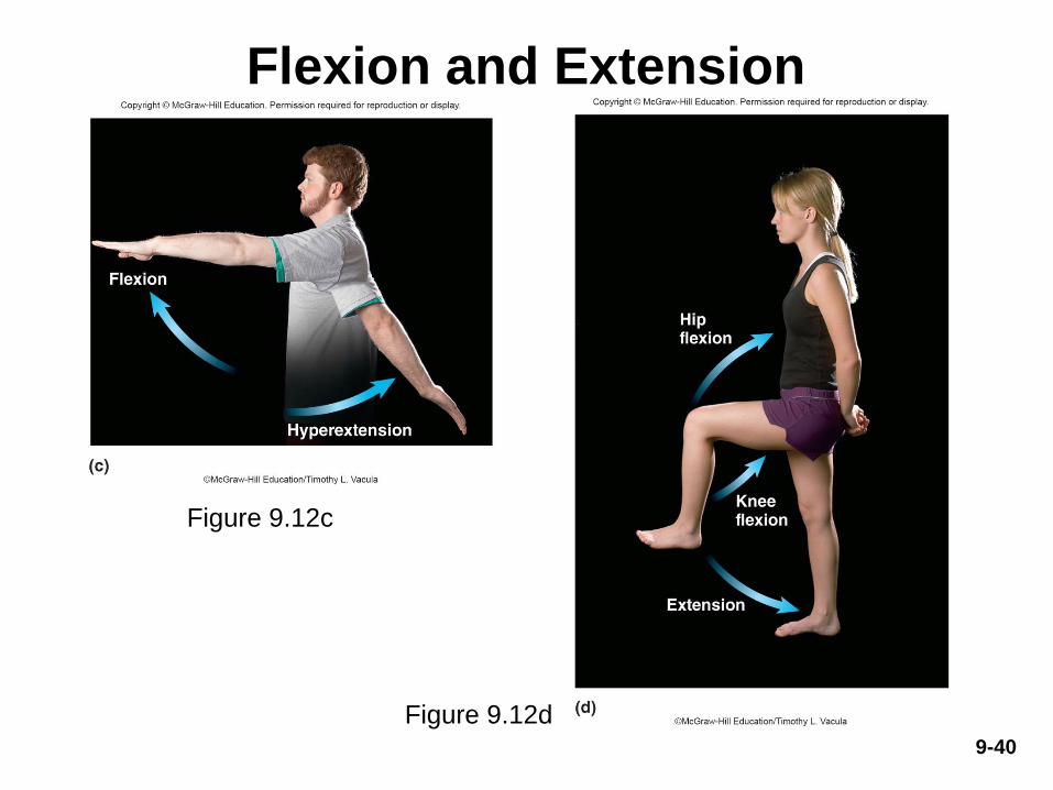
Flexion and Extension
• Flexion—movement that
decreases joint angle
– Common in hinge joints
• Extension—movement that
straightens a joint and returns
a body part to the zero
position
• Hyperextension—extension
of a joint beyond the zero
position
– Flexion and extension occur at
nearly all diarthroses,
hyperextension is limited to a fewFigure 9.12b
Figure 9.12a
9-40
Flexion and Extension
Figure 9.12c
Figure 9.12d
9-41
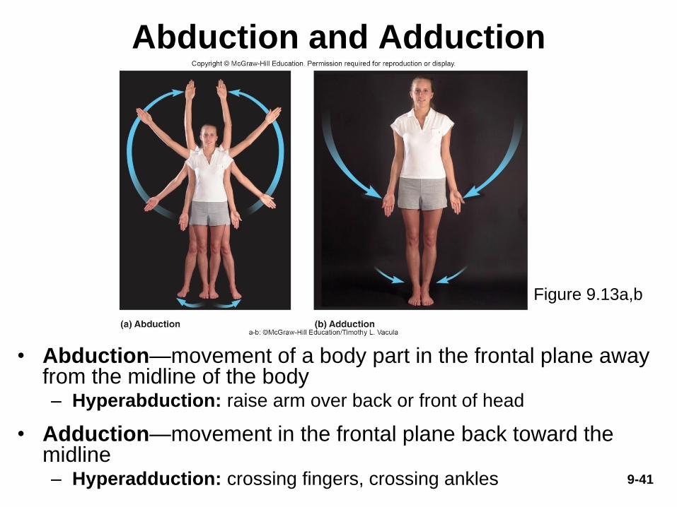
Abduction and Adduction
• Abduction—movement of a body part in the frontal plane away from the midline of the body– Hyperabduction: raise arm over back or front of head
• Adduction—movement in the frontal plane back toward the midline– Hyperadduction: crossing fingers, crossing ankles
Figure 9.13a,b

9-42
Elevation and Depression
• Elevation—movement that raises a body part vertically
in the frontal plane
• Depression—movement that lowers a body part in the
same plane
Figure 9.14a,b

9-43
Protraction and Retraction
• Protraction—the
anterior movement of a
body part in the
transverse (horizontal)
plane
• Retraction—posterior
movement
Figure 9.15a,b

9-44
Circumduction
• Circumduction—one
end of an appendage
remains stationary while
other end makes a
circular motion
– Example: an artist
circumducts upper limb
when painting a circle
on a canvas
Figure 9.16

9-45
Rotation
• Rotation—
movement in which a
bone spins on its
longitudinal axis
– Rotation of trunk,
thigh, head, or arm
• Medial (internal)
rotation turns the
bone inward
• Lateral (external)
rotation turns the
bone outwardFigure 9.17a,b

9-46
Supination and Pronation
• Primarily forearm movements
• Supination—forearm
movement that turns palm to
face anteriorly or upward
– Forearm supinated in
anatomical position
– Radius is parallel to the ulna
• Pronation—forearm movement
that turns palm to face either
posteriorly or downward
– Head of radius spins
– Radius crosses stationary
ulna like an X
Figure 9.18a,b

9-47
Special Movements of Head and Trunk
• Flexion—forward-bending movements at the waist or neck
• Extension—straightens trunk or neck
• Hyperextension—bending over backward
• Lateral flexion—tilting the head or trunk to the right or left at the midline
Figure 9.19a,b,c

9-48
Special Movements of Head and Trunk
• Right and left rotation of trunk and head
Figure 9.19d,e
9-49
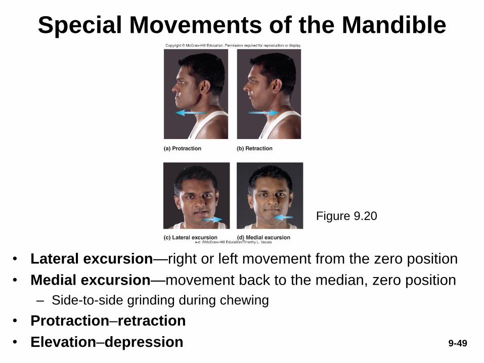
Special Movements of the Mandible
• Lateral excursion—right or left movement from the zero position
• Medial excursion—movement back to the median, zero position
– Side-to-side grinding during chewing
• Protraction–retraction
• Elevation–depression
Figure 9.20
9-50
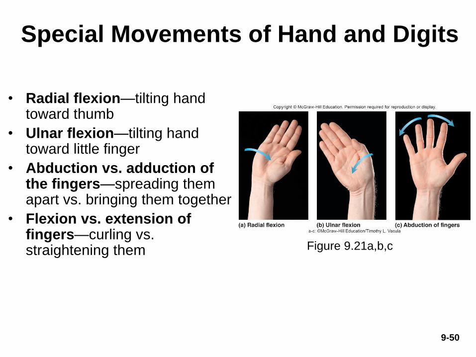
Special Movements of Hand and Digits
• Radial flexion—tilting hand toward thumb
• Ulnar flexion—tilting hand toward little finger
• Abduction vs. adduction of the fingers—spreading them apart vs. bringing them together
• Flexion vs. extension of fingers—curling vs. straightening them Figure 9.21a,b,c

Special Movements of Hand and Digits
• Palmar abduction—moving thumb away from hand and pointing it anteriorly
• Radial abduction—moving thumb away from index finger (90°)
• Flexion of thumb—tip of thumb directed toward palm
• Extension of thumb—straightening the thumb
• Opposition—moving thumb to touch tip of a finger
• Reposition—returning thumb to the zero position
9-51
Figure 9.21d, e
9-52
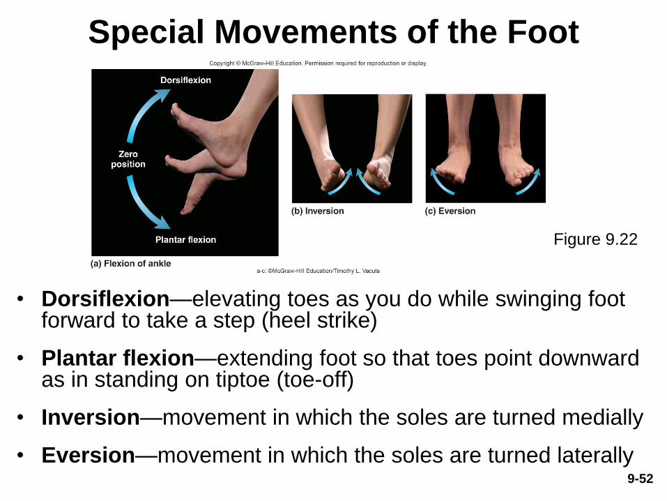
Special Movements of the Foot
• Dorsiflexion—elevating toes as you do while swinging foot forward to take a step (heel strike)
• Plantar flexion—extending foot so that toes point downward as in standing on tiptoe (toe-off)
• Inversion—movement in which the soles are turned medially
• Eversion—movement in which the soles are turned laterally
Figure 9.22

Special Movements of the Foot
• Supination of foot—complex combination of plantar flexion, inversion, and adduction
• Pronation of foot—complex combination of dorsiflexion, eversion, and abduction
9-53

Anatomy of Selected Diarthroses
• Expected Learning Outcomes
– Identify the major anatomical features of the jaw,
shoulder, elbow, hip, knee, and ankle joints.
– Explain how the anatomical differences between
these joints are related to differences in function.
9-54

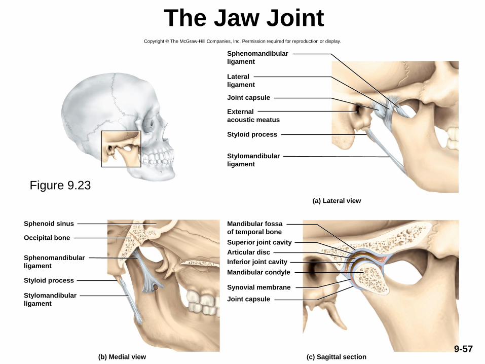
The Jaw Joint
• Temporomandibular (jaw) joint (TMJ)—articulation
of the condyle of the mandible with the mandibular
fossa of the temporal bone
– Combines elements of condylar, hinge, and plane joints
– Synovial cavity of the TMJ is divided into superior and
inferior chambers by an articular disc
9-55
The Jaw Joint
– Two ligaments support joint
• Lateral ligament—prevents posterior displacement of
mandible
• Sphenomandibular ligament—on the medial side
– Deep yawn or strenuous depression can dislocate
the TMJ
• Condyles pop out of fossa and slip forward
• Relocated by pressing down on molar teeth while pushing
the jaw backward
9-56
The Jaw JointCopyright © The McGraw-Hill Companies, Inc. Permission required for reproduction or display.
Joint capsule
Styloid process
(a) Lateral view
(c) Sagittal section(b) Medial view
Occipital bone
Sphenoid sinus
Styloid process
Joint capsule
Synovial membrane
Mandibular condyle
Superior joint cavity
Inferior joint cavity
Articular disc
Sphenomandibular
ligament
Lateral
ligament
External
acoustic meatus
Stylomandibular
ligament
Mandibular fossa
of temporal bone
Sphenomandibular
ligament
Stylomandibular
ligament
Figure 9.23
9-57

TMJ Syndrome
• Temporomandibular joint (TMJ) syndrome
– May affect as many as 75 million Americans
• Signs and symptoms
– Clicking sounds in the jaw, imitation of jaw movement
– Pain radiating from jaw down the neck, shoulders, and back
– Can cause moderate intermittent facial pain, or severe headaches, vertigo (dizziness), tinnitus (ringing in the ears)
• Cause of syndrome
– Caused by combination of psychological tension and malocclusion (misalignment of teeth)
• Treatment
– Psychological management, physical therapy, analgesic and anti-inflammatory drugs, corrective dental appliances to align teeth properly
9-58
9-59
The Shoulder Joint
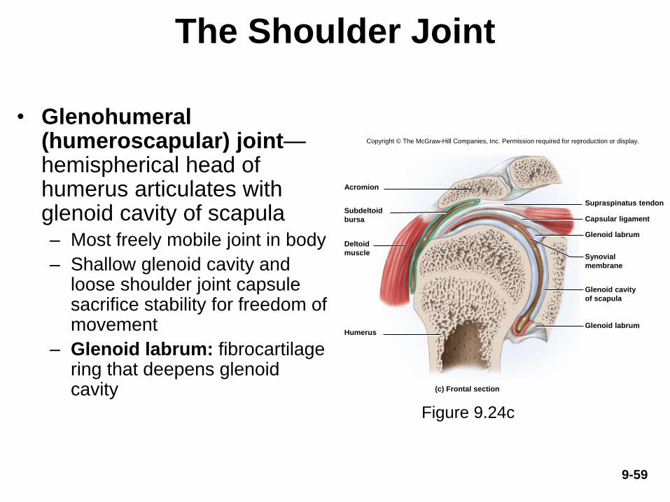
• Glenohumeral (humeroscapular) joint—hemispherical head of humerus articulates with glenoid cavity of scapula– Most freely mobile joint in body
– Shallow glenoid cavity and loose shoulder joint capsule sacrifice stability for freedom of movement
– Glenoid labrum: fibrocartilage ring that deepens glenoid cavity
Figure 9.24c
Copyright © The McGraw-Hill Companies, Inc. Permission required for reproduction or display.
Glenoid labrum
Glenoid labrum
Supraspinatus tendon
Acromion
Capsular ligament
Humerus
(c) Frontal section
Subdeltoid
bursa
Deltoid
muscleSynovial
membrane
Glenoid cavity
of scapula
9-60
The Shoulder Joint
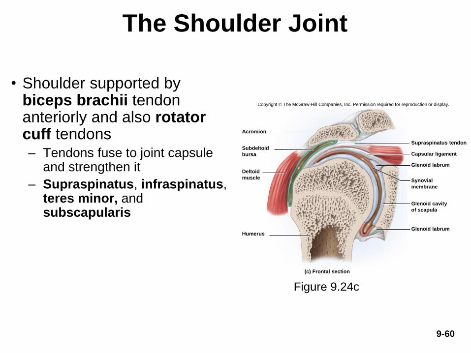
• Shoulder supported by biceps brachii tendon anteriorly and also rotator cuff tendons– Tendons fuse to joint capsule
and strengthen it
– Supraspinatus, infraspinatus, teres minor, and subscapularis
Figure 9.24c
Copyright © The McGraw-Hill Companies, Inc. Permission required for reproduction or display.
Glenoid labrum
Glenoid labrum
Supraspinatus tendon
Acromion
Capsular ligament
Humerus
(c) Frontal section
Subdeltoid
bursa
Deltoid
muscleSynovial
membrane
Glenoid cavity
of scapula
9-61
The Shoulder Joint
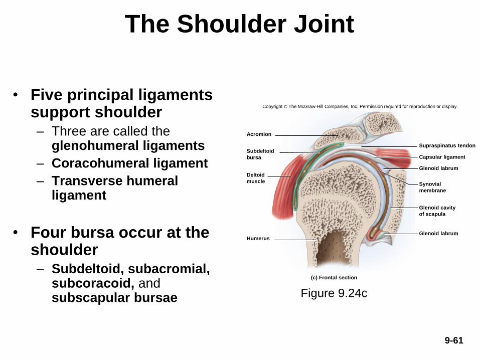
• Five principal ligaments support shoulder– Three are called the
glenohumeral ligaments
– Coracohumeral ligament
– Transverse humeral ligament
• Four bursa occur at the shoulder– Subdeltoid, subacromial,
subcoracoid, and subscapular bursae Figure 9.24c
Copyright © The McGraw-Hill Companies, Inc. Permission required for reproduction or display.
Glenoid labrum
Glenoid labrum
Supraspinatus tendon
Acromion
Capsular ligament
Humerus
(c) Frontal section
Subdeltoid
bursa
Deltoid
muscleSynovial
membrane
Glenoid cavity
of scapula
9-62
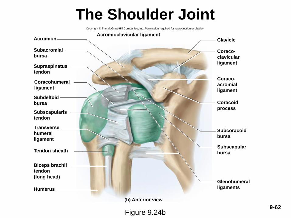
The Shoulder Joint
Figure 9.24b
Copyright © The McGraw-Hill Companies, Inc. Permission required for reproduction or display.
Acromion
Tendon sheath
Humerus
ClavicleAcromioclavicular ligament
Subacromial
bursa
Supraspinatus
tendon
Coracohumeral
ligament
Subdeltoid
bursa
Subscapularis
tendon
Transverse
humeral
ligament
Biceps brachii
tendon
(long head)
(b) Anterior view
Glenohumeral
ligaments
Subscapular
bursa
Subcoracoid
bursa
Coracoid
process
Coraco-
acromial
ligament
Coraco-
clavicular
ligament

9-63
The Shoulder Joint
Figure 9.24d
Copyright © The McGraw-Hill Companies, Inc. Permission required for reproduction or display.
Coracoid process
Coracohumeral ligament
Subscapular bursa
Subscapularis tendon
Acromion
Supraspinatus
tendon
Subdeltoid
bursa
Infraspinatus
tendon
Glenoid cavity
(articular cartilage)
Synovial membrane
(cut)
Teres minor
tendon
(d) Lateral view, humerus removed
Inferior glenohumeral
ligament
Middle glenohumeral
ligament
Biceps brachii tendon
(long head)
Superior glenohumeral
ligament

9-64
The Shoulder Joint
Figure 9.24a
9-65
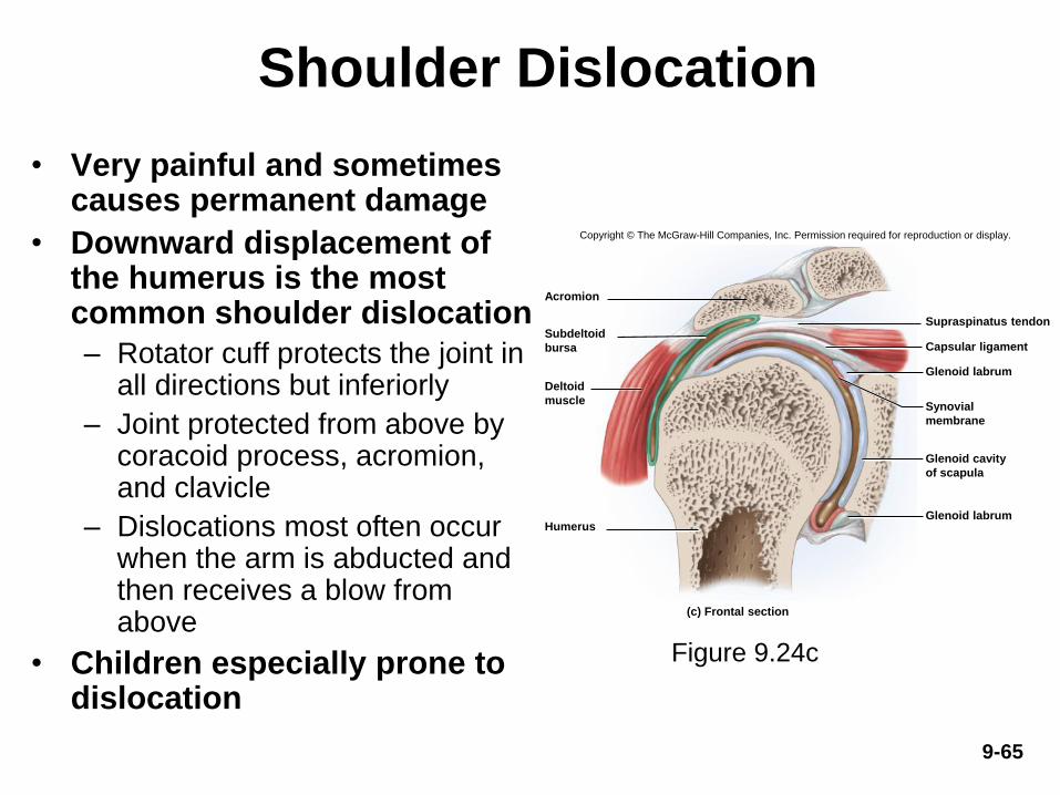
Shoulder Dislocation
• Very painful and sometimes causes permanent damage
• Downward displacement of the humerus is the most common shoulder dislocation
– Rotator cuff protects the joint in all directions but inferiorly
– Joint protected from above by coracoid process, acromion, and clavicle
– Dislocations most often occur when the arm is abducted and then receives a blow from above
• Children especially prone to dislocation
Figure 9.24c
Copyright © The McGraw-Hill Companies, Inc. Permission required for reproduction or display.
Glenoid labrum
Glenoid labrum
Supraspinatus tendon
Acromion
Capsular ligament
Humerus
(c) Frontal section
Subdeltoid
bursa
Deltoid
muscleSynovial
membrane
Glenoid cavity
of scapula

9-66
The Elbow Joint
Figure 9.25c
• Elbow—a hinge that includes two articulations: – Humeroulnar joint: trochlea of
the humerus joins trochlear notch of the ulna
– Humeroradial joint: capitulum of humerus meets head of radius
– Both articulations enclosed in one joint capsule
– Olecranon bursa on posterior
side of elbow eases movements
of tendons
– Radial (lateral) collateral
ligament and ulnar (medial)
collateral ligaments restrict
side-to-side motions
Copyright © The McGraw-Hill Companies, Inc. Permission required for reproduction or display.
(b) Sagittal section
Humerus
Trochlea
Joint capsule
Radius
Olecranon
Articular cartilage
Coronoid process
Ulna
Olecranon
bursa
Figure 9.25b
Copyright © The McGraw-Hill Companies, Inc. Permission required for reproduction or display.
(c) Medial view
Anular ligament
Joint capsule
Humerus
Coronoid process
Radius
Ulna
Tendon of
triceps brachii
Ulnar collateral
ligament
Olecranon
bursa
Tendon of biceps
brachii (cut)
9-67
The Elbow Joint
• Elbow region also contains proximal radioulnar joint– Functions as a pivot, not a hinge
– Head of radius fits into radial notch of ulna
– Held in place by anular ligament encircling radial head
– Allows for pronation and supination
9-68
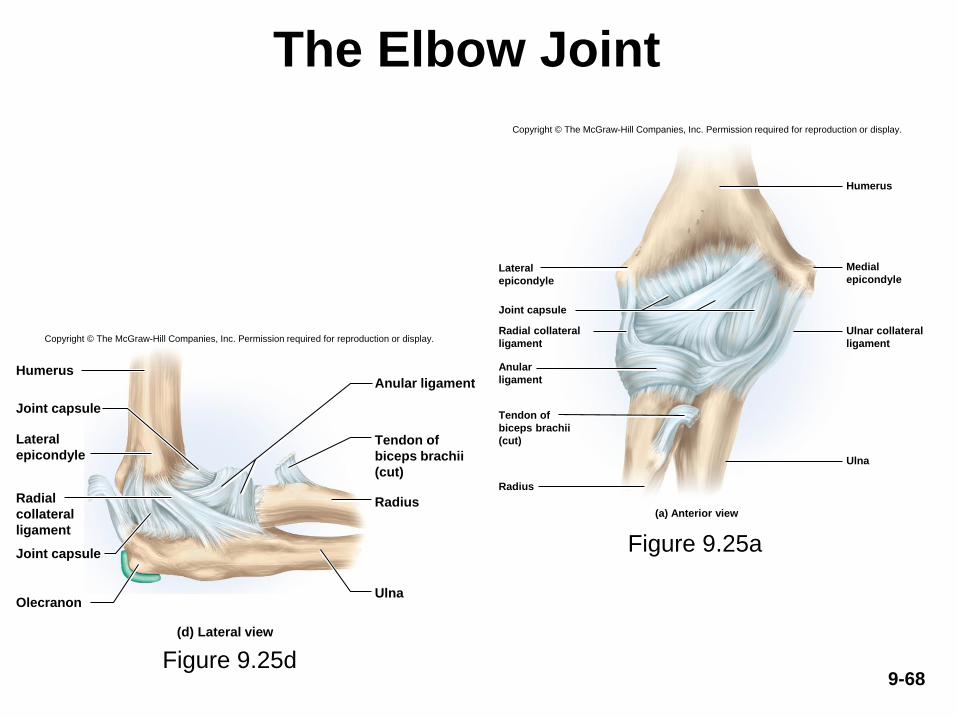
The Elbow Joint
Figure 9.25d
(a) Anterior view
Joint capsule
Humerus
Radius
Ulna
Lateral
epicondyle
Radial collateral
ligament
Anular
ligament
Tendon of
biceps brachii
(cut)
Medial
epicondyle
Ulnar collateral
ligament
Copyright © The McGraw-Hill Companies, Inc. Permission required for reproduction or display.
Figure 9.25a
Copyright © The McGraw-Hill Companies, Inc. Permission required for reproduction or display.
(d) Lateral view
Joint capsule
Humerus
Olecranon
Anular ligament
Radius
Ulna
Joint capsule
Tendon of
biceps brachii
(cut)
Lateral
epicondyle
Radial
collateral
ligament
9-69
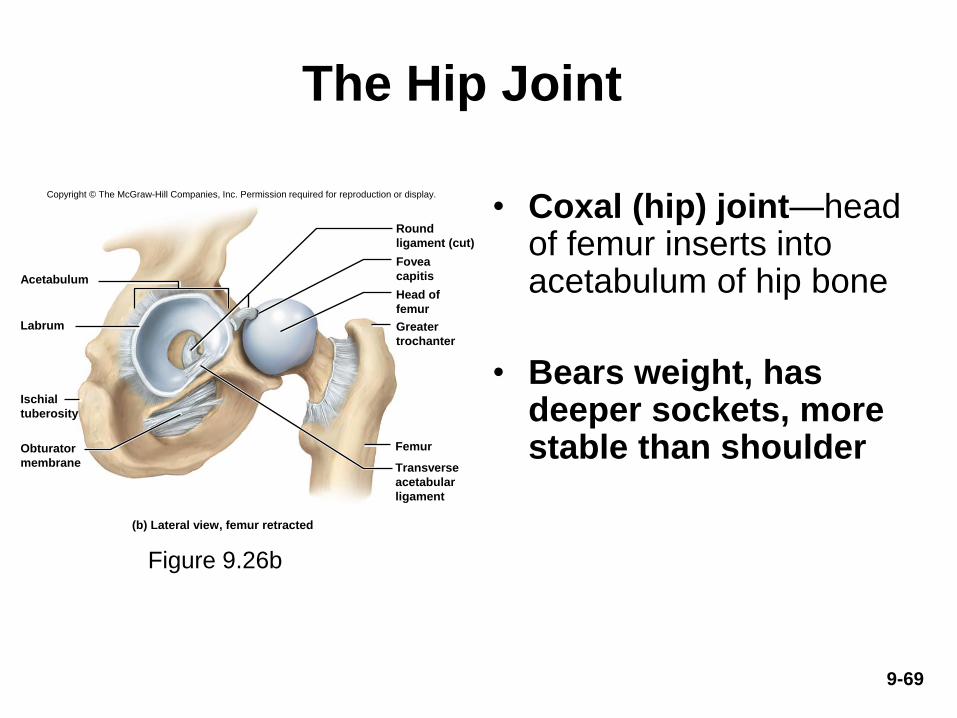
The Hip Joint
• Coxal (hip) joint—head of femur inserts into acetabulum of hip bone
• Bears weight, has deeper sockets, more stable than shoulder
Figure 9.26b
Copyright © The McGraw-Hill Companies, Inc. Permission required for reproduction or display.
Acetabulum
Labrum
Femur
Round
ligament (cut)
Fovea
capitis
Head of
femur
Greater
trochanter
Transverse
acetabular
ligament
Ischial
tuberosity
Obturator
membrane
(b) Lateral view, femur retracted

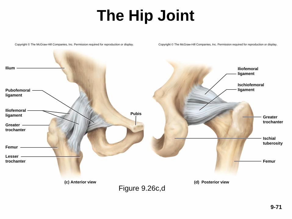
The Hip Joint• Acetabular labrum—horseshoe-shaped ring of
fibrocartilage that deepens socket– Dislocations are rare
• Ligaments supporting hip joint
– Iliofemoral and pubofemoral— anterior
– Ischiofemoral ligament—posterior
– When standing, ligaments become twisted and pull
head of femur tightly into acetabulum
– Transverse acetabular ligament bridges gap on
inferior margin of acetabular labrum
• Round ligament (ligamentum teres)—arises from fovea
capitis and attaches to lower margin of acetabulum
– Contains artery that supplies blood to head of femur9-70
9-71
The Hip Joint
Figure 9.26c,d
Copyright © The McGraw-Hill Companies, Inc. Permission required for reproduction or display.
Ilium
Femur
Pubis
Pubofemoral
ligament
Iliofemoral
ligament
Greater
trochanter
Lesser
trochanter
(c) Anterior view
Femur
(d) Posterior view
Iliofemoral
ligament
Ischiofemoral
ligament
Greater
trochanter
Ischial
tuberosity
Copyright © The McGraw-Hill Companies, Inc. Permission required for reproduction or display.

9-72
The Hip Joint
Figure 9.26a
Acetabular labrum
Acetabulum
Round ligament
Head of femur
Greater trochanter
Shaft of femur
(a) Anterior dissection© The McGraw-Hill Companies, Inc./Timothy L. Vacula, photographer

The Hip Joint
• Dislocation of hip is rare
• Some infants suffer
congenital dislocation
– Acetabulum is not deep
enough to hold head of femur
in place
• Harness, worn for 2 to 4
months can assist with
proper positioning
9-73Figure 9.27
9-74
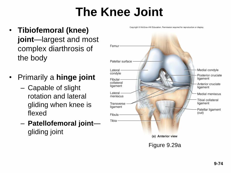
The Knee Joint
• Tibiofemoral (knee)
joint—largest and most
complex diarthrosis of
the body
• Primarily a hinge joint
– Capable of slight
rotation and lateral
gliding when knee is
flexed
– Patellofemoral joint—
gliding joint
Figure 9.29a

9-75
The Knee Joint
Figure 9.29
The Knee Joint
• Joint capsule encloses only the lateral and
posterior aspects of the knee
– Anterior aspect covered by patellar ligament and
lateral and medial retinacula
• All are extensions of the tendon of quadriceps
femoris muscle
• Knee stabilized by:– Quadriceps tendon in front
– Tendon of semimembranosus muscle on rear of thigh
9-76

9-77
The Knee Joint
• Lateral meniscus and medial meniscus—C-shaped cartilages within joint capsule
- Absorb shock and prevent side-to-side rocking
- Joined by transverse ligament
Figure 9.29a, 9.29d

9-78
The Knee Joint
• Popliteal (posterior) region
– Extracapsular ligaments
• Fibular (lateral) collateral
ligament
• Tibial (medial) collateral ligament
– Intracapsular ligaments cross
each other to form X
• Anterior cruciate ligament (ACL)
– Prevents hyperextension of
knee when ACL is pulled tight
– Common site of knee injury
• Posterior cruciate ligament
(PCL)
– Prevents femur from sliding off
tibia
Figure 9.29b

9-79
The Knee Joint
Figure 9.29c
Copyright © The McGraw-Hill Companies, Inc. Permission required for reproduction or display.
Femur
Meniscus
Tibia
Joint cavity
Infrapatellar fat pad
Synovial membrane
Patellar ligament
Patella
Prepatellar bursa
Articular cartilage
Joint capsule
(c) Sagittal section
Bursa under lateral
head of gastrocnemius
Quadriceps
femoris
Quadriceps
femoris tendon
Suprapatellar
bursa
Superficial
infrapatellar bursa
Deep
infrapatellar bursa
• Knee joint has at least 13
bursae
• Four anterior: superficial
infrapatellar, suprapatellar,
prepatellar, and deep
infrapatellar
• Popliteal region: popliteal
bursa and
semimembranosus bursa
• Seven more bursae on
lateral and medial sides of
knee joint
The Knee Joint
9-80
• Ability to lock and unlock knees
– Important aspect of human bipedalism
– When knee fully extended, ACL allows locking
• Femur rotates medially on the tibia, major knee
ligaments taut
– To unlock knee, popliteus contracts and rotates femur
laterally
• Lateral rotation of femur untwists ligaments

9-81
The Knee Joint
Figure 9.28
Femur:
Shaft
Patellar surface
Medial condyle
Lateral condyle
Joint cavity:
Joint capsule
Medial meniscus
Lateral meniscus
Lateral condyle
Tuberosity
Medial condyle
Patellar ligament
Articular facets
Lateral Medial
Anterior cruciate
ligament
Patella
(posterior surface)
Quadriceps
tendon (reflected)
© The McGraw-Hill Companies, Inc./Rebecca Gray, photographer/Don Kincaid, dissections
Tibia:
Copyright © The McGraw-Hill Education. Permission required for reproduction or display.
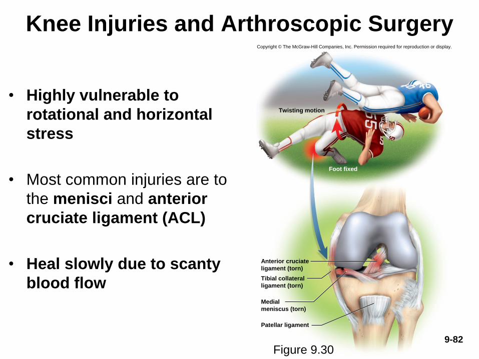
Knee Injuries and Arthroscopic Surgery
• Highly vulnerable to
rotational and horizontal
stress
• Most common injuries are to
the menisci and anterior
cruciate ligament (ACL)
• Heal slowly due to scanty
blood flow
Copyright © The McGraw-Hill Companies, Inc. Permission required for reproduction or display.
Foot fixed
Patellar ligament
Twisting motion
Anterior cruciate
ligament (torn)
Tibial collateral
ligament (torn)
Medial
meniscus (torn)
Figure 9.309-82

Knee Injuries and Arthroscopic
Surgery
• Arthroscopy—procedure in which interior of
joint is viewed with a pencil-thin arthroscope
inserted through a small incision
– Less tissue damage than conventional surgery
– Recover more quickly
– Arthroscopic ACL repair: about 9 months for healing
to be complete
9-83

The Ankle Joint
• Talocrural (ankle) joint—includes two articulations:
– Medial joint: between tibia and talus
– Lateral joint: between fibula and talus
• Both articulations enclosed by one joint capsule
• Malleoli of tibia and fibula overhang the talus on
either side and prevent side-to-side motion
• More restricted range of motion than the wrist
9-84
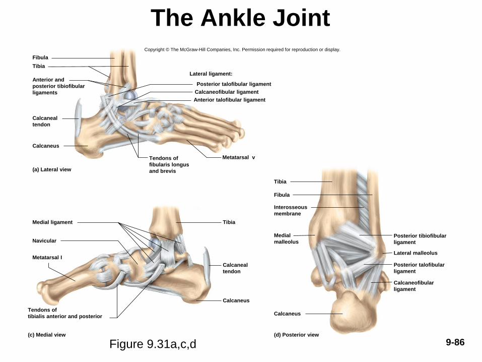
The Ankle Joint
• Ankle ligaments
– Anterior and posterior tibiofibular ligaments: bind
tibia to fibula
– Multipart medial (deltoid) ligament: binds tibia to the
foot on the medial side
– Multipart lateral (collateral) ligament: binds fibula to
the foot on the lateral side
– Calcaneal (Achilles) tendon: extends from the calf
muscles to the calcaneus
• Plantarflexes the foot and limits dorsiflexion
– Sprains (torn ligaments and tendons) are common at
the ankle
• Pain and immediate swelling9-85
9-86
The Ankle Joint
Figure 9.31a,c,d
Copyright © The McGraw-Hill Companies, Inc. Permission required for reproduction or display.
Posterior tibiofibular
ligament
Lateral malleolus
Posterior talofibular
ligament
Calcaneofibular
ligament
Calcaneus
(d) Posterior view
Medial
malleolus
Interosseous
membrane
Fibula
Tibia
Calcaneal
tendon
Calcaneus
Tibia
Tendons of
tibialis anterior and posterior
Metatarsal I
Navicular
Medial ligament
(c) Medial view
Calcaneofibular ligament
Anterior talofibular ligament
Posterior talofibular ligament
Tendons of
fibularis longus
and brevis
Metatarsal v
Calcaneus
Calcaneal
tendon
Anterior and
posterior tibiofibular
ligaments
Tibia
Fibula
(a) Lateral view
Lateral ligament:

9-87
The Ankle Joint
Figure 9.31b

9-88
Arthritis and Artificial Joints
• Arthritis—a broad term for pain and inflammation of joints
• Most common crippling disease in the United States
• Rheumatologists—physicians who treat arthritis and other
joint disorders
• Osteoarthritis (OA)—most common form of arthritis
– “Wear-and-tear arthritis”
– Results from years of joint wear
– Articular cartilage softens and degenerates
– Accompanied by crackling sounds called crepitus
– Bone spurs develop on exposed bone tissue causing pain
9-89
Arthritis and Artificial Joints
• Rheumatoid arthritis (RA)—autoimmune attack
against the joint tissues
– Misguided antibodies (rheumatoid factor) attack
synovial membrane, enzymes in synovial fluid
degrade the articular cartilage, joint begins to ossify
– Ankylosis: solidly fused, immobilized joint
– Remissions occur, steroids and aspirin control
inflammation
• Arthroplasty—replacement of diseased joint with
artificial device called prosthesis

9-90
Rheumatoid Arthritis
Figure 9.32a,b
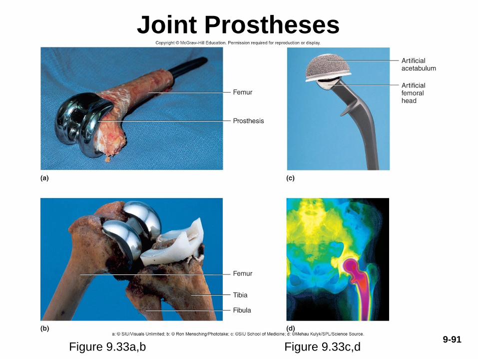
9-91
Joint Prostheses
Figure 9.33a,b Figure 9.33c,d