
Chapter 6, Section 2. Recording Electrodes can be inserted into the brain to record activity. –...
7
How Psychologists Study the Brain Chapter 6, Section 2
-
Upload
stuart-hudson -
Category
Documents
-
view
217 -
download
4
Transcript of Chapter 6, Section 2. Recording Electrodes can be inserted into the brain to record activity. –...

How Psychologists Study the Brain
Chapter 6, Section 2

Recording
• Electrodes can be inserted into the brain to record activity.– Can detect changes that occur when neurons fire.
• EEG: Attached to the scalp so many neurons can be monitored at once.
• Electrical activity of the brain rises and falls rhythmically.– Depends on wakefulness or sleepiness. – They are called brain raves.

Stimulation
• Electrodes can also set off the firing of neurons.
• Wilder Penfield– Stimulated the brains of patients during surgery to
determine functions various parts of the brain perform.

Lesions
• Cutting/destroying parts of the brain.– Sometimes scientists create these in animal
testing.
Accidents
• Learning from tragedy.• Phineas Gage, 1848– Respected railroad foreman, who executed good
judgment, and worked well with others.– Tamped a narrow hole with dynamite, and it
exploded. – Tamping iron weighed 13 lbs and 3 feet long.
Entered Gage’s head below the left eye, and exited through the top of the skull.
– Gage survived, but his personality changed greatly. Caused damaged to the frontal cortex.

• CT Scans– Moving ring passes X-ray
beams around and through a subject’s head. Radiation is absorbed, depending on brain tissue density.
– This produces a 3-D view of the brain.
• PET Scans– Injecting a radioactive
solution into the blood and measuring amount of radiation absorbed.• Active neurons absorb more
solution.
Images
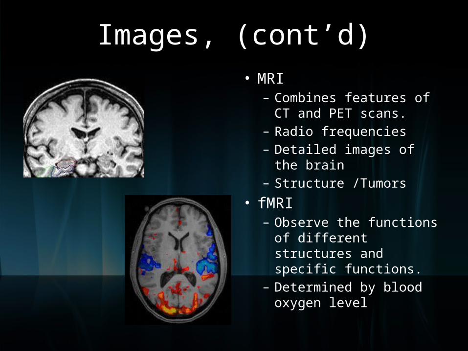
• MRI– Combines features of CT
and PET scans.– Radio frequencies– Detailed images of the
brain– Structure /Tumors
• fMRI– Observe the functions of
different structures and specific functions.
– Determined by blood oxygen level
Images, (cont’d)