CHAPTER 6 Response of Cells to Ionizing Radiation · 2010-08-12 · Abstract: Ionizing radiation is...
59
204 Advances in Biomedical Sciences and Engineering, 204-262 S C Tjong (Ed.) All rights reserved - © 2009 Bentham Science Publishers Ltd. CHAPTER 6 Response of Cells to Ionizing Radiation Wei Han, Kwan Ngok Yu* Department of Physics and Materials Sciences, City University of Hong Kong, Hong Kong *Corresponding author; E-mail: [email protected] Abstract: Ionizing radiation is encountered in our natural environment and is also generated and used by mankind, e.g., for medical uses. A better understanding of the biological effects of ionizing radiation will lead to better use of and better protection from radiation. In this chapter, the response of cells to radiation will be described and discussed. Some basic concepts of ionizing radiation will be briefly given in the beginning. The significant consequences of various types of radiation-induced DNA damages show that DNA is the principle target for biological effects of radiation. The misrepaired or unrepaired DNA damages, in particular DNA double strand breaks, will induce chromosomal aberrations and gene mutations. On the other hand, radiation-induced DNA double strand breaks play an important role in the induction of apoptosis and cell cycle arrest. Radiation- induced bystander response, adaptive response and genomic instability are currently “hot-pots” in the radiobiological research. These three phenomena indicate the complexity of cellular responses to radiation, and will be introduced and discussed in this chapter. BASICS OF RADIOBIOLOGY INTRODUCTION In 1895, the German physicist Wilhelm Conrad Röentgen discovered “a new kind of ray” which could blacken photographic films enclosed in a light-tight box. He named these rays as X-rays, which meant unknown rays. The first medical use of X-rays was reported in 1897, when a German surgeon, Wilhelm Alexander Freud, demonstrated the disappearance of a hairy mold after treatments with X-rays. In 1898, radioactivity was discovered by Antoine Henri Becquerel. In the same year, Pierre and Marie Curie discovered and isolated radium successfully. Just like other new discovers, the potential hazards of radiations or radioactive materials were not adequately acknowledged at the beginning of their discoveries. Along with the applications of radiation in industrial and medical areas, the hazards of radiations were revealed in some unique populations, such as uranium miners who were often exposed to high levels of radon progeny. Most of our understanding of radiation harm comes from several well-known acute exposures. The atomic bombings in Japan in 1945 and the cancer incidence in survivals and their progeny informed us of the potential hazard of nuclear radiations. The leakage of a nuclear
Transcript of CHAPTER 6 Response of Cells to Ionizing Radiation · 2010-08-12 · Abstract: Ionizing radiation is...

204 Advances in Biomedical Sciences and Engineering, 204-262
S C Tjong (Ed.)
All rights reserved - © 2009 Bentham Science Publishers Ltd.
CHAPTER 6
Response of Cells to Ionizing Radiation
Wei Han, Kwan Ngok Yu*
Department of Physics and Materials Sciences, City University of Hong Kong, Hong Kong
*Corresponding author; E-mail: [email protected]
Abstract: Ionizing radiation is encountered in our natural environment and is also generated and used
by mankind, e.g., for medical uses. A better understanding of the biological effects of ionizing
radiation will lead to better use of and better protection from radiation. In this chapter, the response of
cells to radiation will be described and discussed. Some basic concepts of ionizing radiation will be
briefly given in the beginning. The significant consequences of various types of radiation-induced
DNA damages show that DNA is the principle target for biological effects of radiation. The
misrepaired or unrepaired DNA damages, in particular DNA double strand breaks, will induce
chromosomal aberrations and gene mutations. On the other hand, radiation-induced DNA double
strand breaks play an important role in the induction of apoptosis and cell cycle arrest. Radiation-
induced bystander response, adaptive response and genomic instability are currently “hot-pots” in the
radiobiological research. These three phenomena indicate the complexity of cellular responses to
radiation, and will be introduced and discussed in this chapter.
BASICS OF RADIOBIOLOGY
INTRODUCTION
In 1895, the German physicist Wilhelm Conrad Röentgen discovered “a new kind of
ray” which could blacken photographic films enclosed in a light-tight box. He named
these rays as X-rays, which meant unknown rays. The first medical use of X-rays was
reported in 1897, when a German surgeon, Wilhelm Alexander Freud, demonstrated
the disappearance of a hairy mold after treatments with X-rays. In 1898, radioactivity
was discovered by Antoine Henri Becquerel. In the same year, Pierre and Marie
Curie discovered and isolated radium successfully.
Just like other new discovers, the potential hazards of radiations or radioactive
materials were not adequately acknowledged at the beginning of their discoveries.
Along with the applications of radiation in industrial and medical areas, the hazards
of radiations were revealed in some unique populations, such as uranium miners who
were often exposed to high levels of radon progeny. Most of our understanding of
radiation harm comes from several well-known acute exposures. The atomic
bombings in Japan in 1945 and the cancer incidence in survivals and their progeny
informed us of the potential hazard of nuclear radiations. The leakage of a nuclear

Response of Cells to Ionizing Radiation Advances in Biomedical Sciences and Engineering 205
power plant in Chernobyl, Ukraine, in 1986 demonstrated a risk of acute nuclear
exposure during the peacetime. The application of radiation in industry, medical
diagnostics and cancer therapy shows an indispensable role of radiation in our daily
lives. The present chapter will provide basic understanding of radiation biology to
enable safer and more effective applications of radiation. In this section, the basic
concepts of radiation are introduced [1].
TYPES OF IONIZING RADIATION
The raising of an electron in an atom or molecule to a higher energy level without
actual ejection of the electron is called excitation. If a radiation has sufficient energy
to eject one or more orbital electrons from an atom or molecule, the process is called
ionization, and the radiation is called an ionizing radiation. Ionizing radiation can be
broadly categorized into electromagnetic or particulate radiations.
Electromagnetic Radiation
X- and -rays are two forms of electromagnetic radiation, which are commonly
employed in medical and biological applications. X- and -rays do no differ in nature
or properties; the designations x- or -rays merely reflect the way they are produced.
X-rays are produced outside the atomic nucleus while -rays are produced within the
nucleus.
Fig. 1. The wavelengths of various electromagnetic radiations.
������������� �
�������������������������
������������� �
��
������
����
����
���
���
���
���
���
���
��
��
���
��
�
�!���!���"#
!���"#
$�%%���"#
&����"#
#�%
������
'����(
)����
*�+�
,����
&����-,
!���-,
���� ��-,
#�%�������
$����(����
"�.����%��"#�
-����������-,�
/0��1�
)� �#�1�
2�� ��#�1�
,���3������
��
��
��
��
��
��
��
�
��
��
��
��
��
4��� �����5 �
$����� �
$���� ����� �
$���� ������ �
&��� ������ �
6����� ��6�
��
��
��
7
�
�
�
0�
0�
0�
07
0��

206 Advances in Biomedical Sciences and Engineering Han and Yu
X-rays are usually produced in an X-ray tube when accelerated electrons hit a metal
target like tungsten and are decelerated, thereby emitting a spectrum of
bremsstrahlung radiation. Here, part of the kinetic energy of the electrons is
converted into X-rays. (Bremsstrahlung photons are generated when a charged
particle is accelerated or decelerated.) The emitted spectrum is filtered or modulated
to produce a clinically useful X-ray beam. Gamma-rays, in contrast, are produced
spontaneously. They are emitted by radioactive isotopes when their nuclei in excited
states give off the excess energy and return to the ground state. Radio waves, infrared
and visible light are also electromagnetic radiation. All electromagnetic radiations
have the same velocity, but with different frequencies and wavelengths (Figure 1).
The photon energy is proportional to the frequency and inversely proportional to the
wavelength. Usually, electromagnetic radiations are considered ionizing if they have
photon energies larger than 124 eV, which corresponds to a wavelength shorter than
about 10-8
m. In this way, only X-rays and -rays are considered to be involved in
electromagnetic-radiation induced biological effects. Other electromagnetic
radiations, of which the wavelengths are smaller than about 10-8
m, are not covered
(Figure 1).
Particulate Radiations
Another type of ionizing radiation encountered in our nature, or used experimentally
or clinically is particles such as electrons, protons, particles, heavy charged ions and
neutrons.
ABSORPTION OF RADIATION
The process through which X-ray photons are absorbed depends on the energy of the
photons and the chemical composition of the absorbing material. In the high-energy
range most widely used in radiotherapy (viz., 100 keV-25 MeV), the Compton effect
dominates the energy deposition in tissues. Part of the photon energy is given to the
electron as kinetic energy and the photon with the remaining energy continues on its
way but is deflected from its original path. In diagnostic radiology, lower photon
energies are used, for which both Compton and photoelectric absorption processes
occur, with Compton absorption dominating at the higher end of the energy range and
photoelectric effect being more important at lower energies. In the photoelectric
absorption process, the photon gives up all its energy to a bound electron; a part is
used to overcome the binding energy of the electron while the remaining energy is
given to the electron as kinetic energy.
Gamma-ray photons with energy >1.02 MeV may interact with a nucleus to form an
electron-positron pair. Beyond the energy provided to the rest masses of the electron
and positron (0.51 MeV), the excess amount will be carried away equally by these
two particles. The positron is eventually captured by an electron, and annihilation of

Response of Cells to Ionizing Radiation Advances in Biomedical Sciences and Engineering 207
the two particles releases two photons (each with an energy of 0.51 MeV). These two
photons can further lose energy through Compton scattering or photoelectric effect.
DIRECT AND INDIRECT ACTIONS OF RADIATION
The biological effect of radiation can be related to damages of the DNA. Any form of
radiation has a potential to directly interact with target structures to cause ionization,
thus initiating the chain of events that leads to biological changes. This is called the
direct action of radiation, which is the dominant process for radiations with high
linear energy transfer (LET), such as neutrons or particles.
Radiation can also interact with atoms or molecules in a cell (particularly with water)
to produce free radicals, which are able to diffuse far enough to interact with the
critical targets and cause damages. This is called indirect action of radiation. Free
radicals have unpaired electrons, which result in high chemical reactivity. Most of the
energy deposited in cells is absorbed initially in water, which is the main component
of cells, leading to a rapid production of oxidizing and reducing reactive hydroxyl
radicals. Hydroxyl radicals (·OH) may diffuse over distances to interact with DNA to
cause damages. Fortunately, some defensive systems or responses in cells can protect
the cells from the damages. Thiol compounds in cells, such as glutathione and
cysteine, which contain sulfhydryl groups, can react chemically with the free radicals
to reduce their damaging effects. Vitamins C and E and intracellular manganese
superoxide dismutase (MnSOD) also have an ability to scavenge free radicals. In
general, the contribution of free radical processes for sparsely ionizing radiation
exceeds that for direct action of radiation.
RADIATION-INDUCED DNA DAMAGES IN CELLS
Accumulated evidence in radiological studies indicates that DNA is the principle
target for the biologic effects of radiation. Radiation-induced DNA lesions through
induction of gene mutation and chromosome aberration are fundamental to
investigating and understanding radiation-induced cell killing, cell transformation and
carcinogenesis. In this section, we focus on radiation-induced DNA lesions, repair,
and damage signal transduction, as well as cellular response to DNA damages [3].
DNA STRUCTURE AND RADIATION-INDUCED DNA DAMAGES
Introduction to DNA structure
DNA is a large molecule with a double helix structure. It consists of two long
polynucleotide chains, each of them containing four types of nucleotide subunits. The
two chains are held together by hydrogen bonds between the bases of the nucleotides.
Nucleotides form the “backbone” of the DNA, and are composed of a five-carbon

208 Advances in Biomedical Sciences and Engineering Han and Yu
sugar to which one or more phosphate groups and a nitrogen-containing base are
attached. The genetic codes of DNA are represented by groups of four kinds of bases
with specific sequences. In DNA, the sugar is deoxyribose (and hence the name
deoxyribonucleic acid), which is attached to a single phosphate group and the base
can be adenine (A), cytosine (C), guanine (G) or thymine (T). The nucleotides are
covalently joined together in a chain through the sugars and phosphates, and form a
backbone of alternating sugar-phosphate-sugar-phosphate chain.
Each sugar has a phosphate group at the 5’ position on one side and a hydroxyl group
at the 3’ on the other, and each completed chain is formed by linking the 5’ phosphate
to the 3’ hydroxyl so that all of the subunits are lined up in the same orientation. This
polarity of the DNA chain is defined by referring to one end as the 3’ end and the
other as the 5’ end. The bases, A, C, T, or G are arranged in specified sequences to
form the code that stores the biological messages, and the DNA messages in turn
encode proteins to perform biological functions. The complete set of information in
the DNA of an organism is called its genome. In other words, the genome carries the
information for all the proteins the organism will synthesize.
The three-dimensional structure of DNA, viz., the double helix, arises from the
chemical and structural features of the two polynucleotide chains. Since the two
chains are held together by hydrogen bonds between the bases (A and T; G and C) on
the opposite side, all the bases are on the inside of the double helix while the sugar-
phosphate backbones are on the outside. In each case, a bulkier two-ring base (a
purine) is paired with a single-ring base (a pyrimidine). The two sugar-phosphate
backbones wind around each other to form the double helix, with one complete turn
every 10 base pairs. The members of each base pair can fit together only if the two
strands are anti-parallel, i.e., if the polarity of one strand is oriented in a direction
opposite to that of the other. In other words, each strand contains a sequence of
nucleotides which is exactly complementary to that of the opposing strand.
Introduction to radiation-induced DNA damages
In the early days, radiation-induced DNA damages were studied under two different
conditions, viz., irradiating the DNA molecule itself (direct effect) or irradiating it in
a dilute aqueous solution (indirect effect). The direct effect was a result of direct
deposition of energy in the DNA, while the indirect effect was a result from reactions
of radicals produced by ionizing the molecules in the solution.
An ionization can occur in any molecule in the cell creating a cation radical and an
electron. The produced electron can attach to another molecule or it can become
solvated before further reactions. The radical site can be transferred to another nearby
molecule. At the same time, the cation radicals can react and become neutralized
through losing a proton, such as in the case of the cation produced by ionization of

Response of Cells to Ionizing Radiation Advances in Biomedical Sciences and Engineering 209
water. H2O+ reacts with a neighboring water molecule immediately (10-14 s) to
produce the (·OH). As a result of these reactions, cellular DNA can be damaged in
different ways, including direct ionization of the DNA, reactions between the DNA
and electrons or solvated electrons, ·OH or H2O+, or other radicals.
Radiation damages leading to cell deaths were studied in Escherichia coli by
Johansen [4]. In this study, Escherichia coli was protected against radiation killing
through scavengers of free radicals added to the medium. Approximately 65% of the
cell killing was in fact found to be caused by the ·OH radicals, while no
radioprotection was found for scavengers of electrons. Subsequently, similar
experiments [5,6] were carried out in mammalian cells and the same conclusion was
reached, i.e., 65% of the cell killing was caused by ·OH radicals. These studies were
then extended to other systems. For low LET radiation such as X-rays or -rays, high
concentrations of ·OH radical scavengers can reduce the yield of DNA breaks,
chromosome aberration and mutations besides protection against cell killing. Based
on these data, damages can also be classified as scavengable and non-scavengable.
TYPES OF DNA DAMAGES
It is now well known that radiation produces a wide spectrum of DNA lesions,
including damages to nucleotide bases (base damages), DNA single and double-
strand breaks (SSBs and DSBs).
Base damages or modifications
Damages to bases by ionizing radiation have been extensively studied in vitro by
irradiation of free bases, nucleosides, oligonucleotides or DNA in aqueous solution or
in the solid state. The chemistry of lesion formation is already relatively well
understood, and detailed accounts can be found in the literature [7-9]. Although
certain types of DNA base damages such as 8-hydroxydeoxyguanosine have
significant biological significance, available data indicate that such isolated base
damages probably have a minor role in radiation mutagenesis [10]. As such, we will
not go into detailed discussions on radiation-produced base damages here. In general,
the damaged bases can be repaired through the base excision repair pathway.
DNA single strand break
An SSB is formed by abstraction of any of the deoxyribose hydrogens [10]. The
initial radical reactions involve abstraction of a hydrogen atom from the deoxyribose
moiety by an ·OH radical [7]. The radical can then react with an oxygen molecule and
form a peroxy radical. Damages to DNA bases will result in destabilization of the N-
glycosidic bond and abasic deoxyribose residues are then formed. Other lesions,
which are generated by ·OH radicals, can be converted into strand breaks by

210 Advances in Biomedical Sciences and Engineering Han and Yu
treatment with hot alkalis, so these break sites are referred to as alkali-labile sites.
Irradiation in the presence of oxygen will increase the number of alkali-labile sites
[11]
Studies on radiation damages to individual sites in DNA suggest that these lesions are
not important in mammalian cells. The DNA SSBs generated in mammalian cells
through hydrogen peroxide treatment at 0 oC were found not to cause cell killing
[12]. On the other hand, production of these breaks is inhibited by an ·OH radical
scavenger, implying an ·OH radical as an intermediate reactive species causing the
breaks. At a lethal dose, the number of singly damaged sites present in each cell is in
the order of 105-10
6, while the number of multiply damaged sites (MDS) such as
DSB is smaller than l00. It has been established that most of the strand breaks
induced by ionizing radiation are repaired by a DNA ligation step [7].
DNA double strand break
A DSB is the most important lesion produced by ionizing radiation where the breaks
in the two strands are opposite to each another, or separated by only a few base pairs.
Unrepaired or mis-repaired DSBs are the most important lesions in the induction of
chromosomal abnormalities and gene mutations [13,14]. Furthermore, the close
association of radiation damages, which create “clustered” damages, has recently
become recognized as an important feature of radiation damage. Such clustered
damages can arise from the combination of direct damages induced by the track of
radiation, and indirect damages induced by secondary reactive species which are
produced by subsequent ionization events [15,16]. Clustered damages can involve
SSBs or DSBs associated with base damages, or more complex associations including
multiple closely scattered DSBs. The LET of the radiation determines the frequency
and complexity of clustered damages. Modeling studies have shown that about 30%
and 70% of DSBs induced by low and high LET radiations, respectively, are of a
complex form involving two or more DSBs. If the breaks associated with base
damages are included, the proportions will become 60% and 90% for low and high
LET radiations, respectively [17-19]. When the damages are more complex, it is
more difficult for them to be repaired and they are more likely to lead to biological
consequences.
The similarity between the damages induced by exposures to low dose irradiation and
those induced endogenously is important for the studies of the damages. It is known
that a large amount of oxidative damages can be inflicted in the cells by reactive
oxygen species (ROS) generated during normal cellular metabolism. These ROS
induced damages appear to be similar to those induced by ionizing radiations (IRs),
although there are also important differences. One important aspect is the nature of
the termini for ROS- and IR-induced damages, which can affect the repair process.
The breaks induced by restriction enzymes have 3’-hydroxyl and 5’-phosphate

Response of Cells to Ionizing Radiation Advances in Biomedical Sciences and Engineering 211
moieties at the termini, which is a prerequisite for enzymatic ligation. On the other
hand, the majority of breaks generated by ROS and IR have “damaged” termini, most
frequently with 3’-phosphate or 3’-phosphoglycolate end groups [10]. Some 5’
termini with hydroxyl end groups are also formed. Such termini will require
processing before ligation. Excision of a damaged nucleotide will likely lead to base
loss at the break. The types of DNA damages induced by ROS and IR are also
different. Base damages and SSBs dominate the ROS-induced damages. The
frequency of DSBs generated by ROS varies for different reactive species but is
typically less that 0.5% of the induced damages, and these DSBs are distributed
relatively uniformly throughout the DNA. On the contrary, even low doses of IR can
lead to complex lesions with clustered damages due to inhomogeneous energy
deposition.
DNA-protein cross-links (DPC)
Cross-linking of DNA to nuclear proteins will affect DNA processes such as
replication, transcription and repair, but the role of DPCs in the response to IR is not
clear. Twenty-nine proteins were found to have cross-linked to DNA, including
structural proteins, regulators of transcription, stress response, and cell cycle
regulatory proteins [20]. A linear dose-response relationship was identified for
hamster and human cells in the low dose range (0-1.5 Gy), and no difference in the
DPC induction was revealed under aerated and hypoxic conditions.
DNA DOUBLE-STRAND BREAK REPAIR
DSBs induced by ionizing radiation and other carcinogenic chemicals are considered
the most relevant lesion for mutations and carcinogenesis. DSBs can also be
generated in a number of natural processes including replication, meiosis, and
production or formation of antibodies.
Unrepaired and misrepaired DSBs are serious threats to the genomic integrity [21].
DSBs lead to chromosomal aberrations which can simultaneously affect many genes
and cause malfunction and death in cells [22]. It is noted here that DSBs are
continuously induced in normal living organisms as a consequence of oxidative
metabolisms [23,24].
Genome protection requires the capability to repair DSBs and to ensure that repair is
performed with sufficient fidelity. There are two main pathways of DSB repair,
namely, homologous recombination (HR) and nonhomologous end joining (NHEJ),
which are error-free and error-prone, respectively. The pathways are conserved from
Saccharomy cescervisae to mammalian cells, despite the different relative
importance. It is generally considered that HR and NHEJ dominate DSB repairs in

212 Advances in Biomedical Sciences and Engineering Han and Yu
yeast and in mammalian cells, respectively. DSB repair has been the subject of a
number of excellent reviews [25-31].
Homologous recombination
HR is a high-fidelity and efficient mechanism to repair DNA DSBs. HR retrieves the
information lost at the break site from the undamaged sister chromatid or homologous
chromosome. In the course of HR, the damaged DNA physically contacts an
undamaged DNA with a homologous sequence, and uses it as a template for repair.
From yeast to mammalian cells, HR is mediated through the Rad 52 epistasis group
of proteins that includes Rad 50, Rad 51, Rad 52, Rad 54, and meiotic recombination
11 (Mre11). Rad family proteins are Ras-related proteins which lack typical C-
terminal amino acid motifs for isoprenylation.
HR starts with a nucleolytic resection of the DSB in the 5’-3’ direction by the Mre11-
Rad50-Nbs1 (MRN) complex. The 3’ single-stranded DNA is bound by a heptameric
ring complex formed by Rad 52 proteins, which protects it against exonucleolytic
digestion. The competition between Rad 52 and the Ku complex for binding to DNA
ends may determine whether the DSB is repaired via the HR or the NHEJ pathway.
Single-strand annealing (SSA) is a process for rejoining DSBs by exploiting the
homology between the two ends of the joined sequences. The process relies on
regions of homology to align the DNA strands to be rejoined. Single stranded regions
are created adjacent to the break that extends to the repeated sequences. When this
process has proceeded far enough to reveal the complementary sequences, the two
DNAs are annealed and then ligated. The genes which define SSA belong to the Rad
52 epistasis group of HR.
Nonhomologous end joining (NHEJ)
DNA repair via the NHEJ pathway is rough and emergent. The process rejoins the
two ends of a DSB without the requirement of sequence homology between the two
ends. The process can be described as a series of steps:
Initial step: binding of a heterodimeric complex to the damaged DNA. The complex
consists of the proteins Ku70 and Ku80 (alias XRCC5). Binding of the complex will
protect the DNA from exonuclease digestion. In the binding, Ku80 is distal to while
Ku70 is proximal to the DNA break. The Ku70/Ku80 heterodimer can translocate
from the DNA end in an ATP-independent manner.
Formation of DNA-PKcs. Following the binding, the Ku heterodimer associates with
the catalytic subunit of DNA-PK (XRCC7, DNA-PKcs) to form the active DNA-PK
holoenzyme. DNA-PKcs is activated by interaction with a single-strand DNA at the
site of DSB and displays Ser/Thr kinase activity.

Response of Cells to Ionizing Radiation Advances in Biomedical Sciences and Engineering 213
Linkage of two ends. XRCC4 forms a stable complex with DNA ligase IV, and this
complex binds to the ends of DNA molecules and links together duplex DNA
molecules with complementary but non-ligatable ends. The XRCC4–ligase IV
complex cannot directly re-ligate most DSBs generated by DSB inducing agents.
Instead, they have to be processed first. In yeast, DSB processing is performed by the
MRN complex which removes excess DNA at 3’ flaps. Furthermore, 5’ flaps are
removed by the flap endonuclease 1 (FEN1). Deficiency of this protein will result in
reducing the usage of the NHEJ pathway.
Removal of NHEJ related factors. The NHEJ related factors must be removed from
the DNA before the re-ligation of the DSBs. The auto-phosphorylation of DNA-PKcs
and/or DNA-PK mediating the phosphorylation of accessory factors are important in
the release of DNA-PKcs and Ku from the DSB before end-joining. Finally, the DSB
repair is completed, although nucleotides are often lost, which results in an inaccurate
repair.
SENSING AND SIGNALING TRANSDUCTION OF DNA DOUBLE-STRAND
BREAK AFTER RADIATION
Sensing and Signaling: Activation of Related Factors
The core components of responses to DNA damages include the signal, sensors of the
signal, transducers and effectors. Recent studies have also identified another class of
molecules, mediators (also called adaptors), which do not possess catalytic activities
but facilitate signaling through promoting physical interactions between other
proteins. In the next part, the main sensors, mediators and effectors will be briefly
introduced.
(I) Role of ATM/ATR
In response to DSBs, ataxia-telangiectasia mutated (ATM) shows distinct changes
that include monomerization of dimers/oligomers and intermolecular auto-
phosphorylation on Ser1981. ATM was first discovered in ataxia-telangiectasia (A-
T), a multi-system disorder associated with diverse characteristics that include cancer
predisposition and clinical radiosensitivity [32]. The cells derived from A-T patients
show defects in the cellular response to DSBs in the activation signaling pathways,
including the ability to arrest at the G1/S, S and G2/M cell cycle checkpoints [33-35].
ATM can play as a Ser-Thr protein kinase both in vivo and in vitro, and can
phosphorylate the serine 15 residue of p53 specifically. p53 regulates the cell cycle
and thus functions as a tumor suppressor which that is involved in preventing cancers
[36-38]. In contrast to normal cells, p53 levels in A-T cell lines are not elevated
following radiation. This suggests that ATM acts in the upstream of p53 as an early

214 Advances in Biomedical Sciences and Engineering Han and Yu
damage sensor in response to radiation-induced DSBs [39,40]. Further studies
showed the complex function of ATM in sensing DSBs. Phosphorylation of serine 15
residue was not a key factor in controlling the p53 stability, and ATM could also
phosphorylate other sites on p53. ATM could also phosphorylate other kinases such
as Chk1 and Chk2, and kinases could phosphorylate p53 on serine 20 residue to keep
the stability of p53. Other kinases including DNA-PK and ATR could phosphorylate
the serine 15 residue of p53. Furthermore, ATM could also phosphorylate MDM2,
which could affect the stability of p53. Thus, ATM/ATR were important through
phosphorylation in regulating p53 in a direct or indirect manner. These findings
suggested that ATM was critical in sensing DSBs and in initiating signal transduction
pathways by phosphorylation to control cell cycle arrest.
The MRN complex is required in many mammalian cell lines to activate ATM in
response to DSBs under normal conditions [41-43]. Recent studies indicated that
MRN/MRX complexes activated ATM by independently promoting both
monomerization and auto-phosphorylation. First, the MRN complex mediated
monomerization of ATM in vitro in the occurrence of dense DSBs [44,45]. One role
of MRN/X might be to tether DNA and to increase the local concentration of DSBs to
allow ATM monomerization, which was revealed based on the findings that MRX
proteins in budding yeast could associate with DSBs in vivo and purified MRN
complexes could tether DNA molecules in vitro [46-49]. Another role of MRN was to
promote ATM kinase activity via binding of Nbs1 to ATM. Binding to Nbs1 was
required in the auto-phosphorylation of ATM on Ser1981 residue in vitro regardless
of the DSB concentration [44]. Association of MRN/X with DSB sites would help
ATM target the proper sites to execute its function.
In contrast to ATM, ATR showed no changes in modification or activity after a
genotoxic stress. ATR relocalized on DSB sites via the association with ssDNA-RPA
complexes. ToBP1 (Topoisomerase II Binding Protein 1) was found to act as a
mediator, binding to and stimulating the kinase activity of Xenopus and human ATR
in vitro [50,51].
(II) Role of Nbs1, hMre11 and hRad50
Nijmegen Breakage Syndrome (NBS) is a phenomenon associated with cancer
predisposition and radiosensitivity [52,53]. The cell lines derived from A-T and NBS
have similar phenotypes such as radiosensitivity, cell cycle checkpoint deficiency and
decreased ability to stabilize p53. The gene defective in NBS was indicated to encode
protein Nbs1 or p95 [54,55]. Nbs1 interacts strongly with hMre11 and hRad50 to
form an MRN complex in sensing and repairing of DSBs. The finding that mutations
in the ATM gene resulted in A-T and Mre11 gene mutations and caused A-T-like
disorder (ATLD) further strengthened the link between A-T and NBS [56]. hMre11
and hRad50 null mice showed embryonic lethality. Mutations in hMre11 in ATLD

Response of Cells to Ionizing Radiation Advances in Biomedical Sciences and Engineering 215
impaired but did not inactivate hMre11 function, a feature which was consistent with
the milder clinical features of this variant class of A-T. NBS cells also had cell-cycle
checkpoint defects in response to ionizing radiation, and these defects were notably in
the S-phase checkpoint to show the radioresistant DNA synthesis [57]. The complex
of hMre11, hRad50 and p95 (MRN complex) co-localized at the DSB sites [58]. In
yeast and vertebrates, MRX played an important role in both HR and NHEJ pathways
[59]. However, in mammalian cells, it was not an essential component of the NHEJ
pathway [60]. There was also evidence that the MRN complex played an important
role in either directly activating ATM or in aiding ATM-dependent phosphorylation
events [61,43]. As such, MRN is activated with a number of damage response factors
including p53 via dependent and independent processes, and acts with ATM as an
early sensor complex.
(III) Role of H2AX
Mice lacking H2AX are viable but show genomic instability and radiosensitivity [62].
H2AX is a member of the histone H2A family, and the histone H2A together with the
other members of the histone family provide the backbone for wrapping of the DNA.
H2AX becomes phosphorylated to -H2AX in response to DNA damages and is
critical in the recruitment of repair factors at DSB sites [62,63]. H2AX
phosphorylation is a rapid response following formation of DSBs. Phosphorylation
will extend rapidly to H2AX molecules located up to 3 megabase pairs beyond the
DSB site [64]. -H2AX can be observed as discrete foci under a fluorescent
microscope through immuofluorescence with the use of phosphospecific antibodies.
All DSBs are marked by the presence of such foci and the number of -H2AX foci
equals the number of DSBs [65]. -H2AX is also important in recruiting some repair
factors such as Rad 51 and MRN complex to localize at the site of broken DNA [66].
After the DSB repair, -H2AX will dephosphorylate to its original form, i.e., H2AX.
The analysis of foci number is used as a tool to monitor the formation and repair of
DSBs.
(IV) BRCA1 and BRCA2
BRCA1 and BRCA2 are genes deficient in familial breast cancer patients. Germline
mutations in BRCA1 and BRCA2 induced a high risk of breast and ovarian tumors
[67,68]. There were evidences that both gene products were involved in damage
response mechanisms and that cells deficient in either protein showed pronounced
genomic instability [69]. BRCA1 deficient cells showed marked genomic instability,
impaired ability in the HR pathway [70] and impaired checkpoint function, including
impaired S and G2/M checkpoint arrest [71]. BRCA1 was phosphorylated after DSB
induction and localized to H2AX foci after DNA damage and thus co-localizes with
MRN complex, 53BP1 and the mediator of DNA damage checkpoint 1 (MDC1) [63].
BRCA1 was also found necessary to facilitate some ATM-dependent phosphorylation
events to help other repair related factors localize to the H2AX foci [72,73]. All these

216 Advances in Biomedical Sciences and Engineering Han and Yu
results suggested that BRCA1 was critical in controlling checkpoint signaling and in
triggering the HR pathway. On the other hand, cells deficient in BRAC2 did not
appear to show cell cycle checkpoint defects but showed an impaired ability in the
HR pathway [74]. BRCA2 was proposed to be required for the localization of Rad 51
to the sites of single stranded DNA since Rad 51 foci did not form in BRCA2
defective cells [75,76].
(V) MDC1 and 53BP1
53BP1 was originally identified through its ability to bind to p53 via C-terminal
BRCT repeats present in 53BP1 [77]. MDC1 was identified as a binding partner of
the Mre11 complex simultaneously by several laboratories. The encoded protein is
required to activate the intra-S phase and G2/M phase cell cycle checkpoints in
response to DNA damages [78,79]. Evidence showed that both proteins form foci and
co-localize with H2AX and MRN foci at the sites of DSB after irradiation [80-82]
and these proteins were required for the normal function of checkpoint responses.
Lack of these proteins would lead to at least some radiosensitivity.
(VI) Role of p53
An increase in the p53 levels was found as an early response of mammalian cells
(within minutes) to DNA damages [83]. p53, known as protein 53 or tumor protein
53, is a transcription factor which in humans is encoded by the TP53 gene. Changes
in p53 expression result in the transcription of some key proteins involved in a
number of distinct damage response pathways, since the capability of p53 functioning
as a transcriptional activator may also be increased [84,85] after the formation of
damages. The role of p53 in the response to radiation damages is complex since it
affects some aspects of DNA repair, controls checkpoint cell cycle arrest and initiates
apoptosis, etc. [86]. Studies on patients deficient in p53 (Li-Fraumeni syndrome
patients) and p53 knock-out mice [87-89] demonstrated the importance of p53 in
damage response mechanisms. In addition, mutations in p53 are found in around 40%
of tumors covering all the cancer types.
Regulation of p53 is strict and the network of p53 regulation in mammalian cells is
complex [84,85]. Mdm2 was found as a key protein in controlling p53 [90], and
binding of Mdm2 to the amino-terminus of p53 would target it for ubiquitination and
subsequent degradation by ubiquitin controlled proteosomes ([91]. Knock-out mice
for Mdm2 were found to be embryonic lethal due to the high endogenous levels of
p53, but double mutant p53/Mdm2 knock out mice were found to be viable.
Mutations in Mdm2 were commonly found in tumors, in particular those with normal
p53 function. In normal undamaged cells, p53 is maintained at low levels via Mdm2
binding and ubiquitin-dependent degradation. In response to radiation damages,
Mdm2 negatively regulates both stabilization of p53 and its function. Changes to p53

Response of Cells to Ionizing Radiation Advances in Biomedical Sciences and Engineering 217
and/or Mdm2 decrease their binding potential with subsequent increase in the lifetime
of p53, and Mdm2 binding suppresses p53 as a transcription activator [92].
Cell Cycle Arrest after Radiation
Mammalian cell cycle contains G1, S, G2 and M phases, and variations in
radiosensitivity are shown in the different phases. The following views concerning
the different radiosensitivities associated with different phases of the cell cycle are
widely accepted in cellular radiobiology [93,94]:
• mitotic cells are the most radiosensitive;
• if G1 has an considerable length, there is normally a period with a resistance,
which declines towards the S phase;
• the resistance increases in the S phase, with a maximum increase in the latter
part of the phase;
• the G2 phase is almost as sensitive as the mitotic phase.
Progression from one phase to the next occurs by phosphorylation or
dephosphorylation of cyclin dependent kinases (Cdks). DNA damages will often lead
to arrest at cell cycle checkpoints, which are also called DNA integrity checkpoints.
Besides checkpoints at the boundaries between adjacent phases, there is also an S
phase checkpoint that recognizes a stalled replication fork. These checkpoint
responses have been extensively studied in Saccharomyces cerevisiae or
Schizosacchromyces pombe, but the operation of checkpoints is also evident in
mammalian cells. In the following, the processes in mammalian cells will be briefly
reviewed.
The checkpoint responses are conserved between organisms, although the multiple
steps and mechanisms are less well understood in mammalian cells [95,96]. In
mammalian cells, PI3-K related protein kinases (PIKK kinases), ATM (ataxia
telangiectasia mutated protein)/ATR (ataxia telangiectasia and Rad3-related protein)
are important in controlling checkpoint responses. These pathways target p53 which
then mediate permanent cell cycle arrest or apoptosis besides induction of transient
delays at cell cycle transitions.
(I) G1/S arrest
Two types of G1/S arrest were observed in mammalian cells, namely, prolonged
arrest and a more transient response [97,98]. The former is a p53-dependent response
and the latter is similar to the G1/S response observed in yeast. Protein 21 (p21) is a
major p53 response protein required for G1/S arrest [99], and p53 regulates strictly
p21 as a transcription factor. Neither p53 nor ATM null cells showed a prolonged
radiation induced G1/S arrest. p21 is an inhibitor of cyclin dependent kinases. It plays
an important role in the G1/S arrest by binding to the cyclin D- and cyclin E-
associated kinases, and by inhibiting the ability to phosphorylate the retinoblastoma

218 Advances in Biomedical Sciences and Engineering Han and Yu
gene product (pRb) [100]. p21 has another role in controlling growth arrest by
preventing proliferating cell nuclear antigen (PCNA) from activating DNA
polymerase, which is essential in DNA replication. G1/S arrest after radiation does
not help in the survival of cells, based on the observation that p53 null cell lines or
transformed fibroblasts (which normally lack this arrest due to p53 inactivation)
showed elevated radioresistance compared to p53 wild cells [101].
(II) S phase arrest
Cell cycle arrest in the S phase enables repairs of the radiation-induced damages
before these are permanently fixed by DNA replication into irreparable chromosomal
breaks. Among all cellular checkpoints, the S-phase checkpoint is the most complex.
Evidence showed that early S phase arrest after irradiation was ATM-dependent but
later S phase arrest was associated with ATR [102]. Cells deficient in genes of ATM
and NBS displayed a phenotype called radioresistant DNA synthesis, and S phase
arrest was not observed before the damages were repaired [103]. S phase arrest
included inhibition of ongoing replication forks, stabilization of replication forks and
inhibition of late firing replicons [104,105]. Chk2 and Chk1 were involved in
mediating S phase arrest via Cdc25A degradation [106,107]. Chk1 and Chk2 are two
serine/threonine kinases involved in the induction of cell cycle checkpoints.
Phosphorylation of Chk1 by ATM was required for effective radiation-induced
degradation of Cdc25A, and stabilization of p53 was required in this process. The
irreversible slower response to DNA damages required p53 stabilization [108].
(III) G2/M arrest
The contribution of G2/M arrest to cell survival after induction of damages by
radiation remains unclear, although it is generally agreed that the arrest enhances cell
survival and reduces the probability of genomic alterations. The G2/M checkpoint is
regulated by protein kinases Chk1 and Chk2. Phosphorylation of Chk and its
activation as a result of the induced damages are both ATM-dependent. Evidence
suggests that ATM phosphorylates Cds1 and/or Chk1, which in turn phosphorylates
and inactivates Cdc25. Some differences in G2/M arrest were identified between the
normal cells and ATM deficient cells after -irradiation. After irradiation, the normal
cells demonstrated a delay in entering the M phase from the G2 phase, and A-T cells
showed a reduced delay compared to normal cells. This indicated that the G2/M
arrest was at least partially ATM dependent [109]. After a high-dose irradiation,
asynchronous A-T and normal cells could have a permanent arrest at G2/M, which
was showed to be ATR-dependent [110].
RADIATION-INDUCED CHROMOSOME ABERRATION
Radiation-induced chromosome aberrations can be classified as chromatid or
chromosome-type aberrations. When cells in the early interphase (i.e., in the G1

Response of Cells to Ionizing Radiation Advances in Biomedical Sciences and Engineering 219
phase) are exposed to radiation, the first mitoses exclusively give chromosome-type
aberrations, which are results from damages to the G1 chromosome. When this
chromosome is replicated during the S phase, the lesion is replicated and both sister
chromatids at the same site are affected. In contrast, if the cells are irradiated in the S
or G2 phase, i.e., after the DNA is replicated, aberrations with defects in only one
chromatid are produced, and are thus referred to as chromatid-type aberrations [1,2].
CHROMOSOME AND CHROMATID ABERRATIONS
Chromosome aberrations produced by IR
Radiation-induced G1 or early S phase (pre- or early DNA replication) chromosome
aberrations can be categorized as symmetrical or asymmetrical. Symmetrical
rearrangements involve reciprocal translocations (Figure 2A) and inversions.
Reciprocal translocations are generated by the exchange of the broken fragments of
two pre-replication chromosomes. On the other hand, inversions (Figure 2B) are
formed when two breaks occur within the same chromosome. The inversion is either
paracentric if both breaks occur in the same chromosome arm, or pericentric
(encompassing the centromere) if the breaks occur in both chromosome arms.
Symmetrical aberrations do not produce gross distortions in the chromosome
structure and are thus likely compatible with cell survival.
In contrast, asymmetrical chromosome aberrations are more likely lethal due to
incorrect distribution of genetic materials between the daughter cells at mitosis. An
arm of a pre-replicated chromosome with two breaks may rejoin in a way that an
interstitial fragment is lost, leading to an interstitial deletion (Figure 2C) and thus loss

220 Advances in Biomedical Sciences and Engineering Han and Yu
Fig. 2. Chromosome aberrations [2].
of genetic materials. On the other hand, if two breaks occur in two separate
chromosomes in the early interphase, and if the ends are close to each other, they can
rejoin as shown in Figure 2D. The entire structure can be replicated during the S
phase to form a distorted chromosome with two centromeres, i.e., a dicentric,
together with a fragment without centromeres (acentric fragment). Yet there is
another possible structure if the breaks occur in both arms of one chromosome and
the ends rejoin to form a ring and a fragment (Figure 2E). Here, when the
chromosome replicates, two overlapping rings with one centromere are formed
together with two acentric fragments.
�6��8�����������
�*��"��������
�2��"���������%������
�9��9�������
����#���

Response of Cells to Ionizing Radiation Advances in Biomedical Sciences and Engineering 221
Chromatid Aberrations produced by IR
After DNA replication, each chromosome will consist of a pair of chromatids which are joined at the centromere. Chromatid aberrations can be produced at this time, which can be induced by interactions between two chromatid arms of different chromosomes or the same chromosome. Figure 3 summarizes the different possibilities. Figure 3A depicts a chromatid deletion where a terminal fragment of one chromatid arm is deleted. Figure 3B illustrates the formation of an anaphase bridge, which occurs when breaks occur in each of the two chromatid arms of one chromosome and the pairs of broken ends join to form a sister union and an acentric fragment. Figure 3C is a triradial which involves deletions and rejoining of terminal fragments from separate chromosomes. Figure 3D describes an asymmetrical
Fig. 3. Chromatid aberrations [2].
interchange where two chromosomes are formed through deletion of a fragment from
one chromatid arm of each, which leads to the formation of a dicentric structure and
an acentric fragment. Finally, Figure 3E describes a symmetrical interchange between
terminal fragments of chromatid arms from each of the two chromosomes. At the
anaphase, when the sister chromatids try to separate from each other, the two
centromeres of the dicentric will go towards the two opposite poles of the cell, but
separation of the daughter cells cannot be completed. At the same time, the acentric
fragment is lost because of the absence of a centromere.
�6��2�� ��%�%������
�*��6��:����3��%��
�2��8����%����9��6�1 �������������

222 Advances in Biomedical Sciences and Engineering Han and Yu
Mechanisms of Aberration
Possible mechanisms of aberration formation were reviewed by Bryant [111,112].
Briefly, some debates focused on the three pathways as shown in Figure 4. These
pathways are not necessarily exclusive and can exist in parallel.
1. The breakage and reunion mechanism (Figure 4 A) corresponded at the molecular
level to a process of double-strand breakage followed by joining of the broken ends
through a process of NHEJ.
2. The one-hit model (Figure 4 C) depicted that a single radiation-induced DSB
would be enough to initiate an exchange with an otherwise undamaged portion of the
genome [113]. This could involve an HR process with interaction between limited
sequence homologies at different sites. This process is also known as recombinational
misrepair.
Fig. 4. Models for formation of exchanges: (A) breakage-and-reunion, (B) exchange theory, and (C) recombinational misrepair (one hit)[2].
3. The third pathway (Figure 4B) [114] viewed primary lesions destined for pairwise
interaction as damages that did not immediately compromise the integrity of the
chromosome. After the primary lesions had attempted the exchange process, the
chromosomal structure was disrupted in such a way that the aberrations were visible
�
�
�
������
��
� ��������
�������������������
���������������
���������� ��

Response of Cells to Ionizing Radiation Advances in Biomedical Sciences and Engineering 223
at mitosis. Failures in the exchange process, instead of unrejoined breaks, led to most
terminal deletions.
RADIATION-INDUCED CELL KILLING: NECROSIS AND APOPTOSIS
Radiation-induced cell killing has been known for nearly one century since radiation
was discovered. In fact, this is the basis for radiotherapy, in which radiation is used to
kill tumor cells. Following irradiation, mammalian cells can die in a number of ways,
including necrosis and apoptosis. The fate of mammalian cells following irradiation is
illustrated in Figure 5.
Fig. 5. Fate of irradiated cells [117].
Apoptosis
Apoptosis is a form of programmed cell death, which is a physiological “cell-suicide”
program essential for embryonic development, immune-system function, and the
maintenance of tissue homeostasis in multicellular organisms. Dysregulation of
apoptosis has been involved in numerous pathological conditions, including
neurodegenerative diseases, autoimmunity and cancer. Apoptosis in mammalian cells
is mediated by a family of cysteine proteases, which are known as the caspases. To
control the apoptotic program, caspases are initially expressed in cells as inactive
procaspase precursors that are activated by oligomerization, and they cleave the
precursor forms of effector caspases. Activated effector caspases then cleave a
specific set of cellular substrates, leading to a group of biochemical and
��������� ���
��������������
�����������������
���� ����������������������
������������
�������������� ���
�����������������������������
���� ���������������
������������������� � �����!
"��������������
��������
��������������
������������
��������������������� ������!
�����������������
# $ %
224 Advances in Biomedical Sciences and Engineering Han and Yu
morphological changes associated with the apoptotic phenotype. Caspase activation
can be triggered by extrinsic and intrinsic apoptotic pathways.
Necrosis
Necrosis is a disorganized and unregulated process of traumatic cell destruction. The
process completes by the release of intracellular components. A distinctive set of
morphological features is observed, including membrane distortion, organelle
degradation and cellular swelling. Necrosis is usually a consequence of a
pathophysiological condition, including infection, inflammation or ischaemia.
Radiation-Induced Apoptosis
The pathways and possible mechanisms of radiation-induced apoptosis were
reviewed in the past decades [115,116]. Here, some of them will be briefly
introduced.
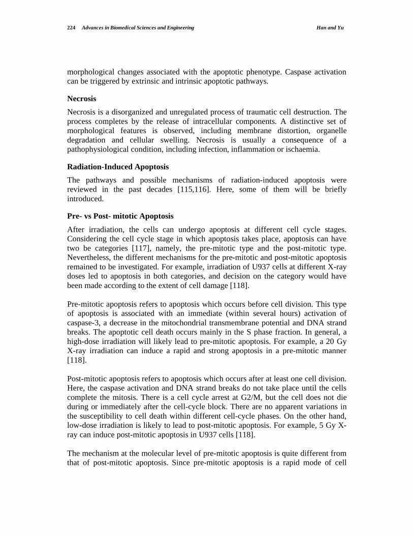
Pre- vs Post- mitotic Apoptosis
After irradiation, the cells can undergo apoptosis at different cell cycle stages.
Considering the cell cycle stage in which apoptosis takes place, apoptosis can have
two be categories [117], namely, the pre-mitotic type and the post-mitotic type.
Nevertheless, the different mechanisms for the pre-mitotic and post-mitotic apoptosis
remained to be investigated. For example, irradiation of U937 cells at different X-ray
doses led to apoptosis in both categories, and decision on the category would have
been made according to the extent of cell damage [118].
Pre-mitotic apoptosis refers to apoptosis which occurs before cell division. This type
of apoptosis is associated with an immediate (within several hours) activation of
caspase-3, a decrease in the mitochondrial transmembrane potential and DNA strand
breaks. The apoptotic cell death occurs mainly in the S phase fraction. In general, a
high-dose irradiation will likely lead to pre-mitotic apoptosis. For example, a 20 Gy
X-ray irradiation can induce a rapid and strong apoptosis in a pre-mitotic manner
[118].
Post-mitotic apoptosis refers to apoptosis which occurs after at least one cell division.
Here, the caspase activation and DNA strand breaks do not take place until the cells
complete the mitosis. There is a cell cycle arrest at G2/M, but the cell does not die
during or immediately after the cell-cycle block. There are no apparent variations in
the susceptibility to cell death within different cell-cycle phases. On the other hand,
low-dose irradiation is likely to lead to post-mitotic apoptosis. For example, 5 Gy X-
ray can induce post-mitotic apoptosis in U937 cells [118].
The mechanism at the molecular level of pre-mitotic apoptosis is quite different from
that of post-mitotic apoptosis. Since pre-mitotic apoptosis is a rapid mode of cell

Response of Cells to Ionizing Radiation Advances in Biomedical Sciences and Engineering 225
death, a prompt activation of pre-existing cytoplasmic caspase-3 may be involved.
Shinomiya et al. verified this hypothesis. In the study, caspase-3 inhibitor was added
to the culture media at different time points, and apoptosis-related events such as
PARP cleavage and DNA fragmentation were examined [118]. Regarding the 20 Gy-
irradiated U937 cells, suppression on PARP cleavage was effective when the caspase-
3 inhibitor was added before irradiation, but not effective if it was added after
irradiation (even within 1 h). This clearly indicated that activation of caspase-3 was a
very early event in pre-mitotic apoptosis and that pre-existing caspases were
important here. Furthermore, for the 20 Gy-irradiated cells, DNA fragmentation was
significantly decreased when the casapase-3 activity was suppressed by its inhibitor.
In contrast, for the 5 Gy-irradiated cells (i.e., the case of post-mitotic apoptosis), this
inhibitor did not reduce the apoptotic rate. This proposed an alternative pathway for
the post-mitotic apoptosis. It is known that post-mitotic apoptosis requires a transient
G2/M blockade. Furthermore, post-mitotic apoptosis took a longer incubation period
(more than 24 h) to execute when compared to pre-mitotic apoptosis. As such, the
post-mitotic apoptosis was suggested not only due to primary damages, but also due
to accumulation of secondary changes that occurred during the cytostatic phase. In
this way, down-regulation of anti-apoptosis genes and up-regulation of apoptosis-
related genes were considered likely to be involved in post-mitotic apoptosis.
DNA damage-dependent or independent apoptosis
(I) DNA damage-dependent apoptosis
One type of apoptosis is initiated via intrinsic signaling pathways and is often DNA
damage-dependent [119]. The intrinsic signaling pathways initiate apoptosis by
producing mitochondrial-initiated intracellular signals and act directly on targets
within the cell. Here, DNA damages are regarded to trigger the apoptosis. The
stimuli, including radiation, toxins, hypoxia and free radicals etc., that initiate the
intrinsic pathways produce intracellular signals that may act in either a positive or
negative way. All these stimuli can lead to changes in the inner mitochondrial
membrane, then an opening of the mitochondrial permeability transition pore, loss of
the mitochondrial trans-membrane potential and release of two main groups of
normally sequestered pro-apoptotic proteins from the inter-membrane space into the
cytosol [120].
The first group consists of cytochrome c, Smac/DIABLO, and the serine protease
HtrA2/Omi [121], and the first group proteins function to activate the caspase-
dependent mitochondrial pathway. Cytochrome c binds and activates Apaf-1 as well
as procaspase-9 to form an “apoptosome” [122,123] and the clustering of procaspase-
9 in this manner leads to caspase-9 activation. Smac/DIABLO and HtrA2/Omi can
inhibit the activity of apoptosis inhibitor proteins to promote apoptosis [124,125].
The second group of pro-apoptotic proteins, apoptosis-inducing factor (AIF),

226 Advances in Biomedical Sciences and Engineering Han and Yu
endonuclease G and endonuclease CAD, are released from the mitochondria during
apoptosis. The release of these proteins occurs in the late phase of apoptosis and they
cause DNA fragmentation. The released AIF, CAD and endonuclease G translocate
to the nucleus and initiate DNA fragmentation in two steps. In the first step, AIF
causes DNA fragmentation into ~50-300 kb pieces and condensation of peripheral
nuclear chromatin [126], and Endonuclease G cleaves nuclear chromatin to produce
oligonucleosomal DNA fragments [127]. In the second step, CAD is cleaved by
caspase-3, and then it leads to oligonucleosomal DNA fragmentation and a more
pronounced and advanced chromatin condensation [128,129].
Members of the Bcl-2 family of proteins control and regulate the apoptotic
mitochondrial events [130], and the tumor suppressor protein p53 has a critical role in
regulation of the Bcl-2 family of proteins [131]. The exact mechanisms have not yet
been completely understood. The Bcl-2 family of proteins governs mitochondrial
membrane permeability and can be either pro-apoptotic or anti-apoptotic. These
proteins are very important in that they can determine whether a cell commits
apoptosis or aborts the process. A possible action of the Bcl-2 family of proteins is to
regulate cytochrome c release from the mitochondria via alteration of mitochondrial
membrane permeability.
(II) DNA damage-independent apoptosis
Another type of apoptosis is not initiated by DNA damages, but instead by reception
of apoptosis related factors on the cellular membrane. This type of apoptosis is also
referred to as mediated by extrinsic signaling pathways. The involved factors are
members of the tumor necrosis factor (TNF) receptor gene superfamily [132]. To
date, the best-characterized ligands and corresponding death receptors include
FasL/FasR, TNF- /TNFR1, Apo3L/DR3, Apo2L/DR4 and Apo2L/DR5 [133]. The
members of the TNF receptor family have similar cyteine-rich extracellular domains
and a cytoplasmic domain, called the “death domain”. This death domain plays a
critical role in transmitting the death signal from the cell surface to the intracellular
signaling pathways after interacting with the membrane receptors.
The extrinsic apoptosis signaling pathway consists of two steps, namely, binding with
receptor and activation of caspase 8. Clustering of receptors is found on the cell
membrane. Upon ligand binding, cytoplasmic adapter proteins are recruited, which
exhibit corresponding death domains that bind with the receptors. The binding of Fas
ligand to Fas receptor leads to the binding of the Fas-associated death domain
(FADD) protein, and the binding of TNF ligand to TNF receptor leads to the binding
of the TNF receptor-associated death domain (TRADD) protein, with recruitment of
FADD and RIP [134]. FADD then associates with procaspase-8 via dimerization of
the death effector domain, resulting in the auto-catalytic activation of procaspase-8
[135]. Once caspase-8 is activated, the execution of apoptosis is triggered.

Response of Cells to Ionizing Radiation Advances in Biomedical Sciences and Engineering 227
A simplified model of radiation-induced apoptotic pathways is shown in Figure 6.
Surviving fraction = Colonies counted
Cells seeded (plating effeciency/100)
Fig. 6. A simplified model of radiation-induced apoptotic pathways. 1: Radiation-induced DNA damage initiates apoptosis via p53-dependent mechanisms, e.g. by regulating the expression of Bcl-2 family members. 2: Mitochondria are triggered to release caspase-activating factors, such as cytochrome c, by various stimuli, including Bax, reactive oxygen intermediates. 3: Activation of the pro-apoptotic SAPK/JNK pathway may occur downstream of membrane-derived signals, including ceramide and Daxx, a CD95-binding protein [116].
CELL-SURVIVAL CURVES
Experimental radiation survival curves are based on the clonogenic assay, which does
not differentiate among different modes of cells killed in an irradiated cell population
[1,2]. From the viewpoint of radiotherapy, loss of clonogenicity represents the most
significant consequence of exposure to ionizing radiation. Generally speaking, the
surviving fraction is given by
�������� ���
� ������� ��
����
����
���������� �����
�����
��������
��!��"#�
��"$������������ ����
���%��$&�����
������������ ��������
��� ��
������
���
��������� ��������
�����
����'�(�
���)
�
*
���� �����
���

228 Advances in Biomedical Sciences and Engineering Han and Yu
Target Theory: The target theory of cell survival is based on the model in which a
number of critical targets have to be inactivated for a cell to be killed. For example,
the single-target survival curve is a straight line on a semi-logarithmic plot (Figure 7
A). This kind of survival curves has been identified for inactivation of viruses and
bacteria, and can be applicable to the response of certain very radiosensitive
mammalian cells (normal and malignant) to very low-dose rates, as well as to the
response to high LET radiation.
Fig. 7. Illustration of survival curves. (A) Single-hit, single-target survival curve; (B) Multi-target survival curve; (C) Two-component survival curve; (D) Linear quadratic model of cell killing [2].
The target theory and derivation of simple cell-survival relationships in terms of
targets and hits have been in use for a long time. A drawback, however, is that
specific or idiographic radiation targets have not been identified in mammalian cells.
Furthermore, although the two-component model (Figure 7 C) predicts cell killing in
the low-dose region, the nearly linear change in cell survival in the dose range from 0
to Dq implies no sparing when the fraction size is below 2 Gy, which does not agree
with experimental or clinical data.
RADIATION-INDUCED CELL MUTATIONS
Although the mutagenic capability of radiation was first described by Muller in 1927,
it is only within the past two decades that information has been obtained regarding
the molecular changes involved in mutations of mammalian cells. Radiation can
induce a wide spectrum of mutations, from point mutations in single genes to
�����
��
��
��
��
��
��
�
� ��
�
�
� ��
�
�����
��
��
��
��
��
��
�
��
��
��
�����
��
��
��
�
��
��
��
�����
��
��
��
�
��
��
��
� �
��
����� �� ������� ����� �� �������
��
��
�
� ��
�
�
� ��
�

Response of Cells to Ionizing Radiation Advances in Biomedical Sciences and Engineering 229
deletions of several genes, based on the earlier studies with the hemizygous X-linked
HPRT gene [136]. The mutation spectrum induced by radiation is different from
spontaneous mutations induced by ultraviolet light and chemical mutagens, the
majority of the latter being consequences of point mutations. Most evidence at the
molecular level indicated that the DNA deletions resulting in gene loss were the
primary events leading to mutagenic effects of ionizing radiation. Since mutations or
deletions of some essential genes are closely related to survival of cells, this limits the
ability to detect large deletions in certain genes, especially some lethal genes.
Radon is a naturally occurring radioactive gas and alpha particles are emitted from
radon and its progeny. The US Environmental Protection Agency estimated radon in
indoor environments to have caused 21,600 deaths per year through radon-induced
lung cancers [137]. To have a better quantitative assessment of the lung cancer risk
associated with residential radon exposure, Hei’s group investigated the mutation
inducing capability of single alpha particles, by using the microbeam facility at the
Columbia University to deliver an exact number of alpha particles to the nuclear
region of cells [138]. They examined the frequencies and molecular spectrum of S1-
mutants induced in human-hamster hybrid (AL) cells by 1, 2, 4, or 8 particles.
Although single-particle traversal was only slightly cytotoxic to AL cells (survival
fraction ~0.82), it was very mutagenic, and the average induced mutant fraction was
110 mutants per 105 survivors. Furthermore, both toxicity and mutant induction were
dose-dependent. The presence or absence of marker genes among the mutants was
determined by multiplex PCR, and the proportion of mutants with multi-locus
deletions was found to increase with the number of particle traversals.
Earlier studies in a number of different biological systems indicated that nuclear
irradiation was critical for cytotoxic effects. As such, the nucleus was considered the
main target of radiation. In a further study by Hei’s group, mutagenesis of
cytoplasmic irradiation with low fluences of alpha particles was studied with their
microbeam facility [139]. The results showed that cytoplasmic traversals by alpha
particles led to more mutations in AL cells, but had relatively little effect on cell
survival. An approximately doubled spontaneous mutation frequency was observed
for a single alpha-particle traversal, and a 2- to 3-fold enhancement in the mutation
frequency was observed with up to four particle traversals per cell. No further
increase in the mutation frequency was found for larger particle fluences. These
results are in contrast to their earlier studies involving nuclear irradiation. For nuclear
traversals, mutation frequencies were 2- to 3-fold higher than those for the same
number of cytoplasmic traversals. Furthermore, in the case of nuclear radiation, the
mutation frequency kept increasing with the fluence up to eight or more particles per
cell. It was particularly interesting to note that the spectrum of molecular-structural
changes was significantly different between the two types of irradiation. Nuclear
irradiation mainly led to large-scale changes such as those described previously for
X-rays. On the other hand, cytoplasmic irradiation mainly led to point mutations with

230 Advances in Biomedical Sciences and Engineering Han and Yu
the spectrum resembling that of spontaneously arising mutants. In both cases, an
enhanced production of reactive oxygen species was involved [140]. These results
concluded that nuclear radiation was not required for the production of important
genetic effects.
As regards the mechanism of radiation mutagenesis, the mutagenesis is considered a
result of the cell attempting to repair damages based on analyses of the induced
mutations. DSBs are regarded as an important initiating lesion in the pathogenesis of
large deletions characteristic of ionizing radiation and the involvement of DSB repair
pathways in the mutagenic process [141]. DSBs can be repaired by HR in an error-
free manner. Nevertheless, most DSBs are repaired by an error-prone process which
likely accounts for many of the potentially mutagenic DNA lesions. Multiple DSBs in
a cell may thus lead to chromosomal rearrangements and other large-scale changes,
which are commonly present in irradiated cells. Repeat sequences in the genome also
suggest that NHEJ of DSBs is usually responsible for the mutagenic process when
large deletions are involved.
Radiation-induced base damages are also important. It is known that base damages
can often lead to base substitutions (point mutations) and that certain repair pathways
involved in base damage repair can also be mutagenic.
RADIATION-INDUCED BYSTANDER EFFECT
OVERVIEW: OBSERVATION AND SIGNIFICANCE OF BYSTANDER
EFFECT
Biological effects of ionizing radiation have long been considered a consequence of
DNA damages in the irradiated cells. Here, unrepaired or misrepaired DNA damages
in the irradiated cells are responsible for the genetic effects. At the same time, no
effects are expected in cells in the population that have not received radiation
exposure. This conventional dogma was, however, challenged by the occurrence of
the radiation-induced bystander effect (RIBE).
RIBE was reported back in 1954, when cells exposed to doses of low LET radiation
were found to have an indirect effect in producing a plasma-borne factor, which led
to chromosome breakage and cytogenetic abnormalities in human bone marrow or
lymphocytes and caused tumors in rats [142]. Plasma from high-dose radiotherapy
patients were also found to induce various aberrations, including dicentrics and
chromatid and chromosome breaks, in normal non-irradiated lymphocytes in short-
term culture. The radiation-induced clastogenic factors in the plasma of irradiated
patients had low molecular mass, and their production involved lipid peroxidation
and oxidative stress pathways. These factors either had long lives or were regenerated

Response of Cells to Ionizing Radiation Advances in Biomedical Sciences and Engineering 231
continuously since they persisted in the plasma of atomic bomb survivors even 31
years after exposure.
From the early 1990s, developments in single-cell irradiations either with low -
particle fluences or with microbeam facility have led to a large amount of
experimental data and an immense interest in the bystander effects. In the following,
more detailed information or findings for RIBE will be introduced.
Generally, RIBE can be defined as the phenomenon that the irradiated cells (by
particles, X- or -ray, heavy ions etc.) can release some signaling molecule(s), which
is transferred via the medium or gap-junctions, so that the same cytotoxicity or
genotoxicity can be observed in the non-irradiated cells, which are either close to the
irradiated cells or shared the conditioned medium harvested from the irradiated cells.
RIBE has challenged the conventional dogma of radiation protection, the guidelines
for which are based on prediction of biological effects of low doses of radiation by
extrapolating from known epidemiological datasets. These datasets are mainly in the
high-dose regions and the main source of information came from Japanese atomic-
bomb survivors. The simplest way to perform the extrapolation is to assume a linear
no-threshold relationship between the dose and the biological effect even at very low
doses. In other words, a dose, however small, always has a finite probability of
causing a biological effect. Environmental radon has been suggested to cause about
21,600 lung-cancer deaths in USA each year. The presence of bystander effects
implies no direct correlation between the number of cells exposed to radiation and the
number of cells at risk of mutation, chromosomal damage or apoptosis. Instead,
biological effects depend on complex interactions between the irradiated cells and the
bystander cells. The risk is no longer that of a single cell resulted from its radiation
damages; instead the risk is “amplified” by the bystander effect and a simple dose–
effect relationship is no longer valid.
RIBE in a system of low fluence LPHA-particle irradiation
One way to study RIBE is to use a broad beam of particles with a low fluence, in
which case a small proportion of cells are traversed by particles. J.B. Little’s lab
was the first to report a bystander effect resulting from high LET irradiation. In this
study, the frequency of sister chromatid exchanges (SCEs) reached 30% in a cultured
population of Chinese hamster ovary (CHO) cells when less than 1% of the cell
nuclei were actually traversed by an particle (a dose corresponding to 0.31 mGy)
[143]. Similar results were later reported for normal human lung fibroblasts. An
enhanced frequency of HPRT locus mutations was also found in bystander CHO cells
in cultures following an exposure to very low fluences of particles. Changes in gene
expression in non-irradiated cells were also observed after low fluence of particles.
It was noted that up-regulation of the p53 damage response pathway occurred in
bystander cells in monolayer cultures exposed to very low fluences of particles

232 Advances in Biomedical Sciences and Engineering Han and Yu
[144]. Phosphorylation of p53 in bystander cells suggested that p53 up-regulation in
bystander cells was a consequence of DNA damages. Activation of the p53 damage
response pathways in bystander cells was confirmed by in situ immunofluorescence
studies which showed up-regulation of p21Waf1
in clusters of cells in the monolayer
population, of which only 12% of the cell nuclei had been traversed by an particle
[145].
Direct measurements of DNA damages in bystander cells after exposure to low
fluence particles have been made possible through advancement of the associated
experimental techniques. Phosphorylated H2AX, a member of the histone family, is a
marker of DSBs. With in situ immunofluorescence of -H2AX, excessive DSBs were
measured in the bystander cells after even 1 cGy -particle irradiation, for which less
than 1/10 nuclei were hit by the particles [146].
Studies of RIBE with Charged Particle Microbeam Facility
Microbeam techniques can deliver exactly one particle (or more) to irradiate precisely
the cell nucleus or cytoplasm [147]. This technique has been used to investigate the
bystander effect for -particle irradiation for a variety of biological end points,
including cell lethality, DNA damage, mutagenesis and oncogenic transformation
[148]. One endpoint used in the RIBE study with the microbeam facility at the Gray
Cancer Institute, UK, was micronuclei assay. In their study, human glioblastoma
T98G cell nuclei were individually irradiated with an exact number of helium ions. It
was found that when only 1 cell in a population of ~1200 cells was targeted, with one
or five ions, the cellular damage measured as induced micronuclei was increased by
20%. When a fraction from 1% to 20% of cells was individually targeted, the
micronuclei yield in the population greatly exceeded the yield predicted with no
bystander effect. Another end point investigated with the Columbia microbeam
facility was mutagenesis. When a near-lethal dose of 20 alpha particles were directed
to each nucleus of 20% of human–hamster hybrid (AL) cells, the surviving fraction
was found to be less than 1%. On the other hand, by employing mutations in human
chromosome 11 as the endpoint, the mutation yield was found to be four times that of
the background. Since the irradiated cells were exposed to a lethal dose, the
mutations should come from the non-irradiated bystander cells. The mutation
spectrum evaluated for the bystander cells was significantly different from the
spontaneous spectrum, which hinted different mutagenic mechanisms [149].
In another study of bystander-mediated oncogenesis mouse fibroblast (C3H10T12),
cells were plated in a monolayer and either every cell or every tenth cell was
irradiated with 1 to 8 alpha particles directed to the cell nucleus. The cells were
subsequently replated at low density and the transformed foci, based on the
morphology, were counted 6 weeks later. More cells were found inactivated than the
number actually traversed by alpha particles. Furthermore, when 10% of the cells on

Response of Cells to Ionizing Radiation Advances in Biomedical Sciences and Engineering 233
a dish were exposed to 2 or more alpha particles, the frequency of induced oncogenic
transformation was indistinguishable from that observed when all the cells on the dish
were exposed to the same number of alpha particles [148].
In another study, Shao et al. [150] suggested that direct targeting of nuclear DNA was
not always required for expression of a radiation-induced bystander effect. A helium-
ion (3He
2+) microbeam was employed to target individual cells within a population of
radioresistant glioma cells cultured alone or in co-culture with primary human
fibroblasts. Even when only a single cell was traversed with one 3He
2+ ion through its
cytoplasm, bystander responses were observed in the non-irradiated glioma or
fibroblast cells. Specifically, the micronuclei yield was increased by 36% for the
glioma population and 78% for the bystander fibroblast population. Furthermore, they
did not depend on whether the cell cytoplasm or nucleus was targeted. This is very
important in redefining the source of the bystander effect signal(s).
RIBE after Transfer of Conditioned Medium from Irradiated Cells
As previously described, irradiated individuals release their plasma clastogenic
factors that will induce chromosomal damages in cultured cells from non-irradiated
donors. Although this phenomenon had been known for half a century, the underlying
reasons have remained unknown. In the recent ten years, considerable evidence for
the toxic effects of bystander signals has been gathered from studies where the
culture media from irradiated cells have been transferred to cultures of non-irradiated
cells [142,151,152]. The first study was carried out by Mothersill and Seymour’s
group. They demonstrated that irradiated epithelial cells, but not fibroblasts, secreted
a toxic signal or the so-called bystander factor into the culture medium which, if
transferred to non-irradiated cells, could significantly reduce the plating efficiency of
the latter [153]. In relation to this phenomenon, further studies showed that a medium
irradiated in the absence of cells had no toxic effects on survival when transferred to
non-irradiated cells. The effect was dependent on the cell number at the time of
irradiation, could be observed as soon as 30 min post-irradiation, and was still
effective when medium transfer occurred 60 h after irradiation. On the other hand, the
effect was independent of dose in the range from 0.5 to 5 Gy. The factor involved
appeared to be a protein since it was heat-labile but stable if frozen, and did not
require cell-to-cell contact to induce its effect in the recipient cells. The first
detectable effect was a rapid (1-2 min) calcium pulse in the medium receptor cells,
which was then followed (30 min to 2 h later) by changes in the mitochondrial
membrane permeability and the induction of ROS. The critical role of mitochondrial
metabolism was suggested by the lack of signal production by cells that did not have
a functional glucose-6-phosphate dehydrogenase enzyme [154].
Held’s group demonstrated that normal human fibroblast cells, when irradiated by
broad field 250 kVp X-rays, could also release bystander factor(s) into the medium

234 Advances in Biomedical Sciences and Engineering Han and Yu
0 5 10 15 20 25 30
14
16
18
20
22
24
26
28
30
32
34
* p<0.05
** p<0.01
*
**
*
*
Fra
ctio
n o
f D
SB
Po
sitiv
e C
ells (
%)
Time Post Irradiation (min)
with cells
without cells
Fig. 8. Timing of bystander factor(s) by normal human fibroblast cells AG1522 in the initiation of RIBE. The conditioned medium harvested from the irradiated culture induced a time-dependent DSB in the medium receptor cells. The irradiated medium without cells did not show distinct DSB-inducing activity [156].
[155]. They used a transwell insert culture dish system to demonstrate the X-ray
induced medium-mediated bystander effects in AG1522 normal human fibroblasts.
The micronuclei yield in non-irradiated bystander cells was found to be
approximately doubled that of the background in the dose range from 0.1 to 10 Gy.
Induction of p21Waf1
protein and foci of -H2AX in bystander cells were also
independent of the dose delivered to the irradiated cells above 0.1 Gy. The ROS
levels were increased persistently in irradiated cells up to 60 h after irradiation and in
bystander cells up to 30 h. The clonogenic assay of bystander cells showed a decrease
in their survival after receiving doses up to 0.5 Gy, but was then independent of the
dose above 0.5 Gy.
Wu’s group also revealed that the normal human fibroblast cells AG1522, when
irradiated with low-dose (1 cGy) alpha particles, could release bystander factor(s)
into the medium [156]. Medium transfer experiments showed that the conditioned
medium harvested from the irradiated culture induced excessive DNA DSBs in the
cells receiving the medium, and the capability of the medium of DSB induction was
time-dependent (Figure 8). The results indicated that the bystander signaling
molecule(s) had been generated very quickly (probably less than 2.5 min) after
irradiation and persisted continuously up to 30 min although the production of
signaling molecule(s) decreased after 10 min post irradiation. Their findings revealed
an initiation and early process of bystander response induced by low dose alpha-

Response of Cells to Ionizing Radiation Advances in Biomedical Sciences and Engineering 235
particle irradiation, and the findings were very important for understanding the
mechanisms underlying the bystander response.
RIBE in 3-DimensionAL Tissue
The bystander effect experiments described above made use of cultured cell
populations. It is also pertinent to study the effect in vivo. To simulate the bystander
effects in a tissue, Belyakov and coworkers [157] used reconstructed normal human
three-dimensional skin tissue systems. One of the systems used was a full thickness
skin model corresponding to the epidermis and dermis of normal human skin. A
charged-particle microbeam was used to irradiate cells at defined locations while
guaranteeing that cells located more than a few micrometers away received no
radiation exposure. The studied endpoints were the induction of micronucleated and
apoptotic cells. As a result, significant effects were detected in non-irradiated cells up
to 1 mm away from the irradiated cells, namely, an average increase of 1.7-fold for
micronuclei and 2.8-fold for apoptosis (Figure 9).
Fig. 9. Induction of (A) apoptotic and (B) micronucleated cells in non-irradiated bystander cells at different distances from the plane of irradiated cells in a 3-D human epidermal skin model (EPI-200) [157]. (Copyright 2005 National Academy of Sciences, U.S.A.)
In their further study, DSBs were detected in the bystander cells in the 3-D tissue
[158]. Occurrences of DSBs in irradiated cells and bystander cells are markedly
different: the former reached a maximum 30 min after irradiation, while the latter
reached a maximum 12 to 48 h after irradiation and gradually decreased over a 7-day
time course. At the maxima, 40% to 60% of bystander cells were affected, i.e., a 4- to
6-fold increase over the controls. The rise in bystander DSB occurrences were
followed by a larger number of apoptosis and micronucleus formation, by loss of
nuclear DNA methylation, and by an increased fraction of senescent cells. These
showed that DNA DSBs were involved in tissue bystander responses and that they
were precursors to downstream effects in human tissues. Bystander cells which
����
����
����
����
����
���
������ ��� ��� �� ��� ��
� ������������ ���� ������������� ������
����� ������������� ������� �� ���� ����
!��������
"�������
�����
�����
�����
�����
����
������ ��� ��� �� ��� ��
"�������
�� ���� ���� !��������
� ������������ ���� ������������� ������
����� ������������# �$�� ����%���
&

236 Advances in Biomedical Sciences and Engineering Han and Yu
showed genomic instability after irradiation were also found more likely to become
cancerous than unaffected cells.
Bystander Effects in Mouse and Fish Models
The existence of RIBE in vitro is well established and supported by solid
experimental evidence. On the other hand, however, clear experimental
demonstrations of bystander effects in vivo are limited. One of these came from a
series of experiments performed by Brooks and coworkers [159]. Chinese hamsters
were injected with different-sized particles internally deposited with alpha-particle
emitting plutonium. The radioactive particles concentrated in the liver and caused
chronic low-dose radiation exposure, causing the highest absorbed dose and dose rate
to cells located closest to the largest particles. However, analysis of induced
chromosome damages in these livers revealed increased cytogenetic damages but no
changes in the aberration frequency as a function of the local dose. These indicated
that all the cells in the liver had the same risk of induced chromosome damage
despite the small fraction of liver cells that were irradiated.
In another study, Kovalchuk’s group reported RIBE in mouse after X-ray irradiation
of one-half of the mouse body. They showed that RIBE increased DNA strand breaks
and Rad51 levels in the unexposed bystander tissue [160]. In terms of epigenetic
changes, unilateral radiation suppressed global methylation in directly irradiated
tissue, but not in bystander tissue at studied time points. They observed a significant
reduction in the levels of the de novo DNA methyl transferases DNMT3a and 3b and
a concurrent increase in the levels of the maintenance DNA methyl transferase
DNMT1 in bystander tissues. Furthermore, the levels of two methyl-binding proteins
known to be involved in transcriptional silencing, MeCP2 and MBD2, were also
increased in bystander tissue. These findings illustrated radiation induced DNA
damages in bystander tissue more than a centimeter away from directly irradiated
tissues, and suggested that epigenetic transcriptional regulation might be involved in
RIBE.
RIBE in nervous system was studied by Saran and co-workers [161], who regarded
the neonatal mouse cerebellum as an accurate in vivo model to detect, quantify, and
understand radiation-bystander responses. They conducted experiments using
specially designed lead shields for protecting mouse heads and observed marked
enhancement of medulloblastoma in mice irradiated with their brains shielded. They
further analyzed DSBs in the shielded cerebella through the -H2AX foci, and the
fractions of apoptotic cells. Besides these genetic events, they also reported
bystander-related tumor induction in the cerebellum of radiosensitive Patched-1
(Ptch1) heterozygous mice after X-ray irradiation of the remainder of the body. They
showed that genetic damage was critical for in vivo oncogenic bystander responses,

Response of Cells to Ionizing Radiation Advances in Biomedical Sciences and Engineering 237
and suggested the involvement of gap-junction intercellular communication (GJIC) in
RIBE in the central nervous system.
Mothersill’s Group reported RIBE in a cultured rainbow trout population [162]. They
found that fish irradiated to 0.5 Gy X-ray (100 kVp) total body dose released factors
into the water that could induce bystander effects in other unexposed fish. The
unexposed fish were either arranged to share the same aquarium with the irradiated
fish, or accommodated in the water previously used to hold the irradiated fish. Five
organs were removed from each fish and tissue explants were cultured. The RIBE
was expressed as increased cell deaths in a reporter system. The responses varied for
different cell types, with the gill and fin showing the most pronounced responses.
These results suggested that bystander signals involved chemical messengers secreted
by an irradiated fish into the water, which could be passed to other fish.
POSSIBLE MECHANISMS
Possible mechanisms of RIBE have been studied widely. Most experimental results
broadly pointed to three types of mechanisms:
(i) soluble transmissible factor(s) generated by irradiated cells;
(ii) GJIC-mediated transmission of RIBE;
(iii) oxidative metabolism-mediated transmission of RIBE.
These mechanisms are not independent of one another. In fact, in most cases, these
mechanisms can co-exist. In the next part, these three mechanisms will be discussed
in more details.
RIBE Mediated by Soluble Transmissible Factor(s) Secreted by Irradiated Cells
In the previous sections, we introduced RIBE after medium transfer. The conditioned
medium harvested from -irradiated keratinocytes and transferred to control
keratinocytes or fibroblasts resulted in a toxic effect in the latter. Effects of
transmissible factor(s) can also be observed in subconfluent cell cultures irradiated
with alpha particles. Irradiation of the nuclei of a few mammalian cells in a
subconfluent culture with alpha particles resulted in the induction of genetic damages
(micronucleus formation and apoptosis) in a larger fraction of cells [163]. In these
studies, targeting the alpha particles outside the cells did not have the effect, which
suggested that RIBE was due to transmissible factor(s) released from the irradiated
cells.
As regards the nature of the soluble factor(s), studies showed that alpha-particle
irradiation of cultured cells generated a factor(s) capable of inducing SCEs in the
bystander cells, and the factor(s) survived freeze thawing and was heat labile [164].
In parallel experiments, the same group showed that alpha-particle irradiation of the
culture medium devoid of cells also caused generation of SCE-inducing factor(s);

238 Advances in Biomedical Sciences and Engineering Han and Yu
such factors however were short-lived. In both situations, the supernatant from
irradiated cells or irradiated medium caused the induction of excessive SCEs in non-
irradiated cells to the same extent observed with direct alpha-particle irradiated cell
cultures. Interestingly, both the short lived medium- and cell-derived SCE-inducing
activities were inhibited by the anti-oxidant enzyme superoxide dismutase (SOD),
suggesting that reactive oxygen species (ROS) were involved.
Secreted transforming growth factor 1 (TGF- 1) [165] or Interleukin-8 (Il-8) [166]
in the medium of alpha-particle irradiated cultures were suggested to play a role in
mediating the bystander response-induced cell proliferation. Shao’s recent study
suggested that secreted TGF- 1 played a role in mediating the micronuclei induction
in the bystander cells [167]. In this study, a fraction of cells within a glioblastoma
population were individually irradiated with microbeam alpha particles, and the
micronuclei yield in the non-targeted cells was found to have increased. These RIBE
was mitigated by treating the cells with either aminoguanidine, which was an
inhibitor of inducible nitric oxide (NO) synthase, or anti-TGF- 1, indicating that NO
and TGF- 1 were involved in the RIBE. Intracellular NO was detected in the
bystander cells, and additional TGF- 1 was detected in the medium from irradiated
T98G cells, but it was diminished by aminoguanidine. Conversely, treatment with
diethylamine nitric oxide, which was an NO donor, induced TGF- 1 generation in
T98G cells. In relation to these experiments, treatment with recombinant TGF- 1 also
induced NO and micronuclei. Treatment with anti-TGF- 1 inhibited the NO
production when only 1% of cells were irradiated, but not when 100% of cells were
irradiated. In general, downstream of radiation-induced NO, TGF- 1 could be
released from irradiated T98G cells and played a key role as a signaling factor in
RIBE by further inducing free radicals and DNA damages in the bystander cells.
In another study, Han and coworkers investigated the possible nature of secreted
soluble bystander signal(s) in the initiation and propagation of the early processes of
bystander signaling induced by low-dose alpha-particle irradiation [168]. They
focused on the 10 min time point after irradiation, when the secreted signal(s) reached
the maximum. Their former studies showed that the secreted signal(s), which rapidly
induced phosphorylated H2AX in non-irradiated cells that had received the
conditioned medium, might be short-lived and was released in a time-dependent
manner in the first 30 min after irradiation [156]. Using NG-methyl-L-arginine, 4-
amino-5-methylamino-2',7'-difluorofluorescein diacetate and N -nitro-L-arginine (L-
NNA) treatment before exposure to 1 cGy particles, they showed that NO produced
in the irradiated cells was important and necessary for the DNA DSB inducing
activity (DIA) of conditioned medium, and the generation of NO in irradiated
confluent AG1522 cells was in a time-dependent manner and that almost all NO was
generated within 15 min post-irradiation. Concurrently, the kinetics of NO production
in the medium of irradiated cells after irradiation was rapid and in a time dependent

Response of Cells to Ionizing Radiation Advances in Biomedical Sciences and Engineering 239
manner as well, with a maximum yield observed at 10 min after irradiation,
determined with electron spin resonance analysis. Furthermore, they also showed that
7-Nitroindazole and L-NNA, but not aminoguanidine hemisulfate, treatment before
exposure to 1 cGy particles significantly decreased the DIA of the conditioned
medium, and this suggested that constitutive NO from the irradiated cells possibly
acted as an intercellular signaling molecule to initiate and activate the early process
(<30 min) of bystander response after low-dose irradiation.
Role of GJIC in the Transmission of RIBE
Little’s group first studied the possible role of GJIC in RIBE. Direct evidence for the
participation of GJIC in RIBE from alpha-particle irradiated to non-irradiated
mammalian cells was obtained when GJIC proficient or deficient confluent cultures
of human and rodent cells were exposed to very low fluences of alpha particles [144].
In situ immunofluorescence studies of p21Waf1
expression in AG1522 cultures
exposed to 0.3 cGy of alpha particles (a dose at which about 2% of the nuclei would
be irradiated) showed that the typical aggregate pattern of induction of the protein
disappeared in the presence of a chemical inhibitor lindane of GJIC. In irradiated
cultures treated with lindane, p21Waf1
was induced primarily only in single cells. The
WB-F344 cells were GJIC competent as was demonstrated with the transfer of the
Lucifer yellow dye among contiguous adjacent cells. The WM-aB1 cells were
derived from WB-F344 cells and were deficient in GJIC function [144]. An increase
in p21Waf1
levels in confluent WB-F344 cultures was observed after exposure to mean
doses as low as 0.3 cGy, and at this dose 1% or less of the cells would have their
nuclei traversed by alpha particles. While small clusters of responding cells were
observed in WB-F344 cells, only single isolated and presumably irradiated WM-aB1
cells showed up-regulation of p21Waf1
after exposure of 0.3-1.0 cGy. The p21Waf1
levels increased significantly only at mean doses of 5 cGy or higher in WM-aB1 cell
cultures. These findings were confirmed in low passage mouse embryo fibroblasts
from wild type and isogenic knockout embryos for connexin 43. Similar to WB-F344
and WM-aB1 cells, western blot analyses demonstrated lack of detectable increase in
p21Waf1
expression in connexin43-/-
cells exposed to mean doses less than 10 cGy.
Conversely, p21Waf1
was induced in wild type cells after exposure to mean doses as
low as 0.6 cGy. All these observations strongly supported the involvement of GJIC in
the bystander gene expression response in confluent, density-inhibited cell cultures
exposed to alpha particles. When GJIC was inhibited, micronuclei induction was also
inhibited in bystander cells in confluent cultures exposed to alpha particles.
The role of GJIC in mediating the alpha-particle induced bystander response was
further confirmed by mutation studies using microbeam irradiation [169]. Microbeam
facility was used to irradiate the nuclei of a precise fraction (either 100% or <20%) of
the cells in a confluent population with exactly one alpha particle each. Irradiation of
10% of the cells resulted in a mutant yield similar to that from irradiation of 100% of

240 Advances in Biomedical Sciences and Engineering Han and Yu
the cells. The effect was significantly eliminated when the cells were pretreated with
a 1 mM of octanol which inhibited GJIC, or in cells carrying a dominant negative
connexin 43 vector.
The Role of Oxidative Metabolism in Mediating the Radiation-Induced
Bystander Response
Radiation-induced ROS were known to participate in damages to various cellular
components, and to produce DSBs in addition to base damages and single-strand
breaks. Alpha-particle-induced metabolic ROS production was also involved in the
activation of signaling pathways in bystander cells [145]. An induction of SCEs in
bystander cells after exposure to very low fluences of alpha particles was inhibited by
SOD, a superoxide radical scavenger [165]. Further studies directly showed that
alpha particles (0.4-19 cGy) initiated the intracellular production of ROS (superoxide
anions and hydrogen peroxide) in human cells through involvement of the plasma
bound NADPH-oxidase [164]. These studies suggested that the ROS response did not
require direct nuclear or even cellular transverses by alpha particles [164].
After exposing confluent cell cultures to very low fluences of alpha particles, the
ROS formed were shown to induce stress-inducible proteins in the p53 and mitogen
activated protein kinase pathways in bystander cells. Active SOD and catalase
enzymes were capable of suppressing these effects and also inhibiting the activation
of redox sensitive transcription factors in bystander cells [145]. Diphenyliodonium
(DPI) is an inhibitor of flavin containing oxidase enzymes such as NAD(P)H-oxidase.
NAD(P)H-oxidase enzymes are known to produce ROS in quantities capable of
stimulating signaling pathways and these enzymes are rapidly activated by a variety
of soluble mediators and engagement of cell-surface receptors. Treatment with DPI
led to a significant reduction in the production of micronuclei by low doses of alpha
particles. This suggested that activation of NAD(P)H-oxidase might induce the
production of ROS in bystander cells.
The role of flavoprotein oxidases in the alpha-particle-induced bystander effect was
extended when the enhanced accumulation of p53 and p21Waf1
occurring after mean
doses of 1 to 3 cGy was significantly reduced by the treatment with DPI [145]. These
suggested that the increased numbers of SCEs in bystander cells from cultures
exposed to low fluences of alpha particles involved membrane bound NAD(P)H-
oxidase [165]. In general, these results supported the hypothesis that a DPI sensitive
flavin containing oxidase activity represented a significant source of ROS production
in human fibroblast cultures exposed to low fluences of alpha particles. They also
suggested that ROS production from the activation of membrane bound NAD(P)H
oxidase(s) might trigger the signaling pathway leading to the accumulation of p21Waf1

Response of Cells to Ionizing Radiation Advances in Biomedical Sciences and Engineering 241
and p53 as well as induction of micronuclei and SCEs following radiation exposure
to these doses.
Cell membrane involvement in the bystander response to low fluences of alpha
particles was further confirmed by the complete suppression of SCEs and HPRT
mutations in CHO cells exposed to very low fluences of alpha particles in the
presence of Filipin, a drug that disrupted lipid rafts [143]. Importantly, at a dose of 10
cGy, when most effects occurred in directly irradiated cells, no suppressive effect of
Filipin was observed. It was also interesting to note that gap-junctions were reported
to partition in lipid rafts [170]. Of further interest is the finding that ROS-activated
kinase(s) (e.g. member(s) of the mitogen-activated protein kinase (MAPK)
superfamily) also have a role in activation of gap-junction proteins [171].
Furthermore, binding sites for the redox-sensitive AP-1 and NF- B transcription
factors, which are activated by low fluences of alpha particles have been shown to
exist in the connexin 43 gene promoter region [172]. It is tempting to speculate that
the mechanisms described above act in concert to promote the bystander effect.
There were some contradictory results regarding the involvement of oxidative
metabolism in the induction of a bystander mutagenic effect. For example, in
microbeam alpha-particle experiments with human-hamster hybrid cells, DMSO
failed to suppress the induction of mutations in bystander cells, suggesting that ROS
were not involved in the mutagenic bystander effect [149]. In contrast, induction of
HPRT mutations in CHO bystander cells from cultures exposed to low fluences of
broad beam alpha particles was consistent with the involvement of oxidative
metabolism in the effect [173]. Further evidence of upregulation of oxidative
metabolism in bystander cells was gathered from gene expression studies in human
diploid fibroblast cultures exposed to very low fluences of alpha particles [145].
Oxidative metabolism was also implied in toxic bystander effects observed in media
transfer experiments involving -radiation [174,175]. Treatment of the irradiated
cultures with the antioxidants, L-lactate and L-deprenyl or with drugs that inhibited
collapse of mitochondrial membrane potential prevented the cytotoxic effects from
the irradiated cell conditioned medium. Similar observations were obtained in out-of-
field in vivo experiments examining the genetic effects of partial organ irradiation.
For example, DNA damages detected in the shielded apex region of rat lung when the
lung base was irradiated was blocked by intravenously injected Cu-Zn SOD [176].
The involvement of oxidative metabolism in the early and initiation process was
studied by Wu’s group [156]. After irradiating half of the AG1522 cells cultured on a
mylar dish with 1 cGy alpha particles, time-dependent DNA DSBs were induced
shortly after irradiation in bystander AG1522 cells as observed through in situ
detection. The induction of DSB in bystander cells was greatly reduced after
treatment with DMSO. The capability of the conditioned medium to induce DSB can

242 Advances in Biomedical Sciences and Engineering Han and Yu
also be inhibited by DMSO treatment. These results demonstrated the importance of
oxidative metabolism in the early and initiation process of RIBE.
Other Pathways Involved in the Transduction of RIBE
(I) Cyclooxygenase-2 as a central component of the bystander signaling scheme
Experiments have been conducted in Hei’s lab with normal human lung fibroblasts
(NHLF) to identify genes that are expressed differently in directly irradiated and
bystander cells [177]. Among the 96 genes represented on the platform, the
abundance of one message, COX-2, was found to be consistently higher by more than
three-fold, while the RNA level of insulin growth factor binding protein-3 was found
to be consistently lower by more than seven-fold. Addition of the COX-2 inhibitor
NS-398 (50 mM) suppressed COX-2 activity in NHLF cells and bystander
mutagenesis at the HPRT locus. These results indicated that COX-2 expression was
involved in the bystander effect.
(II) Role of nuclear factor NF- B in the bystander response
Since NF- B is an important transcription factor for many signaling genes, including
COX-2, it is likely that NF- B also participates in the bystander response. There was
strong evidence that alpha-particle irradiation up-regulated NF- B binding activity in
both directly irradiated and bystander cells, while Bay 11-7082, an inhibitor of I B
kinase (IKK)/NF- B, efficiently suppressed this up-regulation and also reduced
levels to below the basal amount [178]. This inhibitor of NF- B activity also
efficiently down-regulated the expression of COX-2 and inducible NO synthase in
both directly irradiated and bystander fibroblasts. Earlier studies using confluent
human skin fibroblasts exposed to low fluences of alpha particles showed a rapid up-
regulation of NF- B, c-Jun N-terminal kinase (JNK) and extracellular signal-related
kinase (ERK) in the exposed population [145] and suggested activation of these
stress-inducible signaling pathways in bystander cells. Furthermore, addition of the
antioxidant SOD was found to suppress the induction. Since induction of NF- B
binding activity could be found in both directly irradiated and bystander cells, its role
in the bystander response in this study is equivocal.
(III) Important Role of Mitochondria in RIBE
Mitochondria play an important role in the generation of free radicals and in the
regulation of apoptosis [179]. Ionizing radiation induced mitochondrial damages
through increases of ROS production, depolarization of mitochondrial membrane
potential, and release of cytochrome c in directly irradiated cells [180]. The
involvement and the role of mitochondria in RIBE were studied by three independent
labs almost simultaneously.

Response of Cells to Ionizing Radiation Advances in Biomedical Sciences and Engineering 243
By using charged particle microbeams, Hei’s group found that mitochondrial DNA
depleted human skin fibroblasts (o), when a fraction was irradiated with lethal doses,
showed a higher bystander mutagenic response in confluent monolayers compared to
their parental mitochondrial functional cells (+) [178]. However, when mixed
cultures of o and
+ cells were employed and only one population of cells were
irradiated with a lethal dose of alpha particles, a decreased bystander mutagenesis
was uniformly found in non-irradiated bystander cells of both cell types, which
suggested that signals from one cell type could modulate expression of bystander
response in another cell type. Furthermore, they found that Bay 11-7082, an inhibitor
of NF- B activation, and 2-(4-carboxyphenyl)-4,4,5,5-tetramethylimidazoline-1-
oxyl-3-oxide, a scavenger of NO, significantly decreased the mutation frequency in
both bystander o and
+ cells. In addition, they found that the NF- B activity and its
dependent proteins, COX-2 and iNOS, were lower in bystander o cells when
compared with their + counterparts. These results indicated that mitochondria played
an important role in the regulation of RIBE and that mitochondria dependent NF-
B/iNOS/NO and NF- B/COX-2/prostaglandin E2 signaling pathways were
important to the process.
Prise’s group found that mitochondria might be the source of damage signals of RIBE
when the cytoplasm was irradiated with a microbeam facility [181]. The p53 binding
protein 1 (53BP1) formed foci at DNA DSB sites and was an important sensor of
DNA damages. They used the microbeam facility to irradiate specifically the nucleus
or cytoplasm of a cell and quantified the response in irradiated and bystander cells by
studying the 53BP1 foci. Their results showed that irradiating only the cytoplasm of a
cell led to formation of 53BP1 foci in both hit and bystander cells, which did not
depend on the dose or the number of cells targeted. The use of common inhibitors
against ROS and reactive nitrogen species prevented the formation of 53BP1 foci in
hit and bystander cells. On the other hand, treatment with Filipin to disrupt
membrane-dependent signaling did not prevent the formation of 53BP1 foci in the
irradiated cells, but did prevent signaling to bystander cells. Finally, active
mitochondrial function was shown as a requirement for these responses because o
cells could not produce a bystander signal, although they could respond to a signal
from normal + cells.
The role of mitochondria in the early process of RIBE was investigated by Wu’s
group [182]. They used either o human-hamster hybrid AL or
+ AL cells as
irradiated donor cells and normal human skin fibroblast AG1522 cells as receptor
cells in a series of medium transfer experiments to investigate the mitochondria-
related signal process. The mitochondrial-DNA depleted cells or normal AL cells
treated with inhibitors of mitochondrial respiratory chain function had an attenuated
-H2AX induction, which indicated that mitochondria played a functional role in
bystander effects. Moreover, treatment of + AL donor cells with specific inhibitors of

244 Advances in Biomedical Sciences and Engineering Han and Yu
NO synthase, or inhibitor of mitochondrial calcium uptake (ruthenium red)
significantly decreased -H2AX induction and that radiation could stimulate cellular
NO and superoxide anion production in irradiated + AL cells, but not in
o AL cells.
These results suggested that radiation-induced NO derived from mitochondria might
be an intracellular bystander factor and calcium-dependent mitochondrial NO
synthase might play an essential role in the process.
A Model of RIBE transmission
A unifying model proposed by Hei [183] summarizes the signaling pathways
involved in the transduction of RIBE (Figure 10). Expression/secretion of the
inflammatory cytokines are profoundly increased after exposure to ionizing radiation
or oxidants. Secreted or membrane-associated forms of cytokines such as TNF-
activate I B kinase (IKK)-mediated phosphorylation of I B, which releases nuclear
factor NF- B. NF- B enters the nucleus and acts as a transcription factor for COX-2
and inducible NO synthase genes. TNF- also activates MAPK pathways (ERK, JNK
and p38) that, via the activation protein AP-1 transcription factor, additionally up-
regulate expression of COX-2 and inducible NO synthase, which stimulates
production of NO. Activation of COX-2 provides a continuous supply of reactive
radicals and cytokines for the propagation of bystander signals through either gap
junctions or medium. Furthermore, mitochondrial damages lead to the production of
hydrogen peroxide, which migrates freely across plasma membranes.
Fig. 10. A model of the signaling pathways involved in RIBE [183].
����
�������
���������
����
����
����
�
������������ �
���
�����
�����
�����
������ ��!""
����#
��
����
"�������
$�%&��'&()�
������������ ���
������
(*��(�+����,�
������(-./.01-�����(-./.01-
��������������
� �
�����
�230 4,1-�-13(.431
�230 4,1-�3�54 ��(-.( 5 013
����������� �� �������������
������������ ��� (�67480�.43
�

Response of Cells to Ionizing Radiation Advances in Biomedical Sciences and Engineering 245
LOW DOSE RADIATION-INDUCED ADAPTIVE RESPONSE
OVERVIEW: LOW DOSE RADIATION-INDUCED ADAPTIVE RESPONSE
Adaptive responses are another response which has challenged traditional thinking in
radiation effects besides the bystander effect. The adaptive response was first
observed for chromosomal aberrations [184]. Pre-exposing cells to a low “priming”
dose of radiation appeared to protect these cells from the effects of a second larger
“challenging” dose typically given several hours later.
The earliest studies were performed in human lymphocytes. Those pretreated with a
small priming dose would have decreases up to 50% in the frequency of aberrations
induced by the challenging dose in these cells [185,186]. Adaptive responses in mice
in vivo were also detected in terms of cancer induction [187]. Since the initial report
appeared about 20 years ago, literally hundreds of reports have been published
describing this phenomenon in various experimental systems and for various
biological endpoints including micronucleus formation, mutations and neoplastic
transformation. Adaptive response is evolutionarily conserved and has been observed
in eukaryotes, human and other mammalian cells, and in humans and animals. This
effect increases the rate of DNA repair [188], reduces the frequency of radiation-
induced and spontaneous neoplastic transformation in rodent [189] and human cells
[190], and has been shown to increase tumor latency in mice [191]. Adaptive
response has been reported in response to both low-LET (X-rays, -rays, particles)
and to high-LET (neutrons, particles) radiation [192]. The effect is not consistently
seen in all cell types, and there has been considerable donor variation in studies with
human lymphocytes.
In earlier studies of the adaptive response regarding chromosomal aberrations in
lymphocytes, low dose-rate exposure from tritiated thymidine was used as a priming
dose, but acute exposure to X-rays was also subsequently shown to induce the effect
[193]. Priming doses of 5-100 mGy were generally required to induce the protective
effect [194], and these doses were high enough to produce significant damages in all
cells irradiated. The adaptive effect occurred within 3-6 hours when the cells became
resistant to the higher challenging dose. The magnitude of the effect depended on
many factors including dose, dose-rate, cell and tissue type and the endpoint
measured.
SIGNIFICANCE OF ADAPTIVE RESPONSE IN RISK ASSESSMENT
Several studies investigated the adaptive response and neoplastic transformation
[195]. The results showed that pre-exposure to low priming doses of radiation
reduced the effectiveness of a subsequent challenging dose in inducing neoplastic
transformation, and thus suggested the adaptation induced by the priming dose.

246 Advances in Biomedical Sciences and Engineering Han and Yu
Adaptive responses in relation to radiation induced cancer and stimulatory effects on
the immune system were also studied in the past decades. There was insufficient
information on the role and mechanisms of adaptive responses to influence judgment
on low dose cancer risk. Recent animal carcinogenesis studies relating to adaptive
responses [196] suggested that adaptive-like responses might increase tumor latency
whilst not affecting the life-time risk. These data are of scientific interest but the
relevance to radiological protection has remained relatively uncertain. The
phenomenon clearly appears to be a real one in many cellular systems. However, it
will be important to determine the extent to which it is active for human exposures in
vivo at relevant dose and dose-rate levels before it can be considered a factor in risk
estimation. In the absence of molecular mechanisms, it is not straightforward to
evaluate the potential significance of the adaptive response in the risk assessment of
ionizing radiation exposure to human populations.
BYSTANDER EFFECT AND ADAPTIVE RESPONSE
Both the bystander effect and the adaptive response have been measured for
immediate or short-term effects, such as gene-expression alterations, apoptosis, DNA
double strand breaks, and neoplastic transformation, etc. These two responses seem to
have opposite effects on exposure to low dose radiation, namely, the bystander effect
would increase the risk while the adaptive response would reduce the risk. However,
evidence showed that bystander effect mechanisms might be involved in adaptive
responses as well. The study made by Mothersill’s group [197] demonstrated,
through using 13 different human cell lines, a weak inverse relationship between the
adaptive response and bystander effect. The cell lines showing the weakest bystander
response and the strongest adaptive response were the least efficient at cell-to-cell
communication, the most malignant and the most rapidly dividing among all the cell
lines.
POSSIBLE MECHANISMS OF ADAPTIVE RESPONSE
Adaptive responses vary significantly for different cell systems and end-points used.
Despite the proposed involvement of repair processes and antioxidant activity,
detailed molecular mechanisms remain unclear. Adaptive responses have also been
linked to radiation hormesis which refers to beneficial radiation responses at low
doses. Radiation hormesis is characterized with a threshold dose above which the risk
increases, in contrast to the linear no-threshold hypothesis. In fact, there are not a lot
of data in support of the linear no-threshold hypothesis, and it is common to estimate
the risk at low dose through extrapolation from existing epidemiological data [198]. It
is interesting that many adaptive responses were observed for very low priming doses
(0.2 Gy), which could also induce bystander signaling pathways. Further studies on
the interactions between these two processes appear to be pertinent [199]. One
possibility is that non-targeted responses to low-dose irradiation are simply stress
response mechanisms of biological systems. These are outlined in the following.

Response of Cells to Ionizing Radiation Advances in Biomedical Sciences and Engineering 247
(I) P53 plays a key role in the adaptive response
It is now known that low doses of radiation can modulate the expression of a variety
of genes [200,201]. Sasaki et al. found that p53 played a key role in the adaptive
response, and proposed adaptive response and apoptosis to constitute a
complementary defense mechanism. In cultured cells in vitro and in the spleens of
mice in vivo, through a priming low-dose irradiation, p53-dependent apoptosis after
exposure to high-dose radiation could be suppressed [202]. Induction of heat shock
proteins was also reported to be involved in the adaptive response [203,204].
(II) Increased DNA repair ability
While the phenomenon reflected that the induction of some types of DNA repair
process required a certain level of damage in the cell, no such inducible DNA repair
mechanism for DNA strand breaks had been clearly demonstrated in mammalian
cells. Restriction enzymes that produced DSBs would induce adaptation in human
lymphocytes [185], and the rate of repair was reported to be more efficient in adapted
cells [205]. Some studies also gave evidence of involvement of DNA repair in the
adaptive response in yeast [206]. Transcription and translation of genes related to
DNA repair and in cell-cycle regulation were also found to be required for the
adaptive response in human lymphocytes [207].
(III) Activation of protein kinase C
Activation of protein kinase C was required for radiation-induced adaptive responses,
and the intracellular signal transduction pathway induced by protein phosphorylation
with protein kinase C was a key step in the signal transduction pathways induced by
low-dose irradiation [208].
DELAYED EFFECT OF EXPOSURE TO IONIZING RADIATION
RADIATION-INDUCED GENOMIC INSTABILITY: IN VITRO/IN VIVO
STUDIES AND POSSIBLE MECHANISMS
Genomic instability is an all-embracing concept to describe the increased rate of
alterations in the genome. Radiation-induced genomic instability is observed in cells
at a delayed time after irradiation and manifests in the progeny of the irradiated cells
after several generations (Figure 11). Instability is measured with various biological
endpoints such as gene mutations and amplifications, chromosomal alterations,
changes in ploidy, micronucleus formation, microsatellite instabilities, and/or
decreased plating efficiency [209]. Genomic instability is considered a hallmark in
the process of carcinogenesis and most human tumors express instability as multiple,
unbalanced chromosomal aberrations.

248 Advances in Biomedical Sciences and Engineering Han and Yu
Fig. 11. A schematic presentation for radiation-induced genomic instability [203].
DEMONSTRATION OF RADIATION-INDUCED GENOMIC INSTABILITY
The first demonstration of the existence of radiation-induced genomic instability was
derived from a measurement of the kinetics of radiation-induced malignant
transformation of cells in vitro [210-212]. These results suggested that the
transformed foci did not arise from a single radiation-damaged cell. Instead, radiation
actually induced a type of instability in 20-30% of the irradiated cell population and
this instability enhanced the probability of occurrence of a second neoplastic
transforming event. This transforming event occurred with a constant frequency per
cell per generation, and had the characteristics of a mutagenic event [213]. Wright’s
group found that exposure to alpha particles (but not X-rays) produced a high
frequency of non-clonal chromosomal aberrations in the clonal descendants, and this
finding was compatible with alpha-particle-induced lesions in stem cells that resulted
in the transmission of chromosomal instability to their progeny. These findings led to
the hypothesis that radiation induced genomic instability would enhance the rate of
malignant transformation, chromosomal aberrations, or other genetic events that
would occur in the descendants of the irradiated cells after many generations of cell
division.
���������������� �������������������� ��������������
� ��������� ���� ���������������������������������������������������������� ������������������!
"�����#����������$����%�� ��
&����#����������$����%�� ��
'�� ������
����(�� ����������

Response of Cells to Ionizing Radiation Advances in Biomedical Sciences and Engineering 249
This hypothesis was confirmed subsequently in a number of experiment systems with
the measurement of various genetic endpoints [214,215]. In terms of mutagenesis
[216,217] approximately 10% of clonal populations showed a significant elevation in
the frequency of spontaneous mutations when compared to that derived from non-
irradiated cells, and this increased mutation rate persisted for approximately 30
generations post-irradiation, and then gradually subsided. These delayed mutations
resembled spontaneous mutations in that the majority of them were point mutations.
In this way, they were formed by a mechanism different from that involved in direct
X-ray-induced mutations which primarily involved deletions. Enhancements of both
mini-satellite [218] and micro-satellite [219] instabilities were also observed in the
descendants of irradiated cells selected for mutations at the thymidine kinase locus.
The instability measured by mini-satellite sequences in X-ray-transformed mouse
10T cells was also significantly increased [220]. Nonclonal chromosome
aberrations arising in the progeny of irradiated cells many generations after
irradiation were revealed by a number of laboratories [151]. The genomic instability
could be induced by high or low LET radiation in a variety of cell types. For low-
dose irradiation, a dose-response relationship was not observed as the responses
saturated at low doses. The instability-induced cytogenetic abnormalities resembled
those that occurred spontaneously as a consequence of endogenous damaging
processes. These unstable aberrations might result in apoptosis and thus accounted for
a component of the delayed death phenotype in some cell systems. The most
significant aberrations were gross chromosomal rearrangements, particularly
chromosomal duplications, and partial trisomies which seemed to involve
amplification and recombination of large chromosomal regions by a yet unknown
mechanism.
Radiation-induced genomic instability was also observed in mouse models. After
whole-body irradiation, Watson et al. [221] observed a constant fraction (10-13.4%)
of cells with stable chromosomal aberrations up to 17.5 months and this increased to
49.8% at 24 months in samples from three CBA/H mice. However, Bouffler et al.
[222] failed to find evidence of transmissible chromosomal instability 50 or 100 days
after in vivo exposure of CBA/H mice to alpha particles from the bone-seeking
radionuclide 224
Ra or X-rays. Similarly, chromosomal instability was also not
detected in peripheral blood lymphocytes from C57BL/6 mice for up to 30 days
[223] or up to 21 months [224] following whole-body irradiation.
Chromosomal instability was induced in human lymphocytes after irradiation and
culture in vitro [225]. Using the same lymphocyte culture protocol, chromosomal
instability was reported in blood samples from individuals exposed during the
radiation accident in Estonia in 1994 [226]. Chromosome analysis of G-banded
peripheral blood lymphocytes from two groups of plutonium workers with 20-50%
and >50% maximum permissible body burdens of plutonium from the British Nuclear
Fuels facility at Sellafield (in West Cumbria, United Kingdom) revealed a significant
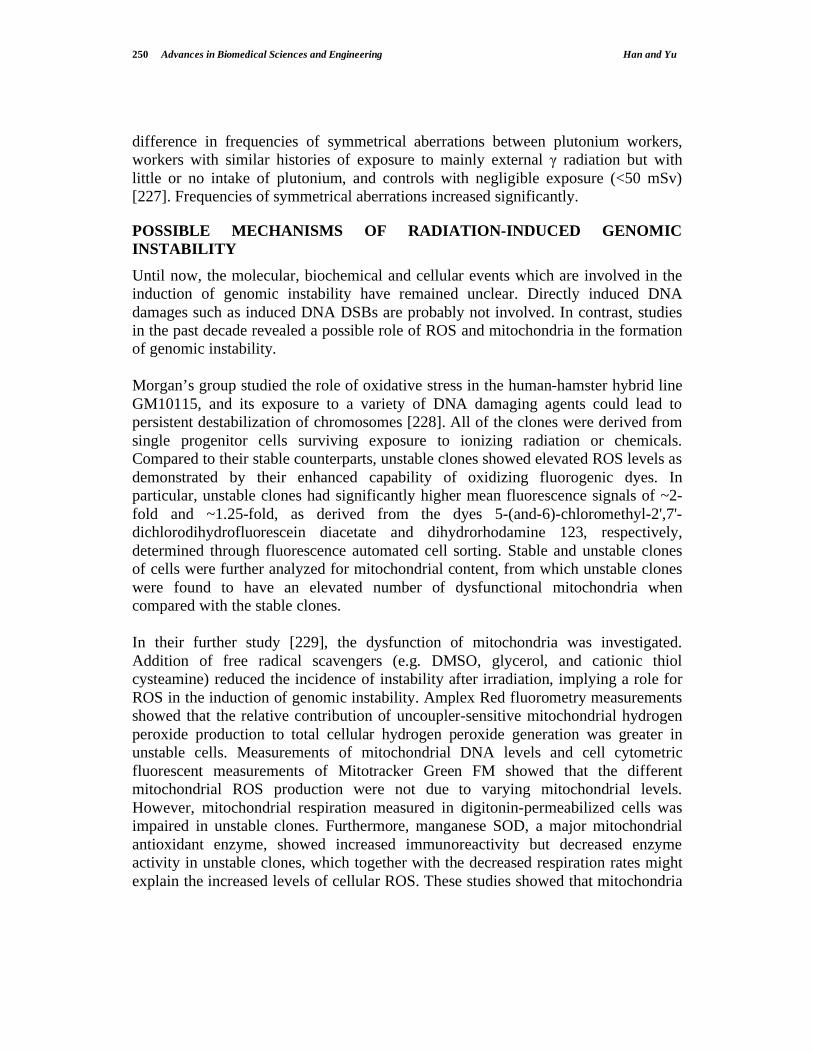
250 Advances in Biomedical Sciences and Engineering Han and Yu
difference in frequencies of symmetrical aberrations between plutonium workers,
workers with similar histories of exposure to mainly external radiation but with
little or no intake of plutonium, and controls with negligible exposure (<50 mSv)
[227]. Frequencies of symmetrical aberrations increased significantly.
POSSIBLE MECHANISMS OF RADIATION-INDUCED GENOMIC
INSTABILITY
Until now, the molecular, biochemical and cellular events which are involved in the
induction of genomic instability have remained unclear. Directly induced DNA
damages such as induced DNA DSBs are probably not involved. In contrast, studies
in the past decade revealed a possible role of ROS and mitochondria in the formation
of genomic instability.
Morgan’s group studied the role of oxidative stress in the human-hamster hybrid line
GM10115, and its exposure to a variety of DNA damaging agents could lead to
persistent destabilization of chromosomes [228]. All of the clones were derived from
single progenitor cells surviving exposure to ionizing radiation or chemicals.
Compared to their stable counterparts, unstable clones showed elevated ROS levels as
demonstrated by their enhanced capability of oxidizing fluorogenic dyes. In
particular, unstable clones had significantly higher mean fluorescence signals of ~2-
fold and ~1.25-fold, as derived from the dyes 5-(and-6)-chloromethyl-2',7'-
dichlorodihydrofluorescein diacetate and dihydrorhodamine 123, respectively,
determined through fluorescence automated cell sorting. Stable and unstable clones
of cells were further analyzed for mitochondrial content, from which unstable clones
were found to have an elevated number of dysfunctional mitochondria when
compared with the stable clones.
In their further study [229], the dysfunction of mitochondria was investigated.
Addition of free radical scavengers (e.g. DMSO, glycerol, and cationic thiol
cysteamine) reduced the incidence of instability after irradiation, implying a role for
ROS in the induction of genomic instability. Amplex Red fluorometry measurements
showed that the relative contribution of uncoupler-sensitive mitochondrial hydrogen
peroxide production to total cellular hydrogen peroxide generation was greater in
unstable cells. Measurements of mitochondrial DNA levels and cell cytometric
fluorescent measurements of Mitotracker Green FM showed that the different
mitochondrial ROS production were not due to varying mitochondrial levels.
However, mitochondrial respiration measured in digitonin-permeabilized cells was
impaired in unstable clones. Furthermore, manganese SOD, a major mitochondrial
antioxidant enzyme, showed increased immunoreactivity but decreased enzyme
activity in unstable clones, which together with the decreased respiration rates might
explain the increased levels of cellular ROS. These studies showed that mitochondria

Response of Cells to Ionizing Radiation Advances in Biomedical Sciences and Engineering 251
in unstable cells were abnormal and they probably caused persistent oxidative stress
in the unstable clones.
TRANS-GENERATIONAL EFFECTS OF RADIATION EXPOSURE
Trans-generational effects are those effects observed in offspring born after one or
both parents had been irradiated prior to conception [209]. Potential trans-
generational effects might play a significant role in reproductive legacy of individuals
living in radiation-contaminated areas.
Wiley and colleagues used a pre-implantation embryo chimera assay to demonstrate
adverse effects in progeny embryos after acute whole-body paternal irradiation, and
illustrated the trans-generational effects of radiation exposure. They measured the
competitive cell-proliferation disadvantage of an embryo with a radiation history after
challenge by direct cell-to-cell contact with a normal embryo in an aggregation
chimera [230,231]. The F1 embryos conceived 6–7 weeks after paternal F0
irradiation were the most likely to display the phenotype, indicating that the type B
spermatogonia were the most sensitive. Baulch et al. [232] evaluated F3 mouse
offspring from F0 paternal mice exposed to 1 Gy 137
Cs -rays for gene products that
could modulate cell proliferation rate including receptor tyrosine kinase, protein
kinase C and MAP kinases and protein levels of nuclear p53 (Trp53) and p21Waf1
(Cdkn1a). All three protein kinase activities were changed, and nuclear levels of
Trp53 and Cdkn1a protein were higher in F3 offspring with a paternal F0 radiation
history than in unirradiated littermates.
Fortunately, no radiation-induced genetic, i.e. hereditary, diseases have been
demonstrated in a human population exposed to ionizing radiation until now. No
excess in cancer incidence in children born to parents exposed by the atomic
bombings in Japan [233,234] or in the offspring of cancer patients treated with
radiotherapy [235] have been reported. Nevertheless, the risk of cancer in the
offspring of humans irradiated prior to conception remains controversial.
REFERENCES
[1] Hall EJ. Radiobiology for the Radiologist (5th Edition), Lippincott Williams & Wilkins,
Philadelphia, 2000.
[2] Lehnert S. Biomolecular Action of Ionizing Radiation, Taylor & Francis Group, LLC, 2007.
[3] Valentin J. Low-dose Extrapolation of Radiation-Related Cancer Risk, Elsevier, 2006.
[4] Johansen L. The contribution of water-free radicals to the X-ray inactivation of bacteria. In:
Cellular Radiation Biology, Williams & Wlikins, Baltimore, Maryland, 1965.
[5] Roots R, Okada S. Protection of DNA molecules of cultured mammalian cells from radiation-
induced single-strand scissions by various alcohols and SH compounds. Int J Radiat Biol 1972;
21: 329–42.
[6] Chapman JD, Reuvers A, Borsa J, and Greenstock CL. Chemical radioprotection and
radiosensitization of mammalian cells growing in vitro. Radiat Res 1995; 56: 291–306.
[7] von Sonntag C. The Chemical Basis of Radiation Biology. Taylor and Francis, London, 1987.,

252 Advances in Biomedical Sciences and Engineering Han and Yu
[8] Te´oule R. Radiation-induced DNA damage and its repair. Int J Radiat Biol 1987; 51: 573–89.
[9] Nicoloff JA, Hoekstra MF. DNA Repair in Higher Eukaryotes, Vol. 2. Humana Press Inc.,
Totowa, New Jersey, 1996.
[10] Ward, J.E. Nature of lesions formed by ionizing radiation. In DNA damage and Repair. Human
Press, Totowa NJ, Vol. 2, 1998.
[11] Hutchison F. Chemical changes produced in DNA by ionizing radiation. Prog. Nucleic Acid
Res 1985; 32: 115–54.
[12] Ward JF. Biochemistry of DNA lesions. Radiat Res 1985; 104: S103–S111.
[13] Goodhead DT. Initial events in the cellular effects of ionising radiations: clustered damage in
DNA. Int J Radiat Biol 1994; 65: 7-17.
[14] Ward JF. Radiation mutagenesis: the initial DNA lesion responsible. Radiat Res 1995; 142:
362-8.
[15] Nikjoo H, O'Neill P, Terrissol M, Goodhead DT. Quantitative modelling of DNA damage using
Monte Carlo track structure method. Radiat Environ Biophys 1999; 38: 31-38.
[16] Boudaiffa B, Hunting D, Cloutier P, Huels MA, Sanche L. Induction of single- and double-
strand breaks in plasmid DNA by 100-1500 eV electrons. Int J Radiat Biol 2000; 9: 1209-21.
[17] Nikjoo H, Bolton CE, Watanabe R, Terrissol M, O'Neill P, Goodhead DT. Modelling of DNA
damage induced by energetic electrons (100 eV to 100 keV). Radiat Prot Dosimetry 2002; 99:
77-80.
[18] Nikjoo H, Munson RJ, Bridges BA, RBE-LET relationships in mutagenesis by ionizing
radiation. Radiat Res 2000: 40 Suppl; 85-105.
[19] Nikjoo H, O'Neill P, Wilson, WE, Goodhead DT. Computational approach for determining the
spectrum of DNA damage induced by ionizing radiation. Radiat Res 2001; 156: 577-83.
[20] Barker S, Weinfeld M, Zheng J, Li L, Murray D. Identification of mammalian proteins cross-
linked to DNA by ionizing radiation. J Biol Chem 2005; 280: 33826–38.
[21] Hoeijmakers JH, Genome maintenance mechanisms for preventing cancer. Nature 2001; 411:
366–74.
[22] Rich T, Allen RL, Wyllie AH, Defying death after DNA damage. Nature 2000; 96: 643–49.
[23] Chaudhry MA, Weinfeld M, Reactivity of human apurinic/apyrimidinic endonuclease and
Escherichia coli exonuclease III with bistranded abasic sites in DNA. J Biol Chem 1997; 272:
15650–55.
[24] Dahm-Daphi J, Sass C, Albert W, Comparison of biological effects of DNA damage induced by
ionizing radiation and hydrogen peroxide in CHO cells. Int J Radiat Biol 2000; 76: 67–75.
[25] Jeggo PA. DNA breakage and repair. Adv Genet 1998; 38: 185–218.
[26] Karran P. DNA double strand break repair in mammalian cells. Curr Opin Genet Dev 2000; 10:
144–150.
[27] Khanna KK, Jackson SP. DNA double-strand breaks: Signaling, repair and the cancer
connection. Nat Genet 2001; 27: 247–54.
[28] Jackson SP. Sensing and repairing DNA double-strand breaks. Carcinogenesis 2002; 23: 687–
96.
[29] Sancar A, Lindsey-Boltz LA, Unsal-Kacmaz K, Linn S. Molecular mechanisms of mammalian
DNA repair and the DNA damage checkpoints. Annu Rev Biochem 2004; 73: 39–85.
[30] Kurz EU, L-MS. DNA damage-induced activation of ATM and ATM-dependent signaling
pathways. DNA Repair 2004; 3: 889–900,.
[31] Collis SJ, DeWeese TL, Jeggo PA, Parker AR. The life and death of DNA-PK. Oncogene 2005;
24: 949–61.
[32] Taylor AMR, Metcalfe JA, Thick J, Mak YF. Leukemia and lymphoma in ataxia telangiectasia.
Blood 1996; 87: 423-38.
[33] Goodarzi AA, Block WD, Lees-Miller SP. The role of ATM and ATR in DNA damage-induced
cell cycle control. Prog Cell Cycle Res 2003; 5: 393-411.

Response of Cells to Ionizing Radiation Advances in Biomedical Sciences and Engineering 253
[34] Shiloh Y. ATM and ATR: networking cellular responses to DNA damage. Curr Opin Genet
Dev 2001; 11: 71-77.
[35] Shiloh Y. ATM and related protein kinases: safeguarding genome integrity. Nat Rev Cancer
2003; 3: 155-68.
[36] Banin S, Moyal L, Shieh S, Taya Y, Anderson CW, Chessa L, Smorodinsky NI, Prives C, Reiss
Y, Shiloh Y, Ziv Y. Enhanced phosphorylation of p53 by ATM in response to DNA damage.
Science 1998; 281: 1674-77.
[37] Canman CE, Lim DS, Cimprich KA, Taya Y, Tamai K, Sakaguchi K, Appella E, Kastan MB,
Siliciano JD. Activation of the ATM kinase by IR and phosphorylation of p53. Science 1998;
281: 1677-79.
[38] Khanna KK., Keating KE, Kozlov S, Scott S, Gatei M, Hobson K, Taya Y, Gabrielli B, Chan
D, Lees-Miller SP, Lavin MF. ATM associates with and phosphorylates p53: mapping the
region of interaction. Nature Genetics 1998; 20: 398 - 400.
[39] Kastan MB, Zhan Q, El-Deiry WS, Carrier F, Jacks T, Walsh WV, Plunkett BS, Vogelstein B,
Fornace AJ. A mammalian cell cycle checkpoint pathway utilizing p53 and GADD45 is
defective in ataxia-telangiectasia. Cell 1992; 71: 587-97.
[40] Lu X, Lane DP. Differential induction of transcriptionally active p53 following UV or ionizing
radiation: defects in chromosome instability syndromes? Cell 1993; 75: 765-78.
[41] Carson CT, Schwartz RA, Stracker TH, Lilley CE, Lee DV, Weitzman MD. The Mre11
complex is required for ATM activation and the G2/M checkpoint. EMBO J 2003; 22: 6610–
20.
[42] Mochan TA, Venere M, DiTullio RAJ, Halazonetis TD. 53BP1 and NFBD1/MDC1-Nbs1
function in parallel interacting pathways activating ataxiatelangiectasiabmutated (ATM) in
response to DNA damage. Cancer Res 2003; 63: 8586–91.
[43] Uziel T, Lerenthal Y, Moyal L, Andegeko Y, Mittelman L, Shiloh Y. Requirement of the MRN
complex for ATM activation by DNA damage. EMBO J 2003; 22: 5612–21.
[44] Dupre A, Boyer-Chatenet L, Gautier H. Two-step activaiton of ATM by DNA and the Mre11-
Rad50-Nbs1 comples. Nat Struct Mol Biol 2006; 13: 451–57.
[45] Lee JH, Paull TT. ATM activation by DNA double-strand breaks through the Mre11-Rad50-
Nbs1 complex. Science 2005; 308: 551 54.
[46] Aten JA, Stap J, Krawczyk PM, van Oven CH, Hoebe RA, et al. Dynamics of DNA double-
strand breaks revealed by clustering of damaged chromosome domains. Science 2004; 303: 92–
95.
[47] Lisby M, Barlow JH, Burgess RC, Rothstein R. Choreography of the DNA damage response:
spatiotemporal relationships among checkpoint and repair proteins. Cell 2004; 118: 699–713.
[48] Moreno-Herrero F, de Jager M, Dekker NH, Kanaar R, Wyman C, Dekker C. Mesoscale
conformational changes in the DNA-repair complex Rad50/Mre11/Nbs1 upon binding DNA.
Nature 2005; 437: 440–43.
[49] Shroff R, Arbel-Eden A, Pilch D, Ira G, Bonner WM, et al. Distribution and dynamics of
chromatin modification induced by a defined DNA double-strand break. Curr Biol 2004; 14:
1703–11.
[50] Kumagai A, Lee J, Yoo HY, Dunphy WG. TopBP1 activates the ATR-ATRIP complex. Cell
2006; 124: 943–55.
[51] Yamane K, Wu X, Chen J. A DNA damage-regulated BRCT-containing protein, TopBP1, is
required for cell survival. Mol Cell Biol 2002; 22: 555–66.
[52] International Nijmegen Breakage Syndrome Study Group. Nijmegen breakage syndrome. Arch
Dis Child 2000; 82: 400-06.
[53] Shiloh, Y. Ataxia-telangiectasia and the Nijmegen breakage syndrome: related disorders but
genes apart. Annu Rev Genet 1997; 31: 635-62.

254 Advances in Biomedical Sciences and Engineering Han and Yu
[54] Carney JP, Maser RS, Olivares H, Davis EM, Le Beau M, Yates III JR, Hays L, Morgan WF,
Petrini, JHJ. The hMre11/hRad50 protein complex and Nijmegen breakage-syndrome: linkage
of double-strand break repair to the cellular DNA damage response. Cell 1998; 93: 477-86.
[55] Varon R, Vissinga C, Platzer M, Cerosaletti KM, Chrzanowska KH, Saar K, Beckmann G,
Seemanova E, Cooper PR, Nowak NJ, Stumm M, Weemaes CMR, Gatti RA, Wilson RK,
Digweed M, Rosenthal A, Sperling K, Concannon P, Reis A. Nibrin, a novel DNA double-
strand break repair protein, is mutated in Nijmegen breakage syndrome. Cell 1998; 93: 467-76.
[56] Stewart GS, Maser RS, Stankovic T, Bressan DA, Kaplan MI, Jaspers NG, Raams A, Byrd PJ,
Petrini, JH, Taylor AM. The DNA double-strand break repair gene hMRE11 is mutated in
individuals with an ataxia-telangiectasia-like disorder. Cell 1999; 9: 577-87.
[57] Cerosaletti K, Concannon P. Independent roles for Nibrin and Mre11 in the activation and
function of ATM. J Biol Chem 2004; 279: 38813–9.
[58] Kobayashi J, Tauchi H, Sakamoto S, Nakamura A, Morishima K, Matsuura S, Kobayashi T,
Tamai K, Tanimoto K, Komatsu K. NBS1 localizes to gamma-H2AX foci through interaction
with the FHA/BRCT domain. Curr Biol 2002; 12: 1846-51.
[59] Tauchi H, Kobayashi J, Morishima K, van Gent DC, Shiraishi T, Verkaik NS, vanHeems D, Ito
E, Nakamura A, Sonoda E, Takata M, Takeda S, Matsuura S, Komatsu K. Nbs1 is essential for
DNA repair by homologous recombination in higher vertebrate cells. Nature 2002; 420: 93-98.
[60] O'Driscoll M, Cerosaletti KM, Girard PM, Dai Y, Stumm M, Kysela B, Hirsch B, Gennery A,
Palmer SE, Seidel J, Gatti RA, Varon R, Oettinger MA, Sperling K, Jeggo PA, Concannon P. ()
DNA Ligase IV mutations identified in patients exhibiting development delay and
immunodeficiency. Mol Cell 2001; 8: 1175-85.
[61] Girard PM, Riballon E, Begg A, Waugh A, Jeggo PA. Nbs1 promotes ATM dependent
phosphorylation events including those required for G1/S arrest. Oncogene 2002; 21: 4191-99.
[62] Celeste A, Petersen S, Romanienko PJ, Fernandez-Capetillo O, Chen HT, Sedelnikova OA,
Reina-San-Martin B, Coppola V, Meffre E, Difilippantonio MJ, Redon C, Pilch DR, Olaru A,
Eckhaus M, Camerini-Otero RD, Tessarollo L, Livak F, Manova K, Bonner WM, Nussenzweig
MC, Nussenzweig A. Genomic Instability in Mice Lacking Histone H2AX. Science 2002; 296:
922 – 27.
[63] Yamazaki V, Kirchgessner CU, Gellert M, Bonner WM. A critical role for histone H2AX in
recruitment of repair factors to nuclear foci after DNA damage. Curr Biol 2000; 10: 886-95.
[64] Rogakou EP, Boon C, Redon C, Bonner WM. Megabase chromatin domains involved in DNA
double-strand breaks in vivo. J Cell Biol 1999; 146: 905-16.
[65] Rothkamm K, Kruger I, Thompson LH, Lobrich M. Pathways of DNA double-strand break
repair during the mammalian cell cycle. Mol Cell Biol 2003; 23: 5706-15.
[66] Chowdhury D, Keogh MC, Ishii H, Peterson CL, Buratowski S, Lieberman J. gamma-H2AX
dephosphorylation by protein phosphatase 2A facilitates DNA doublestrand break repair. Mol
Cell 2005; 20: 801–9.
[67] Miki Y, Swensen J, Shattuck-Eidens D, Futreal PA, Harshman K, Tavtigian S, Liu Q, Cochran
C, Bennett LM, Ding W. A strong candidate for the breast and ovarian cancer susceptibility
gene BRCA1. Science 1994; 266: 66-71.
[68] Wooster R, Bignell G, Lancaster J, Swift S, Seal S, Mangion J, Collins N, Gregory S, Gumbs
C, Micklem G. Identification of the breast cancer susceptibility gene BRCA2. Nature 1995;
378: 89-792.
[69] Venkitaraman AR. Cancer susceptibility and the functions of BRCA1 and BRCA2. Cell 2002;
108: 171-82.
[70] Moynahan ME, Chiu JW, Koller BH, Jasin M. Brca1 controls homology-directed DNA repair.
Mol Cell 1999; 4: 511-18.
[71] Xu B, Kim S, Kastan MB. Involvement of brca1 in s-phase and g(2)-phase checkpoints after
ionizing irradiation. Mol Cell Biol 2001; 21: 3445-50.

Response of Cells to Ionizing Radiation Advances in Biomedical Sciences and Engineering 255
[72] Foray N, Marot D, Gabriel A, Randrianarison V, Carr A, Perricaudet M, Ashworth A, Jeggo P.
() A subset of ATM and ATR-dependent phosphorylation events requires the BRCA1 protein.
EMBO 2003; 22: 2860-71.
[73] Lee JS, Collins KM, Brown AL, Lee CH, Chung JH. hCds1-mediated phosphorylation of
BRCA1 regulates the DNA damage response. Nature 2000; 404: 201-04.
[74] Moynahan ME, Pierce AJ, Jasin M. BRCA2 is required for homology-directed repair of
chromosomal breaks. Mol Cell 2001; 7: 263-72.
[75] Pellegrini L, Yu DS, Lo T, Anand S, Lee M, Blundell TL, Venkitaraman AR. Insights into
DNA recombination from the structure of a RAD51-BRCA2 complex. Nature 2002; 420: 287-
293.
[76] Yang H, Jeffrey PD, Miller J, Kinnucan E, Sun Y, Thoma NH, Zheng N, Chen PL, Lee WH,
Pavletich NP. BRCA2 function in DNA binding and recombination from a BRCA2-DSS1-
ssDNA structure. Science 2002; 297: 1837-48.
[77] Wang B, Matsuoka S, Carpenter PB, Elledge SJ. 53BP1, a mediator of the DNA damage
checkpoint. Science 2002; 298: 1435-38.
[78] Goldberg M, Stucki M, Falck J, D'Amours D, Rahman D, Pappin D, Bartek J, Jackson SP.
MDC1 is required for the intra-S-phase DNA damage checkpoint. Nature 2003; 421: 952-56.
[79] Lou Z, Chini CC, Minter-Dykhouse K, Chen J. MDC1 regulates BRCA1 localization and
phosphorylation in DNA damage checkpoint control. J Biol Chem 2003; 278: 13599-602.
[80] Abraham RT. Checkpoint signalling: focusing on 53BP1. Nat Cell Biol 2002; 4: E277-79.
[81] Fernandez-Capetillo O, Chen HT, Celeste A, Ward I, Romanienko PJ, Morales JC, Naka K, Xia
Z, Camerini-Otero RD, Motoyama N, Carpenter PB, Bonner WM, Chen J, Nussenzweig A.
DNA damage-induced G2-M checkpoint activation by histone H2AX and 53BP1. Nat Cell Biol
2002; 4: 993-97.
[82] Stewart GS, Wang B, Bignell CR, Taylor AM, Elledge SJ. MDC1 is a mediator of the
mammalian DNA damage checkpoint. Nature 2003; 421: 961-66.
[83] Kastan MB, Onyekewere O, Sidransky D, Vogelstein B, Craig RW. Participation of p53 protein
in the cellular response to DNA damage. Cancer Res 1991, 51, 6304-11.
[84] Ashcroft M, Kubbutat MH, Vousden KH. Regulation of p53 function and stability by
phosphorylation. Mol Cell Biol 1999; 19: 1751-58.
[85] Lakin ND, Jackson SP. Regulation of p53 in response to DNA damage. Oncogene 1999; 18:
7644-55.
[86] Fei P, El-Deiry WS. P53 and radiation responses. Oncogene 2003; 22: 5774-83.
[87] Donehower LA, Harvey M, Slagle BL, McArthur MJ, Montgomery CA, Butel JS, Bradley A.
Mice deficient for p53 are developmentally normal but susceptible to spontaneous tumours.
Nature 1992; 356: 215-21.
[88] Malkin D, Li FP, Strong LC, Fraumeni JF, Nelson CE, Kim DH, Kassel J, Gryka MA, Bischoff
FZ, Tainsky MA. Germ line p53 mutations in a familial syndrome of breast cancer, sarcomas,
and other neoplasms. Science 1990: 250; 1233-38.
[89] Srivastava S, Zou ZQ, Pirollo K, Blattner W, Chang EH. Germ-line transmission of a mutated
p53 gene in a cancer-prone family with Li-Fraumeni syndrome. Nature 1990; 348: 747-49.
[90] Deb SP. Cell cycle regulatory functions of the human oncoprotein MDM2. Mol Cancer Res
2003; 1: 1009-16.
[91] Kubbutat MH, Ludwig RL, Ashcroft M, Vousden KH. Regulation of Mdm2-directed
degradation by the C terminus of p53. Mol Cell Biol 1998; 18: 5690-98.
[92] Momand J, Zambetti GP, Olson DC, George D, Levine AJ. The mdm2 oncogene product forms
a complex with the p53 protein and inhibits p53-mediated transactivation. Cell 1992; 69: 1237-
45.
[93] Sinclair WK. Cyclic x-ray responses in mammalian cells in vitro. Radiat Res 1968; 33: 620–43.
[94] Su TT. Cellular Responses to DNA Damage: One Signal, Multiple Choices. Annu Rev Genet
2006; 40: 187 208.

256 Advances in Biomedical Sciences and Engineering Han and Yu
[95] Durocher D, Jackson SP. DNA-PK, ATM and ATR as sensors of DNA damage: variations on a
theme? Curr Opin Cell Biol 2001; 13: 225-31.
[96] Rouse J, Jackson SP. Interfaces between the detection, signaling, and repair of DNA damage.
Science 2002; 297: 547-51.
[97] Di Leonardo A, Linke SP, Clarkin K, Wahl GM. DNA damage triggers a prolonged p53-
dependent G1 arrest and long-term induction of Cip1 in normal human fibroblasts. Genes Dev
1994; 8: 2450-51;
[98] Little JB. Delayed initiation of DNA synthesis in irradiated human diploid cells. Nature 1968;
146: 1064-65.
[99] Wahl GM, Carr AM. The evolution of diverse biological responses to DNA damage: insights
from yeast and p53. Nature Cell Biol 2001; 3: E277-E286.
[100] Ko LJ, Prives C. p53: puzzle and paradigm. Genes Dev 1996; 10: 1054-72.
[101] Lee JM, Bernstein A. p53 mutations increase resistance to ionizing radiation. PNAS 1993; 90:
5742-46.
[102] Zhou XY, Wang X, Hu B, Guan J, Iliakis G, Wang Y. An ATM-independent S-phase
checkpoint response involves CHK1 pathway. Cancer Res 2002; 62: 1598-1603.
[103] Jackson S. Sensing and repairing DNA double strand breaks. Carcinogenesis 2002; 23: 687-96.
[104] Feijoo C, Hall-Jackson C, Wu R, Jenkins D, Leitch J, Gilbert DM, Smythe C. Activation of
mammalian Chk1 during DNA replication arrest: a role for Chk1 in the intra-S phase
checkpoint monitoring replication origin firing. J Cell Biol 2001; 154: 913-23.
[105] Tercero JA, Longhese MP, Diffley JF. A central role for DNA replication forks in checkpoint
activation and response. Mol Cell 2003; 11: 1323-36.
[106] Iliakis G, Wang Y, Guan J, Wang H. DNA damage checkpoint control in cells exposed to
ionizing radiation. Oncogene 2003; 22: 5834-47.
[107] Xiao Z, Chen Z, Gunasekera AH, Sowin TJ, Rosenberg SH, Fesik S, Zhang H. Chk1 mediates
S and G2 arrests through Cdc25A degradation in response to DNA-damaging agents. J Biol
Chem 2003; 278: 21767-73.
[108] Gottifredi V, Prives C. The S phase checkpoint: When the crowd meets at the fork. Semin. Cell
Dev. Biol 2005; 16: 355–68,
[109] Beamish H, Lavin MF. (Radiosensitivity in ataxia-telangiectasia: anomalies in radiation-
induced cell cycle delay. Int J Radiat Biol 1994; 65: 175-84.
[110] Wang X, Khadpe J, Hu B, Iliakis G, Wang Y. An overactivated ATR/CHK1 pathway is
responsible for the prolonged G2 accumulation in irradiated AT cells. J Biol Chem 2008, 278:
30869-74.
[111] Bryant PE. Repair and chromosomal damage. Radiother Oncol 72; 2004: 251–256.
[112] Hlatky LR, Sachs RK, Vazquez M, Cornforth MN. Radiation-induced chromosome aberrations:
insights gained from biophysical modeling. Bioessays 2002; 24: 714–723.
[113] Cucinotta FA, Nikjoo H, O’Neill P, Goodhead DT. Kinetics of DSB rejoining and formation of
simple chromosome exchange aberrations. Int J Radiat Biol Relat Stud Phys Chem Med 76:
1463–1474, 2000.
[114] Revell SH. The accurate estimation of chromatid breakage and its relevance to a new
interpretation of chromatid aberrations induced by ionizing radiation. Proc Roy Soc Lond B
Biol Sci 150: 563–589, 1959.
[115] McConkey DJ, Orrenius S. Signal Transduction Pathways in Apoptosis. Stem Cells 1996; 14:
19-631;
[116] Verheij M, Bartelink H. Radiation-induced apoptosis. Cell Tissue Res 2000; 301: 133–42.
[117] Shinomiya N. New concepts in radiation-induced apoptosis: 'premitotic apoptosis' and
'postmitotic apoptosis', J Cell Mol Med 2001; 5: 240-53.
[118] Shinomiya N, Kuno Y, Yamamoto F, Fukasawa M, Okumura A. Uefuji M., Rokutanda M.,
Different mechanisms between premitotic apoptosis and postmitotic apoptosis in X-irradiated
U937 cells. Int J Radiat Oncol Biol Phys 2000; 47: 767-77.

Response of Cells to Ionizing Radiation Advances in Biomedical Sciences and Engineering 257
[119] Elmore S. Apoptosis: A Review of Programmed Cell Death, Toxicol Pathol 2007; 35: 495-516.
[120] Saelens X, Festjens N, Vande Walle L, van Gurp M, van Loo G, Vandenabeele P. Toxic
proteins released from mitochondria in cell death. Oncogene 2004; 23:2861-74.
[121] Du C, Fang M, Li Y, Li L, Wang X. Smac, a mitochondrial protein that promotes cytochrome
c-dependent caspase activation by eliminating IAP inhibition. Cell 2000; 102: 33-42.
[122] Chinnaiyan AM. The apoptosome: heart and soul of the cell death machine. Neoplasia. 1999; 1:
5-15.
[123] Hill MM, Adrain C, Duriez PJ, Creagh EM, Martin SJ. Analysis of the composition, assembly
kinetics and activity of native Apaf-1 apoptosomes. Embo J 2004; 23: 2134-45.
[124] van Loo G, van Gurp M, Depuydt B, Srinivasula SM, Rodriguez I, Alnemri ES, Gevaert K,
Vandekerckhove J, Declercq W, Vandenabeele P. The serine protease Omi/HtrA2 is released
from mitochondria during apoptosis. Omi interacts with caspase-inhibitor XIAP and induces
enhanced caspase activity. Cell Death Differ. 2002a; 9: 20-6.;
[125] Schimmer AD. Inhibitor of apoptosis proteins: translating basic knowledge into clinical
practice. Cancer Res 2004; 64: 7183-90.
[126] Joza N, Susin SA, Daugas E, Stanford WL, Cho SK, Li CY, Sasaki T, Elia AJ, Cheng HY,
Ravagnan L, Ferri KF, Zamzami N, Wakeham A, Hakem R, Yoshida H, Kong YY, Mak TW,
Zuniga- Pflucker JC, Kroemer G, Penninger JM. Essential role of the mitochondrial apoptosis-
inducing factor in programmed cell death. Nature 2001; 410: 549-54.
[127] Li LY, Luo X, Wang X. Endonuclease G is an apoptotic DNase when released from
mitochondria. Nature. 2001; 412: 95-9.
[128] Enari M, Sakahira H, Yokoyama H, Okawa K, Iwamatsu A, Nagata S. A caspase-activated
DNase that degrades DNA during apoptosis, and its inhibitor ICAD. Nature. 1998; 391: 43-50.;
[129] Susin SA, Daugas E, Ravagnan L, Samejima K, Zamzami N, Loeffler M, Costantini P, Ferri
KF, Irinopoulou T, Prevost MC, Brothers G, Mak TW, Penninger J, Earnshaw WC, Kroemer
G. Two distinct pathways leading to nuclear apoptosis. J Exp Med. 2000; 192: 571-80.
[130] Cory S, Adams JM. The Bcl2 family: regulators of the cellular life-or-death switch. Nat Rev
Cancer 2002; 2: 647-56.
[131] Schuler M, Green DR. Mechanisms of p53-dependent apoptosis. Biochem Soc Trans 2001; 29:
684-8.
[132] Locksley RM, Killeen N, Lenardo MJ. The TNF and TNF receptor superfamilies: integrating
mammalian biology. Cell 2001; 104: 487-501.
[133] Ashkenazi A, Dixit VM. Death receptors: signaling and modulation. Science. 1998; 281: 1305-
8.
[134] Hsu H, Xiong J, Goeddel DV. The TNF receptor 1-associated protein TRADD signals cell
death and NF-kappa B activation. Cell. 1995; 81: 495-504.
[135] Kischkel FC, Hellbardt S, Behrmann I, Germer M, Pawlita M, Krammer PH, Peter ME.
Cytotoxicity-dependent APO-1 (Fas/CD95)-associated proteins form a death-inducing signaling
complex (DISC) with the receptor. Embo J. 1995; 14: 5579-88.
[136] Thacker J. The nature of mutants induced by ionising radiation in cultured hamster cells. Mutat
Res 1986; 160: 267-75.
[137] National Research Council Committee on Health Risks of Exposure to Radon (BEIR VI)
(1994) Health Effects of Exposure to Radon: Time for Reassessment (National Academy of
Sciences, Washington, DC.
[138] Hei TK, Wu LJ, Liu SX, Vannais D, Waldren CA, Randers-Pehrson G. Mutagenic effects of a
single and an exact number of particles in mammalian cells. PNAS 1997; 94: 3765-70.
[139] Wu LJ, Randers-Pehrson G, Xu A, Waldren CA, Geard CR, Yu ZL, Hei TK. Targeted
cytoplastic irradiation with alpha particles induces mutations in mammalian cells. PNAS 1999;
96: 4959-64.

258 Advances in Biomedical Sciences and Engineering Han and Yu
[140] Limoli CL, Hartmann A, Shepard L, Yang CR, Boothman DA, Bartholomew J, Morgan WF.
Apoptosis, reproductive failure and oxidative stress in Chinese hamster ovary cells with
compromised genomic integrity. Cancer Res 1998; 58: 3712-18.
[141] Little JB. Radiation carcinogenesis. Carcinogenesis 2000; 21: 397-404.
[142] Mothersill C, Seymour C. Radiation-induced bystander effects: Past history and future
directions. Radiat Res 2001; 155: 759–67.
[143] Nagasawa H, Cremesti A, Kolesnick R, Fuks Z, Little JB. Involvement of membrane signaling
in the bystander effect in irradiated cells. Cancer Res 2002; 62: 2531-4.
[144] Azzam EI, de Toledo SM, Little JB. Direct evidence for the participation of gap junction-
mediated intercellular communication in the transmission of damage signals from a particle
irradiated to non-irradiated cells. PNAS 2001; 98: 473-8.
[145] Azzam EI, de Toledo SM, Spitz DR, Little JB. Oxidative metabolism modulates signal
transduction and micronucleus formation in bystander cells from -particle irradiated normal
human fibroblasts. Cancer Res 2002; 62: 5436-42.
[146] Hu B, Han W, Wu L, Feng H, Liu X, Zhang L, Xu A, Hei TK, Yu Z, In Situ Visualization of
DSBs to Assess the Extranuclear/Extracellular Effects Induced by Low-Dose a-Particle
Irradiation, Radia Res 2005; 164: 286–91.
[147] Randers-Pehrson G, Geard CR, Johnson G, Elliston CD, Brenner DJ, The Columbia University
Single-Ion Microbeam, Radia Res 2001; 156: 210–214.
[148] Hall EJ, Hei TK. Genomic instability and bystander effects induced by high LET radiation.
Oncogene 2003; 22: 7034-42.
[149] Zhou H, Randers-Pehrson G, Waldren CA, Vannais D, Hall EJ, Hei TK. Induction of a
bystander mutagenic effect of alpha particles in mammalian cells. PNAS 2000; 97: 2099–104.
[150] Shao C, Folkard M, Michael BD, Prise KM, Targeted cytoplasmic irradiation induces bystander
responses, PNAS 2004; 101: 13495-500.
[151] Little J. Genomic instability and bystander effects: A historical perspective. Oncogene 2003;
22: 6978–87.
[152] Morgan WF. Non-targeted and delayed effects of exposure to ionizing radiation:I. Radiation-
induced genomic instability and bystander effects in vitro. Radiat Res 2003; 159: 567–80.
[153] Seymour CB, Mothersill C. Relative contribution of bystander and targeted cell killing to the
low-dose region of the radiation dose–response curve. Radiat Res 2000; 153: 508–11.
[154] Mothersill C, Stamato TD, Perez ML, Cummins R, Mooney R, Seymour CB. Involvement of
energy metabolism in the production of ‘bystander effects’ by radiation. Br J Cancer 2000; 82:
1740–6.
[155] Yang H, Asaad N, Held KD. Medium-mediated intercellular communication is involved in
bystander responses of X-ray-irradiated normal human fibroblasts. Oncogene 2005; 24: 2096–
103.
[156] Han W, Wu L, Hu B, Zhang L, Chen S, Bao L, Zhao Y, Xu A, Yu Z. The Early and Initiation
Processes of Radiation-Induced Bystander Effects Involved in the Induction of DNA Double
Strand Breaks in Non-Irradiated Cultures, Br J Radiology 2007; 80: S7–S12.
[157] Belyakov OV, Mitchell SA, Parikh D, Randers-Pehrson G, Marino SA, Amundson SA, Geard
CR, Brenner DJ. Biological effects in unirradiated human tissue induced by radiation damage
up to 1 mm away. PNAS 2005; 102: 14203-8.
[158] Sedelnikova OA, Nakamura A, Kovalchuk O, Koturbash I, Mitchell SA, Marino SA, Brenner
DJ, Bonner WM. DNA Double-Strand Breaks Form in Bystander Cells after Microbeam
Irradiation of Three-dimensional Human Tissue Models, Cancer Res 2007; 67: 4295-320.
[159] Brooks AL, Retherford JC, McClellan RO. Effect of 239
PuO2 particle number and size on the
frequency and distribution of chromosome aberrations in the liver of the Chinese hamster.
Radiat Res 1974; 59: 693–709.

Response of Cells to Ionizing Radiation Advances in Biomedical Sciences and Engineering 259
[160] Koturbash I, Rugo RE, Hendricks CA, Loree J, Thibault B, Kutanzi K, Pogribny I, Yanch JC,
Engelward BP, Kovalchuk O. Irradiation induces DNA damage and modulates epigenetic
effectors in distant bystander tissue in vivo. Oncogene 2006; 25: 4267-75.
[161] Mancuso M, Pasquali E, Leonardi S, Tanori M, Rebessi S, Majo VD, Pazzaglia S, Toni MP,
Pimpinella M, Covelli V, Saran A. Oncogenic bystander radiation effects in Patched
heterozygous mouse cerebellum. PNAS 2008; 105: 12445-50.
[162] Mothersill C, Bucking C, Smith RW, Agnihotri N, O’Neill A, Kilemade M, Seymour CB.
Communication of Radiation-Induced Stress or Bystander Signals between Fish in Vivo,
Environ. Sci. Technol 2006; 40: 6859-64.
[163] Prise KM, Belyakov OV, Folkard M, Micheal BD. Studies on Bystander Effects in Human
Fibroblasts Using a Charged Particle Microbeam. Int J Radiat Biol 1998; 74: 793-98.
[164] Lehnert BE, Goodwin EH. Extracellular Factor(S) Following Exposure to Alpha Particles Can
Cause Sister Chromatid Exchanges in Normal Human Cells. Cancer Res 1997; 57: 2164-71.
[165] Narayanan PK, Goodwin EH, Lehnert BE. Alpha Particles Initiate Biological Production of
Superoxide Anions and Hydrogen Peroxide in Human Cells. Cancer Res 1997; 57: 3963-71.
[166] Narayanan PK, LaRue KE, Goodwin EH. Lehnert, B. E. Alpha Particles Induce the Production
of Interleukin-8 by Human Cells. Radiat Res 1999; 152: 57-63.
[167] Shao C, Folkard M, Prise KM. Role of TGF- 1 and nitric oxide in the bystander response of
irradiated glioma cells. Oncogene 2008; 27: 434-40.
[168] Han W, Wu L, Chen S, Bao L, Zhang L, Jiang E, Zhao Y, Xu A, Hei TK, Yu Z. Constitutive
nitric oxide acting as a possible intercellular signaling molecule in the initiation of radiation-
induced DNA double strand breaks in non-irradiated bystander cells. Oncogene 2007; 26: 2330-
39.
[169] Zhou H, Suzuki M, Randers-Pehrson G, Vannais D, Chen G, Trosko JE, Waldren CA, Hei TK.
Radiation risk to low fluences of particles may be greater than we thought. PNAS 2001; 98:
14410-5.
[170] Schubert AL, Schubert W, Spray DC, Lisanti MP. Connexin Family Members Target to Lipid
Raft Domains and Interact with Caveolin-1. Biochemistry 2002; 41: 5754-64.
[171] Lampe PD, Lau AF. Regulation of Gap Junctions by Phosphorylation of Connexins. Arch
Biochem Biophys 2000; 384: 205-15.
[172] Echetebu CO, Ali M, Izban MG, MacKay L, Garfield RE. Localization of Regulatory Protein
Binding Sites in the Proximal Region of Human Myometrial Connexin 43 Gene. Mol Hum
Reprod 1999; 5: 757-66.
[173] Huo L, Nagasawa H, Little JB. Hprt Mutants Induced in Bystander Cells by Very Low
Fluences of Alpha Particles Result Primarily from Point Mutations. Radiat Res 2001; 156: 521-
25.
[174] Lyng FM, Seymour CB, Mothersill C. Production of a Signal by Irradiated Cells Which Leads
to a Response in Non-irradiated Cells Characteristic of Initiation of Apoptosis. Br J Cancer
2000; 83: 1223-30.
[175] Lyng FM, Seymour CB, Mothersill C. Oxidative Stress in Cells Exposed to Low Levels of
Ionizing Radiation. Biochem Soc Trans 2001; 29: 350-53.
[176] Khan MA, Hill RP. Van Dyk J. Partial Volume Rat Lung Irradiation: An Evaluation of Early
DNA Damage. Int J Radiat Oncol Biol Phys. 1998; 40: 467-76.
[177] Zhou H, Ivanov VN, Gillespie J, Geard CR, Amundson SA, Brenner DJ, Yu Z, Lieberman HB,
Hei TK. Mechanism of radiation-induced bystander effect: role of the cyclooxygenase-2
signaling pathway. PNAS 2005; 102: 14641-6.
[178] Zhou H, Ivanov VN, Lien Y, Davidson M, Hei TK. Mitochondrial Function and Nuclear
Factor- B Mediated Signaling in Radiation-Induced Bystander Effects,. Cancer Res 2008; 68:
2233-40.
[179] Balaban RS, Nemoto S, Finkel T. Mitochondria, oxidants, and aging. Cell 2005; 120: 483-95.

260 Advances in Biomedical Sciences and Engineering Han and Yu
[180] Leach JK, Tuyle GV, Lin PS, Schmidt-Ullrich R, Mikkelsen RB. Ionizing radiation-induced,
mitochondria-dependent generation of reactive oxygen/nitrogen. Cancer Res 2001; 61: 3894-
901.
[181] Tartier L, Gilchrist S, Burdak-Rothkamm S, Folkard M, Prise KM. Cytoplasmic Irradiation
Induces Mitochondrial-Dependent 53BP1 Protein Relocalization in Irradiated and Bystander
Cells. Cancer Res 2007; 67: 5872-9.
[182] Chen S, Zhao Y, Han W, Zhao G, Zhu L, Wang J, Bao L, Jiang E, Xu A, Hei TK, Yu Z Wu L.
Mitochondria-dependent signalling pathway are involved in the early process of radiation-
induced bystander effects. Br J Cancer 2008; 98: 1839-44.
[183] Hei TK, Zhou H, Ivanov VN, Hong M, Lieberman HB, Brenner DJ, Amundson SA and Geard
CR. Mechanism of radiation-induced bystander effects: a unifying model. J Pharm Pharmacol
2008; 60: 943-50.
[184] Olivieri G, Bodycote J, Wolff S. Adaptive response of human lymphocytes to low
concentrations of radioactive thymidine. Science 1984; 233: 594-7.
[185] Wolff S. Aspects of the adaptive response to very low doses of radiation and other agents.
Mutat. Res 1996; 358: 135-42.
[186] Sugahara T, Sagan LA, Aoyama T. Low-dose irradiation and biological defense mechanisms.
1992. Elsevier, Amsterdam.
[187] Mitchel RE, Jackson JS, McCann RA, Boreham DR. The adaptive response modifies latency
for radiationinduced myeloid leukemia in CBA/H mice. Radiat Res 1999; 152:273-79.
[188] Broome EJ, Brown DL, and Mitchel REJ. Adaption of human fibroblasts to radiation alters
biases in DNA repair at the chromosome level. Int J Radiat Biol 1999; 75: 681-90.
[189] Azzam EI, de Toledo SM, Raaphorst GP, and Mitchel REJ. Low-dose ionizing radiation
decreases the frequency of neoplastic transformation to a level below the spontaneous rate in
C3H 10T1/2 cells. Radiat Res 1996; 146: 369-73.
[190] Redpath JL, Antoniono RJ. Induction of an adaptive response against lowdose gamma
radiation. Radiat Res 1998; 149: 517-20.
[191] Mitchel REJ, Jackson JS, McCann RA, Boreham DR. Adaptive response modification of
latency for radiation-induced myeloid leukemia in CBA/H mice. Radiat Res 1999; 152: 27-9.
[192] Marples B, Skov KA. Small doses of high-linear energy transfer radiation increase the
radioresistance of Chinese hamster V79 cells to subsequent X irradiation. Radiat Res 1996;
146: 382-7.
[193] Shadley JD, Wiencke JK. Induction of the adaptive response by X-rays is dependant on
radiation intensity. Int J Radiat Biol 1989; 56: 107-18.
[194] Sasaki MS. On the reaction kinetics of the radioadaptive response in cultured mouse cells. Int J
Radiat Biol 1995; 68: 281-91.
[195] Redpath JL, Liang D, Taylor TH, Christie C, Elmore E. The shape of the dose–response curve
for radiation-induced neoplastic transformation in vitro: Evidence for an adaptive response
against neoplastic transformation at low doses of low-LET radiation. Radiat Res 2001; 156:
700-7.
[196] Mitchel RE, Jackson JS, Morrison DP, Carlisle SM. Low doses of radiation increase the latency
of spontaneous lymphomas and spinal osteosarcomas in cancer-prone, radiation-sensitive Trp53
heterozygous mice. Radiat Res 2003; 159: 320-7.
[197] Mothersill C, Seymour CB, Joiner MC. Relationship between radiationinduced low-dose
hypersensitivity and the bystander effect. Radiat Res 2002; 157: 526-32.
[198] Upton AC. Radiation hormesis: data and interpretations. Crit Rev Toxicol 2001; 31:68-695.
[199] Sawant SG, Randers-Pehrson G, Metting NF, Hall EJ. Adaptive response and the bystander
effect induced by radiation in C3H 10T(1/2) cells in culture. Radiat Res 2001; 156: 177-80.
[200] Hallahan DE, Sukhatme VP, Sherman ML, Virudachalam S, Kufe D, Weichselbaum RR.
(1991) Protein kinase C mediates x-ray inducibility of nuclear signal transducers EGRI and
JUN. PNAS 87, 5663-6.
Response of Cells to Ionizing Radiation Advances in Biomedical Sciences and Engineering 261
[201] Sasaki MS, Ejima Y, Tachibana A, Yamada T, Ishizaki K, Shimizu T,Nomura T. DNA damage
response pathway in radioadaptive response. Mutat Res 2002; 504: 101-18.
[202] Takahashi A. Different inducibility of radiation- or heat-induced p53-dependent apoptosis after
acute or chronic irradiation in human cultured squamous cell carcinoma cells. Int J Radiat Biol
2001; 77: 215-24.
[203] Lee YJ, Park GH, Cho HN, Cho CK, Park YM, Lee SJ, Lee YS. Induction of adaptive response
by low-dose radiation in RIF cells transfected with Hspb1 (Hsp25) or inducible Hspa (Hsp70).
Radiat Res 2002; 157: 371-7.
[204] Kang CM, Park KP, Cho CK, Seo JS, Park WY, Lee SJ, Lee YS. Hspa4 (HSP70) is involved in
the radioadaptive response: results from mouse splenocytes. Radiat Res 2002; 157: 650-5.
[205] Ikushima T, Aritomi H, Morisita J. Radioadaptive response: efficient repair of radiation-
induced DNA damage in adapted cells. Mutat Res 1996; 358: 193-8.
[206] Dolling JA, Boreham DR, Bahen ME, Mitchel RE. Role of RAD9-dependent cell-cycle
checkpoints in the adaptive response to ionizing radiation in yeast, Saccharomyces cerevisiae.
Int J Radiat Biol 2000; 76: 1273-9.
[207] Wolff S. Is radiation all bad? The search for adaptation. Radiat Res 1992; 131: 117-32.
[208] Matsumoto H, Takahashi A, Ohnishi T. Radiation-induced adaptive responses and bystander
effects. Biol Sci Space 2004; 18: 247-54.
[209] Morgan WF. Non-targeted and Delayed Effects of Exposure to Ionizing Radiation: II. Radia
Res 2003; 159: 581-6.
[210] Sinclair WK. X ray induced Heritable Damage (small colony formation) in Cultured
Mammalian Cells. Radiat Res 1964; 21: 584-611.
[211] Kennedy AR, Fox M, Murphy G, Little JB. Relationship between x-ray exposure and malignant
transformation in C3H 10T1/2 cells. PNAS 1980; 77: 7262-6.
[212] Kennedy AR, Little JB. Evidence that a second event in x-ray induced oncogenic
transformation in vitro occurs during cellular proliferation. Radiat Res 1984; 99: 228-48.
[213] Kennedy AR, Cairns J, Little JB. Timing of the steps in transformation of C3H 10T1/2 cells by
x-irradiation. Nature 1984; 307: 85-6.
[214] Little JB, Radiation-induced genomic instability. Int J Radiat Biol 1998; 6: 663-71.
[215] Baverstock K. Radiation-induced genomic instability: a paradigm-breaking phenomenon and its
relevance to environmentally induced cancer. Mutat Res 2000; 454: 89-109.
[216] Grosovsky AJ, Parks KK, Giver CR, Nelson SL. Clonal analysis of delayed karyotypic
abnormalities and gene mutations in radiation-induced genetic instability. Mol Cell Biol 1996;
16: 6252-62.
[217] Little JB, Nagasawa H, Pfenning T, Vetrovs H. Radiation-induced genomic instability: Delayed
mutagenic and cytogenetic effects of X rays and alpha particles. Radiat Res 1997; 148: 299-
307.
[218] Li CY, Yandell DW, Little JB, Evidence for coincident mutations in human lymphoblast clones
selected for functional loss of a thymidine kinase gene. Molec Carcinogen 1992; 5: 270-7.
[219] Romney CA, Paulauskis JD, Little JB. X-ray induction of microsatellite instability at autosomal
loci in human lymphoblastoid WTK1 cells. Mutat Res 2001; 478: 97-106.
[220] Paquette B, Little JB. In vivo enhancement of genomic instability in minisatellite sequences of
mouse C3H 10T1/2 cells transformed in vitro by x-rays. Cancer Res 1994; 54: 3173-8.
[221] Watson GE, Lorimore SA, Wright EG, Long-term in vivo transmission of alpha-particle-
induced chromosomal instability in murine haemopoietic cells. Int J Radiat Biol 1996; 69: 175-
82.
[222] Bouffler SD, Haines JW, Edwards AA, Harrison JD, Cox R, Lack of detectable transmissible
chromosomal instability after in vivo or in vitro exposure of mouse bone marrow cells to 224
Ra
alpha particles. Radiat Res 2001; 155: 345-52.

262 Advances in Biomedical Sciences and Engineering Han and Yu
[223] Spruill MD, Ramsey MJ, Swiger RR, Nath J, Tucker JD. The persistence of aberrations in mice
induced by gamma radiation as measured by chromosome painting. Mutat Res 1996; 356: 135-
45.
[224] Spruill MD, Nelson DO, Ramsey MJ, Nath J, Tucker JD. Lifetime persistence and clonality of
chromosome aberrations in the peripheral blood of mice acutely exposed to ionizing radiation.
Radiat Res 2000; 153: 110-21.
[225] Holmberg K, Meijer AE, Harms-Ringdahl M, Lambert B. Chromosomal instability in human
lymphocytes after low dose rate gamma-irradiation and delayed mitogen stimulation. Int J
Radiat Biol 1998; 73: 21-34.
[226] Salomaa S, Holmberg K, Lindholm C, Mustonen R, Tekkel M, Veidebaum T, Lambert B.
Chromosomal instability in in vivo radiation exposed subjects. Int J Radiat Biol 1998; 74: 771-
9.
[227] CA Whitehouse, EJ Tawn, AE Riddell, Chromosome aberrations in radiation workers with
internal deposits of plutonium. Radiat Res 1998; 150: 459-68.
[228] CL Limoli, EG, WF Morgan, SG Swarts, GDD Jones, W Hyun. Persistent Oxidative Stress in
Chromosomally Unstable Cells. Cancer Res 2003; 63: 3107-11.
[229] GJ Kim, GM Fiskum, WF Morgan. A Role for Mitochondrial Dysfunction in Perpetuating
Radiation-Induced Genomic Instability. Cancer Res 2006; 66: 10377-83.
[230] Obasaju MF, Wiley LM, Oudiz DJ, Raabe O, Overstreet JW, A chimera embryo assay reveals a
decrease in embryonic cellular proliferation induced by sperm from X-irradiated male mice.
Radiat Res 1989; 118: 246-56.
[231] Wiley LM, Baulch JE, Raabe OG, Straume T. Impaired cell proliferation in mice that persists
across at least two generations after paternal irradiation. Radiat Res 1997; 148: 145-51.
[232] Baulch JE, Raabe OG, Wiley LM. Heritable effects of paternal irradiation in mice on signaling
protein kinase activities in F3 offspring. Mutagenesis 2001; 16: 17-23.
[233] Yoshimoto Y, Cancer risk among children of atomic bomb survivors. J Am Med Assoc 1990;
264: 596-600.
[234] Yoshimoto Y, Neel JV, Schull WJ, Kato H, Soda M, Eto R, K Mabuchi, Malignant tumors
during the first 2 decades of life in the offspring of atomic bomb survivors. Am J Hum Genet
1990; 46: 1041-52.
[235] Hawkins MM, Draper GJ, Winter DL, Cancer in the offspring of survivors of childhood
leukaemia and non-Hodgkin lymphoma. Br J Cancer 1995; 71: 1335-9.