
CHAPTER 5 PREPARATION AND CHARACTERIZATION OF PHYTOSOME...
29
155 CHAPTER 5 PREPARATION AND CHARACTERIZATION OF PHYTOSOME NANOPARTICLES 5.1 INTRODUCTION The 21 st century has embraced the appearing of the age of nanomedicine era and has already seen explosive growth in this multidisciplinary field such as biomedical applications, advanced drug carriers, new therapies, and in vivo imaging, and even possible future applications of nanopharmaceutics to biomolecular nanotechnology. Exciting biomedical applications for a wide variety of diseases such as cancers, human immunodeficiency virus (HIV), fungal infections etc can be anticipated using liposome nanomedicine. Of particular interest is the field of drug delivery, in which recent advances in the liposome nanoparticles (LNs) have led to carrier systems capable of encapsulating therapeutic agents ranging from conventional drugs to the new genetic drugs (Ryo et al 2005; Fenske et al 2008; Whittenton et al 2008). The preparation, characterization, formulation and evaluation of liposomes and their applications as chemotherapeutic and generic drug carriers, cosmetic and food technologies were reviewed (Torchilin 2005; Immordino et al 2006; Wagner et al 2011; Mufamadi et al 2011). These liposomes are composed of one or more lipid membranes surrounding discrete aqueous compartments and can encapsulate water-soluble drugs in
Transcript of CHAPTER 5 PREPARATION AND CHARACTERIZATION OF PHYTOSOME...
155
CHAPTER 5
PREPARATION AND CHARACTERIZATION OF
PHYTOSOME NANOPARTICLES
5.1 INTRODUCTION
The 21st century has embraced the appearing of the age of
nanomedicine era and has already seen explosive growth in this
multidisciplinary field such as biomedical applications, advanced drug
carriers, new therapies, and in vivo imaging, and even possible future
applications of nanopharmaceutics to biomolecular nanotechnology. Exciting
biomedical applications for a wide variety of diseases such as cancers, human
immunodeficiency virus (HIV), fungal infections etc can be anticipated using
liposome nanomedicine. Of particular interest is the field of drug delivery, in
which recent advances in the liposome nanoparticles (LNs) have led to carrier
systems capable of encapsulating therapeutic agents ranging from
conventional drugs to the new genetic drugs (Ryo et al 2005; Fenske et al
2008; Whittenton et al 2008).
The preparation, characterization, formulation and evaluation of
liposomes and their applications as chemotherapeutic and generic drug
carriers, cosmetic and food technologies were reviewed (Torchilin 2005;
Immordino et al 2006; Wagner et al 2011; Mufamadi et al 2011). These
liposomes are composed of one or more lipid membranes surrounding discrete
aqueous compartments and can encapsulate water-soluble drugs in
156
their aqueous spaces and lipid-soluble drugs within the membrane itself
(Balazs et al 2011). These vesicles release their drug contents by interacting
with cells in any one of these four ways: adsorption, endocytosis, lipid
exchange and fusion. These vesicles-entrapped drugs are distributed within
the body much differently than free drugs; when administered intravenously
to healthy animals and humans, most of the injected liposome accumulate in
the liver, spleen, lungs, bone marrow and lymph nodes (Moghimi et al 2003;
Umalkar et al 2010). Figure 5.1 depicts the schematic representation of gene
delivery using liposome. These vesicles also accumulate preferentially at the
sites of inflammation and infection and in some solid tumors; however, the
reason for this accumulation is not clear (Manjappa et al 2011). Four major
factors influencing liposomes in vivo behavior and biodistribution are as
follows; (1) tendency for liposomes to leak if cholesterol (CH) is not included
in the vesicle membrane, (2) small liposomes are cleared more slowly than
large liposomes, (3) the half-life of a liposome increases as the lipid dose
increases and (4) charged liposomes are cleared more rapidly than uncharged
systems. One concern in this use of this drug carrier in pharmaceutics is the
stability of the liposome during storage and the stability in the blood system
(Mufamadi et al 2011). Though CH increases the stability of liposomes it
unfortunately, creates certain problems when used for the treatment of
atherosclerosis, as it oxidizes, thereby creating stability problems (Samuni et
al 2000; Goyal et al 2005). These vesicles can be made in a particular size
range that makes them viable delivery vehicle through the mechanism of
digestion by phospholipase, phagocytosis by the reticuloendothelial system
and thus releasing its drug contents (Mozafari 2005; Ahsan et al 2002).

157
Figure 5.1 Schematic representation of gene delivery using liposome
As an alternative method to liposome nanomedicine, a number of
researches have been performed to design and characterize the CH-free
liposome or phytosome (phyto-plant derived) drug carriers to pharmaceutics
application (Ickenstein et al 2003; Semalty et al 2010). To date, little is
known about the application of phytosome nanoparticles (PNs) for retention
of entrapped solutes and these reasons alone were sufficient to propose that
PNs may be relevant carriers for agents that are not currently retained in
conventional formulations. While in most cases natural and synthetic PLs are
the major components, different types of biocompatible plant extract can be
employed for improving the stability, encapsulation and delivering efficiency
of conventional liposome nanoparticles (LNs) (Fenske et al 2008).
158
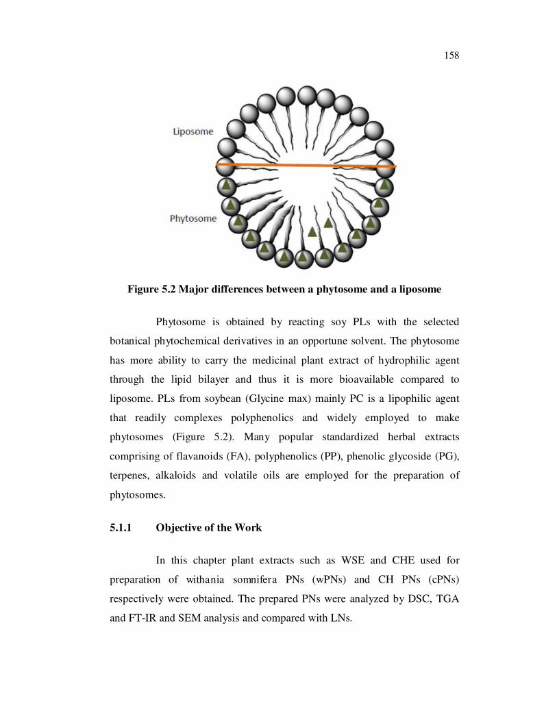
Figure 5.2 Major differences between a phytosome and a liposome
Phytosome is obtained by reacting soy PLs with the selected
botanical phytochemical derivatives in an opportune solvent. The phytosome
has more ability to carry the medicinal plant extract of hydrophilic agent
through the lipid bilayer and thus it is more bioavailable compared to
liposome. PLs from soybean (Glycine max) mainly PC is a lipophilic agent
that readily complexes polyphenolics and widely employed to make
phytosomes (Figure 5.2). Many popular standardized herbal extracts
comprising of flavanoids (FA), polyphenolics (PP), phenolic glycoside (PG),
terpenes, alkaloids and volatile oils are employed for the preparation of
phytosomes.
5.1.1 Objective of the Work
In this chapter plant extracts such as WSE and CHE used for
preparation of withania somnifera PNs (wPNs) and CH PNs (cPNs)
respectively were obtained. The prepared PNs were analyzed by DSC, TGA
and FT-IR and SEM analysis and compared with LNs.

159
Thus obtained wPNs and cPNs are expected to be used as an
injectable solution for controlled and targeted drug carrier system.
5.2 MATERIALS AND METHODS
5.2.1 Materials
All reagents and chemicals used were of analytical grade. Soya
Lecithins and cholesterol were purchased from Hi-media Ltd, India. All other
reagents and chemicals used for the study were sourced from SRL Ltd., India.
5.2.2 Preparation of PNs
The PNs were prepared by the following modified method
(Mozafari et al 2007). A known amount of PNs ingredients was added to a
preheated (60°C, 5 min) glycerol (final concentration 3%, v/v). The mixture
was further heated (60°C) while stirring (approx. 1000 rpm) on a hotplate
stirrer for a period of 45–60 min under N2 atmosphere. For the preparation of
CH-containing formulations, CH was first dissolved in the aqueous phase of
Milli Q water (autoclaved) at elevated temperatures (120°C) while stirring
(approx. 6500-24000 rpm) for a period of 15–30 min under N2 atmosphere
before adding the other components mentioned above. After preparation of
PNs samples, they were left at room temperature under N2 for 30 min to
stabilize. These PNs were produced by passing the emulsion repeatedly
through a polycarbonate membrane having pores 400 and 200 nm in diameter.
The diameters of these PNs were determined by the pore size of the
membrane used during the extrusion method. These PNs produced are
unilamellar, the residual solvent is removed by subjecting the phytosome film
to a high vacuum on a lyophilizer for 90-120 min.

160
5.2.3 PSD and ZP Analyses
The PSD and ZP of LNs, wPNs and cPNs were determined by
Dynamic laser light scattering technology using a size measurer and Laser
Doppler electrophoretic mobility using the Zetasizer at 25 °C. The concentrations
of these PNs were diluted to 0.1 % (w/v) by pH 7.0 PBS (0.05 M).
5.2.4 DSC Analyses
The thermal properties of LNs, wPNs and cPNs were analyzed
using Differential Scanning Calorimetric (DSC, TA-DSC Q 200) analysis.
The thermal behaviour was studied by heating 4±0.5 mg of each individual
sample in a covered sample pan under nitrogen gas flow. The investigations
were carried out over the temperature range 25-300 °C with a heating rate of
10 °C min-1
.
5.2.5 TGA Analysis
The thermal properties of LNs, wPNs and cPNs were analyzed
using Thermal Gravimetric (TGA, TA-TGA Q 50) Analysis. The thermal
behavior was studied by heating 4±0.5 mg of each individual sample in a
sample pan under nitrogen gas flow. The investigations were carried out over
the temperature range 25-800 °C with a heating rate of 20 °C min-1
.
5.2.6 XRD Analysis
The crystalline states of LNs, wPNs and cPNs were evaluated by
XRD. Diffraction patterns were obtained on a Rigaku miniflex II desktop X-
ray diffractometer. The X-ray generator was operated at 40 KV tube voltages
and 40 mA of tube current, using the Ka lines of copper as the radiation
source. The scanning angle ranged from 1 to 60 of 45 min in step scan mode
(step width 1°/min).
161
5.2.7 FT-IR Analysis
The FT-IR analyses of LNs, wPNs and cPNs were carried out using
Perkin Elmer spectrometer. The spectra represented the average of 50 scans.
All spectra were recorded from 600 to 4000 cm1 with a resolution of 4 cm
1.
KBr pellets were prepared by gently mixing 1mg sample with 100mg KBr.
5.2.8 Light Microscopic and SEM Analyses
The LNs, wPNs and cPNs were prepared for light microscopic
analysis. The PNs was diperesed in phosphate buffer saline (PBS). Finally,
the samples were observed under the light microscope.
The LNs, wPNs and cPNs were prepared for SEM. Gold coating
was performed by sputter coater. Finally, cell samples were observed under
the SEM [SEM, FEI-Quanta 200] at a beam voltage of 10 kV.
5.2.9 DPPH Radical Scavenging Activity
The capacity of wPNs and wPNs to scavenge the DPPH radical was
estimated according to the method (Siddhuraju et al 2002). To 2ml of LNs,
wPNs and cPNs, 2ml solution of DPPH 0.1mM was added separately. The
reaction mixture was shaken and incubated in the dark for 30 min, at room
temperature and the absorbance was recorded at 517 nm against methanol.
Controls containing methanol instead of the antioxidant solution and blanks
containing methanol instead of DPPH solution were also made. The
experiment was performed in triplicate. The inhibition of the DPPH radical by
the samples was calculated with reference to control absorbance. The % of
DPPH radical scavenging activity was plotted against the sample
concentration. Scavenging activity was expressed as the inhibition %
calculated using the following formula,

162
100xAbsControl
AbsSampleAbsControlactivityiradicalAnt%
5.2.10 Elution of the WSE and CHE
Determination of solubility characteristics of LNs, wPNs and cPNs
were obtained by adding excess of the samples to 5ml of water in sealed glass
container at room temperature. The liquids were shaken for 24 h and
centrifuged at 5000 rpm for 10 min. The supernatant was filtered and 1ml of
filtrate mixed with 9 ml of methanol. 1 ml of aliquot of the resulting solution
was measured at 360 nm.
5.2.11 Cell Viability Assay
Monolayers of fibroblast cell line NIH 3T3 purchased from
National Centre for Cell Science (NCCS), Pune, India, were grown on
dispersion of LNs, wPNs and cPNs on 96 well culture plate (Corning, NY)
and maintained in Dulbecco's Modified Eagles Medium (DMEM) with 10%
Fetal Calf Serum (FCS) supplemented with antibiotics (Sigma), penicillin
(120 units/ml), streptomycin (75 mg/ml), gentamycin (160 mg/ml) and
amphotericin B (3 mg/ml) at 37 °C humidified with 5 % CO2. After 24 hrs,
the MTT assay was performed to see the % cell viability.
5.2.12 Blood Compatibility Evaluation
The blood compatibility of LNs, wPNs and cPNs were evaluated
with in vitro blood perfusion approach. Equipment for continuously blood
perfusion was assembled. The systems including these PNs were rinsed with
sterilized 0.2 ml/L NaCl prior to the perfusion. 5 ml human blood (containing
5 ml/L EDTA to anti-coagulate) was circularly perfused into these PNs (the
adsorbents were previously swollen in sterilized 0.2 ml/L NaCl solution). The
163
changes in the amounts of white blood cell (WBC), red blood cell (RBC),
haemoglobin (HGB) and blood platelet (PLT) of the blood were monitored.
5.2.13 Storage Stability
The LNs, wPNs and cPNs were stored in a refrigerator at 4 °C.
Samples of 0.2 ml were taken at predetermined intervals.
5.2.14 Statistical Analysis
Results were expressed as mean values and standard deviations (±SD)
5.3 RESULTS
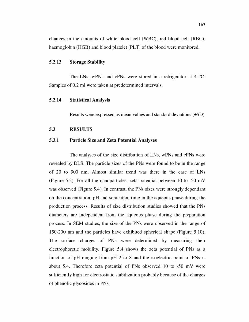
5.3.1 Particle Size and Zeta Potential Analyses
The analyses of the size distribution of LNs, wPNs and cPNs were
revealed by DLS. The particle sizes of the PNs were found to be in the range
of 20 to 900 nm. Almost similar trend was there in the case of LNs
(Figure 5.3). For all the nanoparticles, zeta potential between 10 to -50 mV
was observed (Figure 5.4). In contrast, the PNs sizes were strongly dependant
on the concentration, pH and sonication time in the aqueous phase during the
production process. Results of size distribution studies showed that the PNs
diameters are independent from the aqueous phase during the preparation
process. In SEM studies, the size of the PNs were observed in the range of
150-200 nm and the particles have exhibited spherical shape (Figure 5.10).
The surface charges of PNs were determined by measuring their
electrophoretic mobility. Figure 5.4 shows the zeta potential of PNs as a
function of pH ranging from pH 2 to 8 and the isoelectric point of PNs is
about 5.4. Therefore zeta potential of PNs observed 10 to -50 mV were
sufficiently high for electrostatic stabilization probably because of the charges
of phenolic glycosides in PNs.
164
0 2 4 6 8 10
0
200
400
600
800
1000
Siz
e (
nm
)
T im e (M in )
LN s
wP N s
cP N s
Figure 5.3 Particle size analyses of LNs, wPNs and cPNs
4 5 6 7 8 9 1 0
-5 0
-4 0
-3 0
-2 0
-1 0
0
1 0
2 0
Ze
ta p
ote
ntia
l (m
V)
p H
L N s
w P N s
cP N s
Figure 5.4 Zeta potential of LNs, wPNs and cPNs
10 9 8 7 6 5 4 3 2 1
165
5.3.2 DSC Analysis
Figure 5.5 depicts the DSC thermograms of LNs, wPNs and cPNs.
This thermal behaviour may be ascribed to the presence of plant extract in an
amorphous form or molecularly dispersed. This effect on the crystalline habit
of WSE and CHE may be related to the preparative method of the PNs, in
which WSE and CHE may be turned from a crystalline state to an amorphous
one. It can be observed that LNs and PNs showed similar Td 46.61, 59.56 and
63.51 ºC, respectively (Table 5.1).
35 40 45 50 55 60 65 70 75
-5
-4
-3
-2
-1
0
1
Heat F
low
(W
/g)
Temperature (°C)
LNs
wPNs
cPNs
Figure 5.5 DSC analysis of LNs, wPNs and cPNs
While significant differences in main phase transition temperatures
were not observed, the enthalpy change in the PNs were reduced. This
phenomenon could be attributed to packing differences in the bilayer after
lyophilization. When the liposomes are dehydrated, the packing density of the
head groups increases, thereby increasing opportunities for van der Waals

166
interactions between the hydrocarbon chains. As a result, PNs have a higher
transition temperature and enthalpy changes than LNs. These results indicate
that the addition of WSE and CHE to LNs in some respects mimics the
addition of water, confirming the interaction between the extract and
phosphate groups. From the size and phase transition results, it can be
concluded that WSE and CHE protect the PNs membrane, but some fusion
still occurs, producing slight differences in the mean diameter, size
distribution and phase transition properties. In all these studies, the
thermogram of the complex also exhibited a single peak which was different
from the peak of phytoconstituents and PLs. This interaction may be due to
hydrophobic interaction and hydrogen bonding. The hydroxyl groups of the
phenol rings of WSE and CHE may be involved in hydrogen bonding whereas
the aromatic rings may be involved in hydrophobic interaction. As a result,
the major sharp peaks of PLs disappear and lower the phase transition
temperature. After the combination of the PLs with WSE and CHE molecule
polarity parts, the carbon–hydrogen chain in PLs could turn freely and enwrap
the PLs molecule polarity parts, which made the sequence decrease between
PLs aliphatic hydrocarbon chains.
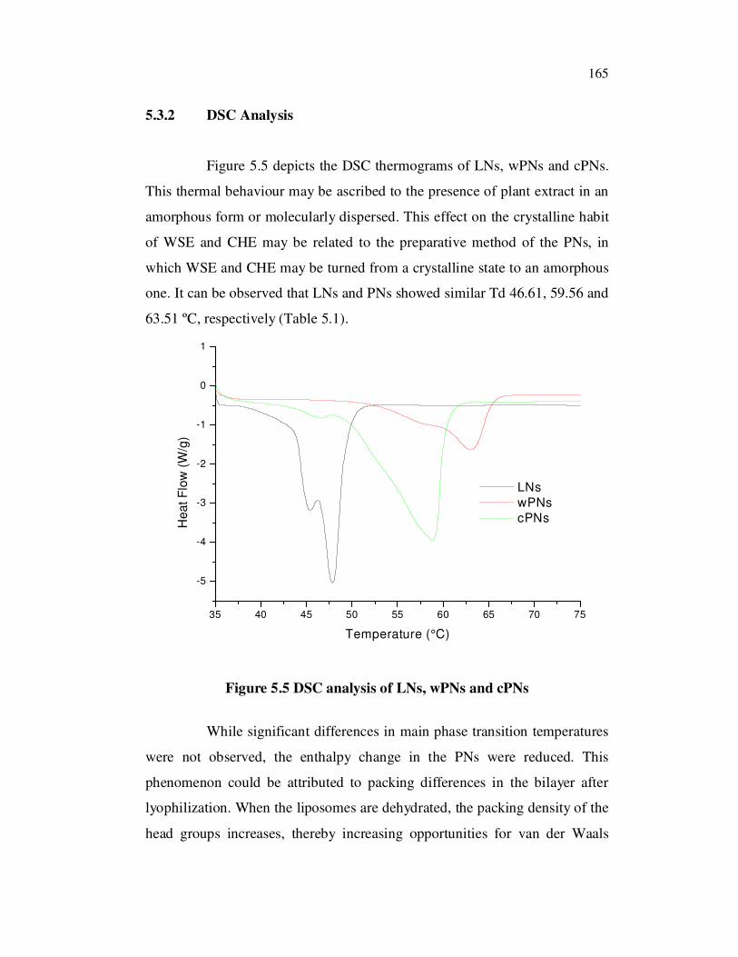
5.3.3 TGA Analysis
The TGA analyses of LNs, wPNs and cPNs are shown in Figure
5.6. The % weight losses are presented in the Table 5.1. The TGA profile of
PNs shows an initial loss on drying below 100 °C (about 4%) and a second
weight loss between 175 °C - 250 °C due to bound water in sample. Around
80%, 72% and 70 % weight losses were observed for LNs, wPNs and cPNs
samples respectively. The results show the improved thermal stability of
wPNs and cPNs compared to LNs.
167
100 200 300 400 500 600 700
20
40
60
80
100
We
igh
t (%
)
Temperature (°C)
LNs
wPNs
cPNs
Figure 5.6 TGA analysis of LNs, wPNs and cPNs
In the above cases, PL-WSE and PL-CHE molecular interactions in
the complex lead to a radically different thermal profile and behavior of PNs
compared to LNs.
Table 5.1 DSC and TGA properties of LNs, wPNs and cPNs
Process Td (°C) % weight loss
LNs 46 3 79.75
wPNs 59 4 75.59
cPNs 63 3 72.18
5.3.4 XRD Analysis
The X-ray diffraction patterns for LNs, wPNs and cPNs are shown
in Figure 5.7. While the diffraction patterns for wPNs and cPNs show various
sharp peaks, diffraction pattern for LNs did not show any peaks in the same
region of the spectrum, indicating the crystalline and amorphous characteristic
of LNs. The appearance of PNs crystalline diffraction peaks confirmed.

168
10 20 30 40 50 60
0
200
400
600
800
1000
1200
1400
1600
1800
2000
LNs
2q [o]
Inte
nsity [A
rb.
Un
it]
0
1000
2000
3000
4000
5000
wPNs
Inte
nsity [A
rb.
Un
it]
0
1000
2000
3000
4000
5000
cPNs
Inte
nsity [
Arb
. U
nit]
Figure 5.7 XRD analysis of LNs, wPNs and cPNs
The crystallinity of wPNs and cPNs sample may be attributed to the
interaction between PLs and the WSE and CHE. The disappearance of LNs
crystalline diffraction peaks confirmed the formation of PNs. Unlike LNs,
bonding between WSE and CHE with the PLs in development of PNs, might
have resulted in the significant changes in their XRD.
169
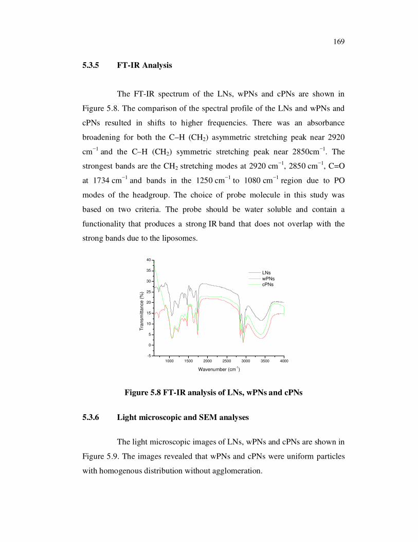
5.3.5 FT-IR Analysis
The FT-IR spectrum of the LNs, wPNs and cPNs are shown in
Figure 5.8. The comparison of the spectral profile of the LNs and wPNs and
cPNs resulted in shifts to higher frequencies. There was an absorbance
broadening for both the C–H (CH2) asymmetric stretching peak near 2920
cm1 and the C–H (CH2) symmetric stretching peak near 2850cm
1. The
strongest bands are the CH2 stretching modes at 2920 cm1, 2850 cm
1, C=O
at 1734 cm1 and bands in the 1250 cm
1 to 1080 cm
1 region due to PO
modes of the headgroup. The choice of probe molecule in this study was
based on two criteria. The probe should be water soluble and contain a
functionality that produces a strong IR band that does not overlap with the
strong bands due to the liposomes.
1000 1500 2000 2500 3000 3500 4000
-5
0
5
10
15
20
25
30
35
40
Tra
nsm
itta
nce (
%)
Wavenumber (cm-1)
LNs
wPNs
cPNs
Figure 5.8 FT-IR analysis of LNs, wPNs and cPNs
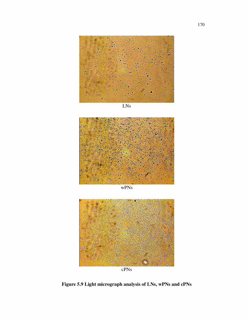
5.3.6 Light microscopic and SEM analyses
The light microscopic images of LNs, wPNs and cPNs are shown in
Figure 5.9. The images revealed that wPNs and cPNs were uniform particles
with homogenous distribution without agglomeration.
170
LNs
wPNs
cPNs
Figure 5.9 Light micrograph analysis of LNs, wPNs and cPNs
171
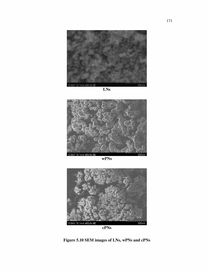
LNs
wPNs
cPNs
Figure 5.10 SEM images of LNs, wPNs and cPNs
172
The SEM images of PNs are shown in Figure 5.10. SEM images of
the PNs revealed that wPNs and cPNs were spherical particles with a
homogenous distribution with smooth surfaces. The PNs existed dispersedly
in PNs system, and they were not agglomerated. In all the cases, the presence
of spherical-shaped vesicles was predominant and in most cases, they were
less than 100 nm in diameter.
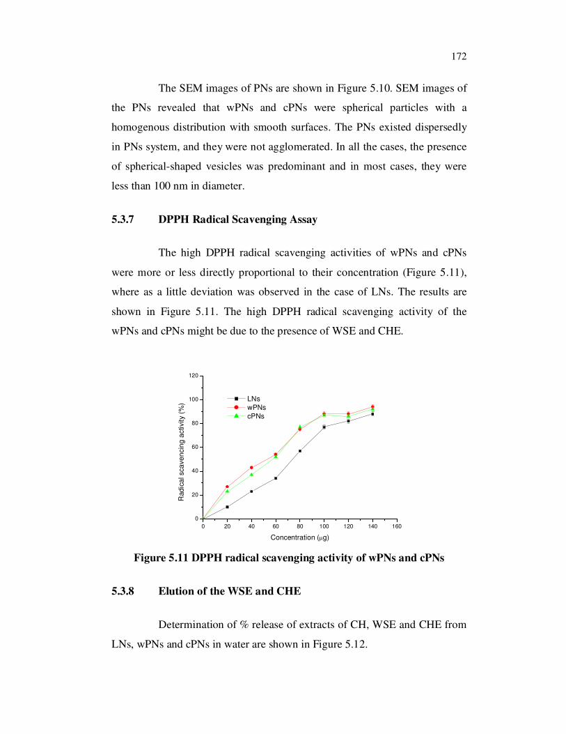
5.3.7 DPPH Radical Scavenging Assay
The high DPPH radical scavenging activities of wPNs and cPNs
were more or less directly proportional to their concentration (Figure 5.11),
where as a little deviation was observed in the case of LNs. The results are
shown in Figure 5.11. The high DPPH radical scavenging activity of the
wPNs and cPNs might be due to the presence of WSE and CHE.
0 20 40 60 80 100 120 140 160
0
20
40
60
80
100
120
Rad
ica
l sca
ve
ncin
g a
ctivity (
%)
Concentration ( g)
LNs
wPNs
cPNs
Figure 5.11 DPPH radical scavenging activity of wPNs and cPNs
5.3.8 Elution of the WSE and CHE
Determination of % release of extracts of CH, WSE and CHE from
LNs, wPNs and cPNs in water are shown in Figure 5.12.
173
0 2 4 6 8 10 12 14
0
5
10
15
20
25
30
35
40
45
50
55
60
65
70
75
80
85
90
95
100
% r
ele
ase
Time (h)
CH
WSE
CHE
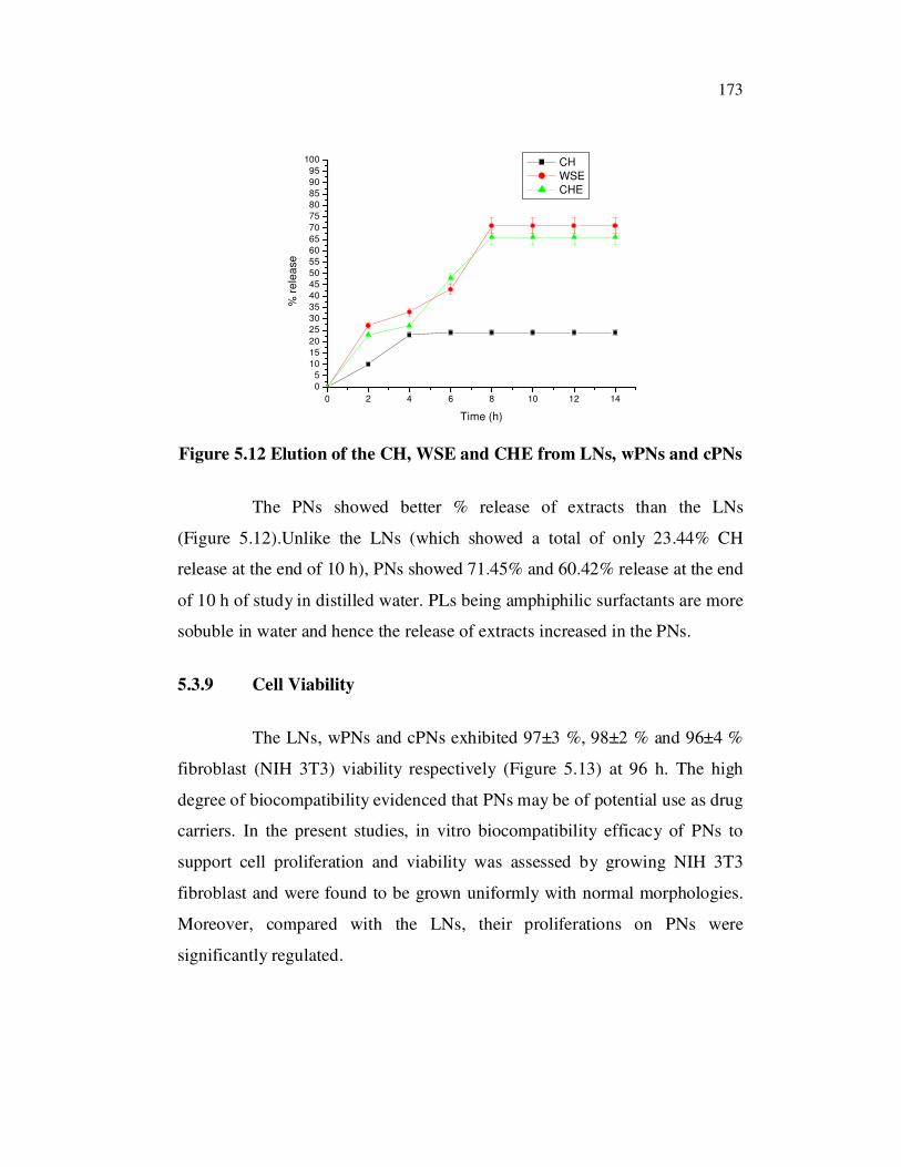
Figure 5.12 Elution of the CH, WSE and CHE from LNs, wPNs and cPNs
The PNs showed better % release of extracts than the LNs
(Figure 5.12).Unlike the LNs (which showed a total of only 23.44% CH
release at the end of 10 h), PNs showed 71.45% and 60.42% release at the end
of 10 h of study in distilled water. PLs being amphiphilic surfactants are more
sobuble in water and hence the release of extracts increased in the PNs.
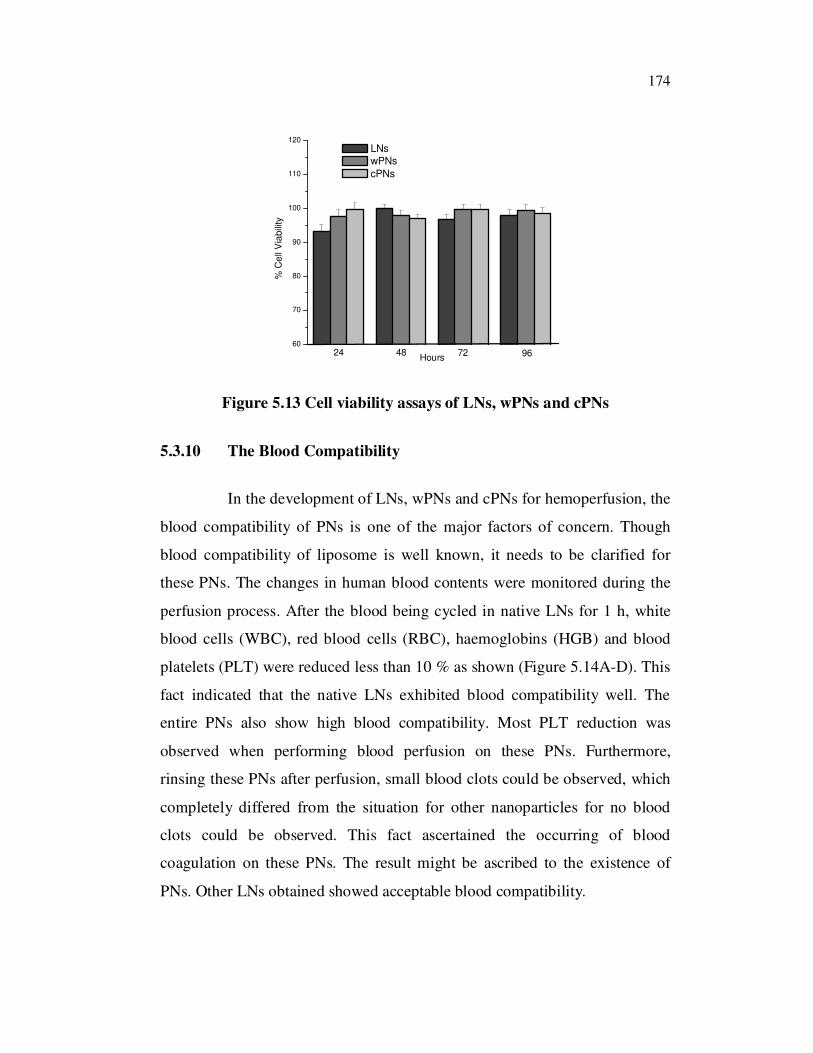
5.3.9 Cell Viability
The LNs, wPNs and cPNs exhibited 97±3 %, 98±2 % and 96±4 %
fibroblast (NIH 3T3) viability respectively (Figure 5.13) at 96 h. The high
degree of biocompatibility evidenced that PNs may be of potential use as drug
carriers. In the present studies, in vitro biocompatibility efficacy of PNs to
support cell proliferation and viability was assessed by growing NIH 3T3
fibroblast and were found to be grown uniformly with normal morphologies.
Moreover, compared with the LNs, their proliferations on PNs were
significantly regulated.
174
60
70
80
90
100
110
120
96724824
% C
ell
Via
bili
ty
Hours
LNs
wPNs
cPNs
Figure 5.13 Cell viability assays of LNs, wPNs and cPNs
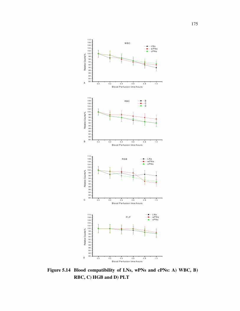
5.3.10 The Blood Compatibility
In the development of LNs, wPNs and cPNs for hemoperfusion, the
blood compatibility of PNs is one of the major factors of concern. Though
blood compatibility of liposome is well known, it needs to be clarified for
these PNs. The changes in human blood contents were monitored during the
perfusion process. After the blood being cycled in native LNs for 1 h, white
blood cells (WBC), red blood cells (RBC), haemoglobins (HGB) and blood
platelets (PLT) were reduced less than 10 % as shown (Figure 5.14A-D). This
fact indicated that the native LNs exhibited blood compatibility well. The
entire PNs also show high blood compatibility. Most PLT reduction was
observed when performing blood perfusion on these PNs. Furthermore,
rinsing these PNs after perfusion, small blood clots could be observed, which
completely differed from the situation for other nanoparticles for no blood
clots could be observed. This fact ascertained the occurring of blood
coagulation on these PNs. The result might be ascribed to the existence of
PNs. Other LNs obtained showed acceptable blood compatibility.
175
0.0 0.2 0.4 0.6 0 .8 1.0
80
82
84
86
88
90
92
94
96
98
100
102
104
106
108
110
A
W B C
Rela
tive C
oun
ts/%
B lo od P erfus ion t im e /hours
LN s
w P N s
c P N s
0.0 0.2 0.4 0.6 0 .8 1.0
80
82
84
86
88
90
92
94
96
98
100
102
104
106
108
110
B
R B C
Re
lative C
ou
nts
/%
B lo od P erfus ion t im e /hours
B
C
D
0.0 0.2 0.4 0.6 0 .8 1.0
80
82
84
86
88
90
92
94
96
98
100
102
104
106
108
110
C
R G B
Rela
tive C
oun
ts/%
B lo od P erfus ion t im e /hours
L N s
w P N s
c P N s
0.0 0 .2 0 .4 0 .6 0 .8 1 .0
80
82
84
86
88
90
92
94
96
98
100
102
104
106
108
110
D
P LP
Rela
tive C
ounts
/%
B lo od P e rfus ion t im e /hou rs
LN s w P N s
cP N s
Figure 5.14 Blood compatibility of LNs, wPNs and cPNs: A) WBC, B)
RBC, C) HGB and D) PLT

176
The time courses of blood cell counts during the perfusion process
are shown (Figure 5.14a-d). The results revealed that relatively sharp change
in the blood cells occurred at the initial stage of perfusion and gradually
reached stable values. It was opposed that reduction in blood cell may result
from the cell trapping by these PNs. Once these PNs were filled with blood
cells, no more cells in the blood would be reduced.
5.3.11 Storage Stability
The PNs were subjected to storage stability study for a period of 6
months. The storage stability of LNs, wPNs and cPNs at 4 °C and at pH 7.0 is
presented in the Figure 5.15a and b.
0 1 2 3 4 5 6 7 8
40
50
60
70
80
90
100
110
120
130
A
Siz
e (
nm
)
Tim e (Day)
LNs wPNs
cPNs
0 20 40 60 80 100 120 140 160 180 200 220 240 260
40
45
50
55
60
65
70
75
80
85
90
95
100
B
Siz
e (
nm
)
T ime (Days)
LNs
wPNs
cPNs
Figure 5.15 Storage stability of LNs, wPNs and cPNs
177
It can be seen from retention ratio of LNs tended to decrease with
increasing storage period and it was decreased to 88.83% after 3 months
storage. It indicated that more plant extract leaked out from PNs with increase
of storage period. The leakage of extract from PNs might be attributed to
hydroxylation and degradation of bilayer membranes and/or vesicle
fusion/aggregation. However, PNs were stable for 3 months, during that
period, no significant differences in size distribution and mean diameter
occurred.
5.4 DISCUSSION
Development of novel drug carrier from natural resources is very
much needed because of the beneficial role of herbal drug in the management
of varied diseases. The bioavailability of lipophilic drugs when administered
orally as solid dosage forms is low. There are usually several factors
responsible for this but a particularly widespread problem is poor absorption
due to slow and incomplete drug dissolution. In this case, improved
bioavailability can be achieved by the use of carriers with medicinal plant
extract which can enhance the rate and the extent of therapeutic drug
solubilizing into aqueous intestinal fluids. PLs play a major role in drug
delivery technology. There are numerous advantages of PLs in addition to
solubilizing property while considering them for a carrier system. In the
present study, wPNs and cPNs were prepared in the presence of N2
atmosphere. The physicochemical investigations showed that WSE and CHE
formed complexes with PLs.
This work was focused on the preparation and characterization of
PNs for drug carriers, with the aim of establishing their utility for delivery of
hydrophobic drugs. Prior to establishment of these PNs formulations were
designed for drug delivery applications. There is a great deal of existing
literature on the in vitro physical and chemical properties of these PNs
178
prepared without CH, the existing literature provides a solid foundation for
the development of PNs as intravenous drug carrier systems. Despite
extensive studies of CH-free formulations, there has been little emphasis on
their application as drug carriers other than CH-rich liposome formulations
being considered as thermo sensitive formulations (Santos et al 2002 and
2004). Others have focused on the physico-chemical and biological attributes
of CH-free liposome including phase transition temperature determination by
DSC, XRD, protein binding, permeability and pharmacokinetic studies
(Ulrich et al 2003). Collectively, this study provides conclusive evidence that
these PNs have distinctive properties that may be beneficial for drug carriers.
However, when this information has been applied to drugs, the CH-free
formulation, even when stabilized by plant extracts such as PC-WSE and PC-
CHE incorporation, exhibit poor drug retention when compared to CH-
containing formulations. The use of novel PNs are an advanced dosage
formulation technology to deliver chemotherapeutics such herbal and
synthetic drugs by improved absorption and, as a result, produce better results
than those obtained by conventional herbal extracts (Huh et al 1996; Acharya
et al 2011 and Singh et al 2011; Manthena et al 2010). Water-soluble phyto-
constituent molecules (mainly WSC and CHE) can be converted into lipid-
compatible molecular complexes. These complexes are more bioavailable as
compared to simple herbal extracts owing to their enhanced capacity to cross
the lipid rich biomembranes and finally reach the circulatory system
(Bhattacharya et al 2009; Saraf, 2010; Vinod et al 2010; Sindhumol et al
2010; Kumar et al 2010; Jain et al 2010; Patela et al 2010).
This study provides, for the first time, evidence that PNs can
exhibit improved properties, thus providing the opportunity to develop such
formulations for drugs that are poorly retained in CH-containing liposomes. A
direct comparison of CH-rich LNs with other successful drug carriers
including conventional PL:CH and sterically stabilized PC-WSE and PC-CHE
179
based PNs are provided. The antioxidant activities of the PNs were
significantly higher than LNs.
That the PNs could have improved pharmacokinetics and
pharmacological parameters can advantageously be used in the treatment of
liver diseases. It can also be used in anti-inflammatory agents as well as in
pharmaceutical and cosmetic compositions. PNs are obtained by reacting soy
PLs with the selected botanical derivatives such WSE and CHE. On the basis
of their physicochemical and spectroscopic characteristics, these complexes
can be considered novel entities. Likewise PNs, LNs are formed by mixing a
water soluble substance with PC in definite ratio under specific conditions.
Here, no chemical bond is formed; the PC molecules surround the water
soluble substance. There may be hundreds or even thousands of PC molecules
surrounding the water-soluble compound. The PNs process the PC with plant
components actually, form a 1:1 or a 2:1 molecular complexes depending on
the substance complexed involving chemical bonds. This difference results in
PNs being much better absorbed than LNs showing better bioavailability. PNs
have been found superior to liposomes in topical and skin care products.
PNs are a complex between a natural product and PLs, like soy
PLs. This complex is obtained by reaction of PL and the medicinal plant
extract in an appropriate solvent. PC is a bifunctional compound with the
phosphatidyl moiety being lipophilic and the choline moiety being
hydrophilic in nature. Specifically the choline head of the PC molecule binds
to these herbal phytoconstituent while the lipid soluble phosphatidyl portion
comprising the body and tail envelopes the choline bound material. Hence,
the phytoconstituents produce a lipid compatible molecular complex with
PLs, also called as phyto-PL complex. The complexes are anchored through
chemical bonds to the polar choline head of the PLs, as can be demonstrated
by specific spectroscopic techniques. Precise chemical analysis indicates that
180
units of PNs are usually flavonoids, polyphenolics and phenolic glycosides
linked with at least one PC molecule. This results in a little micro to nano
sphere or cell being produced. The PNs produces a little cell, whereby the
plant extract or its active constituent could be protected from destruction
owing to the property of PC. On the basis of spectral analysis it has been
shown that the main PC-WSE and PC-CHE interaction is due to the formation
of H-bonds and hydrophobic interaction between the polar head of PLs (i.e.
phosphate and ammonium groups) and the polar functionalities of the
substrate. When treated with water, PNs assume micellar shape forming
liposomial-like structures. In liposomes the active principle is dissolved in the
floating in the layer membrane, while in PNs the active principle is anchored
to the polar head of PLs, becoming an essential and integral part of the lipid
membrane for example in the case of the PC-WSE and PC-CHE complex,
there is formation of H-bonds between the PP, FA and PG of the flavone
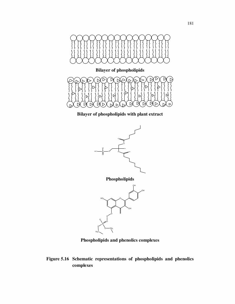
moiety and the phosphate ion on the PC side. The Schematic representations
of phospholipids and phenolics complexes are shown in Figure 5.16. PNs are
advanced forms of herbal products which show better absorption, utilization
and as a result will produce better results than conventional LNs, the
increased bioavailability of the PNs by pharmacokinetics and
pharmacodynamic tests in experimental animals and in human subjects.
Preparation and characterization of PLs complexes of flavonoids
for effective drug delivery have been studied (Oteiza et al 2005; Semalty et al
2010). The sterol structure-function relationships in natural and artificial
membranes have been reviewed (Bloch et al 1983). There are two important
observations pertaining to PNs that warrant further discussion. First and
foremost, flavonoids, polyphenolics and phenolic glycosides are essential
components.
181
Bilayer of phospholipids
Bilayer of phospholipids with plant extract
O
OOPHO O
O
O
O-H
Phospholipids
O
OH
OH
O
H
HO
OH
O
P
O
OO
H2C
CH2
Phospholipids and phenolics complexes
Figure 5.16 Schematic representations of phospholipids and phenolics
complexes

182
Consistent with CH-containing liposomes, drug loading for the
liposome is dependant on liposome composition as well as the specific
physico-chemical properties of the drug being used (Liang et al
2004).Modulation of liposomal membrane fluidity by flavonoids,
isoflavonoids and CH derivatives have been studied (Arora et al 2000; Kisoon
et al 2002). The effect of flavonoids on the PLs bilayers interaction has been
studied (Semalty et al 2010; Tedeschi et al 2010). Importantly, the CH-free
formulations may be particularly well suited for the more hydrophobic drugs.
Removing CH from the PNs may facilitate even greater flexibility and control
of drug leakage rates. The PNs technology is a breakthrough model for
marked enhancement of bioavailability, assured delivery to the tissues and no
compromise on nutrient safety.
The PNs have following advantages 1) PNs are better bioavailable
botanical extracts, dramatically enhance bioavailability due to their complex
with PLs and deliver faster and improved absorption. 2) They could enhance
the absorption of lipid insoluble polar medicinal phytochemicals through oral
as well as topical route showing better bioavailability with significantly better
therapeutic benefit 3) Dose requirement can be minimized as the
bioavailability is increased. 4) PC used in preparation of PNs could act as a
carrier and hepatoprotective substance showing the synergistic effect when
hepatoprotective substances like flavanoids are employed to form complex. 5)
PNs could be widely be used in cosmetics due to more skin penetration and
high lipid profile and 6) PNs show better stability profile owing to the
chemical bonds formed between PC molecule and phytoconstituents.
183
5.5 CONCLUSIONS
PNs were successfully prepared through interactions between PL
with WSE and CHE. ZP measurement confirmed the particle sizes and
charges and the SEM analysis confirmed the surface morphology of these
PNs. It was concluded that the PLs complex with WSE and CHE might be of
potential use for improving its bioavaibility. The results showed that PLs in
PLs-WSE and CHE complex were joined by non-covalent bond and did not
form a new compound. It has been observed that the complex formed an
effective scavenger of DPPH radicals and showed strong antioxidant activity.
With the sustained release pro le, it may be possible to get a sustained action
with smaller dose. This value added herbal drug carriers system can pave the
way for large molecules to pass through the lipophilic biological membrane
and get absorbed into the systemic circulation. These PNs are supposed to
support the attachment and proliferation of cells better. These PNs based drug
carriers could be used for targeted and controlled/sustained release of drug to
lymphatic system and nervous system, blood-brain barriers and blood-
placental barriers and cellular and sub-cellular organelles etc., It would offer
several advantages including an increase in drug bioavailability and retention
at the target site and improving the adherence or adhesion to the designated
target and sustaining drug release depots. It might be effective in the
treatment of some infectious diseases such as cancer and arthritis.