Chapter 5 Abdelfattah A. Touman and Grigoris K. Stratakos
24
Chapter 5 Long-Term Complications of Tracheal Intubation Abdelfattah A. Touman and Grigoris K. Stratakos Additional information is available at the end of the chapter http://dx.doi.org/10.5772/intechopen.74160 Abstract Endotracheal intubation is an intervention frequently performed in the hospital setting in order to protect the central airway and provide mechanical support of ventilation. Many health care providers are expected to be able to intubate the patients for different indica- tions. As the case in any medical intervention, endotracheal intubation can cause compli- cations. These complications are categorized as early or late according to the time of onset of the presenting symptoms. This chapter will discuss the long term complications of endotracheal intubation that might be encountered by the treating physicians. The chapter will stress on the predisposing factors for these complications and the available methods to avoid and treat them. Keywords: intubation related complications, obstructive fibrinous tracheal pseudomembranes, post-intubation tracheal stenosis, tracheomalacia, tracheoesophageal fistula 1. Introduction Intubation is an invasive, however, lifesaving procedure which established its role in the daily management of critically ill patients. Physicians treating patients with endotracheal tubes can encounter complications during or immediately after the procedure. Complication appearance or identification can be delayed for days to weeks. They can be categorized into three categories: 1. Long term complications that occur immediately during endotracheal tube (ETT) insertion 2. Complications of the prolonged intubation 3. Late onset complications © 2018 The Author(s). Licensee IntechOpen. This chapter is distributed under the terms of the Creative Commons Attribution License (http://creativecommons.org/licenses/by/3.0), which permits unrestricted use, distribution, and reproduction in any medium, provided the original work is properly cited.
Transcript of Chapter 5 Abdelfattah A. Touman and Grigoris K. Stratakos

Chapter 5
Long-Term Complications of Tracheal Intubation
Abdelfattah A. Touman and Grigoris K. Stratakos
Additional information is available at the end of the chapter
http://dx.doi.org/10.5772/intechopen.74160
Provisional chapter
Long-Term Complications of Tracheal Intubation
Abdelfattah A. Touman and Grigoris K. Stratakos
Additional information is available at the end of the chapter
Abstract
Endotracheal intubation is an intervention frequently performed in the hospital setting inorder to protect the central airway and provide mechanical support of ventilation. Manyhealth care providers are expected to be able to intubate the patients for different indica-tions. As the case in any medical intervention, endotracheal intubation can cause compli-cations. These complications are categorized as early or late according to the time of onsetof the presenting symptoms. This chapter will discuss the long term complications ofendotracheal intubation that might be encountered by the treating physicians. The chapterwill stress on the predisposing factors for these complications and the available methodsto avoid and treat them.
Keywords: intubation related complications, obstructive fibrinous trachealpseudomembranes, post-intubation tracheal stenosis, tracheomalacia, tracheoesophagealfistula
1. Introduction
Intubation is an invasive, however, lifesaving procedure which established its role in the dailymanagement of critically ill patients. Physicians treating patients with endotracheal tubes canencounter complications during or immediately after the procedure. Complication appearanceor identification can be delayed for days to weeks. They can be categorized into three categories:
1. Long term complications that occur immediately during endotracheal tube (ETT) insertion
2. Complications of the prolonged intubation
3. Late onset complications
© 2016 The Author(s). Licensee InTech. This chapter is distributed under the terms of the Creative Commons
Attribution License (http://creativecommons.org/licenses/by/3.0), which permits unrestricted use,
distribution, and eproduction in any medium, provided the original work is properly cited.
DOI: 10.5772/intechopen.74160
© 2018 The Author(s). Licensee IntechOpen. This chapter is distributed under the terms of the CreativeCommons Attribution License (http://creativecommons.org/licenses/by/3.0), which permits unrestricted use,distribution, and reproduction in any medium, provided the original work is properly cited.

2. Risk assessment and general preventive measures
Risk assessment and preventive measures of the intubation-related complications should beundertaken immediately after the decision for an endotracheal tube (ETT) insertion is taken forthe patient’s management. These measures can be divided into three main groups:
I. Measures that should be taken before intubation:
1. Routine assessment for potentially difficult intubation cases.
2. Choosing the proper sized tube.
3. Using proper technique
4. Choose an experienced, skilled physician to perform the procedure.
5. Preparing alternative plans in cases of unanticipated difficulties. The Difficult AirwaySociety (DAS) guidelines recommended a four steps plans A–D as the following [1]:
A. Preparing an initial tracheal intubation plan that includes using the direct laryn-goscopy.
B. Preparing a secondary intubation plan which includes using a dedicatedsupraglottic airway device such as the classic laryngeal mask airway (LMA) incase of plan A has failed.
C. When plan B fails; the physician should be prepared to oxygenate and ventilatethe patient, postpone the surgery, and awaken the patient.
D. In cases where physician ‘cannot intubate, cannot ventilate’ (CICV) rescue tech-niques such as cannula or surgical cricothyroidotomy should be available at thefacility.
II. Measures that applied during intubation:
1. Intubating the patients under direct vision.
2. Using intubation assisting devices as the ETT stylet, Eschmann tracheal tube intro-ducer (gum-elastic bougie tube), video laryngoscope or the flexible bronchoscopy indifficult to intubate cases [2].
3. Applying appropriate cuff pressure not to exceed 20 mm H2O.
4. Stabilization the ETT using fixation tapes or devices.
III. Measures which should be applied after intubation:
1. Frequent suctioning of the oral and endotracheal secretions.
2. Application of antiseptics as chlorhexidine to decontaminate the oral cavity.
3. Assessments of skin integrity around lips and adhesive tape at least twice daily.
Tracheal Intubation90

3. Complications that occur immediately during endotracheal tube (ETT)insertion
Complications of tracheal intubation might occur at any stage during the intubation withdevastating consequences which may last as long as patients survive.
Table 1A lists the chronic complications that usually result from trauma during intubation.
3.1. Post intubation prolonged voice hoarseness
Temporary hoarseness is a common complain that occur in one third [3] to half [4] of theintubated patients and usually resolve spontaneously within 1 week; however, the incidenceof prolonged hoarseness (>7 days) is estimated to be less than 1% [4, 5]. Vocal cord edema orlacerations, epiglottic hematoma and vocal cord paralysis secondary to compression of ante-rior branch of the recurrent laryngeal nerve are all potential etiologies of prolonged hoarseness[6]. In a prospective study 7 out of 25 cases (28%) of prolonged hoarseness had no identifiablecauses [4]. Permanent hoarseness is even more rare, and being caused by granuloma of thevocal cord or arytenoid dislocation. The risk factors for this complication found to be longerduration of intubation, older age [4] and female gender [7]. Using a smaller size tube especiallyfor females has been suggested to decrease the incidence of prolonged voice hoarseness [4, 8].
3.2. Arytenoid dislocation
Arytenoid dislocation is a rare cause of vocal cord paralysis with less than 0.1% incidence rate[9]. The commonest mechanism of intubation related vocal cord paralysis is compression of theanterior branch of the recurrent laryngeal nerve as a result of prolonged intubation which willbe discussed separately under complication of prolonged intubation subtitle. Risk factors forthis complication can either be related to patients or the procedure. Patients related factorsinclude retrognathia, dental malocclusion, a large tongue base, and cricoarytenoid jointinvolvement by rheumatoid arthritis. While; Procedure related risk factors include traumaticand/or prolonged intubation, protrusion of the endotracheal tube stylet, pressure from thedistal curved part of the ETT and inexperience and poor techniques by the performance [10].The majority of the patients present with dysphagia [11]; prolonged hoarseness, sore throat,and cough are less frequent symptoms. There are two types of dislocation, the posterolateraland the anteromedial dislocation; the latter being the most dangerous as it can compromise the
Prolonged voice hoarseness
Arytenoid dislocation
Cervical spine and spinal cord injuries
Traumatic dental injury
Table 1A. Chronic complications that occur immediately at ETT insertion.
Long-Term Complications of Tracheal Intubationhttp://dx.doi.org/10.5772/intechopen.74160
91

airway causing acute stridor and acute respiratory distress soon after extubation [12]. Figure 1shows example of anteromedial left arytenoid cartilage dislocation [13].
Diagnosis can be confirmed by laryngoscopy and/or CT scan [14]. Laryngeal electromyogra-phy is sometimes used to differentiate arytenoid dislocation from recurrent laryngeal nerveparalysis. Tracheotomy is required in cases which present with acute airway compromise,other cases may have spontaneous repositioning of arytenoid cartilage thus do not needtreatment [15]. Many surgical laryngoscopic techniques have been described as the definitivetreatment [16, 17]. Late treatment options include Teflon injection, cricoarytenoid arthrodesis[18] and laryngeal framework procedure [19].
3.3. Cervical spine and spinal cord injuries
Urgent intubation to ensure protection of the airways is frequently required for poly-traumapatients. Careful manipulation of the neck by an experience health care provider is of crucialimportance as such patients potentially have cervical spine injuries. Predictors of spinal injuryin poly-trauma cases are accident by motor vehicle collision or fall down. Presence of pelvicfracture or Injury Severity Score (ISS) >15 should alert the physician to a possible cervical spineinvolvement [20]. Patients with systemic diseases which potentially affect the cervical spine asrheumatoid arthritis or ankylosing spondylitis must be handled as high risk for intubationrelated cervical spine injuries. Once the health care provider suspects a high risk cervical spineinjury, he should maintain the patient’s head in a neutral position throughout the intubationprocedure. Cervical hyperextension in such cases can traumatize the spinal cord resulting inparaplegia. Manual In-Line Stabilization (MILS) is the recommended method to stabilize thehead and neck during high risk intubation. The caveats of this method are: firstly, the need of asecond health care provider who stabilizes the patient’s occiput and mastoid processes usinghis both hands. Secondly, it decreases the laryngoscopic view by 45% [21]. Application ofcervical collar is another important protective measure; available collars include soft collar,hard collar, extrication collar, Philadelphia collar. Again, application of collar significantly limitmouth opening and make intubation harder [22]. Cervical spine can also be stabilized using abilateral sandbags with 3-inch-wide cloth tape across the forehead. Adding Philadelphia collarto the sand bags further reduced the extension movement [23].
Figure 1. Laryngoscopic image of anteromedial dislocation of left arytenoid cartilage. Courtesy of Oh et al. [13].
Tracheal Intubation92

In cases of difficult intubation the available choices are:
1. Video or fiberoptic laryngoscopy intubation.
2. Gum elastic bougie which has been shown in a clinical study to facilitate the intubationwhile applying MILS [21].
3.4. Traumatic dental injury
The incidence of traumatic dental injury found to be 1:2805 in a large analysis of more than 500thousand surgeries. The most common tooth to be traumatized is the upper incisors; further-more, 13% of the cases had multiple teeth involved. Different type of teeth injuries have beendescribed, crown fracture and partial dislocation being the commonest [24]. Difficult intuba-tion is the main risk factor for dental injury, other risk factor include poor dentation andpreexisting craniofacial abnormalities [24].
4. Complications of the prolonged intubation
Prolonged intubation is defined as intubation exceeding 7 days [25]. Clinical studies haveshown that prolonged intubation is a risk factor for many complications.
Table 1B lists complications of prolonged intubation that present while patient is still onmechanical ventilator or early at extubation.
4.1. Mucosal/dermal pressure ulcer
Mucosal/dermal pressure ulcers are caused either by the tube itself or by the securing taps anddevices. It can affect the lips, mouth, gums, and tongue. This complication is very commondespite it is preventable; its incidence reported to be 20% in mechanically ventilated patients[26]. The identified risks for pressure ulcer are [27, 28]:
1. Hypoalbuminemia, older age, catabolic diseases.
2. Increase pressure, shearing forces and friction over bony prominence.
3. Moisture.
Pressure ulcer around adhesive tapes
Vocal cord paralysis
Ventilator-associated pneumonia (VAP)
Sinusitis
Tracheomalacia
Laryngotracheal stenosis
Table 1B. Complications of prolonged intubation.
Long-Term Complications of Tracheal Intubationhttp://dx.doi.org/10.5772/intechopen.74160
93

Prevention of ETT related pressure ulcer can be summarized in the following steps [29]:
1. To Proper size of the tube before the insertion.
2. To adequately secure the tube to prevent dislodgment without creating additional pressure.
3. To inspect the skin beneath the tube at least twice daily.
4. To keep the skin under the tube dry and clean as possible.
5. To reposition the ETT to redistribute pressure while make sure that the ETT depth did notchange.
6. To remove the tube once it is not absolutely necessary.
Pressure ulcers prevention is crucial as they are painful and their healing process is slow.Generally, removal of the cause of the pressure, cleaning, application of antiseptics and debride-ment is the available treatment options.
4.2. Vocal cord paralysis
Intubation related vocal cord paralysis is rare with estimated incidence of 0.03%; nevertheless, itconstitutes 22.6% of all causes of vocal cord paralysis [30]. It is usually a result of compression ofthe anterior branch of the recurrent laryngeal nerve between the inflated cuff of the ETT and thethyroid cartilage [31]. Prolonged intubation is the major risk factor for vocal cord paralysis whichcan be unilateral (left vocal cord is more commonly involved than the right) or bilateral [6].Unilateral vocal cord paralysis present immediately after extubation with hoarseness of voiceand dysphonia; while, bilateral paralysis presented as sever stridor which lead to reintubation.Re-establishment of vocal cord motion is a good prognostic sign of improvement of the voice[32]. In bilateral vocal cord palsy pulmonary function testing shows variable extrathoracicobstruction on flow volume loop [33]. Direct laryngoscopy and/or laryngeal electromyographyare the diagnostic test of choice. Intervention is not required for unilateral paralysis as spontane-ous resolution usually occurs on average of about 10 weeks [6], however, if no resolution occursin 12 months then recovery is unlikely [32]. Temporary injection of the affected vocal cord is anoption to improve the voice [32]. Unfortunately, tracheostomy is necessary in many cases suffer-ing from bilateral vocal cord paralysis for long term management.
4.3. Ventilator-associated pneumonia (VAP)
Ventilator-associated pneumonia (VAP) is defined in the latest Infectious Diseases Society ofAmerica (IDSA) and the American Thoracic Society (ATS) guideline as a pneumonia occurring>48 h after endotracheal intubation [34]. VAP is subsequently divided for practical reasons into an early onset pneumonia and late-onset pneumonia; the earliest is a pneumonia thatdevelops within the first 4 days of admission in the mechanically ventilated patients [35]. Asystematic review has estimated that between 10 and 20% of patients receiving >48 h ofmechanical ventilation develop VAP [36]; up to 50% of them develop an early VAP (withinthe first 4 days after admission) [35]. Interestingly, the VAP hazard is decreasing over days,this has been shown in a prospective multicentre Canadian study where VAP rate was approx-imately 3% per day in the first week of mechanical ventilation, then the rate dropped to 2%
Tracheal Intubation94

and 1% per day in the second and the third week respectively [37]. Gastric colonization,oropharyngeal and tracheal colonization, and cross colonization of the patients by contami-nated hands of hospital personnel are all risks for VAP. Endotracheal tubes itself appear to bean independent risk factor for VAP [38]. Its presence impairs the host natural defenses againstinfections; furthermore, bacteria adhere to the plastic tube forming a complete or partialbiofilm in 84 and 95% respectively [39]. Intubated patients are having recurrent aspirations ofdislodged parts of this biofilm or from the pooled secretions above the tube cuff.
Studies have shown that the bacterial colonization is changed from community to nosocomialpattern after the fourth day of the admission [34]. In early-onset pneumonia the commonestpathogens include, Streptococcus pneumoniae, Haemophilus influenzae and methicillin sensitiveStaphylococcus aureus (MSSA); while in the late onset methicillin resistant Staphylococcus aureus(MRSA), Gram-negative bacilli such as Pseudomonas aeruginosa, Escherichia coli, Klebsiellapneumoniae, and Acinetobacter baumannii are frequently encountered [40]. Moreover, it is notuncommon for VAP to have polymicrobial infection. VAP diagnosis is achieved by using thefollowing diagnostic criteria [41]:
1. Clinical sign of infection as fever, purulent tracheal secretions, and leucocytosis.
2. Bacteriologic evidence of infection.
3. Radiologic suggestion of infection.
Application of strict prevention measures in the intensive care units is practical approach todeceases the burden of VAP on the patients and the health care system. VAP prevention mea-sures include:
1. Noninvasive positive-pressure ventilation in selected groups of patients.
2. Silver covered tube: it has been shown in a Cochrane review that silver coated tube decreasethe risk of VAP in the first 10 days of mechanical ventilation possibly due to the antimi-crobial effect of silver [42].
3. Mouth hygiene with chlorhexidine as a mouthrinse or a gel: this has shown in a Cochranereview to reduce the risk of VAP from 24% to about 18% [43].
4. Positioning the patient in semirecumbent position: decrease the risk of VAP from 35 to 8%comparing to supine position [44].
5. Applying an endotracheal tube cuff pressure of 20 cm H2O to prevent aspiration.
6. Continuous aspiration of subglottic secretions (CASS): it reduce both early and late onset VAP [45].
7. Discontinuation of mechanical ventilation as early as possible.
IDSA/ATS updated guideline recommends empirical coverage of both S. aureus and P. aeruginosain patients with suspected VAP. They recommended either vancomycin or linezolid to coverMRSA in at risk patients or in patients who are treated in units with high methicillin resistantrate (>10–20%). Single antipseudomonal antibiotic is generally suggested unless the patient is athigh risk for antimicrobial resistance or being treated in a unit with high resistant rate forpseudomonas (>10%). A 7-day course is generally recommended [34].
Long-Term Complications of Tracheal Intubationhttp://dx.doi.org/10.5772/intechopen.74160
95

4.4. Sinusitis
Incidence of nosocomial sinusitis is estimated to be 12 cases per 1000 patient-days [46]. Prolongedintubation is the main risk factor as the majority of the infections occur after 7 days of thehospitalization [47]. The rout of intubation either orotracheal or nasotracheal does not alter thenosocomial sinusitis rate as shown in a randomized clinical trial of patients needed intubation formore than 7 days [48]. Other identified risk factor include sedative use, nasoenteric feeding tube,Glasgow Coma Score (GCS) less than 7 and nasal colonization with Gram-negative bacteria [46].Nosocomial sinusitis usually presents with fever; the disease should be suspected in all intubatedpatients who have a fever without an obvious source. Most patients have leucocytosis [47];purulent nasal discharge is a useful clinical sign that raise the suspicion of the sinusitis as a sourceof the infection. Air-fluid level or opacification are the radiological signs seen on the sinus CTscan images. Microbiological confirmation of the infection can be achieved by culture of sinusfluid in 75 and 90% of the cases using antral tap or endoscopic tissue culture respectively [49].Infective bacteria that cause nosocomial sinusitis are similar to those of VAP, furthermore, up to62% of patients with confirmed sinusitis had evidence of concomitant ventilator associatedpneumonia (VAP); thus, assessment for VAP is warranted [50]. Nosocomial sinusitis requires 5–7 days of empirical broad spectrum systemic antibiotic that cover the common hospital acquiredpathogens until the result of the sinus fluid culture is available [51]. Additional local measuresinclude removal of nasogastric tubes and using nasal decongestants [51]. Surgical drainage of theinfected sinuses is indicated if the condition failed to response to the appropriate course ofantibiotic therapy within 2–5 days of treatments [51].
5. Late onset complications
In this sectionwe discuss the complicationswhich usually present days toweeks after extubation.Many of the late onset complications are consequences to prolonged intubation. Table 2 summa-rizes the late onset complications.
5.1. Obstructive fibrinous tracheal pseudomembrane
Formation of an obstructive fibrinous tracheal pseudomembrane (OFTP) is a rarely reportedbut potentially fatal complication. OFTP has been reported in 53 patients (39 adult and 15
Obstructive fibrinous tracheal pseudomembrane formation
Post-intubation tracheal stenosis
Laryngeal stenosis
Tracheomalacia
Tracheoesophageal fistula
Tracheoarterial fistula
Table 2. Late onset complications.
Tracheal Intubation96

pediatric) until September 2016 [52]. The OFTP is not just a thick mucus plug that impact onthe tracheal wall; it is a well-formed, well-organized fibrinous membrane.
The exact mechanism of this fibrinous material formation is not completely known. It is hypothe-sized that OFTP is a consequences of tracheal ischemic injury which lead to mucosal injury. Thiscause desquamation and necrosis of the tracheal epithelium in addition to polymorphonuclear cellsinfiltration; and subsequently fibrinous material formation [53, 54]. OFTP has been reported inpatients intubated with different types of ETT including cuffed rubber tubes, double lumen tube,and cuffed and uncuffed silicone tubes. Clinical studies have suggested several risk factors such asincrease pressure of ETT cuff, usage of large tracheal tubes, traumatic intubation and prolongedintubation [54]. Presence of one or more of the previously mentioned risk factors is not absolutelynecessary forOFTP to develop. Patients havingOFTPmost commonly presentwith post extubationstridor, cough and/or hoarseness with 74.1%, 13% and 9.3% respectively. Respiratory failure maydevelop in almost one third of the cases. Themedian time to onset of symptoms after extubationwas30 h in adult population. Nevertheless, OFTP is still in the differential diagnosis for early extubationfailure; it has been presented immediately after extubation in few cases [52]. CT scan can show theintratracheal pseudomembrane, however, the definitive diagnosis is usually obtained by flexiblebronchoscopy. Spontaneous expulsion of the fibrinous tracheal pseudomembrane is uncommon(11.1%), and flexible bronchoscopy is a very useful option for removal of the pseudomembraneespecially in the pediatric population. Rigid bronchoscopy is another important tool which wasreported to be necessary for a successfulmanagement inmore than 50%of the adult patients [52].
5.2. Post intubation tracheal stenosis (PITS)
The incidence of PITS was previously estimated to occur in 1% of the intubated patients;however, the use of low pressure high volume cuffs has reduced its incidence by 10-fold [55].The pathogenesis of PITS starts by a mucosal ischemic injury due to excessive ETT cuff pressure(>30 mm Hg) [56]. This compression injury on the tracheal cartilages causes chondritis withsubsequent cicatricial fibrosis and progressive stenosis [57]. Figure 2 shows the ETT cuff relatedcompression injury in a canine module [58].
Figure 2. A. Congestion of the external tracheal wall compressed by cuff was observed immediately after extubation. B.Tracheal cartilage underwent necrosis and collapsed at 2 weeks after extubation. Courtesy of Su et al. [58].
Long-Term Complications of Tracheal Intubationhttp://dx.doi.org/10.5772/intechopen.74160
97

PITS is categorized into three types [55]:
1. Simple (web-like) stenosis (Figure 3A and D):
• The length is less than 1 cm
• No or minimal damage to tracheal cartilages.
2. Complex stenosis (Figure 3B and E):
• Longer than 1 cm.
• Tracheal cartilages are damaged.
• Usually complicated with tracheomalacia.
3. Pseudoglottic (A-shaped or tent-shaped) stenosis (Figure 3C) [59]:
• It is a particular subtype of the complex stenosis that usually follows tracheostomyinsertion.
• The stenosis is due to dislocation or fracture of the tracheal cartilaginous rings withlocalized tracheomalacia
Increased ETT cuff pressure is considered to be the most important cause of PITS;other proposed risk factors are [60]:
1. Oversizing the ETT in comparison to the tracheal lumen.
2. Prolonged intubation. It should be noted that a brief intubation does not exclude thediagnosis of PITS as it has been reported to follow intubation of less than 24 h [61].
Figure 3. A–C: Bronchoscopic images show the different types of the PITS (G. Stratakos; Intubation/Long-Term Compli-cations of Tracheal Intubation). D and E: Schematic illustration of simple and complex PITS on coronal cut of chest CT (A.Touman; Intubation/Long-Term Complications of Tracheal Intubation).
Tracheal Intubation98

3. Hypotension
4. Steroid use and local tracheal infection are potential risk factors.
Patients with PITS usually complain of dyspnea with or without stridor. Expira-tory wheeze at exertion is also a feature of PITS which can be easily confused withasthma. Therefore, the diagnosis is not initially suggested in up to 44% of thecases [56]. Furthermore, PITS manifest lately during patients’ recovery from theICU admission; during which patients’ physical activity is minimal. Thus dys-pnea and stridor may not be clinically evident until the disease reaches anadvanced stage i.e. trachea has lost 70% of its diameter [55, 62].
PITS symptoms usually start within the first 3 months after extubation. Thestenotic segment can be visualized by the computed tomography or directlyduring bronchoscopy. If pulmonary function test is requested the characteris-tic plateaued expiratory curve of the flowvolume loops exhibit only if thetracheal lumen is critically stenosed [55]. Management of PITS requires amultidisciplinary approach including thoracic surgeon and interventional pul-monologist experience in the treatment of such cases. Simple web-like steno-ses can be treated by radial incisions at 3 and 9-o’clock positions usingneodymium-yttrium aluminum garnet (Nd-YAG) laser, photo-dissection orelectro-knife [63]. Circumferential sleeve resection with end to end anastomo-sis is the treatment of choice if applicable for the Complex stenosis and incases of multiple relapses of the web like stenoses. In non-operable patientsballoon dilatation, Nd-YAG laser, photo-dissection and/or stent placement arealternative options [55].
5.3. Tracheomalacia
Malacia derives from the Greek world “μαλακία”which means softness. Tracheomalacia (TM)describes the condition where the anterolateral wall of the trachea collapses during respiratorycycle due to softening of the cartilaginous rings that maintain the contour of the trachea. If thedisease extends to involve the trachea and the main bronchi the term tracheobronchomalacia(TBM) is used. In cases where the posterior membranous part of the trachea excessivelydisplaces anteriorly during expiration as a result of atrophy of the longitudinal elastic fibersof the membranous wall, the condition is described as excessive dynamic airway collapse(EDAC) [64]. TM and EDAC may or may not coexist [65].
The tracheal lumen caliber normally varies during the respiratory cycle; decrease in the cross-sectional area of the trachea by more than 50% is considered abnormal [66]. Tracheomalaciamay results from primary (congenital) or secondary (acquired) causes;
The exact incidence of the TM after intubation is unknown; in general the acquired TMis more common than the congenital one [64]. Intubation and tracheostomy are thecommonest secondary causes [66]. Risk factors and the pathogenesis of the TM are similarto PITS where the tracheostomy stoma or the excessive and/or prolonged ET tube cuffpressure causes airway ischemia and chondritis which eventually result in necrosis andsoftening of the tracheal cartilage [66]. Depending on the site of the insult TM can be
Long-Term Complications of Tracheal Intubationhttp://dx.doi.org/10.5772/intechopen.74160
99

intrathoracic or extrathoracic; the intrathoracic (below the thoracic inlet) tracheomalaciabeing the commonest. In cases of intrathoracic tracheomalacia the trachea collapses duringexpiration because the intrathoracic pressure exceeds the intraluminal pressure; while in theextrathoracic tracheomalacia the collapse occur during the inspiration because the atmo-spheric pressure exceed the negative intratracheal pressure [67].
TM might be overseen if the tracheal narrowing is mild; On the contrary, it can be diagnosedearly in case of difficulty to wean a patient from mechanical ventilation [6]. Intubation itselfcan pose diagnostic challenges for diagnosis because the ETT keeps the malacic part of thetrachea patent preventing its collapse. Moreover, the positive-pressure ventilation acts as apneumatic stent [65]. In milder cases, exertional dyspnea, cough, difficulty to clear sputum andhemoptysis are the main symptoms [68]. Barking cough [69] and cough syncope [70] associ-ated with forced cough are also feature of the disease. In severe cases of TM symptoms canprogress to hypercapnic (type 2) respiratory failure [71]. Inspiratory and/or expiratory stridorpresence depends on the severity of the collapse and the location of the malacic trachealsegment. As was the case with PITS, TM poses diagnostic challenge due to overlap betweenits symptoms and asthma symptoms. In fact, dyspnea associated with wheeze is present in51% of TM cases which frequently misdiagnosed as bronchial asthmas. Moreover, physicianmight confuse the recurrent episodes of bronchitis which result from the retained secretions byasthma exacerbation. History of ET intubation or tracheostomy should alert the treatingphysician to consider TM especially if the patient condition is refractory to corticosteroids andbronchodilators therapy [65].
Once TM is suspected a dynamic airway CT scan (during inspiratory and expiratory phase)can confirm the diagnosis. The dynamic flexible bronchoscopy preferably performed on spon-taneously breathing patients under conscious sedation is considered the gold standard diag-nostic method (Figure 4) [72]. Cine fluoroscopy and Cine magnetic resonance imaging arealternative methods for diagnosis.
Different abnormalities on the pulmonary function test can suggest the TM, however, none arecharacteristic. The reported abnormalities include:
Figure 4. Bronchus intermedius with severe bronchomalacia, before and after establishment of C-PAP non-invasivemechanical ventilation (G. Stratakos; Intubation/Long-Term Complications of Tracheal Intubation).
Tracheal Intubation100

1. The commonest abnormality is an obstructive lung physiology with decreased value offorced expiratory volume in 1 s (FEV1); it was reported to occur in approximately 70% ofthe cases [73].
2. Flattening of the expiratory limb of the flow volume curve. This pattern is highly suggestive ofTM and it was recorded in 50% of the moderate severity cases [73].
3. The flow-volume loop shows rapid declines of the maximal expiratory flow following a sharp lowpeak [74] Figure 5.
4. Rarely, flow oscillations (saw-tooth appearance) appear on the flow volume curve (observed in1.4% of the cases) [75, 76].
Incidental discovery of TM or EDAC during bronchoscopy in asymptomatic patients does notrequire medical intervention. Available treatments for the symptomatic cases include:
1. Noninvasive positive-pressure ventilation (NIPPV) using Continues positive airway pressure(CPAP) which act as pneumatic splint for the malacic collapsing airway [65].
2. Bronchoscopic interventions and stent placement are one of the most commonly used treat-ment options in TM. Silicone stents are preferred because of the dynamic nature of theairway during breathing. Patients should be aware of the need of continues care of thestent to prevent obstruction of the stent by retained mucus [65].
3. Tracheostomy which bypasses the malacic segment; if a longer tube used as MontgomeryT-tube the tube itself splint the airway open. In cases of long malacic segment it can becombined with the NIPPV [64].
Figure 5. A. The flow volume loop of a post intubation TM patient shows rapid decline in the maximal expiratoryflow after a sharp peak. B. Flow volume loop of the same patient after treatment with tracheal silicon stent (G.Stratakos; Intubation/Long-Term Complications of Tracheal Intubation).
Long-Term Complications of Tracheal Intubationhttp://dx.doi.org/10.5772/intechopen.74160
101

4. Surgical interventions such as:
a. Tracheal resection and end to end anastomosis can be curative in short segmenttracheomalacia [77].
b. Tracheobronchoplasty: different surgical methods have been described to reinforce thewall of the malacic trachea; surgeon used mesh, autologous grafts from fascia, bone orcostal cartilage as tracheal splint. Biocompatible ceramic rings and plastic prostheseshave also been used [64].
5.4. Tracheoesophageal fistula
Development of a tracheoesophageal fistula (TEF) in an intubated is rare; it is estimated tooccur in less than 1% of the intubated patients [78]. High ETT cuff pressure is the major causeof this complication, it cause the tube to erode through the posterior membranous part of thetrachea in to the esophagus forming a communicating tract Figure 6.
Ischemic necrosis starts to appear after as little as 10 h of intubation with ETT cuff pressures ofgreater than 20–30 cm of water [78]. Other risk factors of TEF include [79]:
1. Prolonged duration of intubation.
2. Excessive repeated ETT manipulations.
3. Use of a rigid nasogastric tube
Figure 6. Illustrates a TEF shown during bronchoscopy. Arrow is pointing the orifice of the esophageal fistula (Gr. Stratakos;Intubation/Long-Term Complications of Tracheal Intubation).
Tracheal Intubation102

4. Diabetes, infection and/or steroids use.
TEF presentations are variable and depend in many cases on the size of the fistulaand the amount of the gastric contents regurged to the airway. TEF may be discovered by thetreating team while the patient on mechanical ventilator in cases the enteral food materials areaspirated from the ETT during suctioning. Physician might also notice that the patient’s abdo-men is distended with air; positive endotracheal cuff leaks might also be the pointer towardthe diagnosis. On the other hand, the patients might present after extubation with choking andcoughing with feedings, copious secretions, progressive dyspnea and recurrent episodes ofaspiration pneumonia and/or recurrent hypoxemic events [80].
Once the condition is suspected clinically it can be confirmed by imaging. CT scanor esophagograph with barium or gastrografin contrast can be used. The communicatingfistulous tract can directly be visualized during Esophagogastroduodenoscopy (EGD) or bron-choscopy. A small or high up fistulas might be missed during bronchoscopy; therefore, bron-choscopist should pull the ETT up and meticulously search the fistula [81]. Bronchoscopicsigns of fistula include redness and swollen of the mucosa and/or presence of whitish materialin the airway [81].
Spontaneous closure of the fistula is rare; therefore, once a TEF has been identified a promptmedical intervention should be undertaken as a bridge until the patient general conditionimproves to allow for definitive surgical interventions. The following management’s steps areabsolutely necessary [82]:
1. Prompt diagnosis and treatment of the aspiration pneumonia, using broad spectrumantibiotics which cover Gram positive and negative pathogens as well as the anaerobes.
2. A tracheostomy tube should be inserted so that its cuff to be below the fistula. If this is notpossible, the ETT cuff to be placed distal to the fistula; its cuff should be inflated withminimal pressure.
3. Insertion of a jejunostomy tube for feeding or using total parenteral nutritions (TBN).
4. Gastrostomy tube insertion to reduce gastric contents reflux to the airway.
Surgical repair is the definitive treatment for most cases of intubation induced TEF.The TEF repair surgeries are high risk surgeries with a mortality rate of 10.9%; therecurrent rate of the fistulas is 7.9% and post-operative delayed tracheal stenosis hasbeen reported to occur on 2.6% [82]. Endoscopic closure of small fistula, or inoperableones using fibrin glue [81], cardiac septal defect occluders [83] or silicon rings [84] havebeen reported in few case reports. Single or dual esophageal and/or tracheal stent place-ment has been used for palliation in cases where TEF is resulted from malignant under-lying diseases [81].
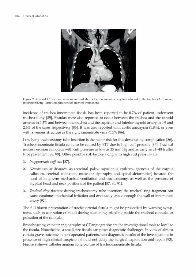
5.5. Tracheoarterial fistula
The term tracheoarterial fistula describes an abnormal communication between the tracheo-bronchial tree and blood vessels. The innominate artery is the most affected vascular structure(72% of the reported cases) due to its close anatomical proximity to the trachea (Figure 7). The
Long-Term Complications of Tracheal Intubationhttp://dx.doi.org/10.5772/intechopen.74160
103
incidence of trachea-innominate fistula has been reported to be 0.7% of patient underwenttracheostomy [85]. Fistulas were also reported to occur between the trachea and the carotidarteries in 4.3% and between the trachea and the superior and inferior thyroid artery in 0.9 and2.6% of the cases respectively [86]. It was also reported with aortic aneurysm (1.8%), or evenwith a venous structure as the right innominate vein <3.5% [86].
Low lying tracheostomy tube insertion is the major risk for this devastating complication [86].Tracheoinnominate fistula can also be caused by ETT due to high cuff pressure [87]. Trachealmucosa erosion can occur with cuff pressure as low as 25 mm Hg and as early as 24–48 h aftertube placement [88, 89]. Other possible risk factors along with high cuff pressure are:
1. Inappropriate cuff size [87].
2. Neuromuscular disorders as (cerebral palsy, myoclonus epilepsy, agenesis of the corpuscallosum, cerebral contusion, muscular dystrophy and spinal deformities) because theneed of long-term mechanical ventilation and tracheostomy, as well as the presence ofatypical head and neck positions of the patient [87, 90, 91].
3. Tracheal ring fracture during tracheostomy tube insertion; the tracheal ring fragment cancause continues mechanical irritation and eventually erode through the wall of innominateartery [92].
The full-blown presentation of tracheoarterial fistula might be proceeded by warning symp-toms, such as aspiration of blood during suctioning, bleeding beside the tracheal cannula, orpulsation of the cannula.
Bronchoscopy, catheter angiography or CTangiography are the investigational tools to localizethe fistula. Nonetheless, a small size fistula can poses diagnostic challenges. In view of almostcertain grave outcome in non-operated patients, non-diagnostic results of the investigations inpresence of high clinical suspicion should not delay the surgical exploration and repair [93].Figure 8 shows catheter angiography picture of tracheoinnominate fistula.
Figure 7. Coronal CT with intravenous contrast shows the innominate artery lies adjacent to the trachea (A. Touman;Intubation/Long-Term Complications of Tracheal Intubation).
Tracheal Intubation104

Warning signs such as sentinel bleeding or pulsating tracheostomy tube are describedin literature and should be checked and reported despite they are frequently absent.Tracheoarterial fistula presentation is of massive bleeding. The majority of the cases (72%)bleed in the first 3 weeks [86], however, bleeding might occur as late as after 20 years [84]. Incases with massive bleeding hemostasis should be insured before pursuing investigations ordefinitive treatment. Hemostasis can be achieved by over inflating the tracheostomy/Endotra-cheal tube cuff until the bleeding stop [90]. The aspirated blood should be suctioned asasphyxiation is more likely to cause death than blood exsanguination. Supportive measuressuch as intravenous volume replacement to correct hypovolemia, or blood transfusion toreplace the blood loss and optimize oxygen delivery should be given as necessary. Thedefinitive treatment is surgical; with various techniques that have been described in theliterature. There is always concern while dissecting the innominate artery to cause arterialinsufficiency to the cerebral circulation. Neurological deficit reported to occur in 4.5% offistula repair survivor [91]; therefore, cerebral blood flow monitoring during the surgery isof utmost important [90].
Author details
Abdelfattah A. Touman1* and Grigoris K. Stratakos2
*Address all correspondence to: [email protected]
1 Cardiovascular Specialist Centre, Dammam, Kingdom of Saudi Arabia
2 1st Department of Respiratory Medicine, National and Kapodistrian University of Athens,Athens, Greece
Figure 8. Angiographic scan of a free contrast agent beside the innominate artery and beside left lateral side of thetracheal cannula. Courtesy of Richter et al. [92].
Long-Term Complications of Tracheal Intubationhttp://dx.doi.org/10.5772/intechopen.74160
105

References
[1] Frerk C, Mitchell VS, McNarry AF, Mendonca C, Bhagrath R, Patel A, O'Sullivan EP,Woodall NM, Ahmad I. Difficult Airway Society 2015 guidelines for management ofunanticipated difficult intubation in adults. BJA: British Journal of Anaesthesia. 1 December2015;115(6):827-848. DOI: 10.1093/bja/aev371
[2] Komasawa N, Hyoda A, Matsunami S, Majima N, Minami T. Utility of a gum-elasticbougie for difficult airway management in infants: A simulation-based crossover analy-sis. BioMed Research International. 2015;2015:5. Article ID 617805. DOI: 10.1155/2015/617805
[3] Jones MW, Catling S, Evans E, Green DH, Green JR. Hoarseness after tracheal intubation.Anaesthesia. 1992;47:213-216
[4] Yamanaka H, Hayashi Y, Watanabe Y, Uematu H, Mashimo T. Prolonged hoarseness andarytenoid cartilage dislocation after tracheal intubation. British Journal of Anaesthesia.2009;103(3):452-455
[5] Peppaerd SB, Dickens JH. Laryngeal injury following short-term intubation. The Annalsof Otology, Rhinology, and Laryngology. 1983;92:327-330
[6] Sue RD, Susanto I. Long-term complications of artificial airways. Clinics in Chest Medi-cine. 2003;24:457-471
[7] Maruyama K, Sakai H, Miyazawa H, Toda N, Iinuma Y, Mochizuki N, Hara K, Otagiri T.Sore throat and hoarseness after total intravenous anaesthesia. British Journal of Anaes-thesia. 2004 Apr;92(4):541-543. Epub 2004 Feb 6
[8] Stout DM, Bishop MJ, Dwersteg JF, Cullen BF. Correlation of endotracheal tube size withsore throat and hoarseness following general anesthesia. Anesthesiology. 1987;67:419-421
[9] Kambic V, Radsel Z. Intubation lesions of the larynx. British Journal of Anaesthesia. 1978;50:587-589
[10] Tolley NS, Cheesman TD, Morgan D, Brookes GB. Dislocated arytenoid: An intubation-induced injury. Annals of the Royal College of Surgeons of England. 1990;72:353-356
[11] Quick CA, Merwin GE. Arytenoid dislocation. Archives of Otolaryngology. 1978;104(5):267-270
[12] Castella X, Gilabert J, Perez C. Arytenoid dislocation after tracheal intubation: Anunusual cause of acute respiratory failure. Anesthesiology. 1991;74:613-615
[13] Oh TK, Yun J-Y, Ryu CH, Park YN, Kim NW. Arytenoid dislocation after uneventfulendotracheal intubation: A case report. Korean Journal of Anesthesiology. 2016;69(1):93-96. DOI: 10.4097/kjae.2016.69.1.93
[14] Alexander AE, Lyons GD, Fazekas-May MA, Rigby PL, Nuss DW, David L, Williams K.Utility of helical computed tomography in the study of arytenoid dislocation and arytenoidsubluxation. The Annals of Otology, Rhinology, and Laryngology. 1997;106(12):1020-1023
Tracheal Intubation106

[15] Gauss A, Treiber HS, Haehnel J, Johannsen HS. Spontaneous reposition of a dislocatedarytenoid cartilage. British Journal of Anaesthesia. 1993;70:591-592
[16] Leelamanit V, Sinkijcharoenchai W. A promising new technique for closed reduc-tion of arytenoid dislocation. The Journal of Laryngology and Otology. 2012;126(2):168-174
[17] Lee SW, Park KN, Welham NV. Clinical features and surgical outcomes following closedreduction of arytenoid dislocation. JAMA Otolaryngology. Head & Neck Surgery. 2014;140(11):1045-1050
[18] Prasertwanitch Y, Schwartz JJH, Vandam LD. Arytenoid cartilage dislocation followingprolonged endotracheal intubation. Anesthesiology. 1971;41:516-517
[19] Hoffman H, Brunberg J, Winter P, Sullivan M, Kileny P. Arytenoid subluxation: Diagno-sis and treatment. The Annals of Otology, Rhinology, and Laryngology. 1991;100:1-9
[20] Clayton JL, Harris MB, Weintraub SL, Marr AB, Timmer J, Stuke LE, et al. Risk factors forcervical spine injury. Injury. 2012 Apr;43(4):431-435
[21] Nolan JP, Wilson ME. Orotracheal intubation in patients with potential cervical spineinjuries. An indication for the gum elastic bougie. Anaesthesia. 1993;48:630-633
[22] Goutcher CM, Lochhead V. Reduction in mouth opening with semi-rigid cervical collars.British Journal of Anaesthesia. 2005;95:344-348
[23] Podolsky S, Baraff LJ, Simon RR, Hoffman JR, Larmon B, Ablon W. Efficacy of cervicalspine immobilization methods. The Journal of Trauma. 1983 Jun;23(6):461-465
[24] Warner ME, Benenfeld SM, Warner MA, Schroeder DR, Maxson PM. Perianesthetic dentalinjuries: Frequency, outcomes, and risk factors. Anesthesiology. 1999 May;90(5):1302-1305
[25] Vogelhut MM, Downs JB. Prolonged endotracheal intubation. Chest. 1979 Jul;76(1):110-111
[26] Pender L, Frazier S. The relationship between dermal pressure ulcers, oxygenation andperfusion in mechanically ventilated patients. Intensive & Critical Care Nursing. 2004;21:29-38
[27] Allman RM. Pressure ulcers among the elderly. The New England Journal of Medicine.1989;320:850
[28] Bourdel-Marchasson I, Barateau M, Rondeau V, Dequae-Merchadou L, Salles-MontaudonN, Emeriau JP, et al. A multicenter trial of the effects of oral nutritional supplementationin critically ill older patients. Nutrition. 2000;16:1-5
[29] National Pressure Ulcer Advisory Panel, European Pressure Ulcer Advisory Panel andPan Pacific Pressure Injury Alliance. In: Haesler E, editor. Prevention and Treatment ofPressure Ulcers: Quick Reference Guide. Perth, Australia: Cambridge Media; 2014
[30] Sariego J. Vocal fold hypomobility secondary to elective endotracheal intubation:A general surgeon's perspective. Journal of Voice. 2010 Jan;24(1):110-112. Epub 2009Jan 29
Long-Term Complications of Tracheal Intubationhttp://dx.doi.org/10.5772/intechopen.74160
107

[31] Ellis PD, Pallister WK. Recurrent laryngeal nerve palsy and endotracheal intubation. TheJournal of Laryngology and Otology. 1975;89:823-826
[32] Young VN, Smith LJ, Rosen C. Voice outcome following acute unilateral vocal foldparalysis. The Annals of Otology, Rhinology, and Laryngology. 2013 Mar;122(3):197-204
[33] Miller RD, Hyatt RE. Evaluation of obstructing lesions of the trachea and larynx by flow-volume loops. The American Review of Respiratory Disease. 1973 Sep;108(3):475-481
[34] Kalil AC, Metersky ML, Klompas M, Muscedere J, Sweeney DA, Palmer LB, et al. Man-agement of adults with hospital-acquired and ventilator-associated pneumonia: 2016Clinical Practice Guidelines by the Infectious Diseases Society of America and the Amer-ican Thoracic Society. Clinical Infectious Diseases. 2016 Sep 1;63(5):e61-e111
[35] Langer M, Cigada M, Mandelli M, Mosconi P, Tognoni G. Early onset pneumonia: Amulticenter study in intensive care units. Intensive Care Medicine. 1987;13:342-346
[36] Safdar N, Dezfulian C, Collard HR, Saint S. Clinical and economic consequences ofventilator-associated pneumonia: A systematic review. Critical Care Medicine. 2005 Oct;33(10):2184-2193
[37] Guyatt GH, Leasa D, Jaeschke RZ, Brun-Buisson C. Incidence of and risk factors forventilator-associated pneumonia in critically ill patients. Annals of Internal Medicine.1998;129(6):433-440
[38] Craven DE, Steger KA. Epidemiology of nosocomial pneumonia. New perspectives on anold disease. Chest. 1995;108:1S-16S
[39] Sottile FD, Marrie TJ, Prough DS, Hobgood CD, Gower DJ, Webb LX, et al. Nosocomialpulmonary infection: Possible etiologic significance of bacterial adhesion to endotrachealtubes. Critical Care Medicine. 1986 Apr;14(4):265-270
[40] Chastre J, Fagon J-Y. Ventilator-associated pneumonia. American Journal of Respiratoryand Critical Care Medicine. 2002;165:867-903
[41] Fagon J, Chastre J, Hance A. Evaluation of clinical judgment in the identification andtreatment of nosocomial pneumonia in ventilated patients. Chest. 1993;103(2):547-553
[42] Tokmaji G, Vermeulen H, Müller MCA, Kwakman PHS, Schultz MJ, Zaat SAJ. Silver-coated endotracheal tubes for prevention of ventilator-associated pneumonia in criticallyill patients. Cochrane Database of Systematic Reviews. 2015;(8). Art. No.: CD009201. DOI:10.1002/14651858.CD009201.pub2
[43] Hua F, Xie H, Worthington HV, Furness S, Zhang Q, Li C. Oral hygiene care for criticallyill patients to prevent ventilator-associated pneumonia. Cochrane Database of SystematicReviews. 2016;(10). Art. No.: CD008367. DOI: 10.1002/14651858.CD008367.pub3
[44] Drakulovic MB, Torres A, Bauer TT, Nicolas JM, Nogué S, Ferrer M. Supine body positionas a risk factor for nosocomial pneumonia in mechanically ventilated patients: Arandomised trial. Lancet. 1999;354(9193):1851-1858
Tracheal Intubation108

[45] Lacherade JC, De Jonghe B, Guezennec P, Debbat K, Hayon J, Monsel A, et al. Inter-mittent subglottic secretion drainage and ventilator-associated pneumonia: A multi-center trial. American Journal of Respiratory and Critical Care Medicine. 2010;182(7):910-917
[46] George DL, Falk PS, Umberto Meduri G, Leeper KV Jr, Wunderink RG, Steere EL, et al.Nosocomial sinusitis in patients in the medical intensive care unit: A prospective epide-miological study. Clinical Infectious Diseases. 1998;27(3):463
[47] Caplan ES, Hoyt NJ. Nosocomial sinusitis. JAMA. 1982;247:639-641
[48] Holzapfel L, Chevret S, Madinier G, et al. Influence of long-term oro or nasotrachealintubation on nosocomial maxillary sinusitis and pneumonia: Results of a prospective,randomized, clinical trial. Critical Care Medicine. 1993;21:1132-1138
[49] Casiano RR, Cohn S, Villasuso E 3rd, Brown M, Memari F, Barquist E, Namias N.Comparison of antral tap with endoscopically directed nasal culture. The Laryngoscope.2001;111(8):1333
[50] Rouby J, Laurent P, Gosnach M, et al. Risk factors and clinical relevance of nosocomialmaxillary sinusitis in the critically ill. American Journal of Respiratory and Critical CareMedicine. 1994;150:776-784
[51] Stein M, Caplan ES. Nosocomial sinusitis: A unique subset of sinusitis. Current Opinionin Infectious Diseases. 2005;18:147-150
[52] Sehgal IS, Dhooria S, Bal A, Aggarwal AN, Behera D, Agarwal R. Obstructive fibrinoustracheal pseudomembrane after endotracheal intubation. Respiratory Care. 2016 Sep;61(9):1260-1266
[53] Deslée G, Brichet A, Lebuffe G, Copin MC, Ramon P, Marquette CH. Obstructive fibri-nous tracheal pseudomembrane. A potentially fatal complication of tracheal intubation.American Journal of Respiratory and Critical Care Medicine. 2000;162:1169-1171
[54] Manassero A, Ugues S, Bertolaccini L, Bossolasco M, Terzi A, Coletta G. A very earlystage of obstructive fibrinous tracheal pseudo-membrane formation. Journal of ThoracicDisease. 2012;4(3):320-322. DOI: 10.3978/j.issn.2072-1439.2012.05.10
[55] Stratakos G. Postintubation tracheal stenosis and endoscopic management. Pneumonologie.2003;16(3):262-270
[56] Spittle N, McCluskey A. Lesson of the week: Tracheal stenosis after intubation. BMJ. Oct21 2000;321(7267):1000-1002
[57] Grillo HC, Cooper JD, Geffin B, et al. A low pressure cuff for tracheostomy tubes tominimize tracheal injury. A comparative clinical trial. The Journal of Thoracic and Car-diovascular Surgery. 1971;62:898-907
[58] Su Z et al. A canine model of tracheal stenosis induced by cuffed endotracheal intubation.Scientific Reports. 2017;7:45357. DOI: 10.1038/srep45357
Long-Term Complications of Tracheal Intubationhttp://dx.doi.org/10.5772/intechopen.74160
109

[59] Plojoux J, Laroumagne S, Vandemoortele T, Astoul PJ, Thomas PA, Dutau H. Manage-ment of benign dynamic “A-shape” tracheal stenosis: A retrospective study of 60 patients.The Annals of Thoracic Surgery. 2015 Feb;99(2):447-453. DOI: 10.1016/j.athoracsur.2014.08.037. Epub 2014 Dec 12
[60] Farzanegan R, Farzanegan B, Alehashem M, Zangi M, Niakan Kalhori SR, Sheikhy K,et al. Item selection and content validity of the risk factors of post-intubation trachealstenosis observation questionnaire for ICU-admitted patients. Tanaffos. 2017;16(1):22-33
[61] Yang KL. Tracheal stenosis after a brief intubation. Anesthesia and Analgesia. 1995 Mar;80(3):625-627
[62] Andrews MJ, Pearson FG. Analysis of 59 cases of tracheal stenosis following tracheos-tomy with cuffed tube and assisted ventilation, with special reference to diagnosis andtreatment. The British Journal of Surgery. 1973;60:208-212
[63] Mehta AC, Lee FY, Cordasco EM, Kirby T, Eliachar I, De Boer G. Concentric tracheal andsubglottic stenosis. Management using the Nd�YAG laser for mucosal sparing followedby gentle dilatation. Chest. 1993;104:673-677
[64] Carden KA, Boiselle PM, Waltz DA, Ernst A. Tracheomalacia and tracheobronchomalaciain children and adults: An in-depth review. Chest. 2005 Mar;127(3):984-1005
[65] Murgu SD, Colt HG. Tracheobronchomalacia and excessive dynamic airway collapse.Respirology. 2006;11:388-406
[66] Feist JH, Johnson TH, Wilson RJ. Acquired tracheomalacia: Etiology and differentialdiagnosis. Chest. 1975;68(3):340-345
[67] Austin J, Ali T. Tracheomalacia and bronchomalacia in children: Pathophysiology, assess-ment, treatment, and anaesthesia management. Paediatric Anaesthesia. 2003;13:3-11
[68] Nuutinen J. Acquired tracheobronchomalacia. European Journal of Respiratory Diseases.1982;63:380
[69] Wright CD. Tracheomalacia. Chest Surgery Clinics of North America. 2003;13:349-357
[70] Koziej M, Gorecka D. Cough-syncope syndrome in tracheobronchomalacia. Pneumonologiai Alergologia Polska. 1992;60:89-91
[71] Collard P, Freitag L, Reynaert MS, et al. Respiratory failure due to tracheobronch-omalacia. Thorax. 1996;51:224-226
[72] Majid A, Gaurav K, Sanchez JM, et al. Evaluation of tracheobronchomalacia by dynamicflexible bronchoscopy.Apilot study.Annals of theAmerican Thoracic Society. 2014;11:951-955
[73] Gilkeson RC, Ciancibello LM, Hejal RB, Montenegro HD, Lange P. Tracheobronch-omalacia: Dynamic airway evaluation with multidetector CT. AJR. 2001;176:205-210
[74] Campbell AH, Faulks LW. Expiratory air-flow pattern in tracheobronchial collapse. TheAmerican Review of Respiratory Disease. 1965;92:781-791
Tracheal Intubation110

[75] Garcia-Pachon E. Tracheobronchomalacia: A cause of flow oscillations on the flow-volume loop. Chest. 2000;118:1519
[76] VinckenW, CosioMG. Flow oscillations on the flow-volume loop: A nonspecific indicator ofupper airway dysfunction. Bulletin Européen de Physiopathologie Respiratoire. 1985;21:559
[77] Grillo HC. Surgical treatment of postintubation tracheal injuries. The Journal of Thoracicand Cardiovascular Surgery. 1979;78:860-875
[78] Mooty RC, Rath P, Self M, Dunn E, Mangram A. Review of tracheo-esophageal fistulaassociated with endotracheal intubation. Journal of Surgical Education. 2007 Jul-Aug;64(4):237-240
[79] Darteville P, Macchiarini P. Management of acquired tracheoesophageal fistula. ChestSurgery Clinics of North America. 1996;6:819-836
[80] ReedMF, Mathisen DJ. Tracheoesophageal fistula. Chest Surgery Clinics of North America.2003 May;13(2):271-289
[81] Ernst A, Herth FJ. Principles and Practice of Interventional Pulmonology. New York:Springer; 2013
[82] Mathisen DJ, Grillo HC, Wain JC, Hilgenberg AD. Management of acquired nonmalignanttracheoesophageal fistula. The Annals of Thoracic Surgery. 1991;52:759-765
[83] Li J, Gao X, Chen J, Lao M, Wang S, Zeng G. Endoscopic closure of acquired oesophagor-espiratory fistulas with cardiac septal defect occluders or vascular plugs. RespiratoryMedicine. 2015 Aug;109(8):1069-1078. Epub 2015 May 5
[84] Erdim I, Sirin AA, Baykal B, Oghan F, Guvey A, Kayhan FT. Treatment of large persistenttracheoesophageal peristomal fistulas using silicon rings. Brazilian Journal of Otorhinolar-yngology. Sep-Oct 2017;83(5):536-540. DOI: 10.1016/j.bjorl.2016.06.011. Epub 2016 Jul 21
[85] Scalise P, Prunk SR, Healy D, Votto J. The incidence of tracheoarterial fistula in patientswith chronic tracheostomy tubes. A retrospective study of 544 patients in a long term carefacility. Chest. 2005;128:3906-3909
[86] Schaefer OP, Irwin RS. Tracheoarterial fistula: An unusual complication of tracheostomy.Journal of Intensive Care Medicine. 1995;10:64-75
[87] Tijana D, Ivana Č, Milenko B, Slobodan S. Iatrogenic tracheoarterial fistula: Case reportand literature review. Medicine, Science, and the Law. 2017 Jul;57(3):143-145. DOI:10.1177/0025802417720567
[88] Stiles PJ. Tracheal lesions after tracheostomy. Thorax. 1965;20:517-522
[89] Nordin U. Trachea and cuff-induced tracheal injury: An experimental study on causativefactors and prevention. Acta Oto-Laryngologica. Supplementum. 1977;345:1-71
[90] Furukawa K, Kamohara K, Itoh M, Morokuma H, Morita S. Operative technique fortracheo-innominate artery fistula repair. Journal of Vascular Surgery. 2014;59:1163-1167
Long-Term Complications of Tracheal Intubationhttp://dx.doi.org/10.5772/intechopen.74160
111

[91] Jones JW, Reynolds M, Hewitt RL, Drapanas T. Tracheo-innominate artery erosion: Suc-cessful surgical management of a devastating complication. Annals of Surgery. 1976 Aug;184(2):194-204
[92] Richter T, Gottschlich B, Sutarski S, Müller R, Ragaller M. Late life-threatening hemor-rhage after percutaneous tracheostomy. International Journal of Otolaryngology. 2011;2011:3. Article ID 890380. DOI: 10.1155/2011/890380
[93] Vaidya N, Strauchler D, Guelfguat M. Computed tomography angiography diagnosis oftracheoinnominate fistula: A case report and review of literature. Quantitative Imaging inMedicine and Surgery. 2013;3(2):121-125
Tracheal Intubation112