Chapter 4Chapter 4 - INFLIBNETshodhganga.inflibnet.ac.in/bitstream/10603/2219/15/15_chapter...
54
Chapter 4 Sachin Shah University of Pune Ph.D. Thesis 115 Chapter 4 Chapter 4 Chapter 4 Chapter 4 Immobilization of whole cells on biocompatible materials: Use as Enzyme Source for the Biotransformation of Arachidonic acid to 19-HETE and 20-HETE
Transcript of Chapter 4Chapter 4 - INFLIBNETshodhganga.inflibnet.ac.in/bitstream/10603/2219/15/15_chapter...
Chapter 4
Sachin Shah University of Pune
Ph.D. Thesis
115
Chapter 4Chapter 4Chapter 4Chapter 4
Immobilization of whole cells on
biocompatible materials: Use as Enzyme
Source for the Biotransformation of
Arachidonic acid to 19-HETE and 20-HETE

Chapter 4
Sachin Shah University of Pune
Ph.D. Thesis
116
4.1. Summary
A variety of materials can be utilized for immobilization of Candida bombicola
based on different immobilization protocols. These biomaterials can be used as enzyme
sources for the transformation of arachidonic acid to vasoactive compounds 19-
hydroxyeicosatetraenoic acid (19-HETE) and 20-hydroxyeicosatetraenoic acid (20-
HETE). In general few points need to be considered before choosing the materials for the
whole cell immobilization.
1. The materials should be stable, robust and inert. It must be biocompatible and
should not interfere with bioreaction.
2. The materials should protect the whole cell containing reactive enzyme against
microbial deterioration and render the enzyme accessible to cofactor, metal ions
etc.
3. The materials must permit substrate accessibility to immobilized whole cells and
thus avoid mass transfer problem.
4. The materials should have high immobilization whole cell loading factor for the
efficient transformation reaction.
5. The immobilization process must be simple, quick, inexpensive and ecofriendly.
The studies in this chapter are focused on immobilization of Candida bombicola
on novel biocompatible supports as an enzyme sources for the transformation of
arachidonic acid to bioactive molecules, 19-hydroxyeicosatetraenoic acid (19-HETE)
and 20- hydroxyeicosatetraenoic acid (20-HETE). This chapter is divided into two
subchapters for three different biocompatible materials used for immobilization of
Candida bombicola.

Chapter 4
Sachin Shah University of Pune
Ph.D. Thesis
117
4.2. Introduction
Impressive advances are being made in the synthesis of chemically functionalized
and patterned biocompatible surfaces for the immobilization of biomolecules such as
whole cells and enzymes of microorganisms such as bacteria and yeast.1,
2 Development
of methodologies for the entrapment and immobilization of whole cells has important
implications in a range of applications, examples of which include basic cell biology,3, 4
biosensing,5 tissue engineering
6 and treatment of diseases by controlled delivery of
biological products.7 One of the challenges in this area is to develop protocols wherein
the immobilization of the cells is to be spatially controlled, preferably on a submicron to
micron scale by designing surfaces of varying “adsorptivity” of the biological system.
This gives an insight into the effect of cell shape and cell function3
which enhances our
ability to control the cellular environment and helps in understanding fundamental cell
biology. Such patterned surfaces for immobilization of cells have been obtained using
microcontact printing (µ-CP) on reactive3
and mixed self-assembled monolayers
(SAMs).8 SAMs of alkanethiolates
9 and alkylsilanes
10 were obtained by the sol-gel
technique11
and using elastomeric membranes.12
Groves et. al., in 2001 have
demonstrated that phospholipid bilayers act as biomimetic surfaces and modulate the
assembly and growth of cells.13
Recently µ-CP of organic monolayers and subsequent
polymer functionalization has been used to develop patterns in the seeding of bacterial
cells.14
An important application of bacterial and fungal cells (genetically engineered and
otherwise) is use as “factories” for the production of industrially and medically important
enzymes and metabolites.15
Here we have been interested in assembly of specific cells on

Chapter 4
Sachin Shah University of Pune
Ph.D. Thesis
118
surfaces from the point of view of using the cells as sources of enzymes for
biotransformations and synthesis of new materials. The enzyme cytochrome P450 present
in the yeast cells was used to catalyze in situ ω and ω -1 hydroxylation of arachidonic
acid. Cytochrome P450, the enzyme of interest is unstable outside the cellular
environment and in such cases, immobilization of the whole cells was important to
catalyze reactions that are dependent on the unstable enzymes. As part of our search for
newer and more versatile materials, tailorable surfaces for cell immobilization has been
used. This chapter presents the synthesis of different biocompatible materials whose
surface may readily be modified to render it compatible for a variety of biocatalytic
applications.

Chapter 4
Sachin Shah University of Pune
Ph.D. Thesis
119
Chapter 4. Chapter 4. Chapter 4. Chapter 4. AAAA
Candida bombicola cells immobilized on patterned lipid films as
enzyme sources for the transformation of arachidonic acid to 19-
HETE and 20-HETE
Chapter 4. Chapter 4. Chapter 4. Chapter 4. BBBB
Nanogold membrane as scaffolds for whole cell immobilization as
enzyme source for biotransformations of arachidonic acid to 19-
HETE and 20-HETE

Chapter 4
Sachin Shah University of Pune
Ph.D. Thesis
120
Chapter 4. A: Candida bombicola cells immobilized on patterned lipid films as
enzyme sources for the transformation of arachidonic acid to 19-HETE and 20-
HETE
4.A.1. Introduction
Chapter 4.A. part presents the assembly of Candida bombicola yeast cells onto
patterned thermally evaporated fatty amine thin films (octadecylamine, ODA) and the use
of the enzyme cytochrome P450 present in the yeast cells to catalyze in-situ the ω-
hydroxylation of arachidonic acid (AA) to 19- hydroxyeicosatetraenoic acid (19-HETE)
and 20-hydroxyeicosatetraenoic acid (20-HETE)16, 17
(see Figure 4.A.1.). Cytochrome
P450 is a membrane bound protein and is known to be highly unstable outside the cells.17
Thus, rather than immobilizing the unstable purified enzyme within a lipid film, we
demonstrate here the immobilization of Candida bombicola yeast cells carrying the
enzyme cytochrome P450.16, 17
This immobilization affords a cheaper, renewable and
more versatile alternative for carrying out the transformation of arachidonic acid to 19-
HETE and 20-HETE. In view of the vasoactive and renal pharmacological activity of 19-
HETE and 20-HETE,18
the large-scale production of this molecule is of commercial
interest and the methodology presented herein assumes added importance.
Part of the work presented in this chapter 4.A. has been published in: Biotechnol. Prog. 2003, 19,
1659-1663.

Chapter 4
Sachin Shah University of Pune
Ph.D. Thesis
121
Figure 4.A.1. Transformation of arachidonic acid to 20-hydroxyeicosatetraenoic
acid (20-HETE) and 19-hydroxyeicosatetraenoic acid (19-HETE) mediated by
cytochrome P450 enzyme present in Candida bombicola cells
The immobilization of the Candida bombicola cells was accomplished by simple
immersion of the patterned ODA film into an aqueous dispersion of the cells as illustrated
in Scheme 4.A.1. The assembly of the cells on the ODA surface occurs possibly through
hydrophobic interactions between the cell wall and the ODA molecules (Scheme 4.A.1.)
and thus provides a rational strategy for assembly of the cells. The adhesion of the cells
to the ODA surface is sufficiently strong and permits reuse of the immobilized cells in
the biochemical transformation of arachidonic acid to 20-HETE.
OH
O
Arachidonic acid
Acid hydrolysis
Candida bombicola cells
cytochrome P450 enzyme
OH
O
OH19-HETE
OH
O
OH
20-HETE
+

Chapter 4
Sachin Shah University of Pune
Ph.D. Thesis
122
Scheme 4.A.1. Illustration of immobilization of Candida bombicola yeast cells on
thermally evaporated ODA film surface
Chapter 4
Sachin Shah University of Pune
Ph.D. Thesis
123
4.A.2. Materials and Methods
4.A.2.1. Chemicals
Arachidonic acid of 99 % purity and octadecylamine (ODA) of 97 % purity were
obtained from Sigma and Aldrich Chemicals respectively and used as received. All
reagents were from standard commercial sources and of highest quality available.
4.A.2.2. Growth of Candida bombicola cells
Candida bombicola cells ATCC 22214 were pre-cultivated in 50 mL medium at
30 0C consisting of (g l
-1): glucose, 100; yeast extract, 1; (NH4)2SO4, 1; MgSO4 · 7H2O,
0.3; Na2HPO4, 2; NaH2PO4, 7; pH 5.5 and shaken at 160 rpm. Cells from the late growth
phase (24 h) were inoculated into 1 lit. flasks containing 400 mL medium. Fermentative
procedure employed for the experiment was same as described previously in Chapter 2
section (2.2.3.).
4.A.2.3. Deposition of octadecylamine films
250 Å thick films of ODA (CH3-(CH2)17–NH2) were thermally evaporated onto 6
MHz AT-cut quartz crystals for quartz crystal microgravimetry (QCM) measurements, Si
(111) wafers (for Fourier transform infrared (FTIR) spectroscopy and scanning electron
microscopy (SEM) measurements) in an Edwards E308 vacuum coating unit. A 40 µm x
40 µm mesh size transmission electron microscope (TEM) grid was used as a mask in the
deposition of patterned ODA films on the Si (111) substrate. The deposition was done at
a pressure of 1 x 10⁻⁷
Torr and the film deposition rate and thickness were monitored in-
situ using an Edwards quartz crystal thickness monitor.
Chapter 4
Sachin Shah University of Pune
Ph.D. Thesis
124
4.A.2.4. Immobilization of Candida bombicola cells onto the thermally evaporated
ODA films
The immobilization of the yeast cells on the ODA surface was followed by QCM
by immersion of the 250 Å thick ODA film on gold-coated AT cut quartz crystals for
different time intervals in an aqueous dispersion of the cells (~ 108 cells/mL) and
measuring the frequency change of the crystals ex-situ after thorough washing and drying
of the crystals. The frequency counter used was an Edwards FTM5 instrument operating
at a frequency stability and resolution of + 1 Hz. For a 6 MHz crystal used in the
investigation, this translates into a mass resolution of 12 ng/cm2. The frequency change
was converted to mass loading using the Sauerbrey formula.19
The 250 Å thick ODA
films on Si (111) substrates were immersed in the Candida bombicola cells dispersed in
deionized water for 4 h. This optimum time of immersion was estimated from QCM
measurements. For the reusability of the cells immobilized on 250 Å thick ODA films
surface, were washed 3 times by deionized water prior to reuse.
4.A.2.5. FTIR measurements
FTIR measurements of the 250 Å thick ODA films deposited on Si (111)
substrates before and after immobilization of the Candida bombicola cells were made on
a Perkin Elmer Spectrum-1 FTIR spectrometer operated in the diffuse reflectance mode
at a resolution of 4 cm-1
.

Chapter 4
Sachin Shah University of Pune
Ph.D. Thesis
125
4.A.2.6. Scanning electron microscopy (SEM)
The immobilization of the Candida bombicola cells on the patterned ODA films
was also studied by scanning electron microscopy (SEM) on a Leica Stereoscan-440
electron microscope. Presence of cells on the patterned ODA film surface after
incubation of the cells immobilized on ODA surface after 96 h in the reaction medium
was confirmed from SEM images.
4.A.2.7. Synthesis of sophorolipids
The yeast cells immobilized on the 250 Å thick ODA films were immersed in the
reaction medium containing 5 mL 10% sterile glucose and 30 mg arachidonic acid and
was incubated for 96 h at 30 0C under slow shaking. After the reaction, the supernatant
was decanted and used for extracting the sophorolipid.17
4.A.2.8. Hydrolysis of glycolipid and isolation of 19-HETE and 20-HETE
Acid hydrolysis of the sophorolipids under N2 with 1M HCl for 12 h at 25 0C
liberated the fatty acids, which were extracted with an equal volume of chloroform.
Hydroxylated fatty acids were purified on 500 mg Aminopropyl Sep-Pak Cartridges
(Waters). Samples in 0.5 mL chloroform were applied to cartridge pre-equilibrated with 5
mL n-hexane. Neutral lipids were eluted with 25 mL chloroform /2-propanol (2:1, v/v),
mono-hydroxylated fatty acids with 25 mL 2% (v/v) acetic acid in diethyl ether and
phospholipids with 25 mL methanol. The hydroxy fatty acid fraction was rotary
evaporated and the residue taken up in a small volume of chloroform. Hydroxy fatty
acids were purified by TLC on standard Kiesel-gel 60 plates and developed with
petroleum ether (b.p. 60-80 0C) diethyl ether/ acetic acid (50:50:1, by volume). Iodine
Chapter 4
Sachin Shah University of Pune
Ph.D. Thesis
126
vapors were used to visualize fatty acids and the corresponding bands were immediately
eluted with methanol/chloroform (2:1 v/v) and derivatized to their methyl ester silyl
ethers.17,
20
4.A.2.9. Gas chromatography mass spectroscopy (GC-MS)
A Shimadzu GCMS QP 5050 automated quadrupole mass spectrometer operating
in the electron impact mode. GC parameters: column used, BP-5 fused silica column
(30m x 0.25 mm, 0.25mm, 0.25mm coatings). He gas as a carrier at 14 kPa head
pressure; injector at 250 0C; column initially at 150
0C for 1 min (rate 35
0C/min) then
increased to 220 0C for 5 min (rate 5
0C/min) and then at 280
0C and then held at these
conditions for 10 min; injection volume 1 µL. MS parameters: Interface temperature 250
0C, ionization mode electron impact, scan range 70 to 450 amu (arbitrary mass units) s
-1.

Chapter 4
Sachin Shah University of Pune
Ph.D. Thesis
127
4.A.3. Results and discussions
4.A.3.1. QCM studies
The kinetics of cell immobilization onto the thermally evaporated ODA films was
monitored by immersion of a 250 Å thick ODA covered QCM crystal for different time
intervals in the cells dispersed in deionized water and monitoring the change in resonance
frequency of the crystal ex-situ after thorough washing and drying of the crystals. Since
the mass of the individual cells is not known, we have used the frequency change alone
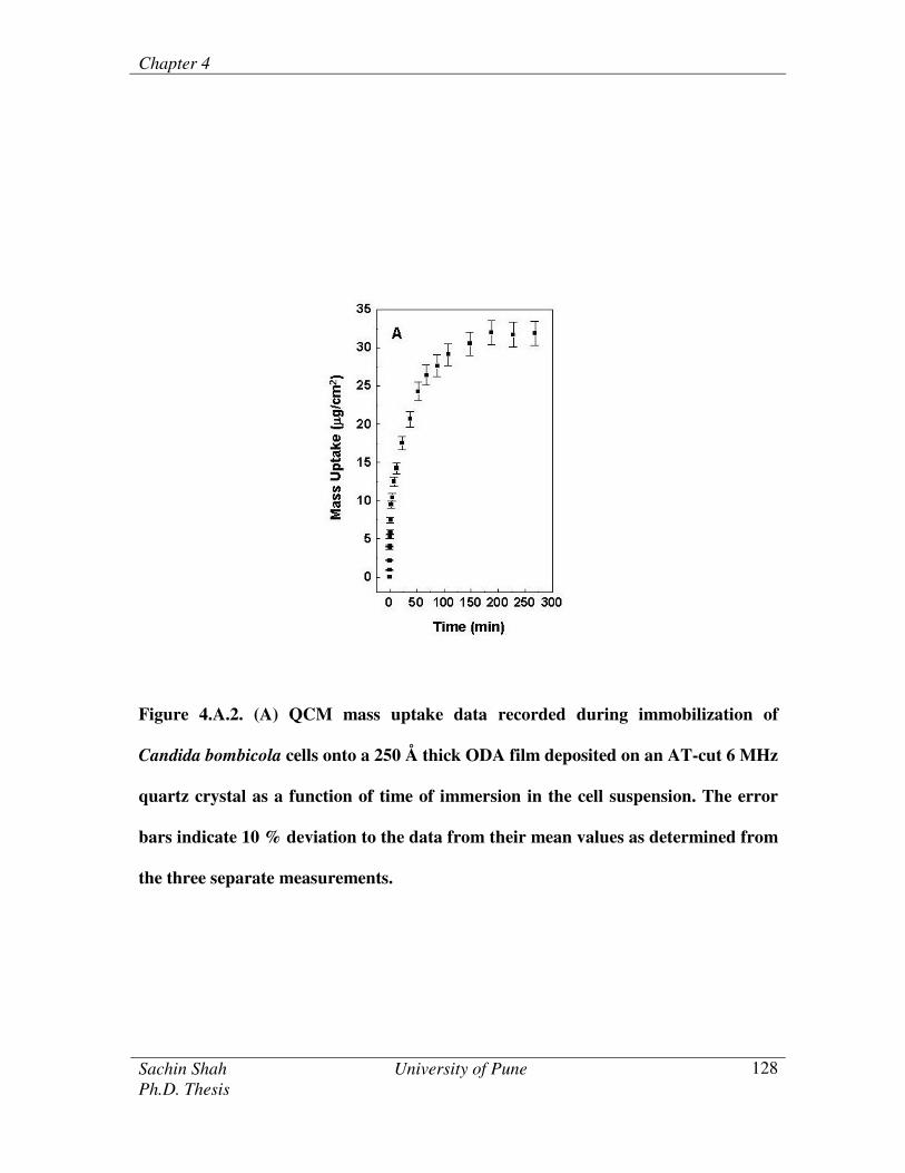
as an indicator of the attachment of the cells on the hydrophobic ODA surface. Figure
4.A.2. (A) has shown the QCM mass uptake data recorded during immobilization of
Candida bombicola cells to the ODA film surface. The error bars are based on an
analysis of three separate QCM measurements. It is observed from the Figure 4.A.2. (A)
that there is a rapid attachment of the cells initially with 90 % of the cells being
immobilized within the first 100 minutes of immersion. The cell density on the ODA film
surface eventually reaches saturation after 4 h of immersion in the yeast cell suspension.
In all further experiments, this optimum time of immersion (4 h) in the Candida
bombicola cells solution was used to obtain films of the immobilized cells.
Chapter 4
Sachin Shah University of Pune
Ph.D. Thesis
128
Figure 4.A.2. (A) QCM mass uptake data recorded during immobilization of
Candida bombicola cells onto a 250 Å thick ODA film deposited on an AT-cut 6 MHz
quartz crystal as a function of time of immersion in the cell suspension. The error
bars indicate 10 % deviation to the data from their mean values as determined from
the three separate measurements.

Chapter 4
Sachin Shah University of Pune
Ph.D. Thesis
129
4.A.3.2. FTIR Studies
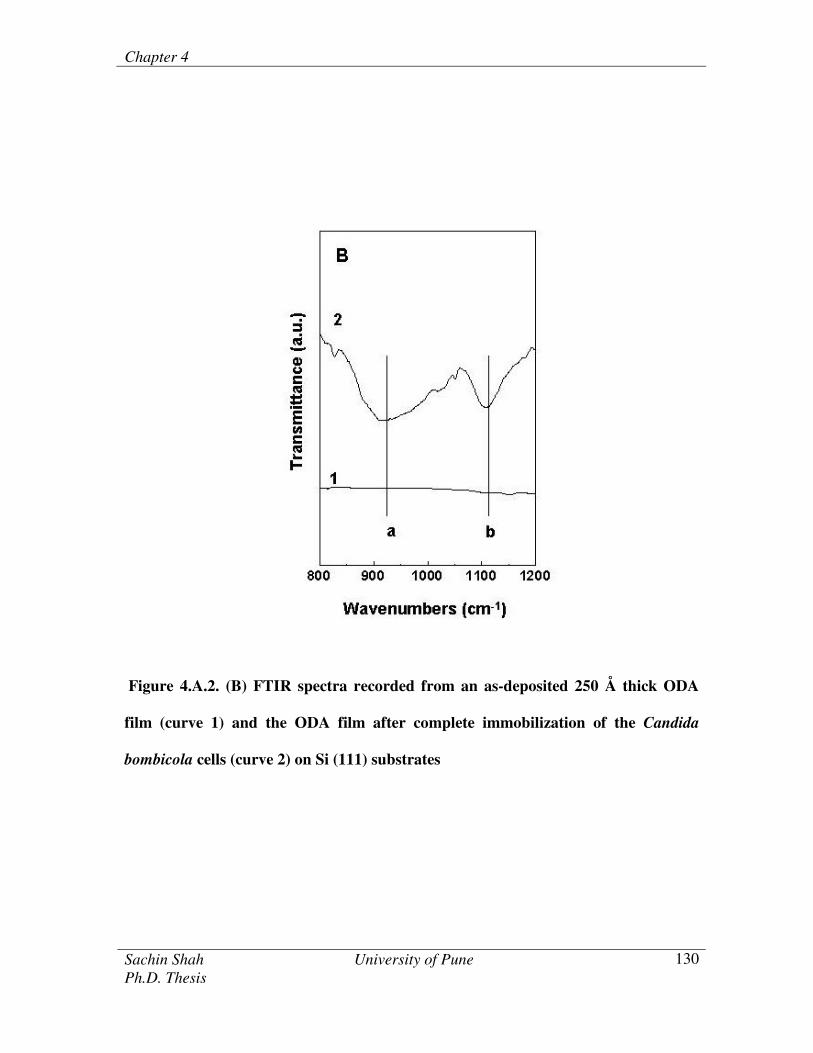
FTIR spectroscopy provides a convenient means of monitoring the attachment of
the Candida bombicola cells via fingerprint signatures of cellular components. Figure
4.A.2. (B) has shown the FTIR spectra recorded from a 250 Å thick as-deposited ODA
film (curve 1) and the ODA film after immersion in Candida bombicola cells solution for
4 h (curve 2). Two prominent features labeled a (917 cm-1
) and b (1110 cm-1
) in the
figure can be seen for the cell-immobilized ODA film (curve 2) which are clearly absent
in the as-deposited ODA film (curve 1). These two absorption bands a and b are
characteristic of excitation of deoxyribose-phosphate vibration modes and vibrations in
the deoxyribose groups in the DNA molecules of the yeast cells respectively.21
The FTIR
results thus present additional evidence for the presence of the Candida bombicola cells
on the ODA film surface.
Chapter 4
Sachin Shah University of Pune
Ph.D. Thesis
130
Figure 4.A.2. (B) FTIR spectra recorded from an as-deposited 250 Å thick ODA
film (curve 1) and the ODA film after complete immobilization of the Candida
bombicola cells (curve 2) on Si (111) substrates

Chapter 4
Sachin Shah University of Pune
Ph.D. Thesis
131
4.A.3.3. SEM measurements
Figure 4.A.3. (A) has shown the SEM image of the as-deposited ODA film on a
Si (111) substrate using a TEM grid as a mask. It is seen that well-defined individual
hexagonal elements of the ODA film have been deposited on the substrate. Figure 4.A.3.
(B) and (C) have shown energy dispersive analysis of x-rays (EDAX) spot profile
analysis on masked (marked as x in Figure 4.A.3. (A)) and exposed substrate (marked as
+ in Figure 4.A.3. (A)) of the patterned surfaces of ODA lipid films. Figure 4.A.3. (C)
has shown nitrogen signal from the exposed surface, hence confirms the deposition of the
ODA, however nitrogen signals were absent from the masked region, this confirms that
ODA is not deposited in this region. Thereafter, this film was immersed in an aqueous
suspension of Candida bombicola cells for 4 h and washed thoroughly prior to imaging
by SEM.
Figures 4.A.4. (A) and (B) have shown the low and high magnification SEM
image recorded after immobilization of the Candida bombicola cells onto the hexagonal
ODA patterns. It is clearly seen from SEM image that the yeast cells are immobilized
extremely faithfully on the ODA elements with negligible binding of the cells to the
exposed silicon surface.

Chapter 4
Sachin Shah University of Pune
Ph.D. Thesis
132
Figure 4.A.3. (A) SEM images recorded from patterned thermally evaporated ODA thin
film. (B) and (C) shows EDAX spot profile analysis on masked. [In Figure 4.A.3. (A)
marked as x and exposed surface of patterned ODA lipid films (marked as + )]
Figures 4.A.4 (A) and (B). low and high magnification of SEM images after
immobilization of Candida bombicola cells onto the ODA film surface.
Chapter 4
Sachin Shah University of Pune
Ph.D. Thesis
133
4.A.3.4. Synthesis of sophorolipids
As mentioned briefly in the introduction, our interest in Candida bombicola cells
centres on the ability of the cells to catalyse the transformation of arachidonic acid to 20-
HETE (Figure 1) and therefore, it is of paramount importance to establish the viability of
the immobilized cells in performing this biochemical function. Films of the immobilized
cells were reacted with arachidonic acid and the sophorolipids were isolated from the
reaction medium and subjected to acid hydrolysis as described in detail in the
experimental section. The immobilized yeast cells transformed 75 % of the arachidonic
acid to sophorolipid, which was then subjected to acid hydrolysis to yield 20-HETE 17
.
The overall reaction leading to the formation of 20-HETE from arachidonic acid was
shown in Figure 4.A.1.
4.A.3.5. Acid hydrolysis of sophorolipids and isolation of 19-HETE and 20-HETE
As mentioned briefly in the introduction, our interest in Candida bombicola cells
centers on the ability of the cells to catalyze the transformation of arachidonic acid to 19-
HETE and 20-HETE. The sophorolipids formed during the biotransformations of
arachidonic acid were subjected to acid hydrolysis to yield 19-HETE and 20-HETE
compounds. The hydroxyecosatetraenoic acids were reacted with diazomethane solution
and thereafter with the bis silyl trimethyl fluroacetamide (BSTFA) to give methyl ester
silyl ether of hydroxyecosatetraenoic acid.
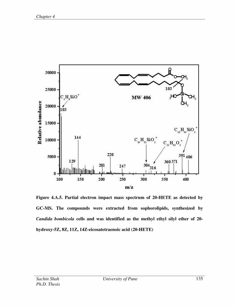
Figure 4.A.5. has shown the partial mass spectrum of 20-HETE as detected by
GC-MS. Significant ions occurred at m/z [406 M+], 391 [M
+ - 15], 316 [M
+ - 90], 304
[(M+ +1) – 103].
17 Selective ion monitoring also showed a prominent signal at m/z 103
[(CH3)3-Si-O+-CH2].
17 The mass spectrum thus clearly indicates that the hydroxyl group

Chapter 4
Sachin Shah University of Pune
Ph.D. Thesis
134
is at the C20 position and that the compound was 20-hydroxy 5Z, 8Z, 11Z, 14Z-
eicosatetraenoic acid (20-HETE). Confirmation of the identity of 20-HETE was obtained
by the observed co-elution of 20-HETE standard by GC-MS with the isolated material.
The mass spectrum indicated a hydroxyl group at position C19. The mass
spectrum of ions occurred at m/z 117 due to cleavage from C19 to C20; [(CH3)3 -Si- O+-
CH-CH3] and 73 [base ion; (CH3)3-Si] and loss of 220 [M+ - 186] due to rearrangement
and loss of –CH=CH (CH2)3-CH[O+-Si-(CH3)3]-CH3, 201 {M
+ - [131 + 74 (silyl group +
H)]}. Figure 4.A.6. has shown the structures of the significant ions occurred of methyl
ester silyl ether of 19-HETE. The compound was identified as 19-
hydroxyeicosatetraenoic acid.17
Also yeast cells such as Candida apicola have been
shown to synthesize sophorolipids in which sophorose, as a diglucoside is linked
glycosidically to the terminal (n-) or sub terminal (n-1) hydroxyl group of a hydroxyl
fatty acid.17
We would like to add that the film of the immobilized cells could be reused
after reaction and thorough washing with only a marginal loss in activity (5 % cells over
5 reuse cycles) indicating that the cells were strongly bound to the underlying ODA film
surface and that there is little leaching out of the cells during reaction. This is confirmed
by imaging the ODA patterned surface after incubating the cells in the reaction medium
for 96 h. the presence of the cells on the ODA film surface after reaction.
Chapter 4
Sachin Shah University of Pune
Ph.D. Thesis
135
Figure 4.A.5. Partial electron impact mass spectrum of 20-HETE as detected by
GC-MS. The compounds were extracted from sophorolipids, synthesized by
Candida bombicola cells and was identified as the methyl ethyl silyl ether of 20-
hydroxy-5Z, 8Z, 11Z, 14Z-eicosatetraenoic acid (20-HETE)

Chapter 4
Sachin Shah University of Pune
Ph.D. Thesis
136
Figure 4.A.6. Partial electron impact mass spectrum of 19-HETE as detected by
GC-MS. The compounds were extracted from sophorolipids, synthesized by
Candida bombicola cells and was identified as the methyl ethyl silyl ether of 19-
hydroxy-5Z, 8Z, 11Z, 14Z-eicosatetraenoic acid (19-HETE)

Chapter 4
Sachin Shah University of Pune
Ph.D. Thesis
137
Figure 4.A.7. (A) and (B) have shown SEM images of different regions of the
Candida bombicola cells bound to ODA lipid films after one cycles of reaction
(incubating in the reaction medium for 30 oC for 96 h). Thus, the yeast cells were
strongly bound to the hydrophobic ODA lipid film permitting excellent reuse. The films
of the immobilized cells could be reused after reaction and through washing with only a
marginal loss in biocatalytic activity ca. 10 % after 5 cycles, indicating that the cells are
strongly bound to the ODA lipid films.
Figure 4.A.7. Low (A) and high (B) magnification SEM images of Candida bombicola
whole cells immobilized on thermally evaporated octadecylamime lipid films after one
cycle of reaction.

Chapter 4
Sachin Shah University of Pune
Ph.D. Thesis
138
4.A.4. Conclusion
In this study, we have demonstrated the immobilization of Candida bombicola
yeast cells on patterned thermally evaporated ODA films, the assembly of cells to ODA
films possibly driven by hydrophobic interactions between cell walls and ODA
molecules. The immobilized yeast cells were biologically active and cytochrome P450
enzyme present in the Candida bombicola cells could be used to transform arachidonic
acid to 19-hydroxyeicosatetraenoic acid (19-HETE) and 20-hydroxyeicosatetraenoic acid
(20-HETE). The biocomposite films are easily separated from the reaction medium for
additional reuse.

Chapter 4
Sachin Shah University of Pune
Ph.D. Thesis
139
Chapter 4. B: Nanogold membrane as scaffolds for whole cell immobilization as
enzyme source for biotransformations of arachidonic acid to 19-HETE and 20-
HETE
4.B.1. Introduction
Chapter 4.B. focuses on the preparation of chemically functionalized
biocompatible polymeric membrane embedded by the gold nanoparticles and
hydrophobized by using octadecylamine (ODA). The free standing hydrophobic
nanogold membrane provides a biocompatible surface for the immobilization of whole
cells. The attachment of the cells to the ODA bound to the nanogold membrane occurs
possibly through the nonspecific interactions such as hydrophobic interactions between
the cell walls and the ODA molecules. The enzyme, cytochrome P450 present in the
immobilized yeast cells on the ODA film surface was used for the transformation of the
arachidonic acid (AA) to sophorolipids and thereafter sophorolipids were acid hydrolyzed
to liberate 19- hydroxyeicosatetraneoic acid (19-HETE) and 20-hydroxyeicosatetraneoic
acid (20-HETE). The advantage of using whole cells is that it limits us to use cofactors
such as NADPH for the synthesis of sophorolipids.
The synthesis of a free-standing gold nanoparticle membrane at the interface
between chloroform containing bis (2-(4-aminophenoxy)ethyl)ether (DAEE) and aqueous
chloroauric acid solution.
Part of the work presented in this chapter 4.B. has been published in: Biotechnol. Prog. 2004, 20,
1817-1824.
Chapter 4
Sachin Shah University of Pune
Ph.D. Thesis
140
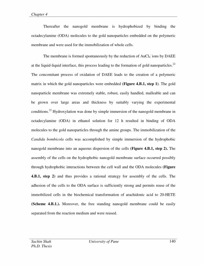
Thereafter the nanogold membrane is hydrophobized by binding the
octadecylamine (ODA) molecules to the gold nanoparticles embedded on the polymeric
membrane and were used for the immobilization of whole cells.
The membrane is formed spontaneously by the reduction of AuCl4- ions by DAEE
at the liquid-liquid interface, this process leading to the formation of gold nanoparticles.22
The concomitant process of oxidation of DAEE leads to the creation of a polymeric
matrix in which the gold nanoparticles were embedded (Figure 4.B.1, step 1). The gold
nanoparticle membrane was extremely stable, robust, easily handled, malleable and can
be grown over large areas and thickness by suitably varying the experimental
conditions.22
Hydroxylation was done by simple immersion of the nanogold membrane in
octadecylamine (ODA) in ethanol solution for 12 h resulted in binding of ODA
molecules to the gold nanoparticles through the amine groups. The immobilization of the
Candida bombicola cells was accomplished by simple immersion of the hydrophobic
nanogold membrane into an aqueous dispersion of the cells (Figure 4.B.1, step 2). The
assembly of the cells on the hydrophobic nanogold membrane surface occurred possibly
through hydrophobic interactions between the cell wall and the ODA molecules (Figure
4.B.1, step 2) and thus provides a rational strategy for assembly of the cells. The
adhesion of the cells to the ODA surface is sufficiently strong and permits reuse of the
immobilized cells in the biochemical transformation of arachidonic acid to 20-HETE
(Scheme 4.B.1.). Moreover, the free standing nanogold membrane could be easily
separated from the reaction medium and were reused.
Chapter 4
Sachin Shah University of Pune
Ph.D. Thesis
141
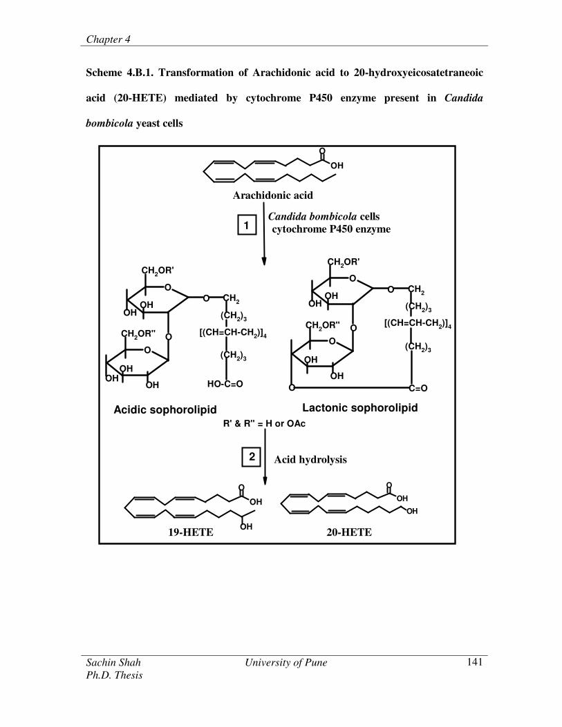
Scheme 4.B.1. Transformation of Arachidonic acid to 20-hydroxyeicosatetraneoic
acid (20-HETE) mediated by cytochrome P450 enzyme present in Candida
bombicola yeast cells
R' & R" = H or OAc
Acidic sophorolipid Lactonic sophorolipid
O
O
O
O CH2
(CH2)3
[(CH=CH-CH2)]4
(CH2)3
HO-C=O
OH
OHOH
OH
OH
O
O
O
O CH2
(CH2)3
[(CH=CH-CH2)]4
(CH2)3
OH
OH
OH
OH
O C=O
CH2OR"
CH2OR'CH2OR'
CH2OR"
OH
O
Arachidonic acid
Candida bombicola cells
cytochrome P450 enzyme
OH
O
OH19-HETE
OH
O
OH
20-HETE
Acid hydrolysis
1
2

Chapter 4
Sachin Shah University of Pune
Ph.D. Thesis
142
Hydrophobic nanogold poly membrane
Figure 4.B.1. Illustration of hydrophobization of nanogold membrane using
octadecylamine and thereafter, immobilization of the Candida bombicola whole cells
on the hydrophobic nanogold membrane
Chapter 4
Sachin Shah University of Pune
Ph.D. Thesis
143
4.B.2. Materials and Methods
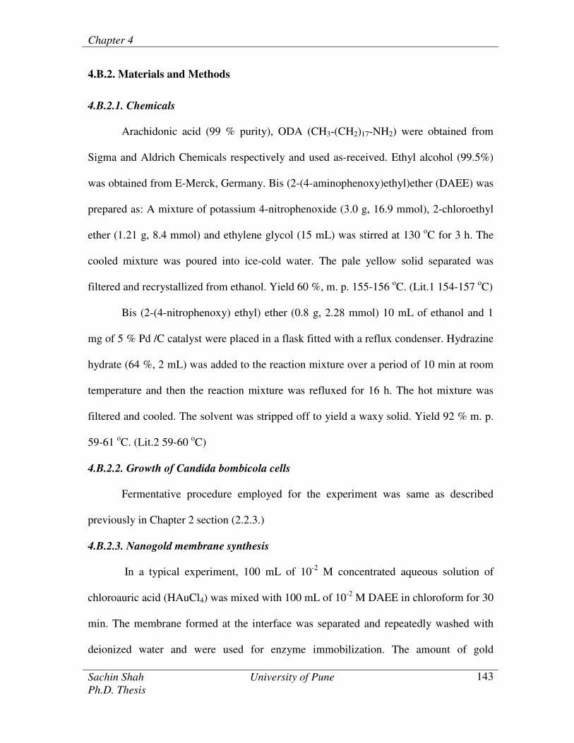
4.B.2.1. Chemicals
Arachidonic acid (99 % purity), ODA (CH3-(CH2)17-NH2) were obtained from
Sigma and Aldrich Chemicals respectively and used as-received. Ethyl alcohol (99.5%)
was obtained from E-Merck, Germany. Bis (2-(4-aminophenoxy)ethyl)ether (DAEE) was
prepared as: A mixture of potassium 4-nitrophenoxide (3.0 g, 16.9 mmol), 2-chloroethyl
ether (1.21 g, 8.4 mmol) and ethylene glycol (15 mL) was stirred at 130 oC for 3 h. The
cooled mixture was poured into ice-cold water. The pale yellow solid separated was
filtered and recrystallized from ethanol. Yield 60 %, m. p. 155-156 oC. (Lit.1 154-157
oC)
Bis (2-(4-nitrophenoxy) ethyl) ether (0.8 g, 2.28 mmol) 10 mL of ethanol and 1
mg of 5 % Pd /C catalyst were placed in a flask fitted with a reflux condenser. Hydrazine
hydrate (64 %, 2 mL) was added to the reaction mixture over a period of 10 min at room
temperature and then the reaction mixture was refluxed for 16 h. The hot mixture was
filtered and cooled. The solvent was stripped off to yield a waxy solid. Yield 92 % m. p.
59-61 oC. (Lit.2 59-60
oC)
4.B.2.2. Growth of Candida bombicola cells
Fermentative procedure employed for the experiment was same as described
previously in Chapter 2 section (2.2.3.)
4.B.2.3. Nanogold membrane synthesis
In a typical experiment, 100 mL of 10-2
M concentrated aqueous solution of
chloroauric acid (HAuCl4) was mixed with 100 mL of 10-2
M DAEE in chloroform for 30
min. The membrane formed at the interface was separated and repeatedly washed with
deionized water and were used for enzyme immobilization. The amount of gold

Chapter 4
Sachin Shah University of Pune
Ph.D. Thesis
144
nanoparticles in the membrane was determined by atomic absorption spectroscopy
(AAS). 10 mg of nanogold membrane was dissolved in 20 mL freshly prepared saturated
I2 solution in KI and volume was made up to 100 mL using deionized water. The solution
was analyzed by a Varian Spectra AA 220 atomic absorption spectrometer (AAS) and
was compared with the standard of gold solution to estimate the weight percent of gold
nanoparticles in the membrane. The gold nanoparticles leached from the polymeric
membrane were also used for the immobilization of whole cells.
4.B.2.4. Hydrophobization of nanogold membrane
10 mg of the nanogold membrane was dispersed in 10-2
M ODA solution prepared
in ethyl alcohol for 12 h. The nanogold membranes were then washed with copious
amount of alcohol and chloroform and dried in the air for further use.
4.B.2.5. UV-Vis spectroscopy studies
UV-visible spectra of gold nanoparticles embedded in the polymeric membrane
were recorded on a quartz substrate using a Jasco V570 UV/VIS/NIR spectrophotometer
operated at a resolution of 1 nm. The probable structure of the nanogold membrane is
illustrated in Figure 4.B.1.
4.B.2.6. Transmission Electron Microscopy (TEM) measurements
TEM measurements were performed on a JEOL Model 1200EX instrument
operated at an accelerating voltage of 120 kV. Samples for TEM analysis were prepared
by transferring a nanogold membrane from the liquid-liquid interface on carbon-coated
TEM copper grids. The mixtures were allowed to dry for 1 min following which the extra
solution was removed using a blotting paper. TEM measurements of gold nanoparticles
leached from the nanogold membranes were also recorded.

Chapter 4
Sachin Shah University of Pune
Ph.D. Thesis
145
4.B.2.7. FTIR measurements
FTIR measurements of the nanogold membrane formed on Si (111) substrates
before and after binding of octadecylamine (ODA) were made on a Perkin Elmer
Spectrum-1 FTIR spectrometer operated in the diffuse reflectance mode at a resolution of
4 cm-1
.
4.B.2.8. Scanning electron microscopy (SEM) and energy dispersive analysis of X-rays
(EDAX) measurements
The immobilization of the Candida bombicola cells on the as prepared and
hydrophobic nanogold membrane were studied by scanning electron microscopy (SEM)
on a Leica Stereoscan-440 electron microscope. Spot-profile energy dispersive analysis
of X-rays (EDAX) measurements were performed to test the faithfulness of cell
immobilization onto the surface of the nanogold membranes using a Phoenix EDAX
attachment connected to the scanning electron microscope. Presence of cells on the
hydrophobic nanogold membrane surface after incubation of the cells after 96 h in the
reaction medium was confirmed by SEM images. SEM images of the gold nanoparticles
leached from the polymeric membrane before and after immobilization of cells were also
recorded.
4.B.2.9. X-ray diffraction measurement (XRD)
XRD measurements of gold nanoparticles bound to the polymeric membrane were
done on a Philips PW 1830 instrument operating at 40 kV and a current of 30 mA with
Cu Kα radiation.

Chapter 4
Sachin Shah University of Pune
Ph.D. Thesis
146
4.B.2.10. Immobilization of Candida bombicola cells onto the hydrophobic nanogold
membrane
20 mg of hydrophobic nanogold membranes were then immersed in an aqueous
dispersion of the cells (~ 108 cells/mL) for 4 h (the optimum time of immersion was
estimated from our earlier experiments).23
The amount of cells immobilized on the
hydrophobic nanogold membrane was estimated from the initial and the final cell counts
after immobilization. To determine the confidence limit, separate measurements were
made for 3 different hydrophobic nanogold membranes. For the reusability of the cells
immobilized on surface of hydrophobic nanogold membrane, it was washed 3 times by
deionized water prior to reuse. Nanogold membrane and gold nanoparticles leached
polymeric membrane were also used for the immobilization of the whole cells.
4.B.2.11. Synthesis of sophorolipids
The yeast cells immobilized on the 20 mg of hydrophobic nanogold membrane were
immersed in the reaction medium containing 5 mL 10% of sterile glucose and 30 mg
arachidonic acid in 200 µl alcohol and was incubated for 96 h at 30 0C under slow
shaking. After the reaction, the supernatant was decanted and used for extracting
sophorolipid.17, 23
4.B.2.12. Hydrolysis of glycolipid and isolation of 20-HETE and 19-HETE
Acid hydrolysis of the sophorolipids under N2 with 1M HCl for 12 h at 25 0C
liberated the fatty acids, which were extracted with an equal volume of in chloroform.17,23
Hydroxylated fatty acids were purified as described in section 4.A.2.8. of chapter 4.

Chapter 4
Sachin Shah University of Pune
Ph.D. Thesis
147
4.B.2.13. Gas chromatography mass spectroscopy (GC-MS)
A Shimadzu GC-MS QP 5050 automated quadrupole mass spectrometer
operating in the electron impact mode was used as described in section 4.A.2.9. of
chapter 4.
4.B.3. Results and discussions
4.B.3.1. Preparation of the nanogold membrane material
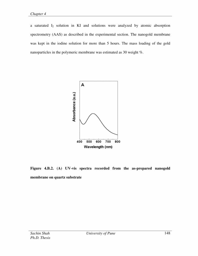
Figure 4.B.2. (A) has shown the UV-Vis spectrum recorded in the transmission
mode from the thin film of nanogold membrane transferred onto a quartz substrate. A
strong absorption band centered at 540 nm is observed. This absorption was due to
excitation of surface plasmons in gold nanoparticles and is responsible for their vivid
pink-purple colour.24
The amine groups of DAEE molecules at the interface were
protonated (pH of HAuCl4 solution ~ 3.2) leading to electrostatic complexation with
AuCl4- ions. That the electrostatic complexation with gold ions was a crucial step in the
formation of the gold nanoparticle membrane was indicated by the control experiment
where a similar interfacial reaction was carried out with the aqueous HAuCl4 solution
maintained at pH 9. At this pH, the amine groups in DAEE would not be protonated and
no membrane formation was observed even after 12 h of reaction. Reduction of
chloroaurate ions takes place at the interface and the oxidized DAEE molecules cap the
spontaneously formed gold nanoparticles preventing their further aggregation. The
nanogold membrane could be formed either by simple cross-linking of the gold
nanoparticles through the terminal groups of oxidized DAEE or through formation of a
polymeric network of the oxidized DAEE molecules. The estimation of gold
nanoparticles in polymeric membrane was done by leaching the gold nanoparticles using
Chapter 4
Sachin Shah University of Pune
Ph.D. Thesis
148
a saturated I2 solution in KI and solutions were analyzed by atomic absorption
spectrometry (AAS) as described in the experimental section. The nanogold membrane
was kept in the iodine solution for more than 5 hours. The mass loading of the gold
nanoparticles in the polymeric membrane was estimated as 30 weight %.
Figure 4.B.2. (A) UV-vis spectra recorded from the as-prepared nanogold
membrane on quartz substrate

Chapter 4
Sachin Shah University of Pune
Ph.D. Thesis
149
4.B.3.2. FTIR studies
Figure 4.B.2. (B) has shown the FTIR spectra recorded from a nanogold
membrane before (curve 1) and after (curve 2) hydrophobization with the ODA
molecules by immersion in 10-2
M ODA solution formed in absolute alcohol for 12 h.
Two prominent features labeled a (2850 cm-1
) and b (2920 cm-1
) were due to the
methylene antisymmetric and symmetric vibrations from the hydrocarbon chains of
octadecylamine molecules bound to the nanogold membranes which were clearly absent
in the as prepared nanogold membranes (curve 1). The frequency of these resonance
indicated the ODA molecules on the gold particle surface were in closed-packed state.
The hydrophobization of gold nanoparticles using octadecylamine molecules and the
binding of the ODA molecules to the nanogold membranes occured through amine
groups.24,
25
Figure 4.B.2. (B) FTIR spectra recorded from the
nanogold membrane before (curve 1) and after
(curve 2) hydrophobization with the
octadecylamine (ODA).

Chapter 4
Sachin Shah University of Pune
Ph.D. Thesis
150
4.B.3.3. XRD and EDAX measurements
Figure 4.B.3. has shown the powder XRD pattern of the nanogold membrane.
The Bragg reflections in the nanogold membrane clearly correspond to presence of
gold.22
The presence of intense (311) reflection in the XRD pattern suggested oriented
growth of the gold nanoparticles in the polymeric membrane along these crystallographic
planes. This confirmed the reduction of chloroaurate ions at the liquid-liquid interface for
the formation of gold nanoparticles. Spot profile EDAX measurements were done on the
nanogold polymeric membrane. The prominent Au signal confirms the fidelity of gold
nanoparticles in the polymeric membrane. However, the chlorine signals were also seen
which were attributed to the unreduced gold ions (AuCl4-) were present in the membrane,
presumably bound to the surface of the gold nanoparticles.
Figure 4.B.3. XRD patterns recorded from the gold nanoparticle membrane. Inset
shows the spot profile EDAX recorded form the gold nanoparticle polymeric
membrane
Chapter 4
Sachin Shah University of Pune
Ph.D. Thesis
151
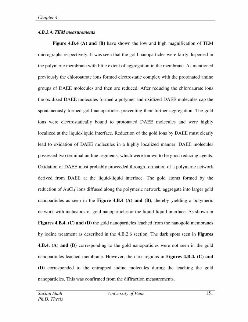
4.B.3.4. TEM measurements
Figure 4.B.4 (A) and (B) have shown the low and high magnification of TEM
micrographs respectively. It was seen that the gold nanoparticles were fairly dispersed in
the polymeric membrane with little extent of aggregation in the membrane. As mentioned
previously the chloroaurate ions formed electrostatic complex with the protonated amine
groups of DAEE molecules and then are reduced. After reducing the chloroaurate ions
the oxidized DAEE molecules formed a polymer and oxidized DAEE molecules cap the
spontaneously formed gold nanoparticles preventing their further aggregation. The gold
ions were electrostatically bound to protonated DAEE molecules and were highly
localized at the liquid-liquid interface. Reduction of the gold ions by DAEE must clearly
lead to oxidation of DAEE molecules in a highly localized manner. DAEE molecules
possessed two terminal aniline segments, which were known to be good reducing agents.
Oxidation of DAEE most probably proceeded through formation of a polymeric network
derived from DAEE at the liquid-liquid interface. The gold atoms formed by the
reduction of AuCl4- ions diffused along the polymeric network, aggregate into larger gold
nanoparticles as seen in the Figure 4.B.4 (A) and (B), thereby yielding a polymeric
network with inclusions of gold nanoparticles at the liquid-liquid interface. As shown in
Figures 4.B.4. (C) and (D) the gold nanoparticles leached from the nanogold membranes
by iodine treatment as described in the 4.B.2.6 section. The dark spots seen in Figures
4.B.4. (A) and (B) corresponding to the gold nanoparticles were not seen in the gold
nanoparticles leached membrane. However, the dark regions in Figures 4.B.4. (C) and
(D) corresponded to the entrapped iodine molecules during the leaching the gold
nanoparticles. This was confirmed from the diffraction measurements.

Chapter 4
Sachin Shah University of Pune
Ph.D. Thesis
152
Figure 4.B.4. (A) and (B) Low and high magnification of TEM micrographs of the free
standing nanogold membrane.
Figures 4.B.4. (C) and (D) The TEM micrographs of the gold nanoparticles leached
from the nanogold membrane

Chapter 4
Sachin Shah University of Pune
Ph.D. Thesis
153
4.B.3.5. SEM
Figure 4.B.5. (A) and (B) have shown the low and high magnification of SEM
images of the nanogold membrane transferred from the liquid-liquid interface on the Si
(111) substrates. It was seen that surface of the nanogold membranes was not smooth.
Moreover, a mesh like structures were seen to form which indicated the simple cross-
linking of the gold nanoparticles through the terminal groups of oxidized DAEE or
through formation of a polymeric network of the oxidized DAEE molecules. As
mentioned briefly earlier, the first step in the formation of the membrane was electrostatic
complexation of AuCl4- ions with protonated amine groups of DAEE molecules at the
liquid-liquid interface and incomplete reduction of the gold ions would explain their
presence in the membrane.
Figure 4.B.5. (A) and (B) Low and high magnification of SEM images of nanogold
membrane synthesized at the liquid-liquid interface and transferred on Si(111) substrate.
Thereafter, this nanogold membrane was immersed in an aqueous suspension of
Candida bombicola cells for 4 h and washed thoroughly prior to imaging by SEM.
Figure 4.B.6. (A) and (B) have shown the SEM images recorded after immobilization of

Chapter 4
Sachin Shah University of Pune
Ph.D. Thesis
154
the Candida bombicola cells on to the hydrophobic nanogold membrane. It was clearly
seen from the SEM images that yeast cells were immobilized extremely faithfully on the
surface of the hydrophobic nanogold membranes. It was well known that these Candida
bombicola cells bound to hydrophobic regions.23
The process of attachment of the cells to
the hydrophobic nanogold membranes has been illustrated in Figure 4.B.1. The low and
high magnification of SEM images recorded after immobilization of the Candida
bombicola cells on to the as prepared nanogold membrane as shown in Figure 4.B.6. (C)
and (D). The density of the cells was less as compared to the cells immobilized on the
hydrophobic nanogold membranes. It was well known that the cells bind to hydrophobic
regions of the surface23
and thus the use of octadecylamine bound to the nanogold
membranes provided the hydrophobic surface for the binding of the Candida bombicola
yeast cells. The process of the attachment of the Candida bombicola yeast cells to the
hydrophobic nanogold membrane has been illustrated in Figure 4.B.1. In order to
understand the interactions between the polymer membrane and the Candida bombicola
yeast cells, the gold nanoparticles leached polymeric membrane was used for the
immobilization of whole cells. Care was taken to wash the polymeric membranes with
copious amount of chloroform and after with deionized water prior to use.

Chapter 4
Sachin Shah University of Pune
Ph.D. Thesis
155
Figure 4.B.6. (A) and (B) The SEM images of the Candida bombicola cell immobilized
on the hydrophobic nanogold membranes.
Figure 4.B.6. (C) and (D). The Candida bombicola cells immobilized on the as prepared
nanogold membrane.

Chapter 4
Sachin Shah University of Pune
Ph.D. Thesis
156
Figure 4.B.7. (A) and (B) have shown the SEM images after immersion of the
gold nanoparticles leached polymeric membrane in aqueous dispersion of Candida
bombicola cells in water for 4 h. It was seen that hardly any number of the cells attached
to the polymeric surface. This confirmed the role of hydrophobic gold nanoparticles
embedded in the polymeric membrane were responsible for the attachment of the cells to
the surface of the nanogold membrane.
Figure 4.B.7. (A) and (B) The SEM images after the immobilization of the Candida
bombicola cells on gold nanoparticles leached polymeric membrane.

Chapter 4
Sachin Shah University of Pune
Ph.D. Thesis
157
4.B.3.6. Synthesis of sophorolipids
Enzymes of the cytochrome P450 monooxygenase family are known to
metabolize hydroxylation of arachidonic acid.26
Expressions of cytochrome P450 have
been documented in liver, kidney and cerebral microvasculars. The earlier biochemical
studies of the cytochrome P450 dependent arachidonic acid monooxygenase reaction
showed that catalytic activity turnover was NADPH dependent.27
This was an advantage
of using whole cells, which limited us to use cofactors such as NADPH for the synthesis
of sophorolipids. Moreover, the advantage in the use of immobilized whole cell system
with cell bound activity was that this obviated the need for enzyme extraction and
removal of the unwanted macromolecules released during the extraction process which
were laborious and expensive.
Candida bombicola yeast cells are known to produce extracellular biosurfactants
known as sophorolipids. Sophorolipids are produced as a mixture of acidic and lactonic
forms. Sophorolipids obtained from the Candida bombicola yeast cells; the lactonic form
represented the largest fraction of the products.28
Scheme 4.B.1. has shown the
transformation of arachidonic acid using Candida bombicola yeast cells to the acidic and
lactonic form of the sophorolipids (Scheme 4.B.1. step 1). Thereafter, acid hydrolysis of
these sophorolipids yields 20-HETE (Scheme 4.B.1. step 2). Nanogold membranes of the
immobilized Candida bombicola yeast cells were reacted with arachidonic acid and the
sophorolipids were isolated from the reaction medium and subjected to acid hydrolysis as
described in details in the experimental section. The amount of cells immobilized on the
20 mg of hydrophobic nanogold membrane was estimated from the initial (~ 108
cells/mL) and the final cell counts after immobilization as described in the experimental
Chapter 4
Sachin Shah University of Pune
Ph.D. Thesis
158
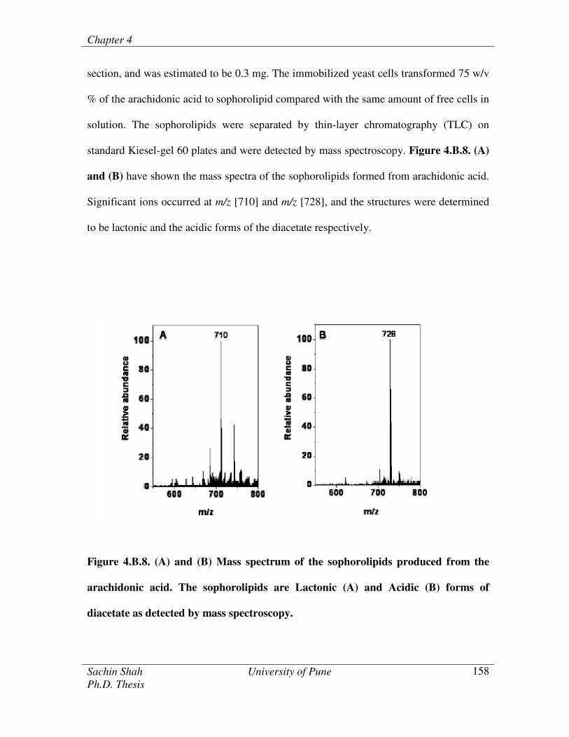
section, and was estimated to be 0.3 mg. The immobilized yeast cells transformed 75 w/v
% of the arachidonic acid to sophorolipid compared with the same amount of free cells in
solution. The sophorolipids were separated by thin-layer chromatography (TLC) on
standard Kiesel-gel 60 plates and were detected by mass spectroscopy. Figure 4.B.8. (A)
and (B) have shown the mass spectra of the sophorolipids formed from arachidonic acid.
Significant ions occurred at m/z [710] and m/z [728], and the structures were determined
to be lactonic and the acidic forms of the diacetate respectively.
Figure 4.B.8. (A) and (B) Mass spectrum of the sophorolipids produced from the
arachidonic acid. The sophorolipids are Lactonic (A) and Acidic (B) forms of
diacetate as detected by mass spectroscopy.

Chapter 4
Sachin Shah University of Pune
Ph.D. Thesis
159
The binding of the cells to the nanogold membranes was strong enough with
marginal leaching of cells during the sophorolipid production. Hence, the immobilized
cells on the hydrophobic nanogold membrane were reused for the five successive reaction
cycles. The yeast cells bound to the hydrophobic nanogold membrane transformed 70
w/v % of the arachidonic acid to sophorolipid for the third cycle of reaction, while
transformed 60 w/v % for the fifth cycle of reaction.
Figure 4.B.9. (A) and (B) have shown the low and high magnification of SEM
images of different regions of Candida bombicola cells immobilized on hydrophobic
nanogold membrane after one cycle of reaction (incubating in the reaction medium for 30
0C for 96 h). SEM images have shown the registry of the cells attached to the surface of
the hydrophobic nanogold membranes. SEM confirms the fidelity of the yeast cells on the
surface of the hydrophobic nanogold membrane after one cycle of reaction.
Figure 4.B.9. (A) and (B) Low and High magnification of SEM images of the
immobilized Candida bombicola cells on the hydrophobic nanogold membrane after
one reaction cycle
Chapter 4
Sachin Shah University of Pune
Ph.D. Thesis
160
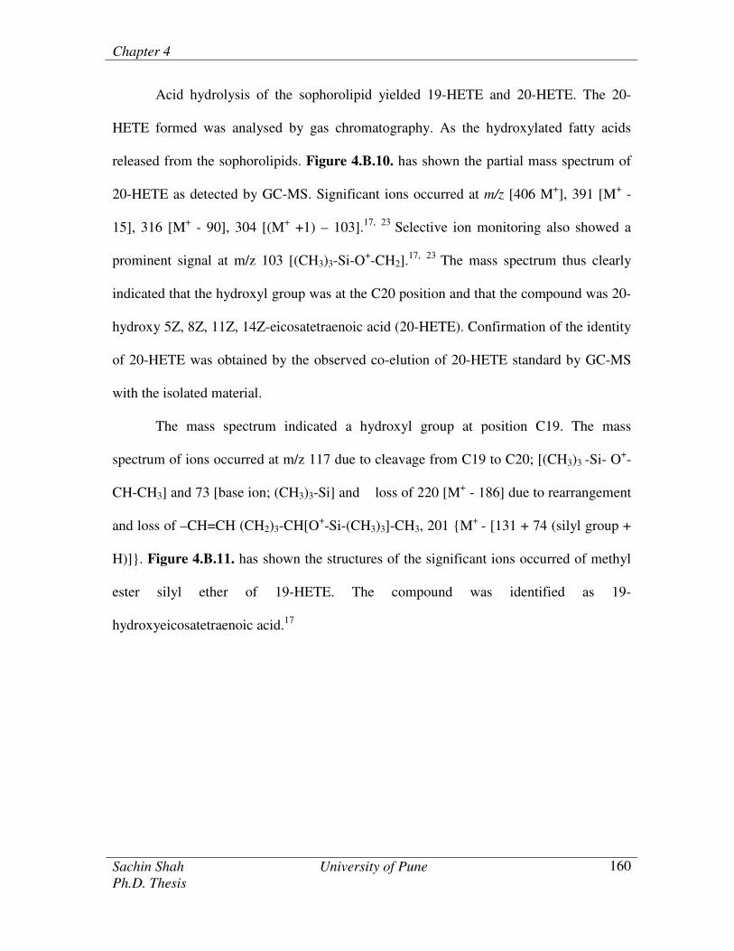
Acid hydrolysis of the sophorolipid yielded 19-HETE and 20-HETE. The 20-
HETE formed was analysed by gas chromatography. As the hydroxylated fatty acids
released from the sophorolipids. Figure 4.B.10. has shown the partial mass spectrum of
20-HETE as detected by GC-MS. Significant ions occurred at m/z [406 M+], 391 [M
+ -
15], 316 [M+ - 90], 304 [(M
+ +1) – 103].
17,
23 Selective ion monitoring also showed a
prominent signal at m/z 103 [(CH3)3-Si-O+-CH2].
17,
23 The mass spectrum thus clearly
indicated that the hydroxyl group was at the C20 position and that the compound was 20-
hydroxy 5Z, 8Z, 11Z, 14Z-eicosatetraenoic acid (20-HETE). Confirmation of the identity
of 20-HETE was obtained by the observed co-elution of 20-HETE standard by GC-MS
with the isolated material.
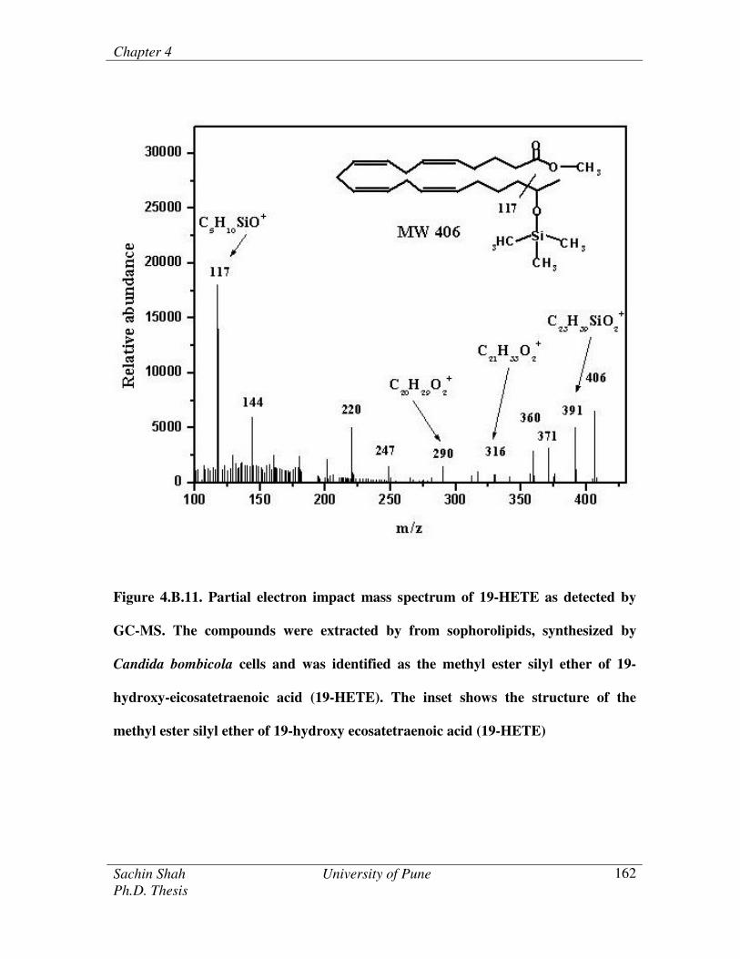
The mass spectrum indicated a hydroxyl group at position C19. The mass
spectrum of ions occurred at m/z 117 due to cleavage from C19 to C20; [(CH3)3 -Si- O+-
CH-CH3] and 73 [base ion; (CH3)3-Si] and loss of 220 [M+ - 186] due to rearrangement
and loss of –CH=CH (CH2)3-CH[O+-Si-(CH3)3]-CH3, 201 {M
+ - [131 + 74 (silyl group +
H)]}. Figure 4.B.11. has shown the structures of the significant ions occurred of methyl
ester silyl ether of 19-HETE. The compound was identified as 19-
hydroxyeicosatetraenoic acid.17

Chapter 4
Sachin Shah University of Pune
Ph.D. Thesis
161
Figure 4.B.10. Partial electron impact mass spectrum of 20-HETE as detected by
GC-MS. The compounds were extracted by from sophorolipids, synthesized by
Candida bombicola cells and was identified as the methyl ester silyl ether of 20-
hydroxy-eicosatetraenoic acid (20-HETE). The inset shows the structure of the
methyl ester silyl ether of 20-hydroxy ecosatetraenoic acid (20-HETE)
Chapter 4
Sachin Shah University of Pune
Ph.D. Thesis
162
Figure 4.B.11. Partial electron impact mass spectrum of 19-HETE as detected by
GC-MS. The compounds were extracted by from sophorolipids, synthesized by
Candida bombicola cells and was identified as the methyl ester silyl ether of 19-
hydroxy-eicosatetraenoic acid (19-HETE). The inset shows the structure of the
methyl ester silyl ether of 19-hydroxy ecosatetraenoic acid (19-HETE)

Chapter 4
Sachin Shah University of Pune
Ph.D. Thesis
163
4.B.4. Conclusion
In this study, we have demonstrated the synthesis of hydrophobic nanogold
membrane and thereafter used for the immobilization of Candida bombicola yeast cells
driven by hydrophobic interactions between the cell walls and the ODA molecules.
Enzymes of the cytochrome P450 monooxygenase family present in the yeast cells were
used for the hydroxylation of arachidonic acid for the production of sophorolipids. The
binding of the cells to the nanogold membrane was strong enough to prevent the leaching
of the whole cells from the surface. Hence the whole cells immobilized membranes were
reused for the biotransformation.

Chapter 4
Sachin Shah University of Pune
Ph.D. Thesis
164
4.3. References
1. a. W. M. Albers, I. Vikholm, T. Viitala, J. Peltonen, 2001, Interfacial and materials
aspects of the immobilization of biomolecules onto solid surfaces, Handbook of
Surfaces and Interfaces of Materials, Ed. H.S. Nalwa, (Academic Press, San Diego)
5, Chapter 1. b. G. Gilardi, A. Fantuzzi, 2001, Manipulating redox systems:
applications to nanotechnology. Trends Biotechnol. 19, 468-476.
2. M. Sastry, 2002, Entrapment of proteins and DNA in thermally evaporated lipid films.
Trends Biotechnol. 20, 185-188.
3. R. Singhvi, A. Kumar, G. P. Lopez, G. N. Stephanopoulos, D. I. C. Wang, G. M.
Whitesides, D. E. Ingber, 1994, Engineering cell shape and function. Science 264,
696-698.
4. G. S. Chen, M. Mrksich, S. Huang, G. M. Whitesides, D. E. Ingber, 1997, Geometric
control of cell life and death. Science 276, 1425.
5. a. P.M. John, R. St. Davis, N. Cady, J. Czajka, C. A. Batt, H. G. Craighead, 1998,
Diffraction-based cell detection using a microcontact printed antibody grating. Anal.
Chem. 70, 1108-1111. b. L. Bousse, 1996, Whole cell biosensors. Sensors and
Actuators B, 34, 270-275.
6. P. Ortenwall, H. Wandenwick, J. Kutti, B. Risberg, 1987, Reduction in deposition of
indium 111 labeled platelets after autologus endothelial cells seeding of Dacron aortic
bifurcation grafts in humans: A preliminary report. J. Vasc. Surg. 6, 17-25.

Chapter 4
Sachin Shah University of Pune
Ph.D. Thesis
165
7. G. Orive, R. M. Hernandez, A. R. Gascon, M. Igartua, J. L. Pedraz, 2002,
Encapsulated cell technology: from research to market. Trends Biotechnol. 20, 382-
387 and references therein.
8. P. Ghosh, M. L. Amirpour, W. M. Lackowski, M. V. Pishko, R. M. Crooks, 1999, A
simple lithographic approach for preparing patterned, micron scale corrals for
controlling cell growth. Angew. Chem. Int. Ed. 38, 1592-1595.
9. C. D. Tidwell, S. I. Ertei, B. D. Ratner, 1997, Endothelial cell growth and protein
adsorption on terminally functionalized, self assembled monolayers of alkanethiolates
on gold. Langmuir 13, 3404-3413.
10. J. H. Stenger, C. S. Georger, C. S. Dulcey, J. J. Hickman, A. S. Rudolph, T. B
Nielsen, S. M. McCort, J. M. Calvert, 1992, Coplaner molecular assemblies of amino
and perfluorinated alkylsilanes: characterization and geometric definition of
mammalian cell adhesion and growth. J. Am. Chem. Soc. 114, 8435-8442.
11. S. Chia, J. Urano, F. Tamanoi, B. Dunn, J. I. Zink, 2000, Patterned hexagonal arrays
for living cells in sol-gel silica films. J. Am. Chem. Soc. 122, 6488-6489.
12. E. Ostuni, R. Kane, C. S. Chen, D. E. Ingber, G. M. Whitesides, 2000, Patterning
mammalian cells using elastomeric membranes. Langmuir 16, 7811-7819.
13. J. T. Groves, L. K. Mahal, C. R. Bertozzi, 2001, Control of cell adhesion and growth
with micropatterned supported lipid membranes. Langmuir 17, 5129-5133.
14. B. Rowan, M. A. Wheeler, R. M. Crooks, 2002, Patterning bacteria within
hyperbranched polymer film templates. Langmuir 18, 9914-9917.
Chapter 4
Sachin Shah University of Pune
Ph.D. Thesis
166
15. P .J. Punt, N. van Biezen, A. Conesa, A. Albers, J. Mangnus, C. van den Hondel,
2002, Filamentous fungi as cell factories for heterologous protein production. Trends
Biotechnol. 20, 200-206.
16. R. K. Hommel, S. Stegner, K. Huse, H. Kleber, 1994, Cytochrome P-450 in the
sophorose-lipid-producing yeast Candida (Torulopsis) apicola. Appl. Microbiol.
Biotechnol. 40, 724-728.
17. A. Prabhune, S. R. Fox, C. Ratledge, 2002, Transformation of arachidonic acid to 19-
hydroxy and 20-hydroxy-eicosatetraenoic acid using Candida bombicola.
Biotechnol. Letters. 24, 1041-1044.
18. M. Alonso-Galicia, J. R. Falck, K. M. Reddy, R. Roman, 1999, 20-HETE agonist and
antagonist in the renal circulation. Am. J. Physiol. 277, 790-796.
19. a. G. Sauerbrey, 1959, Verwendung von Schwingquarzen zur Wägung dünner
Schichten und zur Mikrowägung. Z. Phys. (Munich) 155, 206-222. b. D. A. Buttry,
M. D. Ward, 1992, Measurement of interfacial processes at electrode surfaces with
the electrochemical quartz crystal microbalance. Chem.Rev. 92, 1356-1379. c. J.
Wang, L. M. Frostman, M. D. Ward, 1992, Self-assembled thiol monolayers with
carboxylic acid functionality: measuring pH-dependent phase transitions with the
quartz crystal microbalance . J. Phys. Chem. 96, 5224-5228.
20. J. MacLauf, M. Rigaud 1982, Open tubular glass capillary gas chromatography for
separating eicosanoids. Methods Enzymology 86, 612-631.
21. E. Taillandier, J. Liquier, 1992, Infrared spectroscopy of DNA. Methods Enzymology
211, 307-335.

Chapter 4
Sachin Shah University of Pune
Ph.D. Thesis
167
22. P. R. Selvakannan, P. Senthil Kumar, A. S. More, R. D. Shingte, P. P. Wadgaonkar,
M. Sastry, 2004, Free-standing gold nanoparticle membrane by the spontaneous
reduction of aqueous chloroaurate ions by oxyethylene linkage gearing diamines at a
liquid-liquid interface. Adv. Mater.16, 12, 966-971.
23. S. Phadtare, P. Parekh, S. Shah, A. Tambe, R. Joshi, S. R. Sainkar, A. Prabhune, M.
Sastry, 2003, Candida bombicola Cells Immobilized on Patterned Lipid Films as
Enzyme Sources for the Transformation of Arachidonic Acid to 20-HETE.
Biotechnol. Prog. 19, 1659-1663.
24. a. V. Patil, R. B. Malvankar, M. Sastry, 1999, Role of particle size in individual and
competitive diffusion of carboxylic acid derivatized colloidal gold particles in
thermally evaporated fatty amine films. Langmuir 15, 8197-8206. b. M. M. Alvarez,
J. T. Khoury, T. G. Schaaff, M. N. Shafigullin, I. Vezmar, R. L. 1997, Whetten,
Optical absorption spectra of nanocrystal gold molecules. J. Phys. Chem. B 101,
3706-3712. c. P. P. Selvakannan, S. Mandal, R. Pasricha, S. D. Adyanthaya, M. 2002,
Sastry One-step synthesis of hydrohobized gold nanoparticles of controllable size by
the reduction of aqueous chloroaurate ions by hexadecylaniline at the liquid-liquid
interface. Chem. Commun. 1334-1335.
25. M. Sastry, A. Kumar, P. Mukherjee, 2001, Phase transfer of aqueous colloidal gold
particles into organic solutions containing fatty amine molecules. Coll. Surf. A. 181,
255-259.
26. J. H. Capdevila, L. Parkhill, N. Chacos, R. Okita, B. S. Masters, R. W. Estabrook,
1981, The oxidative metabolism of arachidonic acid by purified cytochrome P-450.
Biochem. Biophys. Res. Commun. 101, 1357-1363.
Chapter 4
Sachin Shah University of Pune
Ph.D. Thesis
168
27. Bolcato, C. A. Frye, R. F. Zemaitis, M. A. Poloyac, S. M. 2003, Determination of 20-
hydroxyeicosatetraenoic acid in microsomal incubates using high-performance liquid
chromatography-mass spectrometry (HPLC-MS). J. Chromatography B, 794, 363-
372.
28. Y. Hu, L. K. Ju, 2001, Purification of lactonic sophorolipids by crystallization. J.
Biotechnol. 87, 263-272.