CHAPTER 48 NERVOUS SYSTEMS Copyright © 2002 Pearson Education, Inc., publishing as Benjamin...
96
CHAPTER 48 NERVOUS SYSTEMS Copyright © 2002 Pearson Education, Inc., publishing as Benjamin Cummings Section A: An Overview Of Nervous Systems 1. Nervous systems perform the three overlapping functions of sensory input, integration, and motor output 2. Networks of neurons with intricate connections form nervous systems
-
Upload
lilian-booth -
Category
Documents
-
view
218 -
download
2
Transcript of CHAPTER 48 NERVOUS SYSTEMS Copyright © 2002 Pearson Education, Inc., publishing as Benjamin...

CHAPTER 48NERVOUS SYSTEMS
Copyright © 2002 Pearson Education, Inc., publishing as Benjamin Cummings
Section A: An Overview Of Nervous Systems
1. Nervous systems perform the three overlapping functions of sensory input,
integration, and motor output
2. Networks of neurons with intricate connections form nervous systems

• Peripheral nervous system (PNS).
• Sensory receptors a responsive to external and iternal stimuli.
• Such sensory input is conveyed to integration centers, where the input is interpreted and associated with a response.
1. Nervous systems perform the three overlapping functions of sensory input, integration, and motor output
Copyright © 2002 Pearson Education, Inc., publishing as Benjamin Cummings

Copyright © 2002 Pearson Education, Inc., publishing as Benjamin Cummings
Fig. 48.1

• Motor output is the conduction of signals from integration centers to effector cells.
• Effector cells carry out the body’s response to a stimulus.
Copyright © 2002 Pearson Education, Inc., publishing as Benjamin Cummings
• The central nervous system (CNS) is responsible for integration.
Copyright © 2002 Pearson Education, Inc., publishing as Benjamin Cummings
• The signals of the nervous system are conducted by nerves.
Copyright © 2002 Pearson Education, Inc., publishing as Benjamin Cummings

• Neuron Structure and Synapses.
• The neuron is the structural and functional unit of the nervous system.
• Nerve impulses are conducted along a neuron.
• Dentrite cell body axon hillock axon
• Some axons are insulated by a myelin sheath.
2. Networks of neurons with intricate connections form nervous systems
Copyright © 2002 Pearson Education, Inc., publishing as Benjamin Cummings

Copyright © 2002 Pearson Education, Inc., publishing as Benjamin Cummings
Fig. 48.2
• Axon endings are called synaptic terminals.
• They contain neurotransmitters which conduct a signal across a synapse.
• A synapse is the junction between a presynaptic and postsynaptic cell.
Copyright © 2002 Pearson Education, Inc., publishing as Benjamin Cummings

• A Simple Nerve Circuit – the Reflex Arc.
• A reflex is an autonomic response.
Copyright © 2002 Pearson Education, Inc., publishing as Benjamin Cummings
Fig. 48.3

• A ganglion is a cluster of nerve cell bodies within the PNS.
• A nucleus is a cluster of nerve cell bodies within the CNS.
Copyright © 2002 Pearson Education, Inc., publishing as Benjamin Cummings
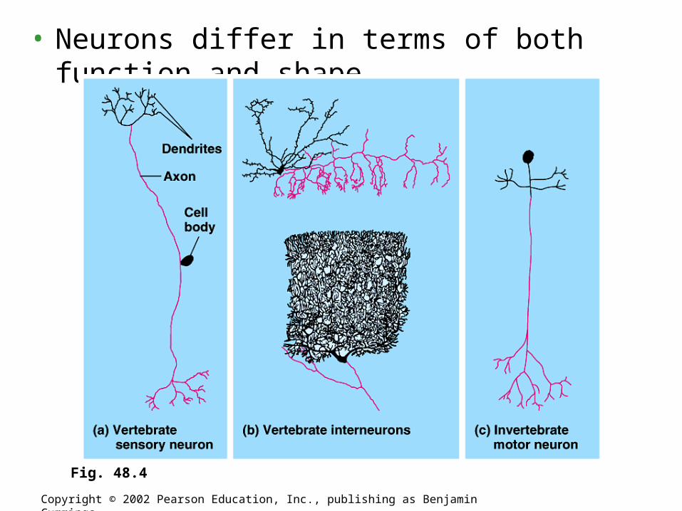
• Neurons differ in terms of both function and shape.
Copyright © 2002 Pearson Education, Inc., publishing as Benjamin Cummings
Fig. 48.4
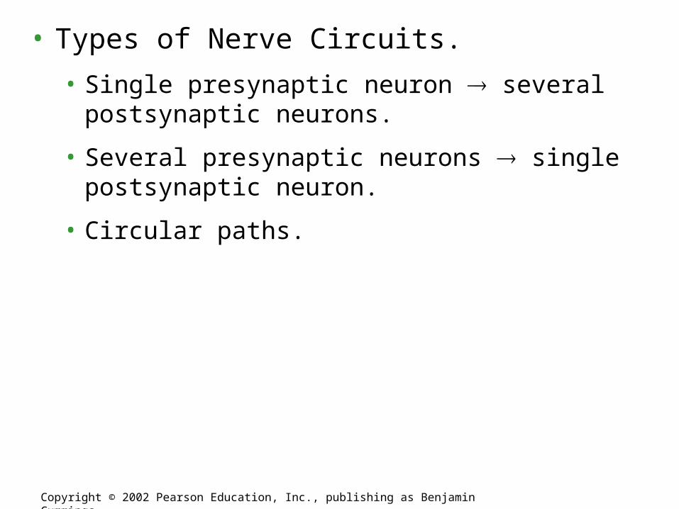
• Types of Nerve Circuits.
• Single presynaptic neuron several postsynaptic neurons.
• Several presynaptic neurons single postsynaptic neuron.
• Circular paths.
Copyright © 2002 Pearson Education, Inc., publishing as Benjamin Cummings

• Supporting Cells (Glia).
• There are several types of glia.
• Astrocytes are found within the CNS.
• Structural and metabolic support.
• By inducing the formation of tight junctions between capillary cells astrocytes help form the blood-brain barrier.
• Like neurons, astrocytes communicate with one another via chemical signals.
Copyright © 2002 Pearson Education, Inc., publishing as Benjamin Cummings

• Oligodendrocytes are found within the CNS.
• Form a myelin sheath by insulating axons.
Copyright © 2002 Pearson Education, Inc., publishing as Benjamin Cummings

• Schwann cells are found within the PNS.
• Form a myelin sheath by insulating axons.
Copyright © 2002 Pearson Education, Inc., publishing as Benjamin Cummings
Fig. 48.5

CHAPTER 48NERVOUS SYSTEMS
Copyright © 2002 Pearson Education, Inc., publishing as Benjamin Cummings
Section B1: The Nature Of Nerve Signals
1. Every cell has a voltage, or membrane potential, across its plasma
membrane
2. Changes in the membrane potential of a neuron give rise to nerve impulses
3. Nerve impulses propagate themselves along an axon

• A membrane potential is a localized electrical gradient across membrane.
• Anions are more concentrated within a cell.
• Cations are more concentrated in the extracellular fluid.
1. Every cell has a voltage, or membrane potential, across its plasma membrane
Copyright © 2002 Pearson Education, Inc., publishing as Benjamin Cummings

• Measuring Membrane Potentials.
Copyright © 2002 Pearson Education, Inc., publishing as Benjamin Cummings
Fig. 48.6a
• An unstimulated cell usually have a resting potential of -70mV.

• How a Cell Maintains a Membrane Potential.
• Cations.
• K+ is the principal intracellular cation.
• Na+ is the principal extracellular cation.
• Anions.
• Proteins, amino acids, sulfate, and phosphate are the principal intracellular anions.
• Cl– is the principal extracellular anion.
Copyright © 2002 Pearson Education, Inc., publishing as Benjamin Cummings
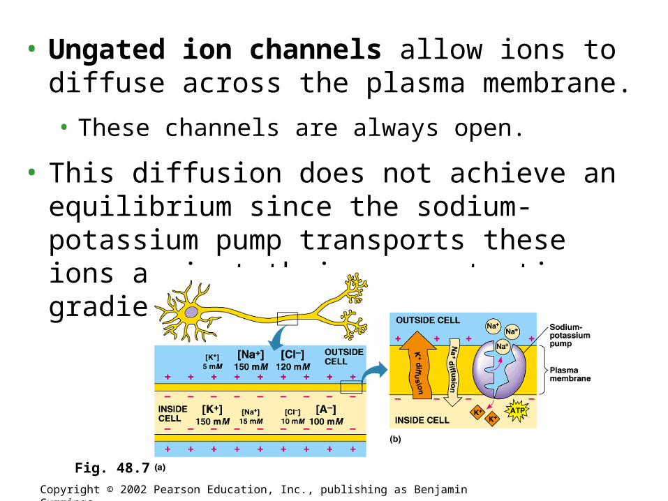
• Ungated ion channels allow ions to diffuse across the plasma membrane.
• These channels are always open.
• This diffusion does not achieve an equilibrium since the sodium-potassium pump transports these ions against their concentration gradients.
Copyright © 2002 Pearson Education, Inc., publishing as Benjamin Cummings
Fig. 48.7

• Excitable cells have the ability to generate large changes in their membrane potentials.
• Gated ion channels open or close in response to stimuli.
• The subsequent diffusion of ions leads to a change in the membrane potential.
2. Changes in the membrane potential of a neuron give rise to nerve impulses
Copyright © 2002 Pearson Education, Inc., publishing as Benjamin Cummings

• Types of gated ions.
• Chemically-gated ion channels open or close in response to a chemical stimulus.
• Voltage-gated ion channels open or close in response to a change in membrane potential.
Copyright © 2002 Pearson Education, Inc., publishing as Benjamin Cummings

• Graded Potentials: Hyperpolarization and Depolarization
• Graded potentials are changes in membrane potential
Copyright © 2002 Pearson Education, Inc., publishing as Benjamin Cummings

• Hyperpolarization.
• Gated K+ channels open K+ diffuses out of the cell the membrane potential becomes more negative.
Copyright © 2002 Pearson Education, Inc., publishing as Benjamin Cummings
Fig. 48.8a

• Depolarization.
• Gated Na+ channels open Na+ diffuses into the cell the membrane potential becomes less negative.
Copyright © 2002 Pearson Education, Inc., publishing as Benjamin Cummings
Fig. 48.8b

• The Action Potential: All or Nothing Depolarization.
• If graded potentials sum to -55mV a threshold potential is achieved.
• This triggers an action potential.
• Axons only.
Copyright © 2002 Pearson Education, Inc., publishing as Benjamin Cummings
Fig. 48.8c

• In the resting state, closed voltage-gated K+ channels open slowly in response to depolarization.
• Voltage-gated Na+ channels have two gates.
• Closed activation gates open rapidly in response to depolarization.
• Open inactivation gates close slowly in response to depolarization.
Copyright © 2002 Pearson Education, Inc., publishing as Benjamin Cummings
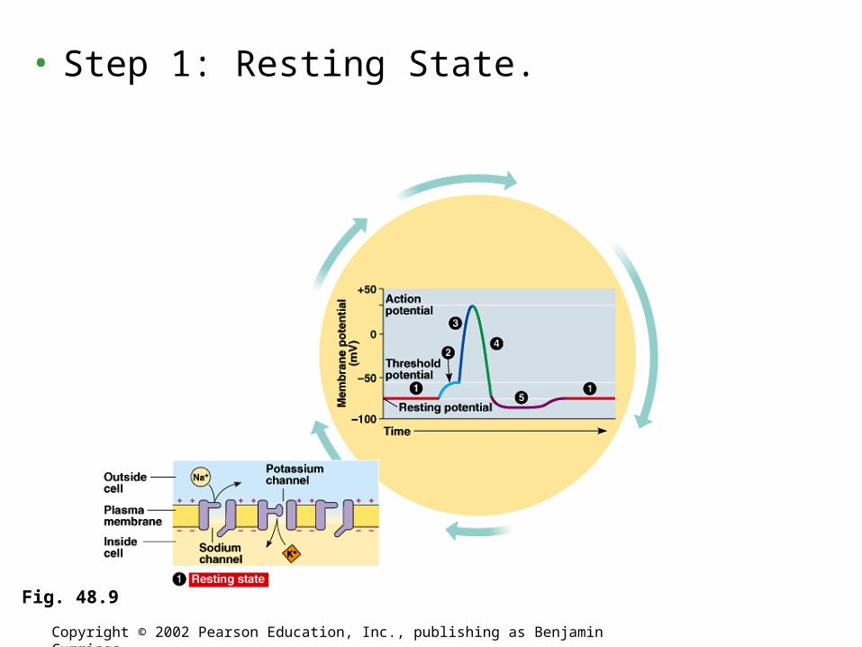
• Step 1: Resting State.
Copyright © 2002 Pearson Education, Inc., publishing as Benjamin Cummings
Fig. 48.9

• Step 2: Threshold.
Copyright © 2002 Pearson Education, Inc., publishing as Benjamin Cummings
Fig. 48.9

• Step 3: Depolarization phase of the action potential.
Copyright © 2002 Pearson Education, Inc., publishing as Benjamin Cummings
Fig. 48.9

• Step 4: Repolarizing phase of the action potential.
Copyright © 2002 Pearson Education, Inc., publishing as Benjamin Cummings
Fig. 48.9

• Step 5: Undershoot.
Copyright © 2002 Pearson Education, Inc., publishing as Benjamin Cummings
Fig. 48.9

• During the undershoot both the Na+ channel’s activation and inactivation gates are closed.
• At this time the neuron cannot depolarize in response to another stimulus: refractory period.
Copyright © 2002 Pearson Education, Inc., publishing as Benjamin Cummings

• The action potential is repeatedly regenerated along the length of the axon.
• An action potential achieved at one region of the membrane is sufficient to depolarize a neighboring region above threshold.
• Thus triggering a new action potential.
• The refractory period assures that impulse conduction is unidirectional.
3. Nerve impulses propagate themselves along an axon
Copyright © 2002 Pearson Education, Inc., publishing as Benjamin Cummings

Copyright © 2002 Pearson Education, Inc., publishing as Benjamin Cummings
Fig. 48.10
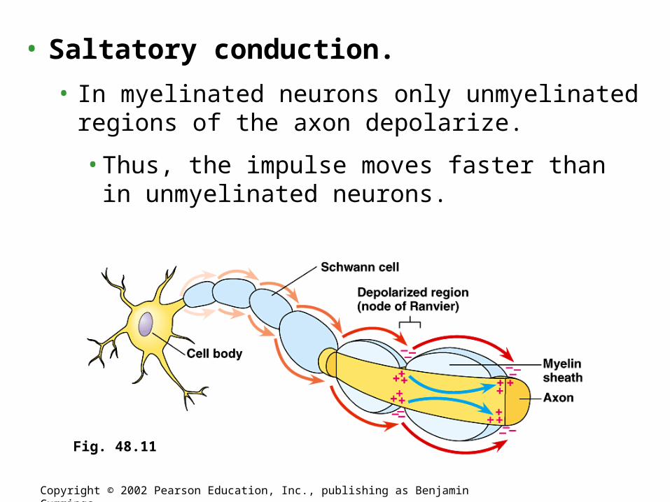
• Saltatory conduction.
• In myelinated neurons only unmyelinated regions of the axon depolarize.
• Thus, the impulse moves faster than in unmyelinated neurons.
Copyright © 2002 Pearson Education, Inc., publishing as Benjamin Cummings
Fig. 48.11

CHAPTER 48NERVOUS SYSTEMS
Copyright © 2002 Pearson Education, Inc., publishing as Benjamin Cummings
Section B2: The Nature Of Nerve Signals
4. Chemical or electrical communication between cells occurs at synapses
5. Neural integration occurs at the cellular level
6. The same neurotransmitter can produce different effects on different types
of cells

• Electrical Synapses.
• Action potentials travels directly from the presynaptic to the postsynaptic cells via gap junctions.
4. Chemical or electrical communication between cells occurs at synapses
Copyright © 2002 Pearson Education, Inc., publishing as Benjamin Cummings

• Chemical Synapses.
• More common than electrical synapses.
• Postsynaptic chemically-gated channels exist for ions such as Na+, K+, and Cl-.
• Depending on which gates open the postsynaptic neuron can depolarize or hyperpolarize.
Copyright © 2002 Pearson Education, Inc., publishing as Benjamin Cummings

Copyright © 2002 Pearson Education, Inc., publishing as Benjamin Cummings
Fig. 48.12

• Excitatory postsynaptic potentials (EPSP) depolarize the postsynaptic neuron.
• The binding of neurotransmitter to postsynaptic receptors opens gated channels that allow Na+ to diffuse into and K+ to diffuse out of the cell.
5. Neural integration occurs at the cellular level
Copyright © 2002 Pearson Education, Inc., publishing as Benjamin Cummings

• Inhibitory postsynaptic potential (IPSP) hyperpolarizes the postsynaptic neuron.
• The binding of neurotransmitter to postsynaptic receptors open gated channels that allow K+ to diffuse out of the cell and/or Cl- to diffuse into the cell.
Copyright © 2002 Pearson Education, Inc., publishing as Benjamin Cummings

• Summation: graded potentials (EPSPs and IPSPs) are summed to either depolarize or hyperpolarize a postsynaptic neuron.
Copyright © 2002 Pearson Education, Inc., publishing as Benjamin Cummings
Fig. 48.14

• Acetylcholine.
• Excitatory to skeletal muscle.
• Inhibitory to cardiac muscle.
• Secreted by the CNS, PNS, and at vertebrate neuromuscular junctions.
6. The same neurotransmitter can produce different effects on different types of cells
Copyright © 2002 Pearson Education, Inc., publishing as Benjamin Cummings

• Biogenic Amines.
• Epinephrine and norepinephrine.
• Can have excitatory or inhibitory effects.
• Secreted by the CNS and PNS.
• Secreted by the adrenal glands.
Copyright © 2002 Pearson Education, Inc., publishing as Benjamin Cummings

• Dopamine
• Generally excitatory; may be inhibitory at some sites.
• Widespread in the brain.
• Affects sleep, mood, attention, and learning.
• Secreted by the CNS and PNS.
• A lack of dopamine in the brain is associated with Parkinson’s disease.
• Excessive dopamine is linked to schizophrenia.
Copyright © 2002 Pearson Education, Inc., publishing as Benjamin Cummings

• Serotonin.
• Generally inhibitory.
• Widespread in the brain.
• Affects sleep, mood, attention, and learning
• Secreted by the CNS.
Copyright © 2002 Pearson Education, Inc., publishing as Benjamin Cummings

• Amino Acids
• Gamma aminobutyric acid (GABA).
• Inhibitory.
• Secreted by the CNS and at invertebrate neuromuscular junctions.
Copyright © 2002 Pearson Education, Inc., publishing as Benjamin Cummings

• Glycine.
• Inhibitory.
• Secreted by the CNS.
Copyright © 2002 Pearson Education, Inc., publishing as Benjamin Cummings

• Glutamate.
• Excitatory.
• Secreted by the CNS and at invertebrate neuromuscular junctions.
Copyright © 2002 Pearson Education, Inc., publishing as Benjamin Cummings

• Aspartate.
• Excitatory.
• Secreted by the CNS.
Copyright © 2002 Pearson Education, Inc., publishing as Benjamin Cummings

• Neuropeptides.
• Substance P.
• Excitatory.
• Secreted by the CNS and PNS.
Copyright © 2002 Pearson Education, Inc., publishing as Benjamin Cummings

• Met-enkephalin (an endorphin).
• Generally inhibitory.
• Secreted by the CNS.
Copyright © 2002 Pearson Education, Inc., publishing as Benjamin Cummings

• Gasses that act as local regulators.
• Nitric oxide.
• Carbon monoxide.
Copyright © 2002 Pearson Education, Inc., publishing as Benjamin Cummings

CHAPTER 48NERVOUS SYSTEMS
Copyright © 2002 Pearson Education, Inc., publishing as Benjamin Cummings
Section C: Evolution And Diversity Of Nervous Systems
1. The ability of cells to respond to the environment has evolved over billions
of years
2. Nervous systems show diverse patterns of organization

1. The ability of cells to respond to the environment has evolved over billions of years
Copyright © 2002 Pearson Education, Inc., publishing as Benjamin Cummings

• Nerve nets.
2. Nervous systems show diverse patterns of organization
Copyright © 2002 Pearson Education, Inc., publishing as Benjamin Cummings
Fig. 48.15a, b

• With cephalization comes more complex nervous systems.
Copyright © 2002 Pearson Education, Inc., publishing as Benjamin Cummings
Fig. 48.15c-h

CHAPTER 48NERVOUS SYSTEMS
Copyright © 2002 Pearson Education, Inc., publishing as Benjamin Cummings
Section D1: Vertebrate Nervous Systems1. Vertebrate nervous systems have central and peripheral components
2. The divisions of the peripheral nervous system interact in maintaining
homeostasis
3. Embryonic development of the vertebrate brain reflects its evolution from three
anterior bulges of the neural tube
4. Evolutionarily older structures of the vertebrate brain regulate essential
automatic and integrative functions

1. Vertebrate nervous systems have central and peripheral components
Copyright © 2002 Pearson Education, Inc., publishing as Benjamin Cummings
• Central nervous system (CNS).• Brain and spinal cord.
• Both contain fluid-filled spaces which contain cerebrospinal fluid (CSF).
• The central canal of the spinal cord is continuous with the ventricles of the brain.
• White matter is composed of bundles of myelinated axons
• Gray matter consists of unmyelinated axons, nuclei, and dendrites.
• Peripheral nervous system.• Everything outside the CNS.

• Structural composition of the PNS.
• Paired cranial nerves that originate in the brain and innervate the head and upper body.
• Paired spinal nerves that originate in the spinal cord and innervate the entire body.
• Ganglia associated with the cranial and spinal nerves.
2. The divisions of the peripheral nervous system interact in maintaining homeostasis
Copyright © 2002 Pearson Education, Inc., publishing as Benjamin Cummings

• Functional composition of the PNS.
Copyright © 2002 Pearson Education, Inc., publishing as Benjamin Cummings
Fig. 48.17

• A closer look at the (often antagonistic) divisions of the autonomic nervous system.
Copyright © 2002 Pearson Education, Inc., publishing as Benjamin Cummings
Fig. 48.18
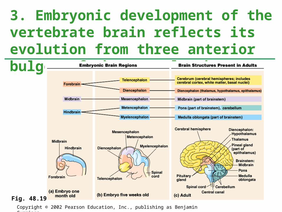
3. Embryonic development of the vertebrate brain reflects its evolution from three anterior bulges of the neural tube
Copyright © 2002 Pearson Education, Inc., publishing as Benjamin Cummings
Fig. 48.19

Copyright © 2002 Pearson Education, Inc., publishing as Benjamin Cummings
Fig. 48.20

• The Brainstem.
• The “lower brain.”
• Consists of the medulla oblongata, pons, and midbrain.
• Derived from the embryonic hindbrain and midbrain.
• Functions in homeostasis, coordination of movement, conduction of impulses to higher brain centers.
4. Evolutionary older structres of the vertebrate brain regulate essential automatic and integrative functions
Copyright © 2002 Pearson Education, Inc., publishing as Benjamin Cummings

• The Medulla and Pons.
• Medulla oblongata.
• Contains nuclei that control visceral (autonomic homeostatic) functions.
• Breathing.
• Heart and blood vessel activity.
• Swallowing.
• Vomiting.
• Digestion.
• Relays information to and from higher brain centers.Copyright © 2002 Pearson Education, Inc., publishing as Benjamin Cummings
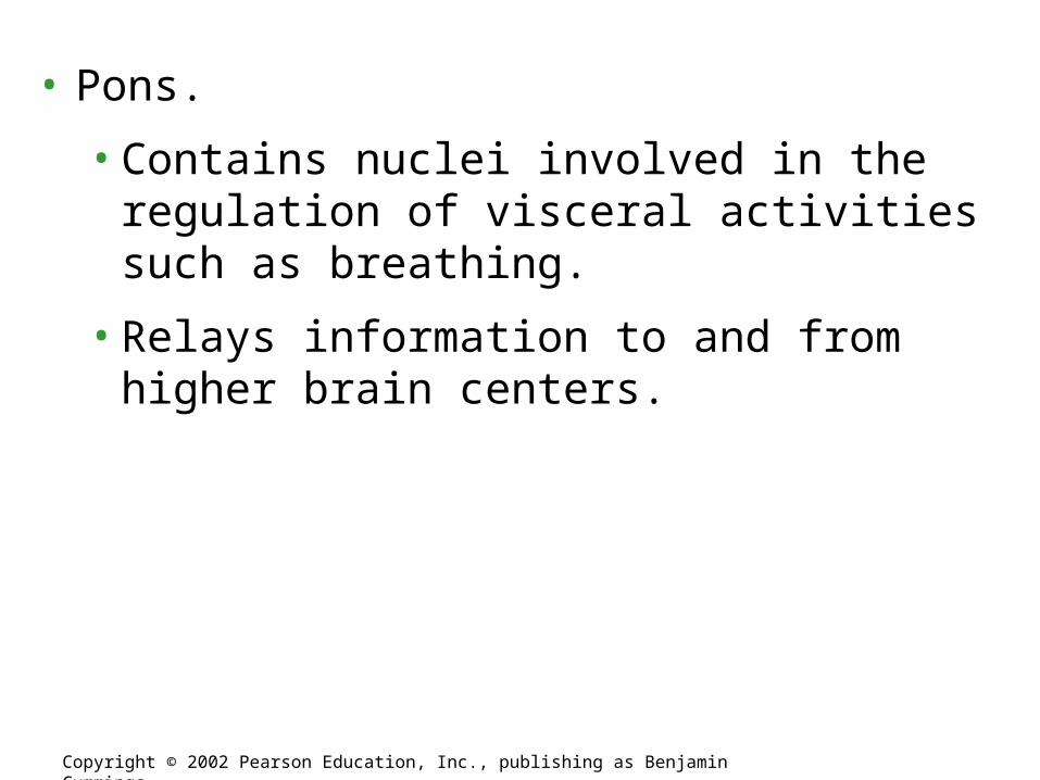
• Pons.
• Contains nuclei involved in the regulation of visceral activities such as breathing.
• Relays information to and from higher brain centers.
Copyright © 2002 Pearson Education, Inc., publishing as Benjamin Cummings

• The Midbrain.
• Contains nuclei involved in the integration of sensory information.
• Superior colliculi are involved in the regulation of visual reflexes.
• Inferior colliculi are involved in the regulation of auditory reflexes.
• Relays information to and from higher brain centers.
Copyright © 2002 Pearson Education, Inc., publishing as Benjamin Cummings

• The Reticular System, Arousal, and Sleep.
• The reticular activating system (RAS) of the reticular formation.
• Regulates sleep and arousal.
• Acts as a sensory filter.
Copyright © 2002 Pearson Education, Inc., publishing as Benjamin Cummings
Fig. 48.21

• Sleep and wakefulness produce patterns of electrical activity in the brain that can be recorded as an electroencephalogram (EEG).
• Most dreaming occurs during REM (rapid eye movement) sleep.
Copyright © 2002 Pearson Education, Inc., publishing as Benjamin Cummings
Fig. 48.22b-d

• The Cerebellum.
• Develops from part of the metencephalon.
• Functions to error-check and coordinate motor activities, and perceptual and cognitive factors.
• Relays sensory information about joints, muscles, sight, and sound to the cerebrum.
• Coordinates motor commands issued by the cerebrum.
Copyright © 2002 Pearson Education, Inc., publishing as Benjamin Cummings

• The thalamus and hypothalamus.
• The epithalamus, thalamus, and hypothalamus are derived from the embryonic diencephalon.
Copyright © 2002 Pearson Education, Inc., publishing as Benjamin Cummings

• Epithalamus.
• Includes a choroid plexus and the pineal gland.
Copyright © 2002 Pearson Education, Inc., publishing as Benjamin Cummings

• Thalamus.
• Relays all sensory information to the cerebrum.
• Contains one nucleus for each type of sensory information.
• Relays motor information from the cerebrum.
• Receives input from the cerebrum.
• Receives input from brain centers involved in the regulation of emotion and arousal.
Copyright © 2002 Pearson Education, Inc., publishing as Benjamin Cummings

• Hypothalamus.
• Regulates autonomic activity.
• Contains nuclei involved in thermoregulation, hunger, thirst, sexual and mating behavior, etc.
• Regulates the pituitary gland.
Copyright © 2002 Pearson Education, Inc., publishing as Benjamin Cummings

• The Hypothalamus and Circadian Rhythms.
• The biological clock is the internal timekeeper.
• The clock’s rhythm usually does not exactly match environmental events.
• Experiments in which humans have been deprived of external cues have shown that biological clock has a period of about 25 hours.
• In mammals, the hypothalamic suprachiasmatic nuclei (SCN) function as a biological clock.
• Produce proteins in response to light/dark cycles.
• This, and other biological clocks, may be responsive to hormonal release, hunger, and various external stimuli.
Copyright © 2002 Pearson Education, Inc., publishing as Benjamin Cummings

CHAPTER 48NERVOUS SYSTEMS
Copyright © 2002 Pearson Education, Inc., publishing as Benjamin Cummings
Section D2: Vertebrate Nervous Systems5. The cerebrum is the most highly evolved structure of the mammalian brain
6. Regions of the cerebrum are specialized for different functions
7. Research on neuron development and neural stem cells may lead to new
approaches for treating CNS injuries and diseases

• The cerebrum is derived from the embryonic telencephalon.
5. The cerebrum is the most highly evolved structure of the mammalian brain
Copyright © 2002 Pearson Education, Inc., publishing as Benjamin Cummings
Fig. 48.24a
• The cerebrum is divided into left and right cerebral hemispheres.
• The corpus callosum is the major connection between the two hemispheres.
• The left hemisphere is primarily responsible for the right side of the body.
• The right hemisphere is primarily responsible for the left side of the body.
• Cerebral cortex: outer covering of gray matter.
• Neocortex: region unique to mammals.
• The more convoluted the surface of the neocortex, the more surface area the more neurons.
• Basal nuclei: internal clusters of nuclei.Copyright © 2002 Pearson Education, Inc., publishing as Benjamin Cummings

• The cerebrum is divided into frontal, temporal, occipital, and parietal lobes.
6. Regions of the cerebrum are specialized for different functions
Copyright © 2002 Pearson Education, Inc., publishing as Benjamin Cummings
Fig. 48.24b

• Frontal lobe.
• Contains the primary motor cortex.
• Parietal lobe.
• Contains the primary somatosensory cortex.
Copyright © 2002 Pearson Education, Inc., publishing as Benjamin Cummings

Copyright © 2002 Pearson Education, Inc., publishing as Benjamin Cummings
Fig. 48.25

• Integrative Function of the Association Areas.
• Much of the cerebrum is given over to association areas.
• Areas where sensory information is integrated and assessed and motor responses are planned.
Copyright © 2002 Pearson Education, Inc., publishing as Benjamin Cummings

• The brain exhibits plasticity of function.
• For example, infants with intractable epilepsy may have an entire cerebral hemisphere removed.
• The remaining hemisphere can provide the function normally provided by both hemispheres.
Copyright © 2002 Pearson Education, Inc., publishing as Benjamin Cummings

• Lateralization of Brain Function.
• The left hemisphere.
• Specializes in language, math, logic operations, and the processing of serial sequences of information, and visual and auditory details.
• Specializes in detailed activities required for motor control.
• The right hemisphere.
• Specializes in pattern recognition, spatial relationships, nonverbal ideation, emotional processing, and the parallel processing of information.
Copyright © 2002 Pearson Education, Inc., publishing as Benjamin Cummings

• Language and Speech.
• Broca’s area.
• Usually located in the left hemisphere’s frontal lobe
• Responsible for speech production.
• Wernicke’s area.
• Usually located in the right hemisphere’s temporal lobe
• Responsible for the comprehension of speech.
• Other speech areas are involved in generating verbs to match nouns, grouping together related words, etc.Copyright © 2002 Pearson Education, Inc., publishing as Benjamin Cummings

• Emotions.
• In mammals, the limbic system is composed of the hippocampus, olfactory cortex, inner portions of the cortex’s lobes, and parts of the thalamus and hypothalamus.
• Mediates basic emotions (fear, anger), involved in emotional bonding, establishes emotional memory
• For example, the amygdala is involved in recognizing the emotional content of facial expression.
Copyright © 2002 Pearson Education, Inc., publishing as Benjamin CummingsFig. 48.27

• Memory and Learning.
• Short-term memory stored in the frontal lobes.
• The establishment of long-term memory involves the hippocampus.
• The transfer of information from short-term to long-term memory.
• Is enhanced by repetition (remember that when you are preparing for an exam).
• Influenced by emotional states mediated by the amygdala.
• Influenced by association with previously stored information.
Copyright © 2002 Pearson Education, Inc., publishing as Benjamin Cummings

• Different types of long-term memories are stored in different regions of the brain.
• Memorization-type memory can be rapid.
• Primarily involves changes in the strength of existing nerve connections.
• Learning of skills and procedures is slower.
• Appears to involves cellular mechanisms similar to those involved in brain growth and development.
Copyright © 2002 Pearson Education, Inc., publishing as Benjamin Cummings

• Functional changes in synapses in synapses of the hippocampus and amygdala are related to memory storage and emotional conditioning.
• Long-term depression (LTD) occurs when a postsynaptic neuron displays decreased responsiveness to action potentials.
• Induced by repeated, weak stimulation.
• Long-term potentiation (LTP) occurs when a postsynaptic neuron displays increased responsiveness to stimuli.
• Induced by brief, repeated action potentials that strongly depolarize the postsynaptic membrane.
• May be associated with memory storage and learning.
Copyright © 2002 Pearson Education, Inc., publishing as Benjamin Cummings

• Human Consciousness.
• Brain imaging can show neural activity associated with:
• Conscious perceptual choice
• Unconscious processing
• Memory retrieval
• Working memory.
• Consciousness appears to be a whole-brain phenomenon.
Copyright © 2002 Pearson Education, Inc., publishing as Benjamin Cummings

• The mammalian PNS has the ability to repair itself, the CNS does not.
• Research on nerve cell development and neural stem cells may be the future of treatment for damage to the CNS.
7. Research on neuron development and neural stem cells may lead to new approaches for treating CNS injuries and diseases
Copyright © 2002 Pearson Education, Inc., publishing as Benjamin Cummings

• Nerve Cell Development.
Copyright © 2002 Pearson Education, Inc., publishing as Benjamin Cummings
Fig. 48.28

• Neural Stem Cells.
• The adult human brain does produce new nerve cells.
• New nerve cells have been found in the hippocampus.
• Since mature human brain cells cannot undergo cell division the new cells must have arisen from stem cells.
Copyright © 2002 Pearson Education, Inc., publishing as Benjamin Cummings