BIOLOGY Chapter 41 OSMOTIC REGULATION AND EXCRETION PowerPoint Image Slideshow.

41 | OSMOTICREGULATION ANDEXCRETION
Figure 41.1 Just as humans recycle what we can and dump the remains into landfills, our bodies use and recycle whatthey can and excrete the remaining waste products. Our bodies’ complex systems have developed ways to treat wasteand maintain a balanced internal environment. (credit: modification of work by Redwin Law)
Chapter Outline
41.1: Osmoregulation and Osmotic Balance
41.2: The Kidneys and Osmoregulatory Organs
41.3: Excretion Systems
41.4: Nitrogenous Wastes
41.5: Hormonal Control of Osmoregulatory Functions
Introduction
The daily intake recommendation for human water consumption is eight to ten glasses of water. In order to achieve ahealthy balance, the human body should excrete the eight to ten glasses of water every day. This occurs via the processesof urination, defecation, sweating and, to a small extent, respiration. The organs and tissues of the human body are soakedin fluids that are maintained at constant temperature, pH, and solute concentration, all crucial elements of homeostasis. Thesolutes in body fluids are mainly mineral salts and sugars, and osmotic regulation is the process by which the mineral saltsand water are kept in balance. Osmotic homeostasis is maintained despite the influence of external factors like temperature,diet, and weather conditions.
Chapter 41 | Osmotic Regulation and Excretion 1189
41.1 | Osmoregulation and Osmotic Balance
By the end of this section, you will be able to:
• Define osmosis and explain its role within molecules
• Explain why osmoregulation and osmotic balance are important body functions
• Describe active transport mechanisms
• Explain osmolarity and the way in which it is measured
• Describe osmoregulators or osmoconformers and how these tools allow animals to adapt to different environments
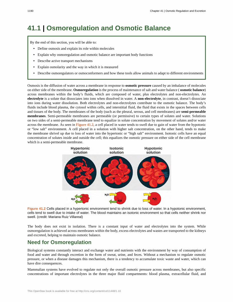
Osmosis is the diffusion of water across a membrane in response to osmotic pressure caused by an imbalance of moleculeson either side of the membrane. Osmoregulation is the process of maintenance of salt and water balance ( osmotic balance)across membranes within the body’s fluids, which are composed of water, plus electrolytes and non-electrolytes. Anelectrolyte is a solute that dissociates into ions when dissolved in water. A non-electrolyte, in contrast, doesn’t dissociateinto ions during water dissolution. Both electrolytes and non-electrolytes contribute to the osmotic balance. The body’sfluids include blood plasma, the cytosol within cells, and interstitial fluid, the fluid that exists in the spaces between cellsand tissues of the body. The membranes of the body (such as the pleural, serous, and cell membranes) are semi-permeablemembranes. Semi-permeable membranes are permeable (or permissive) to certain types of solutes and water. Solutionson two sides of a semi-permeable membrane tend to equalize in solute concentration by movement of solutes and/or wateracross the membrane. As seen in Figure 41.2, a cell placed in water tends to swell due to gain of water from the hypotonicor “low salt” environment. A cell placed in a solution with higher salt concentration, on the other hand, tends to makethe membrane shrivel up due to loss of water into the hypertonic or “high salt” environment. Isotonic cells have an equalconcentration of solutes inside and outside the cell; this equalizes the osmotic pressure on either side of the cell membranewhich is a semi-permeable membrane.
Figure 41.2 Cells placed in a hypertonic environment tend to shrink due to loss of water. In a hypotonic environment,cells tend to swell due to intake of water. The blood maintains an isotonic environment so that cells neither shrink norswell. (credit: Mariana Ruiz Villareal)
The body does not exist in isolation. There is a constant input of water and electrolytes into the system. Whileosmoregulation is achieved across membranes within the body, excess electrolytes and wastes are transported to the kidneysand excreted, helping to maintain osmotic balance.
Need for Osmoregulation
Biological systems constantly interact and exchange water and nutrients with the environment by way of consumption offood and water and through excretion in the form of sweat, urine, and feces. Without a mechanism to regulate osmoticpressure, or when a disease damages this mechanism, there is a tendency to accumulate toxic waste and water, which canhave dire consequences.
Mammalian systems have evolved to regulate not only the overall osmotic pressure across membranes, but also specificconcentrations of important electrolytes in the three major fluid compartments: blood plasma, extracellular fluid, and
1190 Chapter 41 | Osmotic Regulation and Excretion
This OpenStax book is available for free at http://cnx.org/content/col11448/1.10

intracellular fluid. Since osmotic pressure is regulated by the movement of water across membranes, the volume of the fluidcompartments can also change temporarily. Because blood plasma is one of the fluid components, osmotic pressures have adirect bearing on blood pressure.
Transport of Electrolytes across Cell Membranes
Electrolytes, such as sodium chloride, ionize in water, meaning that they dissociate into their component ions. In water,sodium chloride (NaCl), dissociates into the sodium ion (Na+) and the chloride ion (Cl–). The most important ions,whose concentrations are very closely regulated in body fluids, are the cations sodium (Na+), potassium (K+), calcium(Ca+2), magnesium (Mg+2), and the anions chloride (Cl-), carbonate (CO3
-2), bicarbonate (HCO3-), and phosphate(PO3
-).Electrolytes are lost from the body during urination and perspiration. For this reason, athletes are encouraged to replaceelectrolytes and fluids during periods of increased activity and perspiration.
Osmotic pressure is influenced by the concentration of solutes in a solution. It is directly proportional to the number ofsolute atoms or molecules and not dependent on the size of the solute molecules. Because electrolytes dissociate into theircomponent ions, they, in essence, add more solute particles into the solution and have a greater effect on osmotic pressure,per mass than compounds that do not dissociate in water, such as glucose.
Water can pass through membranes by passive diffusion. If electrolyte ions could passively diffuse across membranes, itwould be impossible to maintain specific concentrations of ions in each fluid compartment therefore they require specialmechanisms to cross the semi-permeable membranes in the body. This movement can be accomplished by facilitateddiffusion and active transport. Facilitated diffusion requires protein-based channels for moving the solute. Active transportrequires energy in the form of ATP conversion, carrier proteins, or pumps in order to move ions against the concentrationgradient.
Concept of Osmolality and Milliequivalent
In order to calculate osmotic pressure, it is necessary to understand how solute concentrations are measured. The unit formeasuring solutes is the mole. One mole is defined as the gram molecular weight of the solute. For example, the molecularweight of sodium chloride is 58.44. Thus, one mole of sodium chloride weighs 58.44 grams. The molarity of a solution isthe number of moles of solute per liter of solution. The molality of a solution is the number of moles of solute per kilogramof solvent. If the solvent is water, one kilogram of water is equal to one liter of water. While molarity and molality are usedto express the concentration of solutions, electrolyte concentrations are usually expressed in terms of milliequivalents perliter (mEq/L): the mEq/L is equal to the ion concentration (in millimoles) multiplied by the number of electrical charges onthe ion. The unit of milliequivalent takes into consideration the ions present in the solution (since electrolytes form ions inaqueous solutions) and the charge on the ions.
Thus, for ions that have a charge of one, one milliequivalent is equal to one millimole. For ions that have a charge of two(like calcium), one milliequivalent is equal to 0.5 millimoles. Another unit for the expression of electrolyte concentration isthe milliosmole (mOsm), which is the number of milliequivalents of solute per kilogram of solvent. Body fluids are usuallymaintained within the range of 280 to 300 mOsm.
Osmoregulators and Osmoconformers
Persons lost at sea without any fresh water to drink are at risk of severe dehydration because the human body cannot adaptto drinking seawater, which is hypertonic in comparison to body fluids. Organisms such as goldfish that can tolerate onlya relatively narrow range of salinity are referred to as stenohaline. About 90 percent of all bony fish are restricted to eitherfreshwater or seawater. They are incapable of osmotic regulation in the opposite environment. It is possible, however, fora few fishes like salmon to spend part of their life in fresh water and part in sea water. Organisms like the salmon andmolly that can tolerate a relatively wide range of salinity are referred to as euryhaline organisms. This is possible becausesome fish have evolved osmoregulatory mechanisms to survive in all kinds of aquatic environments. When they live infresh water, their bodies tend to take up water because the environment is relatively hypotonic, as illustrated in Figure41.3a. In such hypotonic environments, these fish do not drink much water. Instead, they pass a lot of very dilute urine,and they achieve electrolyte balance by active transport of salts through the gills. When they move to a hypertonic marineenvironment, these fish start drinking sea water; they excrete the excess salts through their gills and their urine, as illustratedin Figure 41.3b. Most marine invertebrates, on the other hand, may be isotonic with sea water ( osmoconformers). Theirbody fluid concentrations conform to changes in seawater concentration. Cartilaginous fishes’ salt composition of the bloodis similar to bony fishes; however, the blood of sharks contains the organic compounds urea and trimethylamine oxide(TMAO). This does not mean that their electrolyte composition is similar to that of sea water. They achieve isotonicitywith the sea by storing large concentrations of urea. These animals that secrete urea are called ureotelic animals. TMAOstabilizes proteins in the presence of high urea levels, preventing the disruption of peptide bonds that would occur in other
Chapter 41 | Osmotic Regulation and Excretion 1191

animals exposed to similar levels of urea. Sharks are cartilaginous fish with a rectal gland to secrete salt and assist inosmoregulation.
Figure 41.3 Fish are osmoregulators, but must use different mechanisms to survive in (a) freshwater or (b) saltwaterenvironments. (credit: modification of work by Duane Raver, NOAA)
Dialysis TechnicianDialysis is a medical process of removing wastes and excess water from the blood by diffusion andultrafiltration. When kidney function fails, dialysis must be done to artificially rid the body of wastes. This isa vital process to keep patients alive. In some cases, the patients undergo artificial dialysis until they areeligible for a kidney transplant. In others who are not candidates for kidney transplants, dialysis is a life-longnecessity.
Dialysis technicians typically work in hospitals and clinics. While some roles in this field include equipmentdevelopment and maintenance, most dialysis technicians work in direct patient care. Their on-the-jobduties, which typically occur under the direct supervision of a registered nurse, focus on providing dialysistreatments. This can include reviewing patient history and current condition, assessing and responding topatient needs before and during treatment, and monitoring the dialysis process. Treatment may includetaking and reporting a patient’s vital signs and preparing solutions and equipment to ensure accurate andsterile procedures.
1192 Chapter 41 | Osmotic Regulation and Excretion
This OpenStax book is available for free at http://cnx.org/content/col11448/1.10

41.2 | The Kidneys and Osmoregulatory Organs
By the end of this section, you will be able to:
• Explain how the kidneys serve as the main osmoregulatory organs in mammalian systems
• Describe the structure of the kidneys and the functions of the parts of the kidney
• Describe how the nephron is the functional unit of the kidney and explain how it actively filters blood and generatesurine
• Detail the three steps in the formation of urine: glomerular filtration, tubular reabsorption, and tubular secretion
Although the kidneys are the major osmoregulatory organ, the skin and lungs also play a role in the process. Water andelectrolytes are lost through sweat glands in the skin, which helps moisturize and cool the skin surface, while the lungsexpel a small amount of water in the form of mucous secretions and via evaporation of water vapor.
Kidneys: The Main Osmoregulatory Organ
The kidneys, illustrated in Figure 41.4, are a pair of bean-shaped structures that are located just below and posterior to theliver in the peritoneal cavity. The adrenal glands sit on top of each kidney and are also called the suprarenal glands. Kidneysfilter blood and purify it. All the blood in the human body is filtered many times a day by the kidneys; these organs useup almost 25 percent of the oxygen absorbed through the lungs to perform this function. Oxygen allows the kidney cellsto efficiently manufacture chemical energy in the form of ATP through aerobic respiration. The filtrate coming out of thekidneys is called urine.
Figure 41.4 Kidneys filter the blood, producing urine that is stored in the bladder prior to elimination through theurethra. (credit: modification of work by NCI)
Kidney Structure
Externally, the kidneys are surrounded by three layers, illustrated in Figure 41.5. The outermost layer is a tough connectivetissue layer called the renal fascia. The second layer is called the perirenal fat capsule, which helps anchor the kidneysin place. The third and innermost layer is the renal capsule. Internally, the kidney has three regions—an outer cortex, amedulla in the middle, and the renal pelvis in the region called the hilum of the kidney. The hilum is the concave part ofthe bean-shape where blood vessels and nerves enter and exit the kidney; it is also the point of exit for the ureters. The renalcortex is granular due to the presence of nephrons—the functional unit of the kidney. The medulla consists of multiplepyramidal tissue masses, called the renal pyramids. In between the pyramids are spaces called renal columns through
Chapter 41 | Osmotic Regulation and Excretion 1193
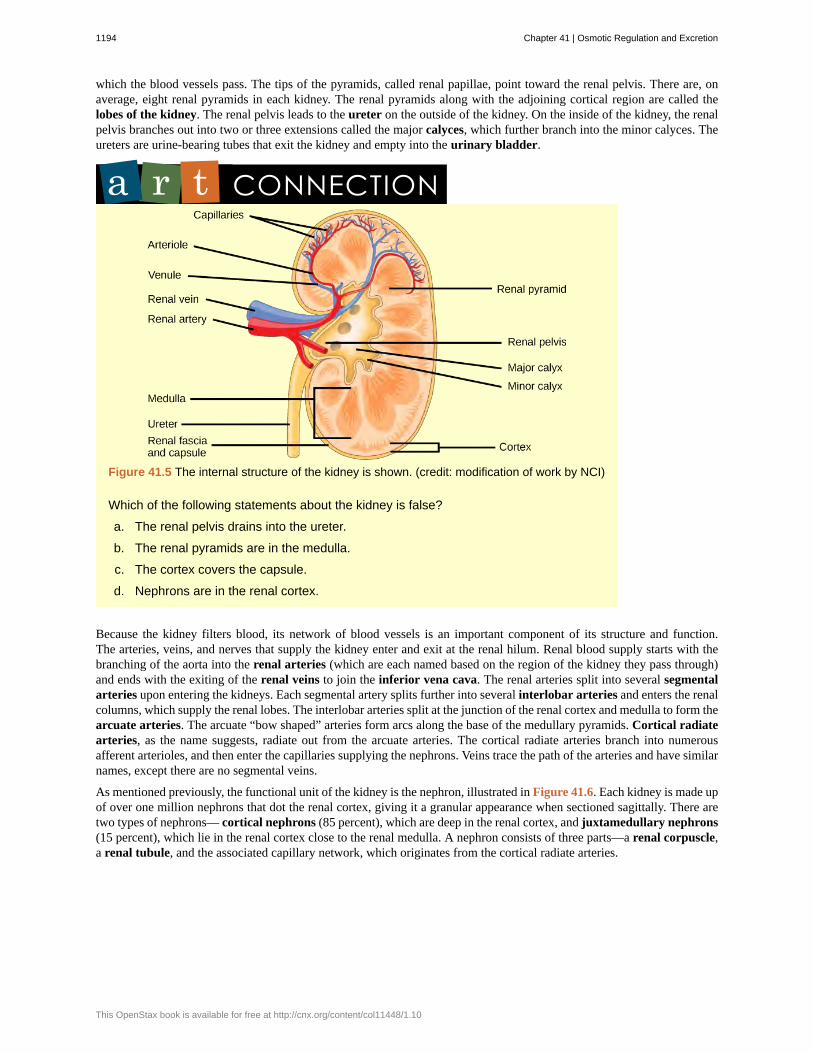
which the blood vessels pass. The tips of the pyramids, called renal papillae, point toward the renal pelvis. There are, onaverage, eight renal pyramids in each kidney. The renal pyramids along with the adjoining cortical region are called thelobes of the kidney. The renal pelvis leads to the ureter on the outside of the kidney. On the inside of the kidney, the renalpelvis branches out into two or three extensions called the major calyces, which further branch into the minor calyces. Theureters are urine-bearing tubes that exit the kidney and empty into the urinary bladder.
Figure 41.5 The internal structure of the kidney is shown. (credit: modification of work by NCI)
Which of the following statements about the kidney is false?
a. The renal pelvis drains into the ureter.
b. The renal pyramids are in the medulla.
c. The cortex covers the capsule.
d. Nephrons are in the renal cortex.
Because the kidney filters blood, its network of blood vessels is an important component of its structure and function.The arteries, veins, and nerves that supply the kidney enter and exit at the renal hilum. Renal blood supply starts with thebranching of the aorta into the renal arteries (which are each named based on the region of the kidney they pass through)and ends with the exiting of the renal veins to join the inferior vena cava. The renal arteries split into several segmentalarteries upon entering the kidneys. Each segmental artery splits further into several interlobar arteries and enters the renalcolumns, which supply the renal lobes. The interlobar arteries split at the junction of the renal cortex and medulla to form thearcuate arteries. The arcuate “bow shaped” arteries form arcs along the base of the medullary pyramids. Cortical radiatearteries, as the name suggests, radiate out from the arcuate arteries. The cortical radiate arteries branch into numerousafferent arterioles, and then enter the capillaries supplying the nephrons. Veins trace the path of the arteries and have similarnames, except there are no segmental veins.
As mentioned previously, the functional unit of the kidney is the nephron, illustrated in Figure 41.6. Each kidney is made upof over one million nephrons that dot the renal cortex, giving it a granular appearance when sectioned sagittally. There aretwo types of nephrons— cortical nephrons (85 percent), which are deep in the renal cortex, and juxtamedullary nephrons(15 percent), which lie in the renal cortex close to the renal medulla. A nephron consists of three parts—a renal corpuscle,a renal tubule, and the associated capillary network, which originates from the cortical radiate arteries.
1194 Chapter 41 | Osmotic Regulation and Excretion
This OpenStax book is available for free at http://cnx.org/content/col11448/1.10

Figure 41.6 The nephron is the functional unit of the kidney. The glomerulus and convoluted tubules are locatedin the kidney cortex, while collecting ducts are located in the pyramids of the medulla. (credit: modification of workby NIDDK)
Which of the following statements about the nephron is false?
a. The collecting duct empties into the distal convoluted tubule.
b. The Bowman’s capsule surrounds the glomerulus.
c. The loop of Henle is between the proximal and distal convoluted tubules.
d. The loop of Henle empties into the distal convoluted tubule.
Renal Corpuscle
The renal corpuscle, located in the renal cortex, is made up of a network of capillaries known as the glomerulus and thecapsule, a cup-shaped chamber that surrounds it, called the glomerular or Bowman's capsule.
Renal Tubule
The renal tubule is a long and convoluted structure that emerges from the glomerulus and can be divided into three partsbased on function. The first part is called the proximal convoluted tubule (PCT) due to its proximity to the glomerulus;it stays in the renal cortex. The second part is called the loop of Henle, or nephritic loop, because it forms a loop (withdescending and ascending limbs) that goes through the renal medulla. The third part of the renal tubule is called the distalconvoluted tubule (DCT) and this part is also restricted to the renal cortex. The DCT, which is the last part of the nephron,connects and empties its contents into collecting ducts that line the medullary pyramids. The collecting ducts amass contentsfrom multiple nephrons and fuse together as they enter the papillae of the renal medulla.
Capillary Network within the Nephron
The capillary network that originates from the renal arteries supplies the nephron with blood that needs to be filtered. Thebranch that enters the glomerulus is called the afferent arteriole. The branch that exits the glomerulus is called the efferentarteriole. Within the glomerulus, the network of capillaries is called the glomerular capillary bed. Once the efferent arterioleexits the glomerulus, it forms the peritubular capillary network, which surrounds and interacts with parts of the renaltubule. In cortical nephrons, the peritubular capillary network surrounds the PCT and DCT. In juxtamedullary nephrons, theperitubular capillary network forms a network around the loop of Henle and is called the vasa recta.
Chapter 41 | Osmotic Regulation and Excretion 1195

Go to this website (http://openstaxcollege.org/l/kidney_section) to see another coronal section of the kidney and toexplore an animation of the workings of nephrons.
Kidney Function and Physiology
Kidneys filter blood in a three-step process. First, the nephrons filter blood that runs through the capillary network in theglomerulus. Almost all solutes, except for proteins, are filtered out into the glomerulus by a process called glomerularfiltration. Second, the filtrate is collected in the renal tubules. Most of the solutes get reabsorbed in the PCT by a processcalled tubular reabsorption. In the loop of Henle, the filtrate continues to exchange solutes and water with the renalmedulla and the peritubular capillary network. Water is also reabsorbed during this step. Then, additional solutes andwastes are secreted into the kidney tubules during tubular secretion, which is, in essence, the opposite process to tubularreabsorption. The collecting ducts collect filtrate coming from the nephrons and fuse in the medullary papillae. From here,the papillae deliver the filtrate, now called urine, into the minor calyces that eventually connect to the ureters through therenal pelvis. This entire process is illustrated in Figure 41.7.
1196 Chapter 41 | Osmotic Regulation and Excretion
This OpenStax book is available for free at http://cnx.org/content/col11448/1.10

Figure 41.7 Each part of the nephron performs a different function in filtering waste and maintaining homeostaticbalance. (1) The glomerulus forces small solutes out of the blood by pressure. (2) The proximal convoluted tubulereabsorbs ions, water, and nutrients from the filtrate into the interstitial fluid, and actively transports toxins and drugsfrom the interstitial fluid into the filtrate. The proximal convoluted tubule also adjusts blood pH by selectively secretingammonia (NH3) into the filtrate, where it reacts with H+ to form NH4
+. The more acidic the filtrate, the more ammoniais secreted. (3) The descending loop of Henle is lined with cells containing aquaporins that allow water to pass fromthe filtrate into the interstitial fluid. (4) In the thin part of the ascending loop of Henle, Na+ and Cl- ions diffuse into theinterstitial fluid. In the thick part, these same ions are actively transported into the interstitial fluid. Because salt but notwater is lost, the filtrate becomes more dilute as it travels up the limb. (5) In the distal convoluted tubule, K+ and H+ ionsare selectively secreted into the filtrate, while Na+, Cl-, and HCO3
- ions are reabsorbed to maintain pH and electrolytebalance in the blood. (6) The collecting duct reabsorbs solutes and water from the filtrate, forming dilute urine. (credit:modification of work by NIDDK)
Glomerular Filtration
Glomerular filtration filters out most of the solutes due to high blood pressure and specialized membranes in the afferentarteriole. The blood pressure in the glomerulus is maintained independent of factors that affect systemic blood pressure.The “leaky” connections between the endothelial cells of the glomerular capillary network allow solutes to pass througheasily. All solutes in the glomerular capillaries, except for macromolecules like proteins, pass through by passive diffusion.There is no energy requirement at this stage of the filtration process. Glomerular filtration rate (GFR) is the volumeof glomerular filtrate formed per minute by the kidneys. GFR is regulated by multiple mechanisms and is an importantindicator of kidney function.
To learn more about the vascular system of kidneys, click through this review (http://openstaxcollege.org/l/kidneys)and the steps of blood flow.
Chapter 41 | Osmotic Regulation and Excretion 1197

Tubular Reabsorption and Secretion
Tubular reabsorption occurs in the PCT part of the renal tubule. Almost all nutrients are reabsorbed, and this occurs eitherby passive or active transport. Reabsorption of water and some key electrolytes are regulated and can be influenced byhormones. Sodium (Na+) is the most abundant ion and most of it is reabsorbed by active transport and then transported tothe peritubular capillaries. Because Na+ is actively transported out of the tubule, water follows it to even out the osmoticpressure. Water is also independently reabsorbed into the peritubular capillaries due to the presence of aquaporins, or waterchannels, in the PCT. This occurs due to the low blood pressure and high osmotic pressure in the peritubular capillaries.However, every solute has a transport maximum and the excess is not reabsorbed.
In the loop of Henle, the permeability of the membrane changes. The descending limb is permeable to water, not solutes;the opposite is true for the ascending limb. Additionally, the loop of Henle invades the renal medulla, which is naturallyhigh in salt concentration and tends to absorb water from the renal tubule and concentrate the filtrate. The osmotic gradientincreases as it moves deeper into the medulla. Because two sides of the loop of Henle perform opposing functions, asillustrated in Figure 41.8, it acts as a countercurrent multiplier. The vasa recta around it acts as the countercurrentexchanger.
Figure 41.8 The loop of Henle acts as a countercurrent multiplier that uses energy to create concentrationgradients. The descending limb is water permeable. Water flows from the filtrate to the interstitial fluid, soosmolality inside the limb increases as it descends into the renal medulla. At the bottom, the osmolality is higherinside the loop than in the interstitial fluid. Thus, as filtrate enters the ascending limb, Na+ and Cl- ions exit throughion channels present in the cell membrane. Further up, Na+ is actively transported out of the filtrate and Cl- follows.Osmolarity is given in units of milliosmoles per liter (mOsm/L).
Loop diuretics are drugs sometimes used to treat hypertension. These drugs inhibit the reabsorption of Na+
and Cl- ions by the ascending limb of the loop of Henle. A side effect is that they increase urination. Why doyou think this is the case?
By the time the filtrate reaches the DCT, most of the urine and solutes have been reabsorbed. If the body requires additionalwater, all of it can be reabsorbed at this point. Further reabsorption is controlled by hormones, which will be discussed in alater section. Excretion of wastes occurs due to lack of reabsorption combined with tubular secretion. Undesirable productslike metabolic wastes, urea, uric acid, and certain drugs, are excreted by tubular secretion. Most of the tubular secretionhappens in the DCT, but some occurs in the early part of the collecting duct. Kidneys also maintain an acid-base balance bysecreting excess H+ ions.
1198 Chapter 41 | Osmotic Regulation and Excretion
This OpenStax book is available for free at http://cnx.org/content/col11448/1.10

Although parts of the renal tubules are named proximal and distal, in a cross-section of the kidney, the tubules are placedclose together and in contact with each other and the glomerulus. This allows for exchange of chemical messengers betweenthe different cell types. For example, the DCT ascending limb of the loop of Henle has masses of cells called macula densa,which are in contact with cells of the afferent arterioles called juxtaglomerular cells. Together, the macula densa andjuxtaglomerular cells form the juxtaglomerular complex (JGC). The JGC is an endocrine structure that secretes the enzymerenin and the hormone erythropoietin. When hormones trigger the macula densa cells in the DCT due to variations in bloodvolume, blood pressure, or electrolyte balance, these cells can immediately communicate the problem to the capillaries inthe afferent and efferent arterioles, which can constrict or relax to change the glomerular filtration rate of the kidneys.
NephrologistA nephrologist studies and deals with diseases of the kidneys—both those that cause kidney failure (such asdiabetes) and the conditions that are produced by kidney disease (such as hypertension). Blood pressure,blood volume, and changes in electrolyte balance come under the purview of a nephrologist.
Nephrologists usually work with other physicians who refer patients to them or consult with them aboutspecific diagnoses and treatment plans. Patients are usually referred to a nephrologist for symptoms suchas blood or protein in the urine, very high blood pressure, kidney stones, or renal failure.
Nephrology is a subspecialty of internal medicine. To become a nephrologist, medical school is followedby additional training to become certified in internal medicine. An additional two or more years is spentspecifically studying kidney disorders and their accompanying effects on the body.
41.3 | Excretion Systems
By the end of this section, you will be able to:
• Explain how vacuoles, present in microorganisms, work to excrete waste
• Describe the way in which flame cells and nephridia in worms perform excretory functions and maintain osmoticbalance
• Explain how insects use Malpighian tubules to excrete wastes and maintain osmotic balance
Microorganisms and invertebrate animals use more primitive and simple mechanisms to get rid of their metabolic wastesthan the mammalian system of kidney and urinary function. Three excretory systems evolved in organisms before complexkidneys: vacuoles, flame cells, and Malpighian tubules.
Contractile Vacuoles in Microorganisms
The most fundamental feature of life is the presence of a cell. In other words, a cell is the simplest functional unit of alife. Bacteria are unicellular, prokaryotic organisms that have some of the least complex life processes in place; however,prokaryotes such as bacteria do not contain membrane-bound vacuoles. The cells of microorganisms like bacteria, protozoa,and fungi are bound by cell membranes and use them to interact with the environment. Some cells, including someleucocytes in humans, are able to engulf food by endocytosis—the formation of vesicles by involution of the cell membranewithin the cells. The same vesicles are able to interact and exchange metabolites with the intracellular environment. Insome unicellular eukaryotic organisms such as the amoeba, shown in Figure 41.9, cellular wastes and excess water areexcreted by exocytosis, when the contractile vacuoles merge with the cell membrane and expel wastes into the environment.Contractile vacuoles (CV) should not be confused with vacuoles, which store food or water.
Chapter 41 | Osmotic Regulation and Excretion 1199

Figure 41.9 Some unicellular organisms, such as the amoeba, ingest food by endocytosis. The food vesicle fuses witha lysosome, which digests the food. Waste is excreted by exocytosis.
Flame Cells of Planaria and Nephridia of Worms
As multi-cellular systems evolved to have organ systems that divided the metabolic needs of the body, individual organsevolved to perform the excretory function. Planaria are flatworms that live in fresh water. Their excretory system consists oftwo tubules connected to a highly branched duct system. The cells in the tubules are called flame cells (or protonephridia)because they have a cluster of cilia that looks like a flickering flame when viewed under the microscope, as illustratedin Figure 41.10a. The cilia propel waste matter down the tubules and out of the body through excretory pores that openon the body surface; cilia also draw water from the interstitial fluid, allowing for filtration. Any valuable metabolites arerecovered by reabsorption. Flame cells are found in flatworms, including parasitic tapeworms and free-living planaria. Theyalso maintain the organism’s osmotic balance.
Figure 41.10 In the excretory system of the (a) planaria, cilia of flame cells propel waste through a tubule formedby a tube cell. Tubules are connected into branched structures that lead to pores located all along the sides of thebody. The filtrate is secreted through these pores. In (b) annelids such as earthworms, nephridia filter fluid from thecoelom, or body cavity. Beating cilia at the opening of the nephridium draw water from the coelom into a tubule. Asthe filtrate passes down the tubules, nutrients and other solutes are reabsorbed by capillaries. Filtered fluid containingnitrogenous and other wastes is stored in a bladder and then secreted through a pore in the side of the body.
Earthworms (annelids) have slightly more evolved excretory structures called nephridia, illustrated in Figure 41.10b. Apair of nephridia is present on each segment of the earthworm. They are similar to flame cells in that they have a tubulewith cilia. Excretion occurs through a pore called the nephridiopore. They are more evolved than the flame cells in thatthey have a system for tubular reabsorption by a capillary network before excretion.
Malpighian Tubules of Insects
Malpighian tubules are found lining the gut of some species of arthropods, such as the bee illustrated in Figure 41.11. Theyare usually found in pairs and the number of tubules varies with the species of insect. Malpighian tubules are convoluted,which increases their surface area, and they are lined with microvilli for reabsorption and maintenance of osmotic balance.Malpighian tubules work cooperatively with specialized glands in the wall of the rectum. Body fluids are not filtered asin the case of nephridia; urine is produced by tubular secretion mechanisms by the cells lining the Malpighian tubules thatare bathed in hemolymph (a mixture of blood and interstitial fluid that is found in insects and other arthropods as well asmost mollusks). Metabolic wastes like uric acid freely diffuse into the tubules. There are exchange pumps lining the tubules,which actively transport H+ ions into the cell and K+ or Na+ ions out; water passively follows to form urine. The secretion
1200 Chapter 41 | Osmotic Regulation and Excretion
This OpenStax book is available for free at http://cnx.org/content/col11448/1.10

of ions alters the osmotic pressure which draws water, electrolytes, and nitrogenous waste (uric acid) into the tubules. Waterand electrolytes are reabsorbed when these organisms are faced with low-water environments, and uric acid is excreted as athick paste or powder. Not dissolving wastes in water helps these organisms to conserve water; this is especially importantfor life in dry environments.
Figure 41.11 Malpighian tubules of insects and other terrestrial arthropods remove nitrogenous wastes and othersolutes from the hemolymph. Na+ and/or K+ ions are actively transported into the lumen of the tubules. Water thenenters the tubules via osmosis, forming urine. The urine passes through the intestine, and into the rectum. There,nutrients diffuse back into the hemolymph. Na+ and/or K+ ions are pumped into the hemolymph, and water follows.The concentrated waste is then excreted.
Visit this site (http://openstaxcollege.org/l/malpighian) to see a dissected cockroach, including a close-up look at itsMalpighian tubules.
41.4 | Nitrogenous Wastes
By the end of this section, you will be able to:
• Compare and contrast the way in which aquatic animals and terrestrial animals can eliminate toxic ammonia fromtheir systems
• Compare the major byproduct of ammonia metabolism in vertebrate animals to that of birds, insects, and reptiles
Of the four major macromolecules in biological systems, both proteins and nucleic acids contain nitrogen. During thecatabolism, or breakdown, of nitrogen-containing macromolecules, carbon, hydrogen, and oxygen are extracted and storedin the form of carbohydrates and fats. Excess nitrogen is excreted from the body. Nitrogenous wastes tend to form toxicammonia, which raises the pH of body fluids. The formation of ammonia itself requires energy in the form of ATP andlarge quantities of water to dilute it out of a biological system. Animals that live in aquatic environments tend to releaseammonia into the water. Animals that excrete ammonia are said to be ammonotelic. Terrestrial organisms have evolvedother mechanisms to excrete nitrogenous wastes. The animals must detoxify ammonia by converting it into a relativelynontoxic form such as urea or uric acid. Mammals, including humans, produce urea, whereas reptiles and many terrestrial
Chapter 41 | Osmotic Regulation and Excretion 1201

invertebrates produce uric acid. Animals that secrete urea as the primary nitrogenous waste material are called ureotelicanimals.
Nitrogenous Waste in Terrestrial Animals: The Urea Cycle
The urea cycle is the primary mechanism by which mammals convert ammonia to urea. Urea is made in the liver andexcreted in urine. The overall chemical reaction by which ammonia is converted to urea is 2 NH3 (ammonia) + CO2 + 3ATP + H2O → H2N-CO-NH2 (urea) + 2 ADP + 4 Pi + AMP.
The urea cycle utilizes five intermediate steps, catalyzed by five different enzymes, to convert ammonia to urea, as shownin Figure 41.12. The amino acid L-ornithine gets converted into different intermediates before being regenerated at the endof the urea cycle. Hence, the urea cycle is also referred to as the ornithine cycle. The enzyme ornithine transcarbamylasecatalyzes a key step in the urea cycle and its deficiency can lead to accumulation of toxic levels of ammonia in the body.The first two reactions occur in the mitochondria and the last three reactions occur in the cytosol. Urea concentration in theblood, called blood urea nitrogen or BUN, is used as an indicator of kidney function.
Figure 41.12 The urea cycle converts ammonia to urea.
1202 Chapter 41 | Osmotic Regulation and Excretion
This OpenStax book is available for free at http://cnx.org/content/col11448/1.10

Excretion of Nitrogenous WasteThe theory of evolution proposes that life started in an aquatic environment. It is not surprising to seethat biochemical pathways like the urea cycle evolved to adapt to a changing environment when terrestriallife forms evolved. Arid conditions probably led to the evolution of the uric acid pathway as a means ofconserving water.
Nitrogenous Waste in Birds and Reptiles: Uric Acid
Birds, reptiles, and most terrestrial arthropods convert toxic ammonia to uric acid or the closely related compound guanine(guano) instead of urea. Mammals also form some uric acid during breakdown of nucleic acids. Uric acid is a compoundsimilar to purines found in nucleic acids. It is water insoluble and tends to form a white paste or powder; it is excretedby birds, insects, and reptiles. Conversion of ammonia to uric acid requires more energy and is much more complex thanconversion of ammonia to urea Figure 41.13.
Figure 41.13 Nitrogenous waste is excreted in different forms by different species. These include (a) ammonia, (b)urea, and (c) uric acid. (credit a: modification of work by Eric Engbretson, USFWS; credit b: modification of work by B."Moose" Peterson, USFWS; credit c: modification of work by Dave Menke, USFWS)
Chapter 41 | Osmotic Regulation and Excretion 1203
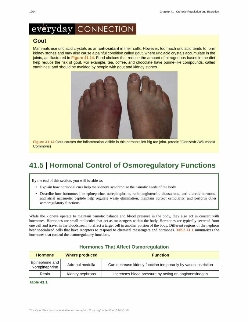
GoutMammals use uric acid crystals as an antioxidant in their cells. However, too much uric acid tends to formkidney stones and may also cause a painful condition called gout, where uric acid crystals accumulate in thejoints, as illustrated in Figure 41.14. Food choices that reduce the amount of nitrogenous bases in the diethelp reduce the risk of gout. For example, tea, coffee, and chocolate have purine-like compounds, calledxanthines, and should be avoided by people with gout and kidney stones.
Figure 41.14 Gout causes the inflammation visible in this person’s left big toe joint. (credit: "Gonzosft"/WikimediaCommons)
41.5 | Hormonal Control of Osmoregulatory Functions
By the end of this section, you will be able to:
• Explain how hormonal cues help the kidneys synchronize the osmotic needs of the body
• Describe how hormones like epinephrine, norepinephrine, renin-angiotensin, aldosterone, anti-diuretic hormone,and atrial natriuretic peptide help regulate waste elimination, maintain correct osmolarity, and perform otherosmoregulatory functions
While the kidneys operate to maintain osmotic balance and blood pressure in the body, they also act in concert withhormones. Hormones are small molecules that act as messengers within the body. Hormones are typically secreted fromone cell and travel in the bloodstream to affect a target cell in another portion of the body. Different regions of the nephronbear specialized cells that have receptors to respond to chemical messengers and hormones. Table 41.1 summarizes thehormones that control the osmoregulatory functions.
Hormones That Affect Osmoregulation
Hormone Where produced Function
Epinephrine andNorepinephrine Adrenal medulla Can decrease kidney function temporarily by vasoconstriction
Renin Kidney nephrons Increases blood pressure by acting on angiotensinogen
Table 41.1
1204 Chapter 41 | Osmotic Regulation and Excretion
This OpenStax book is available for free at http://cnx.org/content/col11448/1.10
Hormones That Affect Osmoregulation
Hormone Where produced Function
Angiotensin Liver Angiotensin II affects multiple processes and increases bloodpressure
Aldosterone Adrenal cortex Prevents loss of sodium and water
Anti-diuretichormone
(vasopressin)
Hypothalamus (storedin the posterior
pituitary)Prevents water loss
Atrial natriureticpeptide Heart atrium
Decreases blood pressure by acting as a vasodilator andincreasing glomerular filtration rate; decreases sodium
reabsorption in kidneys
Table 41.1
Epinephrine and Norepinephrine
Epinephrine and norepinephrine are released by the adrenal medulla and nervous system respectively. They are the flight/fight hormones that are released when the body is under extreme stress. During stress, much of the body’s energy is usedto combat imminent danger. Kidney function is halted temporarily by epinephrine and norepinephrine. These hormonesfunction by acting directly on the smooth muscles of blood vessels to constrict them. Once the afferent arterioles areconstricted, blood flow into the nephrons stops. These hormones go one step further and trigger the renin-angiotensin-aldosterone system.
Renin-Angiotensin-Aldosterone
The renin-angiotensin-aldosterone system, illustrated in Figure 41.15 proceeds through several steps to produceangiotensin II, which acts to stabilize blood pressure and volume. Renin (secreted by a part of the juxtaglomerularcomplex) is produced by the granular cells of the afferent and efferent arterioles. Thus, the kidneys control bloodpressure and volume directly. Renin acts on angiotensinogen, which is made in the liver and converts it to angiotensin I.Angiotensin converting enzyme (ACE) converts angiotensin I to angiotensin II. Angiotensin II raises blood pressure byconstricting blood vessels. It also triggers the release of the mineralocorticoid aldosterone from the adrenal cortex, which inturn stimulates the renal tubules to reabsorb more sodium. Angiotensin II also triggers the release of anti-diuretic hormone(ADH) from the hypothalamus, leading to water retention in the kidneys. It acts directly on the nephrons and decreasesglomerular filtration rate. Medically, blood pressure can be controlled by drugs that inhibit ACE (called ACE inhibitors).
Chapter 41 | Osmotic Regulation and Excretion 1205
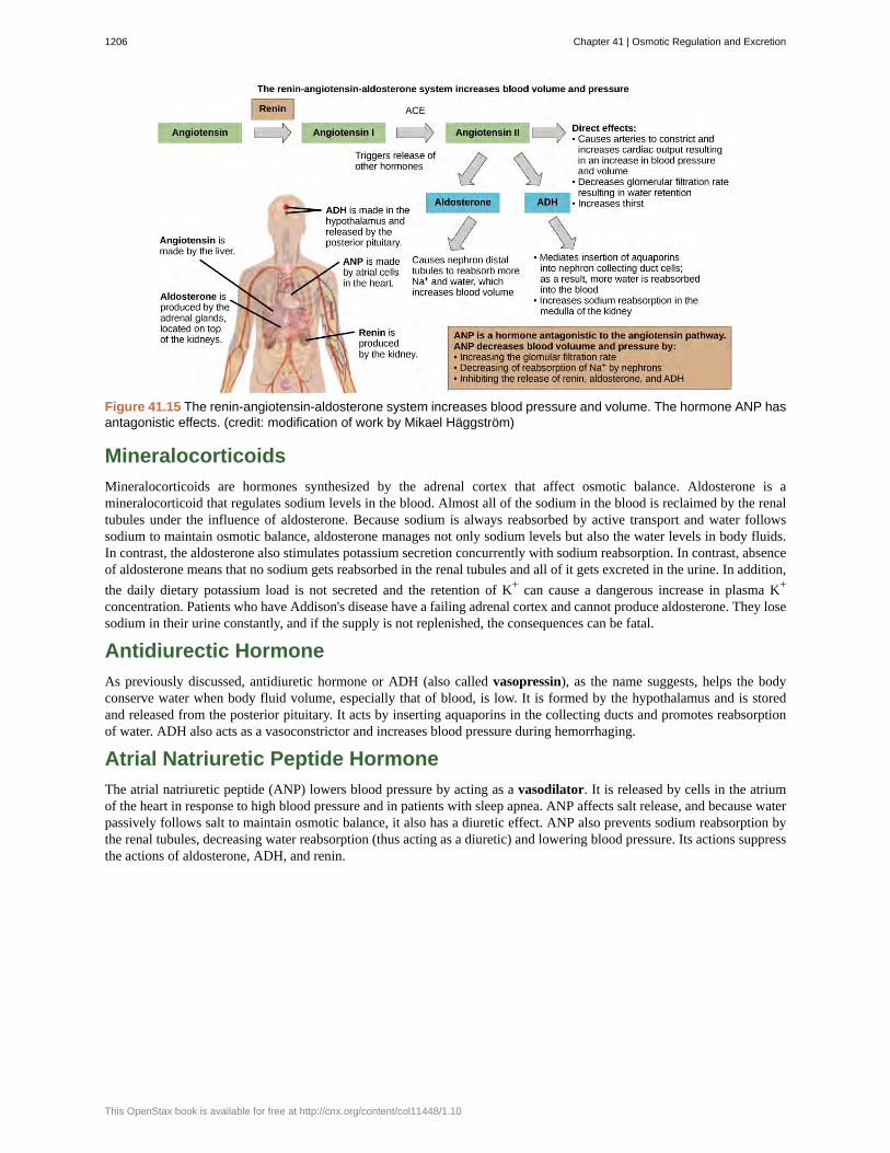
Figure 41.15 The renin-angiotensin-aldosterone system increases blood pressure and volume. The hormone ANP hasantagonistic effects. (credit: modification of work by Mikael Häggström)
Mineralocorticoids
Mineralocorticoids are hormones synthesized by the adrenal cortex that affect osmotic balance. Aldosterone is amineralocorticoid that regulates sodium levels in the blood. Almost all of the sodium in the blood is reclaimed by the renaltubules under the influence of aldosterone. Because sodium is always reabsorbed by active transport and water followssodium to maintain osmotic balance, aldosterone manages not only sodium levels but also the water levels in body fluids.In contrast, the aldosterone also stimulates potassium secretion concurrently with sodium reabsorption. In contrast, absenceof aldosterone means that no sodium gets reabsorbed in the renal tubules and all of it gets excreted in the urine. In addition,the daily dietary potassium load is not secreted and the retention of K+ can cause a dangerous increase in plasma K+
concentration. Patients who have Addison's disease have a failing adrenal cortex and cannot produce aldosterone. They losesodium in their urine constantly, and if the supply is not replenished, the consequences can be fatal.
Antidiurectic Hormone
As previously discussed, antidiuretic hormone or ADH (also called vasopressin), as the name suggests, helps the bodyconserve water when body fluid volume, especially that of blood, is low. It is formed by the hypothalamus and is storedand released from the posterior pituitary. It acts by inserting aquaporins in the collecting ducts and promotes reabsorptionof water. ADH also acts as a vasoconstrictor and increases blood pressure during hemorrhaging.
Atrial Natriuretic Peptide Hormone
The atrial natriuretic peptide (ANP) lowers blood pressure by acting as a vasodilator. It is released by cells in the atriumof the heart in response to high blood pressure and in patients with sleep apnea. ANP affects salt release, and because waterpassively follows salt to maintain osmotic balance, it also has a diuretic effect. ANP also prevents sodium reabsorption bythe renal tubules, decreasing water reabsorption (thus acting as a diuretic) and lowering blood pressure. Its actions suppressthe actions of aldosterone, ADH, and renin.
1206 Chapter 41 | Osmotic Regulation and Excretion
This OpenStax book is available for free at http://cnx.org/content/col11448/1.10

afferent arteriole
ammonia
ammonotelic
angiotensin converting enzyme (ACE)
angiotensin I
angiotensin II
anti-diuretic hormone (ADH)
antioxidant
arcuate artery
ascending limb
blood urea nitrogen (BUN)
Bowman's capsule
calyx
cortex (animal)
cortical nephron
cortical radiate artery
countercurrent exchanger
countercurrent multiplier
descending limb
distal convoluted tubule (DCT)
efferent arteriole
electrolyte
flame cell
glomerular filtration
glomerular filtration rate (GFR)
glomerulus (renal)
hilum
inferior vena cava
interlobar artery
juxtaglomerular cell
juxtamedullary nephron
KEY TERMS
arteriole that branches from the cortical radiate artery and enters the glomerulus
compound made of one nitrogen atom and three hydrogen atoms
describes an animal that excretes ammonia as the primary waste material
enzyme that converts angiotensin I to angiotensin II
product in the renin-angiotensin-aldosterone pathway
molecule that affects different organs to increase blood pressure
hormone that prevents the loss of water
agent that prevents cell destruction by reactive oxygen species
artery that branches from the interlobar artery and arches over the base of the renal pyramids
part of the loop of Henle that ascends from the renal medulla to the renal cortex
estimate of urea in the blood and an indicator of kidney function
structure that encloses the glomerulus
structure that connects the renal pelvis to the renal medulla
outer layer of an organ like the kidney or adrenal gland
nephron that lies in the renal cortex
artery that radiates from the arcuate arteries into the renal cortex
peritubular capillary network that allows exchange of solutes and water from the renaltubules
osmotic gradient in the renal medulla that is responsible for concentration of urine
part of the loop of Henle that descends from the renal cortex into the renal medulla
part of the renal tubule that is the most distant from the glomerulus
arteriole that exits from the glomerulus
solute that breaks down into ions when dissolved in water
(also, protonephridia) excretory cell found in flatworms
filtration of blood in the glomerular capillary network into the glomerulus
amount of filtrate formed by the glomerulus per minute
part of the renal corpuscle that contains the capillary network
region in the renal pelvis where blood vessels, nerves, and ureters bunch before entering or exiting the kidney
one of the main veins in the human body
artery that branches from the segmental artery and travels in between the renal lobes
cell in the afferent and efferent arterioles that responds to stimuli from the macula densa
nephron that lies in the cortex but close to the renal medulla
Chapter 41 | Osmotic Regulation and Excretion 1207
kidney
lobes of the kidney
loop of Henle
macula densa
Malpighian tubule
medulla
microvilli
molality
molarity
mole
nephridia
nephridiopore
nephron
non-electrolyte
osmoconformer
osmoregulation
osmoregulator
osmotic balance
osmotic pressure
perirenal fat capsule
peritubular capillary network
proximal convoluted tubule (PCT)
renal artery
renal capsule
renal column
renal corpuscle
renal fascia
renal pelvis
renal pyramid
renal tubule
organ that performs excretory and osmoregulatory functions
renal pyramid along with the adjoining cortical region
part of the renal tubule that loops into the renal medulla
group of cells that senses changes in sodium ion concentration; present in parts of the renal tubule andcollecting ducts
excretory tubules found in arthropods
middle layer of an organ like the kidney or adrenal gland
cellular processes that increase the surface area of cells
number of moles of solute per kilogram of solvent
number of moles of solute per liter of solution
gram equivalent of the molecular weight of a substance
excretory structures found in annelids
pore found at the end of nephridia
functional unit of the kidney
solute that does not break down into ions when dissolved in water
organism that changes its tonicity based on its environment
mechanism by which water and solute concentrations are maintained at desired levels
organism that maintains its tonicity irrespective of its environment
balance of the amount of water and salt input and output to and from a biological system withoutdisturbing the desired osmotic pressure and solute concentration in every compartment
pressure exerted on a membrane to equalize solute concentration on either side
fat layer that suspends the kidneys
capillary network that surrounds the renal tubule after the efferent artery exits theglomerulus
part of the renal tubule that lies close to the glomerulus
branch of the artery that enters the kidney
layer that encapsulates the kidneys
area of the kidney through which the interlobar arteries travel in the process of supplying blood to the renallobes
glomerulus and the Bowman's capsule together
connective tissue that supports the kidneys
region in the kidney where the calyces join the ureters
conical structure in the renal medulla
tubule of the nephron that arises from the glomerulus
1208 Chapter 41 | Osmotic Regulation and Excretion
This OpenStax book is available for free at http://cnx.org/content/col11448/1.10
renal vein
renin-angiotensin-aldosterone
segmental artery
semi-permeable membrane
transport maximum
tubular reabsorption
tubular secretion
urea cycle
ureotelic
ureter
uric acid
urinary bladder
urine
vasa recta
vasodilator
vasopressin
branch of a vein that exits the kidney and joins the inferior vena cava
biochemical pathway that activates angiotensin II, which increases blood pressure
artery that branches from the renal artery
membrane that allows only certain solutes to pass through
maximum amount of solute that can be transported out of the renal tubules during reabsorption
reclamation of water and solutes that got filtered out in the glomerulus
process of secretion of wastes that do not get reabsorbed
pathway by which ammonia is converted to urea
describes animals that secrete urea as the primary nitrogenous waste material
urine-bearing tube coming out of the kidney; carries urine to the bladder
byproduct of ammonia metabolism in birds, insects, and reptiles
structure that the ureters empty the urine into; stores urine
filtrate produced by kidneys that gets excreted out of the body
peritubular network that surrounds the loop of Henle of the juxtamedullary nephrons
compound that increases the diameter of blood vessels
another name for anti-diuretic hormone
CHAPTER SUMMARY
41.1 Osmoregulation and Osmotic Balance
Solute concentrations across a semi-permeable membranes influence the movement of water and solutes across themembrane. It is the number of solute molecules and not the molecular size that is important in osmosis. Osmoregulationand osmotic balance are important bodily functions, resulting in water and salt balance. Not all solutes can pass through asemi-permeable membrane. Osmosis is the movement of water across the membrane. Osmosis occurs to equalize thenumber of solute molecules across a semi-permeable membrane by the movement of water to the side of higher soluteconcentration. Facilitated diffusion utilizes protein channels to move solute molecules from areas of higher to lowerconcentration while active transport mechanisms are required to move solutes against concentration gradients. Osmolarityis measured in units of milliequivalents or milliosmoles, both of which take into consideration the number of soluteparticles and the charge on them. Fish that live in fresh water or saltwater adapt by being osmoregulators orosmoconformers.
41.2 The Kidneys and Osmoregulatory Organs
The kidneys are the main osmoregulatory organs in mammalian systems; they function to filter blood and maintain theosmolarity of body fluids at 300 mOsm. They are surrounded by three layers and are made up internally of three distinctregions—the cortex, medulla, and pelvis.
The blood vessels that transport blood into and out of the kidneys arise from and merge with the aorta and inferior venacava, respectively. The renal arteries branch out from the aorta and enter the kidney where they further divide intosegmental, interlobar, arcuate, and cortical radiate arteries.
The nephron is the functional unit of the kidney, which actively filters blood and generates urine. The nephron is made upof the renal corpuscle and renal tubule. Cortical nephrons are found in the renal cortex, while juxtamedullary nephrons arefound in the renal cortex close to the renal medulla. The nephron filters and exchanges water and solutes with two sets ofblood vessels and the tissue fluid in the kidneys.
There are three steps in the formation of urine: glomerular filtration, which occurs in the glomerulus; tubular reabsorption,which occurs in the renal tubules; and tubular secretion, which also occurs in the renal tubules.
Chapter 41 | Osmotic Regulation and Excretion 1209

41.3 Excretion Systems
Many systems have evolved for excreting wastes that are simpler than the kidney and urinary systems of vertebrateanimals. The simplest system is that of contractile vacuoles present in microorganisms. Flame cells and nephridia inworms perform excretory functions and maintain osmotic balance. Some insects have evolved Malpighian tubules toexcrete wastes and maintain osmotic balance.
41.4 Nitrogenous Wastes
Ammonia is the waste produced by metabolism of nitrogen-containing compounds like proteins and nucleic acids. Whileaquatic animals can easily excrete ammonia into their watery surroundings, terrestrial animals have evolved specialmechanisms to eliminate the toxic ammonia from their systems. Urea is the major byproduct of ammonia metabolism invertebrate animals. Uric acid is the major byproduct of ammonia metabolism in birds, terrestrial arthropods, and reptiles.
41.5 Hormonal Control of Osmoregulatory Functions
Hormonal cues help the kidneys synchronize the osmotic needs of the body. Hormones like epinephrine, norepinephrine,renin-angiotensin, aldosterone, anti-diuretic hormone, and atrial natriuretic peptide help regulate the needs of the body aswell as the communication between the different organ systems.
ART CONNECTION QUESTIONS
1. Figure 41.5 Which of the following statements aboutthe kidney is false?
a. The renal pelvis drains into the ureter.b. The renal pyramids are in the medulla.c. The cortex covers the capsule.d. Nephrons are in the renal cortex.
2. Figure 41.6 Which of the following statements aboutthe nephron is false?
a. The collecting duct empties into the distalconvoluted tubule.
b. The Bowman’s capsule surrounds theglomerulus.
c. The loop of Henle is between the proximal anddistal convoluted tubules.
d. The loop of Henle empties into the distalconvoluted tubule.
3. Figure 41.8 Loop diuretics are drugs sometimes used totreat hypertension. These drugs inhibit the reabsorption ofNa+ and Cl- ions by the ascending limb of the loop ofHenle. A side effect is that they increase urination. Whydo you think this is the case?
REVIEW QUESTIONS
4. When a dehydrated human patient needs to be givenfluids intravenously, he or she is given:
a. water, which is hypotonic with respect to bodyfluids
b. saline at a concentration that is isotonic withrespect to body fluids
c. glucose because it is a non-electrolyted. blood
5. The sodium ion is at the highest concentration in:
a. intracellular fluidb. extracellular fluidc. blood plasmad. none of the above
6. Cells in a hypertonic solution tend to:a. shrink due to water lossb. swell due to water gainc. stay the same size due to water moving into and
out of the cell at the same rated. none of the above
7. The macula densa is/are:a. present in the renal medulla.b. dense tissue present in the outer layer of the
kidney.c. cells present in the DCT and collecting tubules.d. present in blood capillaries.
8. The osmolarity of body fluids is maintained at________.
a. 100 mOsmb. 300 mOsmc. 1000 mOsmd. it is not constantly maintained
9. The gland located at the top of the kidney is the________ gland.
a. adrenalb. pituitaryc. thyroidd. thymus
10. Active transport of K+ in Malpighian tubules ensuresthat:
1210 Chapter 41 | Osmotic Regulation and Excretion
This OpenStax book is available for free at http://cnx.org/content/col11448/1.10

a. water follows K+ to make urineb. osmotic balance is maintained between waste
matter and bodily fluidsc. both a and bd. neither a nor b
11. Contractile vacuoles in microorganisms:a. exclusively perform an excretory functionb. can perform many functions, one of which is
excretion of metabolic wastesc. originate from the cell membraned. both b and c
12. Flame cells are primitive excretory organs found in________.
a. arthropodsb. annelidsc. mammalsd. flatworms
13. BUN is ________.a. blood urea nitrogenb. blood uric acid nitrogenc. an indicator of blood volumed. an indicator of blood pressure
14. Human beings accumulate ________ before excretingnitrogenous waste.
a. nitrogenb. ammoniac. uread. uric acid
15. Renin is made by ________.a. granular cells of the juxtaglomerular apparatusb. the kidneysc. the nephronsd. All of the above.
16. Patients with Addison's disease ________.a. retain waterb. retain saltsc. lose salts and waterd. have too much aldosterone
17. Which hormone elicits the “fight or flight” response?
a. epinephrineb. mineralcorticoidsc. anti-diuretic hormoned. thyroxine
CRITICAL THINKING QUESTIONS
18. Why is excretion important in order to achieveosmotic balance?
19. Why do electrolyte ions move across membranes byactive transport?
20. Why are the loop of Henle and vasa recta importantfor the formation of concentrated urine?
21. Describe the structure of the kidney.
22. Why might specialized organs have evolved forexcretion of wastes?
23. Explain two different excretory systems other than thekidneys.
24. In terms of evolution, why might the urea cycle haveevolved in organisms?
25. Compare and contrast the formation of urea and uricacid.
26. Describe how hormones regulate blood pressure,blood volume, and kidney function.
27. How does the renin-angiotensin-aldosteronemechanism function? Why is it controlled by the kidneys?
Chapter 41 | Osmotic Regulation and Excretion 1211