CHAPTER 37 MLAB 1415 HEMATOLOGY JOANNA ELLIS, MLS(ASCP) Optical Light Scatter and Flow Cytometry.
15
CHAPTER 37 MLAB 1415 HEMATOLOGY JOANNA ELLIS, MLS(ASCP) Optical Light Scatter and Flow Cytometry
-
Upload
stephen-elliott -
Category
Documents
-
view
222 -
download
7
Transcript of CHAPTER 37 MLAB 1415 HEMATOLOGY JOANNA ELLIS, MLS(ASCP) Optical Light Scatter and Flow Cytometry.
CHAPTER 37MLAB 1415 HEMATOLOGYJOANNA ELLIS, MLS(ASCP)
Optical Light Scatter and
Flow Cytometry

Objectives
Describe the cell anlaysis principles used in flow cytometry.
Define hydrodynamic focusing
List and describe the two types of light scatter.
Identify applications of flow cytometry in the clinical laboratory.
Define Cluster of Differentiation (CD Marker).
Define fluorochrome and describe its use in flow cytometry.
Define immunophenotyping.
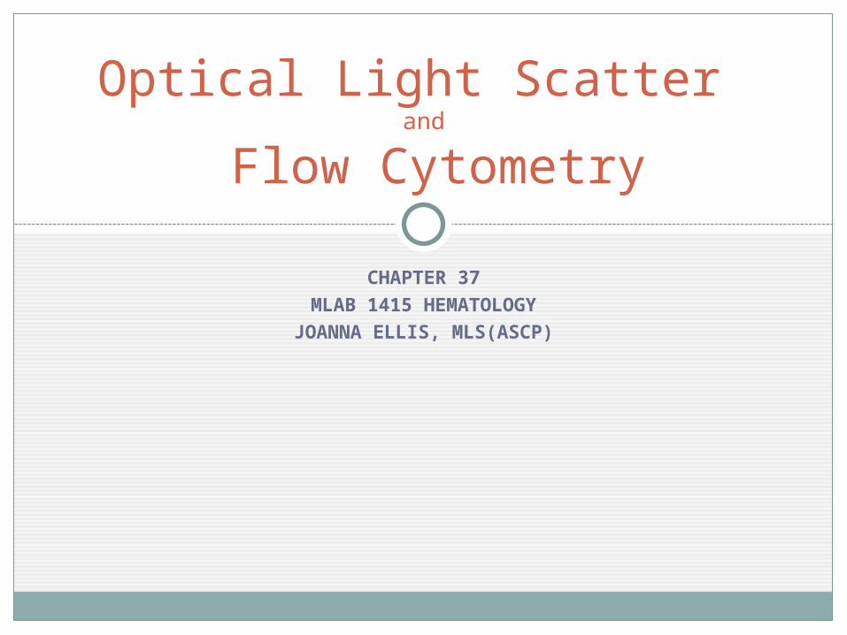
Purpose of Flow Cytometry:
To detect and measure
multiple properties of particles or
cells for identificatio
n and quantitation.
Image from Wikipedia

Introducing the particle or cell into the Flow Cell
Flow cytometry is performed on particles in suspension such as cells or nuclei.
The inner column of the flow cell consists of the liquid sample.
The outer column of sheath fluid controls the diameter of the sample column so that it narrows and isolates single particles (hydrodynamic focusing).
The single particles pass through a laser beam.
The light scatter is detected by a photodetector.
Flow Cell
Image from SonyInsider

Light Scatter
The light from the laser hits the single particle and scatters in different directions.
LASER
Forward Scatter
Side Scatter

Types of Light Scatter
Side light scatter: light scattered at a 90◦ angle from the particle defines internal complexity and granularity of the particle. Neutrophils and eosinophils produce a great deal
of side scatter due to their cytoplasmic granules.Forward scatter: light that continues in the
forward direction relates the particle size. Large cells such as monocytes and neutrophils
produce more forward scatter than nRBCs, and normal lymphs.

Light Scatter Scatterplot
Side Scatter
Forw
ard
Sca
tter
Image from Peripheral Blood Smear Evaluation

Fluorochromes
Fluorochromes are fluorescent compounds that can be bound by antibodies to the particles in the sample. Examples: FITC, PE, Propidium iodide, Acridine orange
Fluorochromes are used in: Immunophenotyping Cell differentiation and enumeration Reticulocyte counting DNA analysis

Detection of Fluorochromes
1. Laser beam excites fluorochrome attached to particle at one wavelength
2. Fluorochrome emits light at different wavelength
3. Light is directed to the photomultiplier tube that detects and quanitifies the light.
4. Computer software analyzes data and generates scatterplot
Photomultiplier Tube
CPU
Cell

Immunophenotyping uses Antibodies and Antigens
Immunophenotyping is the identification of antigens using detection antibodies.
Monoclonal antibodies are manufactured using myeloma cells to bind with a single portion of an antigen (foreign particle/molecule that illicits an immune response).
Polyclonal antibodies are made by injecting antigen into animals and are directed against multiple portions of an antigen.
Most immunophenotyping studies detect cell surface antigens.

Commercial Antibodies are grouped in Clusters of Differentiation (CD Markers)
• CD designations or CD Markers: group of antibodies that recognize the same antigen. Example: Commercially available Leu-4 and OKT3 are
both designated CD3
• CD marker antibodies labeled with fluorochomes have been developed to bind with antigens of identifiable cells. (table 37-3, page 840 in text)
• CD designations are cell markers: Example: CD4+ cells are helper T-cells, CD8+ cells
are Cytotoxic T-cells

Immunophenotyping with Flow Cytometry
The manufactured antibody (CD marker) is attached to a fluorochrome such as FITC and then added to the sample.
If the cell has the antigen that the antibody (Ab) specifically binds (target molecule), the antibody and fluorochrome attach to the cell.
Antibody
Fluorochrome
Antigen (target molecule)
Cell

Immunophenotyping with Flow Cytometry
When the cell/antibody/fluorochrome complex passes through the laser beam, the fluorochrome is excited and fluoresces at a measureable wavelength detected by the photomultiplier tube in the flow cytometer.
Computer software connected with the flow cytometer generate a histogram that visually represent the cells present.
NO TARGET MOLECULE NO FLUORESCENCE
Antibody
Fluorochrome
Antigen (target molecule)
Cell

Clinical Applications of Flow Cytometry
Blood cell enumeration and classification Differentials are based on light scatter as well as fluorescent staining
in some analyzers. Diagnosis and classification leukemia
Immunophenotyping of CD markers specific to certain cell populations can identify the type of leukemia.
Diagnosis and monitoring of HIV progression Immunophenotyping can determine cell deficiencies. The HIV virus
infects and destroys CD4+ cells. Immunophenotyping with Flow Cytometry can determine how many of these cells are in circulation.
Reticulocyte enumeration Fluorochromes that stain RNA directly are used to more accurately
(than manual method) count immature erythrocytes. DNA analysis
Certain fluorochromes attach to DNA directly. The amount of DNA is measured to determine if there are numerical chromosomal abnormalities

References
"Flow Cytometry." Wikipedia. Wikimedia Commons. Web. 17 Aug. 2010. <http://www.wikipedia.org/>. image
McKenzie, Shirlyn B., and J. Lynne. Williams. "Chapter 37." Clinical Laboratory Hematology. Boston: Pearson, 2010. Print.
McManus, Christopher. "Sony Acquires ICyt And Officially Enters Flow Cytometry Business." Sony Insider. 12 Feb. 2010. Web. 17 Aug. 2010. <http://www.sonyinsider.com/2010/02/12/sony-acquires-icyt-and-officially-enters-flow-cytometry-business/>. Image.
Riley, Roger, G. W. James, Sandra Sommer, and Mary Jo Martin. "Peripheral Blood Smear Evaluation." Virginia Commonwealth University Department of Pathology. Medical College of Virginia, 18 Sept. 1999. Web. 17 Aug. 2010. <http://www.pathology.vcu.edu/education/PathLab/pages/hematopath/pbs.html>. Image.