Chapter 34 | Animal Nutrition and the Digestive System 955 ... · 34.4: Digestive System Regulation...
29
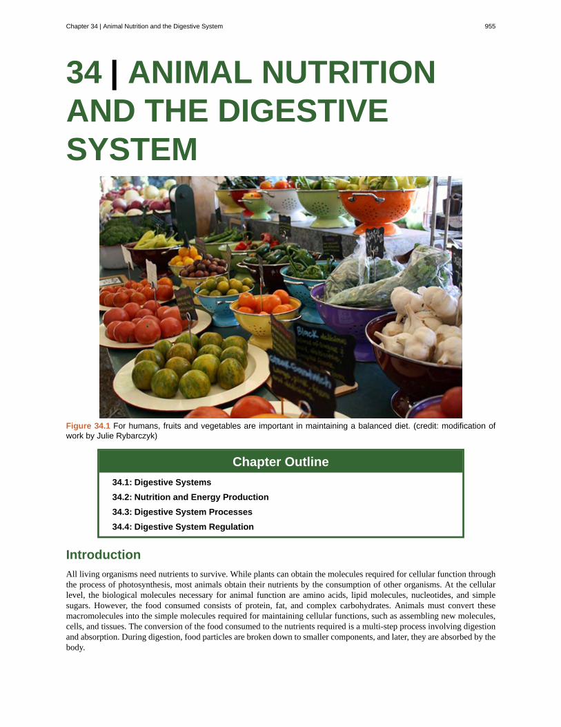
34 | ANIMAL NUTRITION AND THE DIGESTIVE SYSTEM Figure 34.1 For humans, fruits and vegetables are important in maintaining a balanced diet. (credit: modification of work by Julie Rybarczyk) Chapter Outline 34.1: Digestive Systems 34.2: Nutrition and Energy Production 34.3: Digestive System Processes 34.4: Digestive System Regulation Introduction All living organisms need nutrients to survive. While plants can obtain the molecules required for cellular function through the process of photosynthesis, most animals obtain their nutrients by the consumption of other organisms. At the cellular level, the biological molecules necessary for animal function are amino acids, lipid molecules, nucleotides, and simple sugars. However, the food consumed consists of protein, fat, and complex carbohydrates. Animals must convert these macromolecules into the simple molecules required for maintaining cellular functions, such as assembling new molecules, cells, and tissues. The conversion of the food consumed to the nutrients required is a multi-step process involving digestion and absorption. During digestion, food particles are broken down to smaller components, and later, they are absorbed by the body. Chapter 34 | Animal Nutrition and the Digestive System 955
Transcript of Chapter 34 | Animal Nutrition and the Digestive System 955 ... · 34.4: Digestive System Regulation...
34 | ANIMAL NUTRITIONAND THE DIGESTIVESYSTEM
Figure 34.1 For humans, fruits and vegetables are important in maintaining a balanced diet. (credit: modification ofwork by Julie Rybarczyk)
Chapter Outline
34.1: Digestive Systems
34.2: Nutrition and Energy Production
34.3: Digestive System Processes
34.4: Digestive System Regulation
Introduction
All living organisms need nutrients to survive. While plants can obtain the molecules required for cellular function throughthe process of photosynthesis, most animals obtain their nutrients by the consumption of other organisms. At the cellularlevel, the biological molecules necessary for animal function are amino acids, lipid molecules, nucleotides, and simplesugars. However, the food consumed consists of protein, fat, and complex carbohydrates. Animals must convert thesemacromolecules into the simple molecules required for maintaining cellular functions, such as assembling new molecules,cells, and tissues. The conversion of the food consumed to the nutrients required is a multi-step process involving digestionand absorption. During digestion, food particles are broken down to smaller components, and later, they are absorbed by thebody.
Chapter 34 | Animal Nutrition and the Digestive System 955

One of the challenges in human nutrition is maintaining a balance between food intake, storage, and energy expenditure.Imbalances can have serious health consequences. For example, eating too much food while not expending much energyleads to obesity, which in turn will increase the risk of developing illnesses such as type-2 diabetes and cardiovasculardisease. The recent rise in obesity and related diseases makes understanding the role of diet and nutrition in maintaininggood health all the more important.
34.1 | Digestive Systems
By the end of this section, you will be able to:
• Explain the processes of digestion and absorption
• Compare and contrast different types of digestive systems
• Explain the specialized functions of the organs involved in processing food in the body
• Describe the ways in which organs work together to digest food and absorb nutrients
Animals obtain their nutrition from the consumption of other organisms. Depending on their diet, animals can be classifiedinto the following categories: plant eaters (herbivores), meat eaters (carnivores), and those that eat both plants and animals(omnivores). The nutrients and macromolecules present in food are not immediately accessible to the cells. There area number of processes that modify food within the animal body in order to make the nutrients and organic moleculesaccessible for cellular function. As animals evolved in complexity of form and function, their digestive systems have alsoevolved to accommodate their various dietary needs.
Herbivores, Omnivores, and Carnivores
Herbivores are animals whose primary food source is plant-based. Examples of herbivores, as shown in Figure 34.2include vertebrates like deer, koalas, and some bird species, as well as invertebrates such as crickets and caterpillars. Theseanimals have evolved digestive systems capable of handling large amounts of plant material. Herbivores can be furtherclassified into frugivores (fruit-eaters), granivores (seed eaters), nectivores (nectar feeders), and folivores (leaf eaters).
Figure 34.2 Herbivores, like this (a) mule deer and (b) monarch caterpillar, eat primarily plant material. (credit a:modification of work by Bill Ebbesen; credit b: modification of work by Doug Bowman)
Carnivores are animals that eat other animals. The word carnivore is derived from Latin and literally means “meat eater.”Wild cats such as lions, shown in Figure 34.3a and tigers are examples of vertebrate carnivores, as are snakes and sharks,while invertebrate carnivores include sea stars, spiders, and ladybugs, shown in Figure 34.3b. Obligate carnivores are those
956 Chapter 34 | Animal Nutrition and the Digestive System
This OpenStax book is available for free at http://cnx.org/content/col11448/1.10
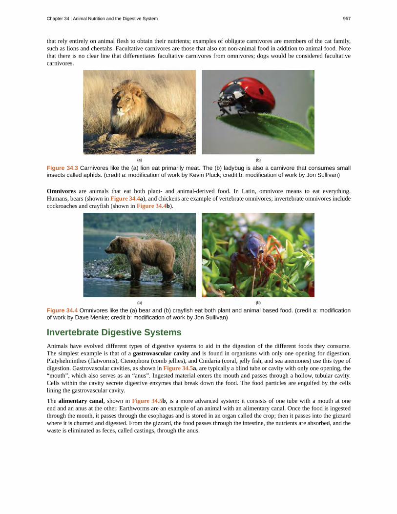
that rely entirely on animal flesh to obtain their nutrients; examples of obligate carnivores are members of the cat family,such as lions and cheetahs. Facultative carnivores are those that also eat non-animal food in addition to animal food. Notethat there is no clear line that differentiates facultative carnivores from omnivores; dogs would be considered facultativecarnivores.
Figure 34.3 Carnivores like the (a) lion eat primarily meat. The (b) ladybug is also a carnivore that consumes smallinsects called aphids. (credit a: modification of work by Kevin Pluck; credit b: modification of work by Jon Sullivan)
Omnivores are animals that eat both plant- and animal-derived food. In Latin, omnivore means to eat everything.Humans, bears (shown in Figure 34.4a), and chickens are example of vertebrate omnivores; invertebrate omnivores includecockroaches and crayfish (shown in Figure 34.4b).
Figure 34.4 Omnivores like the (a) bear and (b) crayfish eat both plant and animal based food. (credit a: modificationof work by Dave Menke; credit b: modification of work by Jon Sullivan)
Invertebrate Digestive Systems
Animals have evolved different types of digestive systems to aid in the digestion of the different foods they consume.The simplest example is that of a gastrovascular cavity and is found in organisms with only one opening for digestion.Platyhelminthes (flatworms), Ctenophora (comb jellies), and Cnidaria (coral, jelly fish, and sea anemones) use this type ofdigestion. Gastrovascular cavities, as shown in Figure 34.5a, are typically a blind tube or cavity with only one opening, the“mouth”, which also serves as an “anus”. Ingested material enters the mouth and passes through a hollow, tubular cavity.Cells within the cavity secrete digestive enzymes that break down the food. The food particles are engulfed by the cellslining the gastrovascular cavity.
The alimentary canal, shown in Figure 34.5b, is a more advanced system: it consists of one tube with a mouth at oneend and an anus at the other. Earthworms are an example of an animal with an alimentary canal. Once the food is ingestedthrough the mouth, it passes through the esophagus and is stored in an organ called the crop; then it passes into the gizzardwhere it is churned and digested. From the gizzard, the food passes through the intestine, the nutrients are absorbed, and thewaste is eliminated as feces, called castings, through the anus.
Chapter 34 | Animal Nutrition and the Digestive System 957

Figure 34.5 (a) A gastrovascular cavity has a single opening through which food is ingested and waste is excreted, asshown in this hydra and in this jellyfish medusa. (b) An alimentary canal has two openings: a mouth for ingesting food,and an anus for eliminating waste, as shown in this nematode.
Vertebrate Digestive Systems
Vertebrates have evolved more complex digestive systems to adapt to their dietary needs. Some animals have a singlestomach, while others have multi-chambered stomachs. Birds have developed a digestive system adapted to eatingunmasticated food.
Monogastric: Single-chambered Stomach
As the word monogastric suggests, this type of digestive system consists of one (“mono”) stomach chamber (“gastric”).Humans and many animals have a monogastric digestive system as illustrated in Figure 34.6ab. The process of digestionbegins with the mouth and the intake of food. The teeth play an important role in masticating (chewing) or physicallybreaking down food into smaller particles. The enzymes present in saliva also begin to chemically break down food. Theesophagus is a long tube that connects the mouth to the stomach. Using peristalsis, or wave-like smooth muscle contractions,the muscles of the esophagus push the food towards the stomach. In order to speed up the actions of enzymes in the stomach,the stomach is an extremely acidic environment, with a pH between 1.5 and 2.5. The gastric juices, which include enzymesin the stomach, act on the food particles and continue the process of digestion. Further breakdown of food takes place in thesmall intestine where enzymes produced by the liver, the small intestine, and the pancreas continue the process of digestion.The nutrients are absorbed into the blood stream across the epithelial cells lining the walls of the small intestines. The wastematerial travels on to the large intestine where water is absorbed and the drier waste material is compacted into feces; it isstored until it is excreted through the rectum.
958 Chapter 34 | Animal Nutrition and the Digestive System
This OpenStax book is available for free at http://cnx.org/content/col11448/1.10
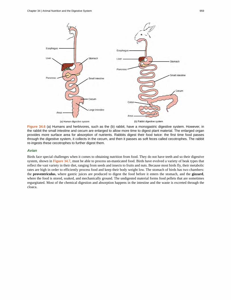
Figure 34.6 (a) Humans and herbivores, such as the (b) rabbit, have a monogastric digestive system. However, inthe rabbit the small intestine and cecum are enlarged to allow more time to digest plant material. The enlarged organprovides more surface area for absorption of nutrients. Rabbits digest their food twice: the first time food passesthrough the digestive system, it collects in the cecum, and then it passes as soft feces called cecotrophes. The rabbitre-ingests these cecotrophes to further digest them.
Avian
Birds face special challenges when it comes to obtaining nutrition from food. They do not have teeth and so their digestivesystem, shown in Figure 34.7, must be able to process un-masticated food. Birds have evolved a variety of beak types thatreflect the vast variety in their diet, ranging from seeds and insects to fruits and nuts. Because most birds fly, their metabolicrates are high in order to efficiently process food and keep their body weight low. The stomach of birds has two chambers:the proventriculus, where gastric juices are produced to digest the food before it enters the stomach, and the gizzard,where the food is stored, soaked, and mechanically ground. The undigested material forms food pellets that are sometimesregurgitated. Most of the chemical digestion and absorption happens in the intestine and the waste is excreted through thecloaca.
Chapter 34 | Animal Nutrition and the Digestive System 959
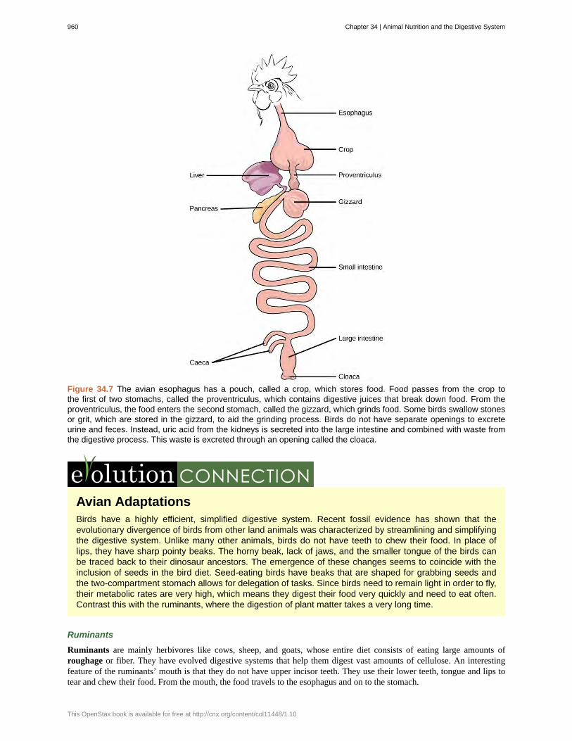
Figure 34.7 The avian esophagus has a pouch, called a crop, which stores food. Food passes from the crop tothe first of two stomachs, called the proventriculus, which contains digestive juices that break down food. From theproventriculus, the food enters the second stomach, called the gizzard, which grinds food. Some birds swallow stonesor grit, which are stored in the gizzard, to aid the grinding process. Birds do not have separate openings to excreteurine and feces. Instead, uric acid from the kidneys is secreted into the large intestine and combined with waste fromthe digestive process. This waste is excreted through an opening called the cloaca.
Avian AdaptationsBirds have a highly efficient, simplified digestive system. Recent fossil evidence has shown that theevolutionary divergence of birds from other land animals was characterized by streamlining and simplifyingthe digestive system. Unlike many other animals, birds do not have teeth to chew their food. In place oflips, they have sharp pointy beaks. The horny beak, lack of jaws, and the smaller tongue of the birds canbe traced back to their dinosaur ancestors. The emergence of these changes seems to coincide with theinclusion of seeds in the bird diet. Seed-eating birds have beaks that are shaped for grabbing seeds andthe two-compartment stomach allows for delegation of tasks. Since birds need to remain light in order to fly,their metabolic rates are very high, which means they digest their food very quickly and need to eat often.Contrast this with the ruminants, where the digestion of plant matter takes a very long time.
Ruminants
Ruminants are mainly herbivores like cows, sheep, and goats, whose entire diet consists of eating large amounts ofroughage or fiber. They have evolved digestive systems that help them digest vast amounts of cellulose. An interestingfeature of the ruminants’ mouth is that they do not have upper incisor teeth. They use their lower teeth, tongue and lips totear and chew their food. From the mouth, the food travels to the esophagus and on to the stomach.
960 Chapter 34 | Animal Nutrition and the Digestive System
This OpenStax book is available for free at http://cnx.org/content/col11448/1.10

To help digest the large amount of plant material, the stomach of the ruminants is a multi-chambered organ, as illustratedin Figure 34.8. The four compartments of the stomach are called the rumen, reticulum, omasum, and abomasum. Thesechambers contain many microbes that break down cellulose and ferment ingested food. The abomasum is the “true” stomachand is the equivalent of the monogastric stomach chamber where gastric juices are secreted. The four-compartment gastricchamber provides larger space and the microbial support necessary to digest plant material in ruminants. The fermentationprocess produces large amounts of gas in the stomach chamber, which must be eliminated. As in other animals, the smallintestine plays an important role in nutrient absorption, and the large intestine helps in the elimination of waste.
Figure 34.8 Ruminant animals, such as goats and cows, have four stomachs. The first two stomachs, the rumen andthe reticulum, contain prokaryotes and protists that are able to digest cellulose fiber. The ruminant regurgitates cudfrom the reticulum, chews it, and swallows it into a third stomach, the omasum, which removes water. The cud thenpasses onto the fourth stomach, the abomasum, where it is digested by enzymes produced by the ruminant.
Pseudo-ruminants
Some animals, such as camels and alpacas, are pseudo-ruminants. They eat a lot of plant material and roughage. Digestingplant material is not easy because plant cell walls contain the polymeric sugar molecule cellulose. The digestive enzymes ofthese animals cannot break down cellulose, but microorganisms present in the digestive system can. Therefore, the digestivesystem must be able to handle large amounts of roughage and break down the cellulose. Pseudo-ruminants have a three-chamber stomach in the digestive system. However, their cecum—a pouched organ at the beginning of the large intestinecontaining many microorganisms that are necessary for the digestion of plant materials—is large and is the site where theroughage is fermented and digested. These animals do not have a rumen but have an omasum, abomasum, and reticulum.
Parts of the Digestive System
The vertebrate digestive system is designed to facilitate the transformation of food matter into the nutrient components thatsustain organisms.
Oral Cavity
The oral cavity, or mouth, is the point of entry of food into the digestive system, illustrated in Figure 34.9. The foodconsumed is broken into smaller particles by mastication, the chewing action of the teeth. All mammals have teeth and canchew their food.
The extensive chemical process of digestion begins in the mouth. As food is being chewed, saliva, produced by the salivaryglands, mixes with the food. Saliva is a watery substance produced in the mouths of many animals. There are three majorglands that secrete saliva—the parotid, the submandibular, and the sublingual. Saliva contains mucus that moistens food andbuffers the pH of the food. Saliva also contains immunoglobulins and lysozymes, which have antibacterial action to reducetooth decay by inhibiting growth of some bacteria. Saliva also contains an enzyme called salivary amylase that begins the
Chapter 34 | Animal Nutrition and the Digestive System 961

process of converting starches in the food into a disaccharide called maltose. Another enzyme called lipase is producedby the cells in the tongue. Lipases are a class of enzymes that can break down triglycerides. The lingual lipase begins thebreakdown of fat components in the food. The chewing and wetting action provided by the teeth and saliva prepare the foodinto a mass called the bolus for swallowing. The tongue helps in swallowing—moving the bolus from the mouth into thepharynx. The pharynx opens to two passageways: the trachea, which leads to the lungs, and the esophagus, which leads tothe stomach. The trachea has an opening called the glottis, which is covered by a cartilaginous flap called the epiglottis.When swallowing, the epiglottis closes the glottis and food passes into the esophagus and not the trachea. This arrangementallows food to be kept out of the trachea.
Figure 34.9 Digestion of food begins in the (a) oral cavity. Food is masticated by teeth and moistened by salivasecreted from the (b) salivary glands. Enzymes in the saliva begin to digest starches and fats. With the help of thetongue, the resulting bolus is moved into the esophagus by swallowing. (credit: modification of work by the NationalCancer Institute)
Esophagus
The esophagus is a tubular organ that connects the mouth to the stomach. The chewed and softened food passes through theesophagus after being swallowed. The smooth muscles of the esophagus undergo a series of wave like movements calledperistalsis that push the food toward the stomach, as illustrated in Figure 34.10. The peristalsis wave is unidirectional—itmoves food from the mouth to the stomach, and reverse movement is not possible. The peristaltic movement of theesophagus is an involuntary reflex; it takes place in response to the act of swallowing.
Figure 34.10 The esophagus transfers food from the mouth to the stomach through peristaltic movements.
A ring-like muscle called a sphincter forms valves in the digestive system. The gastro-esophageal sphincter is located atthe stomach end of the esophagus. In response to swallowing and the pressure exerted by the bolus of food, this sphincteropens, and the bolus enters the stomach. When there is no swallowing action, this sphincter is shut and prevents the contentsof the stomach from traveling up the esophagus. Many animals have a true sphincter; however, in humans, there is no true
962 Chapter 34 | Animal Nutrition and the Digestive System
This OpenStax book is available for free at http://cnx.org/content/col11448/1.10
sphincter, but the esophagus remains closed when there is no swallowing action. Acid reflux or “heartburn” occurs whenthe acidic digestive juices escape into the esophagus.
Stomach
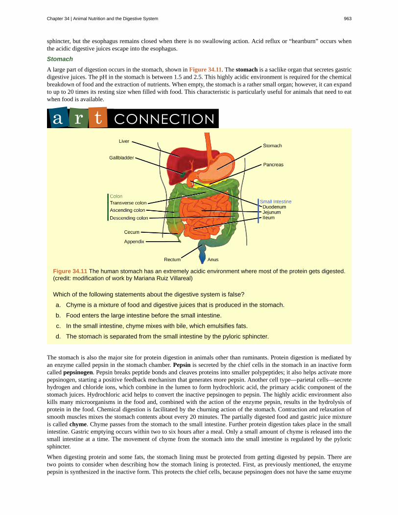
A large part of digestion occurs in the stomach, shown in Figure 34.11. The stomach is a saclike organ that secretes gastricdigestive juices. The pH in the stomach is between 1.5 and 2.5. This highly acidic environment is required for the chemicalbreakdown of food and the extraction of nutrients. When empty, the stomach is a rather small organ; however, it can expandto up to 20 times its resting size when filled with food. This characteristic is particularly useful for animals that need to eatwhen food is available.
Figure 34.11 The human stomach has an extremely acidic environment where most of the protein gets digested.(credit: modification of work by Mariana Ruiz Villareal)
Which of the following statements about the digestive system is false?
a. Chyme is a mixture of food and digestive juices that is produced in the stomach.
b. Food enters the large intestine before the small intestine.
c. In the small intestine, chyme mixes with bile, which emulsifies fats.
d. The stomach is separated from the small intestine by the pyloric sphincter.
The stomach is also the major site for protein digestion in animals other than ruminants. Protein digestion is mediated byan enzyme called pepsin in the stomach chamber. Pepsin is secreted by the chief cells in the stomach in an inactive formcalled pepsinogen. Pepsin breaks peptide bonds and cleaves proteins into smaller polypeptides; it also helps activate morepepsinogen, starting a positive feedback mechanism that generates more pepsin. Another cell type—parietal cells—secretehydrogen and chloride ions, which combine in the lumen to form hydrochloric acid, the primary acidic component of thestomach juices. Hydrochloric acid helps to convert the inactive pepsinogen to pepsin. The highly acidic environment alsokills many microorganisms in the food and, combined with the action of the enzyme pepsin, results in the hydrolysis ofprotein in the food. Chemical digestion is facilitated by the churning action of the stomach. Contraction and relaxation ofsmooth muscles mixes the stomach contents about every 20 minutes. The partially digested food and gastric juice mixtureis called chyme. Chyme passes from the stomach to the small intestine. Further protein digestion takes place in the smallintestine. Gastric emptying occurs within two to six hours after a meal. Only a small amount of chyme is released into thesmall intestine at a time. The movement of chyme from the stomach into the small intestine is regulated by the pyloricsphincter.
When digesting protein and some fats, the stomach lining must be protected from getting digested by pepsin. There aretwo points to consider when describing how the stomach lining is protected. First, as previously mentioned, the enzymepepsin is synthesized in the inactive form. This protects the chief cells, because pepsinogen does not have the same enzyme
Chapter 34 | Animal Nutrition and the Digestive System 963

functionality of pepsin. Second, the stomach has a thick mucus lining that protects the underlying tissue from the action ofthe digestive juices. When this mucus lining is ruptured, ulcers can form in the stomach. Ulcers are open wounds in or onan organ caused by bacteria (Helicobacter pylori) when the mucus lining is ruptured and fails to reform.
Small Intestine
Chyme moves from the stomach to the small intestine. The small intestine is the organ where the digestion of protein, fats,and carbohydrates is completed. The small intestine is a long tube-like organ with a highly folded surface containing finger-like projections called the villi. The apical surface of each villus has many microscopic projections called microvilli. Thesestructures, illustrated in Figure 34.12, are lined with epithelial cells on the luminal side and allow for the nutrients to beabsorbed from the digested food and absorbed into the blood stream on the other side. The villi and microvilli, with theirmany folds, increase the surface area of the intestine and increase absorption efficiency of the nutrients. Absorbed nutrientsin the blood are carried into the hepatic portal vein, which leads to the liver. There, the liver regulates the distribution ofnutrients to the rest of the body and removes toxic substances, including drugs, alcohol, and some pathogens.
Figure 34.12 Villi are folds on the small intestine lining that increase the surface area to facilitate the absorptionof nutrients.
Which of the following statements about the small intestine is false?
a. Absorptive cells that line the small intestine have microvilli, small projections that increase surface areaand aid in the absorption of food.
b. The inside of the small intestine has many folds, called villi.
c. Microvilli are lined with blood vessels as well as lymphatic vessels.
d. The inside of the small intestine is called the lumen.
The human small intestine is over 6m long and is divided into three parts: the duodenum, the jejunum, and the ileum.The “C-shaped,” fixed part of the small intestine is called the duodenum and is shown in Figure 34.11. The duodenumis separated from the stomach by the pyloric sphincter which opens to allow chyme to move from the stomach to theduodenum. In the duodenum, chyme is mixed with pancreatic juices in an alkaline solution rich in bicarbonate thatneutralizes the acidity of chyme and acts as a buffer. Pancreatic juices also contain several digestive enzymes. Digestivejuices from the pancreas, liver, and gallbladder, as well as from gland cells of the intestinal wall itself, enter the duodenum.Bile is produced in the liver and stored and concentrated in the gallbladder. Bile contains bile salts which emulsify lipidswhile the pancreas produces enzymes that catabolize starches, disaccharides, proteins, and fats. These digestive juices breakdown the food particles in the chyme into glucose, triglycerides, and amino acids. Some chemical digestion of food takesplace in the duodenum. Absorption of fatty acids also takes place in the duodenum.
The second part of the small intestine is called the jejunum, shown in Figure 34.11. Here, hydrolysis of nutrients iscontinued while most of the carbohydrates and amino acids are absorbed through the intestinal lining. The bulk of chemicaldigestion and nutrient absorption occurs in the jejunum.
964 Chapter 34 | Animal Nutrition and the Digestive System
This OpenStax book is available for free at http://cnx.org/content/col11448/1.10
The ileum, also illustrated in Figure 34.11 is the last part of the small intestine and here the bile salts and vitamins areabsorbed into blood stream. The undigested food is sent to the colon from the ileum via peristaltic movements of the muscle.The ileum ends and the large intestine begins at the ileocecal valve. The vermiform, “worm-like,” appendix is located at theileocecal valve. The appendix of humans secretes no enzymes and has an insignificant role in immunity.
Large Intestine
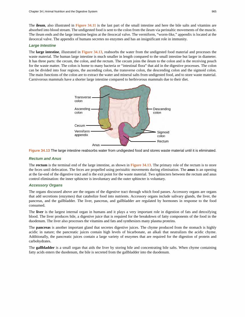
The large intestine, illustrated in Figure 34.13, reabsorbs the water from the undigested food material and processes thewaste material. The human large intestine is much smaller in length compared to the small intestine but larger in diameter.It has three parts: the cecum, the colon, and the rectum. The cecum joins the ileum to the colon and is the receiving pouchfor the waste matter. The colon is home to many bacteria or “intestinal flora” that aid in the digestive processes. The coloncan be divided into four regions, the ascending colon, the transverse colon, the descending colon and the sigmoid colon.The main functions of the colon are to extract the water and mineral salts from undigested food, and to store waste material.Carnivorous mammals have a shorter large intestine compared to herbivorous mammals due to their diet.
Figure 34.13 The large intestine reabsorbs water from undigested food and stores waste material until it is eliminated.
Rectum and Anus
The rectum is the terminal end of the large intestine, as shown in Figure 34.13. The primary role of the rectum is to storethe feces until defecation. The feces are propelled using peristaltic movements during elimination. The anus is an openingat the far-end of the digestive tract and is the exit point for the waste material. Two sphincters between the rectum and anuscontrol elimination: the inner sphincter is involuntary and the outer sphincter is voluntary.
Accessory Organs
The organs discussed above are the organs of the digestive tract through which food passes. Accessory organs are organsthat add secretions (enzymes) that catabolize food into nutrients. Accessory organs include salivary glands, the liver, thepancreas, and the gallbladder. The liver, pancreas, and gallbladder are regulated by hormones in response to the foodconsumed.
The liver is the largest internal organ in humans and it plays a very important role in digestion of fats and detoxifyingblood. The liver produces bile, a digestive juice that is required for the breakdown of fatty components of the food in theduodenum. The liver also processes the vitamins and fats and synthesizes many plasma proteins.
The pancreas is another important gland that secretes digestive juices. The chyme produced from the stomach is highlyacidic in nature; the pancreatic juices contain high levels of bicarbonate, an alkali that neutralizes the acidic chyme.Additionally, the pancreatic juices contain a large variety of enzymes that are required for the digestion of protein andcarbohydrates.
The gallbladder is a small organ that aids the liver by storing bile and concentrating bile salts. When chyme containingfatty acids enters the duodenum, the bile is secreted from the gallbladder into the duodenum.
Chapter 34 | Animal Nutrition and the Digestive System 965

34.2 | Nutrition and Energy Production
By the end of this section, you will be able to:
• Explain why an animal’s diet should be balanced and meet the needs of the body
• Define the primary components of food
• Describe the essential nutrients required for cellular function that cannot be synthesized by the animal body
• Explain how energy is produced through diet and digestion
• Describe how excess carbohydrates and energy are stored in the body
Given the diversity of animal life on our planet, it is not surprising that the animal diet would also vary substantially.The animal diet is the source of materials needed for building DNA and other complex molecules needed for growth,maintenance, and reproduction; collectively these processes are called biosynthesis. The diet is also the source of materialsfor ATP production in the cells. The diet must be balanced to provide the minerals and vitamins that are required for cellularfunction.
Food Requirements
What are the fundamental requirements of the animal diet? The animal diet should be well balanced and provide nutrientsrequired for bodily function and the minerals and vitamins required for maintaining structure and regulation necessary forgood health and reproductive capability. These requirements for a human are illustrated graphically in Figure 34.14
Figure 34.14 For humans, a balanced diet includes fruits, vegetables, grains, and protein. (credit: USDA)
The first step in ensuring that you are meeting the food requirements of your body is an awareness of the food groupsand the nutrients they provide. To learn more about each food group and the recommended daily amounts, explore thisinteractive site (http://openstaxcollege.org/l/food_groups) by the United States Department of Agriculture.
966 Chapter 34 | Animal Nutrition and the Digestive System
This OpenStax book is available for free at http://cnx.org/content/col11448/1.10
Let’s Move! CampaignObesity is a growing epidemic and the rate of obesity among children is rapidly rising in the United States.To combat childhood obesity and ensure that children get a healthy start in life, first lady Michelle Obamahas launched the Let’s Move! campaign. The goal of this campaign is to educate parents and caregiverson providing healthy nutrition and encouraging active lifestyles to future generations. This program aims toinvolve the entire community, including parents, teachers, and healthcare providers to ensure that childrenhave access to healthy foods—more fruits, vegetables, and whole grains—and consume fewer calories fromprocessed foods. Another goal is to ensure that children get physical activity. With the increase in televisionviewing and stationary pursuits such as video games, sedentary lifestyles have become the norm. Learnmore at www.letsmove.gov.
Organic Precursors
The organic molecules required for building cellular material and tissues must come from food. Carbohydrates or sugarsare the primary source of organic carbons in the animal body. During digestion, digestible carbohydrates are ultimatelybroken down into glucose and used to provide energy through metabolic pathways. Complex carbohydrates, includingpolysaccharides, can be broken down into glucose through biochemical modification; however, humans do not producethe enzyme cellulase and lack the ability to derive glucose from the polysaccharide cellulose. In humans, these moleculesprovide the fiber required for moving waste through the large intestine and a healthy colon. The intestinal flora in the humangut are able to extract some nutrition from these plant fibers. The excess sugars in the body are converted into glycogenand stored in the liver and muscles for later use. Glycogen stores are used to fuel prolonged exertions, such as long-distancerunning, and to provide energy during food shortage. Excess glycogen can be converted to fats, which are stored in the lowerlayer of the skin of mammals for insulation and energy storage. Excess digestible carbohydrates are stored by mammals inorder to survive famine and aid in mobility.
Another important requirement is that of nitrogen. Protein catabolism provides a source of organic nitrogen. Amino acidsare the building blocks of proteins and protein breakdown provides amino acids that are used for cellular function. Thecarbon and nitrogen derived from these become the building block for nucleotides, nucleic acids, proteins, cells, and tissues.Excess nitrogen must be excreted as it is toxic. Fats add flavor to food and promote a sense of satiety or fullness. Fatty foodsare also significant sources of energy because one gram of fat contains nine calories. Fats are required in the diet to aid theabsorption of fat-soluble vitamins and the production of fat-soluble hormones.
Essential Nutrients
While the animal body can synthesize many of the molecules required for function from the organic precursors, there aresome nutrients that need to be consumed from food. These nutrients are termed essential nutrients, meaning they must beeaten, and the body cannot produce them.
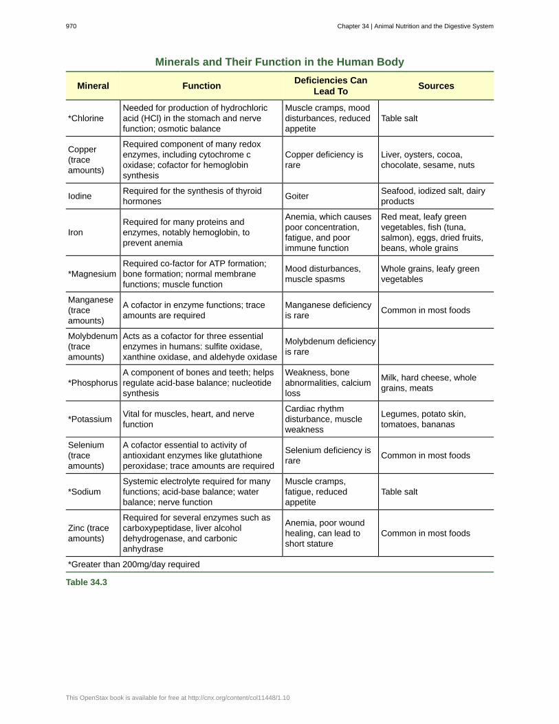
The omega-3 alpha-linolenic acid and the omega-6 linoleic acid are essential fatty acids needed to make some membranephospholipids. Vitamins are another class of essential organic molecules that are required in small quantities for manyenzymes to function and, for this reason, are considered to be co-enzymes. Absence or low levels of vitamins can havea dramatic effect on health, as outlined in Table 34.1 and Table 34.2. Both fat-soluble and water-soluble vitamins mustbe obtained from food. Minerals, listed in Table 34.3, are inorganic essential nutrients that must be obtained from food.Among their many functions, minerals help in structure and regulation and are considered co-factors. Certain amino acidsalso must be procured from food and cannot be synthesized by the body. These amino acids are the “essential” amino acids.The human body can synthesize only 11 of the 20 required amino acids; the rest must be obtained from food. The essentialamino acids are listed in Table 34.4.
Chapter 34 | Animal Nutrition and the Digestive System 967

Water-soluble Essential Vitamins
Vitamin Function Deficiencies Can Lead To Sources
Vitamin B1(Thiamine)
Needed by the body to process lipids,proteins, and carbohydrates Coenzymeremoves CO2 from organic compounds
Muscle weakness, Beriberi: reducedheart function, CNS problems
Milk, meat,dried beans,whole grains
Vitamin B2(Riboflavin)
Takes an active role in metabolism,aiding in the conversion of food to energy(FAD and FMN)
Cracks or sores on the outer surfaceof the lips (cheliosis); inflammationand redness of the tongue; moist,scaly skin inflammation (seborrheicdermatitis)
Meat, eggs,enrichedgrains,vegetables
Vitamin B3(Niacin)
Used by the body to release energy fromcarbohydrates and to process alcohol;required for the synthesis of sexhormones; component of coenzymeNAD+ and NADP+
Pellagra, which can result indermatitis, diarrhea, dementia, anddeath
Meat, eggs,grains, nuts,potatoes
Vitamin B5(Pantothenic
acid)
Assists in producing energy from foods(lipids, in particular); component ofcoenzyme A
Fatigue, poor coordination, retardedgrowth, numbness, tingling of handsand feet
Meat, wholegrains, milk,fruits,vegetables
Vitamin B6(Pyridoxine)
The principal vitamin for processingamino acids and lipids; also helpsconvert nutrients into energy
Irritability, depression, confusion,mouth sores or ulcers, anemia,muscular twitching
Meat, dairyproducts,whole grains,orange juice
Vitamin B7(Biotin)
Used in energy and amino acidmetabolism, fat synthesis, and fatbreakdown; helps the body use bloodsugar
Hair loss, dermatitis, depression,numbness and tingling in theextremities; neuromuscular disorders
Meat, eggs,legumes andothervegetables
Vitamin B9(Folic acid)
Assists the normal development of cells,especially during fetal development;helps metabolize nucleic and aminoacids
Deficiency during pregnancy isassociated with birth defects, such asneural tube defects and anemia
Leafy greenvegetables,whole wheat,fruits, nuts,legumes
Vitamin B12(Cobalamin)
Maintains healthy nervous system andassists with blood cell formation;coenzyme in nucleic acid metabolism
Anemia, neurological disorders,numbness, loss of balance
Meat, eggs,animalproducts
Vitamin C(Ascorbic
acid)
Helps maintain connective tissue: bone,cartilage, and dentin; boosts the immunesystem
Scurvy, which results in bleeding, hairand tooth loss; joint pain andswelling; delayed wound healing
Citrus fruits,broccoli,tomatoes, redsweet bellpeppers
Table 34.1
968 Chapter 34 | Animal Nutrition and the Digestive System
This OpenStax book is available for free at http://cnx.org/content/col11448/1.10

Fat-soluble Essential Vitamins
Vitamin FunctionDeficiencies
Can LeadTo
Sources
Vitamin A(Retinol)
Critical to the development of bones, teeth, andskin; helps maintain eyesight, enhances theimmune system, fetal development, geneexpression
Night-blindness,skin disorders,impairedimmunity
Dark green leafyvegetables, yellow-orange vegetablesfruits, milk, butter
Vitamin D
Critical for calcium absorption for bonedevelopment and strength; maintains a stablenervous system; maintains a normal and strongheartbeat; helps in blood clotting
Rickets,osteomalacia,immunity
Cod liver oil, milk,egg yolk
Vitamin E(Tocopherol)
Lessens oxidative damage of cells,and preventslung damage from pollutants; vital to the immunesystem
Deficiency israre; anemia,nervous systemdegeneration
Wheat germ oil,unrefined vegetableoils, nuts, seeds,grains
Vitamin K(Phylloquinone) Essential to blood clotting Bleeding and
easy bruisingLeafy greenvegetables, tea
Table 34.2
Figure 34.15 A healthy diet should include a variety of foods to ensure that needs for essential nutrients are met.(credit: Keith Weller, USDA ARS)
Minerals and Their Function in the Human Body
Mineral FunctionDeficiencies Can
Lead ToSources
*Calcium
Needed for muscle and neuron function;heart health; builds bone and supportssynthesis and function of blood cells;nerve function
Osteoporosis, rickets,muscle spasms,impaired growth
Milk, yogurt, fish, green leafyvegetables, legumes
Table 34.3
Chapter 34 | Animal Nutrition and the Digestive System 969
Minerals and Their Function in the Human Body
Mineral FunctionDeficiencies Can
Lead ToSources
*ChlorineNeeded for production of hydrochloricacid (HCl) in the stomach and nervefunction; osmotic balance
Muscle cramps, mooddisturbances, reducedappetite
Table salt
Copper(traceamounts)
Required component of many redoxenzymes, including cytochrome coxidase; cofactor for hemoglobinsynthesis
Copper deficiency israre
Liver, oysters, cocoa,chocolate, sesame, nuts
Iodine Required for the synthesis of thyroidhormones Goiter Seafood, iodized salt, dairy
products
IronRequired for many proteins andenzymes, notably hemoglobin, toprevent anemia
Anemia, which causespoor concentration,fatigue, and poorimmune function
Red meat, leafy greenvegetables, fish (tuna,salmon), eggs, dried fruits,beans, whole grains
*MagnesiumRequired co-factor for ATP formation;bone formation; normal membranefunctions; muscle function
Mood disturbances,muscle spasms
Whole grains, leafy greenvegetables
Manganese(traceamounts)
A cofactor in enzyme functions; traceamounts are required
Manganese deficiencyis rare Common in most foods
Molybdenum(traceamounts)
Acts as a cofactor for three essentialenzymes in humans: sulfite oxidase,xanthine oxidase, and aldehyde oxidase
Molybdenum deficiencyis rare
*PhosphorusA component of bones and teeth; helpsregulate acid-base balance; nucleotidesynthesis
Weakness, boneabnormalities, calciumloss
Milk, hard cheese, wholegrains, meats
*Potassium Vital for muscles, heart, and nervefunction
Cardiac rhythmdisturbance, muscleweakness
Legumes, potato skin,tomatoes, bananas
Selenium(traceamounts)
A cofactor essential to activity ofantioxidant enzymes like glutathioneperoxidase; trace amounts are required
Selenium deficiency israre Common in most foods
*SodiumSystemic electrolyte required for manyfunctions; acid-base balance; waterbalance; nerve function
Muscle cramps,fatigue, reducedappetite
Table salt
Zinc (traceamounts)
Required for several enzymes such ascarboxypeptidase, liver alcoholdehydrogenase, and carbonicanhydrase
Anemia, poor woundhealing, can lead toshort stature
Common in most foods
*Greater than 200mg/day required
Table 34.3
970 Chapter 34 | Animal Nutrition and the Digestive System
This OpenStax book is available for free at http://cnx.org/content/col11448/1.10

Essential Amino Acids
Amino acids that must be consumed Amino acids anabolized by the body
isoleucine alanine
leucine selenocysteine
lysine aspartate
methionine cysteine
phenylalanine glutamate
tryptophan glycine
valine proline
histidine* serine
threonine tyrosine
arginine* asparagine
*The human body can synthesize histidine and arginine, but not in the quantities required, especially forgrowing children.
Table 34.4
Food Energy and ATP
Animals need food to obtain energy and maintain homeostasis. Homeostasis is the ability of a system to maintain a stableinternal environment even in the face of external changes to the environment. For example, the normal body temperatureof humans is 37°C (98.6°F). Humans maintain this temperature even when the external temperature is hot or cold. It takesenergy to maintain this body temperature, and animals obtain this energy from food.
The primary source of energy for animals is carbohydrates, mainly glucose. Glucose is called the body’s fuel. The digestiblecarbohydrates in an animal’s diet are converted to glucose molecules through a series of catabolic chemical reactions.
Adenosine triphosphate, or ATP, is the primary energy currency in cells; ATP stores energy in phosphate ester bonds. ATPreleases energy when the phosphodiester bonds are broken and ATP is converted to ADP and a phosphate group. ATP isproduced by the oxidative reactions in the cytoplasm and mitochondrion of the cell, where carbohydrates, proteins, andfats undergo a series of metabolic reactions collectively called cellular respiration. For example, glycolysis is a series ofreactions in which glucose is converted to pyruvic acid and some of its chemical potential energy is transferred to NADHand ATP.
ATP is required for all cellular functions. It is used to build the organic molecules that are required for cells and tissues;it provides energy for muscle contraction and for the transmission of electrical signals in the nervous system. When theamount of ATP is available in excess of the body’s requirements, the liver uses the excess ATP and excess glucose toproduce molecules called glycogen. Glycogen is a polymeric form of glucose and is stored in the liver and skeletal musclecells. When blood sugar drops, the liver releases glucose from stores of glycogen. Skeletal muscle converts glycogen toglucose during intense exercise. The process of converting glucose and excess ATP to glycogen and the storage of excessenergy is an evolutionarily important step in helping animals deal with mobility, food shortages, and famine.
Chapter 34 | Animal Nutrition and the Digestive System 971

ObesityObesity is a major health concern in the United States, and there is a growing focus on reducing obesity andthe diseases it may lead to, such as type-2 diabetes, cancers of the colon and breast, and cardiovasculardisease. How does the food consumed contribute to obesity?
Fatty foods are calorie-dense, meaning that they have more calories per unit mass than carbohydrates orproteins. One gram of carbohydrates has four calories, one gram of protein has four calories, and one gramof fat has nine calories. Animals tend to seek lipid-rich food for their higher energy content.
The signals of hunger (“time to eat”) and satiety (“time to stop eating”) are controlled in the hypothalamusregion of the brain. Foods that are rich in fatty acids tend to promote satiety more than foods that are richonly in carbohydrates.
Excess carbohydrate and ATP are used by the liver to synthesize glycogen. The pyruvate produced duringglycolysis is used to synthesize fatty acids. When there is more glucose in the body than required, theresulting excess pyruvate is converted into molecules that eventually result in the synthesis of fatty acidswithin the body. These fatty acids are stored in adipose cells—the fat cells in the mammalian body whoseprimary role is to store fat for later use.
It is important to note that some animals benefit from obesity. Polar bears and seals need body fat forinsulation and to keep them from losing body heat during Arctic winters. When food is scarce, stored bodyfat provides energy for maintaining homeostasis. Fats prevent famine in mammals, allowing them to accessenergy when food is not available on a daily basis; fats are stored when a large kill is made or lots of foodis available.
34.3 | Digestive System Processes
By the end of this section, you will be able to:
• Describe the process of digestion
• Detail the steps involved in digestion and absorption
• Define elimination
• Explain the role of both the small and large intestines in absorption
Obtaining nutrition and energy from food is a multi-step process. For true animals, the first step is ingestion, the act oftaking in food. This is followed by digestion, absorption, and elimination. In the following sections, each of these steps willbe discussed in detail.
Ingestion
The large molecules found in intact food cannot pass through the cell membranes. Food needs to be broken into smallerparticles so that animals can harness the nutrients and organic molecules. The first step in this process is ingestion. Ingestionis the process of taking in food through the mouth. In vertebrates, the teeth, saliva, and tongue play important roles inmastication (preparing the food into bolus). While the food is being mechanically broken down, the enzymes in saliva beginto chemically process the food as well. The combined action of these processes modifies the food from large particles to asoft mass that can be swallowed and can travel the length of the esophagus.
Digestion and Absorption
Digestion is the mechanical and chemical break down of food into small organic fragments. It is important to break downmacromolecules into smaller fragments that are of suitable size for absorption across the digestive epithelium. Large,complex molecules of proteins, polysaccharides, and lipids must be reduced to simpler particles such as simple sugar beforethey can be absorbed by the digestive epithelial cells. Different organs play specific roles in the digestive process. The
972 Chapter 34 | Animal Nutrition and the Digestive System
This OpenStax book is available for free at http://cnx.org/content/col11448/1.10

animal diet needs carbohydrates, protein, and fat, as well as vitamins and inorganic components for nutritional balance.How each of these components is digested is discussed in the following sections.
Carbohydrates
The digestion of carbohydrates begins in the mouth. The salivary enzyme amylase begins the breakdown of food starchesinto maltose, a disaccharide. As the bolus of food travels through the esophagus to the stomach, no significant digestionof carbohydrates takes place. The esophagus produces no digestive enzymes but does produce mucous for lubrication. Theacidic environment in the stomach stops the action of the amylase enzyme.
The next step of carbohydrate digestion takes place in the duodenum. Recall that the chyme from the stomach enters theduodenum and mixes with the digestive secretion from the pancreas, liver, and gallbladder. Pancreatic juices also containamylase, which continues the breakdown of starch and glycogen into maltose, a disaccharide. The disaccharides are brokendown into monosaccharides by enzymes called maltases, sucrases, and lactases, which are also present in the brush borderof the small intestinal wall. Maltase breaks down maltose into glucose. Other disaccharides, such as sucrose and lactose arebroken down by sucrase and lactase, respectively. Sucrase breaks down sucrose (or “table sugar”) into glucose and fructose,and lactase breaks down lactose (or “milk sugar”) into glucose and galactose. The monosaccharides (glucose) thus producedare absorbed and then can be used in metabolic pathways to harness energy. The monosaccharides are transported acrossthe intestinal epithelium into the bloodstream to be transported to the different cells in the body. The steps in carbohydratedigestion are summarized in Figure 34.16 and Table 34.5.
Figure 34.16 Digestion of carbohydrates is performed by several enzymes. Starch and glycogen are broken down intoglucose by amylase and maltase. Sucrose (table sugar) and lactose (milk sugar) are broken down by sucrase andlactase, respectively.
Digestion of Carbohydrates
Enzyme Produced BySite ofAction
SubstrateActing On
End Products
Salivary amylase Salivary glands Mouth Polysaccharides(Starch)
Disaccharides (maltose),oligosaccharides
Pancreatic amylase Pancreas Smallintestine
Polysaccharides(starch)
Disaccharides (maltose),monosaccharides
Oligosaccharidases Lining of the intestine;brush border membrane
Smallintestine Disaccharides Monosaccharides (e.g.,
glucose, fructose, galactose)
Table 34.5
Protein
A large part of protein digestion takes place in the stomach. The enzyme pepsin plays an important role in the digestionof proteins by breaking down the intact protein to peptides, which are short chains of four to nine amino acids. In theduodenum, other enzymes— trypsin, elastase, and chymotrypsin—act on the peptides reducing them to smaller peptides.Trypsin elastase, carboxypeptidase, and chymotrypsin are produced by the pancreas and released into the duodenum wherethey act on the chyme. Further breakdown of peptides to single amino acids is aided by enzymes called peptidases (thosethat break down peptides). Specifically, carboxypeptidase, dipeptidase, and aminopeptidase play important roles inreducing the peptides to free amino acids. The amino acids are absorbed into the bloodstream through the small intestines.The steps in protein digestion are summarized in Figure 34.17 and Table 34.6.
Chapter 34 | Animal Nutrition and the Digestive System 973
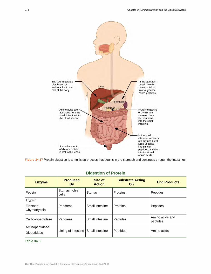
Figure 34.17 Protein digestion is a multistep process that begins in the stomach and continues through the intestines.
Digestion of Protein
EnzymeProduced
BySite ofAction
Substrate ActingOn
End Products
Pepsin Stomach chiefcells Stomach Proteins Peptides
Trypsin
ElastaseChymotrypsin
Pancreas Small intestine Proteins Peptides
Carboxypeptidase Pancreas Small intestine Peptides Amino acids andpeptides
Aminopeptidase
Dipeptidase Lining of intestine Small intestine Peptides Amino acids
Table 34.6
974 Chapter 34 | Animal Nutrition and the Digestive System
This OpenStax book is available for free at http://cnx.org/content/col11448/1.10

Lipids
Lipid digestion begins in the stomach with the aid of lingual lipase and gastric lipase. However, the bulk of lipid digestionoccurs in the small intestine due to pancreatic lipase. When chyme enters the duodenum, the hormonal responses triggerthe release of bile, which is produced in the liver and stored in the gallbladder. Bile aids in the digestion of lipids, primarilytriglycerides by emulsification. Emulsification is a process in which large lipid globules are broken down into several smalllipid globules. These small globules are more widely distributed in the chyme rather than forming large aggregates. Lipidsare hydrophobic substances: in the presence of water, they will aggregate to form globules to minimize exposure to water.Bile contains bile salts, which are amphipathic, meaning they contain hydrophobic and hydrophilic parts. Thus, the bilesalts hydrophilic side can interface with water on one side and the hydrophobic side interfaces with lipids on the other. Bydoing so, bile salts emulsify large lipid globules into small lipid globules.
Why is emulsification important for digestion of lipids? Pancreatic juices contain enzymes called lipases (enzymes thatbreak down lipids). If the lipid in the chyme aggregates into large globules, very little surface area of the lipids is availablefor the lipases to act on, leaving lipid digestion incomplete. By forming an emulsion, bile salts increase the available surfacearea of the lipids many fold. The pancreatic lipases can then act on the lipids more efficiently and digest them, as detailed inFigure 34.18. Lipases break down the lipids into fatty acids and glycerides. These molecules can pass through the plasmamembrane of the cell and enter the epithelial cells of the intestinal lining. The bile salts surround long-chain fatty acidsand monoglycerides forming tiny spheres called micelles. The micelles move into the brush border of the small intestineabsorptive cells where the long-chain fatty acids and monoglycerides diffuse out of the micelles into the absorptive cellsleaving the micelles behind in the chyme. The long-chain fatty acids and monoglycerides recombine in the absorptivecells to form triglycerides, which aggregate into globules and become coated with proteins. These large spheres are calledchylomicrons. Chylomicrons contain triglycerides, cholesterol, and other lipids and have proteins on their surface. Thesurface is also composed of the hydrophilic phosphate "heads" of phospholipids. Together, they enable the chylomicronto move in an aqueous environment without exposing the lipids to water. Chylomicrons leave the absorptive cells viaexocytosis. Chylomicrons enter the lymphatic vessels, and then enter the blood in the subclavian vein.
Figure 34.18 Lipids are digested and absorbed in the small intestine.
Chapter 34 | Animal Nutrition and the Digestive System 975
Vitamins
Vitamins can be either water-soluble or lipid-soluble. Fat soluble vitamins are absorbed in the same manner as lipids. It isimportant to consume some amount of dietary lipid to aid the absorption of lipid-soluble vitamins. Water-soluble vitaminscan be directly absorbed into the bloodstream from the intestine.
This website (http://openstaxcollege.org/l/digest_enzymes) has an overview of the digestion of protein, fat, andcarbohydrates.
976 Chapter 34 | Animal Nutrition and the Digestive System
This OpenStax book is available for free at http://cnx.org/content/col11448/1.10

Figure 34.19 Mechanical and chemical digestion of food takes place in many steps, beginning in the mouth andending in the rectum.
Which of the following statements about digestive processes is true?
a. Amylase, maltase, and lactase in the mouth digest carbohydrates.
b. Trypsin and lipase in the stomach digest protein.
c. Bile emulsifies lipids in the small intestine.
d. No food is absorbed until the small intestine.
Elimination
The final step in digestion is the elimination of undigested food content and waste products. The undigested food materialenters the colon, where most of the water is reabsorbed. Recall that the colon is also home to the microflora called “intestinalflora” that aid in the digestion process. The semi-solid waste is moved through the colon by peristaltic movements of themuscle and is stored in the rectum. As the rectum expands in response to storage of fecal matter, it triggers the neural signalsrequired to set up the urge to eliminate. The solid waste is eliminated through the anus using peristaltic movements of therectum.
Chapter 34 | Animal Nutrition and the Digestive System 977
Common Problems with Elimination
Diarrhea and constipation are some of the most common health concerns that affect digestion. Constipation is a conditionwhere the feces are hardened because of excess water removal in the colon. In contrast, if enough water is not removed fromthe feces, it results in diarrhea. Many bacteria, including the ones that cause cholera, affect the proteins involved in waterreabsorption in the colon and result in excessive diarrhea.
Emesis
Emesis, or vomiting, is elimination of food by forceful expulsion through the mouth. It is often in response to an irritant thataffects the digestive tract, including but not limited to viruses, bacteria, emotions, sights, and food poisoning. This forcefulexpulsion of the food is due to the strong contractions produced by the stomach muscles. The process of emesis is regulatedby the medulla.
34.4 | Digestive System Regulation
By the end of this section, you will be able to:
• Discuss the role of neural regulation in digestive processes
• Explain how hormones regulate digestion
The brain is the control center for the sensation of hunger and satiety. The functions of the digestive system are regulatedthrough neural and hormonal responses.
Neural Responses to Food
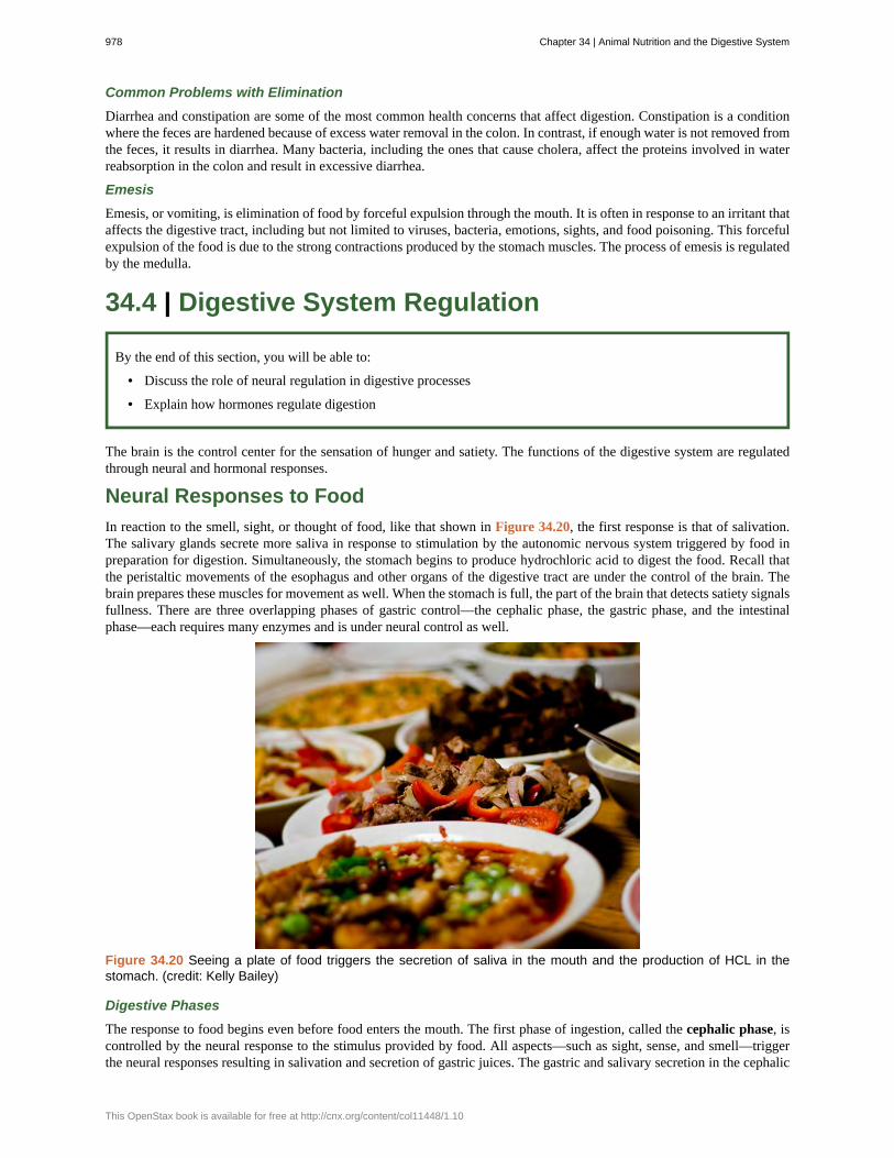
In reaction to the smell, sight, or thought of food, like that shown in Figure 34.20, the first response is that of salivation.The salivary glands secrete more saliva in response to stimulation by the autonomic nervous system triggered by food inpreparation for digestion. Simultaneously, the stomach begins to produce hydrochloric acid to digest the food. Recall thatthe peristaltic movements of the esophagus and other organs of the digestive tract are under the control of the brain. Thebrain prepares these muscles for movement as well. When the stomach is full, the part of the brain that detects satiety signalsfullness. There are three overlapping phases of gastric control—the cephalic phase, the gastric phase, and the intestinalphase—each requires many enzymes and is under neural control as well.
Figure 34.20 Seeing a plate of food triggers the secretion of saliva in the mouth and the production of HCL in thestomach. (credit: Kelly Bailey)
Digestive Phases
The response to food begins even before food enters the mouth. The first phase of ingestion, called the cephalic phase, iscontrolled by the neural response to the stimulus provided by food. All aspects—such as sight, sense, and smell—triggerthe neural responses resulting in salivation and secretion of gastric juices. The gastric and salivary secretion in the cephalic
978 Chapter 34 | Animal Nutrition and the Digestive System
This OpenStax book is available for free at http://cnx.org/content/col11448/1.10

phase can also take place due to the thought of food. Right now, if you think about a piece of chocolate or a crispy potatochip, the increase in salivation is a cephalic phase response to the thought. The central nervous system prepares the stomachto receive food.
The gastric phase begins once the food arrives in the stomach. It builds on the stimulation provided during the cephalicphase. Gastric acids and enzymes process the ingested materials. The gastric phase is stimulated by (1) distension of thestomach, (2) a decrease in the pH of the gastric contents, and (3) the presence of undigested material. This phase consists oflocal, hormonal, and neural responses. These responses stimulate secretions and powerful contractions.
The intestinal phase begins when chyme enters the small intestine triggering digestive secretions. This phase controls therate of gastric emptying. In addition to gastrin emptying, when chyme enters the small intestine, it triggers other hormonaland neural events that coordinate the activities of the intestinal tract, pancreas, liver, and gallbladder.
Hormonal Responses to Food
The endocrine system controls the response of the various glands in the body and the release of hormones at the appropriatetimes.
One of the important factors under hormonal control is the stomach acid environment. During the gastric phase, the hormonegastrin is secreted by G cells in the stomach in response to the presence of proteins. Gastrin stimulates the release ofstomach acid, or hydrochloric acid (HCl) which aids in the digestion of the proteins. However, when the stomach is emptied,the acidic environment need not be maintained and a hormone called somatostatin stops the release of hydrochloric acid.This is controlled by a negative feedback mechanism.
In the duodenum, digestive secretions from the liver, pancreas, and gallbladder play an important role in digesting chymeduring the intestinal phase. In order to neutralize the acidic chyme, a hormone called secretin stimulates the pancreasto produce alkaline bicarbonate solution and deliver it to the duodenum. Secretin acts in tandem with another hormonecalled cholecystokinin (CCK). Not only does CCK stimulate the pancreas to produce the requisite pancreatic juices, it alsostimulates the gallbladder to release bile into the duodenum.
Visit this website (http://openstaxcollege.org/l/enteric_endo) to learn more about the endocrine system. Review thetext and watch the animation of how control is implemented in the endocrine system.
Another level of hormonal control occurs in response to the composition of food. Foods high in lipids take a long timeto digest. A hormone called gastric inhibitory peptide is secreted by the small intestine to slow down the peristalticmovements of the intestine to allow fatty foods more time to be digested and absorbed.
Understanding the hormonal control of the digestive system is an important area of ongoing research. Scientists areexploring the role of each hormone in the digestive process and developing ways to target these hormones. Advances couldlead to knowledge that may help to battle the obesity epidemic.
Chapter 34 | Animal Nutrition and the Digestive System 979
alimentary canal
aminopeptidase
anus
bile
bolus
carboxypeptidase
carnivore
cephalic phase
cholecystokinin
chylomicron
chyme
chymotrypsin
digestion
dipeptidase
duodenum
elastase
endocrine system
esophagus
essential nutrient
gallbladder
gastric inhibitory peptide
gastric phase
gastrin
gastrovascular cavity
gizzard
herbivore
ileum
ingestion
KEY TERMS
tubular digestive system with a mouth and anus
protease that breaks down peptides to single amino acids; secreted by the brush border of small intestine
exit point for waste material
digestive juice produced by the liver; important for digestion of lipids
mass of food resulting from chewing action and wetting by saliva
protease that breaks down peptides to single amino acids; secreted by the brush border of the smallintestine
animal that consumes animal flesh
first phase of digestion, controlled by the neural response to the stimulus provided by food
hormone that stimulates the contraction of the gallbladder to release bile
small lipid globule
mixture of partially digested food and stomach juices
pancreatic protease
mechanical and chemical break down of food into small organic fragments
protease that breaks down peptides to single amino acids; secreted by the brush border of small intestine
first part of the small intestine where a large part of digestion of carbohydrates and fats occurs
pancreatic protease
system that controls the response of the various glands in the body and the release of hormones at theappropriate times
tubular organ that connects the mouth to the stomach
nutrient that cannot be synthesized by the body; it must be obtained from food
organ that stores and concentrates bile
hormone secreted by the small intestine in the presence of fatty acids and sugars; it alsoinhibits acid production and peristalsis in order to slow down the rate at which food enters the small intestine
digestive phase beginning once food enters the stomach; gastric acids and enzymes process the ingestedmaterials
hormone which stimulates hydrochloric acid secretion in the stomach
digestive system consisting of a single opening
muscular organ that grinds food
animal that consumes strictly plant diet
last part of the small intestine; connects the small intestine to the large intestine; important for absorption of B-12
act of taking in food
980 Chapter 34 | Animal Nutrition and the Digestive System
This OpenStax book is available for free at http://cnx.org/content/col11448/1.10
intestinal phase
jejunum
lactase
large intestine
lipase
liver
maltase
mineral
monogastric
omnivore
pancreas
pepsin
pepsinogen
peristalsis
proventriculus
rectum
roughage
ruminant
salivary amylase
secretin
small intestine
somatostatin
sphincter
stomach
sucrase
trypsin
villi
vitamin
third digestive phase; begins when chyme enters the small intestine triggering digestive secretions andcontrolling the rate of gastric emptying
second part of the small intestine
enzyme that breaks down lactose into glucose and galactose
digestive system organ that reabsorbs water from undigested material and processes waste matter
enzyme that chemically breaks down lipids
organ that produces bile for digestion and processes vitamins and lipids
enzyme that breaks down maltose into glucose
inorganic, elemental molecule that carries out important roles in the body
digestive system that consists of a single-chambered stomach
animal that consumes both plants and animals
gland that secretes digestive juices
enzyme found in the stomach whose main role is protein digestion
inactive form of pepsin
wave-like movements of muscle tissue
glandular part of a bird’s stomach
area of the body where feces is stored until elimination
component of food that is low in energy and high in fiber
animal with a stomach divided into four compartments
enzyme found in saliva, which converts carbohydrates to maltose
hormone which stimulates sodium bicarbonate secretion in the small intestine
organ where digestion of protein, fats, and carbohydrates is completed
hormone released to stop acid secretion when the stomach is empty
band of muscle that controls movement of materials throughout the digestive tract
saclike organ containing acidic digestive juices
enzyme that breaks down sucrose into glucose and fructose
pancreatic protease that breaks down protein
folds on the inner surface of the small intestine whose role is to increase absorption area
organic substance necessary in small amounts to sustain life
CHAPTER SUMMARY
34.1 Digestive Systems
Different animals have evolved different types of digestive systems specialized to meet their dietary needs. Humans andmany other animals have monogastric digestive systems with a single-chambered stomach. Birds have evolved a digestivesystem that includes a gizzard where the food is crushed into smaller pieces. This compensates for their inability to
Chapter 34 | Animal Nutrition and the Digestive System 981
masticate. Ruminants that consume large amounts of plant material have a multi-chambered stomach that digestsroughage. Pseudo-ruminants have similar digestive processes as ruminants but do not have the four-compartment stomach.Processing food involves ingestion (eating), digestion (mechanical and enzymatic breakdown of large molecules),absorption (cellular uptake of nutrients), and elimination (removal of undigested waste as feces).
Many organs work together to digest food and absorb nutrients. The mouth is the point of ingestion and the location whereboth mechanical and chemical breakdown of food begins. Saliva contains an enzyme called amylase that breaks downcarbohydrates. The food bolus travels through the esophagus by peristaltic movements to the stomach. The stomach has anextremely acidic environment. An enzyme called pepsin digests protein in the stomach. Further digestion and absorptiontake place in the small intestine. The large intestine reabsorbs water from the undigested food and stores waste untilelimination.
34.2 Nutrition and Energy Production
Animal diet should be balanced and meet the needs of the body. Carbohydrates, proteins, and fats are the primarycomponents of food. Some essential nutrients are required for cellular function but cannot be produced by the animal body.These include vitamins, minerals, some fatty acids, and some amino acids. Food intake in more than necessary amounts isstored as glycogen in the liver and muscle cells, and in fat cells. Excess adipose storage can lead to obesity and serioushealth problems. ATP is the energy currency of the cell and is obtained from the metabolic pathways. Excesscarbohydrates and energy are stored as glycogen in the body.
34.3 Digestive System Processes
Digestion begins with ingestion, where the food is taken in the mouth. Digestion and absorption take place in a series ofsteps with special enzymes playing important roles in digesting carbohydrates, proteins, and lipids. Elimination describesremoval of undigested food contents and waste products from the body. While most absorption occurs in the smallintestines, the large intestine is responsible for the final removal of water that remains after the absorptive process of thesmall intestines. The cells that line the large intestine absorb some vitamins as well as any leftover salts and water. Thelarge intestine (colon) is also where feces is formed.
34.4 Digestive System Regulation
The brain and the endocrine system control digestive processes. The brain controls the responses of hunger and satiety.The endocrine system controls the release of hormones and enzymes required for digestion of food in the digestive tract.
ART CONNECTION QUESTIONS
1. Figure 34.11 Which of the following statements aboutthe digestive system is false?
a. Chyme is a mixture of food and digestive juicesthat is produced in the stomach.
b. Food enters the large intestine before the smallintestine.
c. In the small intestine, chyme mixes with bile,which emulsifies fats.
d. The stomach is separated from the smallintestine by the pyloric sphincter.
2. Figure 34.12 Which of the following statements aboutthe small intestine is false?
a. Absorptive cells that line the small intestinehave microvilli, small projections that increasesurface area and aid in the absorption of food.
b. The inside of the small intestine has many folds,called villi.
c. Microvilli are lined with blood vessels as well aslymphatic vessels.
d. The inside of the small intestine is called thelumen.
3. Figure 34.19 Which of the following statements aboutdigestive processes is true?
a. Amylase, maltase and lactase in the mouthdigest carbohydrates.
b. Trypsin and lipase in the stomach digest protein.c. Bile emulsifies lipids in the small intestine.d. No food is absorbed until the small intestine.
REVIEW QUESTIONS
4. Which of the following is a pseudo-ruminant?
a. cowb. pig
c. crowd. horse
5. Which of the following statements is untrue?a. Roughage takes a long time to digest.
982 Chapter 34 | Animal Nutrition and the Digestive System
This OpenStax book is available for free at http://cnx.org/content/col11448/1.10

b. Birds eat large quantities at one time so that theycan fly long distances.
c. Cows do not have upper teeth.d. In pseudo-ruminants, roughage is digested in the
cecum.
6. The acidic nature of chyme is neutralized by ________.
a. potassium hydroxideb. sodium hydroxidec. bicarbonatesd. vinegar
7. The digestive juices from the liver are delivered to the________.
a. stomachb. liverc. duodenumd. colon
8. Which of the following statements is not true?
a. Essential nutrients can be synthesized by thebody.
b. Vitamins are required in small quantities forbodily function.
c. Some amino acids can be synthesized by thebody, while others need to be obtained from diet.
d. Vitamins come in two categories: fat-soluble andwater-soluble.
9. Which of the following is a water-soluble vitamin?
a. vitamin Ab. vitamin Ec. vitamin Kd. vitamin C
10. What is the primary fuel for the body?
a. carbohydratesb. lipidsc. proteind. glycogen
11. Excess glucose is stored as ________.a. fatb. glucagonc. glycogend. it is not stored in the body
12. Where does the majority of protein digestion takeplace?
a. stomachb. duodenumc. mouthd. jejunum
13. Lipases are enzymes that break down ________.
a. disaccharidesb. lipidsc. proteinsd. cellulose
14. Which hormone controls the release of bile from thegallbladder
a. pepsinb. amylasec. CCKd. gastrin
15. Which hormone stops acid secretion in the stomach?
a. gastrinb. somatostatinc. gastric inhibitory peptided. CCK
CRITICAL THINKING QUESTIONS
16. How does the polygastric digestive system aid indigesting roughage?
17. How do birds digest their food in the absence of teeth?
18. What is the role of the accessory organs in digestion?
19. Explain how the villi and microvilli aid in absorption.
20. What are essential nutrients?
21. What is the role of minerals in maintaining goodhealth?
22. Discuss why obesity is a growing epidemic.
23. There are several nations where malnourishment is acommon occurrence. What may be some of the healthchallenges posed by malnutrition?
24. Explain why some dietary lipid is a necessary part of abalanced diet.
25. Describe how hormones regulate digestion.
26. Describe one or more scenarios where loss ofhormonal regulation of digestion can lead to diseases.
Chapter 34 | Animal Nutrition and the Digestive System 983