
Chapter 3 Ultrafast Chemistry: Using Time-Resolved...
34
NIBBERING ■ FIDDER ■ PINES ULTRAFAST CHEMISTRY AND VIBRATIONS Chapter 3 Ultrafast Chemistry: Using Time-Resolved Vibrational Spectroscopy for Interrogation of Structural Dynamics C O H O electronic spectroscopy vibrational spectroscopy Physics: - linear and nonlinear light field interaction - electronic state coupling - vibrational energy redistribution and dissipation - solvent inertial and diffusional motion Chemistry: - excited state intramolecular hydrogen transfer - bimolecular proton transfer - electron transfer - hydrogen bond rearrangements - bond fission - cis-trans isomerizations Published as: E. T. J. Nibbering, H. Fidder and E. Pines, in Annual Review of Physical Chemistry, Vol. 56, S. R. Leone, P. Alivisatos, A. E. McDermott, eds., pp. 337-367 (Annual Reviews, Palo Alto, USA, 2005).
Transcript of Chapter 3 Ultrafast Chemistry: Using Time-Resolved...

NIBBERING ■ FIDDER ■ PINES ULTRAFAST CHEMISTRY AND VIBRATIONS
Chapter 3
Ultrafast Chemistry: Using Time-Resolved Vibrational
Spectroscopy for Interrogation of Structural Dynamics
CO
HO
electronic spectroscopy vibrational spectroscopy Physics: - linear and nonlinear light field interaction - electronic state coupling - vibrational energy redistribution and dissipation - solvent inertial and diffusional motion Chemistry: - excited state intramolecular hydrogen transfer - bimolecular proton transfer - electron transfer - hydrogen bond rearrangements - bond fission - cis-trans isomerizations
Published as: E. T. J. Nibbering, H. Fidder and E. Pines, in Annual Review of Physical Chemistry, Vol. 56, S. R.
Leone, P. Alivisatos, A. E. McDermott, eds., pp. 337-367 (Annual Reviews, Palo Alto, USA, 2005).

72 NIBBERING ■ FIDDER ■ PINES

ULTRAFAST CHEMISTRY AND VIBRATIONS 73
Annu. Rev. Phys. Chem. 2005. 56:337–67doi: 10.1146/annurev.physchem.56.092503.141314
Copyright c© 2005 by Annual Reviews. All rights reservedFirst published online as a Review in Advance on January 7, 2005
ULTRAFAST CHEMISTRY: Using Time-ResolvedVibrational Spectroscopy for Interrogation ofStructural Dynamics
Erik T.J. Nibbering,1 Henk Fidder,2 and Ehud Pines3
1Max Born Institut fur Nichtlineare Optik und Kurzzeitspektroskopie, D-12489 Berlin,Germany; email: [email protected] of Physical Chemistry, Uppsala Universitet, S-751 23 Sweden;email: [email protected] of Chemistry, Ben-Gurion University of the Negev, Beer-sheva 84105,Israel; email: [email protected]
Key Words hydrogen and proton transfer, hydrogen bonding and solvation,intramolecular vibrational redistribution and vibrational cooling, anharmoniccoupling between vibrational modes, internal conversion
■ Abstract Time-resolved infrared (IR) and Raman spectroscopy elucidates molec-ular structure evolution during ultrafast chemical reactions. Following vibrationalmarker modes in real time provides direct insight into the structural dynamics, asis evidenced in studies on intramolecular hydrogen transfer, bimolecular proton trans-fer, electron transfer, hydrogen bonding during solvation dynamics, bond fission inorganometallic compounds and heme proteins, cis-trans isomerization in retinal pro-teins, and transformations in photochromic switch pairs. Femtosecond IR spectroscopymonitors the site-specific interactions in hydrogen bonds. Conversion between excitedelectronic states can be followed for intramolecular electron transfer by inspection ofthe fingerprint IR- or Raman-active vibrations in conjunction with quantum chemicalcalculations. Excess internal vibrational energy, generated either by optical excita-tion or by internal conversion from the electronic excited state to the ground state,is observable through transient frequency shifts of IR-active vibrations and throughnonequilibrium populations as deduced by Raman resonances.
1. INTRODUCTION: ULTRAFAST STRUCTURALDYNAMICS IN CONDENSED-PHASE CHEMISTRY
Chemistry occurs on a large range of timescales. Rearrangements of biomolecularstructures, such as protein synthesis in ribosomes, or DNA multiplication, involvea large number of chemical transformations, making the timescales of these pro-cesses span on the order of seconds to hours. Bimolecular reaction dynamics inliquid solutions are dominated by relatively slow mutual diffusion (1); typical
0066-426X/05/0505-0337$20.00 337

74 NIBBERING ■ FIDDER ■ PINES
338 NIBBERING � FIDDER � PINES
timescales are on the order of nanoseconds. Elementary steps in chemistry, suchas the dynamical event of a single-bond rearrangement, appear to take place onfemtosecond to picosecond timescales [femtochemistry (2)]. Examples of ultrafastchemistry include hydrogen and proton transfer, electron transfer, bond fissions,and cis-trans isomerizations. In the case of ultrafast condensed-phase chemistrythe dynamics are determined not only by the potential energy surfaces of thereactant-product species. The surrounding solvent also plays a major role in mod-ulating the energy levels of reactants, intermediates, and products, as well as theenergy barriers separating these species (3). In addition, the solvent plays the roleof donor or acceptor of energy (4), often causing an outcome of chemical reactionsdifferent from those for isolated reacting species. Photoinduced chemistry offersthe opportunity of triggering the reaction dynamics at a well-defined point in time.Ultrafast spectroscopy has been an exquisite tool to follow chemical reactions inreal time after applying such an optical trigger pulse.
Since the development of short-pulsed laser systems, ultrafast chemistry in thecondensed phase has been studied mainly using time-resolved electronic spec-troscopy. After the ultrashort optical trigger pulse induces a transition to a higherlying electronic state, the evolution of the molecular system is followed either bytime-resolved fluorescence emission, or by absorbance changes of a probe pulsetuned to electronic resonances in the near-UV or vis regions of the electromagneticspectrum. The disadvantage of probing electronic transitions lies in the fact thatthese are typically strongly broadened owing to coupling to the fluctuating sur-rounding solvent (5–7). As a result, the relatively featureless transient electronicbands of stimulated emission and excited state absorption contributions often over-lap (8), whereas spectral shifting and reshaping caused by solvent reorganization(solvation dynamics) and energy dissipation (vibrational cooling) complicate thesituation even more.
More insight into the dynamics of molecular structures may be achieved us-ing ultrafast structurally resolving techniques. Whereas time-resolved X-ray (9)and electron diffraction (10, 11) and X-ray spectroscopy (12) are still rather tech-nologically demanding, recent developments in ultrafast laser technology haveenabled the efficient generation of tunable femtosecond laser pulses from theUV to the far-infrared regions of the electromagnetic spectrum (13, 14), makingfemtosecond vibrational spectroscopy a versatile tool. Since an early example ofpicosecond infrared (IR) spectroscopy on excited state intramolecular hydrogentransfer (ESIHT) in 1986 (15), the method has mainly been employed on metallo-carbonyl compounds (16–19) and heme proteins (20–22). With the availability ofsensitive mid-infrared detector arrays it is now possible to fully explore the po-tential of time-resolved vibrational spectroscopy for gaining insight into transientmolecular structures involved in chemical reaction pathways. Until now, opticallytriggered chemical reaction dynamics has been studied with femtosecond IR spec-troscopy on cases as diverse as ESIHT (23, 24), bimolecular proton transfer (25,26), electron transfer (27–39), hydrogen bonding in solvation dynamics (40–44),photochemistry of small molecules (17, 45–47), bond fission (48–56) and bond

ULTRAFAST CHEMISTRY AND VIBRATIONS 75
ULTRAFAST CHEMISTRY AND VIBRATIONS 339
activation (57–60) in organometallic compounds, ligand release in heme proteins(9, 61–64), cis-trans isomerization (65–70), and more extensive rearrangementsin photochromic switches (71–73). Picosecond resonance Raman spectroscopyhas been used in electron transfer studies (74–76) and intramolecular vibrationalredistribution studies (77–82). Coherent anti-Stokes Raman spectroscopy (CARS)has been applied to photoinduced dynamics of photochromic switches (83), retinalproteins (84–89), and photoactive yellow protein (90).
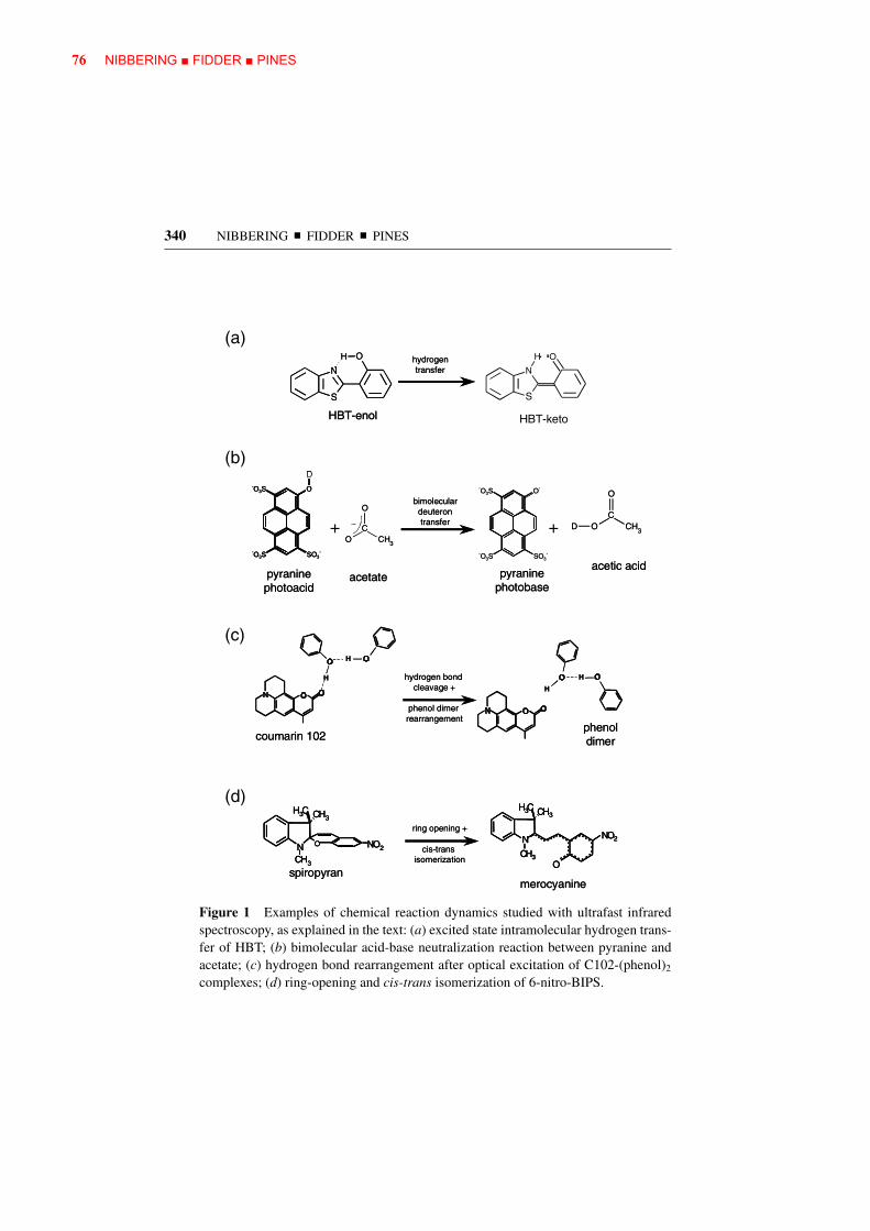
We give an overview of recent achievements in time-resolved vibrational spec-troscopy of ultrafast condensed-phase chemistry. We first describe the advantagesof ultrafast vibrational spectroscopy as a tool to resolve dynamically changingmolecular structures. We then present examples of excited state hydrogen transfer,bimolecular proton transfer dynamics, excited state electron transfer, hydrogenbonding in solvation dynamics, bond fission, and isomerization reactions (also seeFigure 1). We conclude with a prospective outlook.
2. ULTRAFAST INFRARED AND RAMAN SPECTROSCOPY
Vibrational spectroscopy has several advantages over electronic spectroscopy. Vi-brational transitions can often be correlated to specific vibrational motions byinspection of the transition frequencies. In particular the fingerprint region offers awealth of structural information. From identification of these fingerprint vibrationalmodes, conclusions can be drawn on specific structural motifs in the molecules.Vibrational transitions have bandwidths typically smaller (10–20 cm−1) than thosefrom electronic transitions (typically 200–2000 cm−1), due to longer transversedephasing times [the exception to the rule: O–H/O–D stretching vibrations inhydrogen-bonded hydroxyl groups (91)]. It is thus less probable that different tran-sition bands overlap in vibrational spectroscopy than in electronic spectroscopy. Inaddition, small molecular species may always be probed through their vibrations,whereas their electronic transitions often lie in the far-UV (that would normallybe hidden underneath the solvent bands). Major disadvantages of vibrational spec-troscopy on the other hand are the inherent lower cross sections of vibrationaltransitions, and the frequent overlap of the absorption bands with those of the sol-vent. The mismatch in cross sections of electronic and vibrational transitions oftenleads to experimental situations having high excitation densities of samples withabsorber concentrations on the order of 1–20 mM (with often high optical densitiesfor the electronic transitions of OD = 1–2). Deformation of laser pulses whilepropagating through the samples, probe molecule aggregation and sample degra-dation have to be checked when performing ultrafast mid-infrared experiments.Time-resolved Raman experiments often are hampered by unwanted fluorescentemission of the molecules under study, or of impurities in the samples.
In the case that specific vibrational marker modes of reactant, intermediate,and product states are identified, one can follow the outcome of the photoin-duced reaction by inspection of the transitions of these marker modes. Site-specific
76 NIBBERING ■ FIDDER ■ PINES
340 NIBBERING � FIDDER � PINES
H
N
S
O
HBT-enol
H
N
S
O
HBT-enol
hydrogentransfer
hydrogentransfer
HBT-keto
OH
N
S
ONCH3
CH3 CH3
NO2N
CH3
CH3 CH3
O
NO2
spiropyranmerocyanine
ring opening +
cis-trans isomerization
ONCH3
CH3 CH3
NO2ON
CH3
CH3 CH3
NO2N
CH3
CH3 CH3
O
NO2N
CH3
CH3 CH3
O
NO2
spiropyranmerocyanine
ring opening +
cis-trans isomerization
(a)
(b)
(c)
(d)
coumarin 102
hydrogen bond cleavage +
phenol dimer rearrangement
H
H H
H
phenoldimercoumarin 102
hydrogen bond cleavage +
phenol dimer rearrangement
H
H H
H
phenoldimer
-O3S
SO3--O3S
O
HD
pyraninephotoacid
-O3S
SO3--O3S
O
HD-O3S
SO3--O3S
O
HD
pyraninephotoacid
CCH
O
O 3
_
acetate
CCH
O
O 3
_
acetate
-O3S
SO3--O3S
O-
pyraninephotobase
-O3S
SO3--O3S
O-
pyraninephotobase
CCH
O
OD 3
acetic acid
CCH
O
OD 3
acetic acid
++
bimoleculardeuterontransfer
bimoleculardeuterontransfer
Figure 1 Examples of chemical reaction dynamics studied with ultrafast infraredspectroscopy, as explained in the text: (a) excited state intramolecular hydrogen trans-fer of HBT; (b) bimolecular acid-base neutralization reaction between pyranine andacetate; (c) hydrogen bond rearrangement after optical excitation of C102-(phenol)2
complexes; (d) ring-opening and cis-trans isomerization of 6-nitro-BIPS.

ULTRAFAST CHEMISTRY AND VIBRATIONS 77
ULTRAFAST CHEMISTRY AND VIBRATIONS 341
information is obtained in the case of vibrational marker modes that involve nu-clear motions of specific molecular side groups. When vibrational normal modesare involved in nuclear motions of extended parts of the molecules, a direct struc-tural insight is usually not obtained. Isotopic substitution reveals the involvementof certain nuclei in the vibrational motions. When, in comparing the experimen-tally observed vibrational mode pattern with predictions from quantum chemicalcalculations, a full correspondence is possible, the three-dimensional structure isderivable. Current quantum chemical calculational routines, such as density func-tional theory (92), allow for the estimation of the electronic ground state structureof medium-sized molecules. For intermediate and product states in electronic ex-cited states, however, reliable results can be obtained with the routine of ab initiocomplete active space self-consistent field (CASSCF) (93), albeit for mid-sizemolecules not much larger than 20 atoms. New developments in numerical pro-cedures, such as time-dependent density functional theory (TD-DFT) (94), mayprove beneficial in the estimation of larger molecular structures in electronic ex-cited states.
Polarization-dependent UV/vis-pump IR-probe spectroscopy provides insightinto the relative angles between the optical and IR transition dipole moments.From these experiments angular information on transient species is obtainable(20, 61). Because of sensitivity reasons (electronic transitions typically have crosssections 100–1000 times stronger than vibrational transitions), often high energydensities of the electronic pump pulses are used. Care should be taken whenconsidering polarization-dependent measurements because of the effects of finitebleaching, where the ground state bleaching of the sample is significant (a totalbleached fraction on the order of 0.1 or higher) (21). When the vibrational markermode is a local mode, a direct link is present between the optical transition dipolemoment and the vector of the chemical bond modulated by the vibration. Moreoften, for delocalized normal modes one must rely on comparison between theexperimentally found angular information and quantum chemical calculations (64,95).
In the most basic situation the reaction kinetics are deducible by inspection ofvibrational marker modes of reactants, intermediates, and products. The rise anddecay behavior of specific vibrational marker bands indicate the formation and dis-appearance of reactant, intermediate, and product states. Inspection of the spectralshifts and widths of these bands, however, can reveal additional information onlocal interactions, vibrational energy redistribution, and energy dissipation to thesolvent.
Site-specific information is obtainable by inspection of local modes that are af-fected by local interactions. For instance, it is well known that hydrogen bondinginduces marked shifts in O–H, N–H, C=O and C=N bands in the case wherethese structural functionalities are involved in hydrogen bonding (96). Obser-vation of transient spectral shifts reveals important information on changes inhydrogen bond interactions (weakening/strengthening or even hydrogen bondcleavage).

78 NIBBERING ■ FIDDER ■ PINES
342 NIBBERING � FIDDER � PINES
In polyatomic molecules the vibrational normal modes are usually considered inthe harmonic oscillator approximation. In this limit the motions of the vibrationalmodes are independent of each other. In reality anharmonicity of the vibrationalmodes cannot be neglected; anharmonicity is typically treated as perturbation tothe Hamiltonian in the eigenstate representation of the harmonic oscillator normalmodes (97, 98). The first consequence is diagonal anharmonicity, where the en-ergy spacing between the vibrational levels becomes smaller as the correspondingvibrational quantum numbers become higher. Another consequence is that the mo-tions of the vibrational normal modes are no longer independent, as indicated byoff-diagonal anharmonicity terms in the Hamiltonian. Let us assume a representa-tion of the vibrational energy eigenstates of a polyatomic molecule with quadraticanharmonicity only (without degenerate states) (66) as:
E
hc=
∑i
νi
(vi + 1
2
)+
∑i≤ j
xi j
(vi + 1
2
) (v j + 1
2
), 1.
where ν i is the harmonic frequency, and vi is the vibrational quantum number ofmode i, and xij are the anharmonic coupling constants between modes i and j, whichtypically have negative values. The transition frequency for a mode k coupled tothe other modes i �= k is then:
E (Vk → Vk + 1)
hc= ν ′
k + 2xkkvk +∑i �=k
xikvi ; 2.
the first term on the right-hand side of Equation 2 defines the anharmonic correctionfor the vk → vk + 1 transition when the other vibrational modes i are in the groundstate (vi = 0), i.e., the molecule is cold:
ν ′k = νk + 2xkk +
∑i �=k
xik
2. 3.
The second term on the right-hand side of Equation 2 indicates the influenceof the diagonal anharmonicity: The transitions in the vibrational ladder are suc-cessively shifted to lower frequencies. The third term on the right-hand side ofEquation 2 shows the influence of off-diagonal anharmonicity: when other modesi are highly excited (high quantum number vi indicates that the molecule is hot,i.e., it has a large internal vibrational energy), the marker mode k exhibits a red-shifted transition frequency, even for the fundamental vk = 0 → vk = 1 transition.This off-diagonal anharmonicity is explored in multidimensional IR spectroscopicstudies (99). Multidimensional IR spectroscopy has mainly been pursued in the elu-cidation of polypeptide and metallo-carbonyl compounds in the electronic groundstate, where the magnitudes of the couplings give structural information, and thedynamics of these couplings reveal the fluctuations of these structures. Only re-cently has this technique been applied on transient states generated by an electronicpump pulse (69).

ULTRAFAST CHEMISTRY AND VIBRATIONS 79
ULTRAFAST CHEMISTRY AND VIBRATIONS 343
A high degree of internal vibrational energy can be generated under differentcircumstances. For instance, for electronic transitions with a small displacementof Raman-active vibrations most of the electronic transition moment is located inthe electronic 0–0 origin transition. The transition could be excited into the higherlying part of the electronic band by tuning the UV/vis-pump pulse where, uponelectronic excitation, several Raman-active vibrations also change their vibrationalquantum number. When probing an infrared-active marker mode in this electronicexcited state, a red-shifted transition frequency is observable for this marker modeas opposed to where it will show up when exciting in the electronic origin. Formolecular systems with a large displacement for one or several Raman-active vi-brations, the maximum of the absorption band already corresponds to a significantamount of internal vibrational energy. The apparent red-shifted position of the IR-active marker modes already occurs when this excess vibrational energy is onlycontained in a subset of vibrational modes (e.g., the Raman-active modes that showa large displacement upon electronic excitation). Intramolecular vibrational energyredistribution (IVR) will typically equilibrate this excess energy over all vibrationalmodes in the molecule (on timescales ranging from below 100 fs to several picosec-onds for mid-size molecules) (100). In condensed-phase solutions the energy dis-sipation to the solvent (cooling) is irreversible, which translates into a blue-shiftingbehavior of the positions of the vibrational marker modes to the frequency positionobservable when exciting into the electronic origin transition. Vibrational coolingtypically takes place on timescales of several tens of picoseconds (101–103).
When ultrafast internal conversion to other electronic states (reactant groundstate, or product states) occurs, typically a substantial amount of—if not allelectronic—excitation energy is converted into internal vibrational energy beforethe energy dissipation to the solvent can be effective. Transiently red-shifted vibra-tional transitions of fingerprint marker modes have been observed in these cases aswell. In the case of an ultrafast internal conversion of the electronic excited stateto the ground state on a timescale of a few hundreds of femtoseconds, one wouldtypically observe for the vibrational marker mode a bleach signal correspondingto the location of the marker mode transition of the cold molecule, and red-shiftedthe vibrational marker mode transition of the hot molecule. Only upon cooling isthe disappearance of the red-shifted hot ground state absorption accompanied bya refill of the ground state bleach observable. The magnitude of the refill of theground state bleach enables an estimation of the quantum yield of this ultrafastinternal conversion (IC) process back to the electronic ground state (71–73).
Experimentally, transient IR spectroscopy is performed in a spectrally-resolvedconfiguration. Femtosecond IR parametric devices deliver pulses with bandwidthsof 150 cm−1 or more (14). In order to observe shifts as small as the linewidths ofIR-active vibrations, the IR absorbance change must be measured with a detectorafter spectral dispersion with a monochromator. As a side effect of this spectraldispersion, ground state bleach signals often appear to grow at negative pulsedelay with the dephasing time of the transition. This effect, known as perturbed freeinduction decay, is a common feature of spectrally-resolved nonlinear pump-probe

80 NIBBERING ■ FIDDER ■ PINES
344 NIBBERING � FIDDER � PINES
spectroscopy of bleached transitions with dephasing times much longer than thetime resolution of the experiment (104–106). The time resolution of the experimentis given by the cross correlation between the UV/vis-pump and IR-probe pulse(about 100–200 fs), and is typically dominated by group velocity mismatch insamples with thicknesses of about 100 µm.
In principle the same approach can be followed by probing Raman-active vibra-tions. In this case the spectral resolution is determined not only by the monochro-mator through which the spontaneous Raman emission is dispersed, but also bythe bandwidth of the gating pulse by which the Raman effect is induced. As a re-sult UV/vis-pump Raman-probe spectroscopy has a temporal resolution of around1 ps (a compromise between spectral and time resolution) (107). Because of theeven weaker cross sections of Raman transitions, resonance enhancement is oftenused by tuning the gating pulse close to or resonant with an electronic transitionof the state that is probed. This has the advantage of isolating Raman bands of thestate under inspection for observation. The drawback is that fluorescence resultingfrom the resonant electronic excitation often inhibits detection of Raman bandsin extended spectral ranges. Dedicated Raman detection techniques such as Kerr-gates may suppress this unwanted fluorescence emission (108). CARS, being acoherent technique, does not have this drawback, at the cost of a more complexlaser configuration.
Raman spectroscopy offers an additional insight into how chemical reactionsevolve. By comparison of the intensities of anti-Stokes and Stokes lines of a par-ticular vibration, it is possible to derive time-dependent changes in the excitationlevel of this vibration. One can draw conclusions on whether particular modesinitially drive a chemical reaction (state transition promoting modes), or only getexcited after the transition is made, by taking up the excess energy released bythe reaction (accepting modes). In such a way insight is obtained on the energyflow inside a molecular system (IVR) and cooling to the surrounding solvent.Although IR-active transitions exhibit transient frequency shifts due to IVR andcooling as well, it is not easy to deduce transient higher excitation levels of thesemodes.
3. EXCITED STATE INTRAMOLECULARHYDROGEN TRANSFER
Hydrogen bonds determine to a substantial extent the microscopic structure ofa large variety of molecular systems, ranging from hydrogen-bonding liquids toproteins and DNA. In these structures molecular function often involves hydrogenor proton transfer along preformed hydrogen bonds. The most elementary hydro-gen transfer reactions consist of a hydrogen atom H shifting from originally beingpart of a covalent bond A–H to a new binding site B in the same or a neighboringmolecule.
A−H · · · B → A · · · H−B 4.

ULTRAFAST CHEMISTRY AND VIBRATIONS 81
ULTRAFAST CHEMISTRY AND VIBRATIONS 345
In the electronic ground state a transfer can be caused by solvent-induced fluc-tuations of hydrogen-bonded networks. Hydrogen transfer can also be induced af-ter optical excitation. The study of photoinduced ESIHT was pioneered by Weller(109). (For a review of early work on several prototype ESIHT cases, see Reference110; for a recent overview including ultrafast spectroscopic work, see Reference111.) Although traditionally these experiments have been interpreted to in-volve excited state intramolecular proton transfer (ESIPT), in reality the motions ofthe proton are typically accompanied by rearrangements of the electronic chargedensities, and it is more appropriate to describe the observed features as beingcaused by the net transfer of a hydrogen atom, i.e., ESIHT. Elsaesser & Kaiser re-ported a pioneering picosecond IR experiment on 2-(2′-hydroxyphenyl)benzothia-zole (HBT) (15), where a first direct characterization of local changes of moleculargeometries due to hydrogen transfer from the enol to keto state was demonstratedby inspection of IR absorbance changes in the O–H stretching band centred at3000 cm−1 and in the fingerprint region between 1400 and 2000 cm−1. Femtosec-ond time resolution however, is required to resolve the formation dynamics of theproduct state. In UV/vis pump-probe experiments, where e.g., stimulated emis-sion from product states is measured, rise times for ESIHT systems ranging fromseveral tens to hundreds of femtoseconds have been found (111).
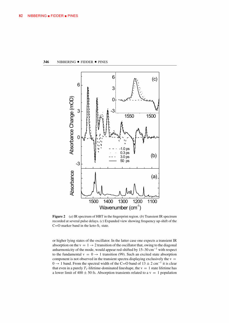
Femtosecond IR spectroscopy has been reported on the ESIHT enol-keto re-action of HBT (23, 24). Here the C=O stretching marker mode at 1530 cm−1, aswell as other fingerprint vibrations, has been monitored after excitation with theUV pump tuned between 310 and 350 nm (see Figure 2). The C=O stretchingmode has a transition frequency significantly lower than free C=O vibrations, asthe carbonyl group is part of the hydrogen-bonded C–N–H · · · O==C moiety andbecause it is part of a larger conjugated π -electronic system. Figure 3 shows therise of the C=O stretching marker mode delayed by 30–50 fs after UV excita-tion of the enol state. This result indicates that the keto-state is generated on a50 fs timescale, hinting at an essentially barrierless excited state potential alongthe reaction coordinate (23). The transfer dynamics however, appears to be muchslower than the period of the O–H stretching vibration of approximately 10 fs. Thisleads to the conclusion that hydrogen transfer does not involve a simple stretchingmotion from donor to acceptor groups.
The transient IR spectra of HBT also provide information on IVR and coolingprocesses (23, 24). During and after hydrogen transfer, the vibronic excess energyoriginating from the UV pump pulse and from the energy difference between theenol-S1 and keto-S1 states is redistributed over numerous intramolecular vibrationsand is eventually dissipated to the solvent. If any of the investigated modes isexcited to the corresponding v = 1 or higher levels, it will immediately give rise tovibrational absorption and stimulated emission from these higher lying levels, andto different signatures in the transient absorbance spectra. The C=O stretchingmode, however, only shows minor changes in spectral envelope and integratedabsorption as a function of pulse delay (23). This points to a formation of theC=O stretching mode in its v = 0 ground state without passing through v = 1
82 NIBBERING ■ FIDDER ■ PINES
346 NIBBERING � FIDDER � PINES
Figure 2 (a) IR spectrum of HBT in the fingerprint region. (b) Transient IR spectrumrecorded at several pulse delays. (c) Expanded view showing frequency up-shift of theC=O marker band in the keto-S1 state.
or higher lying states of the oscillator. In the latter case one expects a transient IRabsorption on the v = 1 → 2 transition of the oscillator that, owing to the diagonalanharmonicity of the mode, would appear red-shifted by 15–30 cm−1 with respectto the fundamental v = 0 → 1 transition (99). Such an excited state absorptioncomponent is not observed in the transient spectra displaying exclusively the v =0 → 1 band. From the spectral width of the C=O band of 13 ± 2 cm−1 it is clearthat even in a purely T1-lifetime-dominated lineshape, the v = 1 state lifetime hasa lower limit of 400 ± 50 fs. Absorption transients related to a v = 1 population

ULTRAFAST CHEMISTRY AND VIBRATIONS 83
ULTRAFAST CHEMISTRY AND VIBRATIONS 347
Figure 3 (a) Delayed rise of the C=O stretching band of HBT in the keto-S1 state(dots, solvent signals substracted; solid curve, cross-correlation as recorded in ZnSe).(b) Transient frequency up-shift of the C=O band caused by intramolecular vibrationalenergy redistribution and cooling.
decay can thus easily be detected with a time resolution of 100 fs. The absence ofsignatures of v = 1 population suggests that the C=O vibration does not act asprimary accepting mode driving the hydrogen transfer reaction by accepting (partof) the vibrational excess energy of about 3000 cm−1 released on transfer fromenol to keto configuration. Similar to the behavior of the C=O stretching mode,all other observed IR-active fingerprint vibrations appear to be generated in theirrespective v = 0 states (24).
All IR-active vibrations in the fingerprint region show after initial appearancea blue-shift in transition frequency that can be fitted with two time constantsranging between 600–750 fs and 14–16 ps (23, 24) (see Figure 3). This spectral
84 NIBBERING ■ FIDDER ■ PINES
348 NIBBERING � FIDDER � PINES
shifting behavior is indicative of IVR and cooling processes, respectively, similarto that observed in other photoinduced chemical studies such as on azobenzene(66) or trans-stilbene (103). The transient spectra do not give direct insight intowhich anharmonically coupled modes cause the initial redshift. However, detailedsteady-state resonance Raman of enol-HBT (112) and femtosecond UV/vis pump-probe studies (113) show the substantial displacement of a subset of low-frequencyRaman-active vibrations in HBT. These modes are strongly elongated upon UVexcitation, i.e., they acquire a significant nonequilibrium excitation. Upon fem-tosecond excitation some of these modes show coherent motions that last up to2 ps. The observed blue-shift of the IR-active fingerprint vibrations then re-flects the decrease of the excess population of these low-frequency Raman-activemodes. It is interesting to note that the damping time T2 = 1–2 ps of the co-herent oscillations observed in femtosecond UV/vis pump-probe studies is abouttwice the fast component in the blue-shift representing IVR, as is expected fora dephasing dominated by population relaxation of excited vibrational levels(using the rule 1/T2 = 1/(2T1) + 1/T∗
2, the pure dephasing rate is negligible1/T∗
2∼= 0).
4. BIMOLECULAR ACID-BASE PROTONTRANSFER REACTIONS
The neutralization reactions between Brønsted acids and bases in liquid solutionare a prototype chemical reaction, as they provide insight into proton exchangephenomena such as autoionization in water (114), the von Grotthuss mechanism ofhigh proton mobility in water (115), and proton pumps through membranes (116).In these reactions a proton is exchanged between the acid and the base:
RO−H + B → RO− + H−B+. 5.
Because these reactions are bimolecular, in liquid solution they are typicallycontrolled by mutual diffusion (1). Eigen and Weller have established that foracid-base reactions in solutions, typical diffusion timescales are on the order ofhundreds of picoseconds or longer. However, when an acid and a base do meetand form a reaction contact pair, the intrinsic on-contact reaction takes place withrates ranging from 10 ps−1 to 1 ps−1 (109, 117). Diffusion prevents a more refinedinsight of bimolecular reaction dynamics. The strategic approach to suppress thedominating role of diffusion is to investigate preformed complexes. In particular,proton transfer requires the formation of acid-base complexes, where the acid andbase are connected via at least one hydrogen bond, which constitutes the protontransfer coordinate.
Proton transfer can be optically triggered by using a photoacid as proton donor.In a photoacid the electronic ground state has a low acidity (indicated by a largepKa value; Ka is the acid dissociation equilibrium constant in water). Excita-tion to an electronic excited state having a larger acidity leads to a jump in pKa
value, typically by 4–6 units, thus facilitating an ultrafast optical triggering of the

ULTRAFAST CHEMISTRY AND VIBRATIONS 85
ULTRAFAST CHEMISTRY AND VIBRATIONS 349
proton transfer reaction. Photoacids have been studied since the pioneering workof Forster, Weller, and Eigen (109, 117, 118). Several classes of photoacids ex-ist, most notably aromatic alcohols (phenols, naphthols, pyrenols) and protonatedaminopyrenes (119–121).
Until now time-resolved studies, using time-correlated fluorescence single pho-ton counting or UV/vis pump-probe spectroscopy, have investigated the protontransfer dynamics from the perspective of the photoacid (120). These studies pro-vide in principle only direct insight into the dynamical process of proton dissoci-ation from the photoacid by monitoring the decay of the photoacid S1 state and/orthe rise of the conjugated photobase S1-species. These studies give insight intothe proton transfer dynamics when the proton is directly scavenged by the ac-cepting base. However, when the proton is first dissociating from the photoacidto the solvent, only to be taken up by an accepting base at later times, both thephotoacid/conjugated photobase and the accepting base require monitoring. Thisis possible by following the dynamics of vibrational marker modes of the protondonating and the proton accepting species.
The acid-base neutralization reaction of pyranine (8-hydroxy-1,3,6-trisulfonate-pyrene; HPTS) with acetate (122, 123) in deuterated water recently has been inves-tigated with femtosecond IR spectroscopy (25, 26). Here the vibrational markermodes were the HPTS photoacid band at 1493 cm−1 and the HPTS photobase bandat 1503 cm−1 and the C=O stretching mode of acetic acid at 1720 cm−1, whichis formed upon deuteron pick-up by acetate. The decay of the HPTS photoacidand the rise of the HPTS photobase band indicate when the deuteron leaves thephotoacid, whereas the rise of the acetic acid band specifies when the deuteronarrives at the deuteron accepting base.
By varying the concentration of the base acetate, and keeping the HPTS con-centration fixed, several acid-base neutralization reaction schemes take place. Itis known from previous studies that HPTS releases a deuteron to be solvated bythe solvent D2O with a time constant of 250 ps at room temperature (124). Withthe time-resolved IR experiment it has been demonstrated that for low acetateconcentrations (<1 M) this solvated deuteron will be picked up by acetate at latertimes (25) (see Figure 4).
RO−D + D2O + Ac− → RO− + (D3O)+ + Ac− → RO− + D2O + D−Ac
solvated deuteron full deuteron transfer
6.
For higher concentrations of acetate (>1 M) the relative encounter times be-tween acetate and HPTS is short enough that a direct deuteron scavenging by theacetate base from the photoexcited HPTS dominates (125) (Figure 5). In the caseof high base concentrations additional signal contributions to the rise of the C=Ostretching mode are caused by preformed HPTS-acetate complexes.
RO−H · · · B → RO− · · · H−B+ 7.

86 NIBBERING ■ FIDDER ■ PINES
350 NIBBERING � FIDDER � PINES
Figure 4 Comparison of the signal rise of the HPTS photobase band at 1503 cm−1
(dots) and the acetic acid marker mode at 1720 cm−1 (solid lines) at low base concen-trations, indicating the initial deuteron release to the solvent and subsequent deuteronpick-up by the base.
The existence of these preformed complexes has been derived from electronicabsorption band shifts (26), and from ultrafast components in the rise of the C=Ostretching band of acetic acid, which makes up more than 50% of the total signalin 4 M acetate solutions (25).
The fraction of HPTS forming a preformed hydrogen-bonded tight complexwith acetate transfers its deuteron within the time resolution of 150 fs. This hintsat an essentially barrierless reaction coordinate for these tight HPTS-acetate com-plexes. In contrast the initially uncomplexed fraction of HPTS, which only reactswith acetate after mutual diffusion, has an on-contact reaction rate that is at leasttwo orders of magnitude smaller (25). A superb correspondence between experi-mental and fitting results is obtained (26) when the Eigen-Weller model for acid-base neutralization, consisting of a diffusional stage and a reaction stage of tightcomplexes, is refined to include an additional reaction stage of loose complexes,presumably made of solvent separated HPTS and acetate (Figure 5).

ULTRAFAST CHEMISTRY AND VIBRATIONS 87
ULTRAFAST CHEMISTRY AND VIBRATIONS 351
Figure 5 Concentration-dependent rise of the C=O stretching marker mode of aceticacid together with fitting results using (a) an instantaneous and a diffusion-controlledcomponent (as published in Reference 25) and using (b) the three-component modelwith the additional static term caused by the loose complexes (26).
A possible reaction scheme involves two types of reactive complexes in sequentialform:
Diffusional Stage Encounter Stage
RO−D + Ac− → RO−D · · · (D2O)n · · · Ac−
loose complex
Reaction Stage
→ RO−D · · · Ac− → RO− · · · D−Ac
tight complex 8.
In the Eigen-Weller model the dynamics are described by an unimolecularon-contact reaction rate kr when the reactants have formed a contact pair aftermutual diffusion (109, 117). In the Collins-Kimball approach (126) the diffusion-controlled reaction dynamics is determined by the von Smoluchowski equationfor the time-dependent concentration of reactants at contact radius and the bi-molecular finite reaction constant upon contact, k0. The Collins-Kimball modelwas refined by Szabo with a Coulombic interaction potential between the reac-tants (127). Adding a second static term representing loose complexes in the fitsof the experimental results allows for an experimental validation (26) of the con-nection between the diffusion-controlled reaction models of Eigen-Weller and ofCollins-Kimball (128).

88 NIBBERING ■ FIDDER ■ PINES
352 NIBBERING � FIDDER � PINES
The initially uncomplexed HPTS molecules react with acetate with a bimolec-ular rate of k0 = 8 · 1010 M−1 s−1. The reaction dynamics of the fraction of looseHPTS. . .acetate complexes already present at the moment of optical excitation ofthe photoacid can be described with a static unimolecular rate constant of kr =(6 ps)−1, whereas the tight complexes have a static rate constant larger than(150 fs)−1. Rini et al. suggest that the relative slower proton transfer of the loosecomplexes is caused by more extensive solvent reorganization dynamics, e.g.,when loose complexes first have to rearrange into tight complexes before the pro-ton transfer from acid to base takes place (25). An alternative explanation mightbe that loose complexes transfer the deuteron through a water wire to the basein a von Grotthuss–type hopping mechanism (129). In the latter case the protontransfer rate is determined by the dynamical properties of the water wire.
5. ELECTRON TRANSFER
Electron transfer is an elementary chemical process of electronic charge redistri-bution between donor and acceptor groups. Photoinduced electron transfer playsa key role in the light harvesting in green plants, and in photovoltaic cells, where alight photon ultimately is converted into electrochemical energy. Electron transferalso plays a role in bimolecular reaction dynamics with the involvement of the har-pooning mechanism. Bimolecular electron transfer between donor and acceptormolecules in solution is diffusion-controlled in the same manner as bimolecularproton transfer. However, the electron transfer reaction can take place with a ratethat is distance dependent. In contrast, proton transfer is more likely to occur ata single on-contact distance, with the rate at zero at all other distances, in linewith the von Smoluchowski and Collins-Kimball boundary conditions. Ultrafastintramolecular electron transfer dynamics has been shown to be often solventcontrolled (130).
Ultrafast photoinduced electron transfer in the reaction centers of Rhodobactersphaeroides has been followed by inspection of fingerprint vibrations of the specialpair P, consisting of bacteriopheophytin HA and quinone QA, revealing transfer andvibrational relaxation times (27–30). Intermolecular electron transfer from a dye,coumarin 337, to the solvent dimethylaniline has been studied through the C=Caromatic ring, and C=O and C≡N vibrations of the dye (131). Vibrational markermodes have been followed for electron injection in dye-sensitized nanocrystallinesemiconductor films (36, 37). Here the metal-to-ligand charge transfer of Ru-complexes in solution has been compared to the transfer of Ru-complexes adsorbedon TiO2, or ZrO2 and SnO2, and electron injection efficiencies have been deter-mined. Polarization-sensitive probing of vibrational marker modes on a molecularsystem consisting of a zinc-porphyrin donor and a pyromellitic diimide accep-tor connected by a spacer moiety has revealed the influence of the torsional an-gle between the donor and acceptors groups on electron transfer rates (39). Ametal-to-ligand charge transfer reaction in a metallo-carbonyl compound has been

ULTRAFAST CHEMISTRY AND VIBRATIONS 89
ULTRAFAST CHEMISTRY AND VIBRATIONS 353
investigated by multidimensional IR spectroscopy after excitation with a UVpump. It has been found that specific CO vibrations can be labeled and thenfollowed during the electron transfer process (38).
A prototype intramolecular charge transfer (ICT) reaction occurs in 4-dimeth-ylaminobenzonitrile (DMABN). Here structural changes are supposed to takeplace when a locally excited (LE) state, reached by optical excitation, convertsto an excited charge transfer (CT) state, responsible for the anomalously red-shifted fluorescence. The mechanisms that underlie the ICT of DMABN and re-lated compounds have been subjects of an intense and long debate, fed by theexperimental and theoretical results of a wide variety of techniques used to probethese compounds (132). Recent efforts in time-resolved vibrational spectroscopyon DMABN have focused on the identification of vibrational marker modes of theLE and CT states. In the rehybridization ICT (RICT) model a rehybridization ofthe cyano-group to a bend C==N geometry occurs (133). From initial femtosec-ond IR studies (31, 32) and theoretical predictions (134) the RICT model couldbe excluded, as this would imply a downshift of the C≡N mode to 1650 cm−1,whereas it experimentally appears at 2100 cm−1. Subsequent time-resolved IR(33, 34, 135) and resonance Raman studies (74–76) on several isotopomers ofDMABN have focused on the identification of the ν(Ph-N) stretching mode. In thetwisted ICT (TICT) model the charge redistribution is accompanied by a twistingof the dimethylamino from a conjugated planar configuration in the LE state toan electronically decoupled perpendicular ICT geometry (136). In the planar ICT(PICT) model the dimethylamino group changes from a pyramidalized LE state toa conjugated PICT state (137). For the TICT model the ν(Ph-N) stretching modeacquires more single-bond character and a downshift of 70 cm−1 has been calcu-lated (134, 138), whereas for the PICT model an upshift of 47 cm−1 is the result ofmore double-bond character (134). Isotopic labeling has enabled the assignmentof the ν(Ph-N) stretching mode to a band located at 1281 cm−1 in transient Ramanexperiments (75) and at 1276 cm−1 in transient IR measurements (34), from whichan experimental downshift of 96 cm−1 compared to the ground state value has beendetermined. These results thus strongly favor the TICT model as the mechanismfor the LE-CT conversion process. A full determination of the excited state struc-ture based on this frequency shift, however, is not trivial, as the ν(Ph-N) stretchingmode also involves motions of ring C–C stretching and C–H in-plane bending. Itis noteworthy that in a recent TD-DFT calculation the structure of the CT statecombines the twisted amino group conformation of TICT with the quinoidal ringstructure of PICT (138).
Time-resolved resonance Raman spectroscopy has been applied to determinethe population kinetics of Raman-active vibrations of betaine-30 (77) and p-nitroaniline (78, 79) that, after electronic excitation to the S1 state with a largechange in electronic charge distribution, respond with a prompt back electrontransfer to the electronic ground state. Because of the large excess vibrational en-ergy after this internal conversion process, large transient nonthermal populationscan be determined, from which vibrational energy flow pathways can be derived.

90 NIBBERING ■ FIDDER ■ PINES
354 NIBBERING � FIDDER � PINES
From the obtained results it is has been concluded that the transient populationsof the Raman-active modes do not follow a Boltzmann distribution on the earliesttimescales. In the case of p-nitroaniline the primary accepting modes are combina-tion/overtones of out-of-plane vibrations, rising with the internal conversion timeconstant of 0.5 ps, whereas the strong Raman active totally symmetric modes showa slower rise with a 2.5 ps time constant, followed by a 6 ps cooling behavior (79).Probing modes of p-nitroaniline in the IR give insight into timescales for IVR andcooling without revealing the IVR pathways (139). Evidence for vibrational exci-tation of C≡N stretching mode in back electron transfer in (CN)3FeCNRu(NH3)5
−
has also been reported in a transient IR study (35).
6. HYDROGEN BONDING IN SOLVATION DYNAMICS
Solute-solvent interactions play a fundamental role in chemical reaction dynamicsin liquids. To study these fluctuating interactions, electronic resonances of nonre-active molecules have been probed via the transient Stokes shift of fluorescence(4) and by coherent photon echo techniques (5–7). From these experiments thetimescales of solvent fluctuations on (and solvent motions due to a change in) theelectronic charge distribution in the solute can be obtained. These fluctuations leadto electronic coherence decay (optical dephasing) and spectral diffusion (solvationdynamics). One obtains, measured in time-resolved Stokes shift measurements, thesolvation correlation function S(t) (140):
S(t) = ωel(t) − ωel(∞)
ωel(0) − ωel(∞), 9.
where ωel(t) is the time-dependent electronic transition frequency between groundand excited state charge distributions. Within the linear response approximationS(t) is identical to the electronic transition frequency fluctuation function C(t), thatis measured in electronic photon echo measurements:
C(t) = 〈δωel(t) δωel(0)〉⟨δωel(0)2
⟩ , 10.
where 〈X〉 denotes an ensemble average of the quantity X, and δωel denotes a fluc-tuation in the electronic energy difference δωel = ωel − 〈ωel〉. In many cases, C(t)consists of ultrafast (100–200 fs) and slower picosecond components. Moleculardynamics simulations have been extremely useful in elucidating that the degreeand timescales of solvation depend not only on the solvent but also strongly onsolute properties such as size, polarizability, charge distribution in ground andexcited states, as well as solvent motion (141–144). From C(t), however, it is notpossible to obtain direct insight into nuclear coordinates, spatial dependencies, andspecific local solute-solvent interactions. In particular, the degree of influence ofsite-specific hydrogen bonding on solvation dynamics has remained a relativelyunexplored territory.

ULTRAFAST CHEMISTRY AND VIBRATIONS 91
ULTRAFAST CHEMISTRY AND VIBRATIONS 355
Site-specific excited state solute-solvent interactions have been probed withfemtosecond IR spectroscopy on hydrogen-bonded complexes of the chromophorecoumarin 102 (C102) (40–43, 145). Experiments on C102 complexed with thesolvent chloroform and with phenol in the nonpolar solvent C2Cl4 have beenperformed. In the case of phenol as hydrogen donor both C102-phenol and C102-(phenol)2 complexes have been probed. In the experiments the C=O stretchingvibrational marker mode of C102 has been followed in time after electronic exci-tation with a UV-pump pulse tuned to the electronic origin transition around 400nm. In the case of C102-(phenol)1,2 complexes the O–H stretching band of thehydrogen donor phenol has also been probed.
C102 forms hydrogen bonds in the electronic ground state (Figure 6), as evi-denced by the frequency downshift of 35 cm−1 of the C==O stretching mode in thecase of C102 in CHCl3 or C102-(phenol)1,2. The broad O–H stretching band be-tween 3200 and 3550 cm−1 indicates hydrogen-bonded hydroxyl groups, whereasthe narrow peak at 3610 cm−1 is caused by uncomplexed phenol molecules. Af-ter electronic excitation a frequency up-shift of the C=O stretching band occurswithin time resolution (<200 fs), from which it can be concluded that with thecharge distribution of the S1 state of C102 a hydrogen bond between C102 and
Figure 6 IR spectra of C102 in C2Cl4 (a), CHCl3 (b), and complexed with phenolin C2Cl4 (c,d). The solid lines show the C==O and O–H stretching marker modeswhen C102 is in the electronic ground state, whereas the dots show the transient bands0.5 ps after electronic excitation.

92 NIBBERING ■ FIDDER ■ PINES
356 NIBBERING � FIDDER � PINES
CHCl3 or (phenol)1,2 is energetically unfavorable and breaks within 200 fs. In thecase of C102-(phenol)1,2 the increase of signal at 3610 cm−1 is consistent with theestimated number of generated free O–H groups.
The C==O stretching mode is a sensitive probe to solvent shell rearrangements(40, 42, 43, 145) (see Figure 7). In the case of C102 dissolved in CHCl3 a fur-ther frequency up-shift of 7 cm−1 has been observed. This vibrational Stokesshift �(t), with a 2.5 ps time constant, can be connected to the solvation corre-lation function S(t) of Equation 9 and the frequency fluctuation correlation func-tion C(t) of Equation 10 (40, 42, 145). Adapting Pullin’s derivation (146) for the
Figure 7 (a) Picosecond rearrangement dynamics of C102 (•: 0.5 ps; ◦: 1 ps; � :2 ps; �: 4 ps; �: 10 ps), showing the vibrational Stokes shift in CHCl3 of the C==Ostretching mode around 1740 cm−1. (b) The Stokes shift fits with a 2.5 ps time constant.(c) The broad O–H stretching band between 3200 and 3550 cm−1 of the releasedphenol-dimers (•: 0.4 ps; ◦: 0.7 ps; � : 1 ps; �: 1.5 ps; �: 2 ps; �: 5 ps) indicates arearrangement to a configuration consistent with a free phenol-dimer (dotted line).

ULTRAFAST CHEMISTRY AND VIBRATIONS 93
ULTRAFAST CHEMISTRY AND VIBRATIONS 357
solvent-induced vibrational frequency shift �νsol with a time-dependent reactionfield for solute-solvent interaction, as defined by the van der Zwan-Hynes relation(147) for the solvation coordinate z(t), it follows that
�(t) = �νsol(t) − �νsol(∞)
�νsol(0) − �νsol(∞)= 1 − z(t) = S(t) ≡ C(t). 11.
The observed time constant for �(t) can be connected to the dielectric responsefunction of the solvent. The experimentally found time constant for �(t) is con-sistent with results from earlier reported time-resolved fluorescence Stokes shiftmeasurements (140). We note that Asbury et. al reported a similar derivation onthe solvent-induced vibrational frequency shifts after electronic excitation of ametallo-carbonyl compound dissolved in several alcohols (148). The hydrogenbonding complex of C102 with aniline has also been monitored through its C==Ostretching mode (44), revealing similar features.
In the case of the hydrogen-bonded complexes of C102-(phenol)1,2 a transientO–H stretching band between 3200–3550 cm−1 indicates a rearrangement havingtaken place with an 800 fs time constant (41–43). From a comparison of the tran-sient IR spectra with the steady-state O–H stretching band of (phenol)2, it followsthat the released phenol-dimer rearranges to its equilibrium configuration. Thisdemonstrates again the potential of ultrafast vibrational spectroscopy of probingdynamics on both the hydrogen bond acceptor C102 and donor (phenol)1,2 sides.Electronic spectroscopic methods would only be able to probe the influence of thesolvent motions affecting the dynamics of the chromophore C102.
These results show the importance of local hydrogen bond interactions anddynamics of solvated chromophores. Until now, it has been argued that theseinteractions have a minor impact on experimentally found correlation functions intime-dependent Stokes shift and photon echo measurements (149, 150). However,substantial solvent-induced shifts in electronic absorption and emission spectraare common for coumarin dyes when hydrogen bonds are formed (43, 151). Itremains a task for future efforts to disentangle the magnitude of the influence ofthese hydrogen bond interactions on solvation dynamics. In this respect we alsorefer to recent gas-phase experiments on coumarin 151 clustered with hydrogenbond–donating solvent molecules (152).
Finally we note that besides probing local fingerprint modes, efforts have beenundertaken to measure the low-frequency solvent modes directly in UV/vis-pumpfar-infrared-(THz)-probe spectroscopy. Until now results have been obtained onlyin the case of betaine-30 and p-nitroaniline (153, 154), where optical excitationinduces large changes in electric dipole moments.
7. BOND FISSION
One of the most elementary photoinduced chemical processes is bond fission,where after absorption of a UV/vis photon one or several chemical bonds breakand the molecular fragments separate from each other. Groundbreaking work on
94 NIBBERING ■ FIDDER ■ PINES
358 NIBBERING � FIDDER � PINES
photodissociation of small molecules using UV/vis pump-probe techniques hasbeen reported since the early days of ultrafast spectroscopy (2). Following thedissociation reactions of small molecules in the IR on the other hand has beenhampered by small absorption cross sections. Early examples include hydrogenabstraction by chlorine radicals (155) and vibrational product state dynamics ofCN radical reactions in solution after photoexcitation of cyanogen iodide (17).Time-resolved resonance Raman spectroscopy has been applied to the dissociationand geminate recombination dynamics of OClO in solution (45, 47). Vibrationalquantum beats led to the conclusion that the reaction dynamics is vibrationallymode specific (156). Interestingly, a transient isomer ClOO has been detected withthis technique (46). Apart from a recent study on peptide conformational dynamicsafter cleavage of a disulfide bond (48), most time-resolved IR work has been doneon metallo-carbonyl compounds, where dissociation, geminate recombination, andvibrational cooling have been studied (16, 18, 19, 55, 56). Rearrangements of themolecular fragments have also been observed (49–54) in a few cases supportedby TD-DFT calculations (52, 53). An interesting line of research deals with theelucidation of light-triggered C–H bond activation of alkanes using organometalliccomplexes (57, 58, 60), later extended to C–Cl bonds and Si–H bonds (58, 59).
Heme proteins have a storage functionality for small ligands such as O2, CO, andNO. These small ligands are attached to the central metal ion in the heme groupsinside the proteins. Optical excitation leads to a metal-ligand bond cleavage, afterwhich the small ligand can recombine, remain inside the protein pocket or leave theprotein. Polarization sensitive UV/vis-pump IR-probe has been utilized to decipherthe relative orientation of ligand to the heme metal, for the cases of CO myoglobin(20, 21, 61–63), CO hemoglobin (20–22, 62), and NO myoglobin (64). Quantumchemical calculations have shown that the transition vibrational dipole moment ofthe bound CO (95) or NO (64) ligand is not parallel to the bond vector of theseligands, and this should be taken into account when interpreting the polarization-sensitive experimental results. Despite much weaker signals the orientation ofunbound CO located in the docking site has also been determined (61, 62). Theseresults can be compared to recent time-resolved X-ray diffraction experiments (9).Picosecond resonance Raman spectroscopy has been used to investigate the IVRand cooling processes of the heme groups (80–82).
8. cis-trans ISOMERIZATIONS AND LARGERREARRANGEMENTS
An important class of photoinduced chemistry of organic molecules involves re-arrangement along a double bond, usually referred to as cis-trans isomerization(157). Optically induced cis-trans isomerization is a key structural dynamical ele-ment for many types of photochromic switches (158) and for photosensor proteins(159). Time-resolved vibrational spectroscopy of optically-induced cis-trans iso-merization has been reported on systems as diverse as azobenzene derivatives

ULTRAFAST CHEMISTRY AND VIBRATIONS 95
ULTRAFAST CHEMISTRY AND VIBRATIONS 359
(66, 69), all-trans retinal (67), retinal proteins such as bacteriorhodopsin (65, 68,84–87), rhodopsin (88, 89), and photoactive yellow protein (70, 90).
A common feature in these cis-trans isomerization reactions is the ultrafastnature of the reaction dynamics taking place in a few picoseconds or less. Oftenoptical excitation leads to formation of the isomerization product in its electronicground state. Therefore a large amount of internal vibrational energy is presentdirectly after isomerization. As a result, the vibrational fingerprint transitions ap-pear often initially red-shifted because of off-diagonal anharmonic coupling withhighly populated low-frequency modes, before vibrational cooling sets in on atimescale of several tens of picoseconds. Explicit examples include azobenzene insolution (66), azobenzene incorporated in a cyclic peptide (69), all-trans retinal insolution (67), and bacteriorhodopsin (65). In the latter case a transient IR exper-iment reported (68) the rise of a C–C stretching mode at 1190 cm−1 within 1 ps,from which the conclusion has been drawn that the all-trans to 13-cis configurationoccurs already when the J state is formed with a 500 fs time constant. In contrast,from dedicated CARS experiments on a collection of artificial bacteriorhodopsinpigments with altered retinal chromophores (84–87), it has been argued that thetrans-cis isomerization is not the first structural change to occur, but takes placeduring the J → K transformation on a picosecond timescale.
The combined ring-opening and cis-trans isomerization reaction of the spiro-pyran-merocyanine switch pair is one of the most studied photochromic reactions.Apart from time-resolved CARS studies on a spironaphthopyran compound (83)that demonstrated generation of the merocyanine product on a timescale of 25–50 ps, most subnanosecond time-resolved studies have used UV/vis-probing tech-niques (160). A recent effort to further elucidate the ultrafast photophysics of thespiropyrans 1′,3′,3′-trimethylspiro-[2H-1-benzopyran-2,2′-indoline] (BIPS) andits derivative 6-nitro-BIPS has been undertaken with femtosecond IR spectroscopy.The choice of solvent influenced not only which merocyanine isomers were formed(72), but also the efficiency of the rather influential internal conversion (IC) path-way causing the relaxation from excited BIPS (71) or 6-nitro-BIPS (72, 73)molecules back to the vibrationally cooled spiropyran electronic ground state;time constants ranged between 10 and 50 ps and an IC quantum yield ranged from0.34 to 0.9 (Figure 8). The solvent-dependent quantum yield for this IC pathwaywas determined from analysis of the partial refilling of bleach signals at frequen-cies corresponding to vibrational absorptions of cold molecules in the electronicground state, for a number of fingerprint modes (Figure 9). Transient absorptions,red-shifted from the cold spiropyran molecule vibrational absorption frequencies,were also observed and illustrated cooling of the hot spiropyran molecules in theelectronic ground state.
IC is traditionally described using the energy gap law, which predicts a lineardependence of the logarithm of the IC rate on the energy gap separating (the min-ima of potential energy surfaces of) the electronic states involved in the internalconversion (161). The physical basis for the energy gap law is a rapid decreaseof the Franck-Condon overlap with increasing vibrational quantum number. The

96 NIBBERING ■ FIDDER ■ PINES
360 NIBBERING � FIDDER � PINES
Figure 8 Transient IR spectra of BIPS in C2Cl4, recorded at several delays after UVexcitation. The efficient IC to the electronic ground state of BIPS is clearly shown by thetransient absorbance of hot BIPS between 1470 and 1485 cm −1 observed at early pulsedelays, followed by cooling with a 17 ps time constant, indicated by disappearance ofthe hot BIPS signals and the bleach recovery of the 1485 cm−1 band of cold BIPS. Amerocyanine product band also appears in the same frequency region, as shown in thespectrum at 100 ps.
energy gap law has been experimentally verified for the T1→ S0 transition of aseries of hydrocarbons. Typical timescales for IC processes associated with theenergy gap law are nanoseconds or longer. In recent years considerably faster ICrates were found for, e.g., DNA (162, 163) and green fluorescent protein (164,165), which have been explained using the concept of conical intersections. For6-nitro-BIPS the energy gap dependence, illustrated in Figure 9a, was found to beweaker than required on the basis of the energy gap law (73), while rise times ofspiropyran hot bands and merocyanine product bands indicate that the IC processtakes less than 10 ps, probably even less than a picosecond. The energy gap wasmodified because of variations in electrostatic interactions for different solvents.The results in Figure 9a disagreed with conventional understanding of the energygap law, whereas the timescale of the IC process was in line with those ascribed toconical intersections. However, a theoretical analysis in the spirit of the energy gaplaw principle demonstrated that large energy gaps do not necessarily lead to small

ULTRAFAST CHEMISTRY AND VIBRATIONS 97
ULTRAFAST CHEMISTRY AND VIBRATIONS 361
Figure 9 (a) Experimentally determined relative IC rates (�) for 6-nitro-BIPS versusthe energy gap. The lines illustrate that the change of the Franck-Condon factor fortransitions v′ = 0 → v = n with increasing v can explain the observed trend. Fitsare shown for three choices of vibrational energy hv, and corresponding requireddisplacements D are hv = 1250 cm−1, D = 5.4 (—); hv = 2500 cm−1, D = 3.3(- -); and hv = 3750 cm−1, D = 2.4 (· · ·). The fits indicate that the data are in aregime where trends deviate from energy gap law behavior. (b) Logarithmic plot ofthe Franck-Condon factor for transitions between the excited state v′ = 0 vibrationalwavefunction and ground state vibrational wavefunctions for the vibrational quantumnumber v = 0–25 and displacements D = 1–8.
Franck-Condon factors (73). The key factor is the displacement between the po-tential energy surfaces. In fact, as illustrated in Figure 9b, for large displacements,which can be expected for (photo)chemical reactions, observed trends becomemore or less opposite to the energy gap law prediction. Interestingly, the theoreti-cal analysis further showed that IC has the fastest rate, and thereby is often mostefficient if the potential energy surface of the lower electronic state crosses through

98 NIBBERING ■ FIDDER ■ PINES
362 NIBBERING � FIDDER � PINES
the bottom of the surface of the higher electronic state (73), a condition that seemsto support the relevance ascribed to conical intersections. The main conclusionsare that the energy gap law is not generally valid, and that high IC quantum yieldscan be expected if an electronic excitation is accompanied by large conformationalchanges (73).
9. CONCLUSIONS AND PROSPECTS
The development of ultrafast IR and Raman spectroscopy has enabled a detailedinsight into the dynamics of molecular structures during ultrafast chemical reac-tions. Probing specific vibrational marker modes provides site-specific informationof particular chemical bonds. Excited state intramolecular hydrogen transfer in theprototype compound 2-(2′-hydroxyphenyl)benzothiazole has been demonstratedto occur on a timescale of 30–50 fs by inspection of the C==O stretching markermode of the product keto-S1 state. Hydrogen bonds of the solute chromophorecoumarin 102 with hydrogen donors have been shown to cleave during solventshell rearrangements induced by optical excitation. Bimolecular proton transferbetween the photoacid pyranine and acetate in water has been monitored in dif-ferent concentration regimes. At low base concentrations (<1 M) proton transferto the solvent takes place before proton pick-up by the acetate base. Direct protonscavenging by the base occurs at higher base concentrations (>1 M). Compari-son of proton transfer rates between directly complexed photoacid and base, andthe on-contact rate of photoacid-base encounter complexes formed after mutualdiffusion has necessitated a refinement of the traditional Eigen-Weller model foracid-base neutralization. Conversion between locally excited and charge trans-fer states of dimethylaminobenzonitrile have been monitored by transient IR andRaman spectroscopy. Comparison with ab initio quantum chemical calculationsindicates the nature of these excited states. Polarization-sensitive spectroscopy hasrevealed the orientation of the CO or NO ligands in myoglobin or hemoglobin,either bound to the heme iron or released into the heme pockets.
Anharmonic coupling between vibrational normal modes leads to transient fre-quency shifts of vibrational transitions, when excess internal vibrational energyis present in the molecular systems. Intramolecular vibrational redistribution andvibrational cooling is reflected by the dynamical shifts of vibrational transitionsas reported on, e.g., azobenzene and 2-(2′-hydroxyphenyl)benzothiazole. Ultra-fast internal conversion to the electronic ground state has been monitored throughtransient red-shifted absorbance signals of hot molecules in the electronic groundstate in the case of the photochromic switch 6-nitro-1′,3′,3′-trimethylspiro[2H-1-benzopyran-2,2′-indoline]. Subsequent decaying of these hot ground state sig-nals and refilling of the bleach signals corresponding to cold molecules enablethe estimation of the quantum yield of ground state recovery, that 6-nitro-1′,3′,3′-trimethylspiro-[2H-1-benzopyran-2,2′-indoline] has been found to be solventdependent.
ULTRAFAST CHEMISTRY AND VIBRATIONS 99
ULTRAFAST CHEMISTRY AND VIBRATIONS 363
Future activities will include the application of polarization-sensitive spec-troscopy on the reaction dynamics of more complex chemical transformations.Transient multidimensional vibrational spectroscopy will explore the structuralinformation obtained from anharmonic couplings between vibrational normalmodes. Developments in quantum chemical numerical procedures will enrich thepotential of the method.
ACKNOWLEDGMENT
This review is the culmination of nine years of research, to whom we acknowl-edge the direct and indirect contributions by Georg Korn, Oliver Duhr, ChristianChudoba, Thomas Elsaesser, Frank Tschirschwitz, Andreas Kummrow, JensDreyer, Peter Hamm, Matteo Rini, Ben-Zion Magnes, Ann-Kathrin Holm, KarstenHeyne, Tomasz Zemojtel, Viivi Lehtovuori, Dina Pines, Omar F. Mohammed, andAnwar Usman.
The Annual Review of Physical Chemistry is online athttp://physchem.annualreviews.org
LITERATURE CITED
1. Rice SA. 1985. Diffusion-Limited Reac-tions. Amsterdam: Elsevier
2. Zewail AH. 1988. Science 242:1645–533. Voth GA, Hochstrasser RM. 1996. J.
Phys. Chem. 100:13034–494. Stratt RM, Maroncelli M. 1996. J. Phys.
Chem. 100:12981–965. Nibbering ETJ, Duppen K, Wiersma DA.
1993. Chem. Phys. 183:167–856. Fleming GR, Cho M. 1996. Annu. Rev.
Phys. Chem. 47:109–347. de Boeij WP, Pshenichnikov MS,
Wiersma DA. 1998. Annu. Rev. Phys.Chem. 49:99–123
8. Kovalenko SA, Eilers-Konig N, Seny-ushkina TA, Ernsting NP. 2001. J. Phys.Chem. A 105:4834–43
9. Schotte F, Lim MH, Jackson TA, SmirnovAV, Soman J, et al. 2003. Science 300:1944–47
10. Siwick BJ, Dwyer JR, Jordan RE, MillerRJD. 2003. Science 302:1382–85
11. Ruan C-Y, Lobastov VA, Vigliotti F, ChenSY, Zewail AH. 2004. Science 304:80–84
12. Plech A, Wulff M, Bratos S, Mirloup F,
Vuilleumier R, et al. 2004. Phys. Rev. Lett.92:125505
13. Cerullo G, De Silvestri S. 2003. Rev. Sci.Instrum. 74:1–18
14. Kaindl RA, Wurm M, Reimann K, HammP, Weiner AM, Woerner M. 2000. J. Opt.Soc. Am. B 17:2086–94
15. Elsaesser T, Kaiser W. 1986. Chem. Phys.Lett. 128:231–37
16. Moore JN, Hansen PA, Hochstrasser RM.1989. J. Am. Chem. Soc. 111:4563–66
17. Raftery D, Gooding E, Romanovsky A,Hochstrasser RM. 1994. J. Chem. Phys.101:8572–79
18. Dougherty TP, Heilweil EJ. 1994. J.Chem. Phys. 100:4006–9
19. Dougherty TP, Grubbs WT, Heilweil EJ.1994. J. Phys. Chem. 98:9396–99
20. Moore JN, Hansen PA, Hochstrasser RM.1988. Proc. Natl. Acad. Sci. USA 85:5062–66
21. Hansen PA, Moore JN, Hochstrasser RM.1989. Chem. Phys. 131:49–62
22. Anfinrud PA, Han C, Hochstrasser RM.1989. Proc. Natl. Acad. Sci. USA 86:8387–91
100 NIBBERING ■ FIDDER ■ PINES
364 NIBBERING � FIDDER � PINES
23. Rini M, Kummrow A, Dreyer J, NibberingETJ, Elsaesser T. 2003. Faraday Discuss.122:27–40
24. Rini M, Dreyer J, Nibbering ETJ, El-saesser T. 2003. Chem. Phys. Lett. 374:13–19
25. Rini M, Magnes B-Z, Pines E, NibberingETJ. 2003. Science 301:349–52
26. Rini M, Pines D, Magnes B-Z, PinesE, Nibbering ETJ. 2004. J. Chem. Phys.121:9593–610
27. Maiti S, Cowen BR, Diller R, Iannone M,Moser CC, et al. 1993. Proc. Natl. Acad.Sci. USA 90:5247–51
28. Maiti S, Walker GC, Cowen BR, Pip-penger R, Moser CC, et al. 1994. Proc.Natl. Acad. Sci. USA 91:10360–64
29. Hamm P, Zurek M, Mantele W, MeyerM, Scheer H, Zinth W. 1995. Proc. Natl.Acad. Sci. USA 92:1826–30
30. Hamm P, Zinth W. 1995. J. Phys. Chem.99:13537–44
31. Chudoba C, Kummrow A, Dreyer J,Stenger J, Nibbering ETJ, et al. 1999.Chem. Phys. Lett. 309:357–63
32. Kummrow A, Dreyer J, Chudoba C,Stenger J, Nibbering ETJ, Elsaesser T.2000. J. Chin. Chem. Soc. 47:721–28
33. Okamoto H. 2000. J. Phys. Chem. A 104:4182–87
34. Okamoto H, Inishi H, Nakamura Y, Ko-htani S, Nakagaki R. 2001. J. Phys. Chem.A 105:4182–88
35. Wang CF, Mohney BK, AkhremitchevBB, Walker GC. 2000. J. Phys. Chem. A104:4314–20
36. Heimer TA, Heilweil EJ, Bignozzi CA,Meyer GJ. 2000. J. Phys. Chem. A 104:4256–62
37. Asbury JB, Ellingson RJ, Ghosh HN, Fer-rere S, Nozik AJ, Lian T-Q. 1999. J. Phys.Chem. B 103:3110–19
38. Bredenbeck J, Helbing J, Hamm P. 2004.J. Am. Chem. Soc. 126:990–91
39. Rubtsov IV, Redmore NP, HochstrasserRM, Therien MJ. 2004. J. Am. Chem. Soc.126:2684–85
40. Chudoba C, Nibbering ETJ, Elsaesser T.1998. Phys. Rev. Lett. 81:3010–13
41. Chudoba C, Nibbering ETJ, Elsaesser T.1999. J. Phys. Chem. A 103:5625–28
42. Nibbering ETJ, Chudoba C, Elsaesser T.1999. Isr. J. Chem. 39:333–47
43. Nibbering ETJ, Tschirschwitz F, Chu-doba C, Elsaesser T. 2000. J. Phys. Chem.A 104:4236–46
44. Palit DK, Zhang TQ, Kumazaki S,Yoshihara K. 2003. J. Phys. Chem. A107:10798–804
45. Hayes SC, Philpott MP, Mayer SG, ReidPJ. 1999. J. Phys. Chem. A 103:5534–46
46. Hayes SC, Thomsen CL, Reid PJ. 2001.J. Chem. Phys. 115:11228–38
47. Hayes SC, Wallace PM, Bolinger JC, ReidPJ. 2002. Int. Rev. Phys. Chem. 21:405–32
48. Volk M, Kholodenko Y, Lu HSM, Good-ing EA, DeGrado WF, Hochstrasser RM.1997. J. Phys. Chem. B 101:8607–16
49. George MW, Dougherty TP, Heilweil EJ.1996. J. Phys. Chem. 100:201–6
50. Jiao TJ, Pang Z, Burkey TJ, Johnston RF,Heimer TA, et al. 1999. J. Am. Chem. Soc.121:4618–24
51. Yeston JS, To TT, Burkey TJ, Heilweil EJ.2004. J. Phys. Chem. B 108:4582–85
52. Yang H, Snee PT, Kotz KT, Payne CK,Harris CB. 2001. J. Am. Chem. Soc.123:4204–10
53. Snee PT, Payne CK, Mebane SD, KotzKT, Harris CB. 2001. J. Am. Chem. Soc.123:6909–15
54. Owrutsky JC, Baronavski AP. 1996. J.Chem. Phys. 105:9864–73
55. Steinhurst DA, Baronavski AP, OwrutskyJC. 2002. Chem. Phys. Lett. 361:513–19
56. Lehtovuori V, Aumanen J, Myllyperkio P,Rini M, Nibbering ETJ, Korppi-TommolaJ. 2004. J. Phys. Chem. A 108:1644–49
57. Bromberg SE, Yang H, Asplund MC, LianT, McNamara BK, et al. 1997. Science278:260–63
58. Yang H, Kotz KT, Asplund MC, WilkensMJ, Harris CB. 1999. Acc. Chem. Res.32:551–60
ULTRAFAST CHEMISTRY AND VIBRATIONS 365
59. Snee PT, Payne CK, Kotz KT, Yang H,Harris CB. 2001. J. Am. Chem. Soc. 123:2255–64
60. Asplund MC, Snee PT, Yeston JS,Wilkens MJ, Payne CK, et al. 2002. J. Am.Chem. Soc. 124:10605–12
61. Lim M, Jackson TA, Anfinrud PA. 1995.Science 269:962–66
62. Lim M, Jackson TA, Anfinrud PA. 1995.J. Chem. Phys. 102:4355–66
63. Sagnella DE, Straub JE, Jackson TA, LimM, Anfinrud PA. 1999. Proc. Natl. Acad.Sci. USA 96:14324–29
64. Zemojtel T, Rini M, Heyne K, DandekarT, Nibbering ETJ, Kozlowski PM. 2004.J. Am. Chem. Soc. 126:1930–31
65. Diller R, Iannone M, Cowen BR, Maiti S,Bogomolni RA, Hochstrasser RM. 1992.Biochemistry 31:5567–72
66. Hamm P, Ohline SM, Zinth W. 1997. J.Chem. Phys. 106:519–29
67. Hamm P, Zurek M, Roschinger T, PatzeltH, Oesterhelt D, Zinth W. 1997. Chem.Phys. Lett. 268:180–86
68. Herbst J, Heyne K, Diller R. 2002. Science297:822–25
69. Bredenbeck J, Helbing J, Behrendt R,Renner C, Moroder L, et al. 2003. J. Phys.Chem. B 107:8654–60
70. Groot ML, van Wilderen LJGW, LarsenDS, van der Horst MA, van StokkumIHM, et al. 2003. Biochemistry 42:10054–59
71. Rini M, Holm A-K, Nibbering ETJ, Fid-der H. 2003. J. Am. Chem. Soc. 125:3028–34
72. Holm A-K, Rini M, Nibbering ETJ, Fid-der H. 2003. Chem. Phys. Lett. 376:214–19
73. Fidder H, Rini M, Nibbering ETJ. 2004.J. Am. Chem. Soc. 126:3789–94
74. Kwok WM, Ma C, Phillips D, MatousekP, Parker AW, Towrie M. 2000. J. Phys.Chem. A 104:4188–97
75. Kwok WM, Ma C, Matousek P, ParkerAW, Phillips D, et al. 2001. J. Phys. Chem.A 105:984–90
76. Ma C, Kwok WM, Matousek P, Parker
AW, Phillips D, et al. 2002. J. Phys. Chem.A 106:3294–305
77. Hogiu S, Werncke W, Pfeiffer M, DreyerJ, Elsaesser T. 2000. J. Chem. Phys.113:1587–94
78. Kozich V, Werncke W, Dreyer J,Brzezinka KW, Rini M, et al. 2002. J.Chem. Phys. 117:719–26
79. Kozich V, Werncke W, Vodchits AI,Dreyer J. 2003. J. Chem. Phys. 118:1808–14
80. Mizutani Y, Kitagawa T. 1997. Science278:443–46
81. Nakashima S, Taniguchi S, Okada T,Osuka A, Mizutani Y, Kitagawa T. 1999.J. Phys. Chem. A 103:9184–89
82. Mizutani Y, Kitagawa T. 2001. J. Phys.Chem. B 105:10992–99
83. Aramaki S, Atkinson GH. 1992. J. Am.Chem. Soc. 114:438–44
84. Ujj L, Zhou Y-D, Sheves M, OttolenghiM, Ruhman S, Atkinson GH. 2000. J. Am.Chem. Soc. 122:96–106
85. Atkinson GH, Ujj L, Zhou YD. 2000. J.Phys. Chem. A 104:4130–39
86. Atkinson GH, Zhou Y, Ujj L, Aharoni A,Sheves M, Ottolenghi M. 2002. J. Phys.Chem. A 106:3325–36
87. Terentis AC, Zhou YD, Atkinson GH, UjjL. 2003. J. Phys. Chem. A 107:10787–97
88. Popp A, Ujj L, Atkinson GH. 1996. Proc.Natl. Acad. Sci. USA 93:372–76
89. Jager F, Lou J-H, Nakanishi K, Ujj L,Atkinson GH. 1998. J. Am. Chem. Soc.120:3739–47
90. Zhou Y, Ujj L, Meyer TE, CusanovichMA, Atkinson GH. 2001. J. Phys. Chem.A 105:5719–26
91. Nibbering ETJ, Elsaesser T. 2004. Chem.Rev. 104:1887–914
92. Koch W, Holthausen MC. 2001. AChemist’s Guide to Density FunctionalTheory. Weinheim, Germany: Wiley-VCH
93. Roos BO, Andersson K, Fulscher MP,Malmqvist P-A, Serrano-Andres L, et al.1996. Adv. Chem. Phys. 93:219–331
ULTRAFAST CHEMISTRY AND VIBRATIONS 101

102 NIBBERING ■ FIDDER ■ PINES
366 NIBBERING � FIDDER � PINES
94. Marques MAL, Gross EKU. 2004. Annu.Rev. Phys. Chem. 55:427–55
95. Spiro TG, Kozlowski PM. 1998. J. Am.Chem. Soc. 120:4524–25
96. Hadzi D, Bratos S. 1976. In The Hydro-gen Bond: Recent Developments in The-ory and Experiments, ed. P Schuster, GZundel, C Sandorfy, pp. 565–611. Ams-terdam: North-Holland
97. Herzberg G. 1945. Molecular Spectra andMolecular Structure. II. Infrared and Ra-man Spectra of Polyatomic Molecules.New York: Van Nostrand
98. Nielsen HH. 1951. Rev. Mod. Phys. 23:90–136
99. Hamm P, Hochstrasser RM. 2001. In Ul-trafast Infrared and Raman Spectroscopy,ed. MD Fayer, pp. 273–347. New York:Marcel Dekker
100. Gruebele M. 2000. Adv. Chem. Phys.114:193–261
101. Elsaesser T, Kaiser W. 1991. Annu. Rev.Phys. Chem. 42:83–107
102. Owrutsky JC, Raftery D, HochstrasserRM. 1994. Annu. Rev. Phys. Chem. 45:519–55
103. Iwata K, Hamaguchi H. 1997. J. Phys.Chem. A 101:632–37
104. Wynne K, Hochstrasser RM. 1995. Chem.Phys. 193:211–36
105. Hamm P. 1995. Chem. Phys. 200:415–29106. Chachisvilis M, Fidder H, Sundstrom V.
1995. Chem. Phys. Lett. 234:141–50107. Towrie M, Parker AW, Shaikh W, Ma-
tousek P. 1998. Meas. Sci. Technol. 9:816–23
108. Matousek P, Towrie M, Stanley A, ParkerAW. 1999. Appl. Spectrosc. 53:1485–89
109. Weller A. 1961. Progr. React. Kinet. 1:187–213
110. Klopffer W. 1977. Adv. Photochem.10:311–58
111. Elsaesser T. 2002. See Ref. 166, pp. 119–53
112. Pfeiffer M, Lenz K, Lau A, ElsaesserT, Steinke T. 1997. J. Raman Spectrosc.28:61–72
113. Lochbrunner S, Wurzer AJ, Riedle E.
2003. J. Phys. Chem. A 107:10580–90
114. Geissler PL, Dellago C, Chandler D,Hutter J, Parrinello M. 2001. Science291:2121–24
115. Marx D, Tuckerman ME, Hutter J, Par-rinello M. 1999. Nature 397:601–4
116. Luecke H, Lanyi JK. 2003. Adv. ProteinChem. 63:111–30
117. Eigen M. 1964. Angew. Chem. Int. Ed.3:1–19
118. Forster T. 1950. Z. Elektrochem. 54:531–35
119. Tolbert LM, Solntsev KM. 2002. Acc.Chem. Res. 35:19–27
120. Pines E, Pines D. 2002. See Ref. 166,pp. 155–84
121. Pines E. 2003. In Chemistry of Phenols,ed. Z Rappoport, pp. 491–529. New York:Wiley
122. Pines E, Magnes BZ, Lang MJ, FlemingGR. 1997. Chem. Phys. Lett. 281:413–20
123. Genosar L, Cohen B, Huppert D. 2000. J.Phys. Chem. A 104: 6689–98
124. Pines E, Huppert D, Agmon N. 1988. J.Chem. Phys. 88:5620–30
125. Goldberg SY, Pines E, Huppert D. 1992.Chem. Phys. Lett. 192:77–81
126. Collins FC, Kimball GE. 1949. J. ColloidSci. 4:425–37
127. Szabo A. 1989. J. Phys. Chem. 93:6929–39
128. Shoup D, Szabo A. 1982. Biophys. J.40:33–39
129. Tanner C, Manca C, Leutwyler S. 2003.Science 302:1736–39
130. Barbara PF, Walker GC, Smith TP. 1992.Science 256:975–81
131. Wang CF, Akhremitchev B, Walker GC.1997. J. Phys. Chem. A 101:2735–38
132. Grabowski ZR, Rotkiewicz K, Rettig W.2004. Chem. Rev. 103:3899–4031
133. Sudholt W, Sobolewski AL, Domcke W.1999. Chem. Phys. 240:9–18
134. Dreyer J, Kummrow A. 2000. J. Am.Chem. Soc. 122:2577–85

ULTRAFAST CHEMISTRY AND VIBRATIONS 367
135. Okamoto H, Kinoshita M, Kohtani S,Nakagaki R, Zachariasse KA. 2002. Bull.Chem. Soc. Jpn. 75:957–63
136. Rotkiewicz K, Grellmann KH, GrabowskiZR. 1973. Chem. Phys. Lett. 19:315–18
137. Zachariasse KA, Grobys M, von derHaar T, Hebecker A, Ilichev YV, et al.1996. J. Photochem. Photobiol. A 102:59–70
138. Rappoport D, Furche F. 2004. J. Am.Chem. Soc. 126:1277–84
139. Schrader T, Sieg A, Koller F, SchreierW, An Q, et al. 2004. Chem. Phys. Lett.392:358–64
140. Horng ML, Gardecki JA, Maroncelli M.1997. J. Phys. Chem. A 101:1030–47
141. Kumar PV, Maroncelli M. 1995. J. Chem.Phys. 103:3038–60
142. Ando K. 1997. J. Chem. Phys. 107:4585–96
143. Cichos F, Brown R, Bopp PA. 2001. J.Chem. Phys. 114:6824–33
144. Cichos F, Brown R, Bopp PA. 2001. J.Chem. Phys. 114:6834–42
145. Nibbering ETJ, Elsaesser T. 2000. Appl.Phys. B 71:439–41
146. Pullin ADE. 1958. Spectrochim. Acta 13:125–38
147. van der Zwan G, Hynes JT. 1985. J. Phys.Chem. 89:4181–88
148. Asbury JB, Wang YQ, Lian T-Q. 2002.Bull. Chem. Soc. Jpn. 75:973–83
149. Moog RS, Bankert DL, Maroncelli M.1993. J. Phys. Chem. 97:1496–501
150. Chapman CF, Fee RS, Maroncelli M.1995. J. Phys. Chem. 99:4811–19
151. Tschirschwitz F, Nibbering ETJ. 1999.Chem. Phys. Lett. 312:169–77
152. Chen Y, Palmer PM, Topp MR. 2002. Int.J. Mass Spectrom. 220:231–51
153. Haran G, WeiDong S, Wynne K,Hochstrasser RM. 1997. Chem. Phys.Lett. 274:365–71
154. McElroy R, Wynne K. 1997. Phys. Rev.Lett. 79:3078–81
155. Raftery D, Iannone M, Phillips CM,Hochstrasser RM. 1993. Chem. Phys.Lett. 201:513–20
156. Fidder H, Tschirschwitz F, Duhr O,Nibbering ETJ. 2001. J. Chem. Phys.114:6781–94
157. Dugave C, Demange L. 2003. Chem. Rev.103:2475–532
158. Feringa BL, ed. 2001. MolecularSwitches. Weinheim, Germany: Wiley-VCH
159. Gai F, Hasson KC, McDonald JC, Anfin-rud PA. 1998. Science 279:1886–91
160. Minkin VI. 2004. Chem. Rev. 104:2751–76
161. Michl J, Bonacic-Koutecky V. 1990.Electronic Aspects of Organic Photo-chemistry. New York: Wiley-Intersci.
162. Pecourt J-ML, Peon J, Kohler B. 2001. J.Am. Chem. Soc. 123:10370–78
163. Crespo-Hernandez CE, Cohen B, HarePM, Kohler B. 2004. Chem. Rev. 104:1977–2019
164. Webber NM, Litvinenko KL, Meech SR.2001. J. Phys. Chem. B 105:8036–39
165. Mandal D, Tahara T, Meech SR. 2004. J.Phys. Chem. B 108:1102–8
166. Elsaesser T, Bakker HJ, eds. 2002. Ul-trafast Hydrogen Bonding Dynamics andProton Transfer Processes in the Con-densed Phase. Dordrecht: Kluwer Acad.
ULTRAFAST CHEMISTRY AND VIBRATIONS 103

104 NIBBERING ■ FIDDER ■ PINES