Chapter 23 Cardiovascular Anatomy & Physiology and ECG...
30
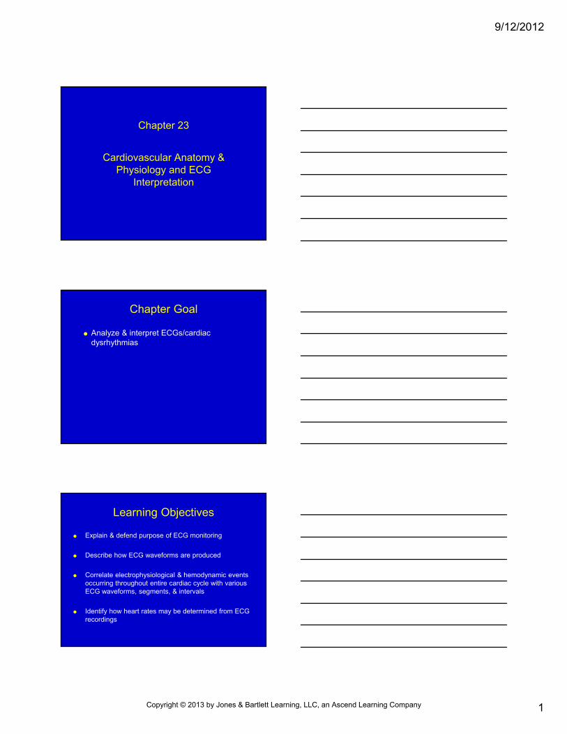
9/12/2012 1 Chapter 23 Cardiovascular Anatomy & Physiology and ECG Interpretation Chapter Goal Analyze & interpret ECGs/cardiac dysrhythmias Learning Objectives Explain & defend purpose of ECG monitoring Describe how ECG waveforms are produced Correlate electrophysiological & hemodynamic events occurring throughout entire cardiac cycle with various ECG waveforms, segments, & intervals Identify how heart rates may be determined from ECG recordings Copyright © 2013 by Jones & Bartlett Learning, LLC, an Ascend Learning Company
Transcript of Chapter 23 Cardiovascular Anatomy & Physiology and ECG...
9/12/2012
1
Chapter 23
Cardiovascular Anatomy & Physiology and ECG
Interpretation
Chapter Goal
Analyze & interpret ECGs/cardiac dysrhythmias
Learning Objectives
Explain & defend purpose of ECG monitoring
Describe how ECG waveforms are produced
Correlate electrophysiological & hemodynamic events occurring throughout entire cardiac cycle with various ECG waveforms, segments, & intervals
Identify how heart rates may be determined from ECG recordings
Copyright © 2013 by Jones & Bartlett Learning, LLC, an Ascend Learning Company
9/12/2012
2
Learning Objectives
List ECG limitations
Describe systematic approach to analysis & interpretation of cardiac dysrhythmias
Explain how to confirm ventricular fibrillation & asystole using 3-lead ECG
Anatomy & Physiology Review
Heart (myocardium) Right & left sides separated by interventricular
septum
Endocardium—inner lining
Pericardium—set of 2 membranes surrounding heart
• Visceral
• Parietal
Atria—upper chambers
Ventricles—lower chambers
Anatomy & Physiology Review
Deoxygenated blood → right atrium (via superior and inferior venae cavae)→tricuspid valve → right ventricle →pulmonary (semilunar) valve →main pulmonary artery → lungs
Oxygenated blood → left atrium (via pulmonary veins) → mitral (bicuspid) valve → left ventricle → aortic (semilunar) valve → aorta
Copyright © 2013 by Jones & Bartlett Learning, LLC, an Ascend Learning Company
9/12/2012
3
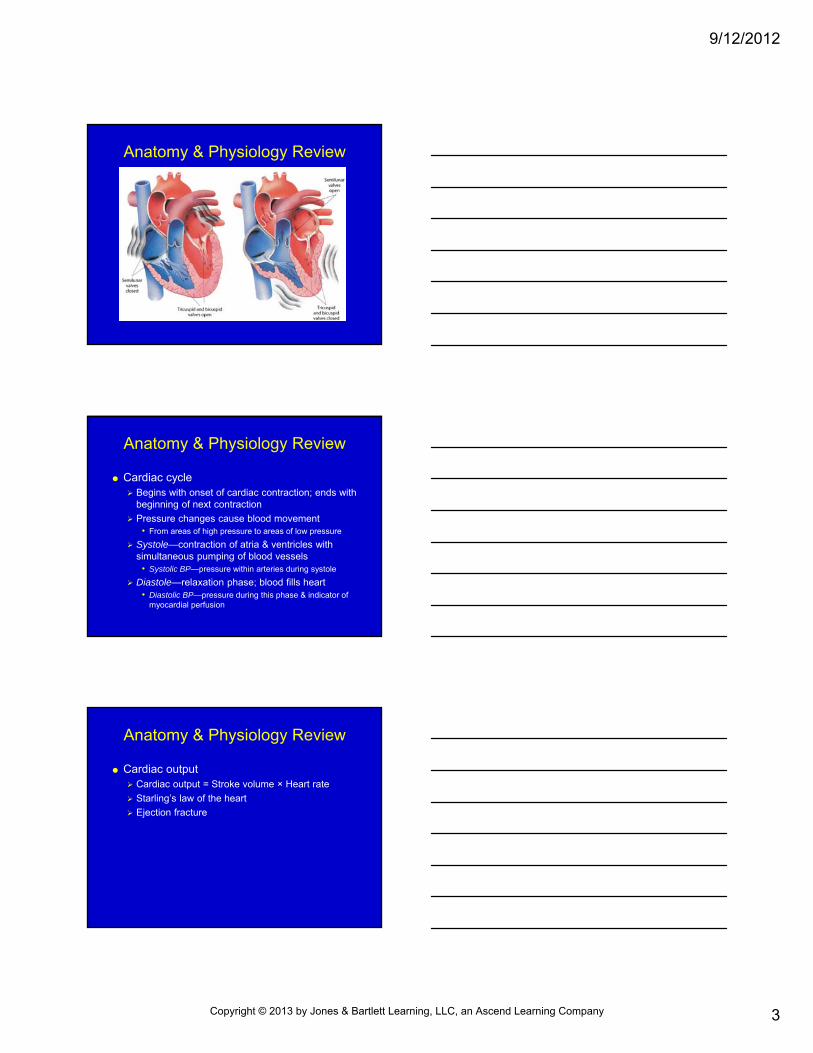
Anatomy & Physiology Review
Anatomy & Physiology Review
Cardiac cycle Begins with onset of cardiac contraction; ends with
beginning of next contraction
Pressure changes cause blood movement• From areas of high pressure to areas of low pressure
Systole—contraction of atria & ventricles with simultaneous pumping of blood vessels
• Systolic BP—pressure within arteries during systole
Diastole—relaxation phase; blood fills heart• Diastolic BP—pressure during this phase & indicator of
myocardial perfusion
Anatomy & Physiology Review
Cardiac output Cardiac output = Stroke volume × Heart rate
Starling’s law of the heart
Ejection fracture
Copyright © 2013 by Jones & Bartlett Learning, LLC, an Ascend Learning Company
9/12/2012
4
Anatomy & Physiology Review
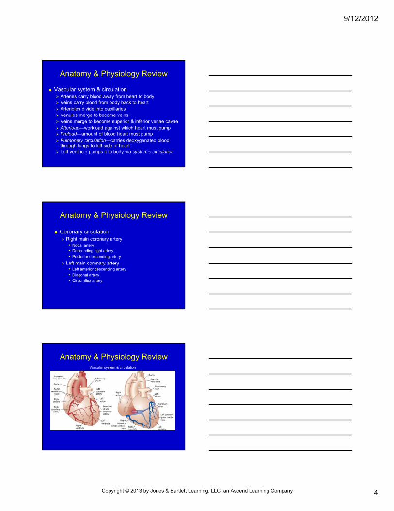
Vascular system & circulation Arteries carry blood away from heart to body Veins carry blood from body back to heart Arterioles divide into capillaries Venules merge to become veins Veins merge to become superior & inferior venae cavae Afterload—workload against which heart must pump Preload—amount of blood heart must pump Pulmonary circulation—carries deoxygenated blood
through lungs to left side of heart Left ventricle pumps it to body via systemic circulation
Anatomy & Physiology Review
Coronary circulation Right main coronary artery
• Nodal artery
• Descending right artery
• Posterior descending artery
Left main coronary artery• Left anterior descending artery
• Diagonal artery
• Circumflex artery
Anatomy & Physiology ReviewVascular system & circulation
Copyright © 2013 by Jones & Bartlett Learning, LLC, an Ascend Learning Company
9/12/2012
5
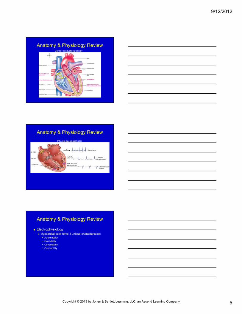
Anatomy & Physiology ReviewCardiac conduction pathway
Anatomy & Physiology Review
Inherent pacemaker rates
Anatomy & Physiology Review
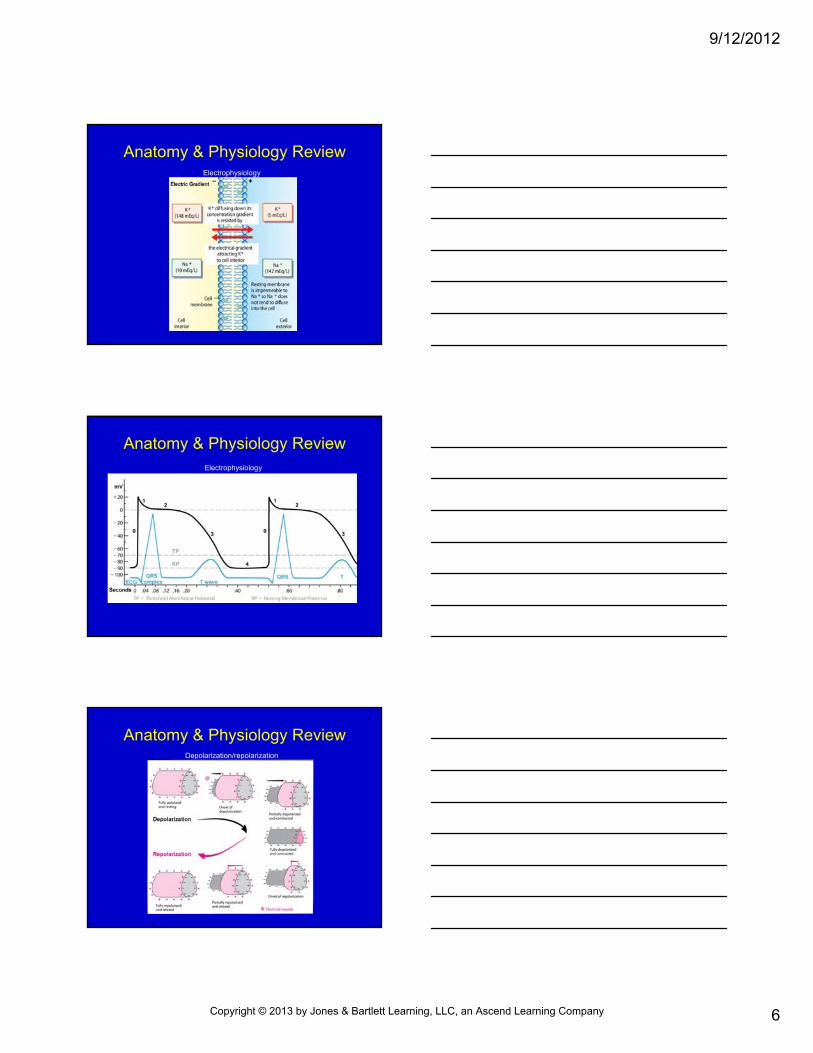
Electrophysiology Myocardial cells have 4 unique characteristics:
• Automaticity
• Excitability
• Conductivity
• Contractility
Copyright © 2013 by Jones & Bartlett Learning, LLC, an Ascend Learning Company
9/12/2012
6
Anatomy & Physiology ReviewElectrophysiology
Anatomy & Physiology ReviewElectrophysiology
Anatomy & Physiology ReviewDepolarization/repolarization
Copyright © 2013 by Jones & Bartlett Learning, LLC, an Ascend Learning Company
9/12/2012
7
Anatomy & Physiology Review
Electrophysiology Absolute refractory period—time when no stimulus
will depolarize myocyte
Relative refractory period—time when sufficiently strong stimulus will depolarize myocardium
Anatomy & Physiology Review
Regulation of heart function Chronotropy
• Heart rate
Dromotropy • Rate of electrical conduction
Inotropy • Strength of contraction
Anatomy & Physiology Review
Regulation of heart function Baroreceptors
• Sensory nerve endings that detect changes in BP & send messages to CNS
• Carotid sinus• Aortic arch• Atria• Vena cava
Chemoreceptors • Receptors in blood vessels that detect changes in chemical
composition of blood• Medulla• Aortic arch• Carotid bodies
Copyright © 2013 by Jones & Bartlett Learning, LLC, an Ascend Learning Company
9/12/2012
8
Anatomy & Physiology Review
Regulation of heart function Parasympathetic stimulation
• ↓ HR• Primarily affects AV node
Sympathetic stimulation • Alpha effects—vasoconstriction• Beta effects— ↑ inotropy, dromotropy, chronotropy
Epinephrine • Greater stimulatory effect on beta receptors
Norepinephrine • Greater stimulatory effect on alpha receptors
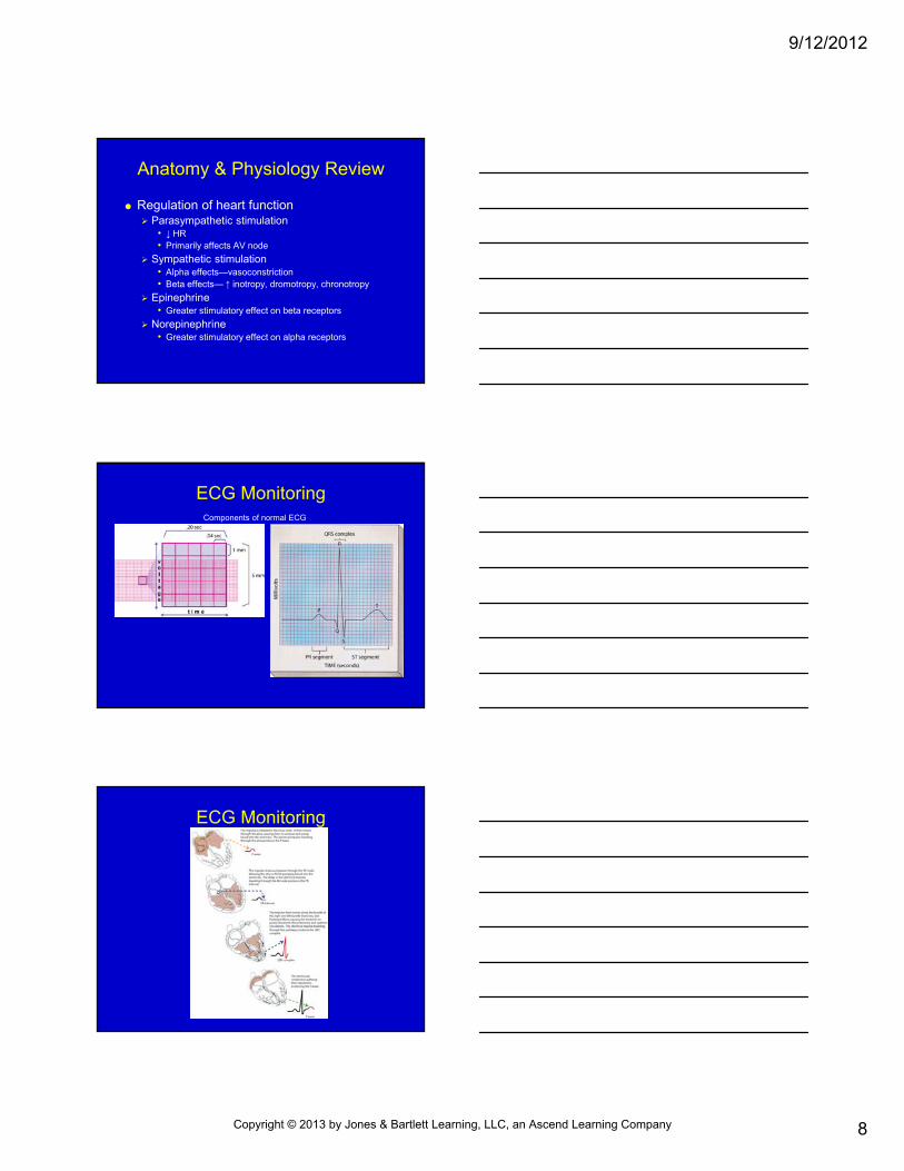
ECG MonitoringComponents of normal ECG
ECG Monitoring
Copyright © 2013 by Jones & Bartlett Learning, LLC, an Ascend Learning Company
9/12/2012
9
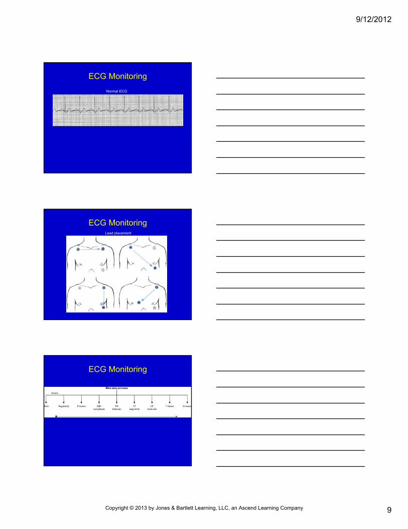
ECG Monitoring
Normal ECG
ECG MonitoringLead placement
ECG Monitoring
Copyright © 2013 by Jones & Bartlett Learning, LLC, an Ascend Learning Company
9/12/2012
10
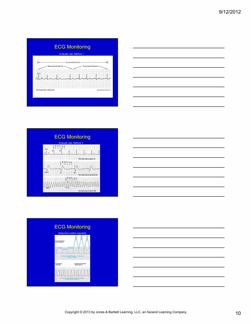
ECG MonitoringEvaluate rate: Method 1
ECG MonitoringEvaluate rate: Method 2
ECG MonitoringDetermine rhythm regularity
Copyright © 2013 by Jones & Bartlett Learning, LLC, an Ascend Learning Company
9/12/2012
11
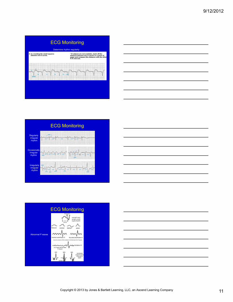
ECG MonitoringDetermine rhythm regularity
ECG Monitoring
Regularly irregularrhythm
OccasionallyIrregularrhythm
IrregularlyIrregularrhythm
ECG Monitoring
Abnormal P waves
Copyright © 2013 by Jones & Bartlett Learning, LLC, an Ascend Learning Company
9/12/2012
12
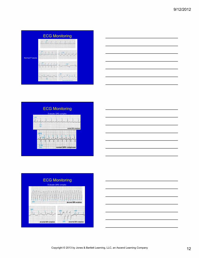
ECG Monitoring
Normal P waves
ECG MonitoringEvaluate QRS complex
ECG MonitoringEvaluate QRS complex
Copyright © 2013 by Jones & Bartlett Learning, LLC, an Ascend Learning Company
9/12/2012
13
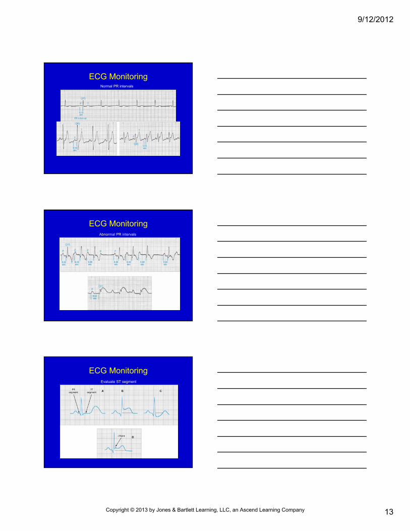
ECG MonitoringNormal PR intervals
ECG MonitoringAbnormal PR intervals
ECG MonitoringEvaluate ST segment
Copyright © 2013 by Jones & Bartlett Learning, LLC, an Ascend Learning Company
9/12/2012
14
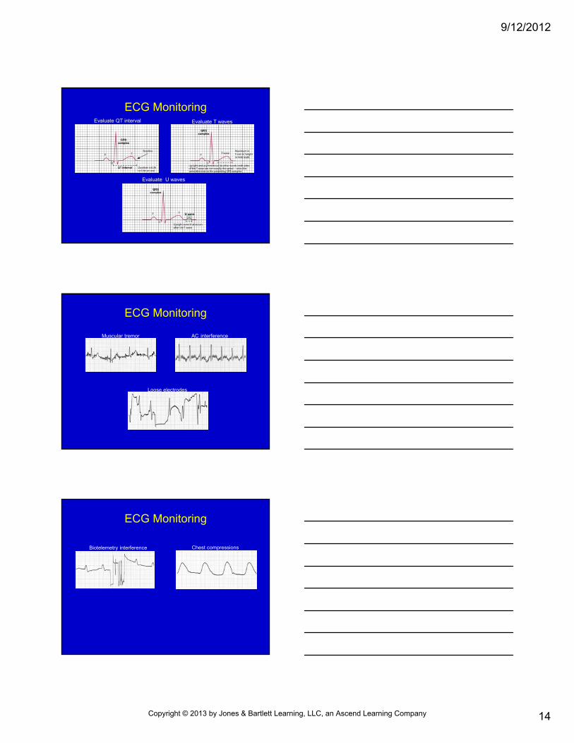
ECG MonitoringEvaluate QT interval Evaluate T waves
Evaluate U waves
ECG Monitoring
Muscular tremor AC interference
Loose electrodes
ECG Monitoring
Biotelemetry interference Chest compressions
Copyright © 2013 by Jones & Bartlett Learning, LLC, an Ascend Learning Company
9/12/2012
15
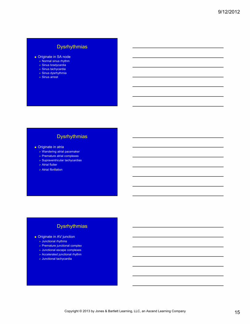
Dysrhythmias
Originate in SA node Normal sinus rhythm Sinus bradycardia Sinus tachycardia Sinus dysrhythmia Sinus arrest
Dysrhythmias
Originate in atria Wandering atrial pacemaker
Premature atrial complexes
Supraventricular tachycardias
Atrial flutter
Atrial fibrillation
Dysrhythmias
Originate in AV junction Junctional rhythms
Premature junctional complex
Junctional escape complexes
Accelerated junctional rhythm
Junctional tachycardia
Copyright © 2013 by Jones & Bartlett Learning, LLC, an Ascend Learning Company
9/12/2012
16
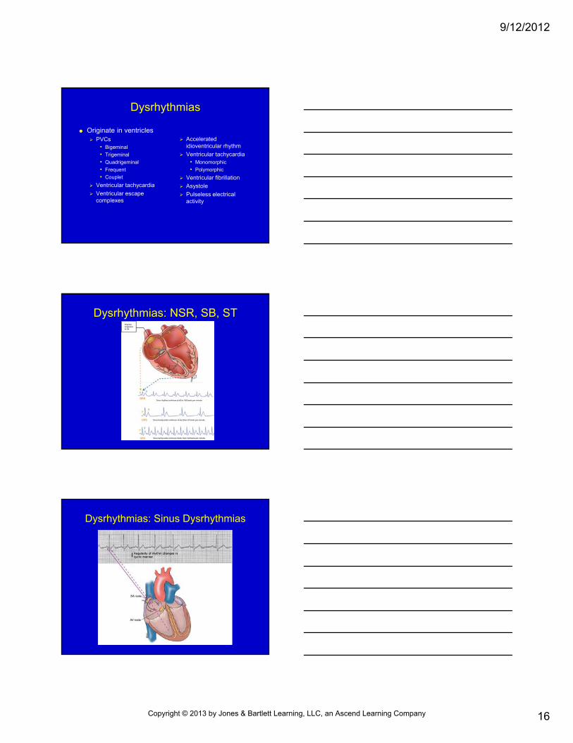
Dysrhythmias
Originate in ventricles PVCs
• Bigeminal
• Trigeminal
• Quadrigeminal
• Frequent
• Couplet
Ventricular tachycardia
Ventricular escape complexes
Accelerated idioventricular rhythm
Ventricular tachycardia • Monomorphic
• Polymorphic
Ventricular fibrillation
Asystole
Pulseless electrical activity
Dysrhythmias: NSR, SB, ST
Dysrhythmias: Sinus Dysrhythmias
Copyright © 2013 by Jones & Bartlett Learning, LLC, an Ascend Learning Company
9/12/2012
17
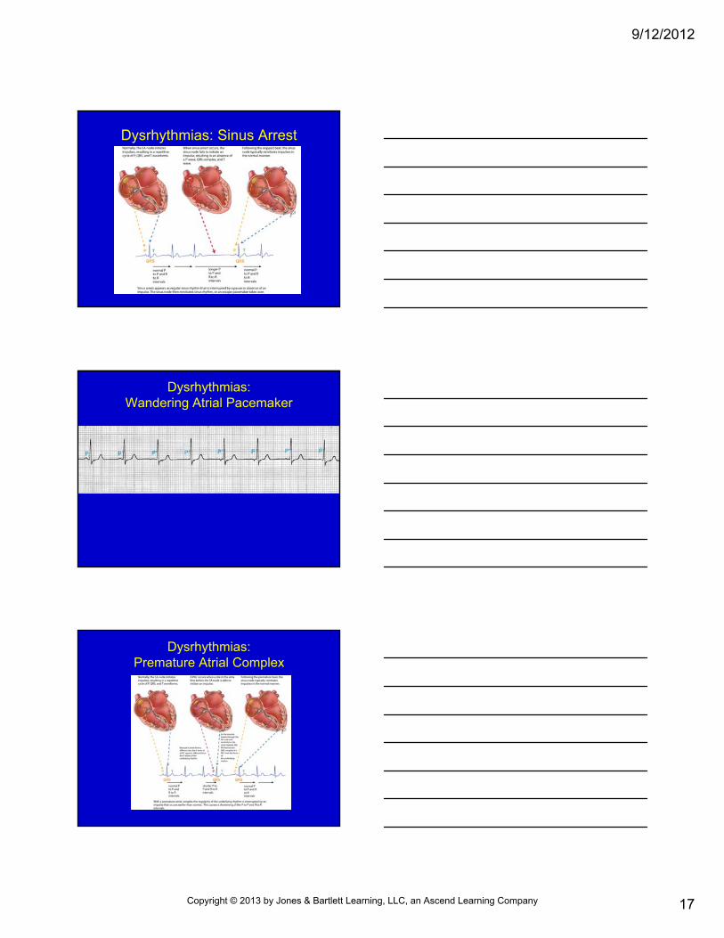
Dysrhythmias: Sinus Arrest
Dysrhythmias:Wandering Atrial Pacemaker
Dysrhythmias:Premature Atrial Complex
Copyright © 2013 by Jones & Bartlett Learning, LLC, an Ascend Learning Company
9/12/2012
18
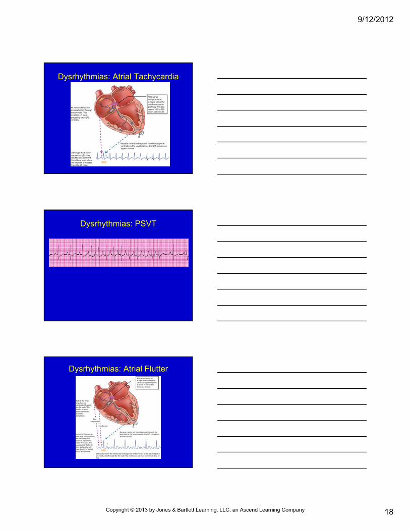
Dysrhythmias: Atrial Tachycardia
Dysrhythmias: PSVT
Dysrhythmias: Atrial Flutter
Copyright © 2013 by Jones & Bartlett Learning, LLC, an Ascend Learning Company
9/12/2012
19
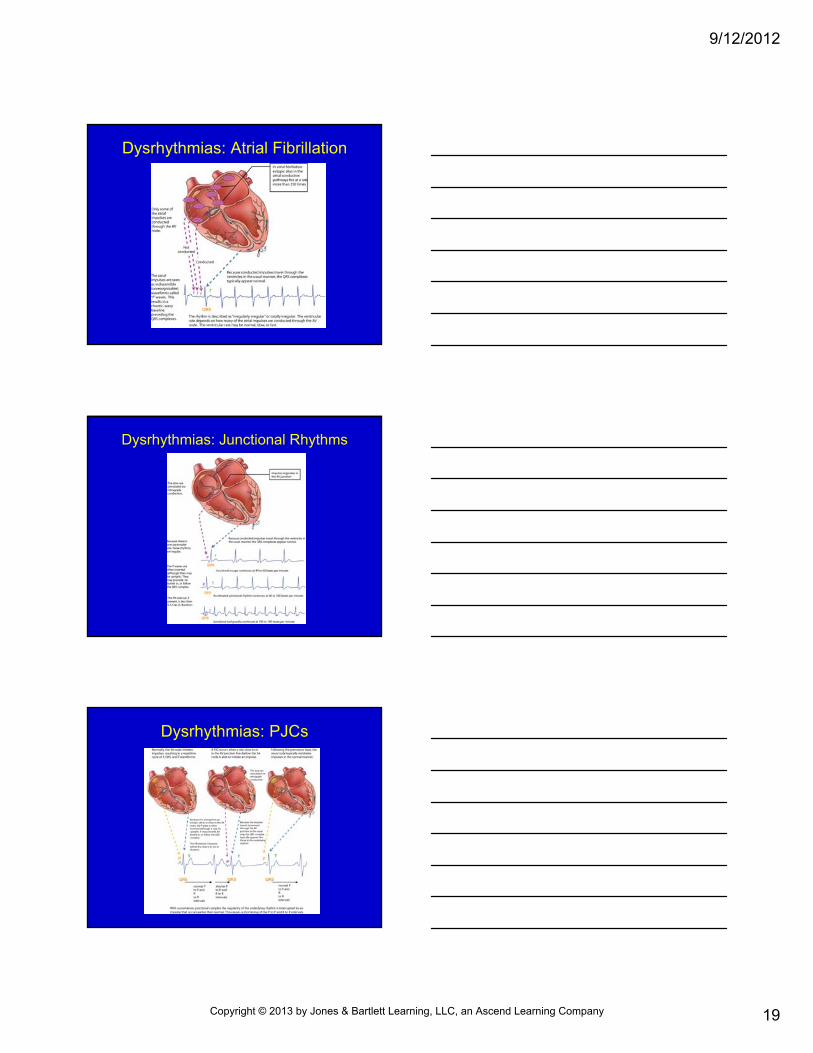
Dysrhythmias: Atrial Fibrillation
Dysrhythmias: Junctional Rhythms
Dysrhythmias: PJCs
Copyright © 2013 by Jones & Bartlett Learning, LLC, an Ascend Learning Company
9/12/2012
20
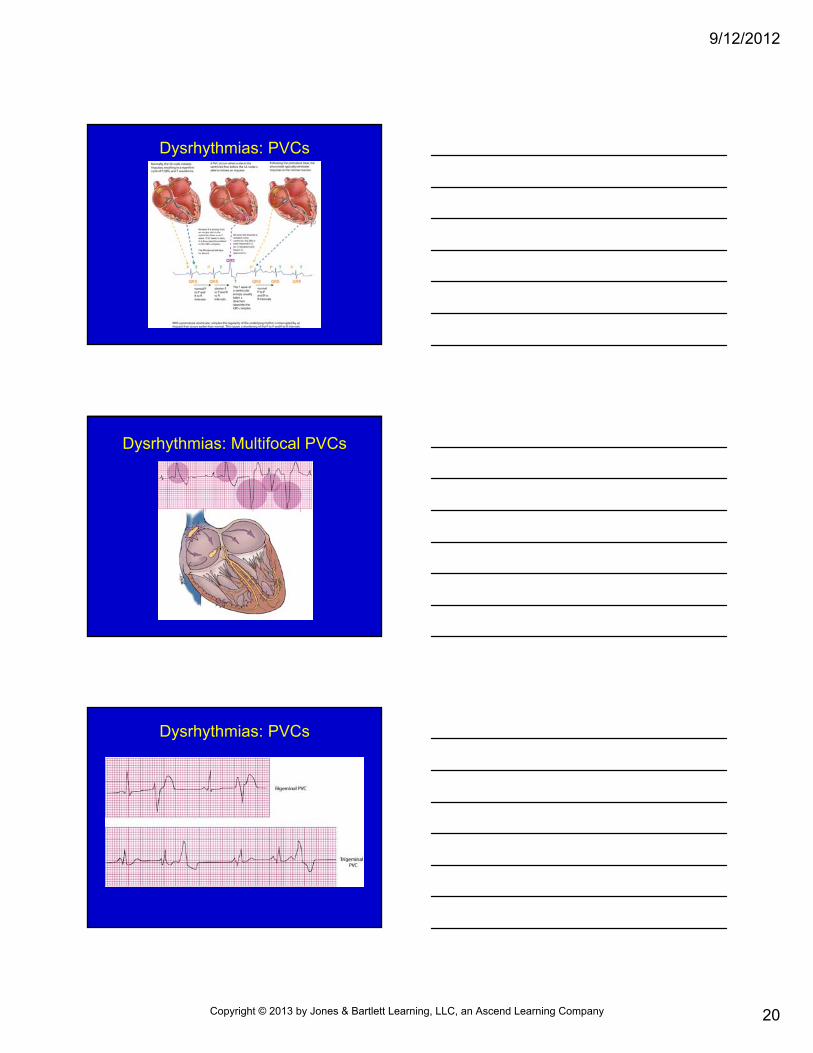
Dysrhythmias: PVCs
Dysrhythmias: Multifocal PVCs
Dysrhythmias: PVCs
Copyright © 2013 by Jones & Bartlett Learning, LLC, an Ascend Learning Company
9/12/2012
21
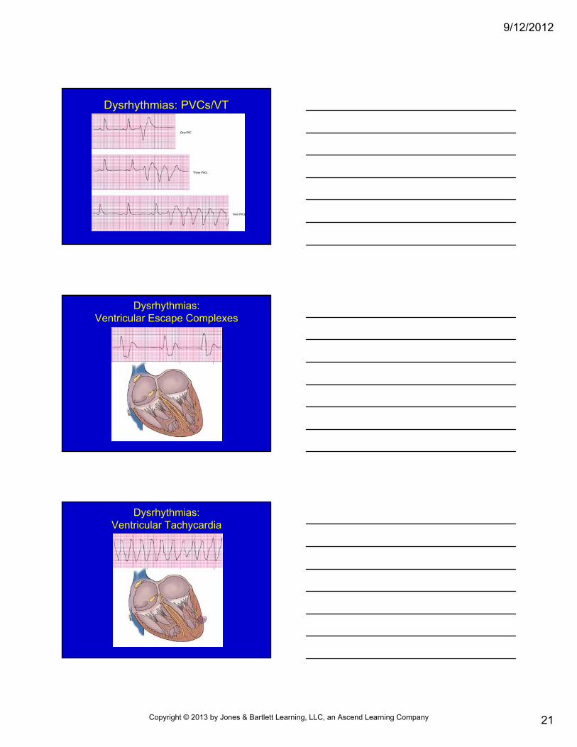
Dysrhythmias: PVCs/VT
Dysrhythmias:Ventricular Escape Complexes
Dysrhythmias:Ventricular Tachycardia
Copyright © 2013 by Jones & Bartlett Learning, LLC, an Ascend Learning Company
9/12/2012
22
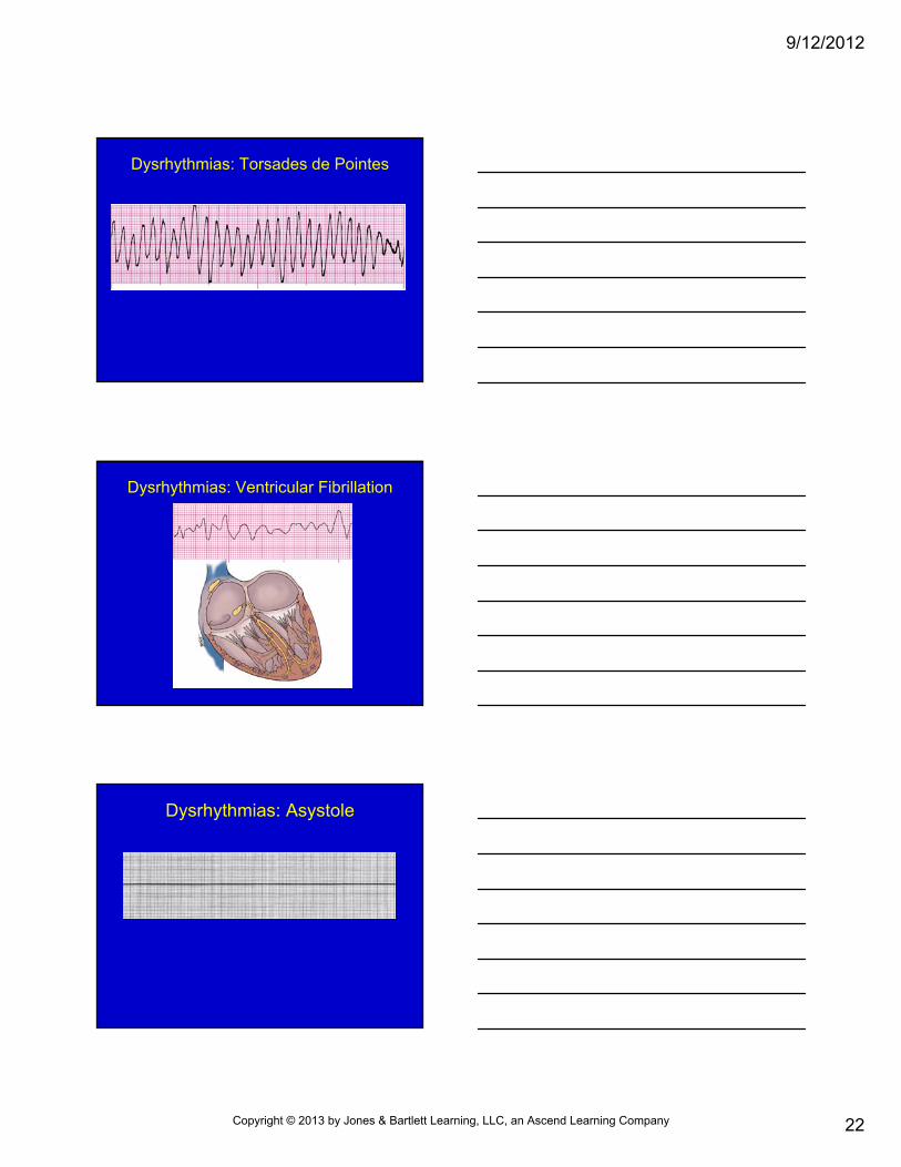
Dysrhythmias: Torsades de Pointes
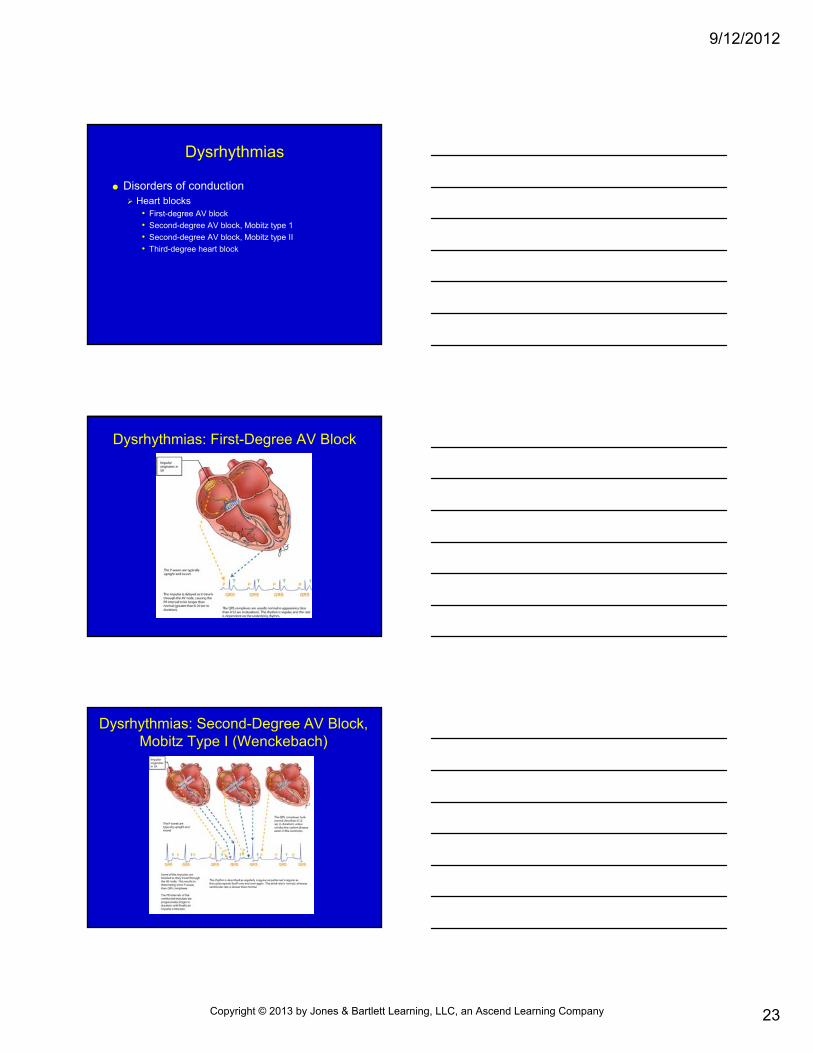
Dysrhythmias: Ventricular Fibrillation
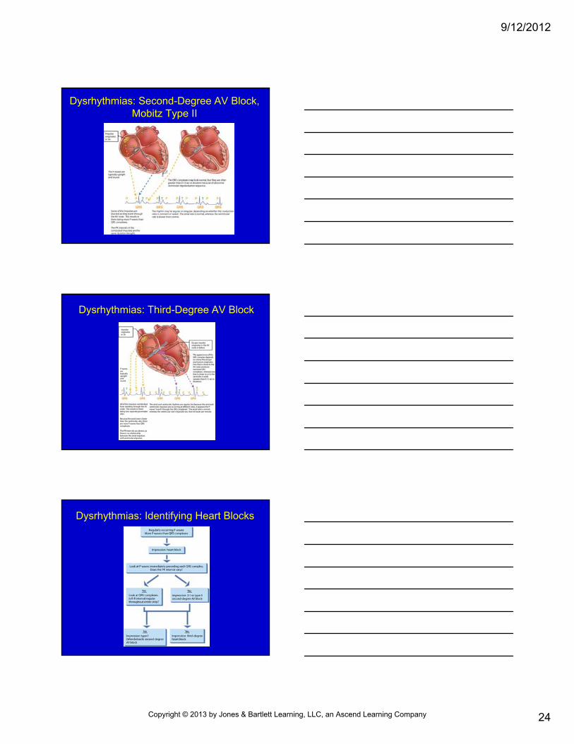
Dysrhythmias: Asystole
Copyright © 2013 by Jones & Bartlett Learning, LLC, an Ascend Learning Company
9/12/2012
23
Dysrhythmias
Disorders of conduction Heart blocks
• First-degree AV block
• Second-degree AV block, Mobitz type 1
• Second-degree AV block, Mobitz type II
• Third-degree heart block
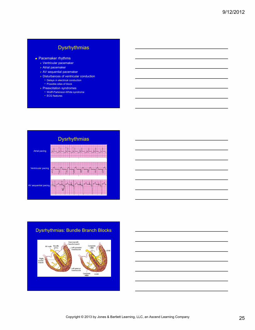
Dysrhythmias: First-Degree AV Block
Dysrhythmias: Second-Degree AV Block, Mobitz Type I (Wenckebach)
Copyright © 2013 by Jones & Bartlett Learning, LLC, an Ascend Learning Company
9/12/2012
24
Dysrhythmias: Second-Degree AV Block, Mobitz Type II
Dysrhythmias: Third-Degree AV Block
Dysrhythmias: Identifying Heart Blocks
Copyright © 2013 by Jones & Bartlett Learning, LLC, an Ascend Learning Company
9/12/2012
25
Dysrhythmias
Pacemaker rhythms Ventricular pacemaker
Atrial pacemaker
AV sequential pacemaker
Disturbances of ventricular conduction• Delays in electrical conduction
• Possible sites of block
Preexcitation syndromes• Wolff-Parkinson-White syndrome
• ECG features
Dysrhythmias
Atrial pacing
Ventricular pacing
AV sequential pacing
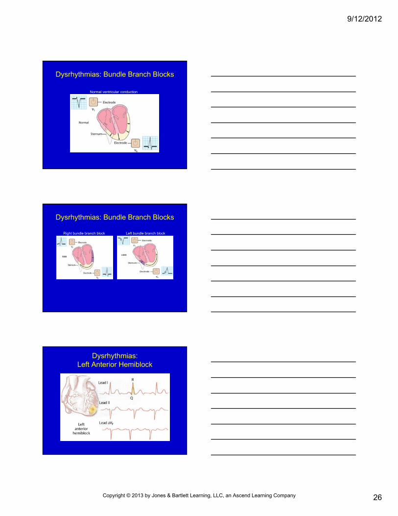
Dysrhythmias: Bundle Branch Blocks
Copyright © 2013 by Jones & Bartlett Learning, LLC, an Ascend Learning Company
9/12/2012
26
Dysrhythmias: Bundle Branch Blocks
Normal ventricular conduction
Dysrhythmias: Bundle Branch Blocks
Left bundle branch blockRight bundle branch block
Dysrhythmias:Left Anterior Hemiblock
Copyright © 2013 by Jones & Bartlett Learning, LLC, an Ascend Learning Company
9/12/2012
27
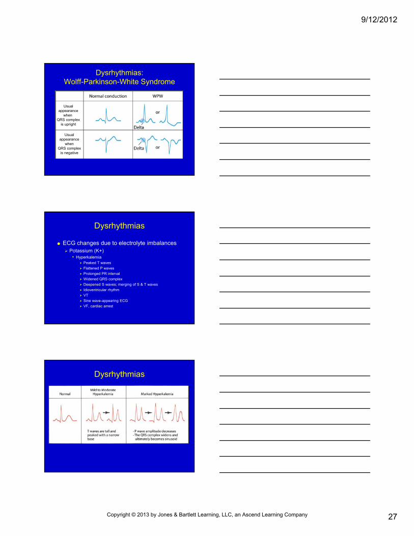
Dysrhythmias:Wolff-Parkinson-White Syndrome
Usualappearance
when QRS complex
is upright
Usualappearance
whenQRS complex
is negative
Dysrhythmias
ECG changes due to electrolyte imbalances Potassium (K+)
• Hyperkalemia Peaked T waves
Flattened P waves
Prolonged PR interval
Widened QRS complex
Deepened S waves; merging of S & T waves
Idioventricular rhythm
VT
Sine wave-appearing ECG
VF, cardiac arrest
Dysrhythmias
Copyright © 2013 by Jones & Bartlett Learning, LLC, an Ascend Learning Company
9/12/2012
28
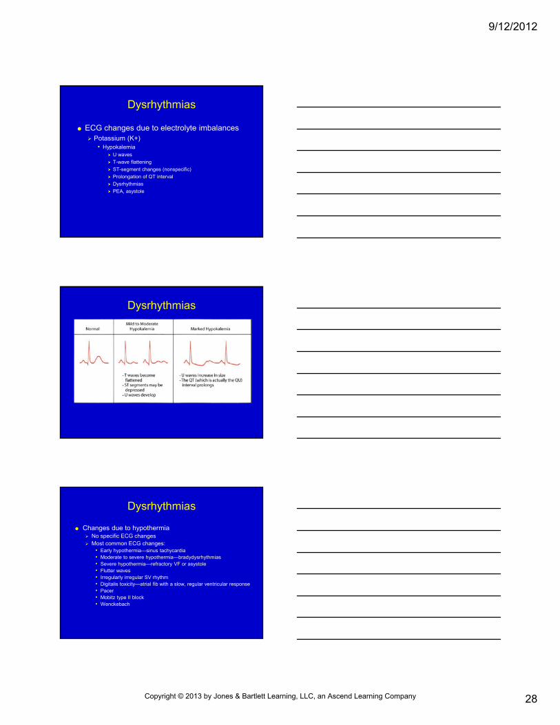
Dysrhythmias
ECG changes due to electrolyte imbalances Potassium (K+)
• Hypokalemia U waves
T-wave flattening
ST-segment changes (nonspecific)
Prolongation of QT interval
Dysrhythmias
PEA, asystole
Dysrhythmias
Dysrhythmias
Changes due to hypothermia No specific ECG changes Most common ECG changes:
• Early hypothermia—sinus tachycardia• Moderate to severe hypothermia—bradydysrhythmias• Severe hypothermia—refractory VF or asystole• Flutter waves• Irregularly irregular SV rhythm • Digitalis toxicity—atrial fib with a slow, regular ventricular response• Pacer• Mobitz type II block • Wenckebach
Copyright © 2013 by Jones & Bartlett Learning, LLC, an Ascend Learning Company
9/12/2012
29
Summary
Heart’s function: pump blood throughout body
Arteries transport blood away from heart
Veins transport blood back to heart
O2 , CO2 & nutrients & waste carried by capillaries
Summary
Electrical nerve impulses cause heart to contract
Autonomic nervous system & hormones control heart rate
ECG is record of electrical activity of heart
P wave represents atrial contraction
Summary
QRS complex represents impulses through ventricles
T wave & possible U wave represent completion of repolarization
PR and ST segments represent electrical pauses
Copyright © 2013 by Jones & Bartlett Learning, LLC, an Ascend Learning Company

9/12/2012
30
Summary
2 common methods of ECG analysis: Observe rhythm on oscilloscope
Print out rhythm
Summary
Dysrhythmias are irregularities of heart rhythm: Originating in:
• Sinus node
• Atria
• AV junction
• Ventricles
• Disorders of conduction
Questions?
Copyright © 2013 by Jones & Bartlett Learning, LLC, an Ascend Learning Company