Chapter 22 Lecture Outline - Palm Beach · PDF file22-3 Introduction • The respiratory...
152
1 Chapter 22 Lecture Outline Copyright © McGraw-Hill Education. Permission required for reproduction or display. See separate PowerPoint slides for all figures and tables pre- inserted into PowerPoint without notes.
Transcript of Chapter 22 Lecture Outline - Palm Beach · PDF file22-3 Introduction • The respiratory...

1
Chapter 22
Lecture Outline
Copyright © McGraw-Hill Education. Permission required for reproduction or display.
See separate PowerPoint slides for all figures and tables pre-
inserted into PowerPoint without notes.

22-2
Introduction
• Breathing represents life!
– First breath of a newborn baby
– Last gasp of a dying person
• All body processes directly or indirectly require ATP
– Most ATP synthesis requires oxygen and
produces carbon dioxide
– Drives the need to breathe to take in oxygen, and
eliminate carbon dioxide
22-3
Introduction
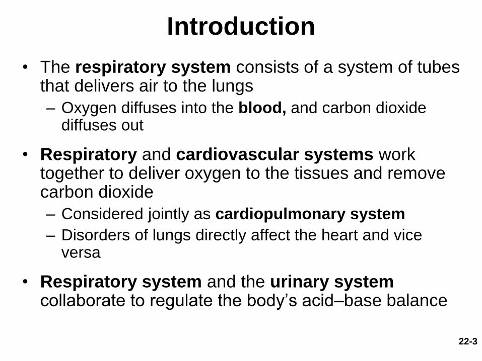
• The respiratory system consists of a system of tubes that delivers air to the lungs
– Oxygen diffuses into the blood, and carbon dioxide diffuses out
• Respiratory and cardiovascular systems work together to deliver oxygen to the tissues and remove carbon dioxide
– Considered jointly as cardiopulmonary system
– Disorders of lungs directly affect the heart and vice versa
• Respiratory system and the urinary system collaborate to regulate the body’s acid–base balance

Anatomy of the Respiratory System
• Expected Learning Outcomes
– State the functions of the respiratory system.
– Name and describe the organs of this system
– Trace the flow of air from the nose to the pulmonary
alveoli.
– Relate the function of any portion of the respiratory tract to
its gross and microscopic anatomy.
22-4
22-5
Anatomy of the Respiratory System
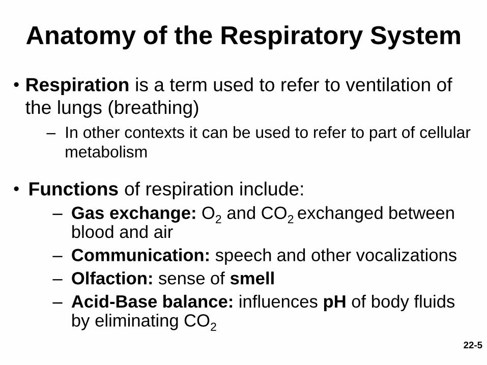
• Respiration is a term used to refer to ventilation of
the lungs (breathing)
– In other contexts it can be used to refer to part of cellular
metabolism
• Functions of respiration include:
– Gas exchange: O2 and CO2 exchanged between blood and air
– Communication: speech and other vocalizations
– Olfaction: sense of smell
– Acid-Base balance: influences pH of body fluids by eliminating CO2
22-6
Anatomy of the Respiratory System
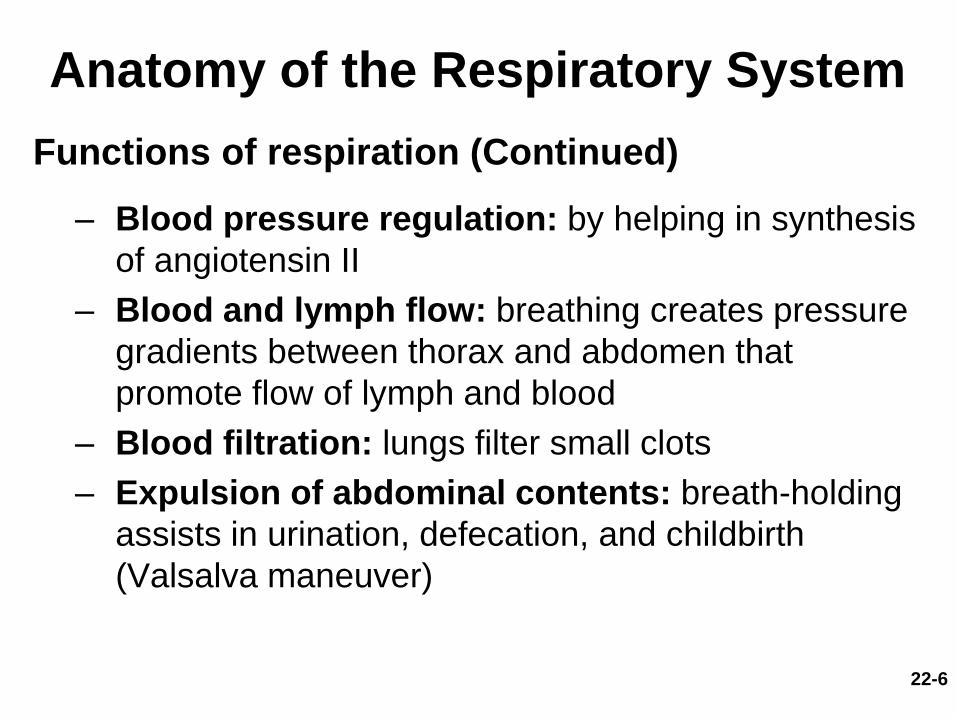
Functions of respiration (Continued)
– Blood pressure regulation: by helping in synthesis
of angiotensin II
– Blood and lymph flow: breathing creates pressure
gradients between thorax and abdomen that
promote flow of lymph and blood
– Blood filtration: lungs filter small clots
– Expulsion of abdominal contents: breath-holding
assists in urination, defecation, and childbirth
(Valsalva maneuver)
22-7
Anatomy of the Respiratory System
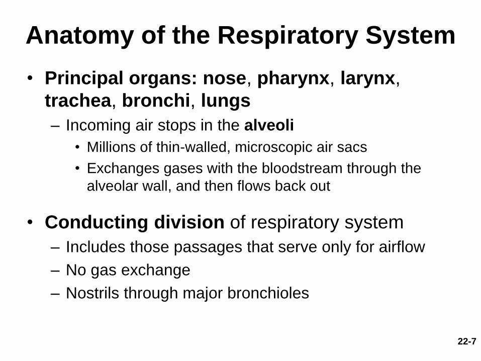
• Principal organs: nose, pharynx, larynx,
trachea, bronchi, lungs
– Incoming air stops in the alveoli
• Millions of thin-walled, microscopic air sacs
• Exchanges gases with the bloodstream through the
alveolar wall, and then flows back out
• Conducting division of respiratory system
– Includes those passages that serve only for airflow
– No gas exchange
– Nostrils through major bronchioles

22-8
Anatomy of the Respiratory System
• Respiratory division of the respiratory system
– Consists of alveoli and other gas exchange regions
• Upper respiratory tract—in head and neck
– Nose through larynx
• Lower respiratory tract—organs of the thorax
– Trachea through lungs
22-9
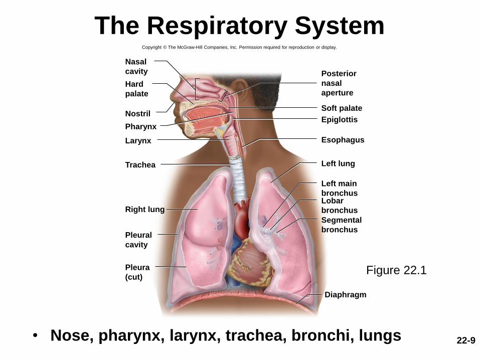
The Respiratory System
• Nose, pharynx, larynx, trachea, bronchi, lungs
Copyright © The McGraw-Hill Companies, Inc. Permission required for reproduction or display.
Nasal
cavity
Nostril
Hard
palate
Larynx
Trachea
Right lung
Posterior
nasal
aperture
Soft palate
PharynxEpiglottis
Esophagus
Left lung
Left main
bronchusLobar
bronchus
Segmental
bronchusPleural
cavity
Pleura
(cut)
Diaphragm
Figure 22.1
22-10
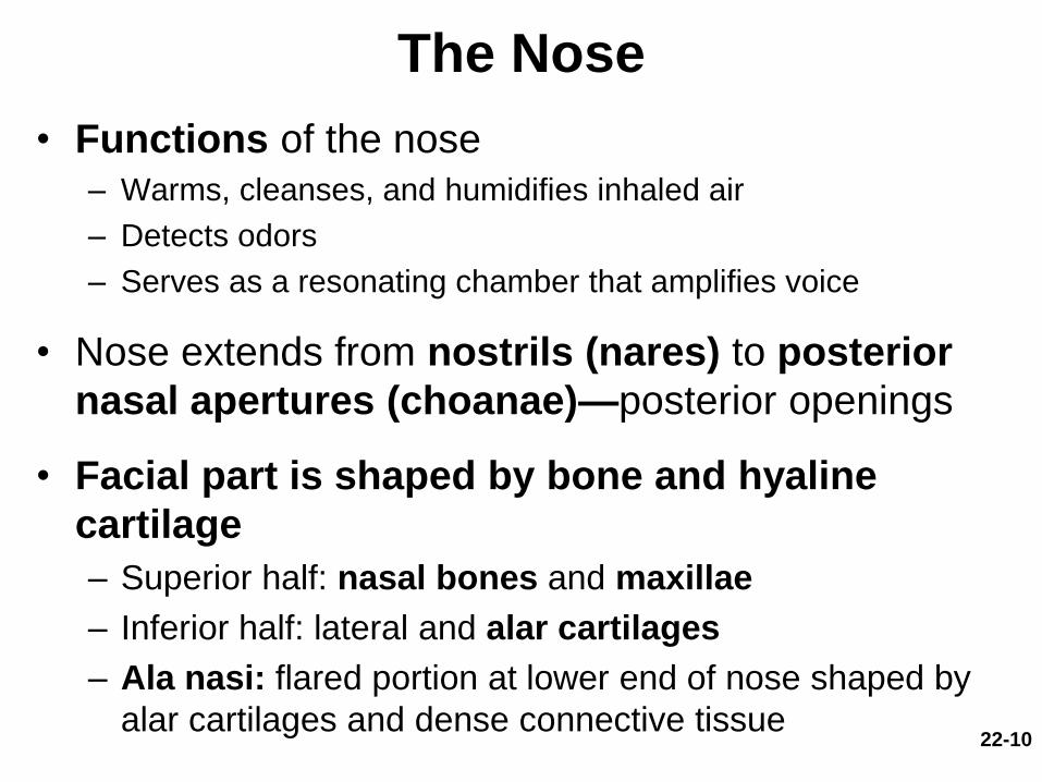
The Nose
• Functions of the nose
– Warms, cleanses, and humidifies inhaled air
– Detects odors
– Serves as a resonating chamber that amplifies voice
• Nose extends from nostrils (nares) to posterior
nasal apertures (choanae)—posterior openings
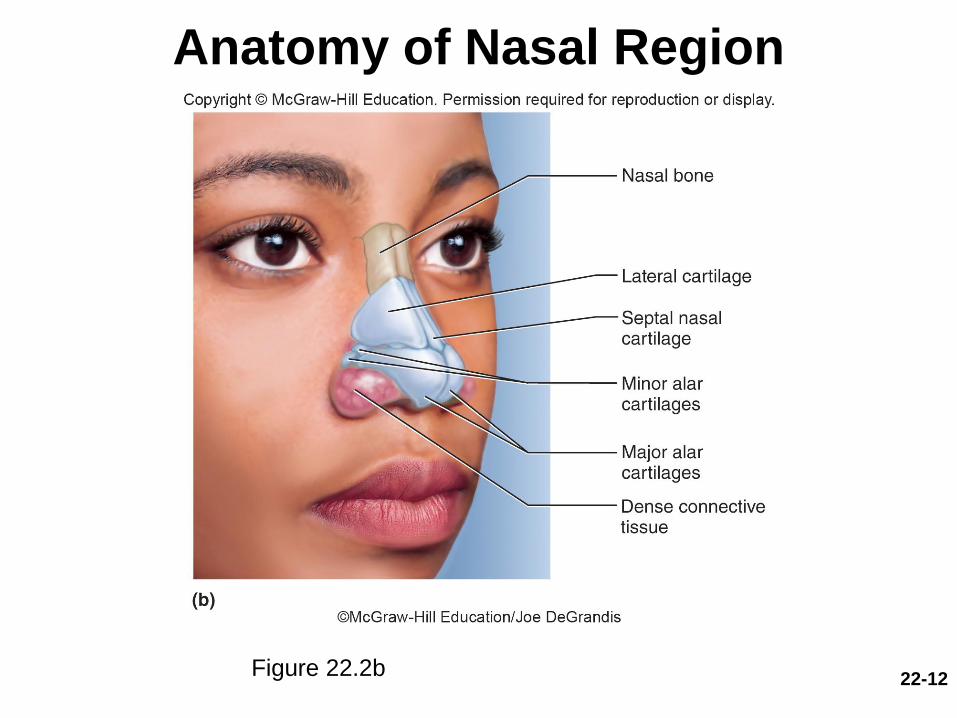
• Facial part is shaped by bone and hyaline
cartilage
– Superior half: nasal bones and maxillae
– Inferior half: lateral and alar cartilages
– Ala nasi: flared portion at lower end of nose shaped by
alar cartilages and dense connective tissue

22-11
Anatomy of the Nasal Region
Figure 22.2a
22-12
Anatomy of Nasal Region
Figure 22.2b

22-13
The Nose
• Nasal fossae—right and left halves of nasal cavity– Nasal septum divides nasal cavity
• Composed of bone and hyaline cartilage
• Vomer forms inferior part
• Perpendicular plate of ethmoid forms superior part
• Septal cartilage forms anterior part
– Roof and floor of nasal cavity
• Ethmoid and sphenoid bones form the roof
• Hard palate forms floor
– Separates the nasal cavity from the oral cavity and allows you to breathe while you chew food
• Paranasal sinuses and nasolacrimal duct drain into nasal cavity

22-14
The Nose
• Vestibule—beginning of nasal cavity; small,
dilated chamber just inside nostrils
– Lined with stratified squamous epithelium
– Vibrissae: stiff guard hairs that block insects and debris
from entering nose
• Posteriorly the nasal cavity expands into a
larger chamber with not much open space
22-15
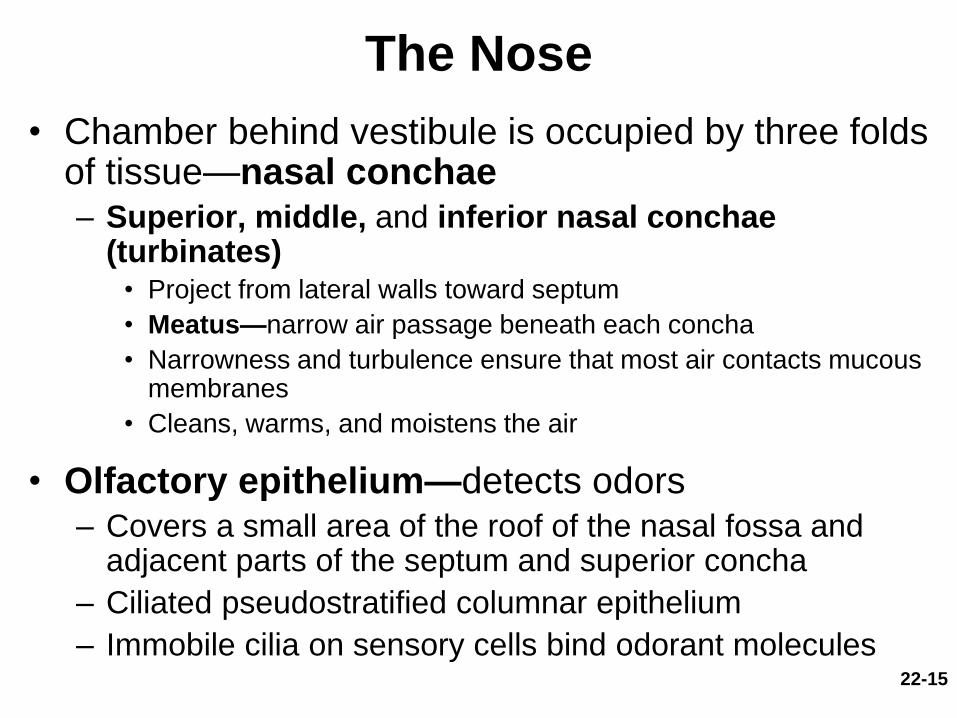
The Nose
• Chamber behind vestibule is occupied by three folds of tissue—nasal conchae– Superior, middle, and inferior nasal conchae
(turbinates)• Project from lateral walls toward septum
• Meatus—narrow air passage beneath each concha
• Narrowness and turbulence ensure that most air contacts mucous membranes
• Cleans, warms, and moistens the air
• Olfactory epithelium—detects odors– Covers a small area of the roof of the nasal fossa and
adjacent parts of the septum and superior concha
– Ciliated pseudostratified columnar epithelium
– Immobile cilia on sensory cells bind odorant molecules

22-16
The Nose
• Respiratory epithelium lines rest of nasal cavity except vestibule– Ciliated pseudostratified columnar epithelium with goblet cells
– Cilia are motile
– Goblet cells secrete mucus and cilia propel the mucus posteriorly toward pharynx
– Swallowed into digestive tract
• Erectile tissue (swell body)—extensive venous plexus in epithelium of inferior concha– Every 30 to 60 minutes, tissue on one side swells with blood
– Restricts airflow through that fossa, so most air directed through other nostril
– Allows engorged side time to recover from drying
– Preponderant flow of air shifts between the right and left nostrils once or twice an hour
22-17
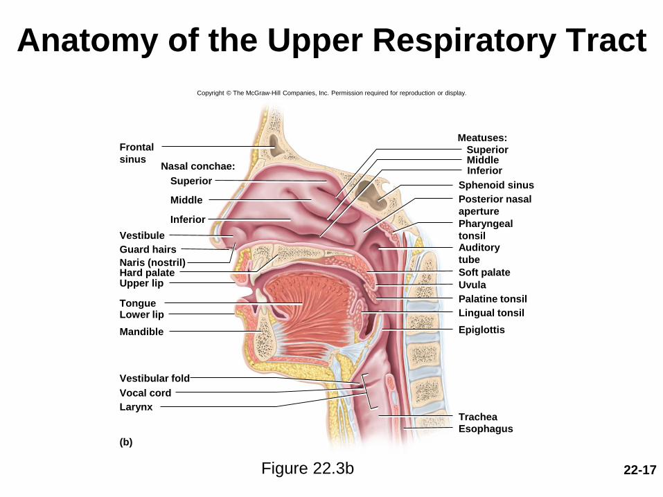
Anatomy of the Upper Respiratory Tract
Copyright © The McGraw-Hill Companies, Inc. Permission required for reproduction or display.
Frontal
sinusNasal conchae:
Superior
Middle
Inferior
Guard hairs
Naris (nostril)Hard palateUpper lip
TongueLower lip
Mandible
Larynx
(b)
SuperiorMiddleInferior
Meatuses:
Sphenoid sinus
Posterior nasal
aperture
Pharyngeal
tonsilAuditory
tube
Soft palate
Uvula
Palatine tonsil
Lingual tonsil
Epiglottis
Vestibular fold
EsophagusTrachea
Vestibule
Vocal cord
Figure 22.3b
22-18
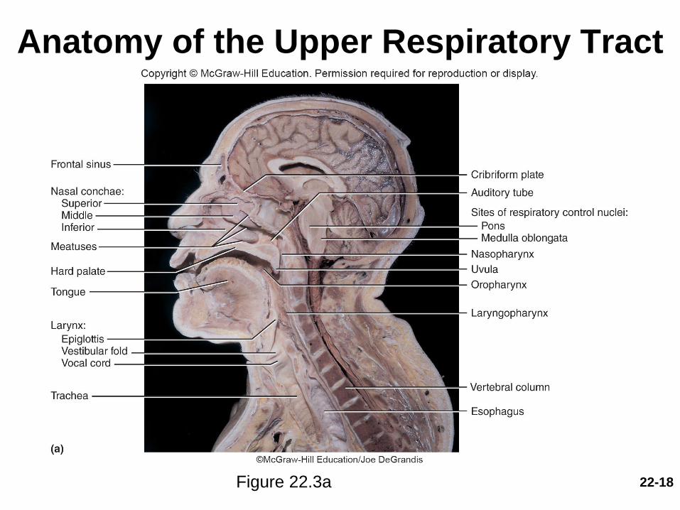
Anatomy of the Upper Respiratory Tract
Figure 22.3a

22-19
Anatomy of the Upper Respiratory Tract
Figure 22.3c
22-20
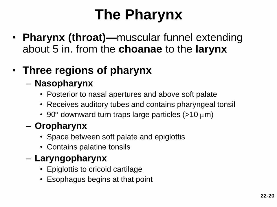
The Pharynx
• Pharynx (throat)—muscular funnel extending about 5 in. from the choanae to the larynx
• Three regions of pharynx– Nasopharynx
• Posterior to nasal apertures and above soft palate
• Receives auditory tubes and contains pharyngeal tonsil
• 90 downward turn traps large particles (>10 m)
– Oropharynx• Space between soft palate and epiglottis
• Contains palatine tonsils
– Laryngopharynx• Epiglottis to cricoid cartilage
• Esophagus begins at that point

22-21
The Pharynx
• Nasopharynx passes only air and is lined by
pseudostratified columnar epithelium
• Oropharynx and laryngopharynx pass air, food,
and drink and are lined by stratified squamous
epithelium
• Muscles of the pharynx assist in swallowing and
speech
22-22
The Larynx
• Larynx (voice box)—cartilaginous chamber
about 4 cm (1.5 in.) long
• Primary function is to keep food and drink out
of the airway
– In several animals it has evolved the additional role of
phonation—the production of sound

22-23
The Larynx
• Epiglottis—flap of tissue that guards the superior
opening of the larynx
– At rest, stands almost vertically
– During swallowing, extrinsic muscles of larynx pull
larynx upward
– Tongue pushes epiglottis down to meet it
– Closes airway and directs food to esophagus behind it
– Vestibular folds of the larynx play greater role in
keeping food and drink out of the airway
22-24
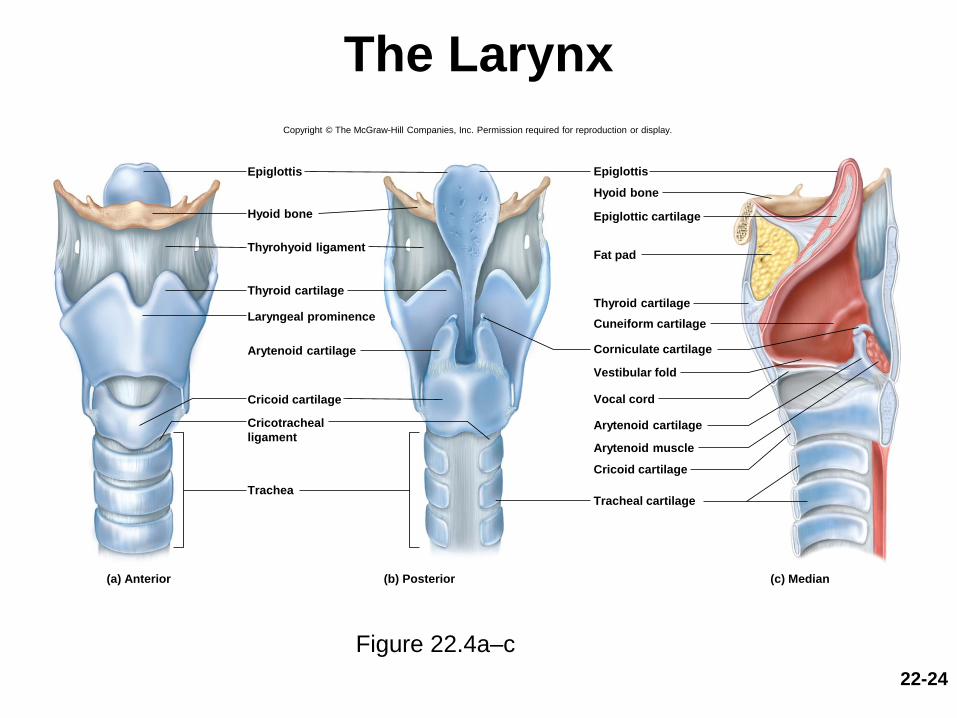
The Larynx
Figure 22.4a–c
Copyright © The McGraw-Hill Companies, Inc. Permission required for reproduction or display.
Epiglottis
Hyoid bone
Thyroid cartilage
Laryngeal prominence
Arytenoid cartilage
Cricoid cartilage
Trachea
(a) Anterior (b) Posterior (c) Median
Tracheal cartilage
Epiglottic cartilage
Epiglottis
Cuneiform cartilage
Corniculate cartilage
Arytenoid cartilage
Arytenoid muscle
Cricoid cartilage
Vocal cord
Thyroid cartilage
Vestibular fold
Fat pad
Hyoid bone
Thyrohyoid ligament
Cricotracheal
ligament

22-25
The Larynx
• Nine cartilages make up framework of larynx
• First three are solitary and relatively large
– Epiglottic cartilage: spoon-shaped supportive plate in
epiglottis; most superior one
– Thyroid cartilage: largest, laryngeal prominence
(Adam’s apple); shield-shaped
• Testosterone stimulates growth, larger in males
– Cricoid cartilage: connects larynx to trachea, ring-like
22-26
The Larynx
• Three smaller, paired cartilages
– Arytenoid cartilages (2): posterior to thyroid cartilage
– Corniculate cartilages (2): attached to arytenoid
cartilages like a pair of little horns
– Cuneiform cartilages (2): support soft tissue between
arytenoids and epiglottis
• Ligaments suspends larynx from hyoid and
hold it together
– Thyrohyoid ligament suspends it from hyoid
– Cricotracheal ligament suspends trachea from larynx
– Intrinsic ligaments hold laryngeal cartilages together
22-27
The Larynx
• Interior wall has two folds on each side that extend from thyroid cartilage in front to arytenoid cartilages in back
– Superior vestibular folds
• Play no role in speech
• Close the larynx during swallowing
– Inferior vocal cords
• Produce sound when air passes between them
• Contain vocal ligaments
• Covered with stratified squamous epithelium
– Suited to endure vibration and contact
• Glottis—the vocal cords and the opening between them
22-28
The Larynx
• Walls of larynx are quite muscular– Deep intrinsic muscles operate the vocal cords
– Superior extrinsic muscles connect larynx to hyoid bone
• Elevate the larynx during swallowing
• Infrahyoid group

The Larynx
• Intrinsic muscles control vocal cords
– Pull on corniculate and arytenoid cartilages causing
cartilages to pivot
– Abduct or adduct vocal cords, depending on direction of
rotation
– Air forced between adducted vocal cords vibrates them
producing high-pitched sound when cords are taut
• Produces lower-pitched sound when cords are more slack
22-29
The Larynx
(Continued)
– Adult male vocal cords, when compared to female cords
• Usually longer and thicker
• Vibrate more slowly
• Produce lower-pitched sound
– Loudness: determined by the force of air passing
between the vocal cords
– Vocal cords produce crude sounds that are formed into
words by actions of pharynx, oral cavity, tongue, and lips
22-30
22-31
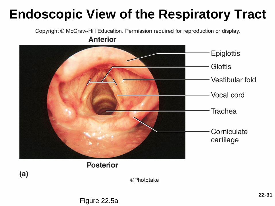
Endoscopic View of the Respiratory Tract
Figure 22.5a
22-32
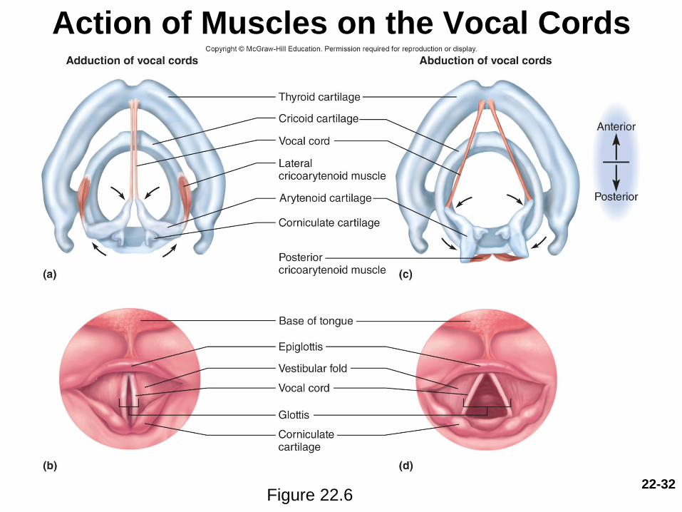
Action of Muscles on the Vocal Cords
Figure 22.6

22-33
The Trachea
• Trachea (windpipe)—a rigid tube about 12 cm
(4.5 in.) long and 2.5 cm (1 in.) in diameter
– Anterior to esophagus
– Supported by 16 to 20 C-shaped rings of hyaline
cartilage that reinforce trachea and prevent collapse
during inhalation
– Opening in rings faces posteriorly toward esophagus
– Trachealis muscle spans opening in rings
• Gap in C allows room for the esophagus to expand as
swallowed food passes by
• Contracts or relaxes to adjust airflow
22-34
The Trachea
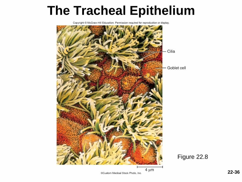
• Inner lining of trachea is ciliated pseudostratified
columnar epithelium
– Composed mainly of mucus-secreting cells, ciliated
cells, and stem cells
– Mucociliary escalator: mechanism for debris removal
• Mucus traps inhaled particles
• Upward beating cilia drives mucus toward pharynx
where it is swallowed
• Middle tracheal layer—connective tissue beneath
the tracheal epithelium
– Contains lymphatic nodules, mucous and serous
glands, and the tracheal cartilages
22-35
The Trachea
• Adventitia—outermost layer of trachea
– Fibrous connective tissue that blends into adventitia of
other organs of mediastinum
• Right and left main bronchi
– Trachea forks at level of sternal angle
– Carina: internal medial ridge in the lowermost tracheal
cartilage
• Directs the airflow to the right and left
22-36
The Tracheal Epithelium
Figure 22.8
Tracheostomy
• Tracheostomy—to make a temporary opening in
the trachea and insert a tube to allow airflow
– Prevents asphyxiation due to upper airway obstruction
– Inhaled air bypasses the nasal cavity and is hot humidified
– If left for long, will dry out mucous membranes of
respiratory tract
– Become encrusted and interfere with clearance of mucus
from tract, thereby promoting infection
22-37
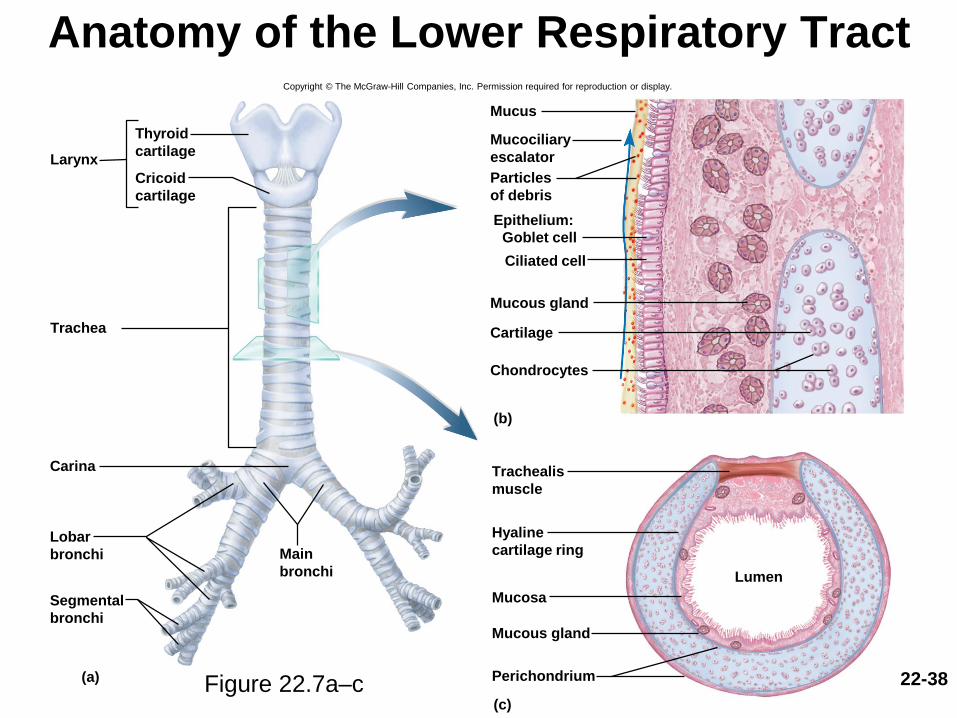
Anatomy of the Lower Respiratory Tract
Larynx
Trachea
Carina
Main
bronchi
Lobar
bronchi
Thyroid
cartilage
Cricoid
cartilage
Trachealis
muscle
Hyaline
cartilage ring
Lumen
Mucosa
Mucous gland
Mucous gland
Perichondrium
(c)
(a)
(b)
Particles
of debris
Cartilage
Chondrocytes
Mucociliary
escalator
Mucus
Ciliated cell
Segmental
bronchi
Epithelium:
Goblet cell
22-38
Copyright © The McGraw-Hill Companies, Inc. Permission required for reproduction or display.
Figure 22.7a–c

22-39
Gross Anatomy of the Lungs
Figure 22.9a,b
Copyright © The McGraw-Hill Companies, Inc. Permission required for reproduction or display.
Lobar bronchi
Apex
Superior lobe
Pulmonary
arteries
Middle lobe
Inferior lobe
(b) Mediastinal surface, right lung
Pulmonary
veins
HilumPulmonary
ligament
Diaphragmatic
surface
(a) Anterior view
Apex of lung
Superior lobe
Mediastinal
surfaces
Middle lobe
Inferior lobar
bronchus
Inferior lobe
Trachea
Main bronchi
Cardiac
impression
Costal
surface
Superior lobar
bronchus
Middle lobar
bronchus
Inferior lobe
Oblique fissure
Base of lung
Oblique
fissure
Superior
lobe
Larynx:
Thyroid cartilage
Cricoid cartilage
Horizontal fissure

22-40
Cross Section Through the Thoracic Cavity
Figure 22.10
22-41
The Lungs and Bronchial Tree
• Lung
– Base: broad concave portion resting on diaphragm
– Apex: tip that projects just above the clavicle
– Costal surface: pressed against the ribcage
– Mediastinal surface: faces medially toward the heart
• Hilum—slit through which the lung receives the main
bronchus, blood vessels, lymphatics, and nerves
• These structures near the hilum constitute the root of the
lung
22-42
The Lungs and Bronchial Tree
• Lungs are crowded by adjacent organs; they neither fill the entire ribcage, nor are they symmetrical
– Right lung
• Shorter than left because liver rises higher on the right
• Has three lobes—superior, middle, and inferior—separated by horizontal and oblique fissure
– Left lung
• Tall and narrow because the heart tilts toward the left and occupies more space on this side of mediastinum
• Has indentation—cardiac impression
• Has two lobes—superior and inferior separated by a single oblique fissure

22-43
The Bronchial Tree
• Bronchial tree—a branching system of air tubes in each lung– From main bronchus to 65,000 terminal bronchioles
• Main (primary) bronchi—supported by C-shaped hyaline cartilage rings– Rt. main bronchus is a branch 2 to 3 cm long, arising
from fork of trachea• Right bronchus slightly wider and more vertical than left
• Aspirated (inhaled) foreign objects lodge in the right main bronchus more often than in the left
– Lt. main bronchus is about 5 cm long• Slightly narrower and more horizontal than the right
22-44
The Bronchial Tree
• Lobar (secondary) bronchi—supported by crescent-shaped cartilage plates– Three rt. lobar (secondary) bronchi: superior, middle,
and inferior• One to each lobe of the right lung
– Two lt. lobar bronchi: superior and inferior• One to each lobe of the left lung
• Segmental (tertiary) bronchi—supported by crescent-shaped cartilage plates– 10 on right, 8 on left
– Bronchopulmonary segment: functionally independent unit of the lung tissue
22-45
The Bronchial Tree
• All bronchi are lined with ciliated pseudostratified
columnar epithelium
– Cells grow shorter and the epithelium thinner as we
progress distally
– Lamina propria has an abundance of mucous glands
and lymphocyte nodules (mucosa-associated
lymphoid tissue, MALT)
• Positioned to intercept inhaled pathogens
– All divisions of bronchial tree have a large amount of
elastic connective tissue
• Contributes to the recoil that expels air from lungs
22-46
The Bronchial Tree
(Conintued)
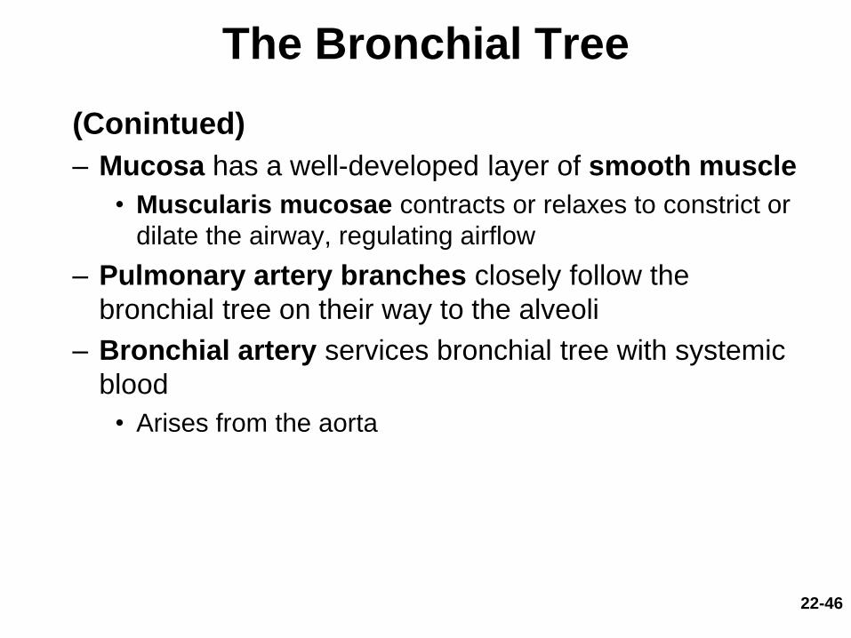
– Mucosa has a well-developed layer of smooth muscle
• Muscularis mucosae contracts or relaxes to constrict or
dilate the airway, regulating airflow
– Pulmonary artery branches closely follow the
bronchial tree on their way to the alveoli
– Bronchial artery services bronchial tree with systemic
blood
• Arises from the aorta
22-47
The Bronchial Tree
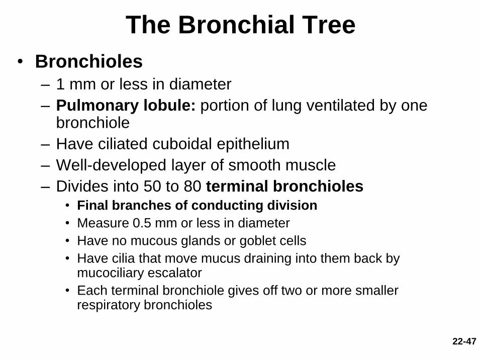
• Bronchioles– 1 mm or less in diameter
– Pulmonary lobule: portion of lung ventilated by one bronchiole
– Have ciliated cuboidal epithelium
– Well-developed layer of smooth muscle
– Divides into 50 to 80 terminal bronchioles• Final branches of conducting division
• Measure 0.5 mm or less in diameter
• Have no mucous glands or goblet cells
• Have cilia that move mucus draining into them back by mucociliary escalator
• Each terminal bronchiole gives off two or more smaller respiratory bronchioles

22-48
The Bronchial Tree
• Respiratory bronchioles
– Have alveoli budding from their walls
– Considered the beginning of the respiratory division
since alveoli participate in gas exchange
– Divide into 2 to 10 alveolar ducts
– End in alveolar sacs: clusters of alveoli arrayed around
a central space called the atrium
22-49
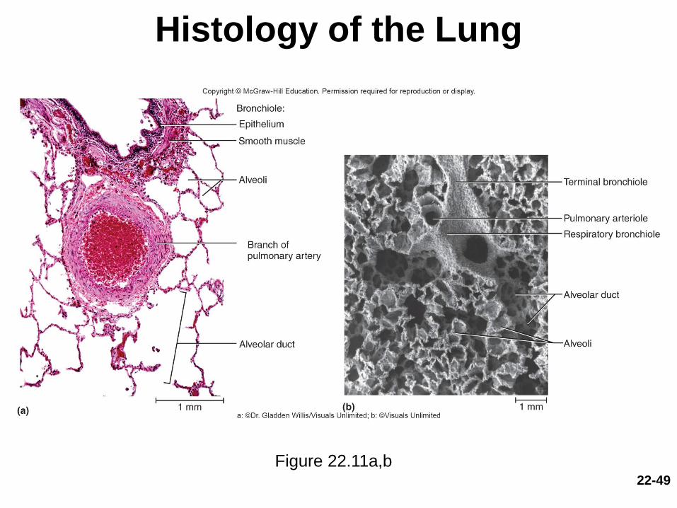
Histology of the Lung
Figure 22.11a,b

22-50
Alveoli
• 150 million alveoli in each lung, providing
about 70 m2 of surface for gas exchange
• Cells of the alveolus
– Squamous (type I) alveolar cells
• Thin, broad cells that allow for rapid gas diffusion
between alveolus and bloodstream
• Cover 95% of alveolus surface area

22-51
Alveoli
Cells of the alveoulus (Continued)
– Great (type II) alveolar cells
• Round to cuboidal cells that cover the remaining 5% of
alveolar surface
• Repair the alveolar epithelium when the squamous (type I)
cells are damaged
• Secrete pulmonary surfactant
– A mixture of phospholipids and proteins that coats the
alveoli and prevents them from collapsing during exhalation

22-52
Alveoli
Cells of the alveoulus (Continued)
– Alveolar macrophages (dust cells)
• Most numerous of all cells in the lung
• Wander the lumens of alveoli and the connective tissue
between them
• Keep alveoli free from debris by phagocytizing dust
particles
• 100 million dust cells die each day as they ride up the
mucociliary escalator to be swallowed and digested with
their load of debris

22-53
Pulmonary Alveoli
Figure 22.12a
Alveoli
• Each alveolus surrounded by a basket of
capillaries supplied by the pulmonary artery
• Respiratory membrane—thin barrier between
the alveolar air and blood
• Respiratory membrane consists of:
– Squamous alveolar cells
– Endothelial cells of blood capillary
– Their shared basement membrane
22-54

Alveoli
• Important to prevent fluid from accumulating in
alveoli
– Gases diffuse too slowly through liquid to sufficiently
aerate the blood
– Alveoli are kept dry by absorption of excess liquid by
blood capillaries
– Lungs have a more extensive lymphatic drainage than
any other organ in the body
– Low capillary blood pressure also prevents rupture of the
delicate respiratory membrane
22-55

22-56
Pulmonary Alveoli
Figure 22.12b,c
(b)
(c)
Respiratory membrane:
Squamous alveolar cellShared basement membraneCapillary endothelial cell
Squamous alveolar cell
Alveolar
macrophage
Respiratory membrane
Fluid with surfactant
Great
alveolar
cell
Air
Lymphocyte
Capillary endothelial cell
CO2
O2
Blood
Copyright © The McGraw-Hill Companies, Inc. Permission required for reproduction or display.

22-57
The Pleurae
• Visceral pleura—serous membrane that covers lungs
• Parietal pleura—adheres to mediastinum, inner surface of
the rib cage, and superior surface of the diaphragm
• Pleural cavity—potential space between pleurae
– Normally no room between the membranes, but contains a film
of slippery pleural fluid
• Functions of pleurae and pleural fluid
– Reduce friction
– Create pressure gradient
• Lower pressure than atmospheric pressure; assists lung inflation
– Compartmentalization
• Prevents spread of infection from one organ in mediastinum to
others

Pulmonary Ventilation
• Expected Learning Outcomes– Name the muscles of respiration and describe their roles.
– Describe brainstem centers that control breathing and the inputs they receive from other parts of the nervous system.
– Explain how pressure gradients account for flow of air into and out of lungs, and how those gradients are produced.
– Identify the sources of resistance to airflow and discuss their relevance to respiration.
– Explain the significance of anatomical dead space to alveolar ventilation.
– Define clinical measurements of pulmonary volume and capacity.
– Define terms for deviations from the normal pattern of breathing.
22-58
22-59
Pulmonary Ventilation
• Breathing (pulmonary ventilation)—consists of a repetitive
cycle of inspiration (inhaling) and expiration (exhaling)
• Respiratory cycle—one complete inspiration and expiration
– Quiet respiration: while at rest, effortless, and automatic
– Forced respiration: deep, rapid breathing, such as during exercise
• Flow of air in and out of lung depends on a pressure
difference between air within lungs and outside body
• Respiratory muscles change lung volumes and create
differences in pressure relative to the atmosphere

22-60
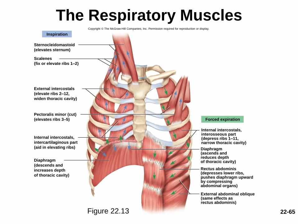
The Respiratory Muscles
• Diaphragm
– Prime mover of respiration
– Contraction flattens diaphragm, enlarging thoracic
cavity and pulling air into lungs
– Relaxation allows diaphragm to bulge upward again,
compressing the lungs and expelling air
– Accounts for two-thirds of airflow
22-61
The Respiratory Muscles
• Internal and external intercostal muscles
– Synergists to diaphragm
– Located between ribs
– Stiffen the thoracic cage during respiration
– Prevent it from caving inward when diaphragm descends
– Contribute to enlargement and contraction of thoracic
cage
– Add about one-third of the air that ventilates the lungs
• Scalenes
– Synergist to diaphragm
– Fix or elevate ribs 1 and 2

22-62
The Respiratory Muscles
• Accessory muscles of respiration act mainly in
forced respiration
• Forced inspiration
– Erector spinae, sternocleidomastoid, pectoralis major,
pectoralis minor, and serratus anterior muscles and
scalenes
– Greatly increase thoracic volume
22-63
The Respiratory Muscles
• Normal quiet expiration – An energy-saving passive process achieved by the
elasticity of the lungs and thoracic cage
– As muscles relax, structures recoil to original shape and original (smaller) size of thoracic cavity, results in airflow out of the lungs
• Forced expiration– Rectus abdominis, internal intercostals, and other
lumbar, abdominal, and pelvic muscles
– Greatly increased abdominal pressure pushes viscera up against diaphragm increasing thoracic pressure, forcing air out
– Important for “abdominal breathing”
22-64
The Respiratory Muscles
• Valsalva maneuver—consists of taking a deep
breath, holding it by closing the glottis, and then
contracting the abdominal muscles to raise
abdominal pressure and push organ contents out
– Childbirth, urination, defecation, vomiting
22-65
Sternocleidomastoid
(elevates sternum)
Scalenes
(fix or elevate ribs 1–2)
External intercostals
(elevate ribs 2–12,
widen thoracic cavity)
Pectoralis minor (cut)
(elevates ribs 3–5)
Internal intercostals,
intercartilaginous part
(aid in elevating ribs)
Diaphragm
(descends and
increases depth
of thoracic cavity)
Inspiration
Internal intercostals,interosseous part(depress ribs 1–11,narrow thoracic cavity)
Diaphragm(ascends andreduces depthof thoracic cavity)
Rectus abdominis(depresses lower ribs,pushes diaphragm upwardby compressingabdominal organs)
External abdominal oblique(same effects asrectus abdominis)
Forced expiration
The Respiratory Muscles
Figure 22.13
Copyright © The McGraw-Hill Companies, Inc. Permission required for reproduction or display.

22-66
Neural Control of Breathing
• No autorhythmic pacemaker cells for respiration,
as in the heart
• Exact mechanism for setting the rhythm of
respiration remains unknown
• Breathing depends on repetitive stimulation of
skeletal muscles from brain and will cease if spinal
cord is severed high in neck
– Skeletal muscles require nervous stimulation
– Interaction of multiple respiratory muscles requires
coordination

22-67
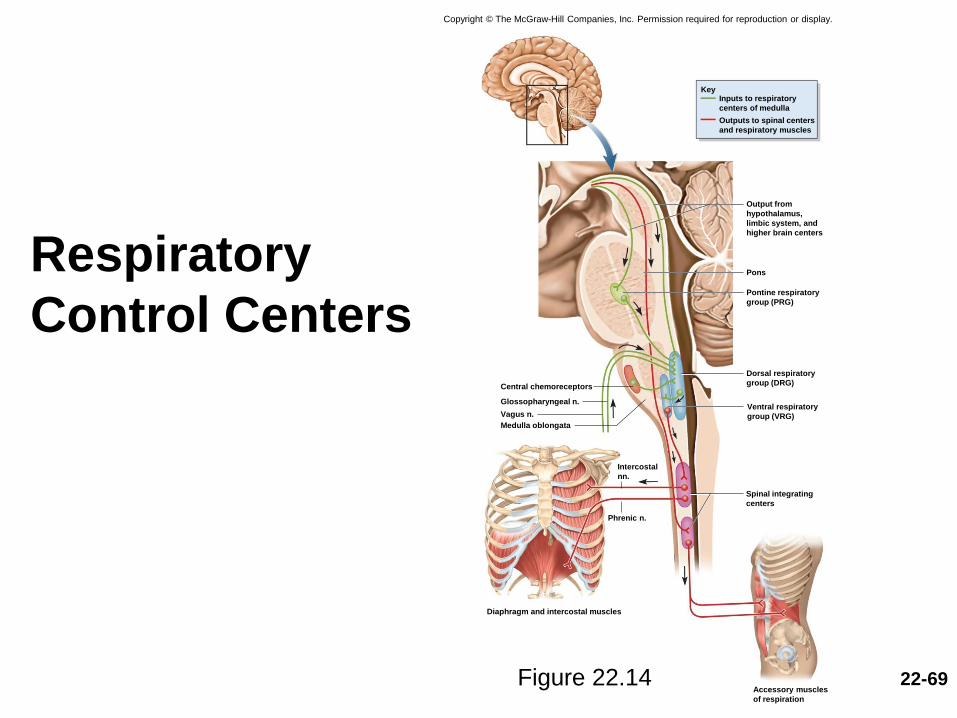
Brainstem Respiratory Centers
• Automatic, unconscious cycle of breathing is controlled
by three pairs of respiratory centers in the reticular
formation of the medulla oblongata and the pons
• Respiratory nuclei in medulla
– Ventral respiratory group (VRG)
• Primary generator of the respiratory rhythm
• In quiet breathing (eupnea), inspiratory neurons fire for about 2
seconds
• Expiratory neurons in eupnea fire for about 3 seconds allowing
inspiratory muscles to relax
• Produces a respiratory rhythm of 12 breaths per minute
– Dorsal respiratory group (DRG)
• Modifies the rate and depth of breathing
• Receives influences from external sources
22-68
Brainstem Respiratory Centers
• Pons
– Pontine respiratory group (PRG)
• Modifies rhythm of the VRG by outputs to both the
VRG and DRG
• Adapts breathing to special circumstances such as
sleep, exercise, vocalization, and emotional
responses
22-69
Respiratory
Control Centers
Copyright © The McGraw-Hill Companies, Inc. Permission required for reproduction or display.
Central chemoreceptors
Spinal integrating
centers
Glossopharyngeal n.
Vagus n.
Diaphragm and intercostal muscles
Accessory muscles
of respiration
Ventral respiratory
group (VRG)
Dorsal respiratory
group (DRG)
Medulla oblongata
Pontine respiratory
group (PRG)
Pons
Output from
hypothalamus,
limbic system, and
higher brain centers
Phrenic n.
Intercostal
nn.
KeyInputs to respiratory
centers of medulla
Outputs to spinal centers
and respiratory muscles
Figure 22.14

22-70
Central and Peripheral Input to the
Respiratory Centers
• Hyperventilation—anxiety-triggered state in
which breathing is so rapid that it expels CO2
from the body faster than it is produced
– As blood CO2 levels drop, the pH rises causing the
cerebral arteries to constrict
– This reduces cerebral perfusion which may cause
dizziness or fainting
– Can be brought under control by having the person
rebreathe the expired CO2 from a paper bag

22-71
Central and Peripheral Input to the
Respiratory Centers
• Central chemoreceptors—brainstem neurons that respond to changes in pH of cerebrospinal fluid– pH of cerebrospinal fluid reflects the CO2 level in the
blood
– By regulating respiration to maintain stable pH, respiratory center also ensures stable CO2 level in blood
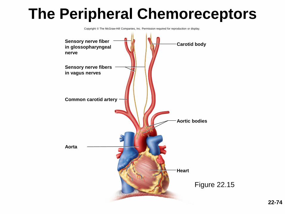
• Peripheral chemoreceptors—located in the carotid and aortic bodies of the large arteries above the heart– Respond to the O2 and CO2 content and the pH of blood
22-72
Central and Peripheral Input to the
Respiratory Centers
• Stretch receptors—found in the smooth muscles
of bronchi and bronchioles, and in the visceral
pleura
– Respond to inflation of the lungs
– Inflation (Hering-Breuer) reflex: triggered by excessive
inflation
• Protective reflex that inhibits inspiratory neurons and stops
inspiration

22-73
Central and Peripheral Input to the
Respiratory Centers
• Irritant receptors—nerve endings amid the
epithelial cells of the airway
– Respond to smoke, dust, pollen, chemical fumes, cold air,
and excess mucus
– Trigger protective reflexes such as bronchoconstriction,
shallower breathing, breath-holding (apnea), or coughing
22-74
The Peripheral Chemoreceptors
Figure 22.15
Copyright © The McGraw-Hill Companies, Inc. Permission required for reproduction or display.
Heart
Aortic bodies
Carotid bodySensory nerve fiber
in glossopharyngeal
nerve
Common carotid artery
Aorta
Sensory nerve fibers
in vagus nerves

22-75
Voluntary Control of Breathing
• Voluntary control over breathing originates in the
motor cortex of frontal lobe of the cerebrum
– Sends impulses down corticospinal tracts to respiratory
neurons in spinal cord, bypassing brainstem
• Limits to voluntary control
– Breaking point: when CO2 levels rise to a point where
automatic controls override one’s will

22-76
Pressure, Resistance, and Airflow
• Respiratory airflow is governed by the same
principles of flow, pressure, and resistance as
blood flow
– The flow of a fluid is directly proportional to the pressure
difference between two points
– The flow of a fluid is inversely proportional to the
resistance
• Atmospheric pressure drives respiration
– The weight of the air above us
– 760 mm Hg at sea level, or 1 atmosphere (1 atm)
• Lower at higher elevations
22-77
Pressure, Resistance, and Airflow
• Boyle’s law—at a constant temperature, the
pressure of a given quantity of gas is inversely
proportional to its volume
– If the lungs contain a quantity of a gas and the lung
volume increases, their internal pressure
(intrapulmonary pressure) falls
• If the pressure falls below atmospheric pressure, air
moves into the lungs
– If the lung volume decreases, intrapulmonary pressure
rises
• If the pressure rises above atmospheric pressure, air
moves out of the lungs

22-78
Pressure, Resistance, and Airflow
• The unit for pressure used by respiratory
physiologists is cm H2O
– This measures how far a column of water would be
moved by a given pressure
– This is more sensitive than mm Hg, since Hg (mercury)
is a heavy liquid
• 1 mm Hg is equal to about 1.4 cm H2O
22-79
Inspiration
• Intrapleural pressure—the slightly negative
pressure that exists between the two pleural
layers
– Recoil of lung tissue and tissues of thoracic cage
causes lungs and chest wall to be pulling in opposite
directions
– The small space between the parietal and visceral
pleura is filled with watery fluid, and so these layers
stay together
– About −5 cm H2O of intrapleural pressure results
22-80
Inspiration
• The two pleural layers cling together due to the
cohesion of water
– When the ribs swing upward and outward during
inspiration, the parietal pleura follows them
– The visceral pleura clings to it by the cohesion of water
and it follows the parietal pleura
– It stretches the alveoli within the lungs
– The entire lung expands along the thoracic cage
– As it increases in volume, its internal pressure drops,
and air flows in

22-81
Inspiration
• Another force that expands the lungs is
explained by Charles’s law
• Charles’s law—volume of a gas is directly
proportional to its absolute temperature
– On a cool day, 16°C (60°F) air will increase its
temperature by 21°C (39°F) during inspiration
– Inhaled air is warmed to 37°C (99°F) by the time it
reaches the alveoli
– Inhaled volume of 500 mL will expand to 536 mL and
this thermal expansion will contribute to the inflation of
the lungs
22-82
Inspiration
• In quiet breathing, the dimensions of the
thoracic cage increase only a few millimeters in
each direction
– Enough to increase its total volume by 500 mL
– Thus, 500 mL of air flows into the respiratory tract

22-83
The Respiratory Cycle
Figure 22.16

22-84
Expiration
• Relaxed breathing
– Passive process achieved mainly by elastic recoil of
thoracic cage
– Recoil compresses the lungs
– Volume of thoracic cavity decreases
– Raises intrapulmonary pressure to about 1 cm H2O
– Air flows down the pressure gradient and out of the lungs
• Forced breathing
– Accessory muscles raise intrapulmonary pressure as
high as +40 cm H2O

22-85
Expiration
• Pneumothorax—presence of air in pleural cavity
– Thoracic wall is punctured
– Inspiration sucks air through the wound into the pleural
cavity
– Potential space becomes an air-filled cavity
– Loss of negative intrapleural pressure allows lungs to
recoil and collapse
• Atelectasis—collapse of part or all of a lung
– Can also result from an airway obstruction as blood
absorbs gases from blood
22-86
Resistance to Airflow
• Increasing resistance decreases airflow
• Two factors influence airway resistance: bronchiole diameter and pulmonary compliance
– Diameter of the bronchioles
• Bronchodilation—increase in diameter of a bronchus or bronchiole
– Epinephrine and sympathetic stimulation stimulate dilation
– Increased airflow
• Bronchoconstriction—decrease in diameter of a bronchus or bronchiole
– Histamine, parasympathetic nerves, cold air, and chemical irritants stimulate bronchoconstriction
– Decreases airflow
– Suffocation can occur from extreme bronchoconstriction brought about by anaphylactic shock and asthma

22-87
Resistance to Airflow
• Two factors influencing airway resistance (Continued)
– Pulmonary compliance: ease with which the lungs can expand
• The change in lung volume relative to a given pressure change
• Compliance is reduced by degenerative lung diseases in which the lungs are stiffened by scar tissue
• Compliance is limited by the surface tension of the water film inside alveoli
– Surfactant secreted by great cells of alveoli disrupts hydrogen bonds between water molecules and thus reduces the surface tension
– Infant respiratory distress syndrome (IRDS)—premature babies lacking surfactant are treated with artificial surfactant until they can make their own
22-88
Alveolar Ventilation
• Only air that enters alveoli is available for gas exchange
• Not all inhaled air gets there—about 150 mL fills the conducting division of the airway
• Anatomic dead space
– Conducting division of airway where there is no gas exchange
– Can be altered somewhat by sympathetic and parasympathetic stimulation
• Sympathetic dilation increases dead space but allows greater flow
• In pulmonary diseases, some alveoli may be unable to exchange gases
– Physiologic (total) dead space—sum of anatomic dead
space and any pathological alveolar dead space
22-89
Alveolar Ventilation
• If a person inhales 500 mL of air, and 150 mL stays
in anatomical dead space, then 350 mL reaches
alveoli
• Alveolar ventilation rate (AVR)
– Air that ventilates alveoli (350 mL) X respiratory rate
(12 bpm) = 4,200 mL/min.
– This measurement is crucially relevant to the body’s ability to get oxygen to the tissues and dispose of carbon dioxide
• Residual volume—1,300 mL that cannot be exhaled with maximum effort

22-90
Spirometry—The Measurement of
Pulmonary Ventilation
• Spirometer—a device that recaptures expired breath
and records such variables as rate and depth of
breathing, speed of expiration, and rate of oxygen
consumption
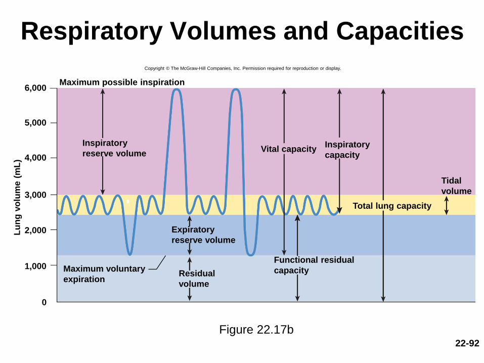
• Respiratory volumes
– Tidal volume: volume of air inhaled and exhaled in one
cycle of breathing (500 mL)
– Inspiratory reserve volume: air in excess of tidal volume
that can be inhaled with maximum effort (3,000 mL)
– Expiratory reserve volume: air in excess of tidal volume
that can be exhaled with maximum effort (1,200 mL)

22-91
Respiratory Volumes and Capacities
Figure 22.17a
22-92
Respiratory Volumes and Capacities
Copyright © The McGraw-Hill Companies, Inc. Permission required for reproduction or display.
Lu
ng
vo
lum
e (
mL
)
6,000
5,000
4,000
3,000
2,000
1,000
0
Maximum possible inspiration
Inspiratory
reserve volume
Expiratory
reserve volume
Residual
volume
Maximum voluntary
expiration
Functional residual
capacity
Total lung capacity
Tidal
volume
Inspiratory
capacityVital capacity
Figure 22.17b

22-93
(Continued)
– Residual volume: air remaining in lungs after maximum
expiration (1,300 mL)
• Allows some gas exchange with blood before next breath of fresh air
arrives
– Vital capacity: total amount of air that can be inhaled and then
exhaled with maximum effort
• VC = ERV + TV + IRV (4,700 mL)
– Important measure of pulmonary health
– Inspiratory capacity: maximum amount of air that can be
inhaled after a normal tidal expiration
• IC = TV + IRV (3,500 mL)
Spirometry—The Measurement of
Pulmonary Ventilation
22-94
(Continued)
– Functional residual capacity: amount of air remaining in
lungs after a normal tidal expiration
• FRC = RV + ERV (2,500 mL)
– Total lung capacity: maximum amount of air the lungs can
contain
• TLC = RV + VC (6,000 mL)
Spirometry—The Measurement of
Pulmonary Ventilation

22-95
Spirometry—The Measurement of
Pulmonary Ventilation
• Spirometry—the measurement of pulmonary function
– Aid in diagnosis and assessment of restrictive and obstructive lung disorders
• Restrictive disorders—those that reduce pulmonary compliance
– Limit the amount to which the lungs can be inflated
– Any disease that produces pulmonary fibrosis
– Black lung disease, tuberculosis

22-96
Spirometry—The Measurement of
Pulmonary Ventilation
• Obstructive disorders—those that interfere with
airflow by narrowing or blocking the airway
– Make it harder to inhale or exhale a given amount
of air
– Asthma, chronic bronchitis
– Emphysema combines elements of restrictive and
obstructive disorders
22-97
Spirometry—The Measurement of
Pulmonary Ventilation
• Forced expiratory volume (FEV)– Percentage of the vital capacity that can be exhaled in a
given time interval
– Healthy adult reading is 75% to 85% in 1 second
• Peak flow– Maximum speed of expiration
– Blowing into a handheld meter
• Minute respiratory volume (MRV)– Amount of air inhaled per minute
– TV x respiratory rate (at rest 500 x 12 = 6,000 mL/min.)
• Maximum voluntary ventilation (MVV)– MRV during heavy exercise
– May be as high as 125 to 170 L/min

22-98
Variations in the Respiratory Rhythm
• Eupnea—relaxed, quiet breathing
– Characterized by tidal volume 500 mL and the
respiratory rate of 12 to 15 bpm
• Apnea—temporary cessation of breathing
• Dyspnea—labored, gasping breathing; shortness
of breath
• Hyperpnea—increased rate and depth of
breathing in response to exercise, pain, or other
conditions
• Hyperventilation—increased pulmonary
ventilation in excess of metabolic demand

22-99
Variations in the Respiratory Rhythm
• Hypoventilation—reduced pulmonary ventilation
leading to an increase in blood CO2
• Kussmaul respiration—deep, rapid breathing
often induced by acidosis
• Orthopnea—dyspnea that occurs when person is
lying down
• Respiratory arrest—permanent cessation of
breathing
• Tachypnea—accelerated respiration

Gas Exchange and Transport
• Expected Learning Outcomes– Define partial pressure and discuss its relationship to a
gas mixture such as air.
– Contrast the composition of inspired and alveolar air.
– Discuss how partial pressure affects gas transport by the blood.
– Describe the mechanism of transporting O2 and CO2.
– Describe the factors that govern gas exchange in the lungs and systemic capillaries.
– Explain how gas exchange is adjusted to the metabolic needs of different tissues.
– Discuss the effect of blood gases and pH on the respiratory rhythm.
22-100
22-101
Composition of Air
• Composition of air
– 78.6% nitrogen, 20.9% oxygen, 0.04% carbon dioxide,
0% to 4% water vapor, depending on temperature and
humidity, and minor gases argon, neon, helium,
methane, and ozone

22-102
Composition of Air
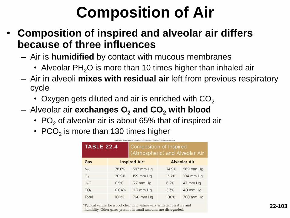
• Dalton’s law—total atmospheric pressure is the
sum of the contributions of the individual gases
– Partial pressure: the separate contribution of each
gas in a mixture
– At sea level 1 atm of pressure = 760 mm Hg
– Nitrogen constitutes 78.6% of the atmosphere, thus
• PN2 = 78.6% x 760 mm Hg = 597 mm Hg
• PO2
= 20.9% x 760 mm Hg = 159 mm Hg
• PH2O = 0.5% x 760 mm Hg = 3.7 mm Hg
• PCO2
= 0.04% x 760 mm Hg = 0.3 mm Hg
• PN2
+ PO2
+ PH2O + PCO2
= 760 mmHg
22-103
Composition of Air
• Composition of inspired and alveolar air differs because of three influences– Air is humidified by contact with mucous membranes
• Alveolar PH2O is more than 10 times higher than inhaled air
– Air in alveoli mixes with residual air left from previous respiratory cycle
• Oxygen gets diluted and air is enriched with CO2
– Alveolar air exchanges O2 and CO2 with blood
• PO2 of alveolar air is about 65% that of inspired air
• PCO2 is more than 130 times higher
22-104
Alveolar Gas Exchange
• Alveolar gas exchange—the swapping of O2 and CO2 across the respiratory membrane– Air in the alveolus is in contact with a film of water
covering the alveolar epithelium
– For oxygen to get into the blood it must dissolve in this water, and pass through the respiratory membrane separating the air from the bloodstream
– For carbon dioxide to leave the blood it must pass the other way, and then diffuse out of the water film into the alveolar air
22-105
Alveolar Gas Exchange
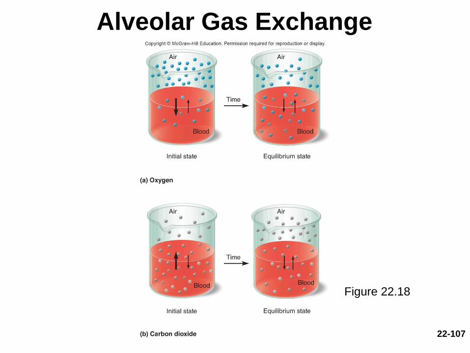
• Gases diffuse down their own gradients until
the partial pressure of each gas in the air is
equal to its partial pressure in water
• Henry’s law—at the air–water interface, for a
given temperature, the amount of gas that
dissolves in the water is determined by its
solubility in water and its partial pressure in air
– The greater the PO2 in the alveolar air, the more O2 the
blood picks up
– Since blood arriving at an alveolus has a higher PCO2
than air, it releases CO2 into the air

22-106
Alveolar Gas Exchange
Henry’s Law (Continued)
– At the alveolus, the blood is said to unload CO2 and
load O2
• Unloading CO2 and loading O2 involves erythrocytes
• Efficiency depends on how long an RBC stays in alveolar
capillaries
– 0.25 second necessary to reach equilibrium
– At rest, RBC spends 0.75 second in alveolar capillaries
– In strenuous exercise, 0.3 second, which is still adequate
– Each gas in a mixture behaves independently
– One gas does not influence the diffusion of another
22-107
Alveolar Gas Exchange
Figure 22.18
22-108
Alveolar Gas Exchange
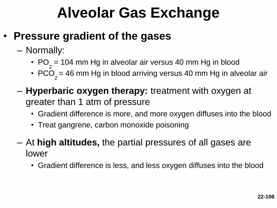
• Pressure gradient of the gases
– Normally:
• PO2
= 104 mm Hg in alveolar air versus 40 mm Hg in blood
• PCO2 = 46 mm Hg in blood arriving versus 40 mm Hg in alveolar air
– Hyperbaric oxygen therapy: treatment with oxygen at
greater than 1 atm of pressure
• Gradient difference is more, and more oxygen diffuses into the blood
• Treat gangrene, carbon monoxide poisoning
– At high altitudes, the partial pressures of all gases are
lower
• Gradient difference is less, and less oxygen diffuses into the blood
22-109
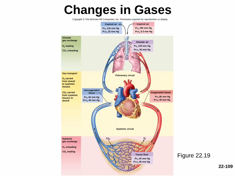
Changes in Gases
Figure 22.19
Copyright © The McGraw-Hill Companies, Inc. Permission required for reproduction or display.
Alveolar
gas exchange
O2 loading
CO2 unloading
Gas transport
O2 carried
from alveoli
to systemic
tissues
CO2 carried
from systemic
tissues to
alveoli
Systemic
gas exchange
O2 unloading
CO2 loading
Expired air Inspired air
PO2 116 mm Hg
PCO2 32 mm Hg
Alveolar air
PO2 104 mm Hg
PCO2 40 mm Hg
Tissue fluid
PO2 40 mm Hg
PCO2 46 mm Hg
Deoxygenated
blood
PO2 40 mm Hg
PCO2 46 mm Hg
Oxygenated blood
PO2 95 mm Hg
PCO2 40 mm Hg
PO2 159 mm Hg
PCO2 0.3 mm Hg
CO2
Pulmonary circuit
Systemic circuit
CO2O2
O2

22-110
Oxygen Loading in Relation to Partial
Pressure GradientCopyright © The McGraw-Hill Companies, Inc. Permission required for reproduction or display.
Pressure gradient of O2
2,500
158
110
40
Am
bie
nt
PO
2(m
m H
g)
Air in hyperbaric chamber
(100% O2 at 3 atm)
Air at 3,000 m
(10,000 ft)
Air at sea level
(1 atm)
Atmosphere Venous blood
arriving at
alveoli
Figure 22.20

22-111
Alveolar Gas Exchange
• Solubility of the gases
– CO2 is 20 times as soluble as O2
• Equal amounts of O2 and CO2 are exchanged across
the respiratory membrane because CO2 is much more
soluble and diffuses more rapidly
• Membrane surface area—100 mL blood in alveolar
capillaries, spread thinly over 70 m2
– Emphysema, lung cancer, and tuberculosis decrease
surface area for gas exchange

22-112
Pulmonary Alveoli in Health and DiseaseCopyright © The McGraw-Hill Companies, Inc. Permission required for reproduction or display.
(a) Normal
(b) Pneumonia
(c) Emphysema
Fluid and
blood cells
in alveoli
Alveolar
walls
thickened
by edema
Confluent
alveoli
Figure 22.21
22-113
Alveolar Gas Exchange
• Membrane thickness—only 0.5 m thick
– Presents little obstacle to diffusion
– Pulmonary edema in left ventricular failure causes edema
and thickening of the respiratory membrane
– Pneumonia causes thickening of respiratory membrane
– When membrane is thicker, gases have farther to travel
between blood and air and cannot equilibrate fast enough
to keep up with blood flow

22-114
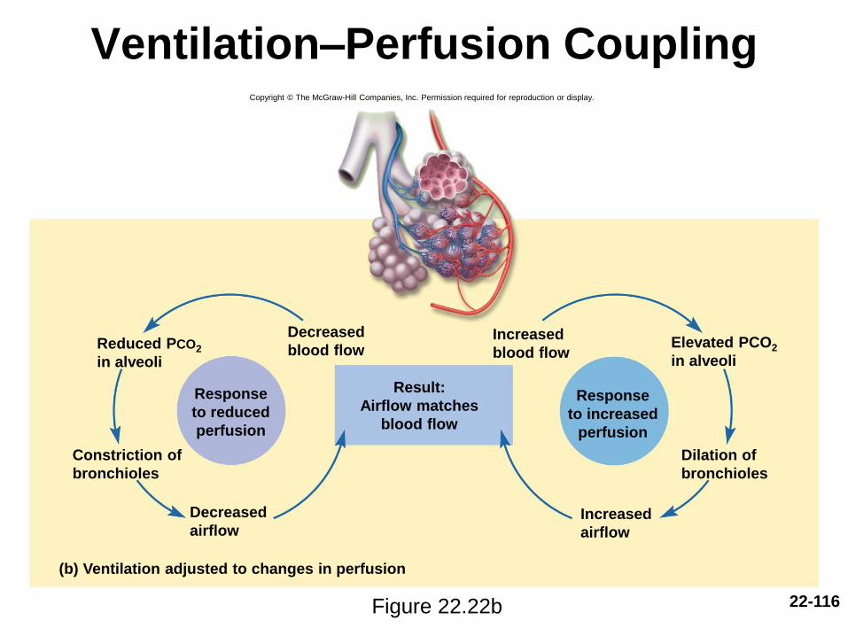
Alveolar Gas Exchange
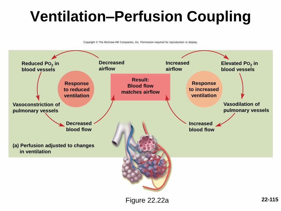
• Ventilation–perfusion coupling—the ability to
match air flow and blood flow to each other
– Gas exchange requires both good ventilation of
alveolus and good perfusion of the capillaries
– Pulmonary blood vessels change diameter depending
on air flow to an area of the lungs
• Example: If an area is poorly ventilated, pulmonary vessels
constrict
– Bronchi change diameter depending on blood flow to
an area of the lungs
• Example: If an area is well perfused, bronchodilation occurs
22-115
Ventilation–Perfusion Coupling
Copyright © The McGraw-Hill Companies, Inc. Permission required for reproduction or display.
(a) Perfusion adjusted to changes
in ventilation
Reduced PO2 in
blood vessels
Elevated PO2 in
blood vessels
Increased
airflow
Vasoconstriction of
pulmonary vessels
Vasodilation of
pulmonary vessels
Decreased
blood flowIncreased
blood flow
Result:
Blood flow
matches airflow
Response
to reduced
ventilation
Response
to increased
ventilation
Decreased
airflow
Figure 22.22a
22-116
Ventilation–Perfusion Coupling
Figure 22.22b
Copyright © The McGraw-Hill Companies, Inc. Permission required for reproduction or display.
Elevated PCO2
in alveoliReduced PCO2
in alveoli
Decreased
blood flow
Result:
Airflow matches
blood flow
Response
to increased
perfusion
Constriction of
bronchioles
Decreased
airflow
Dilation of
bronchioles
Increased
airflow
Response
to reduced
perfusion
(b) Ventilation adjusted to changes in perfusion
Increased
blood flow

22-117
Gas Transport
• Gas transport—the process of carrying gases from
the alveoli to the systemic tissues and vice versa
• Oxygen transport
– 98.5% bound to hemoglobin
– 1.5% dissolved in plasma
• Carbon dioxide transport
– In transport: 90% is hydrated to form carbonic acid
(dissociates into bicarbonate ions); 5% is bound to
proteins; and 5% is dissolved as a gas in plasma
– In exchange: 70% of CO2 comes from carbonic acid;
23% comes from proteins; and 7% comes straight from
plasma

22-118
Oxygen
• Arterial blood carries about 20 mL of O2 per
deciliter
• Hemoglobin—molecule specialized for oxygen
transport
– Four protein (globin) portions
• Each with a heme group that binds one O2 to an iron
atom
• One hemoglobin molecule can carry up to 4 O2
– 100% saturation Hb with 4 O2 molecules per Hb
– 50% saturation Hb with 2 O2 molecules per Hb
• Oxyhemoglobin (HbO2)—O2 bound to hemoglobin
• Deoxyhemoglobin (HHb)—hemoglobin with no O2

22-119
Oxyhemoglobin Dissociation Curve
Figure 22.23
Relationship between hemoglobin saturation and PO2 is
nonlinear (binding facilitates loading; ultimate saturation)
Copyright © The McGraw-Hill Companies, Inc. Permission required for reproduction or display.
100
80
60
40
20
0
20
15
10
5
Systemic tissues Alveoli
Perc
en
tag
e O
2satu
rati
on
of
hem
og
lob
in
mL
O2
/dL
of
blo
od
O2 unloaded
to systemic
tissues
Partial pressure of O2 (PO2) in mm Hg
0 20 40 60 80 100

22-120
Carbon Dioxide
• Carbon dioxide transported in three forms
– Carbonic acid, carbamino compounds, and dissolved in plasma
• 90% of CO2 is hydrated to form carbonic acid
– CO2 + H2O → H2CO3 → HCO3- + H+
– Then dissociates into bicarbonate and hydrogen ions
• 5% binds to the amino groups of plasma proteins and hemoglobin to form carbamino compounds—chiefly carbaminohemoglobin (HbCO2)
– Carbon dioxide does not compete with oxygen
– They bind to different moieties on the hemoglobin molecule
– Hemoglobin can transport O2 and CO2 simultaneously

22-121
Carbon Dioxide
(Continued)
• 5% is carried in the blood as dissolved gas
• Relative amounts of CO2 exchange between the
blood and alveolar air differs
– 70% of exchanged CO2 comes from carbonic acid
– 23% from carbamino compounds
– 7% dissolved in the plasma
• Blood gives up the dissolved CO2 gas and CO2 from the
carbamino compounds more easily than CO2 in
bicarbonate

22-122
Carbon Monoxide Poisoning
• Carbon monoxide (CO)—competes for the O2 binding
sites on the hemoglobin molecule
• Colorless, odorless gas in cigarette smoke, engine
exhaust, fumes from furnaces and space heaters
• Carboxyhemoglobin—CO binds to iron of hemoglobin
(Hb)
– Binds 210 times as tightly as oxygen and ties up Hb for a
long time
– Nonsmokers: less than 1.5% of Hb occupied by CO
– Smokers: 10% of Hb occupied by CO in heavy smokers
– Atmospheric concentration of 0.2% CO is quickly lethal

22-123
Systemic Gas Exchange
• Systemic gas exchange—the unloading of O2
and loading of CO2 at the systemic capillaries
• CO2 loading
– CO2 diffuses into the blood
– Carbonic anhydrase in RBC catalyzes
• CO2 + H2O H2CO3 HCO3− + H+
– Chloride shift
• Keeps reaction proceeding, exchanges HCO3− for Cl−
• H+ binds to hemoglobin

22-124
Systemic Gas Exchange
• Oxygen unloading
– H+ binding to HbO2 reduces its affinity for O2
• Tends to make hemoglobin release oxygen
• HbO2 arrives at systemic capillaries 97% saturated, leaves 75% saturated
– Utilization coefficient: given up 22% of its oxygen load
– Venous reserve: oxygen remaining in the blood after it passes through the capillary beds

22-125
Systemic Gas ExchangeCopyright © The McGraw-Hill Companies, Inc. Permission required for reproduction or display.
Respiring tissue Capillary blood
Dissolved CO2 gas
CO2 + plasma protein
CO2
CO2
O2Dissolved O2 gas
Carbamino compounds
Cl–
7%
23%
70%
98.5%
1.5%
CO2 + Hb
CO2 + H2O
O2 + HHb HbO2+ H+
H2CO3 HCO3– + H+
HbCO2
CAH
Key
Chloride shift
CO2
O2
HbCO2 Carbaminohemoglobin
Hb Hemoglobin
HHb Deoxyhemoglobin
CAH Carbonic anhydrase
HbO2 Oxyhemoglobin
Figure 22.24
22-126
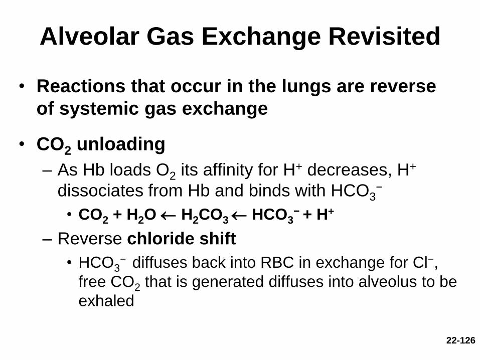
Alveolar Gas Exchange Revisited
• Reactions that occur in the lungs are reverse
of systemic gas exchange
• CO2 unloading
– As Hb loads O2 its affinity for H+ decreases, H+
dissociates from Hb and binds with HCO3−
• CO2 + H2O H2CO3 HCO3− + H+
– Reverse chloride shift
• HCO3− diffuses back into RBC in exchange for Cl−,
free CO2 that is generated diffuses into alveolus to be
exhaled
22-127
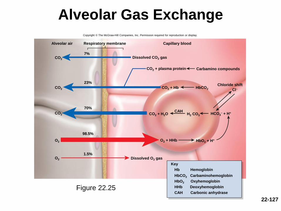
Alveolar Gas ExchangeCopyright © The McGraw-Hill Companies, Inc. Permission required for reproduction or display.
Respiratory membrane Capillary blood
CO2
O2
Alveolar air
Carbamino compounds
7%
23%
70%
98.5%
1.5%
HbCO2
CAH
Key
Cl-Chloride shift
CO2
CO2
O2 Dissolved O2 gas
O2 + HHb HbO2 + H+
HCO3- + H+H2 CO3CO2 + H2O
CO2 + Hb
CO2 + plasma protein
Dissolved CO2 gas
Hb Hemoglobin
HbCO2 Carbaminohemoglobin
HbO2 Oxyhemoglobin
HHb Deoxyhemoglobin
CAH Carbonic anhydraseFigure 22.25
22-128
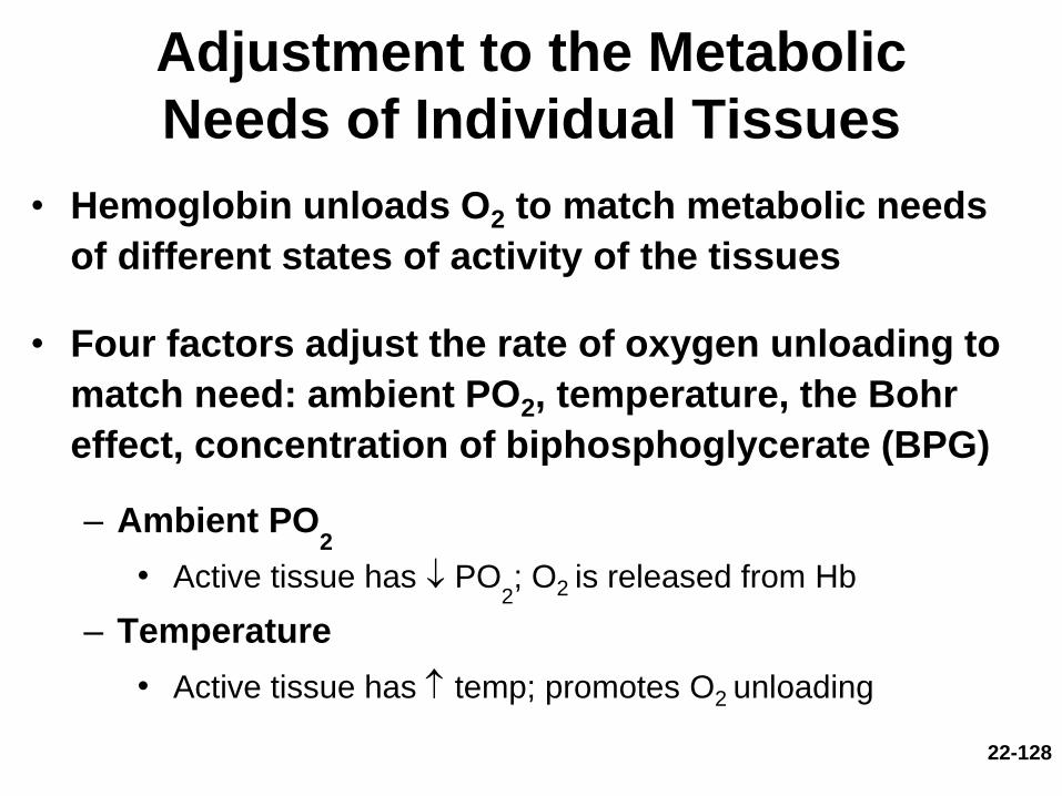
Adjustment to the Metabolic
Needs of Individual Tissues
• Hemoglobin unloads O2 to match metabolic needs
of different states of activity of the tissues
• Four factors adjust the rate of oxygen unloading to
match need: ambient PO2, temperature, the Bohr
effect, concentration of biphosphoglycerate (BPG)
– Ambient PO2
• Active tissue has PO2; O2 is released from Hb
– Temperature
• Active tissue has temp; promotes O2 unloading
22-129
Adjustment to the Metabolic
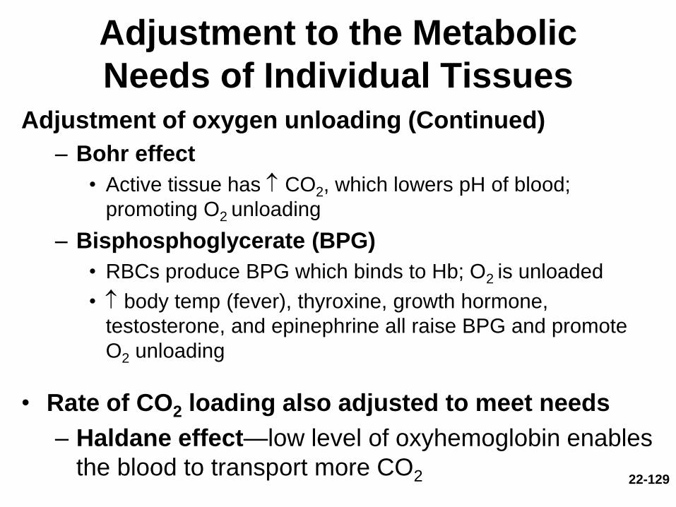
Needs of Individual TissuesAdjustment of oxygen unloading (Continued)
– Bohr effect
• Active tissue has CO2, which lowers pH of blood;
promoting O2 unloading
– Bisphosphoglycerate (BPG)
• RBCs produce BPG which binds to Hb; O2 is unloaded
• body temp (fever), thyroxine, growth hormone,
testosterone, and epinephrine all raise BPG and promote
O2 unloading
• Rate of CO2 loading also adjusted to meet needs
– Haldane effect—low level of oxyhemoglobin enables
the blood to transport more CO2

22-130
Effects of Temperature on
Oxyhemoglobin Dissociation
Figure 22.26a
Copyright © The McGraw-Hill Companies, Inc. Permission required for reproduction or display.
Pe
rce
nta
ge
sa
tura
tio
n o
f h
em
og
lob
in
0
0
PO2 (mm Hg)
100
90
80
70
60
50
40
30
20
10
12010080604020
(a) Effect of temperature
Normal body
temperature
43ºC38ºC
20ºC10ºC

22-131
Effects of pH on
Oxyhemoglobin Dissociation
Bohr effect: release of O2 in response to low pH
Figure 22.26b
0
0
Perc
en
tag
e s
atu
rati
on
of
hem
og
lob
in
PO2 (mm Hg)
100
90
80
70
60
50
40
30
20
10
20 40 60 80 100 120
pH 7.60
pH 7.40
(normal blood pH)
pH 7.20
(b) Effect of pH
Copyright © The McGraw-Hill Companies, Inc. Permission required for reproduction or display.

22-132
Blood Gases and
the Respiratory Rhythm
• Rate and depth of breathing adjust to maintain levels of:
– pH 7.35 to 7.45
– PCO2 40 mm Hg
– PO2 95 mm Hg
• Brainstem respiratory centers receive input from
central and peripheral chemoreceptors that monitor
composition of CSF and blood
• Most potent stimulus for breathing is pH, followed by
CO2, and least significant is O2
22-133
Hydrogen Ions
• Pulmonary ventilation is adjusted to maintain
pH of the brain
– Central chemoreceptors in medulla produce about 75%
of the change in respiration induced by pH shift
• CO2 crosses blood-brain-barrier and reacts with water in CSF to
produce carbonic acid
• The H+ from carbonic acid strongly stimulates central
chemoreceptors, since CSF does not contain much protein buffer
• Hydrogen ions also stimulate peripheral
chemoreceptors which produce 25% of the
respiratory response to pH changes
22-134
Hydrogen Ions
• Acidosis—blood pH lower than 7.35
• Alkalosis—blood pH higher than 7.45
• Hypocapnia—PCO2 less than 37 mm Hg (normal
37 to 43 mm Hg)
• Most common cause of alkalosis
• Hypercapnia—PCO2 greater than 43 mm Hg
• Most common cause of acidosis

22-135
Hydrogen Ions
• Respiratory acidosis and respiratory alkalosis—pH
imbalances resulting from a mismatch between the rate
of pulmonary ventilation and the rate of CO2 production
• Hyperventilation can be a corrective homeostatic
response to acidosis
– “Blowing off” CO2 faster than the body produces it
– Pushes reaction to the left:
CO2 (expired) + H2O H2CO3 HCO3- + H+
– Reduces H+ (reduces acid), raises blood pH toward normal
Hydrogen Ions
• Hypoventilation can be a corrective homeostatic
response to alkalosis
– Allows CO2 to accumulate in body fluids faster than we
exhale it
– Shifts reaction to the right:
CO2 + H2O H2CO3 HCO3- + H+
– Raising the H+ concentration, lowering pH to normal
22-136

Hydrogen Ions
• Ketoacidosis—acidosis brought about by rapid fat
oxidation releasing acidic ketone bodies (seen in
diabetes mellitus)
– Induces Kussmaul respiration: hyperventilation that
reduces CO2 concentration and compensates (to some
degree) for the acidity of ketone bodies
22-137

22-138
Carbon Dioxide
• CO2 has strong indirect effects on respiration
– Through pH, as described previously
• Direct effects
– CO2 at beginning of exercise may directly
stimulate peripheral chemoreceptors and trigger
ventilation more quickly than central
chemoreceptors

22-139
Oxygen
• PO2 usually has little effect on respiration
• Chronic hypoxemia, PO2
less than 60 mm Hg, can
significantly stimulate ventilation
– Hypoxic drive: respiration driven more by low PO2 than
by CO2 or pH
– Emphysema, pneumonia
– High elevations after several days
22-140
Respiration and Exercise
• Causes of increased respiration during exercise
– When the brain sends motor commands to the muscles
• It also sends this information to the respiratory centers
• They increase pulmonary ventilation in anticipation of the needs
of the exercising muscles
– Exercise stimulates proprioceptors of muscles and joints
• They transmit excitatory signals to brainstem respiratory centers
• Increase breathing because they are informed that muscles are
moving
• Increase in pulmonary ventilation keeps blood gas values at their
normal levels in spite of the elevated O2 consumption and CO2
generation by the muscles

Respiratory Disorders
• Expected Learning Outcomes
– Describe the forms and effects of oxygen deficiency
and oxygen excess.
– Describe the chronic obstructive pulmonary diseases
and their consequences.
– Explain how lung cancer begins, progresses, and
exerts its lethal effects.
22-141
22-142
Oxygen Imbalances
• Hypoxia—a deficiency of oxygen in a tissue or the
inability to use oxygen
– A consequence of respiratory diseases
• Hypoxemic hypoxia—state of low arterial PO2
– Usually due to inadequate pulmonary gas exchange
– Oxygen deficiency at high elevations, impaired
ventilation: drowning, aspiration of a foreign body,
respiratory arrest, degenerative lung diseases
• Ischemic hypoxia—inadequate circulation of blood
– Congestive heart failure
22-143
Oxygen Imbalances
• Anemic hypoxia—due to anemia resulting from
the inability of the blood to carry adequate oxygen
• Histotoxic hypoxia—metabolic poisons such as
cyanide prevent tissues from using oxygen
• Cyanosis—blueness of the skin
– Sign of hypoxia

22-144
Oxygen Imbalances
• Although safe to breathe 100% oxygen at 1 atm
for a few hours, oxygen toxicity develops when
pure O2 breathed at 2.5 atm or greater
– Generates free radicals and H2O2
– Destroys enzymes
– Damages nervous tissue
– Leads to seizures, coma, death
• Hyperbaric oxygen
– Formerly used to treat premature infants, caused retinal
damage, was discontinued
22-145
Chronic Obstructive Pulmonary Diseases
• Chronic obstructive pulmonary disease (COPD)—
long-term obstruction of airflow and substantial reduction
in pulmonary ventilation
• Major COPDs are chronic bronchitis and emphysema
– Almost always associated with smoking
– Other risk factors include: air pollution, occupational
exposure to airborne irritants, hereditary defects

22-146
Chronic Obstructive Pulmonary Diseases
• Chronic bronchitis
– Severe, persistent inflammation of lower respiratory tract
– Goblet cells enlarge and produce excess mucus
– Immobilized cilia fail to remove mucus
– Thick, stagnant mucus – ideal for bacterial growth
– Smoke compromises alveolar macrophage function
– Develop chronic cough to bring up sputum (thick mucus
and cellular debris)
– Symptoms include hypoxemia and cyanosis

22-147
Chronic Obstructive Pulmonary Diseases
• Emphysema
– Alveolar walls break down
• Lung has fewer and larger spaces
• Much less respiratory membrane for gas exchange
– Lungs fibrotic and less elastic
• Lungs become flabby and cavitated with large spaces
– Air passages collapse
• Obstructs outflow of air
• Air trapped in lungs; person becomes barrel-chested
– Weaken thoracic muscles
• Spend three to four times the amount of energy just to breathe
22-148
Chronic Obstructive Pulmonary Diseases
• COPD reduces vital capacity
• COPD causes: hypoxemia, hypercapnia, and
respiratory acidosis
– Hypoxemia stimulates erythropoietin release from
kidneys, and leads to polycythemia
• Cor pulmonale
– Hypertrophy and potential failure of right heart due
to obstruction of pulmonary circulation

22-149
Smoking and Lung Cancer
• Lung cancer accounts for more deaths than any
other form of cancer
– Most important cause is smoking (at least 60 carcinogens)
• Squamous-cell carcinoma (most common form)
– Begins with transformation of bronchial epithelium into
stratified squamous from ciliated pseudostratified epithelium
– Dividing cells invade bronchial wall, cause bleeding lesions
– Dense swirls of keratin replace functional respiratory tissue

22-150
Smoking and Lung Cancer
• Adenocarcinoma
– Originates in mucous glands of lamina propria
• Small-cell (oat cell) carcinoma
– Least common, most dangerous
– Named for clusters of cells that resemble oat grains
– Originates in primary bronchi, invades mediastinum,
metastasizes quickly to other organs
22-151
Smoking and Lung Cancer
• 90% originate in mucus membranes of large
bronchi
• Tumor invades bronchial wall, compresses
airway; may cause atelectasis
• Often first sign is coughing up blood
• Metastasis is rapid; usually occurs by time of
diagnosis
– Common sites: pericardium, heart, bones, liver, lymph
nodes, and brain
• Prognosis poor after diagnosis
– Only 7% of patients survive 5 years
22-152
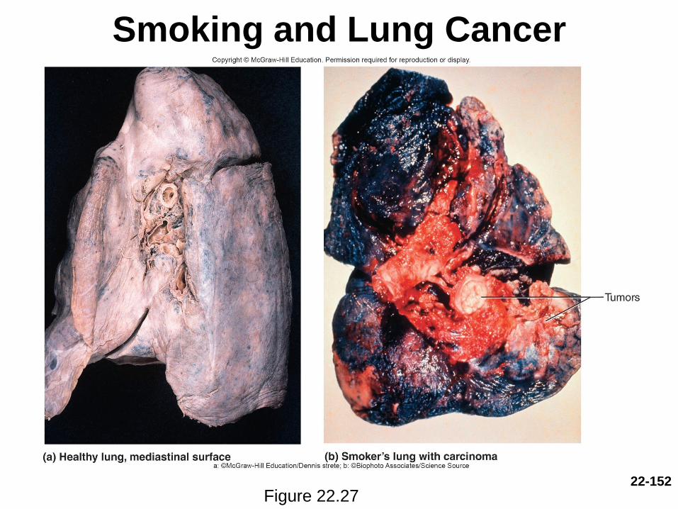
Smoking and Lung Cancer
Figure 22.27