CHAPTER 2 RENAL INJURIES: PENETRATING AND BLUNT
24
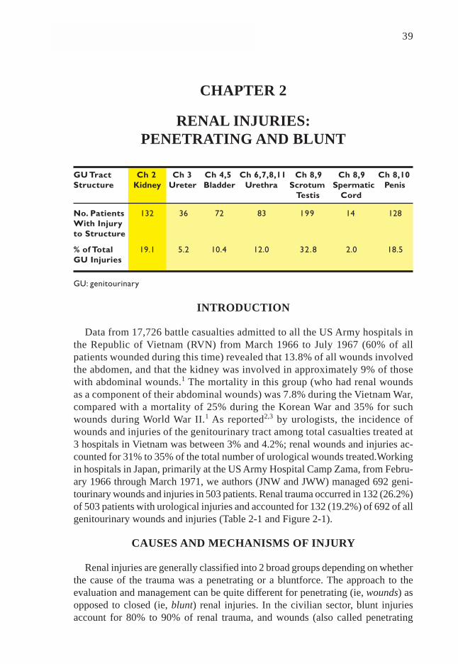
39 CHAPTER 2 RENAL INJURIES: PENETRATING AND BLUNT INTRODUCTION Data from 17,726 battle casualties admitted to all the US Army hospitals in the Republic of Vietnam (RVN) from March 1966 to July 1967 (60% of all patients wounded during this time) revealed that 13.8% of all wounds involved the abdomen, and that the kidney was involved in approximately 9% of those with abdominal wounds. 1 The mortality in this group (who had renal wounds as a component of their abdominal wounds) was 7.8% during the Vietnam War, compared with a mortality of 25% during the Korean War and 35% for such wounds during World War II. 1 As reported 2,3 by urologists, the incidence of wounds and injuries of the genitourinary tract among total casualties treated at 3 hospitals in Vietnam was between 3% and 4.2%; renal wounds and injuries ac- counted for 31% to 35% of the total number of urological wounds treated.Working in hospitals in Japan, primarily at the US Army Hospital Camp Zama, from Febru- ary 1966 through March 1971, we authors (JNW and JWW) managed 692 geni- tourinary wounds and injuries in 503 patients. Renal trauma occurred in 132 (26.2%) of 503 patients with urological injuries and accounted for 132 (19.2%) of 692 of all genitourinary wounds and injuries (Table 2-1 and Figure 2-1). CAUSES AND MECHANISMS OF INJURY Renal injuries are generally classified into 2 broad groups depending on whether the cause of the trauma was a penetrating or a bluntforce. The approach to the evaluation and management can be quite different for penetrating (ie, wounds) as opposed to closed (ie, blunt) renal injuries. In the civilian sector, blunt injuries account for 80% to 90% of renal trauma, and wounds (also called penetrating GU Tract Ch 2 Ch 3 Ch 4,5 Ch 6,7,8,11 Ch 8,9 Ch 8,9 Ch 8,10 Structure Kidney Ureter Bladder Urethra Scrotum Spermatic Penis Testis Cord No. Patients 132 36 72 83 199 14 128 With Injury to Structure % of Total 19.1 5.2 10.4 12.0 32.8 2.0 18.5 GU Injuries GU: genitourinary
Transcript of CHAPTER 2 RENAL INJURIES: PENETRATING AND BLUNT
RENAL INJURIES: PENETRATING AND BLUNT 39
CHAPTER 2
RENAL INJURIES:PENETRATING AND BLUNT
INTRODUCTION
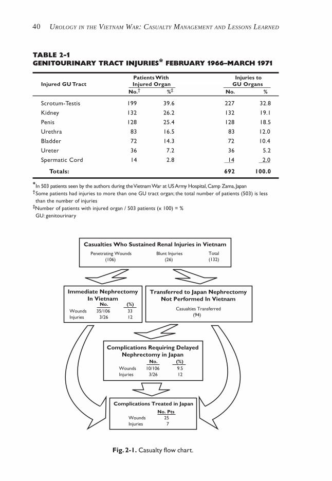
Data from 17,726 battle casualties admitted to all the US Army hospitals inthe Republic of Vietnam (RVN) from March 1966 to July 1967 (60% of allpatients wounded during this time) revealed that 13.8% of all wounds involvedthe abdomen, and that the kidney was involved in approximately 9% of thosewith abdominal wounds.1 The mortality in this group (who had renal woundsas a component of their abdominal wounds) was 7.8% during the Vietnam War,compared with a mortality of 25% during the Korean War and 35% for suchwounds during World War II.1 As reported2,3 by urologists, the incidence ofwounds and injuries of the genitourinary tract among total casualties treated at3 hospitals in Vietnam was between 3% and 4.2%; renal wounds and injuries ac-counted for 31% to 35% of the total number of urological wounds treated.Workingin hospitals in Japan, primarily at the US Army Hospital Camp Zama, from Febru-ary 1966 through March 1971, we authors (JNW and JWW) managed 692 geni-tourinary wounds and injuries in 503 patients. Renal trauma occurred in 132 (26.2%)of 503 patients with urological injuries and accounted for 132 (19.2%) of 692 of allgenitourinary wounds and injuries (Table 2-1 and Figure 2-1).
CAUSES AND MECHANISMS OF INJURY
Renal injuries are generally classified into 2 broad groups depending on whetherthe cause of the trauma was a penetrating or a bluntforce. The approach to theevaluation and management can be quite different for penetrating (ie, wounds) asopposed to closed (ie, blunt) renal injuries. In the civilian sector, blunt injuriesaccount for 80% to 90% of renal trauma, and wounds (also called penetrating
GU Tract Ch 2 Ch 3 Ch 4,5 Ch 6,7,8,11 Ch 8,9 Ch 8,9 Ch 8,10Structure Kidney Ureter Bladder Urethra Scrotum Spermatic Penis
Testis Cord
No. Patients 132 36 72 83 199 14 128With Injuryto Structure
% of Total 19.1 5.2 10.4 12.0 32.8 2.0 18.5GU Injuries
GU: genitourinary
UROLOGY IN THE VIETNAM WAR: CASUALTY MANAGEMENT AND LESSONS LEARNED40
Fig. 2-1. Casualty flow chart.
TABLE 2-1GENITOURINARY TRACT INJURIES* FEBRUARY 1966–MARCH 1971
Patients With Injuries toInjured GU Tract Injured Organ GU Organs
No.† %‡ No. %
Scrotum-Testis 199 39.6 227 32.8
Kidney 132 26.2 132 19.1
Penis 128 25.4 128 18.5
Urethra 83 16.5 83 12.0
Bladder 72 14.3 72 10.4
Ureter 36 7.2 36 5.2
Spermatic Cord 14 2.8 14 2.0
Totals: 692 100.0
*In 503 patients seen by the authors during the Vietnam War at US Army Hospital, Camp Zama, Japan†Some patients had injuries to more than one GU tract organ; the total number of patients (503) is lessthan the number of injuries
‡Number of patients with injured organ / 503 patients (x 100) = %GU: genitourinary
Immediate NephrectomyIn Vietnam
Casualties Who Sustained Renal Injuries in Vietnam
Penetrating Wounds(106)
Blunt Injuries(26)
Total(132)
Transferred to Japan NephrectomyNot Performed In Vietnam
No. (%)Wounds 35/106 33Injuries 3/26 12
Complications Requiring DelayedNephrectomy in Japan
No. (%)Wounds 10/106 9.5Injuries 3/26 12
Complications Treated in Japan No. PtsWounds 25Injuries 7
Casualties Transferred(94)
RENAL INJURIES: PENETRATING AND BLUNT 41
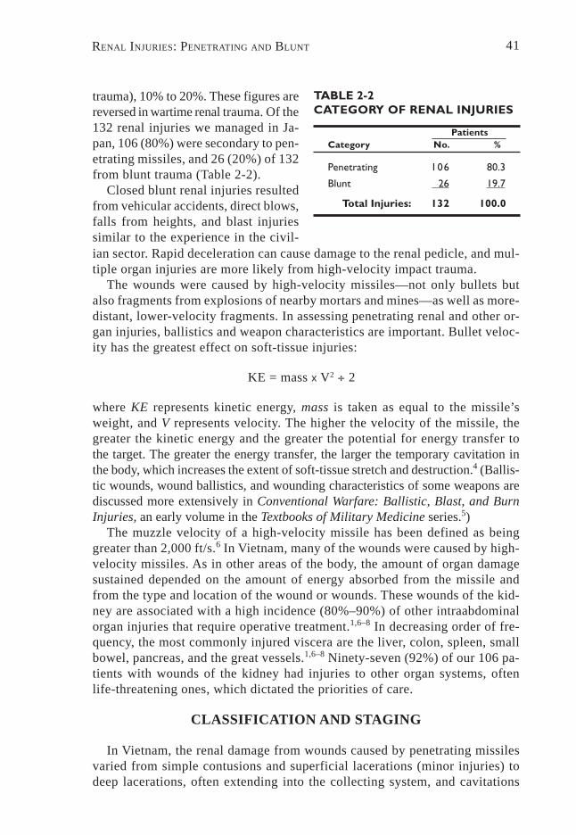
TABLE 2-2CATEGORY OF RENAL INJURIES
PatientsCategory No. %
Penetrating 106 80.3
Blunt 26 19.7
Total Injuries: 132 100.0
trauma), 10% to 20%. These figures arereversed in wartime renal trauma. Of the132 renal injuries we managed in Ja-pan, 106 (80%) were secondary to pen-etrating missiles, and 26 (20%) of 132from blunt trauma (Table 2-2).
Closed blunt renal injuries resultedfrom vehicular accidents, direct blows,falls from heights, and blast injuriessimilar to the experience in the civil-ian sector. Rapid deceleration can cause damage to the renal pedicle, and mul-tiple organ injuries are more likely from high-velocity impact trauma.
The wounds were caused by high-velocity missiles—not only bullets butalso fragments from explosions of nearby mortars and mines—as well as more-distant, lower-velocity fragments. In assessing penetrating renal and other or-gan injuries, ballistics and weapon characteristics are important. Bullet veloc-ity has the greatest effect on soft-tissue injuries:
KE = mass x V2 ÷ 2
where KE represents kinetic energy, mass is taken as equal to the missile’sweight, and V represents velocity. The higher the velocity of the missile, thegreater the kinetic energy and the greater the potential for energy transfer tothe target. The greater the energy transfer, the larger the temporary cavitation inthe body, which increases the extent of soft-tissue stretch and destruction.4 (Ballis-tic wounds, wound ballistics, and wounding characteristics of some weapons arediscussed more extensively in Conventional Warfare: Ballistic, Blast, and BurnInjuries, an early volume in the Textbooks of Military Medicine series.5)
The muzzle velocity of a high-velocity missile has been defined as beinggreater than 2,000 ft/s.6 In Vietnam, many of the wounds were caused by high-velocity missiles. As in other areas of the body, the amount of organ damagesustained depended on the amount of energy absorbed from the missile andfrom the type and location of the wound or wounds. These wounds of the kid-ney are associated with a high incidence (80%–90%) of other intraabdominalorgan injuries that require operative treatment.1,6–8 In decreasing order of fre-quency, the most commonly injured viscera are the liver, colon, spleen, smallbowel, pancreas, and the great vessels.1,6–8 Ninety-seven (92%) of our 106 pa-tients with wounds of the kidney had injuries to other organ systems, oftenlife-threatening ones, which dictated the priorities of care.
CLASSIFICATION AND STAGING
In Vietnam, the renal damage from wounds caused by penetrating missilesvaried from simple contusions and superficial lacerations (minor injuries) todeep lacerations, often extending into the collecting system, and cavitations
UROLOGY IN THE VIETNAM WAR: CASUALTY MANAGEMENT AND LESSONS LEARNED42
with extensive necrosis and shattering renal parenchyma including total abla-tion of the renal unit and major renal vascular damage (major, severe, or criti-cal injuries). The majority of these high-velocity renal wounds can be catego-rized as high grade, major, and often critical and life-threatening requiringimmediate surgery. In Vietnam, the bulk of definitive staging of renal woundswas accomplished by surgical exploration and operative assessment. Renal im-aging was primarily accomplished by either intraoperative or high-dose infu-sion pyelography, which may have defined the side of injury (often apparent priorto the study) and confirmed the probability of a “normal” opposite kidney.
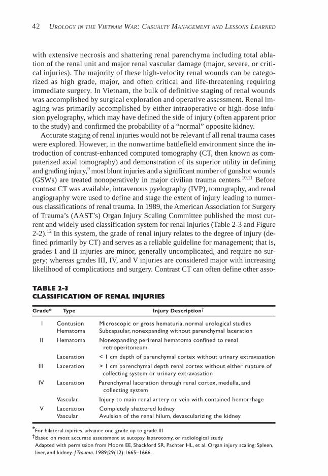
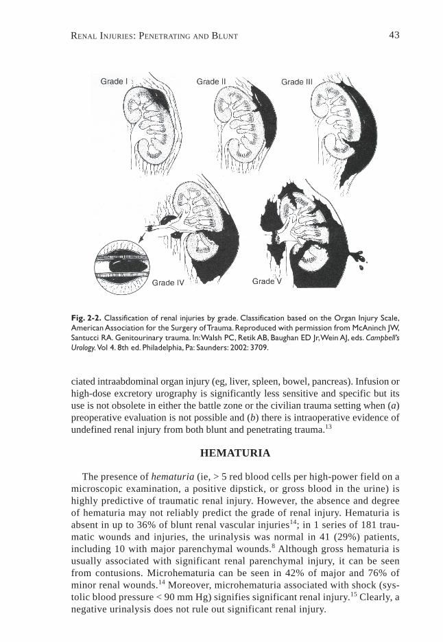
Accurate staging of renal injuries would not be relevant if all renal trauma caseswere explored. However, in the nonwartime battlefield environment since the in-troduction of contrast-enhanced computed tomography (CT, then known as com-puterized axial tomography) and demonstration of its superior utility in definingand grading injury,9 most blunt injuries and a significant number of gunshot wounds(GSWs) are treated nonoperatively in major civilian trauma centers.10,11 Beforecontrast CT was available, intravenous pyelography (IVP), tomography, and renalangiography were used to define and stage the extent of injury leading to numer-ous classifications of renal trauma. In 1989, the American Association for Surgeryof Trauma’s (AAST’s) Organ Injury Scaling Committee published the most cur-rent and widely used classification system for renal injuries (Table 2-3 and Figure2-2).12 In this system, the grade of renal injury relates to the degree of injury (de-fined primarily by CT) and serves as a reliable guideline for management; that is,grades I and II injuries are minor, generally uncomplicated, and require no sur-gery; whereas grades III, IV, and V injuries are considered major with increasinglikelihood of complications and surgery. Contrast CT can often define other asso-
TABLE 2-3CLASSIFICATION OF RENAL INJURIES
Grade* Type Injury Description†
I Contusion Microscopic or gross hematuria, normal urological studiesHematoma Subcapsular, nonexpanding without parenchymal laceration
II Hematoma Nonexpanding perirenal hematoma confined to renalretroperitoneum
Laceration < 1 cm depth of parenchymal cortex without urinary extravasation
III Laceration > 1 cm parenchymal depth renal cortex without either rupture ofcollecting system or urinary extravasation
IV Laceration Parenchymal laceration through renal cortex, medulla, andcollecting system
Vascular Injury to main renal artery or vein with contained hemorrhage
V Laceration Completely shattered kidneyVascular Avulsion of the renal hilum, devascularizing the kidney
*For bilateral injuries, advance one grade up to grade III†Based on most accurate assessment at autopsy, laparotomy, or radiological studyAdapted with permission from Moore EE, Shackford SR, Pachter HL, et al. Organ injury scaling: Spleen,liver, and kidney. J Trauma. 1989;29(12):1665–1666.
RENAL INJURIES: PENETRATING AND BLUNT 43
Fig. 2-2. Classification of renal injuries by grade. Classification based on the Organ Injury Scale,American Association for the Surgery of Trauma. Reproduced with permission from McAninch JW,Santucci RA. Genitourinary trauma. In: Walsh PC, Retik AB, Baughan ED Jr, Wein AJ, eds. Campbell’sUrology. Vol 4. 8th ed. Philadelphia, Pa: Saunders: 2002: 3709.
ciated intraabdominal organ injury (eg, liver, spleen, bowel, pancreas). Infusion orhigh-dose excretory urography is significantly less sensitive and specific but itsuse is not obsolete in either the battle zone or the civilian trauma setting when (a)preoperative evaluation is not possible and (b) there is intraoperative evidence ofundefined renal injury from both blunt and penetrating trauma.13
HEMATURIA
The presence of hematuria (ie, > 5 red blood cells per high-power field on amicroscopic examination, a positive dipstick, or gross blood in the urine) ishighly predictive of traumatic renal injury. However, the absence and degreeof hematuria may not reliably predict the grade of renal injury. Hematuria isabsent in up to 36% of blunt renal vascular injuries14; in 1 series of 181 trau-matic wounds and injuries, the urinalysis was normal in 41 (29%) patients,including 10 with major parenchymal wounds.8 Although gross hematuria isusually associated with significant renal parenchymal injury, it can be seenfrom contusions. Microhematuria can be seen in 42% of major and 76% ofminor renal wounds.14 Moreover, microhematuria associated with shock (sys-tolic blood pressure < 90 mm Hg) signifies significant renal injury.15 Clearly, anegative urinalysis does not rule out significant renal injury.
UROLOGY IN THE VIETNAM WAR: CASUALTY MANAGEMENT AND LESSONS LEARNED44
PENETRATING RENAL TRAUMA
Management
The overall management of the urological casualty in Vietnam started onhospital arrival with prompt preoperative resuscitation and control of shock,relief of respiratory distress, and injury evaluation. If renal injury was sus-pected by either hematuria or location of the wound and missile tract, a high-dose, rapid-infusion (5–10 min) IVP (2 mg/kg iodine contrast) was accom-plished. This usually was adequate to define the site of injury and establish thepresence and “normalcy” of the opposite kidney within 10 to 20 minutes. Be-cause of the high incidence of serious multiple organ wounds, the nonurologicalinjuries were often more obvious and life threatening, dictating priority treat-ment. Such patients often went to immediate surgery with incomplete urologi-cal evaluation.2,3
All patients with penetrating thoracoabdominal wounds and renal wounds wereexplored transabdominally. Intraoperative, high-dose, one-shot IVP was done inincompletely staged patients with obvious retroperitoneal hematoma and suspectedrenal injury. Generally, proximal renal vascular control was accomplished prior torenal exploration. Nephrectomy was accomplished for severely or “irreparably”damaged kidneys and, in instances of hemodynamic instability, even if partial ne-phrectomy or renorrhaphy was technically feasible. In two series2,3 from Vietnaminvolving 4 hospitals, urologists reported that the nephrectomy rate varied from51% to 84%. Debridement, control of bleeding, and partial nephrectomy were othersurgical techniques used in managing these wounds. All renal fossae were exter-nally drained. These urologists reported no significant postoperative complica-tions in Vietnam from the surgical management of these renal wounds.
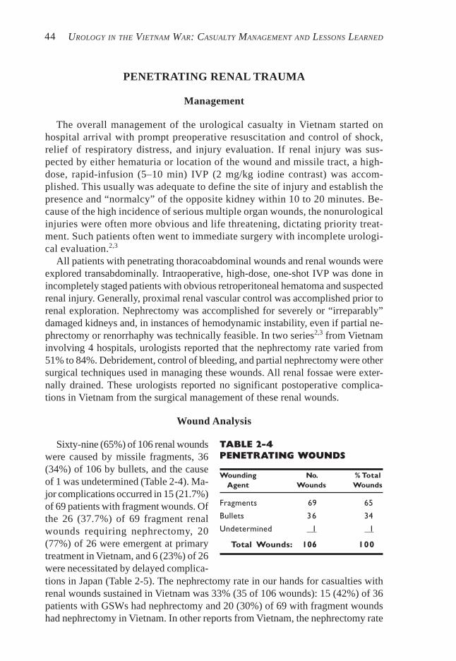
Wound Analysis
TABLE 2-4PENETRATING WOUNDS
Wounding No. % TotalAgent Wounds Wounds
Fragments 69 65
Bullets 36 34
Undetermined 1 1
Total Wounds: 106 100
Sixty-nine (65%) of 106 renal woundswere caused by missile fragments, 36(34%) of 106 by bullets, and the causeof 1 was undetermined (Table 2-4). Ma-jor complications occurred in 15 (21.7%)of 69 patients with fragment wounds. Ofthe 26 (37.7%) of 69 fragment renalwounds requiring nephrectomy, 20(77%) of 26 were emergent at primarytreatment in Vietnam, and 6 (23%) of 26were necessitated by delayed complica-tions in Japan (Table 2-5). The nephrectomy rate in our hands for casualties withrenal wounds sustained in Vietnam was 33% (35 of 106 wounds): 15 (42%) of 36patients with GSWs had nephrectomy and 20 (30%) of 69 with fragment woundshad nephrectomy in Vietnam. In other reports from Vietnam, the nephrectomy rate
RENAL INJURIES: PENETRATING AND BLUNT 45
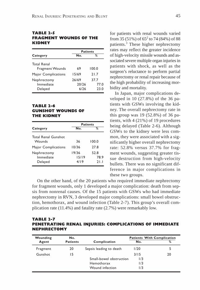
TABLE 2-5FRAGMENT WOUNDS OF THEKIDNEY
PatientsCategory No. %
Total RenalFragment Wounds 69 100.0
Major Complications 15/69 21.7
Nephrectomy 26/69 37.7Immediate 20/26 77.0Delayed 6/26 23.0
TABLE 2-6GUNSHOT WOUNDS OFTHE KIDNEY
PatientsCategory No. %
Total Renal GunshotWounds 36 100.0
Major Complications 10/36 27.8
Nephrectomy 19/36 52.8Immediate 15/19 78.9Delayed 4/19 21.1
TABLE 2-7PENETRATING RENAL INJURIES: COMPLICATIONS OF IMMEDIATENEPHRECTOMY
Wounding No. Patients With ComplicationAgent Patients Complication No. %
Fragment 20 Sepsis leading to death 1/20 5
Gunshot 15 3/15 20Small-bowel obstruction 1/3Hemothorax 1/3Wound infection 1/3
for patients with renal wounds variedfrom 35 (51%) of 652 to 74 (84%) of 88patients.3 These higher nephrectomyrates may reflect the greater incidenceof high-velocity missile wounds and as-sociated severe multiple organ injuries inpatients with shock, as well as thesurgeon’s reluctance to perform partialnephrectomy or renal repair because ofthe high probability of increasing mor-bidity and mortality.
In Japan, major complications de-veloped in 10 (27.8%) of the 36 pa-tients with GSWs involving the kid-ney. The overall nephrectomy rate inthis group was 19 (52.8%) of 36 pa-tients, with 4 (21%) of 19 proceduresbeing delayed (Table 2-6). AlthoughGSWs to the kidney were less com-mon, they were associated with a sig-nificantly higher overall nephrectomyrate: 52.8% versus 37.7% for frag-ment wounds, suggesting greater tis-sue destruction from high-velocitybullets. There was no significant dif-ference in major complications inthese two groups.
On the other hand, of the 20 patients who required immediate nephrectomyfor fragment wounds, only 1 developed a major complication: death from sep-sis from nonrenal causes. Of the 15 patients with GSWs who had immediatenephrectomy in RVN, 3 developed major complications: small bowel obstruc-tion, hemothorax, and wound infection (Table 2-7). This group’s overall com-pli cation rate (11.4%) and fatality rate (2.7%) were remarkably low.
UROLOGY IN THE VIETNAM WAR: CASUALTY MANAGEMENT AND LESSONS LEARNED46
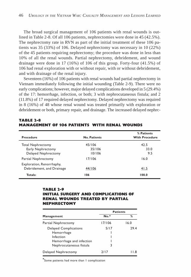
The broad surgical management of 106 patients with renal wounds is out-lined in Table 2-8. Of all 106 patients, nephrectomies were done in 45 (42.5%).The nephrectomy rate in RVN as part of the initial treatment of these 106 pa-tients was 35 (33%) of 106. Delayed nephrectomy was necessary in 10 (22%)of the 45 patients requiring nephrectomy; the procedure was done in less than10% of all the renal wounds. Partial nephrectomy, debridement, and wounddrainage were done in 17 (16%) of 106 of this group. Forty-four (41.5%) of106 had renal exploration with or without repair, with or without debridement,and with drainage of the renal injury.
Seventeen (16%) of 106 patients with renal wounds had partial nephrectomy inVietnam immediately following the initial wounding (Table 2-9). There were noearly complications; however, major delayed complications developed in 5 (29.4%)of the 17: hemorrhage, infection, or both; 3 with nephrocutaneous fistula; and 2(11.8%) of 17 required delayed nephrectomy. Delayed nephrectomy was requiredin 8 (16%) of 48 whose renal wound was treated primarily with exploration ordebridement or both, primary repair, and drainage. The increased delayed nephrc-
TABLE 2-8MANAGEMENT OF 106 PATIENTS WITH RENAL WOUNDS
% PatientsProcedure No. Patients With Procedure
Total Nephrectomy 45/106 42.5Early Nephrectomy 35/106 33.0Delayed Nephrectomy 10/106 9.5
Partial Nephrectomy 17/106 16.0
Exploration, Renorrhaphy,Debridement, and Drainage 44/106 41.5
Totals: 106 100.0
TABLE 2-9INITIAL SURGERY AND COMPLICATIONS OFRENAL WOUNDS TREATED BY PARTIALNEPHRECTOMY
PatientsManagement No.* %
Partial Nephrectomy 17/106 16.0
Delayed Complications 5/17 29.4Hemorrhage 1Infection 1Hemorrhage and infection 1Nephrocutaneous fistula 3
Delayed Nephrectomy 2/17 11.8
*Some patients had more than 1 complication
RENAL INJURIES: PENETRATING AND BLUNT 47
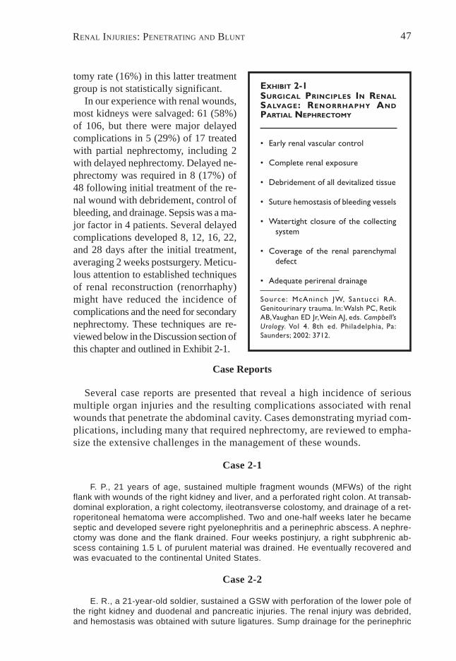
EXHIBIT 2-1SURGICAL PRINCIPLES IN RENALSALVAGE: RENORRHAPHY ANDPARTIAL NEPHRECTOMY
• Early renal vascular control
• Complete renal exposure
• Debridement of all devitalized tissue
• Suture hemostasis of bleeding vessels
• Watertight closure of the collectingsystem
• Coverage of the renal parenchymaldefect
• Adequate perirenal drainage
Source: McAninch JW, Santucc i RA.Genitourinary trauma. In: Walsh PC, RetikAB, Vaughan ED Jr, Wein AJ, eds. Campbell’sUrology. Vol 4. 8th ed. Philadelphia, Pa:Saunders; 2002: 3712.
tomy rate (16%) in this latter treatmentgroup is not statistically significant.
In our experience with renal wounds,most kidneys were salvaged: 61 (58%)of 106, but there were major delayedcomplications in 5 (29%) of 17 treatedwith partial nephrectomy, including 2with delayed nephrectomy. Delayed ne-phrectomy was required in 8 (17%) of48 following initial treatment of the re-nal wound with debridement, control ofbleeding, and drainage. Sepsis was a ma-jor factor in 4 patients. Several delayedcomplications developed 8, 12, 16, 22,and 28 days after the initial treatment,averaging 2 weeks postsurgery. Meticu-lous attention to established techniquesof renal reconstruction (renorrhaphy)might have reduced the incidence ofcomplications and the need for secondarynephrectomy. These techniques are re-viewed below in the Discussion section ofthis chapter and outlined in Exhibit 2-1.
Case Reports
Several case reports are presented that reveal a high incidence of seriousmultiple organ injuries and the resulting complications associated with renalwounds that penetrate the abdominal cavity. Cases demonstrating myriad com-plications, including many that required nephrectomy, are reviewed to empha-size the extensive challenges in the management of these wounds.
Case 2-1
F. P., 21 years of age, sustained multiple fragment wounds (MFWs) of the rightflank with wounds of the right kidney and liver, and a perforated right colon. At transab-dominal exploration, a right colectomy, ileotransverse colostomy, and drainage of a ret-roperitoneal hematoma were accomplished. Two and one-half weeks later he becameseptic and developed severe right pyelonephritis and a perinephric abscess. A nephre-ctomy was done and the flank drained. Four weeks postinjury, a right subphrenic ab-scess containing 1.5 L of purulent material was drained. He eventually recovered andwas evacuated to the continental United States.
Case 2-2
E. R., a 21-year-old soldier, sustained a GSW with perforation of the lower pole ofthe right kidney and duodenal and pancreatic injuries. The renal injury was debrided,and hemostasis was obtained with suture ligatures. Sump drainage for the perinephric
UROLOGY IN THE VIETNAM WAR: CASUALTY MANAGEMENT AND LESSONS LEARNED48
area was instituted. Fourteen days after the injury he was found to have a proximalright-sided nephrocutaneous fistula. This was considered inoperable at the time of ex-ploration owing to dense bowel adhesions and sepsis. An upper right ureteral transec-tion was unrecognized during the initial surgery. After a right nephrectomy, further con-valescence was uneventful.
Case 2-3
R. G. N., a 33-year-old soldier, received a GSW to the abdomen. At laparotomy hehad repair of a tear in the mesoappendix and an appendectomy. A contusion of the rightkidney was noted. The surgeon made no mention of hematuria or of a perirenal he-matoma at that time. Penrose drain was placed in the flank. The initial postoperativecourse was uncomplicated. Eight days later, he developed mild right costovertebral angle(CVA) pain, and gross total hematuria with marked hypotension. Eight units of wholeblood were rapidly administered. An IVP showed no apparent function on the right sidewith a normal appearing left kidney. Transabdominal laparotomy revealed a wound ofthe central portion of the right kidney with considerable necrosis, which had been missedon his initial abdominal exploration. After emergency nephrectomy, his further coursewas uneventful.
Case 2-4
W. H. S., a 21-year-old soldier, suffered fragment wounds of the abdomen from arocket-propelled grenade. At abdominal exploration, small bowel perforations were re-paired. Lacerations of the renal cortex on the right were noted. Penrose drains wereplaced in the kidney and no effort was made to remove the fragments. He was givenantibiotics and 3 units of whole blood. Copious urinary drainage occurred around thePenrose drains for the first few postoperative days. His wound did not appear infectedand he was afebrile. On the 11th postoperative day, he became febrile and septic whiledrainage from the right flank had ceased. His Penrose drains had been partially ad-vanced and were again tweaked, and the wound was explored locally with a large he-mostat and digital exploration. A small amount of drainage was obtained. An IVP showedconsiderable urinary extravasation on the right. A metallic fragment was noted in theregion of the right ureteral pelvic junction (UPJ) and there was a fragment within thekidney. The opposite kidney appeared normal. Drains were placed in the flank and anattempt was made to adequately drain the urine by means of ureteral catheter. Thisdrainage was inadequate and the patient remained febrile. He was explored in Japanthrough a right-flank incision and an examination of the kidney revealed marked ure-teral obstruction with considerable reaction around the metallic fragment in the regionof the UPJ. The patient had wounds in the upper and mid portions of the kidney with agreat deal of necrotic tissue. After nephrectomy, convalescence was uneventful.
Case 2-5
J. E. B., a 26-year-old soldier, incurred fragment wounds of the abdomen. At explo-ration, small bowel perforations were repaired. A wound to the lower pole of the rightkidney was noted, which was not bleeding and nothing further was done. A drain hadbeen placed in the retroperitoneal space and had been removed on the 10th postopera-tive day. There were no early complications. Twenty-seven days postinjury, he suddenlydeveloped total gross painless hematuria. An IVP showed good function bilaterally, anda metallic fragment was noted in the cortex of the lower pole of the right kidney. Therewas no urinary extravasation. Panendoscopy revealed blood coming from the right ure-teral orifice. He had been on antibiotics for 2 weeks following his injury and antibiotics
RENAL INJURIES: PENETRATING AND BLUNT 49
were restarted at the time of his bleeding. The following day he developed right CVApain followed by chills and fever. The repeat IVP was unchanged. He developed classicGram-negative septicemia and shock, and was treated vigorously with fluids, massivedoses of antibiotics, oxygen, and steroids. Blood cultures grew Pseudomonas. He de-veloped hemolysis with a large drop in his hematocrit and an emergency right nephrec-tomy was contemplated, but the patient responded to conservative therapy and eventu-ally became asymptomatic. He was continued on antibiotics until he had been afebrilefor 10 days. Subsequently he was discharged and had no further urological problems.
Comment on Case 2-5
This case illustrates a potential problem with a contaminated retained for-eign body in an undebrided renal wound.
Case 2-6
F. S. F. incurred MFWs of the left chest and abdomen. The lacerated spleen wasremoved; the fragmented upper pole of the left kidney was resected; large bowel perfo-rations were repaired and a colostomy performed; in addition, a transthoracic repair ofthe diaphragm was accomplished. One week after surgery, his subphrenic abscess wasdrained. One month postinjury, he was found to have a left nephrocutaneous fistula.The wound was debrided again and adequately drained, and further convalescencewas without complication.
Case 2-7
E. M. suffered a GSW of the left upper quadrant with the exit wound in the left flank,and a wound of the left arm. At laparotomy, a splenectomy, ligations of bleeding vesselsof the pancreas, closure of the gastric perforations, and resection of the lower pole ofthe left kidney were accomplished. Two weeks postoperatively, the patient became fe-brile and the left perinephric abscess was drained. Follow-up revealed some residualscarring of the left kidney but good renal function.
Case 2-8
B. R. received GSWs of the flank and lacerations of the spleen, left kidney, stom-ach, and tail of the pancreas. A splenectomy was done, the pancreatic wound drained,the wound to the stomach closed, and a perirenal hematoma drained. The patient be-came septic postoperatively and was noted on IVP to have a nephrocutaneous fistula.Seventeen days postinjury, a partial nephrectomy was done. Five days after this, at 22days postinjury, the patient developed severe bleeding which was treated with emer-gency nephrectomy.
Case 2-9
A. J. had MFWs of the right lumbar area with perforation of the lower pole of theright kidney and right colon, treated with colostomy and right nephrostomy, followingdebridement of the kidney. The patient remained septic postoperatively. His nephrostomytube was removed in Vietnam 2 weeks postinjury without radiographic studies. Oneweek later in Japan he had urinary extravasation and became febrile and septic. Fol-lowing emergency nephrectomy, further convalescence was uneventful.
UROLOGY IN THE VIETNAM WAR: CASUALTY MANAGEMENT AND LESSONS LEARNED50
Case 2-10
C. W., a 20-year-old soldier, suffered MFWs of the abdomen. He was in profoundshock when initially resuscitated, and an exploratory laparotomy showed multiple woundsto the small and large bowel, which were repaired with construction of a left colostomy.He had numerous bleeding sites and required transfusion of 23 units of whole blood. Nonote was made of injury to the urinary tract. His postoperative course was complicatedby persistent fever and upper abdominal left-flank pain. He became severely septic inJapan, and exploration on the 10th postoperative day revealed an epigastric abscess,which was drained with Aerobacter and E coli being cultured. A small amount of fluidwas present in the left pericolic gutter but no actual abscess was found. Because of thesevere left-flank pain, a left retrocolic perirenal area was explored, revealing total infarctionof the left kidney. There was a great deal of induration about the renal pedicle and it wasspeculated that the renal artery had been ligated at the time of his abdominal exploration.A nephrectomy was done. His postoperative course was further complicated by recurrentsepsis, abdominal abscesses, and eventual death from generalized sepsis.
Case 2-11
J. H., a 20-year-old soldier, had GSWs of the left abdomen with destruction of thelower pole of the left kidney. At transabdominal surgery, he had a partial nephrectomy,repair of injuries to the small and large bowel, and construction of left colostomy andretroperitoneal Penrose drainage. He drained profuse amounts of urine from his leftflank, and after 10 days the drains were removed. Urinary drainage persisted from theleft flank. On postoperative day 26, urological consultation was obtained in Japan; workuprevealed good bilateral renal function on IVP with evidence of extravasation of urinefrom the lower pole of the left kidney. The patient was given a light anesthetic, and thedrain site was probed with no significant evidence of urinoma. Some drains were placedin the region of the lower pole, and after 3 days the drainage markedly decreased. Thedrains were advanced, removed on the 6th day, and no further drainage ensued. Fol-low-up IVP showed a very small, localized extravasation of urine. This disappeared onserial follow-up studies, and there were no further complications.
DISCUSSION
Renal wounds in Vietnam were encountered in approximately 9% of ab-dominal cavity wounds, with a mortality of 7.8% from abdominal renal wounds.Between 31% and 35% of urological wounds in Vietnam involved the kidney,and, as stated above, the nephrectomy rate reported by urologists was 51% to84%.2,3 Many of these wounds were from high-velocity bullets and fragments,which caused extensive, high-grade, life-threatening, renal, and multiple othercontaminating intraabdominal visceral wounds.
The overall management of the urological casualty in Vietnam and generalmanagement in treating the renal injuries have been discussed above in thischapter in the Management section under Penetrating Renal Trauma. Becauseof the severe, life-threatening, high incidence (80%–100%) of concomitant in-jury to intraperitoneal viscera, transabdominal exploratory laparotomy was believedto be mandatory for all renal wounds in Vietnam,1–3 and, during that time, in thecivilian sector.7,8 The mortality was closely related to the number of intraabdominalviscera damaged, not from the renal wound or wounds per se. Wounds of the
RENAL INJURIES: PENETRATING AND BLUNT 51
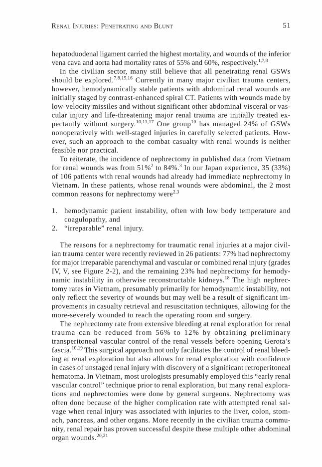
hepatoduodenal ligament carried the highest mortality, and wounds of the inferiorvena cava and aorta had mortality rates of 55% and 60%, respectively.1,7,8
In the civilian sector, many still believe that all penetrating renal GSWsshould be explored.7,8,15,16 Currently in many major civilian trauma centers,however, hemodynamically stable patients with abdominal renal wounds areinitially staged by contrast-enhanced spiral CT. Patients with wounds made bylow-velocity missiles and without significant other abdominal visceral or vas-cular injury and life-threatening major renal trauma are initially treated ex-pectantly without surgery.10,11,17 One group10 has managed 24% of GSWsnonoperatively with well-staged injuries in carefully selected patients. How-ever, such an approach to the combat casualty with renal wounds is neitherfeasible nor practical.
To reiterate, the incidence of nephrectomy in published data from Vietnamfor renal wounds was from 51%2 to 84%.3 In our Japan experience, 35 (33%)of 106 patients with renal wounds had already had immediate nephrectomy inVietnam. In these patients, whose renal wounds were abdominal, the 2 mostcommon reasons for nephrectomy were2,3
1. hemodynamic patient instability, often with low body temperature andcoagulopathy, and
2. “irreparable” renal injury.
The reasons for a nephrectomy for traumatic renal injuries at a major civil-ian trauma center were recently reviewed in 26 patients: 77% had nephrectomyfor major irreparable parenchymal and vascular or combined renal injury (gradesIV, V, see Figure 2-2), and the remaining 23% had nephrectomy for hemody-namic instability in otherwise reconstructable kidneys.18 The high nephrec-tomy rates in Vietnam, presumably primarily for hemodynamic instability, notonly reflect the severity of wounds but may well be a result of significant im-provements in casualty retrieval and resuscitation techniques, allowing for themore-severely wounded to reach the operating room and surgery.
The nephrectomy rate from extensive bleeding at renal exploration for renaltrauma can be reduced from 56% to 12% by obtaining preliminarytransperitoneal vascular control of the renal vessels before opening Gerota’sfascia.10,19 This surgical approach not only facilitates the control of renal bleed-ing at renal exploration but also allows for renal exploration with confidencein cases of unstaged renal injury with discovery of a significant retroperitonealhematoma. In Vietnam, most urologists presumably employed this “early renalvascular control” technique prior to renal exploration, but many renal explora-tions and nephrectomies were done by general surgeons. Nephrectomy wasoften done because of the higher complication rate with attempted renal sal-vage when renal injury was associated with injuries to the liver, colon, stom-ach, pancreas, and other organs. More recently in the civilian trauma commu-nity, renal repair has proven successful despite these multiple other abdominalorgan wounds.20,21
UROLOGY IN THE VIETNAM WAR: CASUALTY MANAGEMENT AND LESSONS LEARNED52
Renal Salvage Procedures
Current recommended techniques of renal salvage after traumatic renal in-jury include renal reconstruction for parenchymal laceration (renorrhaphy) andpartial nephrectomy for renal polar injuries (see Exhibit 2-1).17,22 The key sur-gical techniques employed in renorrhaphy are
• complete exposure of the kidney,• sharp excision with a scalpel blade of all ischemic or devitalized parenchyma
(complete debridement),• hemostasis of bleeding vessels with absorbable 4-0 chromic sutures,• watertight closure of the collecting system, and• coverage or approximation of the parenchymal defect margins (3-0 absorb-
able suture) using any remaining renal capsule and absorbable gelatin bol-sters (GelFoam, Upjohn Co, Kalamazoo, Mich).
Partial nephrectomy is used for polar injuries that cannot be reconstructed. Allnonviable tissue should be sharply excised, and hemostasis and closure of the col-lecting system should be done as in renorrhaphy. Stenting may be used for pelvicor ureteropelvic lacerations. After polar resection of the kidney, in which the cap-sule has been stripped off, the open parenchyma can be covered with a pedicle flapof omentum. When this is not feasible, a peritoneal graft, polyglycolic mesh, orretroperitoneal fat can be used. Warm ischemia time of 30 to 45 minutes is toler-ated if occlusion of the renal artery is needed to control bleeding. External perire-nal drainage should be used after renal reconstruction or partial nephrectomy.
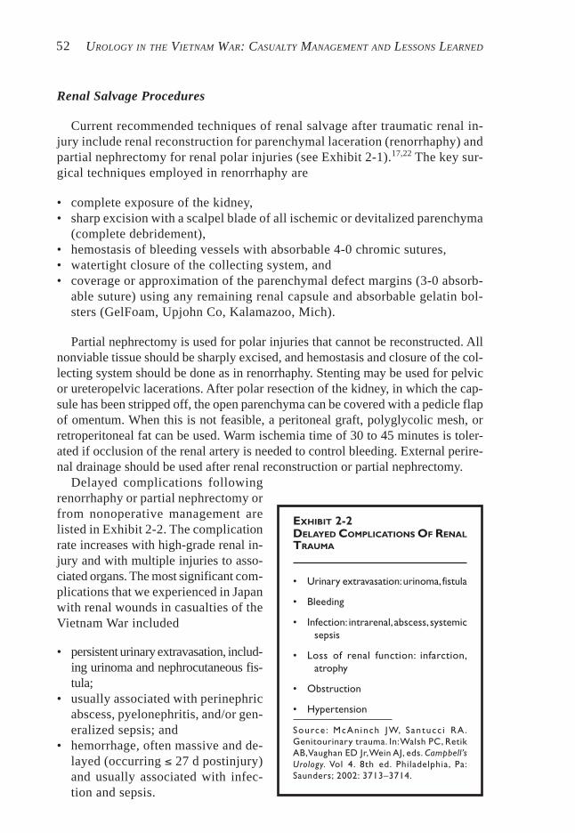
EXHIBIT 2-2DELAYED COMPLICATIONS OF RENALTRAUMA
• Urinary extravasation: urinoma, fistula
• Bleeding
• Infection: intrarenal, abscess, systemicsepsis
• Loss of renal function: infarction,atrophy
• Obstruction
• Hypertension
Source : McAn inch JW, Santucc i RA.Genitourinary trauma. In: Walsh PC, RetikAB, Vaughan ED Jr, Wein AJ, eds. Campbell’sUrology. Vol 4. 8th ed. Philadelphia, Pa:Saunders; 2002: 3713–3714.
Delayed complications followingrenorrhaphy or partial nephrectomy orfrom nonoperative management arelisted in Exhibit 2-2. The complicationrate increases with high-grade renal in-jury and with multiple injuries to asso-ciated organs. The most significant com-plications that we experienced in Japanwith renal wounds in casualties of theVietnam War included
• persistent urinary extravasation, includ-ing urinoma and nephrocutaneous fis-tula;
• usually associated with perinephricabscess, pyelonephritis, and/or gen-eralized sepsis; and
• hemorrhage, often massive and de-layed (occurring ≤ 27 d postinjury)and usually associated with infec-tion and sepsis.
RENAL INJURIES: PENETRATING AND BLUNT 53
The initial management of renal bleeding should be conservative with fluidsand transfusion. But if the conservative approach is unsuccessful, then clearlynephrectomy may be indicated. In the nonbattlefield situation, renal angiogra-phy can often localize the bleeding vessel and bleeding may be controlled byembolization.
Complications That Necessitated Delayed Nephrectomy
Delayed nephrectomy in Japan was required to manage many of these com-plications in 10 (9.5%) of 106 patients with renal wounds: for massive delayedhemorrhage with sepsis (either localized or systemic) in 5 patients, and in 5 forsystemic sepsis associated with intrarenal infection, and perirenal and abdomi-nal abscesses. Most of these patients had significant renal and associated con-taminating abdominal wounds with devitalized tissue; often bathed in urine,blood, and intestinal contents; and often arrived in Japan with obstructing, poorlymanaged Penrose drains. All these factors created an ideal milieu for localabscess, perinephric and intrarenal infection, generalized sepsis, and delayedbleeding. Many of these patients had a prolonged postinjury course and wereinitially depleted of both nutrition and nitrogen, had electrolyte imbalances,and often had various degrees of coagulopathy. Consequently, we found thatnephrectomy was often the wiser choice in managing these complications andoften was lifesaving. There was 1 death in the delayed nephrectomy groupfrom nonrenal cause in Japan.
Of the many patients with nephrocutaneous fistula, only 1 required nephre-ctomy for persistent renal and perirenal infection, generalized sepsis, and ure-teral obstruction. Generally, patients with nephrocutaneous fistula or urinomaor both, and without other organ and systemic problems, healed the urine leakwithout sequelae after debridement and enhanced external perirenal drainage.
Routinely in RVN, perirenal drainage was established with Penrose drains.Patients often arrived in Japan with obstructed Penrose drains (urine and puru-lent material had accumulated behind these drains, contributing to sepsis andpoor healing). The poor management of the Penrose drains was due in part tothe prolonged evacuation system, in which patients were placed in a “holdingarea” (an area of secondary care) prior to evacuation to Japan. Often 48 to 72hours elapsed between when the patient last received definitive care by a urolo-gist or surgeon in Vietnam and when first seen in Japan. Currently, myriad soft,tubular drains with fenestrations and suction capability are available and aremore ideal for wound drainage.
Several factors contributed to the major complications that we managed inJapan, starting with the severe contaminating and devitalizing effects of theinitial wounds, which usually involved multivisceral organ systems. Many ofthe surgeons and urologists who managed these wounds had limited prior traumaexperience. Incomplete debridement, lack of complete hemostasis, poor de-pendent drainage and monitoring of drains, and at times missed organ injuriescontributed to sepsis and delayed hemorrhage. Antibiotics were liberally used
UROLOGY IN THE VIETNAM WAR: CASUALTY MANAGEMENT AND LESSONS LEARNED54
but the variety and spectrum of effective agents was considerably limited—as opposed to the more effective agents currently available today. The urolo-gists’ ability to establish internal and renal drainage was hindered by thepoor quality of tubes and stents available for such drainage, and the gen-eral surgeons’ unfamiliarity with such equipment and techniques contrib-uted to this problem. Imaging and staging of renal injuries were restrictedby the shortcomings of the Vietnam-era IVP. The current expertise of theinterventional radiologist and the large variety of percutaneous techniquesused for urinary diversion and drainage of urinomas and abscess cavitieswere not then available. The lack of longitudinal professional supervisionof the casualty and the need for movement and the time involved in theevacuation of the wounded can be contributing factors to these complica-tions. Notwithstanding these varied factors, however, we believe that the over-all management of patients who incurred renal wounds in Vietnam was superb,as the low mortality rate reflects.
BLUNT TRAUMA
Wound Analysis
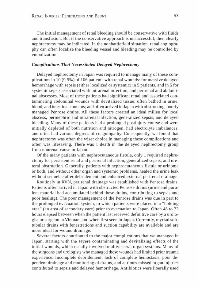
TABLE 2-10CAUSES OF BLUNT RENALINJURIES
Cause of Injury No. Patients
Land Vehicles 6
Blast Injuries 4
Fall 4
Blow 4
Aircraft Accidents 3
Football 2
Crush 1
Unknown 2
Total Patients: 26
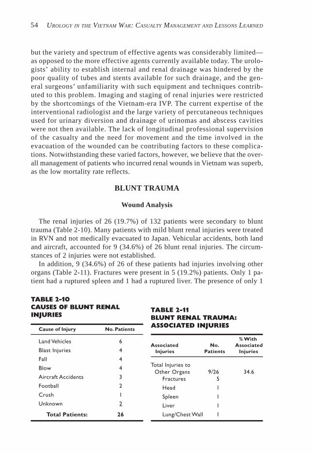
TABLE 2-11BLUNT RENAL TRAUMA:ASSOCIATED INJURIES
% WithAssociated No. Associated
Injuries Patients Injuries
Total Injuries toOther Organs 9/26 34.6
Fractures 5
Head 1
Spleen 1
Liver 1
Lung/Chest Wall 1
The renal injuries of 26 (19.7%) of 132 patients were secondary to blunttrauma (Table 2-10). Many patients with mild blunt renal injuries were treatedin RVN and not medically evacuated to Japan. Vehicular accidents, both landand aircraft, accounted for 9 (34.6%) of 26 blunt renal injuries. The circum-stances of 2 injuries were not established.
In addition, 9 (34.6%) of 26 of these patients had injuries involving otherorgans (Table 2-11). Fractures were present in 5 (19.2%) patients. Only 1 pa-tient had a ruptured spleen and 1 had a ruptured liver. The presence of only 1
RENAL INJURIES: PENETRATING AND BLUNT 55
patient with a head injury might indi-cate that there were presumably manymore fatal crush injuries and blast in-juries of the kidney and other organs(ie, most of the severely wounded ca-sualties also had head injuries thatcaused early deaths). The Japan ex-perience was typical of patients sur-viving blunt trauma.
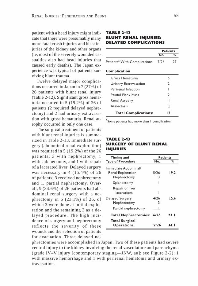
Twelve delayed major complica-tions occurred in Japan in 7 (27%) of26 patients with blunt renal injury(Table 2-12). Significant gross hema-turia occurred in 5 (19.2%) of 26 ofpatients (2 required delayed nephre-ctomy) and 2 had urinary extravasa-tion with gross hematuria. Renal at-rophy occurred in only one case.
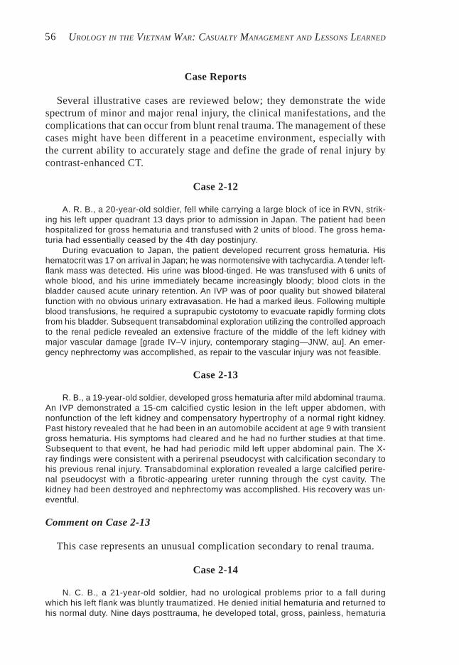
The surgical treatment of patientswith blunt renal injuries is summa-rized in Table 2-13. Immediate sur-gery (abdominal renal exploration)was required in 5 (19.2%) of the 26patients: 3 with nephrectomy, 1with splenectomy, and 1 with repairof a lacerated liver. Delayed surgerywas necessary in 4 (15.4%) of 26of patients: 3 received nephrectomyand 1, partial nephrectomy. Over-all, 9 (34.6%) of 26 patients had ab-dominal renal surgery with a ne-phrectomy in 6 (23.1%) of 26, ofwhich 3 were done at initial explo-ration and the remaining 3 as a de-layed procedure. The high inci-dence of surgery and nephrectomyref lects the sever i ty o f thesewounds and the selection of patientsfor evacuation. Three delayed ne-
TABLE 2-12BLUNT RENAL INJURIES:DELAYED COMPLICATIONS
PatientsNo. %
Patients* With Complications 7/26 27
Complication
Gross Hematuria 5
Urinary Extravasation 2
Perirenal Infection 1
Painful Flank Mass 2
Renal Atrophy 1
Atelectasis 1
Total Complications: 12
*Some patients had more than 1 complication
TABLE 2-13SURGERY OF BLUNT RENALINJURIES
Timing and PatientsType of Procedure No. %
Immediate Abdominal/Renal Exploration 5/26 19.2
Nephrectomy 3
Splenectomy 1
Repair of liverlacerations 1
Delayed Surgery 4/26 15.4Nephrectomy 3
Partial nephrectomy 1
Total Nephrectomies: 6/26 23.1
Total SurgicalOperations: 9/26 34.1
phrectomies were accomplished in Japan. Two of these patients had severecentral injury to the kidney involving the renal vasculature and parenchyma(grade IV–V injury [contemporary staging—JNW, au]; see Figure 2-2): 1with massive hemorrhage and 1 with perirenal hematoma and urinary ex-travasation.
UROLOGY IN THE VIETNAM WAR: CASUALTY MANAGEMENT AND LESSONS LEARNED56
Case Reports
Several illustrative cases are reviewed below; they demonstrate the widespectrum of minor and major renal injury, the clinical manifestations, and thecomplications that can occur from blunt renal trauma. The management of thesecases might have been different in a peacetime environment, especially withthe current ability to accurately stage and define the grade of renal injury bycontrast-enhanced CT.
Case 2-12
A. R. B., a 20-year-old soldier, fell while carrying a large block of ice in RVN, strik-ing his left upper quadrant 13 days prior to admission in Japan. The patient had beenhospitalized for gross hematuria and transfused with 2 units of blood. The gross hema-turia had essentially ceased by the 4th day postinjury.
During evacuation to Japan, the patient developed recurrent gross hematuria. Hishematocrit was 17 on arrival in Japan; he was normotensive with tachycardia. A tender left-flank mass was detected. His urine was blood-tinged. He was transfused with 6 units ofwhole blood, and his urine immediately became increasingly bloody; blood clots in thebladder caused acute urinary retention. An IVP was of poor quality but showed bilateralfunction with no obvious urinary extravasation. He had a marked ileus. Following multipleblood transfusions, he required a suprapubic cystotomy to evacuate rapidly forming clotsfrom his bladder. Subsequent transabdominal exploration utilizing the controlled approachto the renal pedicle revealed an extensive fracture of the middle of the left kidney withmajor vascular damage [grade IV–V injury, contemporary staging—JNW, au]. An emer-gency nephrectomy was accomplished, as repair to the vascular injury was not feasible.
Case 2-13
R. B., a 19-year-old soldier, developed gross hematuria after mild abdominal trauma.An IVP demonstrated a 15-cm calcified cystic lesion in the left upper abdomen, withnonfunction of the left kidney and compensatory hypertrophy of a normal right kidney.Past history revealed that he had been in an automobile accident at age 9 with transientgross hematuria. His symptoms had cleared and he had no further studies at that time.Subsequent to that event, he had had periodic mild left upper abdominal pain. The X-ray findings were consistent with a perirenal pseudocyst with calcification secondary tohis previous renal injury. Transabdominal exploration revealed a large calcified perire-nal pseudocyst with a fibrotic-appearing ureter running through the cyst cavity. Thekidney had been destroyed and nephrectomy was accomplished. His recovery was un-eventful.
Comment on Case 2-13
This case represents an unusual complication secondary to renal trauma.
Case 2-14
N. C. B., a 21-year-old soldier, had no urological problems prior to a fall duringwhich his left flank was bluntly traumatized. He denied initial hematuria and returned tohis normal duty. Nine days posttrauma, he developed total, gross, painless, hematuria
RENAL INJURIES: PENETRATING AND BLUNT 57
that lasted 6 days, ceased, and then recurred 18 days later. Subsequently he had dailygross hematuria that varied in color from light bloody to burgundy wine, and on 2 occa-sions, he had passed painful “wormlike” clots. An IVP, retrograde urogram, and renalarteriography were done and all were thought to be normal. Cystoscopy at the time ofbleeding revealed bloody efflux from the left ureteral orifice. Urine cultures and evalua-tion for tuberculosis were unremarkable. The patient had no evidence of coagulopathy.There was no evidence of sickle cell trait or disease. This patient had no other com-plaints and his gross and microscopic hematuria subsided. He was discharged.
Comment on Case 2-14
The most likely cause of his hematuria was contusion. In well-staged patients,gross hematuria has occasionally been associated with contusion only. Renal an-giography was accomplished to rule out any arteriovenous abnormality.
Case 2-15
M. F., a 22-year-old soldier, fell 25 feet from a tower and incurred blunt trauma tothe left flank. He initially presented with gross hematuria, which subsided on bedrest;his hematocrit and vital signs were normal on presentation. Subsequently, he had a fewepisodes of blood-tinged urine with mild tenderness of the left flank after ambulation; hewas evacuated to Japan 28 days later because of persistent pain. On physical exami-nation he had a tender mass in the left flank with microhematuria and normal renalfunction. An IVP revealed a normal right upper tract and massive extravasation of urinearound the left kidney with a single upper functioning calyx. Transabdominal renal ex-ploration was performed with standard vascular approach to the renal pedicle. Afterreflecting the left colon medially, a large hematoma and a urinoma were evacuated. Thelower two thirds of the left kidney was totally infarcted. Following a nephrectomy, conva-lescence was uneventful.
Comment on Case 2-15
This patient obviously had a grade IV renal injury (contemporary stag-ing—JNW, au) that was missed on initial evaluation in Vietnam. This casereflects the need for accurate staging of patients with blunt renal traumaand gross hematuria.
Case 2-16
R. M., a 23-year-old soldier, was struck by a vehicle and sustained crush injuries ofthe right lateral chest and the right upper abdomen. He had gross hematuria and normaland stable vital signs and hematocrit. The gross hematuria subsided over the first 48hours of bedrest. An IVP revealed a normal-appearing left kidney and good function onthe right with moderate urinary extravasation. He was placed on prophylactic antibacte-rial medications, and his right-flank discomfort improved. On the 7th day postinjury, hedeveloped fever of 101ºF with increasing pain and tenderness. His IVP revealed anexpanding mass on the right with urinary extravasation. An exploration in Vietnam througha right-flank incision revealed a large urinoma, which was appropriately drained. Thekidney appeared viable, and a large renal laceration was repaired. R. M. was main-tained on antibiotics, and his postoperative course was uncomplicated.
UROLOGY IN THE VIETNAM WAR: CASUALTY MANAGEMENT AND LESSONS LEARNED58
DISCUSSION
The causes and mechanisms of blunt renal injuries have been reviewed ear-lier in this chapter. Hematuria is the best indicator of traumatic renal injury.However, the degree of hematuria and the severity of renal injury do not al-ways correlate, although gross hematuria is usually associated with major re-nal parenchymal injury.
Currently, contrast-enhanced CT is the most often used (in civilian centers)and most accurate imaging modality to stage renal trauma according to theanatomical definition of injury and classification of severity (grade) of injury,as discussed earlier in this chapter. Currently, CT is not available in the 1st echelonof treatment of the battlefield casualty, and renal trauma is still staged by IVP.
The Renal Trauma Group at San Francisco General Hospital recognized thatwhen using hematuria as the only indicator for renal injury, IVP and other studiesrevealed a low incidence of renal abnormalities. This group prospectively evalu-ated indications for radiographic imaging. From this study, they concluded thatall adult patients with blunt trauma with gross hematuria, and patients withmicrohematuria and shock (systolic blood pressure < 90 mm Hg anytime dur-ing evaluation and resuscitation), should undergo renal imaging—usually withcontrast-enhanced CT. The incidence of major injuries in the group with grosshematuria was 12.5%. Adults with microscopic hematuria without shock needno imaging but do need clinical follow-up.23,24 These guidelines are the current“gold standard” for the indications for radiographic imaging of the patient withblunt renal trauma.
Of blunt renal injuries, 75% to 85% may be classified as minor, correspond-ing to grades I to III of the Organic Injury Scale (see Table 2-3). There is littlecontroversy today about the usefulness of conservative management of theseinjuries. Major renal injuries comprise the remaining 15% of cases, of which5% are grade V [contemporary staging—JNW, au]. A hemodynamically stablepatient with a well-staged injury by CT can usually be managed without renalexploration. Although grades IV and V injuries more frequently require surgi-cal exploration, many of these patients (without pedicle vascular injury) can bemanaged nonoperatively if their injuries are carefully staged and selected. Pa-tients managed nonoperatively must be hospitalized at bedrest until gross he-maturia abates, and they require periodic reimaging and very close serial moni-toring for complications.11,25 Urological complications in these patients mayoften be approached by minimally invasive endourological technique (ie, ret-rograde ureteral stenting for urinoma). At this time, many systematic inadequa-cies (eg, the lack of equipment, definitive staging by CT, trained trauma per-sonnel and team), and unpredictable variables (eg, the instability of theenvironment—often with the need for distant evacuation) make such an ap-proach impractical in the early management of the combat casualty with majorblunt renal injury or in those with major complications. During the VietnamWar, nonoperative management of major, high-grade, blunt renal injuries wasclearly not a consideration.
RENAL INJURIES: PENETRATING AND BLUNT 59
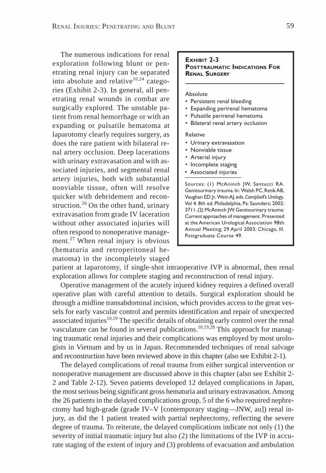
The numerous indications for renalexploration following blunt or pen-etrating renal injury can be separatedinto absolute and relative10,24 catego-ries (Exhibit 2-3). In general, all pen-etrating renal wounds in combat aresurgically explored. The unstable pa-tient from renal hemorrhage or with anexpanding or pulsatile hematoma atlaparotomy clearly requires surgery, asdoes the rare patient with bilateral re-nal artery occlusion. Deep lacerationswith urinary extravasation and with as-sociated injuries, and segmental renalartery injuries, both with substantialnonviable tissue, often will resolvequicker with debridement and recon-struction.26 On the other hand, urinaryextravasation from grade IV lacerationwithout other associated injuries willoften respond to nonoperative manage-ment.27 When renal injury is obvious(hematuria and retroperitoneal he-matoma) in the incompletely staged
EXHIBIT 2-3POSTTRAUMATIC INDICATIONS FORRENAL SURGERY
Absolute• Persistent renal bleeding• Expanding perirenal hematoma• Pulsatile perirenal hematoma• Bilateral renal artery occlusion
Relative• Urinary extravasation• Nonviable tissue• Arterial injury• Incomplete staging
patient at laparotomy, if single-shot intraoperative IVP is abnormal, then renalexploration allows for complete staging and reconstruction of renal injury.
Operative management of the acutely injured kidney requires a defined overalloperative plan with careful attention to details. Surgical exploration should bethrough a midline transabdominal incision, which provides access to the great ves-sels for early vascular control and permits identification and repair of unexpectedassociated injuries10,19 The specific details of obtaining early control over the renalvasculature can be found in several publications.10,19,28 This approach for manag-ing traumatic renal injuries and their complications was employed by most urolo-gists in Vietnam and by us in Japan. Recommended techniques of renal salvageand reconstruction have been reviewed above in this chapter (also see Exhibit 2-1).
The delayed complications of renal trauma from either surgical intervention ornonoperative management are discussed above in this chapter (also see Exhibit 2-2 and Table 2-12). Seven patients developed 12 delayed complications in Japan,the most serious being significant gross hematuria and urinary extravasation. Amongthe 26 patients in the delayed complications group, 5 of the 6 who required nephre-ctomy had high-grade (grade IV–V [contemporary staging—JNW, au]) renal in-jury, as did the 1 patient treated with partial nephrectomy, reflecting the severedegree of trauma. To reiterate, the delayed complications indicate not only (1) theseverity of initial traumatic injury but also (2) the limitations of the IVP in accu-rate staging of the extent of injury and (3) problems of evacuation and ambulation
• Associated injuries
Sources: (1) McAninch JW, Santucci RA.Genitourinary trauma. In: Walsh PC, Retik AB,Vaughan ED Jr, Wein AJ, eds. Campbell’s Urology.Vol 4. 8th ed. Philadelphia, Pa: Saunders; 2002:3711. (2) McAninch JW. Genitourinary trauma:Current approaches of management. Presentedat the American Urological Association 98thAnnual Meeting; 29 April 2003; Chicago, Ill.Postgraduate Course 49.
UROLOGY IN THE VIETNAM WAR: CASUALTY MANAGEMENT AND LESSONS LEARNED60
of patients who have poorly defined major renal injury. Such patients needserial monitoring by an experienced nursing team that is closely supervised bya urologist.
CONCLUSIONS AND EPILOGUE
During the Vietnam War, approximately one third of all battlefield urologi-cal injuries involved the kidney; about 80% of these were renal wounds, mostoften made by fragmentation devices. The overall mortality from abdominalrenal wounds was 7.5% and the incidence of nephrectomy varied from 51% to84%. However, the injury pattern during the Persian Gulf War was different,showing a marked reduction in the incidence of abdominal renal wounds and ashift to pelvic and genital wounds (ie, 17% renal vs 83% pelvic and genital).The most plausible explanation for this shift from abdominal to pelvic–genitalwounds is the ubiquitous wearing of the “flak jacket” by US military person-nel.29 This jacket provides superb protection to the thorax and upper abdomenfrom penetrating fragment wounds. Urologists made several anecdotal reports offragments found in flak jackets at the abdominal and flank locations. An obviousconclusion from this experience is that the high prevalence of use of the flak jacketduring modern combat operations will significantly reduce the incidence ofrenal and associated upper abdominal wounds, substantially decreasing renalinjury, the need for nephrectomy, and death from abdominal renal wounds.
The evaluation of renal trauma has to be tailored to the hospital location andsetting, the available diagnostic and specialty equipment, the expertise of themedical personnel, and ultimately the condition of the patient on presentationat the hospital. The key to the rational management of the renal trauma patientis accurate, precise definition of the extent of renal damage either by (1) opera-tive controlled abdominal renal exploration, primarily in the unstable patientwith penetrating abdominal renal wounds, or (2) radiological imaging (prefer-ably contrast-enhanced CT) of the hemodynamically stable patient, who is mostoften injured from blunt abdominal renal trauma.
In the Vietnam era, the IVP was the prime modality available for renal imag-ing and the staging of renal trauma. This study was usually adequate to definethe side of injury and establish the presence and “normalcy” of the contralat-eral kidney. Since the 1990s, contrast-enhanced CT has become the preferredtechnique for imaging the patient with renal trauma. The limitations of IVP are
• a low sensitivity (approximately 25% of patients with major renal injurieshave “normal” urograms), and
• common nonspecific findings, including delayed visualization of renal col-lecting systems and irregular cortical margins.
On IVP, only nonfunction and extravasation are uniformly associated withmajor renal trauma.15 However, the high-dose, rapid-infusion IVP is still indi-cated and useful in imaging the casualty with renal trauma if
RENAL INJURIES: PENETRATING AND BLUNT 61
• CT is not available;• the patient is severely injured, is hemodynamically unstable, and requires
immediate surgery; and• an unexpected retroperitoneal or perirenal hematoma is found at abdominal
exploration.
Today, the precise staging of renal trauma is best and most often accomplishedby contrast-enhanced CT. This imaging modality is ideally suited for evaluatingthe hemodynamically stable casualty with renal trauma. The current indicationsfor radiographic imaging of all adult patients with blunt trauma are23,24
• gross hematuria,• microhematuria and shock, and• high-risk mechanism of injury.
Currently in the wartime combat medical support units, CT scanning is avail-able in some of the Army’s combat support hospitals and in the Navy’s hospitalships. When indicated and available, contrast-enhanced CT should be used in thedefinitive staging of the renal trauma casualty, so that patients who are candidatesfor operative and nonoperative management can be more clearly defined.
Unstable patients selected for nonoperative management of major renal in-juries should not be placed in the evacuation chain. Additionally, patients treatedprimarily with observation need close serial monitoring and periodic reimagingby an experienced team under the supervision of a urologist. Sometimes theindication for renal exploration will evolve over several days, dictated by subtlechanges in the patient’s status as observed by the urologist. Currently, transab-dominal surgical exploration remains the “standard of care” for combat-in-curred penetrating abdominal renal missile trauma.
REFERENCES
1. Hardaway RM III. Vietnam wound analysis. J Trauma. 1978;18(9):635–643.2. Salvatierra O Jr, Rigdon WO, Norris DM, Brady TW. Vietnam experience with 252 uro-
logical war injuries. J Urol. 1969;101:615–620.3. Selikowitz SM. Penetrating high velocity genito-urinary injuries, II: Ureteral, lower tract,
and genital wounds. Urology. 1977;9(5):493–499.4. Hutton JE, Rich NM. Wounding and wound ballistics. In: McAninch JW, ed. Traumatic
and Reconstructive Urology. Philadelphia, Pa: WB Saunders; 1996: 3–25.5. Bellamy RF, Zajtchuk R, eds. Conventional Warfare: Ballistic, Blast, and Burn Injuries.
In: Zajtchuk R, Bellamy RF, eds. Textbook of Military Medicine. Washington, DC: De-partment of the Army, Office of The Surgeon General, Borden Institute; 1990.
6. Whelan TJ Jr, ed. Missile-caused wounds. In: Whelan TJ Jr, ed. Emergency War Sur-gery. 1st US Rev. Washington, DC: Department of Defense, US Government PrintingOffice; 1975: 9–17.
7. Carlton CE Jr, Scott RS Jr. Penetrating renal injuries: An analysis of 100 cases. J Urol.1960;84:599–603.
8. Scott RS Jr, Carlton CE Jr, Goldman M. Penetrating Injuries of the kidney: An analysisof 181 patients. J Urol. 1969;101:247–253.
UROLOGY IN THE VIETNAM WAR: CASUALTY MANAGEMENT AND LESSONS LEARNED62
9. Bretan PN Jr, McAninch JW, Federle MP, Jeffrey RB Jr. Computerized tomographicstaging of renal trauma: 85 consecutive cases. J Urol. 1986;136:561–565.
10. McAninch JW, Carroll PR, Klosterman PW, et al. Renal reconstruction after injury. JUrol . 1991;115:932–934.
11. Santucci RA, McAninch JW. Diagnosis and management of renal trauma: Past, presentand future. J Am Coll Surg. 2000;191:44326th451.
12. Moore EE, Shackford SR, Pachter HL, et al Organ injury scaling: Spleen, liver, andkidney. J Trauma. 1989;29:1664–1666.
13. Morey AL, McAninch JW, Tiller BK, et al. Single shot intraoperative excretory urogra-phy for the immediate evaluation of renal trauma. J Urol. 1999;161:1008–1092.
14. Cass AS. Renovascular injuries from external trauma. Urol Clin North Am. 1989;16:213–220.
15. Carroll PR, McAninch JW. Operative indications in penetrating renal. J Trauma.1985;25:587–592.
16. Moore EE, Moore JB, Vandozer-Moore S, et al. Mandatory laparotomy for gunshot woundpenetrating the abdomen. Am J Surg. 1980;140:847–851.
17. McAninch JW, Santucci RA. Genitourinary trauma. In: Walsh PC, Retik AB, VaughanED Jr, Wein AJ, eds. Campbell’s Urology. Vol 4. 8th ed. Philadelphia, Pa: Saunders;2002: 3707–3744.
18. Nash PA, Bruce JE, McAninch JW. Nephrectomy for traumatic renal injury. J Urol.1995;153:609–611.
19. McAninch JW, Carroll PR. Renal trauma: Kidney preservation through improved vas-cular control: A refined approach. J Trauma. 1982;22:285–289.
20. Rosen MA, McAninch JW. Management of combined renal and pancreatic trauma. JUrol . 1994;152:22–25.
21. Wessells H, McAninch JW. Effect of colon injury on the management of simultaneousrenal trauma. J Urol. 1996;155:1852–1856.
22. Wessels H. Evaluation and Management of Renal Trauma in the 21st Century. In: Ameri-can Urological Association. AUA Update Series, Lesson 30. Vol 21. Houston, Tex: Ameri-can Urological Association Inc, Office of Education. 2002: 234–239.
23. Miller KS, McAninch JW. Radiographic assessment of renal trauma: Our 15-year expe-rience. J Urol. 1995:154:352–355.
24. McAninch JW. Genitourinary trauma: Current approaches of management. Presented atthe American Urological Association 98th Annual Meeting; 29 April 2003; Chicago, Ill.Postgraduate Course 49.
25. Altman AL, Haas C, Dinchman KH, Spirnak JP, McAninch JW. Selective nonoperativemanagement of blunt grade 5 renal injury. J Urol. 2000;164:27–31.
26. Hussman DA, Gilling PJ, Perry MO, et al. Major renal lacerations with devitalized frag-ments following blunt abdominal trauma: A comparison between nonoperative (expect-ant) versus surgical management. J Urol. 1993;150:1774–1777.
27. Matthews LA, Smith EM, Spirnak JP. Nonoperative treatment of major blunt renal lac-erations with urinary extravasation. J Urol. 1997;157:2056–2058.
28. Scott RF Jr, Selzman HM. Complications of nephrectomy: Review of 450 patients and adescription of a modification of the transperitoneal approach. J Urol. 1966;95:307–312.
29. Thompson IM, Flaherty SF, Morey AF. Battlefield urologic injuries: The Gulf War ex-perience. J Am Coll Surg. 1998;187:139–141.