Chapter 2 Implants and oral anatomy - AQB › english › file › TheBasicsPart2-2.pdfThe maxillary...
40
1 Chapter 2 Implants and oral anatomy Associate Professor of Maxillofacial Anatomy Section, Graduate School of Medical and Dental Sciences, Tokyo Medical and Dental University Tatsuo Terashima In recent years, the development of new materials and improvements in the operative methods used for implants have led to remarkable progress in the field of dental surgery. These methods have been applied widely in clinical practice. The development of computerized medical imaging technologies such as X-ray computed tomography have allowed detailed 3D-analysis of medical conditions, resulting in a dramatic improvement in the success rates of operative intervention. For treatment with a dental implant to be successful, it is however critical to have full knowledge and understanding of the fundamental anatomical structures of the oral and maxillofacial regions. In addition, it is necessary to understand variations in the topographic and anatomical structures among individuals, with age, and with pathological conditions. This chapter will discuss the basic structure of the oral cavity in relation to implant treatment. I. Osteology of the oral area The oral cavity is composed of the maxilla that is in contact with the cranial bone, palatine bone, the mobile mandible, and the hyoid bone. The maxilla and the palatine bones articulate with the cranial bone. The mandible articulates with the temporal bone through the temporomandibular joint (TMJ). The hyoid bone is suspended from the cranium and the mandible by the suprahyoid and infrahyoid muscles. The formation of the basis of the oral cavity by these bones and the associated muscles makes it possible for the oral cavity to perform its various functions. A. Mandible The mandible is a powerful bone which constitutes the lower part of the face; unlike other bones of the cranium, it interacts in a mobile fashion with the temporal bone, through the temporomandibular joint. The mandible is divided into two sections. The parabolic ridge that occupies two-thirds of the frontal section of the mandible, where the teeth are inserted, is called the body of the mandible. The bone that lies perpendicularly behind the body is called the ramus of the mandible. 1. Body of mandible (Fig. 2-2-1-a) The body of the mandible is a curved plate into which the teeth are inserted. a. Alveolus Teeth are embedded in the alveolus of the mandible. The cavity that accommodates the root of the tooth is known as the tooth socket. The edge that contains these tooth sockets is called the alveolar process. The area between each of the tooth sockets is known as the interalveolar septum, and intra-alveolar septa divide the tooth sockets of each molar. b. Mandibular base The inferior border of the mandible is very thick in appearance and is referred to as the mandibular base. c. The exterior The alveolar tubercle that matches the root of the tooth can be observed on the external wall of the
Transcript of Chapter 2 Implants and oral anatomy - AQB › english › file › TheBasicsPart2-2.pdfThe maxillary...

1
Chapter 2 Implants and oral anatomy Associate Professor of Maxillofacial Anatomy Section, Graduate School of Medical and
Dental Sciences, Tokyo Medical and Dental University Tatsuo Terashima
In recent years, the development of new materials and improvements in the operative methods used for implants have led to remarkable progress in the field of dental surgery. These methods have been applied widely in clinical practice. The development of computerized medical imaging technologies such as X-ray computed tomography have allowed detailed 3D-analysis of medical conditions, resulting in a dramatic improvement in the success rates of operative intervention. For treatment with a dental implant to be successful, it is however critical to have full knowledge and understanding of the fundamental anatomical structures of the oral and maxillofacial regions. In addition, it is necessary to understand variations in the topographic and anatomical structures among individuals, with age, and with pathological conditions. This chapter will discuss the basic structure of the oral cavity in relation to implant treatment. I. Osteology of the oral area The oral cavity is composed of the maxilla that is in contact with the cranial bone, palatine bone, the mobile mandible, and the hyoid bone. The maxilla and the palatine bones articulate with the cranial bone. The mandible articulates with the temporal bone through the temporomandibular joint (TMJ). The hyoid bone is suspended from the cranium and the mandible by the suprahyoid and infrahyoid muscles. The formation of the basis of the oral cavity by these bones and the associated muscles makes it possible for the oral cavity to perform its various functions. A. Mandible The mandible is a powerful bone which constitutes the lower part of the face; unlike other bones of the cranium, it interacts in a mobile fashion with the temporal bone, through the temporomandibular joint. The mandible is divided into two sections. The parabolic ridge that occupies two-thirds of the frontal section of the mandible, where the teeth are inserted, is called the body of the mandible. The bone that lies perpendicularly behind the body is called the ramus of the mandible. 1. Body of mandible (Fig. 2-2-1-a) The body of the mandible is a curved plate into which the teeth are inserted. a. Alveolus Teeth are embedded in the alveolus of the mandible. The cavity that accommodates the root of the tooth is known as the tooth socket. The edge that contains these tooth sockets is called the alveolar process. The area between each of the tooth sockets is known as the interalveolar septum, and intra-alveolar septa divide the tooth sockets of each molar. b. Mandibular base The inferior border of the mandible is very thick in appearance and is referred to as the mandibular base. c. The exterior The alveolar tubercle that matches the root of the tooth can be observed on the external wall of the
2
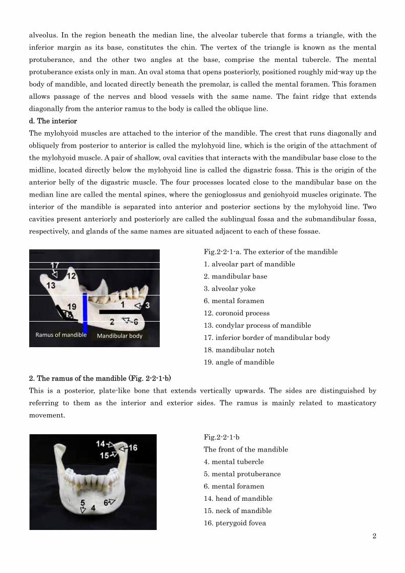
alveolus. In the region beneath the median line, the alveolar tubercle that forms a triangle, with the inferior margin as its base, constitutes the chin. The vertex of the triangle is known as the mental protuberance, and the other two angles at the base, comprise the mental tubercle. The mental protuberance exists only in man. An oval stoma that opens posteriorly, positioned roughly mid-way up the body of mandible, and located directly beneath the premolar, is called the mental foramen. This foramen allows passage of the nerves and blood vessels with the same name. The faint ridge that extends diagonally from the anterior ramus to the body is called the oblique line. d. The interior The mylohyoid muscles are attached to the interior of the mandible. The crest that runs diagonally and obliquely from posterior to anterior is called the mylohyoid line, which is the origin of the attachment of the mylohyoid muscle. A pair of shallow, oval cavities that interacts with the mandibular base close to the midline, located directly below the mylohyoid line is called the digastric fossa. This is the origin of the anterior belly of the digastric muscle. The four processes located close to the mandibular base on the median line are called the mental spines, where the genioglossus and geniohyoid muscles originate. The interior of the mandible is separated into anterior and posterior sections by the mylohyoid line. Two cavities present anteriorly and posteriorly are called the sublingual fossa and the submandibular fossa, respectively, and glands of the same names are situated adjacent to each of these fossae.
Mandibular body
mandibular bodymandibular bodymandibular body
Ramus of mandible
2. The ramus of the mandible (Fig. 2-2-1-b) This is a posterior, plate-like bone that extends vertically upwards. The sides are distinguished by referring to them as the interior and exterior sides. The ramus is mainly related to masticatory movement.
Fig.2-2-1-a. The exterior of the mandible 1. alveolar part of mandible 2. mandibular base 3. alveolar yoke 6. mental foramen 12. coronoid process 13. condylar process of mandible 17. inferior border of mandibular body 18. mandibular notch 19. angle of mandible
Fig.2-2-1-b The front of the mandible 4. mental tubercle 5. mental protuberance 6. mental foramen 14. head of mandible 15. neck of mandible 16. pterygoid fovea
3
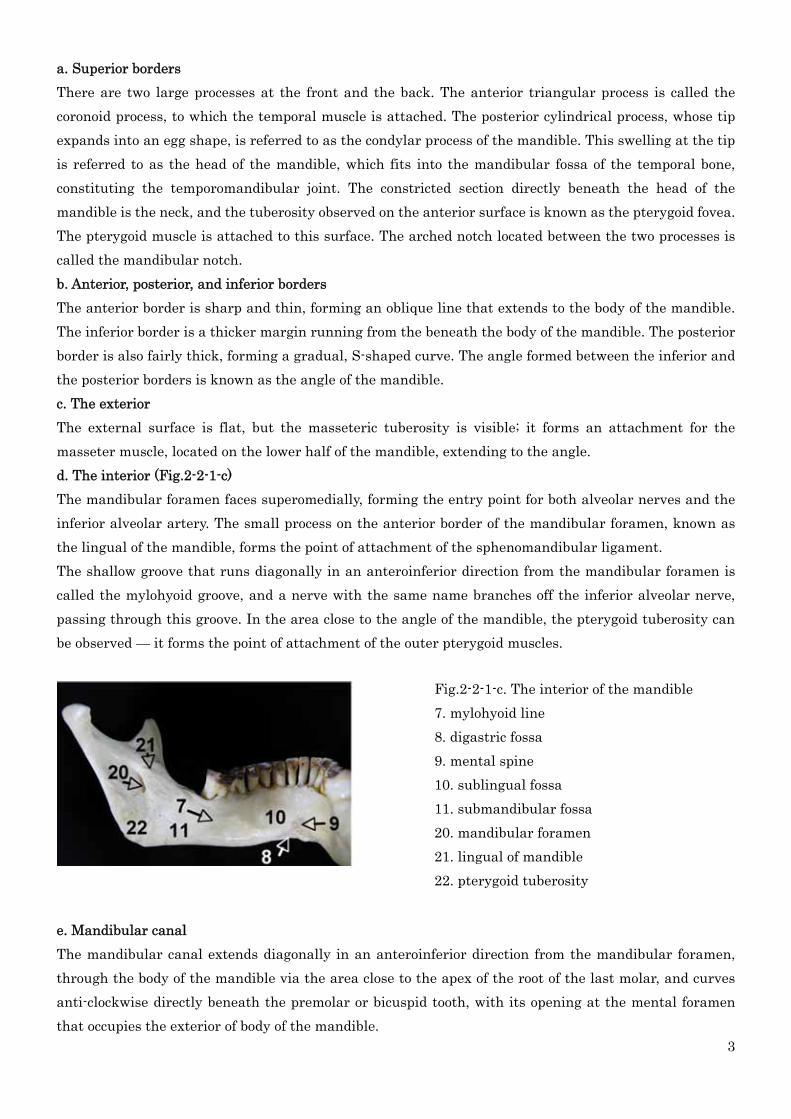
a. Superior borders There are two large processes at the front and the back. The anterior triangular process is called the coronoid process, to which the temporal muscle is attached. The posterior cylindrical process, whose tip expands into an egg shape, is referred to as the condylar process of the mandible. This swelling at the tip is referred to as the head of the mandible, which fits into the mandibular fossa of the temporal bone, constituting the temporomandibular joint. The constricted section directly beneath the head of the mandible is the neck, and the tuberosity observed on the anterior surface is known as the pterygoid fovea. The pterygoid muscle is attached to this surface. The arched notch located between the two processes is called the mandibular notch. b. Anterior, posterior, and inferior borders The anterior border is sharp and thin, forming an oblique line that extends to the body of the mandible. The inferior border is a thicker margin running from the beneath the body of the mandible. The posterior border is also fairly thick, forming a gradual, S-shaped curve. The angle formed between the inferior and the posterior borders is known as the angle of the mandible. c. The exterior The external surface is flat, but the masseteric tuberosity is visible; it forms an attachment for the masseter muscle, located on the lower half of the mandible, extending to the angle. d. The interior (Fig.2-2-1-c) The mandibular foramen faces superomedially, forming the entry point for both alveolar nerves and the inferior alveolar artery. The small process on the anterior border of the mandibular foramen, known as the lingual of the mandible, forms the point of attachment of the sphenomandibular ligament. The shallow groove that runs diagonally in an anteroinferior direction from the mandibular foramen is called the mylohyoid groove, and a nerve with the same name branches off the inferior alveolar nerve, passing through this groove. In the area close to the angle of the mandible, the pterygoid tuberosity can be observed — it forms the point of attachment of the outer pterygoid muscles.
e. Mandibular canal The mandibular canal extends diagonally in an anteroinferior direction from the mandibular foramen, through the body of the mandible via the area close to the apex of the root of the last molar, and curves anti-clockwise directly beneath the premolar or bicuspid tooth, with its opening at the mental foramen that occupies the exterior of body of the mandible.
Fig.2-2-1-c. The interior of the mandible 7. mylohyoid line 8. digastric fossa 9. mental spine 10. sublingual fossa 11. submandibular fossa 20. mandibular foramen 21. lingual of mandible 22. pterygoid tuberosity
4
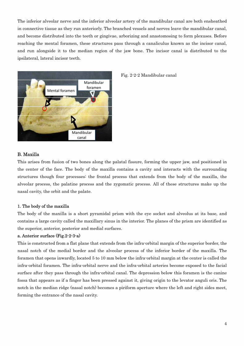
The inferior alveolar nerve and the inferior alveolar artery of the mandibular canal are both ensheathed in connective tissue as they run anteriorly. The branched vessels and nerves leave the mandibular canal, and become distributed into the teeth or gingivae, arborizing and anastomosing to form plexuses. Before reaching the mental foramen, these structures pass through a canaliculus known as the incisor canal, and run alongside it to the median region of the jaw bone. The incisor canal is distributed to the ipsilateral, lateral incisor teeth.
Mental foramen
Mandibularcanal
Mandibularforamen
B. Maxilla This arises from fusion of two bones along the palatal fissure, forming the upper jaw, and positioned in the center of the face. The body of the maxilla contains a cavity and interacts with the surrounding structures though four processes: the frontal process that extends from the body of the maxilla, the alveolar process, the palatine process and the zygomatic process. All of these structures make up the nasal cavity, the orbit and the palate. 1. The body of the maxilla The body of the maxilla is a short pyramidal prism with the eye socket and alveolus at its base, and contains a large cavity called the maxillary sinus in the interior. The planes of the prism are identified as the superior, anterior, posterior and medial surfaces. a. Anterior surface (Fig.2-2-3-a) This is constructed from a flat plane that extends from the infra-orbital margin of the superior border, the nasal notch of the medial border and the alveolar process of the inferior border of the maxilla. The foramen that opens inwardly, located 5 to 10 mm below the infra-orbital margin at the center is called the infra-orbital foramen. The infra-orbital nerve and the infra-orbital arteries become exposed to the facial surface after they pass through the infra-orbital canal. The depression below this foramen is the canine fossa that appears as if a finger has been pressed against it, giving origin to the levator anguli oris. The notch in the median ridge (nasal notch) becomes a piriform aperture where the left and right sides meet, forming the entrance of the nasal cavity.
Fig. 2-2-2 Mandibular canal

5
b. Posterior surface (infratemporal surface) (Fig.2-2-3-b) This forms the anterior wall of the infratemporal fossa, the posterior part of the area separated by a strong ridge that extends downwards from the infrazygomatic crest. The central portion is shaped like an expanded rectangle, with an anterior border that corresponds to the posterior border of the orbit, and an inferior border that extends to the alveolar process of the maxilla. The posterior surface is not visible from behind, as the upper half articulates with the palatal bone, and lower half with the pterygoid process of sphenoid bone. The expanded section at the center is the maxillary tuberosity, containing a few alveolar foramina in the tubercle. This is the point of entry for the alveolar canal that passes through the wall of the maxillary sinus. The posterior superior alveolar nerves and arteries enter the maxilla via this cavity, extending to the region of the molars.
c. Superior surface (The orbital plane) (Fig. 2-2-4) This is the surface that constitutes a large part of the floor of the orbit. The medial border meets both the lacrimal and ethmoid bones. The lateral border articulates with the zygomatic bone in its anterior half; the posterior border is smoothly rounded and forms most of the anterior margin of the inferior orbital fissure and articulates with the palatine bone. The infra-orbital groove runs anteriorly from the center of
Fig. 2-2-3-a,b,c The maxillary a The anterior surface of the maxillary 1. infraorbital margin 2. nasal notch 3. alveolar process of maxilla 4. infraorbital foramen 5. canine fossa 11. frontal process of maxilla
Fig. 2-2-3-b The posterior surface of the maxillary 6. inferior ridge of the zygomatic bone 7. maxillary tuberosity 8. zygomatic process of maxilla 12. alveolar foramen

6
the inferior border of the inferior orbital fissure. This groove becomes the infra-orbital canal, having changed its orientation at the center in an antero-inferior direction, then entering the bone. The infra-orbital canal opens into the infra-orbital foramen of the anterior surface of the body of the maxilla. The infra-orbital groove, the infra-orbital canal and the infra-orbital foramen all permit passage of the infra-orbital arteries and nerves that are distributed to the maxilla and teeth.
d. Interior surface (Fig. 2-2-3-c) This surface forms the external wall of the nasal cavity. The inferior border is transformed into the alveolar process, but two-thirds of the anterior border, the palatine process, protrudes out of this transformation zone horizontally. In the interior, close to the center, a large opening exists that acts as the entrance to the maxillary sinus, called the maxillary hiatus. This large hiatus actually becomes covered by bone inferiorly (the inferior nasal concha), anteriorly (the ethmoid bone) and posteriorly (the palatine bone), therefore eventually becoming more constricted, resulting in a semilunar hiatus. This is covered by mucous membrane in the living body.In the interior, two canals are constructed from two bones that lie adjacent to each other. Half of each canal constitutes a groove in the maxilla. The lacrimal groove that lies at the anterior part of the maxillary hiatus joins the lacrimal bone, forming the nasolacrimal canal that allows tears to flow into the nasal cavity. The greater palatine groove that runs obliquely behind the maxillary hiatus joins the palatine bone to form the greater palatine canal. The greater palatine arteries and nerves pass through these canals, eventually distributing to the palate.
Fig. 2-2-4 The anterior surface of the cranial bone 3. optic nerve 4. superior orbital fissure 5. inferior orbital fissure 6. infraorbital groove 8. infraorbital foramen
Fig. 2-2-3-c The interior of the maxillary 9. maxillary hiatus 10. nasolacrimal canal 13. palatine process of maxilla

7
e. Maxillary sinus This is a hollow cavity that is shaped like an inverted pyramid, similar to the body of the maxilla. The walls of this hollow cavity are extremely thin, 1 to 2 mm in thickness. As the roots of the teeth and the base of the maxillary sinus are in close proximity, inflammation of the root apex can spread to the maxillary sinus. 2. Processes of the maxilla a. Frontal process This faces superiorly, arising from the superomedial edge of the body of the maxilla, and comes into contact with the frontal bone. b. Zygomatic process This forms the anterior plane of the body, and the upper half of the border in the posterior region. It projects laterally, coming into contact with the zygomatic bone. c. Palatine process (Fig.2-2-5) This is a thick, horizontal plate that spreads from the alveolar process of the body of the mandible internally. The medial border forms the anterior two-thirds of the hard palate, formed by articulation of the right and left halves at the median palatine suture. The area of the median palatine suture forms the nasal crest superiorly, and its front edge forms the anterior nasal spine. The posterior border articulates with the horizontal plate of the palatine bone at the traverse palatine suture and the hard palate is constructed from both palatine processes and the horizontal plate of the palatine bone. A tuberosity is present on the inferior surface that faces the oral cavity. In particular, the palatine groove can be observed in the region close to the alveolar process of the molars, which runs anteriorly, with palatine spines present over the central groove. The great palatine nerves and arteries pass through this groove. At the posterior section of the central incisor, the incisive fossa is evident. This depression continues from the incisive foramen to the incisive canal and the incisive canal forks in a Y-shape with openings to both sides of the nasal crest. The nasopalatine nerve and the posterior septal nasal artery run through this incisive canal.
d. Alveolar process This is an arched process that extends from the inferior ridge of the body of the maxilla, protruding
Fig. 2-2-5 The bony palate 1. median palatine suture 2. transverse palatine suture 3. palatine groove 4. greater palatine groove 5. incisive fossa

8
towards the inferior section of the palatine bone. Horseshoe-shaped, it is formed by the combination of the right and left processes, referred to as the alveolar arch, which bears tooth roots. The alveolar indentations that match the tooth roots can be observed on the external wall. 3. Alveolar canal Most of the space within the body of the maxilla is occupied by the maxillary sinus, which is surrounded by a thin outer wall. The alveolar canals contain blood vessels and nerves that distribute to the teeth, running the width of the exterior wall. a. Anterior alveolar canal This arises from the infra-orbital canal and extends along the anterior surface towards the piriform aperture, during which process, it reveals the canaliculi of the premolar teeth. b. Medial alveolar canal This arises from the infra-orbital foramen and extends towards the anterior wall, during which time it gradually changes direction inferiorly, until it reaches the premolar teeth. c. Posterior alveolar canal This originates from the alveolar foramen, extending from posterior to anterior, and eventually distributing to the molar teeth. II. Muscles that form the walls of the oral cavity The muscles associated with functions of the oral cavity can be classed broadly into four muscle groups: A. Muscles of facial expression: a group of small muscles that form facial expressions; B. Masticatory muscles: consist of four large muscles that move the mandible, and are responsible for elevation of, and forward and backward movements of the jaw. C. Suprahyoid muscles: The group of muscles that occupy the space between the hyoid bone and the mandible and form the floor of the oral cavity, as well as performing an important role in opening and closing the mouth. D. Tongue muscles: aid the functions of the tongue. There are extrinsic and intrinsic muscles of the tongue — the former adjust the position of the tongue, whilst the latter change the shape of the tongue. A. Muscles of facial expression (Fig. 2-2-6-a,b) Also known as the facial muscles, these are thin cutaneous muscles that attach to the face. The facial muscles are a conglomerate of small, thin muscles, and facial expressions are constructed by forming various depressions and lines on the face. This group of muscles does not contain fascia, and is controlled by the facial nerves. The facial muscles, distributed subcutaneously, are further divided into smaller muscle groups: 1) Muscles around the oral fissure (muscles of the mouth) 2) Muscles around the orbital fissure (orbicularis oculi muscle, procerus muscle) 3) Muscles of the nasal region (nasalis muscle) 4) Muscles around the auricle (auricular muscle) 5) Muscles around the cranium (occipitofrontalis muscle) 6) Facial portion of the platysma muscle (Not a facial muscle, but functionally, it can be grouped within

9
this category)
The group of muscles that forms the external wall of the oral cavity is called “the muscles of the mouth”. These muscles are located around the oral fissure, and constitute the anterior and the lateral walls. These muscles are subdivided into the muscles that surround the oral fissure in concentric circles, helping to close the mouth, called orbicularis oris, and another group of muscles that spread radially from the anguli oris, the mouth-opening muscles. Those muscles that are involved in opening the mouth are also divided according to location, such that the muscles positioned above, lateral to and below the angulis oris, are each subdivided into two more layers according to their depth. The main muscles are as follows: 1. Orbicularis oris muscle This muscle encircles the oral fissure. A considerable number of muscular bundles are derived from the buccinator muscles, but some of the muscle bundles from all of the mouth muscles join these at some point. In addition, the skin of the posterior rim of the nostrils and a small muscle bundle that arises from the alveolar tubercle of the maxillomandibular incisor teeth connect. Once the mouth is closed, narrowing the oral fissure, if the muscles are further constricted, the mouth becomes pointed. 2. Muscles above anguli oris
Fig. 2-2-6-a,b The muscles of facial expression a 1. greater zygomatic muscle 2. lesser zygomatic muscle 3. levator labii superioris muscle 4. levator labii superioris alaeque nasi muscle 5. levator anguli oris muscle 6. depressor anguli oris muscle 7. depressor labii inferioris muscle 8. mentalis muscle
8. mentalis muscle 9. nasalis muscle 10. frontal belly of occipitofrontalis muscle 11. procerus muscle 12. corrugators supercilii muscle b. 13. buccinators muscle 14. orbicularis oculi muscle
10
a. The greater zygomatic muscle This is the largest muscle in this group, originating from the anterior surface of the zygomatic bone, with the greater part ending up in the skin of the upper lip, and the rest branching out to the skin of the lower lip. The function of this muscle is to draw up the anguli oris in a superolateral direction. b. The lesser zygomatic muscle, levator labii superioris muscle and the levator labii superioris alaeque nasi muscle. The lesser zygomatic muscle arise from the anterior surface of the zygomatic muscle, the levator labii superioris muscle from the anterior surface of the maxilla, and the levator labii superioris alaeque nasi muscle from the frontal process of maxilla, attaching to the skin of the upper lip. These function to pull the upper lip and ala nasi upwards, and deepen the nasolabial sulcus. c. The levator anguli oris muscle This large muscle occupies the depth of the mouth. It originates from the canine fossa, and extends to the anguli oris, partially dividing to provide a part that terminates at the skin of anguli oris, while the greater part connects with the depressor anguli oris muscle below. 3. Muscles lateral to anguli oris a. Risorius muscle This is a slender, triangular muscle placed on the platysma muscle that terminates in the skin close to the anguli oris. Contraction of this muscle results in dimple formation. b. Buccinator muscle This is a large muscle situated in the depth of the mouth that occupies the center of the buccal region. It originates from the exterior of the alveolar bone of the maxillary and the mandibular molars as well as the pterygomandibular raphe. It divides into the upper and lower lip, forming a considerable part of the orbicularis oris muscle. It provides the cheeks with constant tension that is in proportion to the amount of opening and shutting of the mouth, and helps to prevent biting of the cheek during mastication. 4. Muscles below angulis oris a. Depressor anguli oris muscle This forms a triangular muscle plate that has the angulis oris at its tip. This originates from the front half of the mandibular base, is gathered at angulis oris, and terminates in the skin of the top lip. A section of the deep layer connects with the levator anguli oris muscle. It functions to pull the lower lip downwards. b. Depressor labii inferioris muscle This forms a non-rectangular, quadrangular muscle plate that spread extensively within the lower lip, the posterior section of which is covered with the depressor anguli oris muscles. It starts from the anterior section of the mandibular base, and remains in the skin of the lower lip. c. Mentalis muscle This occupies a relatively high position over the median section of the mandible. It originates from the alveolar tubercle of the incisor, and descends and adheres to the skin of the mental region. It functions to pull the chin upwards. 5. The facial component of the platysma muscle This arises from the vicinity of the clavicle, and spreads thinly in the anterior and inferior cervical region. It then ascends over the mandibular base line, and reaches the buccal region of the face.

11
B. Suprahyoid muscles (muscles that form the floor of the mouth) The space between the mandibular base and the tongue muscles, the part corresponding to the base of the oral cavity, is referred to as the floor of the mouth, formed by the suprahyoid muscles. 1. Digastric muscles of the jaw (Fig 2-2-7-a) This digastric structure comprises an anterior belly and a posterior belly, with the intermediate tendon adhering to the hyoid bone. The anterior belly arises from the gastric fossa of the mandibular base, while the posterior belly begins at the mastoid notch in the medial part of the mastoid process. Both of these bellies extend to the intermediate tendon and terminate in the body of the hyoid bone. This muscle effectively pulls up the hyoid bone when the mandible is fixed; alternatively the mandible is pulled up if the hyoid bone is fixed. The anterior belly is controlled by the mandibular nerve (nerve to mylohyoid), and the posterior belly, by the facial nerve (digastric branch), respectively. 2. Stylohyoid muscle (Fig.2-2-7-a) This is a slender muscle that runs obliquely, and roughly parallel to the digastric muscle. It arises at the styloid process and terminates at the anterior end of the thyrohyal, which is at the interior of the intermediate tendon of the digastric muscle. It functions to pull the hyoid muscles upwards in a posterosuperior direction, and fix their position. This muscle is controlled by the facial nerve (stylohyoid branch). 3. The geniohyoid muscle (Fig.2-2-7-b) This is a slender muscle located in the midline of the mouth floor. It arises from the mental spine and terminates on the anterior surface of the body of the hyoid bone. It functions to pull the hyoid bone forwards when the mandible is fixed, whereas, when the hyoid bone is fixed, it effectively pulls the mandible in an inferoposterior direction. It is controlled by the ansa cervicalis (C1) that passes with the hypoglossal nerve. 4. The mylohyoid muscle (Fig.2-2-7-b) This has a plate-like structure that extends to the mandible in a fan shape with the hyoid bone as a pivot. It arises from the mylohyoid bone, with one third attached to the thyrohyoid, with the remaining two-thirds of its anterior structure attaching to the raphe that stretches from the mental spine and body of the hyoid bone. When the mandible is fixed, it acts to pull the hyoid bone upwards, resulting in a shallower mouth floor, but when the hyoid bone is fixed, the mandible is lifted instead. This muscle is also referred to as the diaphragm of the mouth floor because of its location and structure. This muscle is controlled by the nerve to mylohyoid, which arises from the mandibular nerve.

12
Fig. 2-2-7-a,b The suprahyoid muscles a The digastric muscle and the stylohoid muscle 1. posterior belly of the digastric muscle 2. anterior belly of the digastric muscle 3. stylohoid muscle 4. mastoid notch 5. digastrics fossa 6. styloid process 7. hyoid bone C. Masticatory muscles The masseter and temporalis muscles lie on the external surface, and the medial pterygoid and lateral pterygoid muscles occupy the internal surface of the ramus of mandible. Masticatory movements arise due to the contraction of muscles that lift the mandible, and move it back and forth. 1. Temporalis muscle (Fig. 2-2-8-a) This muscle extends in a fan shape over the temporal fossa, originating from the temporal line, becoming confluent, and then terminating in the coronoid process. Muscle fibers can be grouped into bundles based on their orientations: a. Anterior bundle This runs in a vertical direction and pulls the mandible upwards. b. Medial bundle Running obliquely, it acts to pull the mandible in an inferoposterior direction. c. Posterior bundle Running horizontally, when it contracts it pulls the mandible backwards
2. Masseter muscle (Fig. 2-2-8-b) This is the quadrilateral muscle on the external surface of the mandible. It arises from the zygomatic arch, descends and then terminates in the masseteric tuberosity of the external surface of the ramus of the mandible. There are muscle fiber bundles present on the superficial and the deep sections of the masseter
Fig. 2-2-8-a,b The masseter muscle a The temporalis muscle 1. temporalis muscle 2. temporal fossa 3. coronoid process 4. external oblique ridge
b The hyoglossus muscle and the geniohyoid muscle 1. mylohyoid muscle 2. geniohyoid muscle 3. mental spine 4. mylohyoid line 5. hyoid bone

13
muscle. The superficial portion of the muscle is wide, and parallelogram-shaped. The deep portion is smaller and has a more muscular structure. It expands out in a fan shape. Anteriorly, the greater part of the deep portion is concealed by the superficial portion. This muscle pulls the mandible upwards to enable occlusion of the upper and lower teeth. The motions are controlled by the masseteric nerve.
3. Medial pterygoid muscle (Fig.2-2-9-a) This muscle binds the mandibular ramus with the masseter muscle to suspend the mandible in a hammock-like manner. It arises from the pterygoid fossa of the internal surface of the pterygoid process of the sphenoid bone, and it runs in an inferoposterior direction, terminating at the pterygoid tuberosity that is located on the internal surface of the ramus of the mandible. It pulls the mandible upwards, enabling occlusion to be established between the upper and lower teeth, with the help of the masseter muscle. It is controlled by the nerve to the medial pterygoid.
4. Lateral pterygoid muscle (Fig.2-2-9-b) This is located in the superior portion of the interior of the ramus of the mandible, and runs horizontally. The biceps muscle can be divided into a superior and inferior head. The superior head is small and thin, and arises from the infratemporal surface of the sphenoid bone. The inferior head is large and arises from the lateral pterygoid plate. Both run posteriorly, both coalesce and then terminate in the pterygoid fovea. This muscle acts to push the articular process forwards. If one of the lateral pterygoid muscles contracts, the other non-functioning side acts as an axis, which does not move, to allow the functioning head of the mandible to move in an anterior, inferior, and medial direction. Along with the posterior muscle of the
Fig. 2-2-8-b The masseter muscle 1. superficial part 2. deep part 3. zygomatic arch
Fig. 2-2-9-a,b The masticatory muscle a The medial pterygoid muscle 1. medial pterygoid muscle 2. pterygoid tuberosity 3. pterygoid fossa

14
temporal bone, the bilateral motion results in a lateral, reciprocal movement. This muscle is controlled by the nerve to the lateral pterygoid.
III. Arteries that supply the oral cavity The carotid artery is the main vessel that distributes blood to the oral cavity and its surrounding structures. A. External carotid artery (Fig. 2-2-10-a) This branches off from the internal carotid artery at the level of the thyrohyoid or the anterior border of the thyroid cartilage, ascends in front of the internal carotid artery, and crosses the internal surface of the posterior belly of the digastric muscle or the stylohyoid muscle. It then ascends along the posterior border of the ramus of the mandible, forking to form the superficial temporal artery and maxillary artery slightly inferior to the base of the condylar process. The following branches originate during this course: 1) Branches from the anterior wall: superior thyroid artery, lingual artery, facial artery 2) Branches from the posterior wall: occipital artery, posterior auricular artery 3) Branches from the medial wall: ascending pharyngeal artery 4) Branches of the terminal branch: maxillary artery, superficial temporal artery
1. Lingual artery (Fig. 2-2-10-b) This arises from the medial aspect of the anterior wall of the external carotid artery, travels anteriorly,
Fig. 2-2-9-b The lateral pterygoid muscle 1. superior head 2. inferior head 3. pterygoid fovea 4. infratemporal surface 5. lateral pterygoid plate
Fig. 2-2-10-a,b The external carotid artery a The external carotid artery 1. external carotid artery 2. superior thyroid artery 3. lingual artery 4. facial artery 5. ascending pharyngeal artery 6. maxillary artery 7. superficial temporal artery 8. posterior auricular artery 9. occipital artery

15
curves superiorly, and reaches the posterior border of the hyoglossus muscle just at the posterior end of the thyrohyoid, before entering the tongue. It travels forward, sheathed in the hyoglossus muscle, and changes its direction to an anteriosperior direction at the center of the hyoglossus muscle, at which point it is called the deep lingual artery. This artery then continues towards the apex of the tongue on the interior surface of the tongue, branching out the sublingual artery that runs forward near the anterior border of the hyoglossus muscle. In this process, many of the branches reach the dorsum of the tongue, and are distributed in the tongue muscles; these are the dorsal lingual branches.
The main branches are as follows: a. Suprahyoid branch This originates close to the base of the lingual artery, runs beside the thyrohyoid, and anastomoses with the opposite branch. It supplies the hyoid bone, the suprahyoid muscles and infrahyoid muscles. b. Sublingual artery This arises close to the anterior border of the hyoglossus muscle. It travels between the genioglossus muscle and the submandibular gland, ascends along the anterior border of the sublingual gland, anastomoses with the opposite artery near the sublingual caruncle, and is distributed in the lingual gingiva of the anterior tooth region. Branches that ramify in this process pass beneath the alveolar border of the anterior tooth, internal to the mandibular body and the foramen near the mental spine, and anastomose with the artery of the incisive canals. c. Deep lingual artery This is the terminal branch of the lingual artery. It meanders on the medial surface of the tongue and reaches the apex of the tongue. Anastomotic branches are seen between both deep lingual arteries and the dimensions of these arteries on left and right differ substantially among individuals. 2. Facial artery (Fig.2-2-10-c) This arises from the anterior wall of the external carotid artery slightly above the lingual artery, at the center of the retromandibular fossa. It is covered by the posterior belly of the digastric muscle and the stylohyoid muscle. After passing through these muscles, it travels toward the inferior angle of the mandible, and then along the medial, posterior border of the ramus of the mandible. It then passes across the external surface of the submandibular gland, along the mandibular base, changes its direction upwards at the center of the submandibular gland, and appears on the surface of the face at the center of the mandibular base. It then travels towards the angle of the mouth along the posterior border of the
Fig. 2-2-10-b The lingual artery 1. suprahyoid branch 2. sublingual artery 3. tongue 4. deep lingual artery 5. dorsal lingual branch 6. ascending palatine artery

16
depressor labii inferioris muscle. It becomes covered by the greater zygomatic muscle, the lesser zygomatic muscle, the levator labii superioris muscle and the levator labii superioris alaeque nasi muscle, and eventually reaches the ala of nose. It then follows the anterior border of the ala of nose, reaches the apex of the nose, and finally forms the angular artery, progressing towards the medial angle of the eye. Anastomoses are formed with the dorsal nasal arterial branch of the ophthalmic artery.
The names of the main branches are as follows: a. Submental artery This artery arises in the region where the facial artery changes direction towards the face. It travels on the external surface of the mylohyoid muscle, passes between the mylohyoid muscle and the digastric muscle, and reaches the lower part of the chin. Branches that ramify along this course are distributed to the mylohyoid muscle, the digastric muscle, the submandibular gland, the sublingual gland and lymph nodes at the base of the mandible; they anastomose with the sublingual artery and mylohyoid artery. Some of the branches pass through the foramen from the medial surface of the mandible and enter the jawbone. Others pass over the base of the mandible, anastomose with the inferior labial branch of the facial artery and the mental artery, and supply the muscles and skin of the mental region. b. Inferior labial branch of the facial artery This arises from the area slightly beneath the angle of mouth on the posterior border of the depressor anguli oris muscle. It is sheltered by the depressor anguli oris and depressor labii inferioris muscles, and supplies the lower lip. The arteries from both sides disperse in the lips and anastomose to form an arterial circle. c. Superior labial branch of the facial artery This branch arises from an area that is slightly lateral to and above the angle of the mouth, and runs on the inferior surface of the muscles of facial expression within the upper lip. It travels medially along the oral fissure and anastomoses with the contralateral artery of the same name. It supplies the upper lip. d. Angular artery This is the terminal branch of the facial artery. It ascends on the external aspect of the nose, reaches the medial angle of the eye, and anastomoses with the dorsal nasal arterial branch of the ophthalmic artery.
Fig. 2-2-10-c The external carotid artery The facial artery 1. glandular branch 2. submental artery 3. inferior labial branch of facial artery 4. superior labial branch of facial artery 5. angular artery 6. ascending palatine artery
17
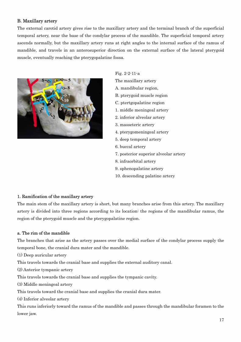
B. Maxillary artery The external carotid artery gives rise to the maxillary artery and the terminal branch of the superficial temporal artery, near the base of the condylar process of the mandible. The superficial temporal artery ascends normally, but the maxillary artery runs at right angles to the internal surface of the ramus of mandible, and travels in an anterosuperior direction on the external surface of the lateral pterygoid muscle, eventually reaching the pterygopalatine fossa.
1. Ramification of the maxillary artery The main stem of the maxillary artery is short, but many branches arise from this artery. The maxillary artery is divided into three regions according to its location: the regions of the mandibular ramus, the region of the pterygoid muscle and the pterygopalatine region. a. The rim of the mandible The branches that arise as the artery passes over the medial surface of the condylar process supply the temporal bone, the cranial dura mater and the mandible. (1) Deep auricular artery This travels towards the cranial base and supplies the external auditory canal. (2) Anterior tympanic artery This travels towards the cranial base and supplies the tympanic cavity. (3) Middle meningeal artery This travels toward the cranial base and supplies the cranial dura mater. (4) Inferior alveolar artery This runs inferiorly toward the ramus of the mandible and passes through the mandibular foramen to the lower jaw.
Fig. 2-2-11-a The maxillary artery A. mandibular region, B. pterygoid muscle region C. ptertgopalatine region 1. middle meningeal artery 2. inferior alveolar artery 3. masseteric artery 4. pterygomeningeal artery 5. deep temporal artery 6. buccal artery 7. posterior superior alveolar artery 8. infraorbital artery 9. sphenopalatine artery 10. descending palatine artery

18
b. Pterygoid muscle region These branches, which arise as the maxillary artery passes over the external surface of the lateral pterygoid muscles, supply the muscles of mastication. (1) Deep temporal artery This travels towards the calvarium and supplies the temporalis muscle. (2) Masseteric artery This travels towards the ramus of the mandible and supplies the masseter muscle. (3) Pterygoid branch This supplies the medial and lateral pterygoid muscles. (4) Buccal artery This travels towards and ultimately supplies the buccinators. c. Pterygopalatine region These branches, which arise as the artery passes through the pterygopalatine fossa, supply the upper jaw, the nasal cavity and the cranial base. (1) Posterior superior alveolar artery This travels towards the posterior surface of the maxilla and supplies the region that extends from the alveolar foramen to the maxilla, as well as the molar region. (2) Infra-orbital artery This travels towards the superior surface of the maxilla, to enter the inferior orbital fissure. It supplies the maxilla and the anterior region of the teeth. (3) Descending palatine artery This travels towards the palate, passing through the greater palatine canal, and supplies the palate. (4) Artery of pterygoid canal This travels towards the cranial base, passes through the pterygoid canal, and supplies the cranial base. (5) Sphenopalatine artery This travels towards the nasal cavity, enters the upper part of the nasal cavity via the sphenopalatine foramen, and forks to give rise to the posterior lateral nasal artery. It then supplies the nasal cavity, and gives rise to the posterior septal branches of the sphenopalatine artery, which supply the nasal septum. 2. Arteries that supply the lower jaw (Fig.2-2-11-b) a. Inferior alveolar artery This arises from the posterior surface of the maxillary artery as it runs along the ramus of the mandible. It descends across the internal surface of the ramus of the mandible together with the inferior alveolar nerve, and enters the mandible via the mandibular foramen. It initially runs in an antero-inferior direction within the mandibular canal, and then runs along the lower third of the mandibular body and along the mandibular base, changing its direction outwards just below the apex of the root of the first premolar tooth. It eventually appears on the surface of the face as the mental artery, passing through the mental foramen after forming the incisive branch, and terminates in the lower lip.

19
Its main branches are as follows. (1) Mylohyoid branch This bifurcates just before it passes through the mandibular foramen; it then descends though the mylohyoid groove, reaches the inferior surface of the mylohyoid muscle, and anastomoses with the submental artery. It supplies the mylohyoid and digastric muscles. (2) Dental branches A considerable number of branches arise as the artery passes through the mandible. They ramify and anastomose, and subsequently supply the teeth and the periodontium. (3) Mental artery This is the terminal branch of the inferior alveolar artery. It exits through the mental foramen, and supplies the mental region and the lower lip. It anastomoses with the inferior labial branch of the facial artery, and the mental artery. (4) Incisive branch This is an extension of the main stem of the inferior alveolar artery. It travels through the anterior dental region of the jawbone towards the midline, and gives rise to the dental branches of the incisive region. b. Deep temporal artery The posterior deep temporal artery and the anterior deep temporal artery join to give rise to this artery. The posterior deep temporal artery originates from the superior wall immediately after exiting from the medial surface of the head of the mandible, and travels in an anterosuperior direction. It eventually supplies the temporal muscle. In contrast, the anterior deep temporal artery arises from the superior wall anterior to the base of the buccal artery. It travels superiorly and supplies the temporalis muscle. c. Masseteric artery This originates close to the base of the posterior deep temporal artery. It descends, passes behind the terminal tendon of the temporal muscle, located on the posterior half of the mandibular notch. It then passes across the external surface of the ramus of the mandible, travels back and forth and supplies the masseter muscle. d. Pterygoid branch This originates from the inferior wall of the maxillary artery, between the base of the posterior deep temporal artery and the base of the buccal artery. It descends and supplies the lateral and medial pterygoid muscles. e. Buccal artery This arises at the inferior wall of the maxillary artery, behind the base of the anterior deep temporal
Fig. 2-2-11-b The inferior alveolar artery 1. inferior alveolar artery 2. mylohyoid branch 3. dental branches 4. incisive branch 5. mental foramen 6. mandibular foramen

20
artery. It travels back and forth between the lateral pterygoid muscle and the terminal tendon of the temporal muscles, passes over the external surface of the buccal muscle, and supplies the buccal muscles. 3. Arteries that supply the upper jaw (Fig.2-2-12-a) a. Posterior superior alveolar artery This may arise from the external wall of the maxillary artery close to the aditus of the pterygopalatine fossa, at the center of the posterior surface of the body of the maxilla; or it may arise with the infra-orbital artery, forming a common stem. It descends on the external surface of the body of the maxilla, enters the maxilla through the alveolar foramen accompanied by the posterior superior alveolar branch, and travels anterior to the posterior superior alveolar canal that runs through the middle of the posterior wall of the maxilla. It anastomoses with the anterior superior alveolar artery, ramifies in this process, and supplies the maxillary sinus and the molar tooth region.
b. Infra-orbital artery (Fig. 2-2-12-b) This is one of the terminal branches of the maxillary artery that originates from the lateral wall of the maxillary artery, close to the aditus of the pterygopalatine fossa. It ascends on the posterior surface of the body of the maxilla, enters the orbit via the inferior orbital fissure with the infra-orbital nerve, and travels anterior to the infra-orbital groove and the infra-orbital canal. It eventually appears on the surface of the face through the infra-orbital foramen, and supplies the nasal region and the gingivae of the anterior teeth of the maxilla, back to the premolar tooth. The anterior superior alveolar artery arises as the infra-orbital artery passes through the infra-orbital canal.
Fig. 2-2-12-a,b The maxillary arterya The Ptertgopalatine Region 1. maxillary artery 2. buccal artery 3. anterior superior alveolar artery 4. infraorbital artery 5. sphenopalatine artery 6. descending palatine artery 7. pterygopalatine fossa 8. greater palatine canal 9. inferior orbital fissure
Fig. 2-2-12-b The infraorbital artery 1. infraorbital groove 2. infraorbital canal 3. infraorbital foramen 4. infraorbital artery 5. inferior orbital fissure

21
c. Anterior superior alveolar artery This originates from the infra-orbital artery in the posterior section of the infra-orbital canal. It travels anteriorly within the anterior superior alveolar canal, reaches the infero-anterior wall of the infra-orbital foramen, and travels towards the piriform aperture. It then descends, after it gives off branches into the nasal cavity, close to the attachment of the middle nasal concha. It ramifies during this process, forming an arterial plexus with the posterior superior alveolar artery, and supplies the teeth and the periodontium. d. Descending palatine artery This forms the terminal branch of the maxillary artery, together with the sphenopalatine artery. It descends in the pterygopalatine fossa, forming the greater palatine artery within the greater palatine canal, and descends further, accompanied by the greater palatine nerve. It then enters the oral cavity through the greater palatine canal, and supplies the hard palate. The lesser palatine arteries and the lesser palatine nerves pass through the lesser palatine canals that arise from the greater palatine canal and supply the soft palate. e. Greater palatine artery The greater palatine artery ramifies after coming out from the greater palatine foramen, giving rise to an anterior branch (that passes through the palatine groove, traveling anteriorly towards the region adjacent to the incisive foramen along the base of the alveolar process, anastomoses with the incisive branch, and supplies the gingivae); a middle branch (that supplies the mucous membrane of the frontal part of the palate); and a posterior branch (that supplies the rear part of the palate and the gingiva of the molar region) f. Sphenopalatine artery This enters the nasal cavity through the sphenopalatine foramen and forms posterior lateral nasal branches (supplying the external wall of the nasal cavity) and posterior septal branches (supplying the nasal septum). The posterior septal nasal artery runs in a diagonal and antero-inferior direction in relation to the nasal septum, passing through the incisive foramen and forming anastomoses with branches of the greater palatine artery. C. Superficial temporal artery This is the terminal branch of the external carotid artery as it ascends. It branches off from the maxillary artery behind the condylar process of the mandible and ascends, passing through the retromandibular fossa, and sheltered by the parotid gland. It exits the gland on its anterior border, and appears under the skin close to the anterior border of the zygomatic arch. It then ascends once more, and supplies the scalp region. IV. Veins that drain the structures that surround the oral cavity The main vessel that runs through the head and the neck region is the internal jugular vein. It descends, draining the veins of the cranium. Most of the veins of the head and neck such as the maxillary vein and the facial vein flow into the internal jugular vein, which joins the subclavian vein in the upper section of

22
the thoracic cage. Veins, usually with names that are the same as those of the branches of the external carotid artery, collect the blood from the capillaries within this area. The retromandibular vein, which has no corresponding artery, is derived from the pterygoid venous plexus that collects blood from the region where the superficial temporal artery and maxillary artery are distributed. This is the most important distinction between the arteries and veins of the head and neck region. In addition, there are abundant junctions and variations in the structure, and combined branches are often present. However, the external carotid artery works as a bypass rather than as an major vein. A. Retromandibular vein The superficial temporal vein that gathers the veins from the territory of the superficial temporal artery as it descends, and the maxillary vein that collects the veins of the pterygoid venous plexus, join to form the retromandibular vein. This supplies the internal jugular vein at the level of the hyoid bone, after traveling through the retromandibular fossa. The main tributaries are as follows: 1. Tributaries that come from the superficial temporal veins. These drain the veins of the temporal region, the face and the masseter muscle. 2. Tributaries that arise from the pterygoid venous plexus. This venous plexus collects blood from tributaries ramified from the maxillary artery. The venous plexus extends into the pterygomandibular space between the medial pterygoid muscle, the lateral pterygoid muscle and the temporalis muscle, fusing posteriorly to form the maxillary vein. This vein joins the superficial temporal vein and forms the retromandibular vein. Part of this vein travels forward, also contributing to the deep facial vein that communicates with the facial vein. 1) The tympanic veins and the stylomastoid vein drain the tympanic cavity. 2) The articular veins drains veins from the temporomandibular joint. 3) The inferior alveolar vein drains veins from the mandible. 4) The middle meningeal veins drain the cranial dura mater. 5) The buccal vein drains the buccinator muscle. 6) The vein of the pterygoid canal drains veins from the cranial base. 7) The masseteric branch, the pterygoid branch, and the deep temporal veins drain the masticatory muscles. 8) The sphenopalatine vein and posterior superior alveolar vein drain the nasal cavity and maxilla. B. Facial vein This collects veins of the frontal and orbital regions, arising as the angular vein in the area close to the medial angle of the eye. It runs obliquely in an inferoposterior direction in the facial plane, reaching the floor of the mouth over the base of the mandible where the anterior border of the masseter muscle and the base of the mandible meet. After traveling through the external surface of the submandibular gland, it joins either the internal carotid vein or the retromandibular vein directly, to form the common facial vein in the area proximal to the angle of the mandible. This subsequently merges with the internal carotid vein or the external jugular vein, and collects all of the facial veins.

23
1) The external nasal veins drain the nasal region. 2) The superior labial vein and the inferior labial vein drain the lips of the mouth. 3) The parotid branches, the pectoral veins, and the masseteric veins drain the correspondingly named regions. 4) The deep facial vein is a communicating branch from the pterygoid venous plexus. 5) The submental vein drains veins from the submental region. C. Lingual vein This arises as the deep lingual vein along the inferior surface of the tongue from the apex, and is fed by the adjoining dorsal lingual vein that drains the dorsum of the tongue. The sublingual vein also joins at the root of the tongue and drains the internal jugular vein. V. Nerves that supply the surroundings of the oral cavity In the head and neck region, the cranial nerves are the predominant nerves. Of these, the trigeminal nerve (the fifth cranial nerve, at least its maxillary and mandibular branches), the facial nerve (the seventh cranial nerve), the glossopharyngeal nerve (the ninth cranial nerve), the vagus nerve (tenth cranial nerve), and the hypoglossal nerve (the twelfth cranial nerve) are distributed in the area surrounding the oral cavity. A. Trigeminal nerve (N. V) The trigeminal nerve is the thickest of the cranial nerves and is a mixed cranial nerve that consists of both sensory and motor fibers. The sensory root forms the trigeminal ganglion in the trigeminal impression of the petrous part of the temporal bone, with three branches extending out of the trigeminal ganglion. The motor root travels forward through the inferior surface of the trigeminal ganglion and joins the mandibular nerve. First branch: The ophthalmic nerve (sensory) This innervates the lacrimal gland and the frontal region of the head. Second branch: The maxillary nerve (sensory) This innervates the maxilla and teeth. Third branch: The mandibular nerve (mixed sensory and motor) It innervates the mandible, teeth, and the masticatory muscles. 1. Ophthalmic nerve (First branch of trigeminal nerve) (Fig. 2-2-13-a) The ophthalmic nerve leaves the trigeminal ganglion, passes through the superior orbital fissure, and branches further upon entering the orbital cavity. a. Lacrimal nerve This travels forward along the superolateral border of the orbital cavity, alongside the lacrimal artery and vein, and is distributed to the skin in the region of the lateral angle of the eye. b. Frontal nerve

24
This is the main branch of the lacrimal nerve. It travels on the levator palpebrae superioris muscle, divides into the supratrochlear nerve and the supra-orbital nerve, passes through the supratrochlear foramen and the supra-orbital foramen on the anterior border of the orbital opening, and innervates the skin of the frontal region of the head. c. Nasociliary nerve This travels in an anteromedial direction beneath the superior rectus muscle and innervates the eyeball and nasal mucosa. d. Ciliary ganglion Nerve branches enter the root via various nerves. The parasympathetic root derives from the oculomotor nerve, the sensory root via the nasociliary ganglion from the communicating branch, and the sympathetic root from the plexus of the ophthalmic artery. From the ganglion, the eyeball receives the short ciliary nerve, which innervates the ciliary muscle and the sphincter pupillae.
2. Maxillary nerve (Second branch of trigeminal nerve) (Fig. 2-2-13-b) This is the second branch that comes off the trigeminal ganglion. It passes through the foramen rotundum and enters the pterygopalatine fossa. It branches again and innervates the maxillary region, the temporal region, the skin of the buccal region and the posterior section of the nasal cavity. a. Zygomatic nerve This passes through the inferior orbital fissure, enters the orbital cavity, travels along its lateral wall, branches into the zygomaticotemporal branch and the zygomaticofacial branch, and innervates the skin of the zygomatic region. b. Pterygopalatine nerve This descends for 3 to 4 mm from the point at which the main stem exits the foramen rotundum, reaching the pterygopalatine ganglion. c. Infra-orbital nerve This is the extension of the main stem. The main branch becomes the infra-orbital nerve and enters the orbital cavity through the inferior orbital fissure after the zygomatic nerve and the pterygopalatine
Fig. 2-2-13-a,b The trigeminal nerve a The ophthalmic nerve 1. lateral branches of supraorbital nerve 2. medial branches of supraorbital nerve 3. supratrochlear nerve 4. infratrochlear nerve 5. nasociliary nerve 6. lacrimal nerve 7. frontal nerve 8. supraorbital foramen (notch) 9. superior orbital fissure 10. ciliary ganglion
25
nerves branch off. It appears on the front surface of the face, and passes through the infra-orbital groove, the infra-orbital canal and the infra-orbital foramen on the floor of the infra-orbital groove. Eight to ten branches originate in the shape of a fan and innervate the ala of the nose, the upper lip and the inferior eyelid. During this process, the anterior superior alveolar branch arises in the infra-orbital canal and the middle superior alveolar branch comes off in the infra-orbital groove; the divided nerves innervate the teeth. d. Posterior superior alveolar branches These arise just before the nerve enters the orbital cavity, and travel inferiorly, entering the maxilla via the alveolar foramen on the infratemporal side of the maxilla. They then run through the infratemporal alveolar canal, constituting the superior dental plexus, to innervate the maxillary molars and the buccal side of the gingivae of this region. e. Branches which form the superior dental nerve plexus (1) Anterior superior alveolar branch This nerve branches off from the main stem as it passes through the infra-orbital canal. It traverses the bone and reaches the anterior wall, having traveled in a direction parallel to the main stem. It then descends through the piriform aperture. During this process, thin branches innervate the area from the first premolar teeth to the central incisor, as well as the gingivae of this region. (2) Middle superior alveolar branch This branches off the main stem in the infra-orbital groove. It runs obliquely in an anterolateral direction through the floor of the orbit and the premolar plexus. The middle superior alveolar branch is not always present, in which case the anterior superior alveolar branch substitutes. It innervates the premolar teeth and the gingivae of this region. (3) Posterior superior alveolar branches (mentioned above) These three branches ramify and anastomose in the maxilla, forming the complex superior dental nerve plexus in the wall of the bone located just above the tooth root. The superior dental branch and the superior gingival branch divide, innervating the teeth, gingivae and the periodontium. f. Pterygopalatine ganglion This ganglion is located in the depths of the upper area of the pterygopalatine fossa. (1) Branches entering the ganglion: The sensory root enters the ganglion as the pterygopalatine nerves, while parasympathetic nerves and the sympathetic root enter the ganglion as the nerves of the pterygoid canal. (2) Branches exiting the ganglion: (a) Posterior nasal nerves These enter the nasal cavity through the sphenopalatine foramen, forming the posterior superior medial nasal branch that innervates the nasal septum. This ultimately becomes the nasopalatine nerve that goes through the incisive canal and innervates the palatine mucous membrane of the incisive region. In contrast, the posterior superior lateral nasal branch innervates the external wall of the nasal cavity. (b) Greater palatine nerve This nerve descends within the greater palatine canal alongside the artery with the same name. It passes through the greater palatine foramen and innervates the hard palate. The lesser palatine nerves branch

26
off from the greater palatine nerve and innervate the soft palate.
3. Mandibular nerve (Fig.2-2-14-a) This is the largest of the three branches of the trigeminal nerve. It is a mixed nerve containing both motor and sensory fibers. It travels through the oval foramen, exits the cranial cavity, and enters the infratemporal fossa. It then descends through the infratemporal fossa, immediately after supplying meningeal branches; it then joins the lingual nerve and the inferior alveolar nerve. The inferior alveolar nerve travels through the mandible, becomes the mental nerve after supplying the teeth and gingiva and passing through the mental foramen. It then innervates the lower lip. The nerve that innervates the masticatory muscles comes off in the region of the infratemporal foramen.
a. Branches that innervate the masticatory muscles (1) Masseteric nerve This travels through the mandibular notch, appears on the external surface of the ramus of the mandible and innervates the masseter muscles. (2) Medial pterygoid nerve This travels in an antero-inferior direction and innervates the medial pterygoid muscle. (3) Lateral pterygoid nerve
Fig. 2-2-13-b The maxillary nerve 1. maxillary nerve 2. infraorbital nerve 3. infraorbital nerve 4. anterior superior alveolar branch 5. middle superior alveolar branch 6. zygomatic nerve 7. posterior superior alveolar branch 8. superior dental nerve plexus 9. greater palatine nerve
Fig. 2-2-14-a,b The trigeminal nerve a The mandibular nerve 1. auriculotemporal nerve 2. deep temporal nerves 3. nerve to lateral pterygoid 4. nerve to medial pterygoid 5. masseteric nerve 6. buccal nerve 7. lingual nerve
8. inferior alveolar nerve 9. nerve to mylohyoid 10. mental nerve 11. inferior dental nerve plexus 12. oval foramen 13. mandibular foramen 14. mental foramen 15. submandibular ganglion
10. foramen rotundum 11. pterygopalatine fossa 12. inferior orbital fissure 13. infraorbital canal 14. infraorbital foramen 15. greater palatine foramen 16. alveolar foramen 17. pterygopalatine ganglion

27
This nerve passes anteriorly and innervates the lateral pterygoid muscles. (4) Deep temporal nerve This divides into branches that travel either anteriorly or posteriorly, and innervates the temporalis muscle. b. Buccal nerve This travels between the two heads of the lateral pterygoid muscle, penetrates the buccinators and innervates the buccal mucosa. c. Auriculotemporal nerve This arises from the same part of the nerve that innervates the masticatory muscle, passing backwards. It reaches the base of the condylar process, traverses the parotid gland in a lateral direction, ascends in front of the auricle with the superficial temporal artery and innervates the skin of the temporal region. d. Lingual nerve This runs between the medial pterygoid muscle and the lateral pterygoid muscle, forms an antero-inferiorly directed arch on the medial surface of the ramus of the mandible, and enters the tongue adjacent to the lingual side of the last molar. The lingual nerve joins the chorda tympani (a branch of the facial nerve), on the internal surface of the ramus of the mandible, forming an arch before the nerves merge. The nerve supplies branches to the fauces and the sublingual nerve. It then becomes the lingual nerve and innervates the mucous membrane of the anterior two-thirds of the tongue. e. Inferior alveolar nerve The main stem changes its name to the inferior alveolar nerve after the lingual nerve branches off. It enters the mandibular canal via the mandibular foramen at the center of the medial surface of the ramus of the mandible. The inferior dental nerve plexus is composed of a considerable number of branches. It forms the inferior dental branches and inferior gingival branches that innervate the teeth and gingivae respectively (Fig. 2-2-14-b).
f. Mylohyoid nerve This nerve arises just before the main stem enters the mandibular foramen, runs through the mylohyoid groove, and innervates the inferior surface of the mylohyoid muscle and the digastric muscle. g. Mental nerve This is the terminal branch of the inferior alveolar nerve. It innervates the mental region and lower lip after reaching the anterior surface of the mandible via the mental foramen.
Fig. 2-2-14-b The inferior alveolar nerve 1. oval foramen 2. buccal nerve 3. lingual nerve 4. nerve to mylohyoid 5. mandibular foramen 6. inferior alveolar nerve 7. otic ganglion

28
h. Inferior dental nerve plexus The considerable number of branches that arise from the mandibular canal intersect with each other to form the inferior dental nerve plexus. The retromolar branch, as with the inferior dental nerve plexus, innervates the molar tooth and the area distal to this; the molar branch innervates the molar region, and the premolar branch is distributed to the canine and the premolar teeth. i. Ganglia of the mandibular nerves Two ganglia belong to the mandibular nerve. (1) Otic ganglion This is located beneath the oval foramen, receiving branches from the mandibular nerve (motor root), the lesser petrosal nerve (sensory root, parasympathetic root), and from the middle meningeal nerve plexus (sympathetic root). The nerve to tensor veli palatini, the nerve to tensor tympani, the ramus communicans of the auriculotemporal nerve, and the ramus communicans of the chorda tympani arise from the ganglion. (2) Submandibular ganglion This is located immediately above the submandibular gland. It receives sensory fibers from the lingual nerve (sensory root), the chorda tympani (parasympathetic root), and branches from the periarterial plexus of the facial artery (sympathetic root). The glandular branches that emerge from the ganglion supply secretory nerves to the submandibular gland and the sublingual gland. B. Facial nerve (N. VII) (Fig. 2-2-15) This is the seventh cranial nerve. It is a mixed nerve that contains the facial nerve (motor fibers), the intermediate nerve (sensory fibers) and parasympathetic fibers (secretory fibers of the lacrimal gland, the submandibular gland and the sublingual gland). It enters bone via the internal acoustic meatus, passes through the facial canal, emerges from the stylomastoid foramen, and enters the retromandibular fossa. It travels through the retromandibular fossa within the substance of the parotid gland, and forms the superior branch and the inferior branch, also creating the parotid nerve plexus. It then supplies the face, innervating the muscles of facial expression. The point at which the facial canal curves sharply in its passage through bone is called the geniculum of facial nerve, and this is where the geniculate ganglion is located.
1. Branches from the nerve passing through the facial canal a. Greater petrosal nerve
Fig. 2-2-15 The facial nerve 1. stylomastoid foramen 2. facial nerve 3. digastric branch 4. stylohyoid branch 5. cervical branch 6. marginal mandibular
7. buccal branches 8. zygomatic branches 9. temporal branches 10. facial canal 11. chorda tympani 12. petrotympanic fissure 13. parotid nerve plexus

29
This originates from the geniculum of the facial nerve. It travels through the anterior surface of the petrous, passes through the foramen lacerum, and emerges on the surface of the cranial base. It anastomoses with the deep petrosal nerve, a branch from the sympathetic plexuses that wrap the internal carotid artery, and becomes the nerve to the pterygoid canal, entering the pterygopalatine ganglion. b. Nerve to stapedius This innervates the stapedius muscle. c. Chorda tympani This originates near the inferior part of the facial canal. It traverses the tympanic cavity, exits the cranium through the petrotympanic fissure of the mandibular fossa, and entering the lingual nerve, forms an arc. It supplies the secretory fibers of the salivary gland, and gustatory fibers to the anterior two-thirds of the tongue. 2. Branches that enter the retromandibular fossa a. Posterior auricular nerve This emerges from the stylomastoid foramen and innervates the muscles behind the auricle. b. Digastric branch This emerges from the stylomastoid foramen and innervates the posterior belly of the digastric muscle, slightly below the foramen. c. Stylohyoid branch This innervates the stylohyoid muscle. 3. Branches to the face a. Temporal branches These arise along the superior border of the parotid gland. They innervate the orbicularis oculi muscle and the muscles of facial expression, located above the former. b. Zygomatic branches and buccal branches These arise along the anterior border of the parotid gland. They innervate the buccinators and the muscles between the oral fissure and the optic fissure. c. Marginal mandibular branch This originates near the inferior angle of the parotid gland. It innervates the muscles of facial expression that are located below the oral fissure. d. Cervical branch It innervates the platysma muscle. 4. Geniculate ganglion Fibers from the geniculate ganglion enter the chorda tympani alongside the parasympathetic secretory nerves, and run to the submandibular gland and the sublingual gland. C. Hypoglossal nerve (N. XII) The twelfth cranial nerve consists exclusively of motor fibers. It arises from the medulla oblongata,

30
passes through the hypoglossal canal of the occipital bone, descends through the retromandibular fossa, descending further along the internal surface of the posterior belly of the digastric muscle at the level of the angle of the mandible, where it changes direction. It then travels forwards, enters the lingual muscles via the root of tongue, and innervates these muscles. 1) The lingual branches: these are the terminal branches of the hypoglossal nerve. They arise as it passes the hyoglossus muscle and the external surface of the geniohyoid muscle and innervate the muscles of the tongue. 2) There are rami communicantes that join other nerves. VI. Important structures of the oral cavity in relation to implants A. Structure of the mandible Fundamentally, the mandible comprises a base, with the muscles and alveolus representing secondary additions to the structure, in terms of mechanical components. The structure of the base has therefore developed in a manner similar to that of typical long bones. More specifically, the base of the mandible is thickly covered with compact bone that provides great mechanical strength. Internally, it consists of the inferior alveolar artery and nerves, with their surroundings covered by thin walls that are made up of compact bone, referred to as the mandibular canal. The compact bone on the lingual side is thinner than that on the buccal side, with the excavation below the mylohyoid line being the thinnest. The bony lamellae of the outermost layer are formed in parallel to its surface. A medullary cavity that is sparsely populated with cancellous bone is contained within the compact bone, and in the fully-grown adult is filled with adipose tissue (Fig. 2-2-16-a,b). When the mandibles of infants and adults are compared, features such as the boundary between the base and the alveolar sections are surprisingly more obscure in infants than in adults, and the interior of the compact bone is filled with more developed cancellous bones, in the infant (Fig. 2-2-17). The surface of the alveolar bone consists of thinner cortical bone in comparison to the base, with developed cancellous bone in its interior to support the cortical bone and the teeth. The alveolar bone in the region of the frontal teeth is generally thicker on the lingual side than that on the labial side, while the alveolar bone is thicker on the buccal side, because the dental arch is directed towards the tongue as it goes backwards in the molar region and an oblique line develops in the buccal side of the jawbone. The tendency of the alveolar bone on the labial side of the incisor to be thin has been shown to be problematic. Absorption of the anterior teeth in this region is not an uncommon occurrence, in which case the alveolar tubercle may also disappear, exposing part of the labial side of the root of the anterior tooth. It is therefore necessary to be aware of any exposure of the implant on performing implant surgery in the frontal portion of the mandible (Fig. 2-2-18-a,b).

31
Fig. 2-2-16-a,b a. The histological mesial-distal cross-section the mandible The internal structure of the jawbone of the region from the canine tooth to the molars can be observed. 1. Inferior alveolar nerve b. The histological buccal-lingual cross-section of the first molar region of the mandible 1. The mandibular canal
1. Alveolar bone The trabeculae of the cancellous bone within the alveolar bone undergo extensive remodeling due to germination of the tooth bud, replacement of deciduous teeth by permanent teeth, and with tooth movement. Cancellous bone with well-developed trabeculae fills the inside of the cortical bone in the area of the premolars mesial to the frontal teeth extending to the opening of the mental foramen, and in the area that is distal to the last molar, extending to the ramus of the mandible. In the molar region, cancellous bone fills the interior of the compact bone up to the apices of the roots of the molar teeth, to withstand the external forces exerted on the teeth. When implanting close to the mandibular canal, however, it is important to be aware that development of cancellous bone in the region around the mandibular canal is slow, and cancellous bone is often absent from this region (Fig. 2-2-19-a to h, Fig. 2-2-20-a,b).
Fig. 2-2-17 The histological mesial-distal cross-section of the mandibular of in a young individual The majority of the jawbone consists of cancellous bones in the region of canines to the molars.
Fig. 2-2-18 a. The histological image of the mesial-distal
cross-section of the mandibular first molarb. The root of tooth is supported by the
alveolar bone composed of cancellous bone.

32
Fig. 2-2-19-a to h The buccal-lingual cross-section in each parts of mandible a. The mandibular canine region b. The mandibular first premolar region 1. incisive canal c. The mandibular second premolar region 2. mental foramen d. Region of mesial root of mandibular first premolar 3. Mandibular canal e. Region of mesial root of mandibular second premolar. The thickness of the buccal side alveolar region
increases. f. Retromolar triangle g. Region of coronoid process h. Region of mandibular foramen 4. mesial border of mandible
Fig. 2-2-20 The mesial-distal cross-section of mandibular molar region a. The mesial-distal cross-section of the buccal side of the mandibular molar dentition. 1. mental foramen b. The mesial-distal cross-section of the center of the mandibular molar dentition. 2. mandibular canal In the region that surrounds the roots of the molar teeth, the development of cancellous bone has been observed to occur uniformly, radiating in all directions from the root surface, as far as the point of bifurcation of the root. In the region close to the apex, cancellous bone composed of the thin trabeculae can be observed in that part of the alveolar bone that surrounds the root of the tooth. However, the cancellous bone is less developed in the supporting alveolar bone located between the roots, the thin trabeculae are seen to be sparse, and bone marrow that consists of adipose tissue fills the space between the trabeculae. It is thus hypothesized that the main role of the trabeculae of the alveolar bone, above the apical area, is to withstand external forces that are exerted on teeth and implants during tooth function (Fig.2-2-21-a,b).

33
Fig. 2-2-21 a. The histological cross-section of the mandibular anterior tooth to the molar region. It is sliced
horizontally near the furcation of the root of molar tooth b. The histological cross-section of the mandibular anterior tooth to the molar region. It is sliced
horizontally near the apical area of the molar tooth. 2. Mandibular canal The mandibular canal runs from the mandibular foramen in an anteroinferior direction at an angle of 150° to the mandibular base and changes direction anteriorly once it reaches the second molar tooth. It then runs horizontally as far as the second premolar, within the lower third of the mandibular body, parallel to the mandibular base. It curves in a posterosuperior direction as it passes below the area between the first and second premolars, and opens at the mental foramen. If the mandibular canal is observed from above, it can be seen to run through one third of the lingual side of the buccolingual diameter, along the internal surface of the mandible, from the mandibular foramen to the first molar tooth or the second molar tooth. It changes direction outward, traveling through a point below the center of the second premolar. After opening at the mental foramen, it bends out and up when it reaches a point between the first and the second premolars. From investigation of the distance between the mandibular canal and each tooth located in the mandible, it has been determined that the approximate distances are: from the second premolar, 7.6 mm; from the mesial root of the first molar, 8.8 mm; from the distal root of the first molar, 7.9 mm; from the mesial root of the second molar, 5.9 mm; from the distal root of the second molar 6.1 mm; and from the third molar 5.0 mm. These findings indicate the closeness of the apex of the molar tooth to the mandibular canal. In the regions from the mandibular foramen to the ramus of the mandible, and around the mental foramen, the wall of the mandibular canal is surrounded by a thin layer of cortical bone; however, as the wall of the mandibular canal in the molar region is made of porous cancellous bone, it is structurally fragile. Present within the canal are the inferior alveolar nerve, artery and veins. It is essential to be aware of the exact location and the course of the canal during implant surgery, as damage to this canal can result in severe accidents, such as serious bleeding and sensory disturbance. If bone resorption is evident in the alveolar bone, the location of the mandibular canal as well as the opening of the mental foramen should be identified before surgery (Fig. 2-2-22-a). The mandibular canal opens into the mental foramen once it has traveled towards the mesial side of the mental foramen and curved in a posterosuperior direction. This curve is called the anterior loop of the mandibular canal. Due to the risk of damaging the neural blood vessels present in the loop, great caution is required when installing implants in the region in front of the mental foramen. The distance of this loop from the mental foramen is typically less than 5 mm; however, there have been reports of cases where a value of over 5 mm was observed. Thus, to ensure safety, the distance should be determined with X-ray examination or CT before an operation. Just before the mandibular canal opens into the mental foramen, the incisive branch arises and travels anteriorly. The incisive branch supplies the region of the mandible that contains the front teeth and the premolar and in its periodontium. The branch that distributes to the region of the front teeth runs in the incisive canal. The incisive canal becomes progressively narrower as it runs from the mental foramen
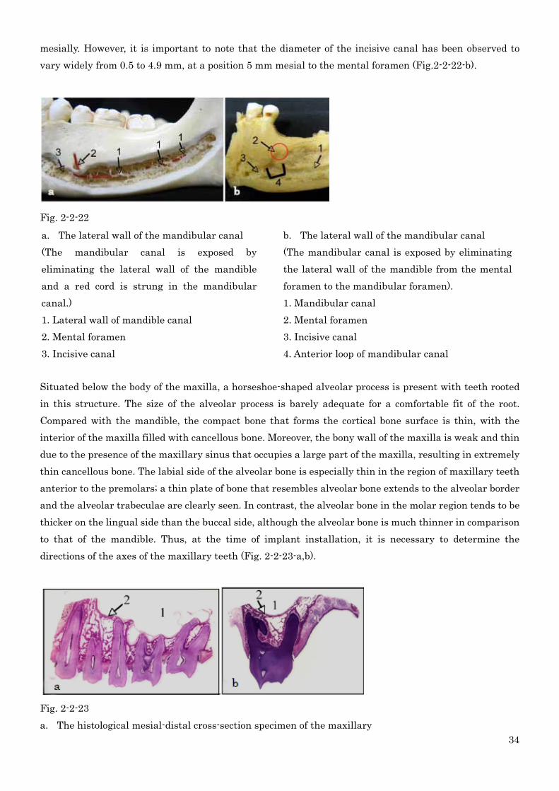
34
mesially. However, it is important to note that the diameter of the incisive canal has been observed to vary widely from 0.5 to 4.9 mm, at a position 5 mm mesial to the mental foramen (Fig.2-2-22-b).
Fig. 2-2-22 B. Structure of the maxilla 1. Alveolar bone Situated below the body of the maxilla, a horseshoe-shaped alveolar process is present with teeth rooted in this structure. The size of the alveolar process is barely adequate for a comfortable fit of the root. Compared with the mandible, the compact bone that forms the cortical bone surface is thin, with the interior of the maxilla filled with cancellous bone. Moreover, the bony wall of the maxilla is weak and thin due to the presence of the maxillary sinus that occupies a large part of the maxilla, resulting in extremely thin cancellous bone. The labial side of the alveolar bone is especially thin in the region of maxillary teeth anterior to the premolars; a thin plate of bone that resembles alveolar bone extends to the alveolar border and the alveolar trabeculae are clearly seen. In contrast, the alveolar bone in the molar region tends to be thicker on the lingual side than the buccal side, although the alveolar bone is much thinner in comparison to that of the mandible. Thus, at the time of implant installation, it is necessary to determine the directions of the axes of the maxillary teeth (Fig. 2-2-23-a,b).
Fig. 2-2-23 a. The histological mesial-distal cross-section specimen of the maxillary
b. The lateral wall of the mandibular canal (The mandibular canal is exposed by eliminating the lateral wall of the mandible from the mental foramen to the mandibular foramen). 1. Mandibular canal 2. Mental foramen 3. Incisive canal 4. Anterior loop of mandibular canal
a. The lateral wall of the mandibular canal (The mandibular canal is exposed by eliminating the lateral wall of the mandible and a red cord is strung in the mandibular canal.) 1. Lateral wall of mandible canal 2. Mental foramen 3. Incisive canal
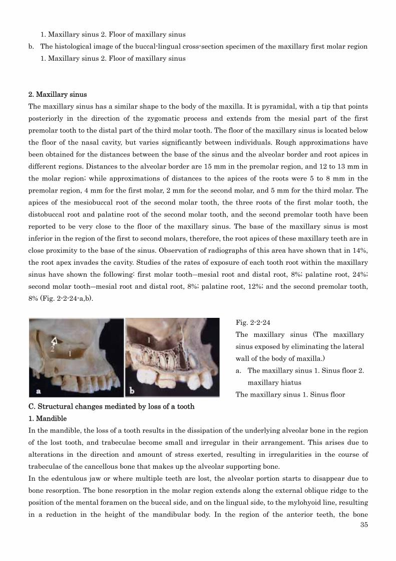
35
1. Maxillary sinus 2. Floor of maxillary sinus b. The histological image of the buccal-lingual cross-section specimen of the maxillary first molar region
1. Maxillary sinus 2. Floor of maxillary sinus 2. Maxillary sinus The maxillary sinus has a similar shape to the body of the maxilla. It is pyramidal, with a tip that points posteriorly in the direction of the zygomatic process and extends from the mesial part of the first premolar tooth to the distal part of the third molar tooth. The floor of the maxillary sinus is located below the floor of the nasal cavity, but varies significantly between individuals. Rough approximations have been obtained for the distances between the base of the sinus and the alveolar border and root apices in different regions. Distances to the alveolar border are 15 mm in the premolar region, and 12 to 13 mm in the molar region; while approximations of distances to the apices of the roots were 5 to 8 mm in the premolar region, 4 mm for the first molar, 2 mm for the second molar, and 5 mm for the third molar. The apices of the mesiobuccal root of the second molar tooth, the three roots of the first molar tooth, the distobuccal root and palatine root of the second molar tooth, and the second premolar tooth have been reported to be very close to the floor of the maxillary sinus. The base of the maxillary sinus is most inferior in the region of the first to second molars, therefore, the root apices of these maxillary teeth are in close proximity to the base of the sinus. Observation of radiographs of this area have shown that in 14%, the root apex invades the cavity. Studies of the rates of exposure of each tooth root within the maxillary sinus have shown the following: first molar tooth―mesial root and distal root, 8%; palatine root, 24%; second molar tooth―mesial root and distal root, 8%; palatine root, 12%; and the second premolar tooth, 8% (Fig. 2-2-24-a,b).
C. Structural changes mediated by loss of a tooth 1. Mandible In the mandible, the loss of a tooth results in the dissipation of the underlying alveolar bone in the region of the lost tooth, and trabeculae become small and irregular in their arrangement. This arises due to alterations in the direction and amount of stress exerted, resulting in irregularities in the course of trabeculae of the cancellous bone that makes up the alveolar supporting bone. In the edentulous jaw or where multiple teeth are lost, the alveolar portion starts to disappear due to bone resorption. The bone resorption in the molar region extends along the external oblique ridge to the position of the mental foramen on the buccal side, and on the lingual side, to the mylohyoid line, resulting in a reduction in the height of the mandibular body. In the region of the anterior teeth, the bone
Fig. 2-2-24 The maxillary sinus (The maxillary sinus exposed by eliminating the lateral wall of the body of maxilla.) a. The maxillary sinus 1. Sinus floor 2.
maxillary hiatus The maxillary sinus 1. Sinus floor

36
resorption extends to the vicinity of the mental spine on the lingual side and the height of the mandibular body is reduced to one-third of its original height (Fig.2-2-25-a, b). With implant surgery, it should be acknowledged that the mandibular canal, which consists of the inferior alveolar nerve, artery and vein, becomes closer to the alveolar crest.
Resorption of the alveolar bone forces the mental foramen to lie closer to the alveolar crest. Where bone resorption extends to involve those parts below the mental foramen, this foramen may become displaced into the inclined, buccal surface of the alveolar crest, with its opening facing upwards. There have been reports of cases in which the extent of bone resorption was so great that it resulted in the mandibular canal being exposed. The displacement of the mandibular canal and mental foramen due to bone resorption suggests that great care must be taken when performing implant surgery in order to avoid any damage to the inferior alveolar nerve, artery and vein, as well as the mental nerve, artery and vein (Fig. 2-2-26-a to d). 2. Maxilla
Fig. 2-2-26 The alveolar bone resorption of the mandible(Note the position of the mental foramen) a. Mandibles with varying degree of
alveolar bone resorption 1. mental foramen 2. border of alveolar crest
b. The bone resorption caused by the localized loss of teeth affecting the mandibular structure
c. The alveolar bone resorption resulting from teeth loss
d. The mandible affected with significant bone resorption.
Fig. 2-2-25 The alveolar bone resorption of the mandiblea. The normal alveolar bone on right side of
mandibular molar region without the loss of teeth
b. The alveolar bone resorption resulting from tooth losses on left side of mandibular molar region. The arrow pointing to the region of bone resorption

37
Maxillary tooth loss also results in resorption of the bone of the alveolar process. The cortical bone thins along with narrowing of the trabeculae of the cancellous bone in the maxillary interior. The bone resorption progresses, with dissipation of most of the anterior tooth region. In the edentulous jaw, losses of roughly 1 cm in height have been reported. In the case of active bone resorption, it can extend to the area right under the anterior nasal spine in the region of the front teeth, as far as the lingual side of the palatine process. There are also situations where bone resorption can extend to the level of the top of the pterygoid process. Resorption of the bone of the alveolar process begins from the labial side. In the edentulous jaw, the alveolar crest is shifted to the lingual side, with the horse shoe-shaped arch that is formed by the alveolar crest becoming smaller and located more centrally than in the toothed jaw. At the median line, the anterior border of the incisive foramen coincides with the alveolar crest. The bony palate also thins, with resorption of the bone of the alveolar process. As bone is resorbed, the cancellous bone on the floor of the sinus becomes narrow and its arrangement appears irregular. However, as the bony wall that surrounds the maxillary sinus is even thinner, it is difficult to identify bone in the molar region that is of sufficient quality for implant insertion (Fig. 2-2-27-a,b). As indicated above, the bony walls of the maxillary molar region that are affected by bone resorption are composed of thin plates. The mucous membrane of the maxillary sinus consists of pseudostratified columnar ciliated epithelium. The tunica propria contains mixed glands but is thinner than the palatine mucous membrane and adhesion to the underlying bone is weak. The mucous membrane of the maxillary sinus is characteristically easily lifted, and easily ruptured at the time of installation of an implant body. It is therefore necessary to ensure that one does not transgress the mucous membrane during implant surgery. Additional care should be taken to avoid any damage to the blood vessels on the lateral walls of the maxillary sinus, such as the posterior superior alveolar branch and the anterior superior alveolar artery that arise from the infra-orbital artery.
D. Other jaw bone structures that need attention 1. Depressions in the jawbones a. The region of mandibular anterior teeth on the labial side In this region, there have been cases where bone resorption has been observed in the region of the labial side of the jawbone, with indentation of the plane of the labial side of the jaw bone close to the root apex to form a concave shape. In such cases, caution is required as there is the possibility of perforating the cortical bone on the labial side if an implant body is inserted in the wrong direction. b. Sublingual fossa
Fig. 2-2-27 The alveolar bone resorption of the maxilla a. The bone resorption of maxillary alveolar
bone. Arrow indicating the alveolar bone becoming increasingly spongy due to bone resorption.
b. The bone resorption of maxillary alveolar bone. ★ Exposure of the maxillary sinus

38
The line of the alveolar crest tends to migrate onto the lingual side as bone is resorbed. Thus, in cases where the sublingual fossa on the internal surface of the mandible is deeply set, the body of an implant inserted in the premolar region may pierce through to the internal surface of the mandible. The sublingual fossa is a shallow basin located in the distal part of the mental spine, above the mylohyoid line, on the lingual side from the mandibular canine tooth to the premolar region. The sublingual fossa is enlarged if a thick sublingual gland is present, in which cases the risk of piercing bone becomes higher at the time of implant insertion. With the increased risk of vascular injury in the floor of the oral cavity, which will inevitably result in serious bleeding, great care is required when drilling into the sublingual fossa (Fig. 2-2-28-a). The sublingual artery that branches off from the lingual artery lies on the lingual side of the region from the mandibular canine to the premolar tooth, travels through the mylohyoid muscle and within the sublingual gland to supply the sublingual gland, the mylohyoid muscle and the soft tissues of the floor of the oral cavity. It then advances to enter the lingual foramen near the mental spine medially, and eventually reaches the incisive canals to anastomose with the inferior alveolar artery. Meanwhile, after branching off from the facial artery, the submental artery passes on the external surface of the mylohyoid muscle along the mandibular base, reaches the submental region, and is distributed to the muscle and the skin of the chin. It enters the mandible from the mandibular base and the internal surface of the mandible via the foramen, and anastomoses with the inferior alveolar artery. These arteries have anastomotic branches and supply blood to their mutual territories. Serious vascular injury can result when drilling into the sublingual fossa because both the sublingual artery and the submental artery pass through the sublingual fossa along the surface, close to the inferior surface of the tongue (Fig.2-2-28-b).
Fig. 2-2-28 c. Submandibular fossa There have been cases where the body of an implant has penetrated the submandibular fossa, injuring the lingual nerve in the mandibular molar region. The lingual nerve passes between the medial and lateral pterygoid muscle and travels in an arcuate fashion in an anteroinferior direction. After emerging from the pterygomandibular space between the mandible and the pterygoid process, it runs through the shallow submucosal layer, on the periosteum adjacent to the lingual side of the last molar, and enters the tongue. It is approximated very closely to the internal wall of the last molar on the lingual side,
The medial surface of the mandible a. The sublingual fossa 1. mylohyoid line 2. sublingual fossa
b. The foramen on lingual surface 1. foramen on lingual surface 2. foramen which is seen on the internal surface of mandible

39
necessitating particular caution at the time of implant insertion, or detaching the mucoperiosteal flap during implant surgery to avoid any damage to the lingual nerve. d. Canine fossa In the region of the anterior teeth of the mandible, perforation of the implant body into the piriform aperture or the canine fossa is permitted in cases where the alveolar process has been affected by bone resorption to a great extent. 2. Structures related to the nerves and blood vessels present in the jawbone a. Incisive fossa, incisive foramen, and incisive canals The incisive fossa is the depression that is seen on the median line of the palate behind the central incisor. The opening of the incisive foramen can be observed here, with the right and the left incisive foramina leading to the incisive canals. The incisive canals ramify in a Y-shape, with openings on both sides of the nasal crest. The nasopalatine nerve and the posterior septal nasal artery run in the incisive canals. If the maxillary anterior tooth region is substantially affected by bone resorption due to tooth loss in the maxilla, the alveolar crest shifts to the palatine side. This can result in the anterior border of the incisive fossa coinciding with the alveolar crest. In such cases, if implant surgery is performed without verifying the location of the incisive fossa, there is an increased risk of damage to the nasopalatine nerve and the posterior septal nasal artery. b. Maxillary tuberosity The loss of teeth in the maxilla causes the alveolar process to undergo extensive bone resorption. Since it inherently has a limited amount of bone substance due to the presence of the maxillary sinus, this portion of the maxilla is the most complicated part involved in installation of implant bodies. Therefore, there are cases where an implant is inserted deeply into the maxillary tuberosity. The pterygopalatine fossa lies behind the maxillary tuberosity. The terminal branch of the maxillary artery is contained in the pterygopalatine fossa, from which the descending palatine artery, posterior superior alveolar branch and infra-orbital artery originate, among other arteries. The buccal artery also arises from the surrounding area. Therefore, when inserting an implant body, there is a risk of injuring the descending palatine artery that supplies the palate. It is a difficult task to stop this artery from bleeding, and extra caution is therefore required. c. Pterygoid venous plexus Upon loss of teeth from the maxilla, bone resorption occurs in the posterior portion, from the maxillary molars to the maxillary tuberosity. The significant structures in the rear part of this portion include the pterygomandibular space between the mandible and the pterygoid process, the maxillary artery, and the medial and lateral pterygoid muscle, both muscles of mastication. The pterygoid venous plexus is present on both surfaces of the pterygoid muscles. As with the insertion of implants into the maxillary tuberosity noted above, injury to the pterygoid venous plexus can result from an unprepared surgical invasion. Bleeding from this venous plexus is also difficult to control, therefore extra caution is required when implanting into the maxillary tuberosity. References 1) Mori O, Ogawa T, Ohuchi H, Mori T. Shared anatomy 1 Introduction/ Osteology/

40
Syndesmology/Myology. (in Japanese) 2) Mori O, Ogawa T, Ohuchi H, Mori T. Shared anatomy 2 Angiology/Nervous system. (in Japanese) 3) Kamijo Y. Illustrated oral anatomy 1, Osteology. Anatome Co., Ltd. (in Japanese) 4) Kamijo Y. Illustrated oral anatomy 2, Myology. Anatome Co., Ltd. (in Japanese) 5) Kamijo Y. Illustrated oral anatomy 3, Angiology. Anatome Co., Ltd. (in Japanese) 6) Kamijo Y. Illustrated oral anatomy 4, Neurology. Anatome Co.,Ltd. (in Japanese) 7) Ito T. Human anatomy. Nanzando Co., Ltd. (In Japanese) 8) Akagawa Y, Matsuura M, Yatani H, Watanabe H. Fundamental concepts and techniques of oral implant. Ishiyaku Pub, Inc. (in Japanese) 9) Katsuki T, Uchida Y. Basis of surgery and surgical anatomy for implant/ oral surgery. Quintessence Publishing Co., Ltd. (in Japanese) 10) Wakita M, Yamashita Y, Akesaka T. Oral histology and embryology. Ishiyaku Pub, Inc. (in Japanese)