Chapter 16 The Molecular Basis of Inheritance. DNA Structure Rosalind Franklin took diffraction...
82
Chapter 16 The Molecular Basis of Inheritance
-
Upload
pauline-charity-mckinney -
Category
Documents
-
view
216 -
download
1
Transcript of Chapter 16 The Molecular Basis of Inheritance. DNA Structure Rosalind Franklin took diffraction...

Chapter 16Chapter 16The Molecular Basis of Inheritance

DNA StructureDNA Structure
• Rosalind Franklin took diffraction x-ray photographs of DNA crystals
• In the 1950’s, Watson & Crick built the first model of DNA using Franklin’s x-rays

Rosalind Franklin Frances Crick & James Watson
X-ray diffraction photograph of DNA, 1953
Proposed double helix model 1953

Concept 16.1: DNA is the genetic material
• Early in the 20th century, the identification of the molecules of inheritance loomed as a major challenge to biologists
Copyright © 2008 Pearson Education Inc., publishing as Pearson Benjamin Cummings

The Search for the Genetic Material: Scientific Inquiry
• When T. H. Morgan’s group showed that genes are located on chromosomes, the two components of chromosomes—DNA and protein—became candidates for the genetic material
• The key factor in determining the genetic material was choosing appropriate experimental organisms
• The role of DNA in heredity was first discovered by studying bacteria and the viruses that infect them
Copyright © 2008 Pearson Education Inc., publishing as Pearson Benjamin Cummings

Evidence That DNA Can Transform Bacteria
• The discovery of the genetic role of DNA began with research by Frederick Griffith in 1928
• Griffith worked with two strains of a bacterium, one pathogenic and one harmless
Copyright © 2008 Pearson Education Inc., publishing as Pearson Benjamin Cummings

• When he mixed heat-killed remains of the pathogenic strain with living cells of the harmless strain, some living cells became pathogenic
• He called this phenomenon transformation, now defined as a change in genotype and phenotype due to assimilation of foreign DNA
Copyright © 2008 Pearson Education Inc., publishing as Pearson Benjamin Cummings

Fig. 16-2
RESULTS
EXPERIMENT

• In 1944, Oswald Avery, Maclyn McCarty, and Colin MacLeod announced that the transforming substance was DNA
• Their conclusion was based on experimental evidence that only DNA worked in transforming harmless bacteria into pathogenic bacteria
• Many biologists remained skeptical, mainly because little was known about DNA

Evidence That Viral DNA Can Program Cells
• More evidence for DNA as the genetic material came from studies of viruses that infect bacteria
• Such viruses, called bacteriophages (or phages), are widely used in molecular genetics research
Copyright © 2008 Pearson Education Inc., publishing as Pearson Benjamin Cummings
Animation: Phage T2 Reproductive CycleAnimation: Phage T2 Reproductive Cycle

Fig. 16-3
Bacterial cell
Phage head
Tail sheath
Tail fiber
DNA
100
nm

• In 1952, Alfred Hershey and Martha Chase performed experiments showing that DNA is the genetic material of a phage known as T2
• To determine the source of genetic material in the phage, they designed an experiment showing that only one of the two components of T2 (DNA or protein) enters an E. coli cell during infection
• They concluded that the injected DNA of the phage provides the genetic information
Copyright © 2008 Pearson Education Inc., publishing as Pearson Benjamin Cummings
Animation: Hershery-Chase ExperimentAnimation: Hershery-Chase Experiment

Fig. 16-4-1
EXPERIMENT
Phage
DNA
Bacterial cell
Radioactive protein
Radioactive DNA
Batch 1: radioactive sulfur (35S)
Batch 2: radioactive phosphorus (32P)

Fig. 16-4-2
EXPERIMENT
Phage
DNA
Bacterial cell
Radioactive protein
Radioactive DNA
Batch 1: radioactive sulfur (35S)
Batch 2: radioactive phosphorus (32P)
Empty protein shell
Phage DNA

Fig. 16-4-3
EXPERIMENT
Phage
DNA
Bacterial cell
Radioactive protein
Radioactive DNA
Batch 1: radioactive sulfur (35S)
Batch 2: radioactive phosphorus (32P)
Empty protein shell
Phage DNA
Centrifuge
Centrifuge
Pellet
Pellet (bacterial cells and contents)
Radioactivity (phage protein) in liquid
Radioactivity (phage DNA) in pellet

Additional Evidence That DNA Is the Genetic Material
• It was known that DNA is a polymer of nucleotides, each consisting of a nitrogenous base, a sugar, and a phosphate group
• In 1950, Erwin Chargaff reported that DNA composition varies from one species to the next
• This evidence of diversity made DNA a more credible candidate for the genetic material
Copyright © 2008 Pearson Education Inc., publishing as Pearson Benjamin Cummings
Animation: DNA and RNA StructureAnimation: DNA and RNA Structure

• Chargaff’s rules state that in any species there is an equal number of A and T bases, and an equal number of G and C bases
Copyright © 2008 Pearson Education Inc., publishing as Pearson Benjamin Cummings

Fig. 16-5Sugar–phosphate
backbone
5 end
Nitrogenous
bases
Thymine (T)
Adenine (A)
Cytosine (C)
Guanine (G)
DNA nucleotide
Sugar (deoxyribose)
3 end
Phosphate
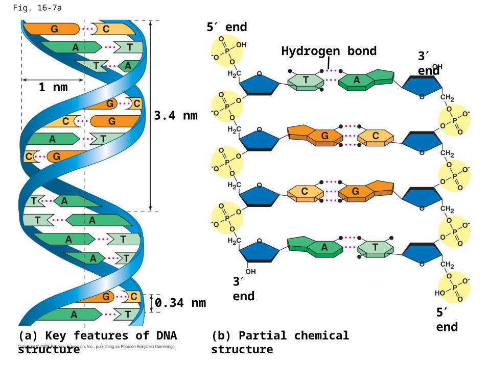
Fig. 16-7a
Hydrogen bond 3 end
5 end
3.4 nm
0.34 nm
3 end
5 end
(b) Partial chemical structure(a) Key features of DNA structure
1 nm

Fig. 16-8
Cytosine (C)
Adenine (A) Thymine (T)
Guanine (G)

Concept 16.2: Many proteins work together in DNA replication and repair
• The relationship between structure and function is manifest in the double helix
• Watson and Crick noted that the specific base pairing suggested a possible copying mechanism for genetic material
Copyright © 2008 Pearson Education Inc., publishing as Pearson Benjamin Cummings

The Basic Principle: Base Pairing to a Template Strand
• Since the two strands of DNA are complementary, each strand acts as a template for building a new strand in replication
• In DNA replication, the parent molecule unwinds, and two new daughter strands are built based on base-pairing rules
Copyright © 2008 Pearson Education Inc., publishing as Pearson Benjamin Cummings
Animation: DNA Replication OverviewAnimation: DNA Replication Overview

Fig. 16-9-1
A T
GC
T A
TA
G C
(a) Parent molecule

Fig. 16-9-2
A T
GC
T A
TA
G C
A T
GC
T A
TA
G C
(a) Parent molecule (b) Separation of strands

Fig. 16-9-3
A T
GC
T A
TA
G C
(a) Parent molecule
A T
GC
T A
TA
G C
(c) “Daughter” DNA molecules, each consisting of one parental strand and one new strand
(b) Separation of strands
A T
GC
T A
TA
G C
A T
GC
T A
TA
G C

• Watson and Crick’s semiconservative model of replication predicts that when a double helix replicates, each daughter molecule will have one old strand (derived or “conserved” from the parent molecule) and one newly made strand
• Competing models were the conservative model (the two parent strands rejoin) and the dispersive model (each strand is a mix of old and new)
Copyright © 2008 Pearson Education Inc., publishing as Pearson Benjamin Cummings

Fig. 16-10
Parent cellFirst replication
Second replication
(a) Conservative model
(b) Semiconserva- tive model
(c) Dispersive model

• Experiments by Matthew Meselson and Franklin Stahl supported the semiconservative model
• They labeled the nucleotides of the old strands with a heavy isotope of nitrogen, while any new nucleotides were labeled with a lighter isotope
Copyright © 2008 Pearson Education Inc., publishing as Pearson Benjamin Cummings

• The first replication produced a band of hybrid DNA, eliminating the conservative model
• A second replication produced both light and hybrid DNA, eliminating the dispersive model and supporting the semiconservative model
Copyright © 2008 Pearson Education Inc., publishing as Pearson Benjamin Cummings

Fig. 16-11a
EXPERIMENT
RESULTS
1
3
2
4
Bacteria cultured in medium containing 15N
Bacteria transferred to medium containing 14N
DNA sample centrifuged after 20 min (after first application)
DNA sample centrifuged after 20 min (after second replication)
Less dense
More dense

Fig. 16-11b
CONCLUSION
First replication Second replication
Conservative model
Semiconservative model
Dispersive model

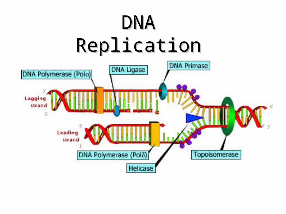
DNA Replication: A Closer Look
• The copying of DNA is remarkable in its speed and accuracy
• More than a dozen enzymes and other proteins participate in DNA replication
Copyright © 2008 Pearson Education Inc., publishing as Pearson Benjamin Cummings

Getting Started
• Replication begins at special sites called origins of replication, where the two DNA strands are separated, opening up a replication “bubble”
• A eukaryotic chromosome may have hundreds or even thousands of origins of replication
• Replication proceeds in both directions from each origin, until the entire molecule is copied
Copyright © 2008 Pearson Education Inc., publishing as Pearson Benjamin Cummings
Animation: Origins of ReplicationAnimation: Origins of Replication

Fig. 16-12a
Origin of replication Parental (template) strand
Daughter (new) strand
Replication fork
Replication bubble
Double-stranded DNA molecule
Two daughter DNA molecules
(a) Origins of replication in E. coli
0.5 µm

Fig. 16-12b
0.25 µm
Origin of replication Double-stranded DNA molecule
Parental (template) strandDaughter (new) strand
Bubble Replication fork
Two daughter DNA molecules
(b) Origins of replication in eukaryotes

3636
DNA ReplicationDNA Replication
• Before new DNA strands can form, there must be RNA primers present to start the addition of new nucleotides
• Primase is the enzyme that synthesizes the RNA Primer
• DNA polymerase can then add the new nucleotides

DNA ReplicationDNA Replication• Begins at Begins at Origins of ReplicationOrigins of Replication
• Two strands open forming Two strands open forming Replication Forks (Y-shaped Replication Forks (Y-shaped region)region)
• New strands grow at the forksNew strands grow at the forks3’
5’
3’
5’
DNA ReplicationDNA Replication

DNA ReplicationDNA Replication
• As the 2 DNA strands open at the origin, Replication Bubbles form
• Prokaryotes (bacteria) have a single bubble
• Eukaryotic chromosomes have MANY bubbles

DNA Replication
• Enzyme Helicase unwinds and separates the 2 DNA strands by breaking the weak hydrogen bonds
• Single-Strand Binding Proteins attach and keep the 2 DNA strands separated and untwisted

DNA Replication
• Enzyme Topoisomerase attaches to the 2 forks of the bubble to relieve stress on the DNA molecule as it separates

Fig. 16-13
Topoisomerase
Helicase
PrimaseSingle-strand binding proteins
RNA primer
55
5 3
3
3

DNA Replication• Before new DNA strands can
form, there must be RNA primers present to start the addition of new nucleotides
• Primase is the enzyme that synthesizes the RNA Primer
• DNA polymerase can then add the new nucleotides

DNA Replication• DNA polymerase can only add
nucleotides to the 3’ end of the DNA • This causes the NEW strand to be built
in a 5’ to 3’ direction
RNAPrimerDNA Polymerase
Nucleotide
5’
5’ 3’
Direction of Replication

Synthesis of the New DNA Strands
The Leading Strand is synthesized as a single strand from the point of origin toward the opening replication fork
RNAPrimerDNA PolymeraseNucleotides
3’5’
5’

Synthesis of the New DNA Strands
The Lagging Strand is synthesized discontinuously against overall direction of replication
This strand is made in MANY short segments It is replicated from the replication fork toward the origin
RNA Primer
Leading Strand
DNA Polymerase
5’
5’
3’
3’
Lagging Strand
5’
5’
3’
3’

Lagging Strand SegmentsLagging Strand Segments
Okazaki Fragments - series of short segments on the lagging strand
Must be joined together by an enzyme
Lagging Strand
RNARNAPrimerPrimer
DNADNAPolymerasePolymerase
3’
3’
5’
5’
Okazaki FragmentOkazaki Fragment

Joining of Okazaki Fragments
The enzyme Ligase joins the Okazaki fragments together to make one strand
Lagging Strand
Okazaki Fragment 2
DNA ligase
Okazaki Fragment 1
5’
5’
3’
3’

Replication of StrandsReplication of Strands
Replication Fork
Point of Origin

Proofreading New DNA
• DNA polymerase initially makes about 1 in 10,000 base pairing errors
• DNA polymerases proofread and correct these mistakes
• The new error rate for DNA that has been proofread is 1 in 1 billion base pairing errors
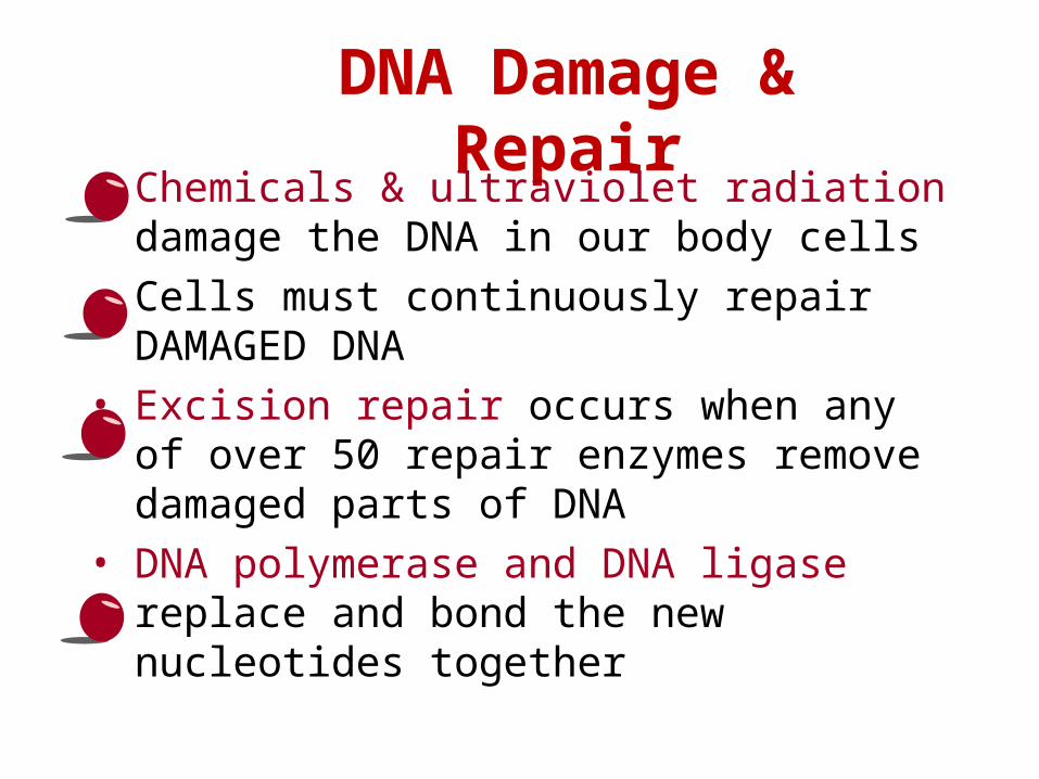
DNA Damage & Repair
• Chemicals & ultraviolet radiation damage the DNA in our body cells
• Cells must continuously repair DAMAGED DNA
• Excision repair occurs when any of over 50 repair enzymes remove damaged parts of DNA
• DNA polymerase and DNA ligase replace and bond the new nucleotides together

Fig. 16-18
Nuclease
DNA polymerase
DNA ligase

Replicating the Ends of DNA Molecules
• Limitations of DNA polymerase create problems for the linear DNA of eukaryotic chromosomes
• The usual replication machinery provides no way to complete the 5 ends, so repeated rounds of replication produce shorter DNA molecules
Copyright © 2008 Pearson Education Inc., publishing as Pearson Benjamin Cummings

Fig. 16-19
Ends of parental DNA strands
Leading strand
Lagging strand
Lagging strand
Last fragment Previous fragment
Parental strand
RNA primer
Removal of primers and replacement with DNA where a 3 end is available
Second round of replication
New leading strand
New lagging strand
Further rounds of replication
Shorter and shorter daughter molecules
5
3
3
3
3
3
5
5
5
5

Fig. 16-15
Leading strand
Overview
Origin of replicationLagging strand
Leading strandLagging strand
Primer
Overall directions of replication
Origin of replication
RNA primer
“Sliding clamp”
DNA poll IIIParental DNA
5
3
3
3
3
5
5
5
5
5

Fig. 16-15a
Overview
Leading strand
Leading strandLagging strand
Lagging strand
Origin of replication
Primer
Overall directions of replication

Fig. 16-15b
Origin of replication
RNA primer
“Sliding clamp”
DNA pol IIIParental DNA
3
5
5
5
5
5
5
3
3
3

Fig. 16-16Overview
Origin of replication
Leading strand
Leading strand
Lagging strand
Lagging strand
Overall directions of replication
Template strand
RNA primer
Okazaki fragment
Overall direction of replication
12
3
2
1
1
1
1
2
2
51
3
3
3
3
3
3
3
3
3
5
5
5
5
5
5
5
5
5
5
53
3

Fig. 16-16a
Overview
Origin of replication
Leading strand
Leading strand
Lagging strand
Lagging strand
Overall directions of replication
12

Fig. 16-16b1
Template strand
5
53
3

Fig. 16-16b2
Template strand
5
53
3
RNA primer 3 5
5
3
1

Fig. 16-16b3
Template strand
5
53
3
RNA primer 3 5
5
3
1
1
3
35
5
Okazaki fragment

Fig. 16-16b4
Template strand
5
53
3
RNA primer 3 5
5
3
1
1
3
35
5
Okazaki fragment
12
3
3
5
5

Fig. 16-16b5
Template strand
5
53
3
RNA primer 3 5
5
3
1
1
3
35
5
Okazaki fragment
12
3
3
5
5
12
3
3
5
5

Fig. 16-16b6
Template strand
5
53
3
RNA primer 3 5
5
3
1
1
3
35
5
Okazaki fragment
12
3
3
5
5
12
3
3
5
5
12
5
5
3
3
Overall direction of replication

Fig. 16-17
OverviewOrigin of replication
Leading strand
Leading strand
Lagging strand
Lagging strandOverall directions
of replication
Leading strand
Lagging strand
Helicase
Parental DNA
DNA pol III
Primer Primase
DNA ligase
DNA pol III
DNA pol I
Single-strand binding protein
5
3
5
5
5
5
3
3
3
313 2
4

The DNA Replication Complex
• The proteins that participate in DNA replication form a large complex, a “DNA replication machine”
• The DNA replication machine is probably stationary during the replication process
• Recent studies support a model in which DNA polymerase molecules “reel in” parental DNA and “extrude” newly made daughter DNA molecules
Copyright © 2008 Pearson Education Inc., publishing as Pearson Benjamin Cummings
Animation: DNA Replication ReviewAnimation: DNA Replication Review

• Eukaryotic chromosomal DNA molecules have at their ends nucleotide sequences called telomeres
• Telomeres do not prevent the shortening of DNA molecules, but they do postpone the erosion of genes near the ends of DNA molecules
• It has been proposed that the shortening of telomeres is connected to aging
Copyright © 2008 Pearson Education Inc., publishing as Pearson Benjamin Cummings

Fig. 16-20
1 µm

• If chromosomes of germ cells became shorter in every cell cycle, essential genes would eventually be missing from the gametes they produce
• An enzyme called telomerase catalyzes the lengthening of telomeres in germ cells
Copyright © 2008 Pearson Education Inc., publishing as Pearson Benjamin Cummings

• The shortening of telomeres might protect cells from cancerous growth by limiting the number of cell divisions
• There is evidence of telomerase activity in cancer cells, which may allow cancer cells to persist
Copyright © 2008 Pearson Education Inc., publishing as Pearson Benjamin Cummings

Concept 16.3 A chromosome consists of a DNA molecule packed together with proteins
• The bacterial chromosome is a double-stranded, circular DNA molecule associated with a small amount of protein
• Eukaryotic chromosomes have linear DNA molecules associated with a large amount of protein
• In a bacterium, the DNA is “supercoiled” and found in a region of the cell called the nucleoid
Copyright © 2008 Pearson Education Inc., publishing as Pearson Benjamin Cummings

• Chromatin is a complex of DNA and protein, and is found in the nucleus of eukaryotic cells
• Histones are proteins that are responsible for the first level of DNA packing in chromatin
Copyright © 2008 Pearson Education Inc., publishing as Pearson Benjamin Cummings
Animation: DNA PackingAnimation: DNA Packing

Fig. 16-21a
DNA double helix (2 nm in diameter)
Nucleosome(10 nm in diameter)
Histones Histone tailH1
DNA, the double helix Histones Nucleosomes, or “beads on a string” (10-nm fiber)

Fig. 16-21b
30-nm fiber
Chromatid (700 nm)
Loops Scaffold
300-nm fiber
Replicated chromosome (1,400 nm)
30-nm fiber Looped domains (300-nm fiber)
Metaphase chromosome

• Chromatin is organized into fibers
• 10-nm fiber
– DNA winds around histones to form nucleosome “beads”
– Nucleosomes are strung together like beads on a string by linker DNA
• 30-nm fiber
– Interactions between nucleosomes cause the thin fiber to coil or fold into this thicker fiber
Copyright © 2008 Pearson Education Inc., publishing as Pearson Benjamin Cummings

• 300-nm fiber
– The 30-nm fiber forms looped domains that attach to proteins
• Metaphase chromosome
– The looped domains coil further
– The width of a chromatid is 700 nm
Copyright © 2008 Pearson Education Inc., publishing as Pearson Benjamin Cummings

• Most chromatin is loosely packed in the nucleus during interphase and condenses prior to mitosis
• Loosely packed chromatin is called euchromatin
• During interphase a few regions of chromatin (centromeres and telomeres) are highly condensed into heterochromatin
• Dense packing of the heterochromatin makes it difficult for the cell to express genetic information coded in these regions
Copyright © 2008 Pearson Education Inc., publishing as Pearson Benjamin Cummings

• Histones can undergo chemical modifications that result in changes in chromatin organization
– For example, phosphorylation of a specific amino acid on a histone tail affects chromosomal behavior during meiosis
Copyright © 2008 Pearson Education Inc., publishing as Pearson Benjamin Cummings

Fig. 16-22
RESULTS
Condensin and DNA (yellow)
Outline of nucleus
Condensin (green)
DNA (red at periphery)
Normal cell nucleus Mutant cell nucleus
You should now be able to:
1. Describe the contributions of the following people: Griffith; Avery, McCary, and MacLeod; Hershey and Chase; Chargaff; Watson and Crick; Franklin; Meselson and Stahl
2. Describe the structure of DNA
3. Describe the process of DNA replication; include the following terms: antiparallel structure, DNA polymerase, leading strand, lagging strand, Okazaki fragments, DNA ligase, primer, primase, helicase, topoisomerase, single-strand binding proteins
Copyright © 2008 Pearson Education Inc., publishing as Pearson Benjamin Cummings

4. Describe the function of telomeres
5. Compare a bacterial chromosome and a eukaryotic chromosome
Copyright © 2008 Pearson Education Inc., publishing as Pearson Benjamin Cummings