Municipal Tennis Facility Proposal April 2006. Overview of KATCH Initiatives.
Upload
nguyendangCategory
view
250download
2
Copyright © 2010 Wolters Kluwer Health | Lippincott Williams & Wilkins
Chapter 16: Cardiovascular Regulation and Integration
McArdle, W.D., Katch, F.I., & Katch, V.L. (2010). Exercise Physiology: Nutrition, Energy, and Human Performance (7 ed.). Baltimore, MD.: Lippincott Williams and Wilkins

• Cardiac muscle maintains its own rhythm and, if left to its inherent rhythmicity, would beat at about 100 bpm
• In the right atrium is the sinoatrial (SA) node, which spontaneously depolarizes and repolarizes
• The impulse spreads across the atria to the atrioventricular (AV) node
• A 0.10-second delay occurs after to allow the atria to contract and propel blood into the ventricles below
The Heart’s Electrical Activity
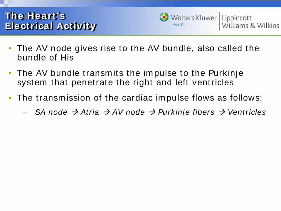
• The AV node gives rise to the AV bundle, also called the bundle of His
• The AV bundle transmits the impulse to the Purkinje system that penetrate the right and left ventricles
• The transmission of the cardiac impulse flows as follows:
– SA node Atria AV node Purkinje fibers Ventricles
The Heart’s Electrical Activity
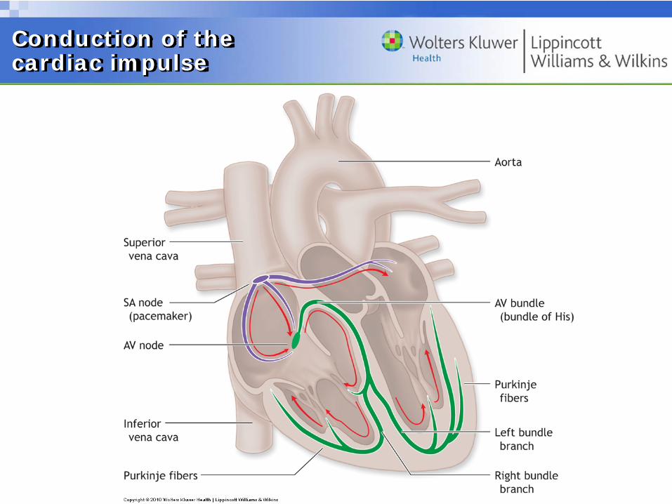
Conduction of the cardiac impulse

• The myocardium’s electrical activity creates an electrical field throughout the body
• The electrocardiogram (ECG) represents a composite record of the heart’s electrical events during a cardiac cycle
• These events provide a way to monitor heart rate during different physical activities and exercise stress testing
• A valid ECG tracing requires proper electrode placement
– ECG leads transmit the electrical signal to a recorder, which creates the composite electrical “picture” of myocardial activity
Electrocardiogram

• Proper skin preparation reduces extraneous electrical “noise” (interference and skeletal muscle artifact)
• Abrade the skin with fine sandpaper or commercially available pads and alcohol to remove surface epidermis and oil
• The skin should appear red, slightly irritated, dry, and clean
Skin Preparation for an ECG

• Provides less sensitivity for diagnostic testing but is useful for routine ECG monitoring in functional exercise testing and radiotelemetry of ECG during physical activity
• Placement:
– The ground (green or black) electrode attaches over the sternum
– The positive (red) electrode attaches on the left side of the chest at the level of the 5th intercostal space adjacent to the midaxillary line
– The positive (white) electrode attaches on the right side of the chest just below the nipple at the level of the 5th intercostal space
Bipolar Configuration

• The standard 12-lead ECG consists of three limb leads, three augmented unipolar leads, and six chest leads
• For improved exercise ECG recordings, electrodes mounted on the torso (abdominal level) replace the conventional ankle and wrist electrodes
Modified 12-Lead Configuration

• Left and Right Leg: Just above the left and right iliac crest on midaxillary line
• Left and Right Arm: Just below the left and right clavicle medial to deltoid
• V1: On right sternal border in 4th intercostal space
• V2: On left sternal border in 4th intercostal space
• V3: At midpoint of a straight line between V2 and V4
• V4: On midclavicular line in 5th intercostal space
• V5: On anterior axillary line and horizontal to V4
• V6: On midaxillary line and horizontal to V4 and V5
Electrode Positioning in the Modified 10-Electrode, Torso-Mounted System

• P wave: Depolarization of the atria when the atria contract
• QRS complex: Signals electrical changes from ventricular depolarization when the ventricles contract
– Atrial repolarization follows the P wave but produces a wave so small that the QRS complex usually obscures it
• T wave: Represents ventricular repolarization that occurs during ventricular diastole
ECG Waves

Phases of an ECG

• Extrinsic Controls: Nerves that directly supply the myocardium and chemical “messengers” that circulate in blood that accelerate the heart in anticipation before exercise begins, and then rapidly adjust to the intensity of physical effort
• Input from the brain and peripheral nervous system continually bombards the cardiovascular control center in the ventrolateral medulla that regulates the heart’s output of blood and blood’s preferential distribution to all the body’s tissues
Extrinsic Regulation of Heart Rate and Circulation
• Neural influences can override the inherent rhythm of the myocardium
– Originate in the cardiovascular center and flow through the sympathetic and parasympathetic components of the autonomic nervous system
– Large numbers of sympathetic and parasympathetic neurons innervate the atria, whereas the ventricles receive sympathetic fibers almost exclusively
Sympathetic and Parasympathetic Neural Input
• Stimulation of the sympathetic cardioaccelerator nerves releases epinephrine and norepinephrine
– Cause chronotropic and inotropic effects
• Sympathetic stimulation also produces vasoconstriction except in the coronary arteries
– Norepinephrine, released by adrenergic fibers, acts as a vasoconstrictor
– Dilation of blood vessels under adrenergic influence occurs from decreased adrenergic activity
Sympathetic Influence
• Parasympathetic neurons release acetylcholine, which delays the rate of sinus discharge to slow heart rate
• Bradycardia results from stimulation of the vagus nerves, which originate in the medulla’s cardioinhibitory center
• Parasympathetic stimulation excites some tissues and inhibits other tissues
• At the start of and during low- to moderate-intensity exercise, HR increases by inhibition of parasympathetic stimulation
• HR in strenuous exercise increases by additional parasympathetic inhibition and direct activation of sympathetic cardioaccelerator nerves
Parasympathetic Influence
• Impulses originating in the brain’s higher somatomotor central command center continually modulate medullary activity
• Central command provides the greatest control over heart rate during exercise
• The heart rapidly “turns on” during exercise by decreasing parasympathetic inhibitory input and increasing stimulating input from the central command
• Central command involvement in cardiovascular regulation explains how variations in emotional state affect cardiovascular response, which creates difficulty obtaining “true” resting values for HR and blood pressure
Central Command
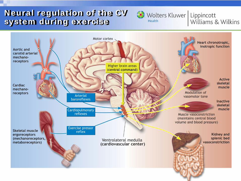
Neural regulation of the CV system during exercise


• Cardiovascular center receives reflex sensory input from peripheral receptors in blood vessels, joints, and muscles
• Chemoreceptors and mechanoreceptors within muscle and its vasculature monitor its chemical and physical state
– This input modifies either parasympathetic or sympathetic outflow to bring about appropriate cardiovascular and respiratory responses to various intensities of physical activity
Peripheral Input
• Three mechanisms continually assess the nature and intensity of exercise and the mass of muscle activated:
– Reflex neural input from mechanical deformation of type III afferents within active muscles
– Chemical stimulation of type IV afferents within active muscles
– Feed-forward outflow from the motor areas of the central command
Peripheral Input cont’d
• Specific mechanoreceptor feedback governs central nervous system’s regulation of blood flow and blood pressure during dynamic exercise
– The aortic arch and carotid sinus contain pressure-sensitive baroreceptors
– Cardiopulmonary receptors assess mechanical activity in left ventricle, right atrium, and large veins
• These function as negative feedback controllers to:
– Inhibit sympathetic outflow from the cardiovascular center
– Blunt an inordinate rise in arterial blood pressure
Peripheral Input cont’d

Review
• Extrinsic controls of cardiac function slow down the heart in anticipation of the upcoming exercise bout.
a. True
b. False
• External pressure against the carotid artery sometimes slows HR from direct baroreceptor stimulation
• Consistently low HR estimation with carotid artery palpation in susceptible individuals would push the person to a higher exercise level
• Vascular disease can affect carotid sinus sensitivity and produce falsely low heart rate values
• A substitute location uses pulse rate at the radial artery or temporal artery
Carotid Artery Palpation

• Blood flows through the vascular circuit generally following physical laws of hydrodynamics applied to rigid, cylindrical vessels
• The volume of flow in any vessel relates to two factors:
– Directly to the pressure gradient between the two ends of the vessels
– Inversely to the resistance encountered to fluid flow
Physical Factors Affecting Blood Flow

• Friction between the blood and internal vascular wall creates resistance or force that impedes blood flow
• Three factors determine resistance:
– Blood thickness or viscosity
– Length of the conducting tube
– Blood vessel radius
Resistance to Blood Flow
• Poiseuille’s Law: Expresses the general relationship among pressure differential, resistance, and flow
– Flow = Pressure gradient x Vessel radius4 ÷ Vessel length x Fluid viscosity
– The transport vessel length remains constant
– Blood viscosity varies only slightly
– The radius affects blood flow the most
• Physiologically, constriction and dilation of the smaller arterial blood vessels provide the crucial mechanism to regulate regional blood flow
Resistance to Blood Flow

• Any increase in energy expenditure requires rapid adjustments in blood flow that impact the entire cardiovascular system
• During exercise, local arterioles of active muscles dilate while vessels to tissues that can temporarily compromise their blood supply constrict
• Factors that contribute to reduced blood flow to non-active tissues:
– Increased sympathetic nervous system outflow
– Local chemicals that directly stimulate vasoconstriction or enhance the effects of other vasoconstrictors
Effect of Exercise on Blood Flow

• Skeletal muscle blood flow couples to metabolic demands
• Regulation occurs from the interaction of neural vasoconstriction activity and locally derived vasoactive substances within the endothelium and red blood cells
• At rest, 1 in 30-40 capillaries in muscles remains open
• The opening of dormant capillaries in exercise serves to:
– Increases total muscle blood flow
– Delivers a large blood volume with only a minimal increase in blood flow velocity
– Increases the effective surface for gas and nutrient exchange between the blood and muscle fibers
Factors Within Active Muscle That Affect Blood Flow

• Vasodilation occurs from local factors related to tissue metabolism that act directly on the smooth muscle bands of small arterioles and precapillary sphincters
– Decreased tissue oxygen, local increases in blood flow, temperature, carbon dioxide, acidity, adenosine, magnesium and potassium ions, and nitric oxide production by the endothelial cells lining the blood vessels
– The venous system may also increase local blood flow by “assessing” increases in the metabolic needs of active muscle and releasing vasodilatory factors

• Nitric oxide serves as an important signal molecule that dilates blood vessels and decreases vascular resistance
• Stimuli from diverse signal chemicals and sheering stress and vessel stretch from increased blood flow through the vessel lumen provoke NO synthesis and release by the vascular endothelium
• In coronary artery disease, the endothelium produces less NO
Nitric Oxide and Autoregulationof Tissue Blood Flow
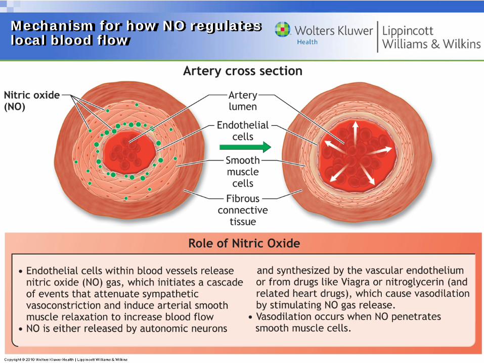
Mechanism for how NO regulates local blood flow
• Transplant patients demonstrate prolonged oxygen uptake kinetics, impaired exercise capacity, and diminished physiologic and hemodynamic function
• Heart transplant recipients can perform relatively intense exercise training and often achieve performance values of moderately trained healthy subjects
• Transplant patients have no stroke volume plateau during graded exercise as the Frank-Starling mechanism causes it to increase throughout exercise
• Exercise response does improve over the post-surgery period but the adaptations exert no meaningful effect on submaximal or peak exercise oxygen consumption
Exercising After Cardiac Transplantation
Review
• Nitric oxide causes what changes in blood vessels?
a. Vasoconstriction and increased vascular resistance
b. Vasodilation and decreased vascular resistance
c. Vasodilation and increased vascular resistance
d. Vasoconstriction and decreased vascular resistance