Chapter 15 Neural Integration I: Sensory Pathways and the Somatic Nervous System.
94
Chapter 15 Neural Integration I: Sensory Pathways and the Somatic Nervous System
-
Upload
lily-bethanie-rogers -
Category
Documents
-
view
228 -
download
3
Transcript of Chapter 15 Neural Integration I: Sensory Pathways and the Somatic Nervous System.
Chapter 15
Neural Integration I:Sensory Pathways and the Somatic Nervous System

fig. 15-1

Sensory Motor
General (15)
Special (17)
Somatic (15)
Autonomic (16)
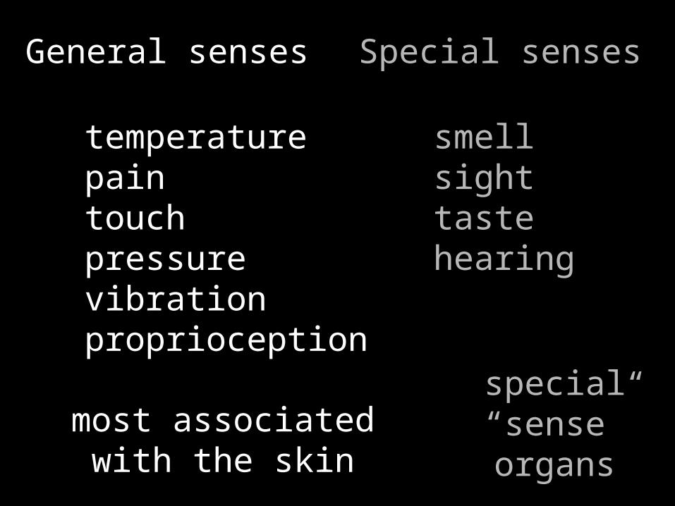
General senses
temperaturepaintouchpressurevibrationproprioception
most associated with the skin
Special senses
smellsighttastehearing
special “sense”organs

General senses
receptors distributed throughout the body
relatively simple

General senses
receptors send info to CNS
arriving info is called sensation
our awareness of it is perception
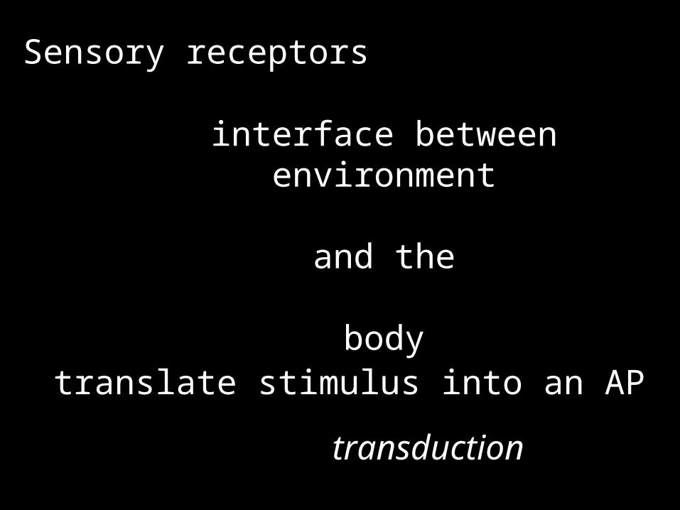
Sensory receptors
interface between environment
and the
body
translate stimulus into an AP
transduction

Sensory receptors
receptors have selective sensivity
chemicalphysical touchlightheat transfer
receptors may or may not have accessory structures associated with them
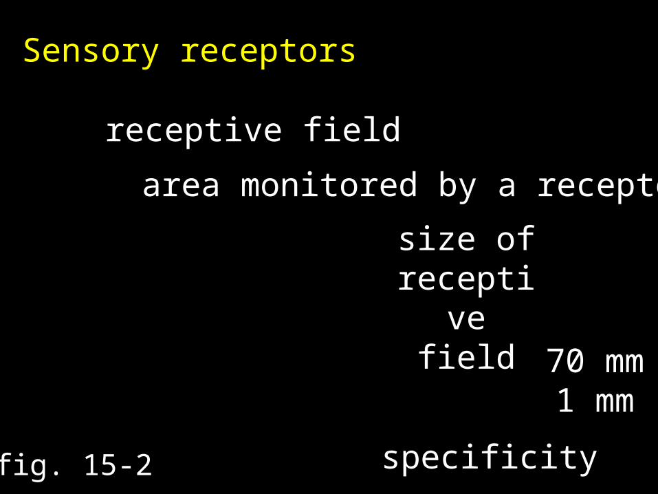
Sensory receptors
receptive field
area monitored by a receptor
fig. 15-2
size of receptive field
70 mm1 mm
specificity
Sensory receptors
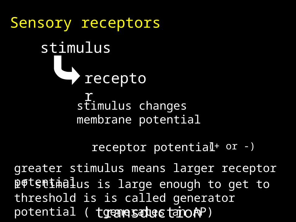
receptor
stimulus
stimulus changesmembrane potential
receptor potential (+ or -)
greater stimulus means larger receptor potentialif stimulus is large enough to get to threshold is is called generator potential (
generates an AP)transduction
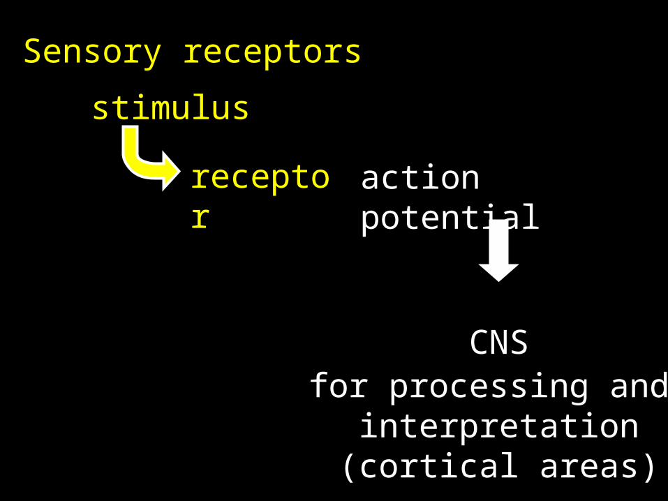
Sensory receptors
receptor
stimulus
action potential
CNSfor processing and
interpretation(cortical areas)

receptor A
cortex
receptor 2receptor Blabeled line

interpretation is based on which “line” information travels on
a “line” carries the same “type” (modality) of information

receptor A
cortex
receptor 2
labeled line
receptor Boptic nerve
shut eyes and rub them gently
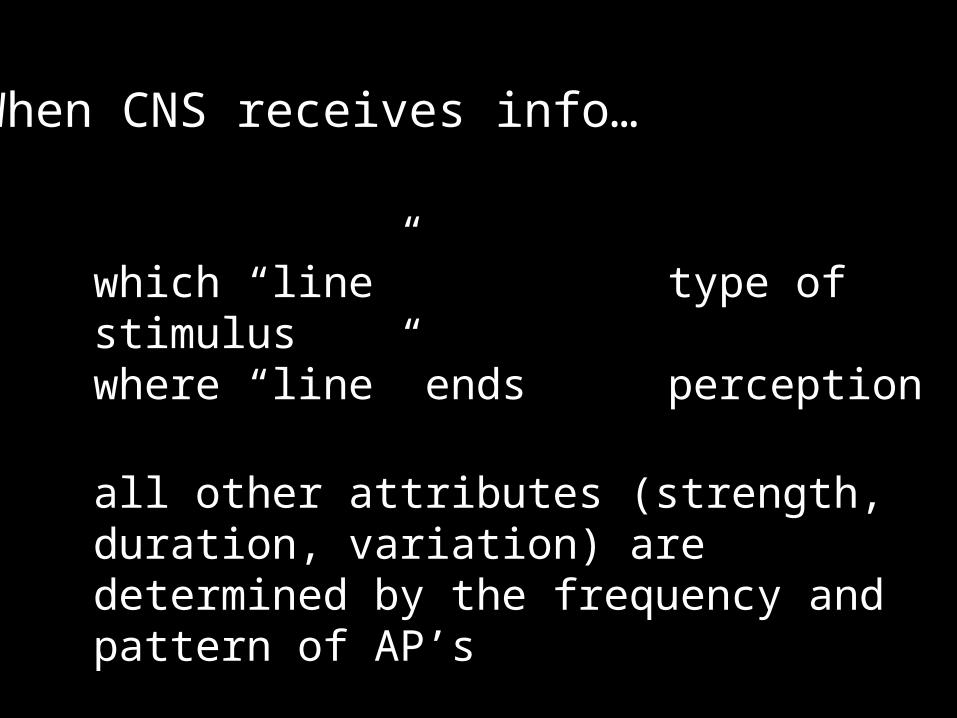
When CNS receives info…
which “line” type of stimuluswhere “line” ends perception
all other attributes (strength, duration, variation) are determined by the frequency and pattern of AP’s

receptor types:
tonic: always “on”
phasic: only on with stimulus
some receptors combine the two
greater stimulus higher freq.lesser stimulus lower freq.

adaptation
reduction in sensitivity in the presence of a constant stimulus
peripheral
centralchange in receptor activity
inhibition of nuclei in pathway

peripheral adaptation
phasic receptors(aka fast-adapting receptors)
example: thermoreceptors
you usually don’t notice roomtemperature unless it changes

central adaptation
example: smell
you walk into a room and notice a new smell…
…but not for long
adaptation reduces the amount of information
reaching the cerebral cortex
about 1% of sensory information coming in reaches our awareness

100 Keys (pg 498)
“Stimulation of a receptor produces action potentials along the axon of a sensory neuron. The frequency or pattern of action potentials contains information about the strength, duration, and variation of the stimulus. Your perception of the nature of that stimulus depends on the path it takes inside the CNS.”

General senses (from chapter 12)
exteroceptors
proprioceptors
interoceptors
outside
position
inside
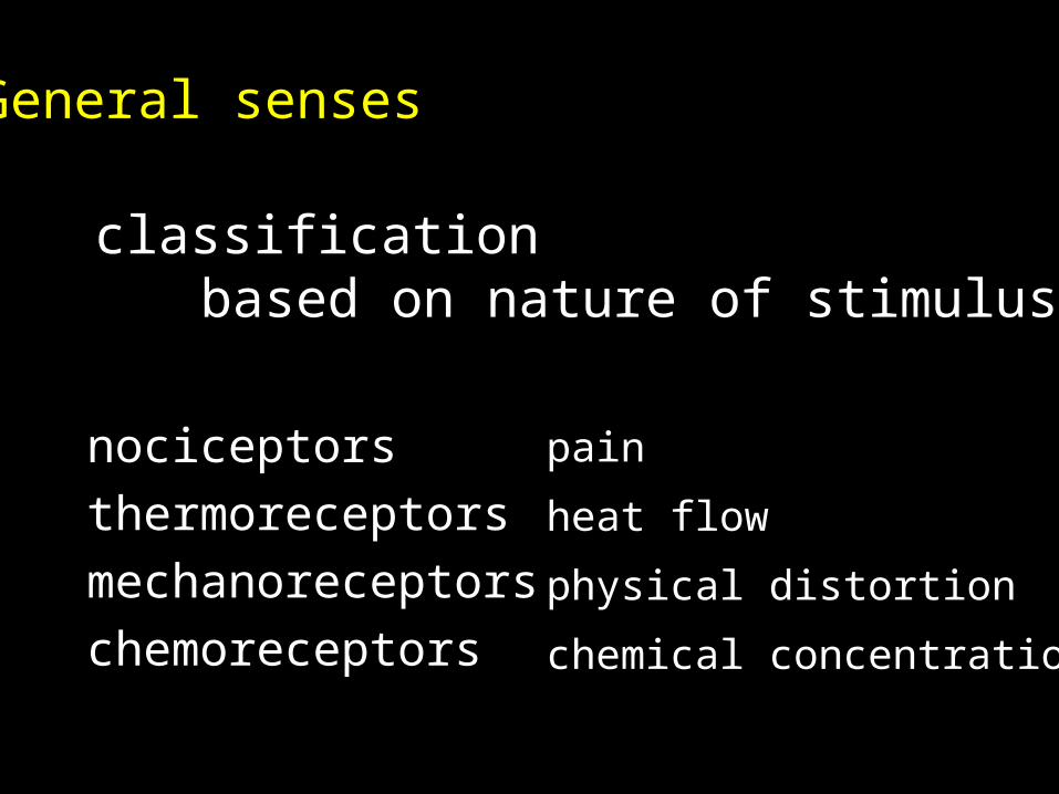
General senses
classificationbased on nature of stimulus
nociceptorsthermoreceptorsmechanoreceptorschemoreceptors
pain
heat flow
physical distortion
chemical concentration

General senses
nociceptorscommon in:
skinjoint capsulescoverings of bonesaround blood vessel walls
free nerve endingslarge receptive fields
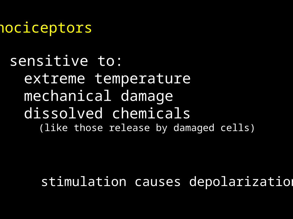
nociceptors
sensitive to:extreme temperature mechanical damagedissolved chemicals
(like those release by damaged cells)
stimulation causes depolarization

two fiber types convey infotype A
fast pain (cut, etc.,)easy to localize
type Cslow pain (“burning, aching”)difficult to localize
nociceptors

tonic receptorsno significant peripheral adaptation
as long as the stimulus is present, it will hurt
but central adaptation can occur(perception of pain may decrease)
nociceptors

sensory neurons bringing in pain info use glutamate and/or substance P as their neurotransmitter
nociceptors
these nts can cause facilitation (?)
pain may be disproportional(feels worse than it should)
pain can be reduced by endorphins and enkephalins (inhibit activity in pathway) [neuromodulators chpt. 12]

nociceptors
endorphins
pain centers use substance Pas nt.
endorphins bind to presynaptic membrane and inhibit substance P release, reducing perception of pain

to here 3/9lec # 24

thermoreceptors
free nerve endings in the dermisskeletal m.hypothalamusliver
warm receptorsor
cold receptors

thermoreceptors
phasic receptorsactive when temperature is changing, quickly adapting to stable temperature
detect transfer of heatheat loss from skin coolheat gain to skin warm

mechanoreceptors
contain mechanically regulated ion channels (chapter 12)
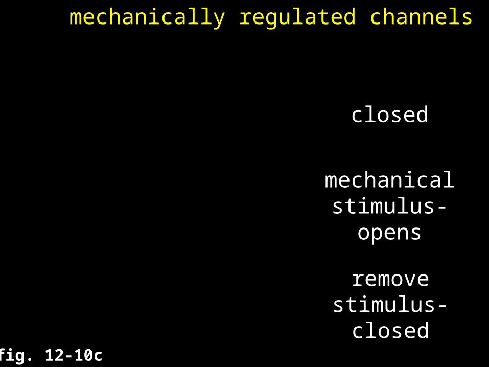
c. mechanically regulated channels
fig. 12-10c
mechanicalstimulus-
opens
removestimulus-
closed
closed

mechanoreceptors
three classes
tactile receptors
baroreceptors
proprioceptors
touch, pressure, vibration
pressure changes(gut, genitourinary)
position of joints/muscles

mechanoreceptors
tactile receptors
fine touch/pressure
crude touch/pressure
small (narrow) receptive fielddetailed informationsensitive
wide receptive fieldpoor localization

fig. 15-3

tactile receptors
range of complexity
free nerve endingsroot hair plexustactile discstactile corpuscles (Meissner’s)lamellated corpuscles (pacinian)Ruffini corpuscles
tactile receptors
free nerve endings
in epidermis of skincornea of eyesensitive to touch and pressuretonic receptorssmall receptive field
tactile receptors
root hair plexus
around each hair folliclesense movement of hairadapt quickly

tactile receptors
tactile discs
sensitive, tonic receptorsin epidermis
fine touch and pressure
tactile receptors
tactile corpuscles (Meissner’s)
fine touch, pressure , vibrationadapt quicklysurrounded by Schwann cellsin dermis of skineyelids, fingertips (sensitive areas)

tactile receptors
lamellated corpuscles (pacinian)
sensitive to deep pressurehigh-frequency vibrations
adapt quicklynerve ending is encapsulatedby layers of supporting cells
(onion)dermis, pancreas, fingers…

tactile receptors
Ruffini corpuscles
pressure and skin distortionlocated deep in the dermistonic, little if any adaptation

fig. 15-3

sensivitity can be altered
infectiondiseasedamage to pathway
e.g., damage to a spinal nervewould affect an entire dermatome

tickle and itch
closely related to touch and pain

baroreceptors
free nerve endings in the walls of organs that stretch
e.g., blood vessels
when pressure changes they expand or contract
changes activity of receptors
proprioceptors
muscle spindles
Golgi tendon organs
receptors in joint capsules
stretch reflex
monitor tendon tension
free nerve endings in joints

proprioceptors
no adaptation
continuously send info to CNS
most processed at subconscious level

chemoreceptors
respond to chemicals dissolved in the surrounding fluidsrespiratory centers in brain
pH, CO2 levels in blood
carotid bodies and aortic bodiespH, CO2, O2 levels in blood
Pathways in the CNS
spinothalamic tractspine to thalamus=sensory
corticospinal tractcortex to spine=moto
r

Pathways in the CNS
sensory pathways
neurons involved
first order neuron
second order neuron
third order neuron
sensory neuron (DRG)
in CNS (crosses over)
in thalamus

Pathways in the CNS
sensory pathways
carry sensory infofrom skin and muscles ofbody wall, head, neck, limbs
Somatic sensory pathways

Pathways in the CNS
sensory pathways
Somatic sensory pathways
posterior column pathwayanterolateral column pathwayspinocerebellar pathway

fig. 15-4

The Posterior Column Pathway
fine touchpressurevibrationsproprioception

The Posterior Column Pathway
inferior half of bodyfirst order neuron in DRG
up the fasciculus gracilis to thenucleus gracilis of med. oblong.
superior half of bodyfirst order neuron in DRG
up the fasciculus cuneatus to thenucleus cuneatus of med. oblong.

The Posterior Column Pathway
second order neuron in nucleus ?cross to other side and ascend to
the ventral nucleus of thalamus
third order neuron in thalamusproject to the primary sensory cortex

fig. 15-4
fig. 15-5a

The Anterolateral Pathway
“crude” touchpressurepaintemperature
The Anterolateral Pathway
first order neuron in DRGsynapses on 2nd order neuron
in dorsal horn of spinal cord
second order neuroncross to opposite side and ascend
The Anterolateral Pathway
second order neuroncross to opposite side and ascend
anterior spinothalamic tract
lateral spinothalamic tract
crude touch and pressureto ventral nucleus of thalamus
pain and temperatureto ventral nucleus of thalamus

The Anterolateral Pathway
second order neuron in spinal cordcross to other side and ascend to
the ventral nucleus of thalamus
third order neuron in ventral thalamusproject to the primary sensory cortex
fig. 15-4
fig. 15-5b

The Anterolateral Pathway
phantom pain ?
referred pain?
activity along pathway, even if “limb” is not there
viceral pains sensations may stimulate neurons of AL pathway

fig. 15-6
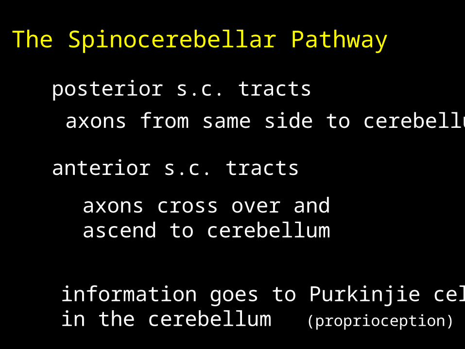
The Spinocerebellar Pathway
posterior s.c. tracts
anterior s.c. tracts
axons from same side to cerebellum
axons cross over andascend to cerebellum
information goes to Purkinjie cellsin the cerebellum (proprioception)

fig. 15-4
fig. 15-7

100 Keys (pg. 507)
Most somatic sensory informationis relayed to the thalamus for processing. A small fraction of the arriving information is projected to the cerebral cortex and reaches our awareness.

to here 3/12lec # 25

Pathways in the CNS
sensory pathways
Somatic sensory pathways
posterior column pathwayanterolateral column pathwayspinocerebellar pathway

fig. 15-4
Pathways in the CNS
sensory pathways
Somatic sensory pathways
posterior column pathwayanterolateral column pathwayspinocerebellar pathway

Pathways in the CNS
sensory pathways
Somatic sensory pathwaysVisceral sensory pathways
info from interoceptors(internal organs)

Pathways in the CNS
Somatic sensory pathwaysVisceral sensory pathways
nociceptors, thermoreceptors,tactile receptors, baroreceptors, chemoreceptors
Pathways in the CNS
Somatic sensory pathwaysVisceral sensory pathways
CN V, VII, IX, X carry info frompharynx, mouth, palate, larynx, trachea and esophagus
project to solitary nucleus(medulla oblongata)
Pathways in the CNS
Somatic sensory pathwaysVisceral sensory pathways
T1 to L2 abdominal organsS2 to S4 pelvic organs
first order neurons project to interneurons which travel up the anterolateral pathway to sol. nuc.
usually subconscious

Pathways in the CNS
sensory pathwaysmotor pathways
the somatic nervous system (SNS)
autonomic nervous system (ANS)voluntary
involuntary

the somatic nervous system (SNS)
always involve at least two neurons
upper motor neuron
lower motor neuroninside CNS (+ or -)
stimulates a motor unit
motor pathways in the CNS
motor information followsone of three main pathways:
corticospinal pathwaymedial pathwaylateral pathway
motor pathways in the CNS
motor pathways in the CNS
corticospinal pathway(aka., pyramidal system)
upper motor neurons arepyramidal cells in primary motor cortex
synapse on lower motor neurons(ventral horn of spinal cord)
also project to other control centers

motor pathways in the CNS
corticospinal pathway
three pairs of tracts:
corticobulbar tracts
to motor nuclei ofCN III, IV, V, VI, VII, IX, XI, XIIconscious control of eye, jaw and face muscles…
motor pathways in the CNS
corticospinal pathway
three pairs of tracts:
corticobulbar tractslateral corticospinal tractsanterior corticospinal tracts

fig. 15-9

Pathways in the CNS
motor pathways
motor information followsone of three main pathways:
corticospinal pathwaymedial pathwaylateral pathway

fig. 15-8

Pathways in the CNS
motor pathways
corticospinal pathwaymedial pathway
muscle tonegross movement
necktrunkproximal
limbs

Pathways in the CNS
motor pathways
medial pathwayUMN in:
vestibular nuclei(hind)
superior colliculus(mid)
reticular formation(brain stem)
posture &balance
reflexive head
position
various

Pathways in the CNS
motor pathways
lateral pathway
control of muscle tone
precise movement of distal limbs
UMN in red nucleus (mid)
descend down rubrospinal tract

Basal Nuclei
background patterns of movement(walking, running, etc.)
adjust activities of UMN in cortex
two populations:
ACh stimulatory
GABA inhibitory
normally:
inactive
active
inhibited

Cerebellum
monitors (sensory):
proprioceptionvisualvestibular (balance)
spinocerebellar tract
superior colliculus
vestibular nucleus
output
continually adjusts UMN activity
Several conditionsALS
amyotrophic lateral sclerosis(aka Lou Gerhig’s disease)degeneration of UMN’s and/or LMN’s
atrophy of musclecerebral palsy
affect voluntary muscle performancetrauma, exposure to drugs etc., genetics
cerebrum, cerebellum, basal nuclei, hippocampus, thalamus
abnormal motor skills, posture, speech…
anencephalylack of higher brain development
100 Keys (pg. 513)
“Neurons of the primary motor cortex (UMN) innervate motor neurons in the brain and spinal cord (LMN) responsible for stimulating skeletal muscles. Higher centers in the brain can suppress or facilitate reflex responses; reflexes can complement or increase the complexity of voluntary movements”