Chapter 15
96
Chapter 15 Exam Six Material
-
Upload
christina-stathis -
Category
Documents
-
view
16 -
download
0
description
Chapter 15. Exam Six Material. Eye and Associated Structures. _______________________________________are in the eye Most of the eye is protected by a cushion of fat and the bony orbit Accessory structures include. Eyebrows. - PowerPoint PPT Presentation
Transcript of Chapter 15

Chapter 15
Exam Six Material

Eye and Associated Structures• _______________________________________
are in the eye• Most of the eye is protected by a cushion of fat
and the bony orbit• Accessory structures include – – – – –

Eyebrows
• ____________________________________ that overlie the supraorbital margins
• Functions include:– – Preventing ______________________________
from reaching the eye• – depresses the _
• – move the eyebrows _

Palpebrae (Eyelids)• Protect the eye anteriorly• – separates _
• – medial and lateral _

Palpebrae (Eyelids)
• – contains _______________________________
that secrete a whitish, oily secretion (Sandman’s eye sand)
• __________________________________ of connective tissue support the eyelids internally
• – gives the _

Palpebrae (Eyelids)• Eyelashes– Project from the free margin of each eyelid–
• Lubricating glands associated with the eyelids– __________________________________________
and sebaceous glands– _________________________________________
lie between the hair follicles

Palpebrae
(Eyelids)

Conjunctiva
• _________________________________ membrane that:– Lines the eyelids as the _– Covers the whites of the eyes as the _
– ______________________________________and protects the eye

Lacrimal Apparatus
• Consists of the lacrimal gland and associated ducts
• Lacrimal glands _• Tears– Contain _– Enter the eye via superolateral excretory ducts – _____________________the eye
__________________________via the lacrimal punctum
– Drain into _
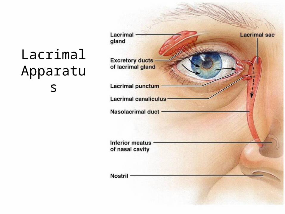
Lacrimal Apparatus
Figure 15.2

Extrinsic Eye Muscles
• Six straplike extrinsic eye muscles– Enable the eye to _– Maintain the _
• Four ________________________________ muscles originate from the annular ring
• Two ______________________________ muscles move the eye in the vertical plane

Extrinsic Eye Muscles

Summary of Cranial Nerves and Muscle Actions
• Names, actions, and cranial nerve innervation of the extrinsic eye muscles

Structure of the Eyeball
• A slightly _____________________________________with anterior and posterior poles
• The wall is composed of three tunics – – – –
• The internal cavity is filled with fluids called _
• The lens separates the internal cavity into anterior and posterior segments

Structure of the Eyeball

Fibrous Tunic
• Forms the outermost coat of the eye and is composed of: – –
• The sclera _____________________________________ and anchors _
• The cornea lets light enter the eye

Vascular Tunic: Choroid Region
• Has three regions: – – –
• Choroid region– A dark brown membrane that forms the posterior
portion of the vascular layer– Supplies _

Vascular Tunic: Ciliary Body
• A thickened ring of tissue _• Composed of _– ciliary muscles
• Anchors the _____________________________________ that holds the lens in place

Vascular Tunic: Iris
• The _________________________________ of the eye
• Pupil – central opening of the iris– • Close vision and bright light
– • Distant vision and dim light
– • Changes in emotional state
– pupils dilate when the subject matter is appealing or requires problem-solving skills

Sensory Tunic: Retina
• A delicate _• Pigmented layer – the outer layer that
________________________________________ and prevents its scattering
• Neural layer, which contains:– ___________________________________________
that transduce light energy– Bipolar cells and ganglion cells– Amacrine and horizontal cells

Sensory Tunic: Retina

The Retina: Ganglion Cells and the Optic Disc
• Ganglion cell axons:– Run along the _– Leave the eye as the _
• The _– Is the site where the optic nerve leaves the eye– Lacks _• the

The Retina: Ganglion Cells and the Optic Disc

The Retina: Photoreceptors
• Rods:– Respond to _– Are used for _
• Cones:– Respond to _– Have
_____________________________________ color vision
– Are found in the _– Are concentrated in the _

Blood Supply to the Retina
• The neural retina receives its blood supply from two sources– The outer third receives its blood from the _– The inner two-thirds is served by the _
• Small vessels radiate out from the optic disc

Inner Chambers and Fluids
• The lens separates the internal eye into _
• The posterior segment is filled with _
– Transmits light– Supports the posterior surface of the lens –
– Contributes to intraocular pressure

Anterior Segment
• Composed of two chambers– • between the cornea and the iris
– • between the iris and the lens
• ____________________________ humor– A plasmalike fluid that fills the anterior segment– Drains via the _
• Supports, nourishes, and _

Anterior Segment

Lens• A biconvex, transparent, flexible,
________________________________structure that:– Allows ____________________________________of light
onto the retina– Is composed of _
• Lens fibers – cells filled with the transparent protein _
• With age, – the lens becomes more compact – – loses its _

Light
• Our eyes respond to a small portion of this spectrum called the _
• Different ___________________________ in the retina respond to different __________________________________ of the visible spectrum

Refraction and Lenses
• When light passes from one transparent medium to another its speed changes and _
• Light passing through a convex lens is bent so that the rays _
• When a convex lens forms an image, the image is _

Focusing Light on the Retina
• Pathway of light entering the eye: –
• Light is refracted:– At the cornea– Entering the lens– Leaving the lens
• The lens curvature and shape allow for fine focusing of an image

Focusing for Distant Vision• Light from a distance
needs ______________________________________ for proper focusing
• Far point of vision – the distance beyond which the lens does not need to change shape to focus (20 ft.)

Focusing for Close Vision
• Close vision requires:– • changing the lens shape by ciliary muscles to increase
refractory power
– • the pupillary reflex constricts the pupils to prevent
divergent light rays from entering the eye
– • medial rotation of the eyeballs toward the object being
viewed

Problems of Refraction
• – normal eye with light focused properly
• – the focal point is in front of the retina– Corrected with a concave lens
• – the focal point is behind the retina– Corrected with a convex lens


Photoreception: Functional Anatomy of Photoreceptors
• – process by which the eye detects light energy
• Rods and cones contain _

Rods
• Functional characteristics– Sensitive to ___________________________ and
best suited for _– Absorb _– Perceived input is in _– Sum of visual input from many rods feeds into a
single ganglion cell – Results in _

Cones
• Functional characteristics – Need ___________________________________
for activation • have low _
– Have pigments that allow a _– Each cone synapses with a _– Vision is _

Excitation of Cones
• There are three types of cones:
• Intermediate colors are perceived by activation of _
• Method of excitation is similar to rods

Signal Transmission in
the Retina

Adaptation• _____________________________________
(going from dark to light) involves:– Dramatic decreases in retinal sensitivity •
– Switching from the rod to the cone system•
• Adaptation to dark is the reverse– – Rhodopsin accumulates in the dark and retinal
sensitivity is restored

Visual Pathways• Axons of retinal ganglion cells form the optic
nerve • Medial fibers of the _
• Most fibers of the optic tracts continue to the _

Visual Pathways
• Other optic tract fibers end in _
• Optic radiations travel from _

Visual Pathways
• Some nerve fibers send tracts to the midbrain ending in the _
• A small subset of visual fibers contain melanopsin (circadian pigment) which:– Mediates papillary light reflexes– Sets daily _

Depth Perception
• Achieved by both eyes viewing the same image from _
• Three-dimensional vision results from _____________________________________ of the slightly different images
• If only one eye is used, _____________________________________and the observer must rely on learned clues to determine depth

Thalamic Processing
• The _____________________________________ of the thalamus:– Relay information on _– Segregate the retinal axons in preparation _
– Emphasize visual inputs from regions of high cone density
– Sharpen the contrast information received by the retina

Cortical Processing
• – Basic dark/bright and _
• Prestriate cortices (association areas) processes– Form, color, and movement
• Visual information then proceeds anteriorly to the:– ___________________________________ –
processes identification of objects– ___________________________________ and
postcentral gyrus – processes spatial location

Chemical Senses
• Chemical senses – –
• Their chemoreceptors respond to chemicals in aqueous solution– Taste• to substances dissolved _
– Smell• to substances dissolved in _

Sense of Smell
• The organ of smell is the _____________________________________, which covers the superior nasal concha
• Olfactory receptor cells are _____________________________________with radiating olfactory cilia
• Basal cells lie at the base of the epithelium

Olfactory Receptors

Physiology of Smell
• Olfactory receptors respond to several different odor-causing chemicals
• When bound to ligand these proteins _
• cAMP (the second messenger) opens ion channels, – causing
___________________________________ of the receptor membrane that then triggers an action potential

Olfactory Pathway
• Olfactory receptor cells _
• Glomerular mitral cells _• Mitral cells send impulses to:– The – The hypothalamus, amygdala, and _

Taste Buds
• _________________of the 10,000 or so taste buds are found on the _
• Taste buds are found in papillae of the tongue mucosa
• Papillae come in three types: – – –
• Fungiform and circumvallate papillae _


Structure of a Taste Bud
• Each _________________________________ taste bud consists of three major cell types– • insulate the receptor
– • dynamic stem cells
– •

Taste Sensations• There are five basic taste sensations– • sugars, saccharin, alcohol, and some amino acids
– • metal ions
– • hydrogen ions
– • alkaloids such as quinine and nicotine
– • elicited by the amino acid glutamate

Physiology of Taste
• In order to be tasted, a chemical:– Must be _– Must contact _
• Binding of the food chemical:– Depolarizes the taste cell membrane, _
– Initiates a generator potential that elicits an _

Gustatory Pathway
• Cranial Nerves _________________________ carry impulses from _
• These impulses then travel to the _____________________________________, and from there fibers branch to the:– – Hypothalamus and limbic system
(________________________________of taste)

Influence of Other Sensations on Taste
• Taste _
• Thermoreceptors, mechanoreceptors, nociceptors also influence tastes
• ________________________________________ enhance or detract from taste

The Ear: Hearing and Balance
• The three parts of the ear are the _• The outer and middle ear are involved with
hearing• The inner ear functions –
• Receptors for hearing and balance: – Respond to _–

The Ear: Hearing and Balance

Outer Ear
• The auricle (pinna) is composed of:– – The _
• External auditory canal– Short, curved tube filled with _

Outer Ear
• Tympanic membrane _– Thin connective tissue membrane that vibrates in
response to sound
– Transfers sound energy to the _
–

Middle Ear (Tympanic Cavity)
• A small, ___________________________, mucosa-lined cavity – Flanked _– Flanked medially by the _
• Epitympanic recess – superior portion of the middle ear
• – connects the middle ear to the nasopharynx– ______________________________________ in
the middle ear cavity with the external air pressure

Middle and Internal Ear

Ear Ossicles
• The tympanic cavity contains three small bones:
• • •
– Transmit _________________________________ of the eardrum to the _
– Dampened by the _


Inner Ear• – Tortuous channels worming their way through _
– Contains the _– Filled with perilymph
• Membranous labyrinth– Series of membranous sacs within the bony
labyrinth– Filled with a _

Inner Ear

The Vestibule
• The __________________________________ of the bony labyrinth
• Suspended in its perilymph are two sacs: – –
• The saccule extends _

The Vestibule
• The utricle extends into the _
• These sacs:– House ___________________________________
called maculae– Respond to _______________________________
and changes in the _

The Semicircular Canals
• Three canals that lie in the _
• Membranous semicircular ducts line each canal and communicate with the utricle
• The _________________________________is the swollen end of each canal and it houses equilibrium receptors in a region called the _
• These receptors respond to _

The Cochlea
• A ______________________________, conical, bony chamber that:– Extends from the anterior vestibule– Coils around a bony pillar called the _– Contains the cochlear duct, which ends at the
cochlear apex– Contains the _

The Cochlea
• The cochlea is divided into three chambers:– Scala _– Scala _– Scala _

The Cochlea
• The scala tympani terminates at the _
• The scalas tympani and vestibuli:– Are filled with – Are continuous with each other via the _
• The scala media is filled with _

The Cochlea
• The “floor” of the cochlear duct is composed of:– The bony spiral lamina– The ____________________________________,
which supports the organ of Corti• The cochlear branch of nerve VIII runs _


Sound and Mechanisms of Hearing• Sound vibrations beat against the eardrum• The eardrum pushes against the ossicles, which
presses fluid in the inner ear against the oval and round windows– This movement sets up _
– Moving hair cells stimulates the cochlear nerve _

Properties of Sound
• – the number of waves that pass a given point in a
given time• – perception of different frequencies (we hear from
20–20,000 Hz)• – intensity of a sound measured in decibels (dB)
• – subjective interpretation of sound intensity

Transmission of Sound to the Inner Ear
• The route of sound to the inner ear follows this pathway:– Outer ear•
– Middle ear•
– Inner ear• scalas vestibuli and tympani to the _• Stimulation of the _• Generation of impulses in the _

Resonance of the Basilar Membrane
• Sound waves of low frequency (inaudible):– Travel around the helicotrema –
• Audible sound waves:– Penetrate through the cochlear duct– Vibrate the _– Excite specific hair cells according to
________________________________________ of the sound

The Organ of Corti
• Is composed of ___________________________________ and outer and _
• _____________________________________ fibers of the cochlear nerve attach to the base of hair cells
• The _– Protrude into the endolymph– Touch the tectorial membrane

Excitation of Hair Cells in the Organ of Corti
• Bending cilia: – Opens __________________________________
ion channels– Causes a
_________________________________________ and the release of a neurotransmitter
– The neurotransmitter causes cochlear fibers to transmit impulses to the brain, where sound is perceived


Auditory Pathway to the Brain• Impulses from the cochlea pass
via the __________________________to the _
• From there, impulses are sent to the:– –
• From there, impulses pass to the _
• Auditory pathways _________________________ so that both cortices receive input from both ears

Deafness• – something hampers sound conduction to the fluids
of the inner ear –
• – results from damage to the
________________________________________ at any point from the cochlear hair cells to the auditory cortical cells

Deafness
• – ringing or clicking sound in the ears in the absence
of auditory stimuli• – labyrinth disorder that affects the cochlea and the
semicircular canals, causing _
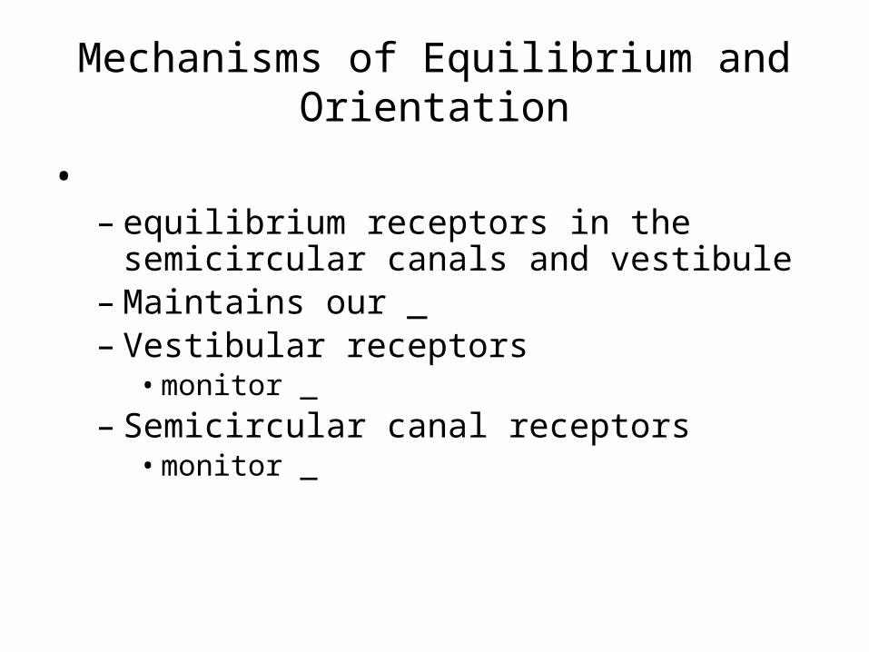
Mechanisms of Equilibrium and Orientation
• – equilibrium receptors in the semicircular canals
and vestibule– Maintains our _– Vestibular receptors • monitor _
– Semicircular canal receptors • monitor _

Anatomy of Maculae• ____________________________________are the sensory
receptors for static equilibrium– Contain supporting cells and hair cells– Each hair cell has stereocilia and kinocilium embedded in the
otolithic membrane•
– jellylike mass studded with tiny stones called _
• __________________________________ hairs respond to _
• __________________________________ hairs respond to _

Effect of Gravity on Utricular Receptor Cells
• Otolithic movement in the _
– Depolarizes vestibular nerve fibers– _________________________________the
number of action potentials generated• Movement in the _– _______________________________________
vestibular nerve fibers– ____________________________________ the
rate of impulse propagation

Effect of Gravity on Utricular Receptor Cells

Crista Ampullaris and Dynamic Equilibrium
• The _– Is the receptor for
_________________________________ equilibrium
– Is located in the ampulla of each _– Responds to angular movements
• Each crista has support cells and hair cells that extend into a gel-like mass called the _
• Dendrites of vestibular nerve fibers encircle the base of the hair cells

Activating Crista Ampullaris Receptors
• Cristae respond to _____________________________________ of rotatory movements of the head
• Directional bending of hair cells in the cristae causes:–
– Hyperpolarizations, and fewer impulses reach the brain
• The result is that the brain is informed of rotational movements of the head

Rotary Head Movement

Balance and Orientation Pathways
• There are ______________________________ for balance and orientation– – –
• These receptors allow our body to respond reflexively