Autonomic Nervous System Chapter 15. Autonomic Nervous System.

330
CHAPTER 13
THE NERVOUS SYSTEM
13.1 SEIZURES R56.8
DESCRIPTION A seizure is a change in movement, attention or level of awareness that is sustained or repetitive and occurs as a result of abnormal and excessive neuronal discharges within the brain.
For recurrent seizures, see section 13.4: Epilepsy. Classification of seizures using International League against Epilepsy (ILAE): Classification of seizures is aetiological and clinical.
Aetiology » Genetic » Metabolic » Structural » Infectious » Immune » Unknown
The causes of seizures are multifactorial. CNS infections are a common cause in the South African setting. The commonest seizures in children are febrile convulsions but the history, examination and investigations must be aimed at excluding the following conditions:
Perinatal conditions Infections Poisoning
» congenital infection » hypoxic-ischaemic
damage » trauma » cerebral haemorrhage
or thrombosis
» meningitis » encephalitis » brain abscess » neurocysticercosis
» accidental ingestion of medicines
» medicine withdrawal » environmental toxins » toxicity of
antiepileptic drugs (AED)
Metabolic conditions Systemic disorders Primary cerebral causes
» hypoglycaemia » hypocalcaemia » hypomagnesaemia » hyponatraemia » hypernatraemia » inborn errors of
metabolism
» vasculitis » hypertensive
encephalopathy » uraemia (renal
failure) » hyperammonaemia
(liver failure)
» cerebral malformation
» genetic/familial (syndromic)
» tumour » idiopathic

CHAPTER 13 THE NERVOUS SYSTEM
331
Clinical Within each of the above categories, generalised, focal or syndromic seizures occur. Generalised seizures: The epileptic focus arises at some point within and rapidly spreads to involve networks in both hemispheres of the brain. Generalised seizures may be: » tonic-clonic, » absence (typical or atypical), » clonic, » tonic or atonic, » myoclonic.
Generalised tonic-clonic seizures (GTCS) that continue or recur for more than 5 minutes in which there is incomplete recovery of consciousness are called Convulsive Status Epilepticus: See section 13.3: Status epilepticus (convulsive). Focal seizures: The epileptic activity arises at some point from a particular focus or networks limited to one hemisphere of the brain.
Focal seizures occur with: » observable aura, motor or autonomic components, » altered consciousness or awareness (previously termed complex partial
seizures).
The presentation of focal seizures depends on the site of origin and may be frontal lobe seizures, temporal lobe seizures, parietal lobe seizures and occipital lobe seizures.
Focal seizures may progress to generalised tonic-clonic seizures and this is known as secondary generalisation. Epileptic Syndromes – See section 13.4: Epilepsy.

CHAPTER 13 THE NERVOUS SYSTEM
332
*Denotes onset of seizure.
International League Against Epilepsy Classification of Seizures1
DIAGNOSTIC CRITERIA Clinical » Obtain a history:
> Eye witness account, aura, video recording. > Perinatal history, drug history, developmental history, school record,
family history and environment. » Examine to exclude obvious aetiology, but in particular, look for occult
causes: > General: skin abnormalities, e.g. Sturge-Weber syndrome and
tuberous sclerosis complex. > CNS examination for loss of consciousness, neck stiffness, localising
signs, head growth, developmental milestones and fundoscopy. > CVS examination: check blood pressure.
Investigations Investigations should be individualised according to clinical indication.
Always consider hypoglycaemia and hypertension as a primary or aggravating cause of any seizure.
Basic investigations: » Blood glucose in all children. » Rapid test for malaria for those who have recently travelled to a malaria
area. » Electrolytes (Na, Ca, Mg) in sick and young children. » Blood culture in febrile children. » Full blood count. » Lumbar puncture: if meningitis is suspected.

CHAPTER 13 THE NERVOUS SYSTEM
333
It is difficult to clinically exclude meningitis in children under 12 months, therefore, a LP may be warranted. Note: If the seizure has progressed to established status epilepticus (i.e. lasted 20–30 minutes), then lumbar puncture is contraindicated until raised intracranial pressure is excluded. For contraindications to LP see section 13.12: Lumbar puncture.
» Neuroimaging: CT scan (brain) – if persistently reduced Glascow coma score (GCS < 12/15) without known cause, raised intracranial pressure or focal intracranial pathology is suspected.
GENERAL AND SUPPORTIVE MEASURES » Ensure an open airway and administer oxygen. » Position to prevent aspiration of vomitus, i.e. recovery position. » Check glucose during the seizure and blood pressure after the seizure. » Obtain intravenous access if seizure duration is > 5 minutes. » Keep child nil per mouth and intravenous fluid volumes at maintenance
rates. » Aetiology will determine further management.
MEDICINE TREATMENT
Urgent medicine treatment is indicated if the seizure is generalised and lasts more than 5 minutes or is causing systemic compromise. Treat as for Status epilepticus: see section 13.3: Status epilepticus (convulsive). If meningitis cannot be excluded, commence antibiotic therapy. See Chapter 8: Infective/Infectious Diseases, section 8.11: Meningitis, acute bacterial.
13.2 SEIZURES, FEBRILE R56.0
DESCRIPTION Seizures occurring in children between the ages of 6 months and 6 years associated with a fever but without evidence of intracranial infection or defined cause for the seizure. Febrile seizures can be classified as simple or complex. Simple febrile seizures: » are generalised tonic-clonic seizures, » are self-limiting, usually less than 5 minutes and always less than
15 minutes, » cause no neurological deficit after the convulsion, » have a good prognosis and very rarely develop into epilepsy, » consist of only one seizure during the febrile illness which needs no
specific treatment, and
CHAPTER 13 THE NERVOUS SYSTEM
334
» there is often a family history of febrile seizures.
Complex febrile seizures – febrile seizures with one or more of the following: » last longer than 15 minutes, » are recurrent within the same febrile illness or occur within 24 hours, » have a focal onset, » have post-ictal, focal neurological abnormalities. Risk factors for recurrent febrile seizures include: » seizure disorder in a first-degree relative, » onset before 12 months of age, » initial complex seizures.
DIAGNOSTIC CRITERIA Clinical » Investigate for intracranial, extracranial and biochemical causes of fever
or seizure. » Signs of meningism are unreliable in children < 2 years of age. » If raised intracranial pressure or meningitis cannot be excluded, the
diagnosis of febrile seizures cannot be made. Treat children empirically for meningitis if suspected.
Investigations
Lumbar puncture » Lumbar puncture is indicated in:
> All children with clinical features of possible meningitis. » Lumbar puncture may be indicated in:
> Children where meningitis cannot be excluded, e.g. < 1 year of age or those who have received a course of antibiotics prior to the event.
» In children > 1 year of age, where a focus of extracranial infection is present and intracranial infection such as meningitis has been excluded clinically, no further investigation is required. Neuroimaging
» Children with complex febrile seizures and persistent lethargy may require neuroimaging and then a lumbar puncture if raised intracranial pressure can reliably be excluded.
» Based on clinical findings, investigate complex febrile seizures for possible underlying conditions such as meningitis, focal brain lesions and epilepsy. Note: An EEG is of no value in simple febrile seizures, but consider in recurrent complex febrile seizures.
GENERAL AND SUPPORTIVE MEASURES » Reassure parents and caregivers.

CHAPTER 13 THE NERVOUS SYSTEM
335
» Educate parents and caregivers regarding the first aid management of seizures.
MEDICINE TREATMENT For fever related symptoms (temperature > 38.5 ˚C):
• Paracetamol, oral, 15 mg/kg/dose 6 hourly. o Paracetamol has no effect on seizure prevention.
If convulsing: See section 13.3: Status epilepticus (convulsive). Continuous anticonvulsant drug prophylactic therapy Routine daily antiepileptic drug prophylaxis is not recommended for patients with simple febrile seizures. For children with recurrent complex febrile seizures, discuss the treatment options with a specialist.
REFERRAL » All patients with recurrent complex febrile seizures without an obvious
cause of the seizure and/or not responding to initial management should be discussed with a specialist.
» Developmental delay/regression.
13.3 STATUS EPILEPTICUS (CONVULSIVE) G41.9
DESCRIPTION ILAE 2015 Convulsive status epilepticus (SE) is characterised by abnormally prolonged seizures lasting more than 5 minutes. It is a medical emergency. After 30 minutes of generalised tonic-clonic seizures, the brain begins to suffer from hypoxia, acidosis, and depletion of local energy stores, cerebral oedema and structural damage. Complications include:
» hyperpyrexia, » disturbances of blood glucose, » respiratory depression, » renal failure, » cerebral oedema, » acidosis, » blood pressure disturbances, » inappropriate antidiuretic hormone (ADH) secretion, » hypoxic ischaemic damage to brain, myocardium and muscles.

CHAPTER 13 THE NERVOUS SYSTEM
336
DIAGNOSTIC CRITERIA Clinical » Convulsive seizure lasting 5 minutes or longer to be managed as status
epilepticus. » The causes of convulsive status epilepticus may be:
• Unknown • Symptomatic with a known cause:
> Acute: secondary to an insult to the brain, e.g. encephalitis, hypoxic episode, trauma and complex febrile seizures; as a result of treatment non-adherence and changes in anticonvulsant therapy.
> Remote: cerebral palsy, post-stroke. > Progressive: brain malignancy, neurodegenerative disease. > Epilepsy syndromes.
GENERAL AND SUPPORTIVE MEASURES » Maintain an open airway. » Place patient on side. » Admit to high- or intensive-care, if possible. » Monitor:
> heart rate, > acid-base status, > respiratory rate, > blood gases, > blood pressure, > SaO2, > electrolytes, > neurological status, > blood glucose, > fluid balance, > antiepileptic drug blood levels, > osmolality.
» Cardiovascular and/or respiratory support if the patient is unable to maintain blood gases and blood pressure within the normal physiological range.
» If it is necessary to ventilate, maintain PaCO2 in the low-normal range, i.e. 4.0–4.5 kPa.
Maintain SaO2 95%: » Oxygen, by facemask or nasal cannulae while convulsing. » Measure antiepileptic drug blood levels if there are breakthrough seizures
on medication, signs of toxicity, drug interactions or concerns about adherence.
MEDICINE TREATMENT Status epilepticus Follow ABCD approach. See flow chart on next page for management of status epilepticus. For buccal midazolam and rectal diazepam, use the intravenous formulation.
For the purpose of rationalising the management of convulsive status epilepticus (SE), it helps to divide or classify it into different stages as below: » Early SE (5–20 minutes). » Established SE (20–30 minutes).

CHAPTER 13 THE NERVOUS SYSTEM
337
» Refractory SE (beyond 30 minutes).
Intravenous fluid:
• Dextrose 5% in sodium chloride 0.9%, IV. o Avoid over-hydration. Keep fluid volume at maintenance. o Maintain normoglycaemia and electrolytes within the normal range.
Other biochemical disorders Correct abnormalities, if present, e.g. glucose, calcium and sodium.
DRUG MANAGEMENT OF STATUS EPILEPTICUS PHASE MANAGEMENT GOALS
EARLY STATUS
0–5 minutes
EMERGENCY INITIAL AED
5 minutes
Early stabilisation phase:
• Immediate ABC
• Diagnose hypoglycaemia
• Establish IV access
If IV access: Lorazepam, IV, 0.1 mg/kg If no IV access: Lorazepam, IM, 0.1 mg/kg OR Diazepam, rectal, 0.5 mg/kg OR Midazolam, buccal, 0.5 mg/kg
• Maintain saturation, cerebral perfusion pressure (CPP)
• Support haemodynamic status
ESTABLISHED STATUS
5–30 minutes
If still convulsing after 5–10 minutes: Repeat Lorazepam, IV, 0.1 mg/kg AND load with Phenytoin, IV, 20 mg/kg (infused in sodium
chloride 0.9% over 20 minutes, not exceeding 3 mg/kg/min with cardiac monitoring) OR Phenobarbital, IV, 20 mg/kg
If still convulsing after 15–20 minutes: (use alternative option to
what was used above) Phenytoin, IV, 20 mg/kg OR Phenobarbital, IV, 20 mg/kg
Refer to ICU
• Stop seizure
PHASE MANAGEMENT GOALS
REFRACTORY STATUS
30–60 minutes
ICU Consideration for:
• Midazolam infusion
• Stop seizure

CHAPTER 13 THE NERVOUS SYSTEM
338
• Endotracheal intubation with neuroprotective ventilation strategy (See Intensive Care Chapter)
• Support haemodynamic status
Note: Once intravenous access is attained, take blood for glucose, blood gas analysis, electrolytes, LFTs, FBC and antiepileptic drug levels if patient is a known epileptic.
Monitor carefully for drug related respiratory depression.
Seizures due to poisoning should PREFERABLY NOT be treated with phenytoin.
Once convulsions are controlled, consider maintenance therapy.
Cerebral oedema Treat when clinically proven. See section 13.13: Raised intracranial pressure.
REFERRAL
Caution: Attempt to control seizures and stabilise the patient before referral.
» Failure to control seizures within 30 minutes. » Where the primary cause is unknown, or if the primary cause itself
requires referral.
13.4 EPILEPSY G40.9
DESCRIPTION Epilepsy is a disease of the brain characterised by any of the following conditions:
» At least two unprovoked (or reflex) seizures occurring > 24 hours apart.
» One unprovoked (or reflex) seizure and a probability of further seizures similar to the general recurrence risk (at least 60%) after two unprovoked seizures, occurring over the next 10 years.
» Diagnosis of an epilepsy syndrome.

CHAPTER 13 THE NERVOUS SYSTEM
339
An epileptic seizure is defined as a transient occurrence of signs and/or symptoms due to abnormal excessive or synchronous neuronal activity in the brain. Generalised epileptic seizures originate within, and rapidly engage, bilaterally distributed networks in the cortical and subcortical structures.
Focal epileptic seizures originate within networks limited to one hemisphere. These may be discretely localized or more widely distributed. Besides the classification according to types, there are also specific seizure syndromes with specific treatment. 1. Childhood Absence Epilepsy. 2. Childhood Epilepsy with Centrotemporal Spikes. 3. Epileptic spasms (West syndrome). 4. Lennox-Gastaut syndrome. 5. Dravet syndrome. 6. Febrile seizures plus (FS+). Epilepsy syndromes include: Childhood Absence Epilepsy » Short spells of sudden onset of motor arrest and impairment of
consciousness lasting between 5 and 30 seconds. » Little or no associated automatic movements. » No post-ictal effect. » Onset from 5–7 years old until puberty. Childhood Epilepsy with Centrotemporal Spikes » Sleep related events of hemifacial clonic spasm. » Inability to speak but retained awareness. » Peak onset at ± 6–10 years. » Usually resolves by late adolescence. Epileptic spasms (West syndrome) » An infantile-onset encephalopathy with epileptic spasms associated with
hypsarrhythmia on the EEG and developmental regression. » Frequent age of onset 3–6 months old. » It is a neurological emergency. Do not delay diagnosis, treatment and
referral. Early intervention reduces subsequent neuro-disability. » Clinically, the child appears to stare, gives a sudden flexion of the trunk
and head, with the limbs in extension or flexion but held in this tonic spasm for a few seconds.
» Events occur in clusters and are most common when the infant is going to sleep or rousing.
» The episodes are distressing to the infant and he/she will often appear red in the face and may cry-out.
» Events are often confused with colic.

CHAPTER 13 THE NERVOUS SYSTEM
340
Lennox-Gastaut syndrome (LGS) » Combinations of GTCS, atypical absences, myoclonic seizures, tonic
seizures, atonic drop attacks and occasionally complex focal seizures. » May occur spontaneously but usually structural. » Onset between 2–3 years of age. » Behavioural problems and neuroregression occurs. Dravet syndrome » A severe form of myoclonic epilepsy with onset in children < 1 year of age
with recurrent clusters of febrile convulsions, severe neuroregression and other non-febrile seizures by 2–3 years.
Febrile seizures plus (FS+) » Children with febrile convulsions that persist beyond 6 years. » These children have epilepsy triggered by fever and may warrant
antiepileptic drug intervention. » There is often a family history of febrile convulsions. Note: West syndrome, Dravet syndrome and Lennox-Gastaut syndrome are regarded as epileptic encephalopathies and are associated with neuroregression and behavioural problems.
DIAGNOSTIC CRITERIA A child may be diagnosed: » with a specific anatomical or systemic cause for the seizure type (see
table of possible causes), » as having an epilepsy syndrome, i.e. a specific seizure type associated with
a characteristic EEG, natural history, response to anticonvulsant therapy and prognosis,
» with epilepsy of unknown aetiology. Investigations: » MRI of the brain is the preferred investigation for recurrent seizures in
children. If not available, a CT scan of the brain is indicated. » EEG is indicated for recurrent or syndromic seizures where a diagnosis
cannot be made on clinical grounds alone. Delay an EEG for at least one week after the convulsive episode.
» If atypical, a 12-lead ECG should be considered in diagnostic uncertainty – it is important to consider prolonged QT interval syndromes.
GENERAL AND SUPPORTIVE MEASURES » Minimise the impact of the epilepsy by obtaining complete seizure
control to maximise the child’s full potential. » Educate the patient and caregiver about epilepsy and associated
complications and comorbidities, i.e. learning difficulties and ADHD.
CHAPTER 13 THE NERVOUS SYSTEM
341
MEDICINE TREATMENT Acute therapy Manage as per seizures/status epilepticus, see sections 13.1: Seizures, and 13.3: Status epilepticus (convulsive). Maintenance therapy
» Monotherapy is preferred. » Combination therapy, if necessary, should be specialist initiated.
Caution: Potential drug-drug interactions. » As a general rule, start with small doses and titrate upwards slowly. » Aim for low-to-mid-therapeutic dose range and accept the lowest dose
at which seizures are controlled. » If seizures continue, titrate to high therapeutic doses, if there are no
unacceptable side-effects. » Measuring drug levels is rarely indicated unless there is concern about
toxicity or adherence and in polytherapy.
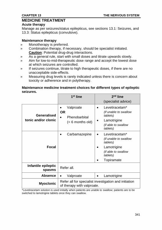
Maintenance medicine treatment choices for different types of epileptic seizures.
1st line 2nd line
(specialist advice)
Generalised
tonic and/or clonic
• Valproate
OR
• Phenobarbital
(< 6 months old)
• Levetiracetam*
(if unable to swallow tablets)
• Lamotrigine
(if able to swallow tablets)
Focal
• Carbamazepine • Levetiracetam*
(if unable to swallow tablets)
• Lamotrigine
(if able to swallow tablets)
• Topiramate
Infantile epileptic spasms
Refer all.
Absence • Valproate • Lamotrigine
Myoclonic Refer all for specialist investigation and initiation of therapy with valproate.
*Levetiracetam solution is used initially when patients are unable to swallow; patients are to be switched to lamotrigine tablets once they can swallow.

CHAPTER 13 THE NERVOUS SYSTEM
342
Caution The choice of AED for girls and women of childbearing potential must be carefully considered. Valproate should be avoided in adolescent women and preadolescent girls who are likely to remain on treatment into their childbearing years unless other treatment is ineffective or effective contraception is in place. This is due to the risk of adverse developmental outcomes to the foetus. If the decision is made to use Valproate in patients this population, complete the ‘Acknowledgement of Risk’ form: https://www.sahpra.org.za/wp-content/uploads/2020/08/6.28_Valproate_Annual_Risk_Acknowledgement_Form_Dec18_v1.pdf
• Valproate, oral, 5 mg/kg/dose (starting dose), 8–12 hourly. o Increase by 5 mg/kg weekly to 15–20 mg/kg/day given 8–12 hourly
over 4 weeks. o Maximum total daily dose: 40 mg/kg/day. o Exclude liver dysfunction prior to initiating therapy (at least ALT), in
children under 2 years or if clinical suspicion of liver dysfunction or metabolic disease.
o Monitor at least clinically for hepatotoxicity.
• Carbamazepine, oral, 5 mg/kg/day (starting dose), 8–12 hourly. o Increase slowly by 0.2 mg/kg at 2 weekly intervals to
5--10 mg/kg/dose 8–12 hourly. o Usual maintenance total daily dose: 10–20 mg/kg/day. o Maximum total daily dose: 20 mg/kg/day. o Dosage intervals: syrup 8 hourly, tablets 12 hourly. o Exacerbates myoclonic seizures and absence seizures.
• Lamotrigine, oral, 0.2 mg/kg/dose (starting daily dose) (specialist initiated). o Increase slowly at 2 weekly intervals to 1–5 mg/kg/dose 12–24 hourly. o Rapid escalation associated with adverse side-effect of skin rash. o Maximum total daily dose when given with valproate: 5 mg/kg/day. o Lamotrigine is given as add-on therapy for different seizure types and
in drug-resistant paediatric epileptic syndromes, such as Lennox-Gastaut syndrome.
o Double the maximum dose of lamotrigine when using carbamazepine or phenobarbital.
o Lamotrigine must be given at a reduced dosage of no more than half the recommended dose in patients using valproate.
• Phenobarbital, oral, 3–5 mg/kg/dose as single dose at night. o May be used in children under six months of age. o Is not recommended as maintenance therapy for children older than
2 years due to undesirable side-effects such as sedation, behaviour

CHAPTER 13 THE NERVOUS SYSTEM
343
disturbances, hyperkinesia and dependence, except in situations where there is poor adherence to other drugs.
o Exacerbates absence seizures.
• Topiramate, oral, 1–3 mg/kg/dose as a single dose at night. o Increase at 1–2 weekly intervals by 0.5–1.5 mg/kg twice daily. o Maximum dose:
▪ ≥ 2 years: 16 mg/kg/day ▪ ≥ 4 years: 30 mg/kg/day
• Levetiracetam, oral, o Infants 1 to < 6 months: Initial: 7 mg/kg/dose twice daily; increase
dosage every 2 weeks by 7 mg/kg/dose twice daily based on response and tolerability to the recommended dose of 21 mg/kg/dose twice daily.
o Infants ≥ 6 months and children < 4 years: Initial: 10 mg/kg/dose twice daily; increase dosage every 2 weeks by 10 mg/kg/dose twice daily based on response and tolerability to the recommended dose of 25 mg/kg/dose twice daily.
o Children > 4 years: initial: 10 mg/kg/dose twice daily; increase dosage every 2 weeks by 10 mg/kg/dose twice daily based on response and tolerability to the recommended dose of 30 mg/kg/dose twice daily.
LoE: III2
REFERRAL » Suspected but undiagnosed secondary cause for seizures. » Focal seizures for neuroimaging (MRI preferred), if facilities or expertise
not available. » All seizures other than simple febrile convulsions in children < 2 years. » Seizures that are not controlled within 2 months on one agent with
minimal side-effects. » Neuroregression. » Mixed seizure types in one patient. » All myoclonic seizures and epileptic spasms at presentation.
13.5 ANTIRETROVIRAL THERAPY (ART) AND ANTIEPILEPTIC DRUGS (AED) Co-administration of antiepileptic drugs in patients on antiretroviral therapy has not been well studied yet, and remains a therapeutic challenge. Drug interactions between AED and ART can arise from a number of mechanisms, including liver metabolism (increased or decreased) and competition for protein binding, resulting in increase in viral replication. There is no strong evidence to guide clinicians at present.

CHAPTER 13 THE NERVOUS SYSTEM
344
The following points are important to remember when treating seizures and epilepsy in patients on ART: » Great caution should be taken when using drugs metabolised in the liver
by the cytochrome P450 enzyme system as this may alter levels of both AED and ART, leading to toxic or sub-therapeutic drug levels. This particularly pertains to the NNRTIs and PIs.
» If clinically indicated, monitor AED levels in patients taking concurrent ART and AED therapy.
» Avoid prescribing carbamazepine, phenobarbital and phenytoin for patients receiving NNRTIs, PIs and InSTIs, as there are serious P450 interactions involved. In this setting, consider lamotrigine, valproate or levetiracetam. See section 13.4: Epilepsy.
» Treat children on ART presenting to casualty with acute seizures or in status epilepticus according to the existing standard status epilepticus or acute seizure protocols.
» Although benzodiazepines, phenytoin and phenobarbital may interact with antiretroviral metabolism, the acute management of acute seizures or SE takes precedence in these instances.
13.6 HEADACHES R51
DESCRIPTION Headache is the most common pain syndrome in children of all ages. Recurrent headaches are a common health problem and can be: » primary, e.g. migraine, or » secondary/symptomatic, e.g. raised intracranial pressure. The actual perception of headache varies according to age and is influenced by factors such as experience, memory and cultural environment. Extract from the International Classification of Headache Disorders (ICHD) Migraine (without aura) Five or more headaches lasting 1–48 hours (duration in children is often shorter, lasting a few hours only) fulfilling at least 2 of the following: » bilateral or unilateral, frontal or parietal in location, » pulsating in character, » moderate or severe, » aggravated by routine activity, » nausea and/or vomiting plus photophobia and/or phonophobia during
headache.
Migraine (with aura) At least 2 attacks fulfilling at least 3 of the following:

CHAPTER 13 THE NERVOUS SYSTEM
345
» one or more reversible aura symptoms, » at least one aura developing over > 4 minutes or 2 or more successive
symptoms, » no aura lasting > 1 hour, » headache follows aura in less than 1 hour.
Episodic tension-type headache At least 10 prior episodes, occurring less than 15 times per month and lasting 30 minutes to 7 days with at least 2 of the following: » pressing or tightening quality, » mild or moderate intensity, » bilateral location, » no aggravation by routine physical activity, » no nausea, vomiting, photophobia or phonophobia. Cluster headache » Severe unilateral sharp headache associated with conjunctival injection
and lacrimation. » Rare in childhood. Paroxysmal Hemicrania Continua » Cluster headache of shorter duration. Each of the above can occur in combination in any patient, i.e. mixed/comorbid headache. Headaches can also be sub-classified according to temporal patterns, i.e. acute, acute recurrent, chronic progressive/non-progressive, episodic or constant.
DIAGNOSTIC CRITERIA » Exclude secondary causes of headache, e.g. raised intracranial pressure. » Red flags in childhood headaches:
> change in pattern (e.g. ‘worst headache ever’), > progressive course over time, > age younger than 3 years, > nocturnal/wakes child from sleep, > early morning vomiting, > ataxia, > focal neurological signs, > alteration of level of consciousness.
GENERAL AND SUPPORTIVE MEASURES » Environmental and lifestyle changes, e.g. avoid precipitants such as bright
lights, sleep deprivation and certain foods, excessive video games. » Adequate hydration. » Avoid skipping meals, excessive caffeine ingestion. » Regular exercise.

CHAPTER 13 THE NERVOUS SYSTEM
346
» Stress alleviation and coping skills training where possible. » Headache diary and identify possible triggers.
MEDICINE TREATMENT Treat non-migraine headaches with analgesics. • Paracetamol, oral, 15 mg/kg/dose 6 hourly as required. For migraine:
• Ibuprofen, oral, 10 mg/kg/dose, 6 hourly. Persistent vomiting and not tolerating oral feeds:
• Metoclopramide, oral, 0.15–0.3 mg/kg as a single dose. OR
• Metoclopramide, IM/IV, 0.1 mg/kg as a single dose. OR
• Ondansetron, oral, 0.1–0.2 mg/kg 12 hourly. Note: Headaches can be an adverse effect associated with the of use ondansetron. Patients with ongoing symptoms should be investigated. Migraine prophylaxis Indicated when headaches occur frequently, impacting on the child’s activity and requiring substantial relief medication. Treat for six months then review.
• Propranolol, oral, 0.5–3 mg/kg/day in 2–3 divided doses. o Contraindicated in asthma and heart block. o Avoid in diabetes and depression.
In children who are unable to take propranolol, e.g. asthma:
• Topiramate, oral, 1–3 mg/kg/day in 1–2 doses (specialist initiated). o Starting dose: 0.5 mg/kg/day. o Titrate dose slowly every 1–2 weeks. o Reinforce behavioural management before considering topiramate.
REFERRAL » Secondary intracranial cause suspected. » Failure to respond to first line treatment.
13.7 NEUROCYSTICERCOSIS B69.0
DESCRIPTION Neurocysticercosis is caused by the cysticercal form, i.e. larval form, of the pork tapeworm, Taenia solium. The larvae may locate in the brain parenchyma, intraventricular and meningeal areas, spinal canal/cord and eye,

CHAPTER 13 THE NERVOUS SYSTEM
347
or a combination of these regions. Viable cysticerci incite little inflammatory response, but dead cysticerci elicit an increased inflammatory response. Cysticerci in the brain may remain dormant or may cause complications such as:
» headache, » focal neurological deficits, » behavioural disorders, » raised intracranial pressure, » visual disturbances, » hydrocephalus, » seizures, » meningitis, » meningo-encephalitis, » spinal cord compression.
DIAGNOSTIC CRITERIA Clinical » Location and stage of the life cycle of the parasite in the brain determines the
clinical features. » Suspect if child from an endemic area, i.e. pig farming area, presents with
neurological abnormalities such as: > seizures, > raised intracranial
pressure/hydrocephalus, > focal neurological deficits, > cranial nerve palsies.
> meningo-encephalitis, > meningitis, > behavioural disorders, > headache,
Investigations » Computed tomography (CT scan) and/or magnetic resonance imaging (MRI
scan) of brain showing cysts, granulomas, peri-lesional oedema or calcification of cysts.
» MRI scan may identify more lesions and viable cystic lesions than the CT scan.
» Soft tissue radiology of muscles of lower limbs may demonstrate calcified cysticerci, i.e. ‘rice grain’ calcifications in muscles.
» Follow-up CT scans and/or MRI scans may help to assess the response to therapy.
GENERAL AND SUPPORTIVE MEASURES Prevention: » Prolonged freezing or thorough cooking of pork to kill the parasite. » Thorough washing of fresh fruit and vegetables in T. solium endemic areas. » Attention to personal hygiene after use of toilet. » Proper sanitation facilities and safe water. » Avoid the use of human excreta as fertiliser. » Look for Taenia ova in the stools of the family members.
MEDICINE TREATMENT Calcified cysticerci and a single dying lesion visible on CT scan require no anti-helminthic treatment.

CHAPTER 13 THE NERVOUS SYSTEM
348
Patients with multiple cysts usually have a mixture of live and dying cysts and are assumed to have active disease and require treatment.
• Albendazole, oral, 7.5 mg/kg/dose 12 hourly for 7 days. o Maximum dose: 400 mg/dose.
Prevention of neurological manifestations In massive infestations, cysticidal therapy may trigger an inflammatory response. Delaying anti-helminthic therapy and adding corticosteroids may lessen the risk.
24 hours prior to albendazole therapy:
• Dexamethasone, IM, 0.15 mg/kg/dose 6 hourly. Then follow with oral therapy as soon as possible:
• Prednisone 1 mg/kg/day for the duration of albendazole therapy, and then taper and discontinue.
Seizure control See section 13.4: Epilepsy. Treat according to the type of seizure. AED treatment for 6–12 months after resolution of lesions on neuroimaging. Recurrent seizures require chronic treatment until seizure-free for 2 years. REFERRAL » Neurocysticercosis not responding to adequate therapy. » Neurocysticercosis with complications, such as hydrocephalus. » Intractable epilepsy.
13.8 NEUROMUSCULAR DISORDERS
13.8.1 INFLAMMATORY POLYNEUROPATHY (GUILLAIN-BARRÉ SYNDROME)* G61.0
* Notifiable condition
DESCRIPTION Guillain-Barré syndrome (GBS) is an acute autoimmune-mediated polyradiculoneuropathy which is precipitated by a preceding viral or other infection. It is the most common acquired polyneuropathy in children. Different forms or variants of Guillain-Barré syndrome are described depending on the clinical and/or neurophysiological characteristics. Acute Inflammatory Demyelinating Polyradiculoneuropathy (AIDP) » This is the most common form, accounting for 80–90% of cases. » Characterised mainly by:

CHAPTER 13 THE NERVOUS SYSTEM
349
> symmetrical, ascending motor weakness, > areflexia, i.e. absence of tendon reflexes, > distal sensory alteration, > pain/paraesthesia.
Acute Motor Axonal Neuropathy (AMAN) » A purely motor form of GBS. » It involves predominantly motor nerves and has an axonal pattern on
electrophysiology (nerve conduction studies). » Although there are similarities with AIDP, the clinical picture tends to be
more severe with more patients suffering from respiratory failure. Acute Motor-Sensory Axonal Neuropathy (AMSAN) » Another axonal form of GBS but with sensory involvement. » It is not frequently found in children. Miller-Fisher syndrome » Patients have external ophthalmoplegia, sensory ataxia, weakness with
areflexia. » Electrophysiological and CSF studies are similar to AIDP. Chronic Inflammatory Demyelinating Polyradiculoneuropathy (CIDP) » May be considered a chronic variant of AIDP. » Most often starts insidiously and progresses slowly, but can have onset
like GBS. » It is managed differently from GBS and should be referred.
DIAGNOSTIC CRITERIA Clinical » Preceding respiratory tract or gastrointestinal infection. » Symmetrical, flaccid muscle weakness with early areflexia. » The muscle weakness may ascend rapidly upwards to involve the trunk,
arms, face and cranial nerves. » Bulbar paralysis and respiratory failure may develop. » Autonomic dysfunction. » Relatively mild, or absence of, sensory signs. » Exclude the following:
> Acute Disseminated Encephalomyelitis (ADEM), > poliomyelitis, a rare cause of hypotonia with abrupt onset of weakness
(usually asymmetrical) in association with a febrile illness, > transverse myelitis:
▪ initial flaccid weakness and areflexia typically involving the lower limbs maximally,
▪ occasionally with pain at the onset, but rapidly progressing to spasticity and hyperreflexia,
▪ a sensory level on the trunk, ▪ bladder and rectal sphincter involvement.

CHAPTER 13 THE NERVOUS SYSTEM
350
> diphtheria, > botulism.
Investigations Follow the Acute Flaccid Paralysis (AFP) investigation protocol » Send two stool specimens taken 24–48 hours apart to the National
Institute of Virology via the local laboratory. » The stool sample needs to be sent within 14 days of onset of paralysis to
exclude poliovirus infection. CSF » CSF findings after 1 week show elevated protein and few or no cells, i.e.
albumino-cytological dissociation. » CSF glucose is normal.
GENERAL AND SUPPORTIVE MEASURES » Notify as AFP. » Admit to a high care or intensive care unit. » Monitor respiratory and autonomic functions closely:
> peak expiratory flow rate, > blood pressure, > respiratory rate, > heart rate, > forced vital capacity (FVC), > bulbar functions, > arterial blood gases.
» Ventilation is recommended when: > rapidly progressing ascending paralysis, including shoulder
weakness, head lag, weak cough and swallowing difficulties, > there is a progressive fall in the peak expiratory flow rate, > tachycardia and sweating occur, > inspiratory efforts are weak (typical signs of respiratory distress will
be absent), > inability to talk, > PaCO2 levels start rising.
Note: These changes precede hypoxaemia detected on blood gas analysis, and ventilation should begin before frank hypoxaemia occurs. Respiratory care must be meticulous.
» To determine fluid losses from autonomic instability, monitor urine output
and degree of sweating. » Provide adequate nutrition. » Provide bladder and bowel care as well as pressure-point care. » Routine physiotherapy for chest and limbs, keep ankles in neutral position
(90°) (may require foot/hand splints). » Protect eyes and keep moist. » Communicate with child as awareness is maintained. Staff should
remember that children may be very frightened but unable to express their emotions and needs.

CHAPTER 13 THE NERVOUS SYSTEM
351
MEDICINE TREATMENT • Immunoglobulin, IV, 1 g/kg/day, slowly over 12–16 hours on two
consecutive days or 0.4 g/kg as a single daily dose on 5 consecutive days early in the disease process. o Use under specialist supervision.
Substantial pain is present (in up to 90%) in the severely affected patients. Pain in this setting is often unrecognised and underestimated. Pain management is essential. See section 20.1: Management of pain. For neuropathic pain:
• Carbamazepine, oral, 5 mg/kg/dose 12 hourly.
REFERRAL » Chronic inflammatory demyelinating polyradiculoneuropathy. » Guillain-Barré syndrome with bulbar paralysis and/or early signs of
respiratory failure. » Patients who have lost or are losing ambulation for management in
consultation with a paediatric neurologist. » Patients with complex Guillain-Barré syndrome.
13.8.2 MYASTHENIA GRAVIS G70.0
DESCRIPTION An auto-immune disorder resulting in muscle fatigue. Mild cases involve the eyes alone, i.e. ptosis and ophthalmoplegia, and severe cases involve proximal muscle groups, respiratory and bulbar control.
DIAGNOSTIC CRITERIA Clinical » Muscle fatigability with exercise and demonstration of this in the clinic
setting: > Lid-lag test, i.e. failure to maintain upward gaze for 1 minute. > Arm-raising test, i.e. failure to maintain the arms at 90° from the trunk
for 1 minute. Note: Myasthenia gravis patients not uncommonly present in a myasthenic crisis, with bulbar and respiratory compromise. Sometimes this may be the first mode of clinical presentation.
MEDICINE TREATMENT • Pyridostigmine, oral, 1–5 mg/kg/day in 4–6 divided doses. (Specialist
initiated).

CHAPTER 13 THE NERVOUS SYSTEM
352
REFERRAL » All for confirmation of diagnosis and initiation of treatment (consideration
of steroids, immuno-modulation therapy). » Myasthenic crisis.
13.8.2.1 MYASTHENIC CRISIS (MC) G70.01
DESCRIPTION Acute onset of respiratory failure due to worsening myasthenia gravis, requiring ICU admission. Respiratory and bulbar insufficiency is common with an inability to breathe or swallow, or may have worsening of existing symptoms. MC is most frequently precipitated by systemic infections.
Myasthenic crisis may be the initial presentation.
DIAGNOSTIC CRITERIA Clinical » Worsening of existing weakness. » Respiratory compromise. » Inability to swallow.
GENERAL AND SUPPORTIVE MEASURES » Admit to ICU for close observation. » Provide ventilatory and feeding support as required.
MEDICINE TREATMENT For consideration of glucocorticoid therapy and intravenous immunoglobulin, in discussion with neurologist.
• Dexamethasone, IV or IM,
• Immunoglobulin, IV, 1 g/kg/day, slowly over 12–16 hours on two consecutive days or 0.4 g/kg as a single daily dose on 5 consecutive days early in the disease process.
REFERRAL » All cases for further management.
13.8.3 DUCHENNE MUSCULAR DYSTROPHY (DMD) G71.01
» An X-linked recessive disorder causing progressive muscle weakness,
typically in males.

CHAPTER 13 THE NERVOUS SYSTEM
353
» Carrier females may have some degree of weakness. » Due to a mutation in the gene coding for dystrophin. » There may be a family history of DMD.
DIAGNOSTIC CRITERIA Clinical » Delayed walking. » Toe walking. » Gowers sign. » Waddling gait. » Lumbar lordosis. » Calf pseudohypertrophy. » Short stature. » Progressive proximal muscle weakness, usually confined to a wheelchair
by age 13 years. » Cardiomyopathy » May have mild cognitive impairment. » Behavioural issues. Investigations » CK markedly elevated. » AST/ALT may be elevated.
GENERAL AND SUPPORTIVE MEASURES » Pain may be present and should be treated appropriately. » Physiotherapy » Occupational therapy. » Nutritional support. » Encourage gentle aerobic exercise. » Psychosocial support for patient and family.
MEDICINE TREATMENT Consider oral corticosteroids once plateau or decline in motor function, in consultation with neurologist.
• Prednisone, oral, 0.75 mg/kg daily. o Maximum dose 30–40 mg.
• Long term use of corticosteroids is associated with various complications. Monitor patients and manage as needed.
If pain is present, refer to Chapter 20: Pain Control, section 20.1.2: Management of pain.
REFERRAL All cases for specialist assessment.

CHAPTER 13 THE NERVOUS SYSTEM
354
13.9 ACUTE DISSEMINATED ENCEPHALOMYELITIS (ADEM) G04.0
DESCRIPTION » Most common demyelinating disorder in childhood. » Affects children with a peak incidence between 5 and 8 years. » May occur following a systemic viral illness or vaccination. » Demyelination of white matter in multiple areas of the brain and spinal
cord.
DIAGNOSTIC CRITERIA » ADEM is a diagnosis of exclusion. » Consider and exclude differential diagnoses: SLE, infectious or
autoimmune encephalitis, metabolic disorders, hypertensive encephalopathy.
Characterised by acute onset of encephalopathy with focal or multifocal neurological deficits. » Seizures » Cranial nerve palsies. » Meningism » Optic neuritis. » Gait disturbances. » Hemiparesis » Pyramidal signs. » Ataxia » Aphasia Symptoms usually peak between 2 and 5 days from onset, but may change or worsen for up to 3 months. CSF » Normal pressure. » May have mild pleocytosis. » Increased protein. » Normal glucose. Diagnostic criteria: 1. A first, polyfocal clinical CNS event with a presumed inflammatory
demyelinating cause. 2. Encephalopathy (alteration in consciousness or behavior unexplained by
fever, systemic illness or postictal syndrome). 3. Brain MRI abnormalities consistent with demyelination during the acute
(initial three-month) phase. 4. No new clinical or MRI findings three months or more after clinical onset.

CHAPTER 13 THE NERVOUS SYSTEM
355
GENERAL AND SUPPORTIVE MEASURES » Supportive care including ICU and mechanical ventilation may be
necessary.
MEDICINE TREATMENT Consider treatment with immune modulators and immunoglobulins in consultation with a neurologist.
REFERRAL » As MRI is required for diagnosis, all patients should be referred prior to
implementing treatment.
13.10 SYDENHAM CHOREA I02.9
DESCRIPTION A movement disorder with rapid involuntary jerks affecting any part of the body often incorporated into a voluntary movement in an attempt to mask it. It is an acute post-streptococcal infection movement disorder and constitutes one of the major criteria for the diagnosis of rheumatic fever. Patient has the appearance of being restless with constant movement which improves with sleep. The movements are classically random in place and random in time.
DIAGNOSTIC CRITERIA Clinical » Exclude drug reactions, hyperthyroidism, systemic lupus erythematosus
and neurodegenerative disorders. Investigations » Cardiac screening, i.e. ECG, echocardiogram. » Serum ASOT, anti-DNAse B. » Erythrocyte sedimentation rate. » Anti-dsDNA, if clinically indicated.
GENERAL AND SUPPORTIVE MEASURES » Emotional support. » School support. » Occupational therapy.
MEDICINE TREATMENT Movement disorders:
• Haloperidol, oral, 0.025 mg/kg/day in 2–3 divided doses. o Increase dose slowly and incrementally to 0.05 mg/kg/day.
PLUS

CHAPTER 13 THE NERVOUS SYSTEM
356
If streptococcal infection:
• Phenoxymethylpenicillin, oral, 500 mg 12 hourly for 10 days. OR
• Amoxicillin, oral, 45 mg/kg/dose 12 hourly for 10 days. THEN Until 21 years of age:
• Benzathine benzylpenicillin (depot formulation), IM, 1.2 million units every 28 days.
OR
• Phenoxymethylpenicillin, oral, 250 mg 12 hourly.
REFERRAL » All patients for specialist assessment.
13.11 CEREBROVASCULAR DISEASE/STROKE I67.9
DESCRIPTION Cerebrovascular disease can be ischaemic (thrombotic or embolic) or haemorrhagic, arterial or venous. Arterial ischaemic stroke must always be considered in any child with sudden onset of hemiparesis or other focal neurological disturbance. The clinical features of cerebral venous thrombosis (CVT) include headache, papilloedema, focal neurological signs, seizures (often focal), and alteration of consciousness. Risk factors: » cardiac disorders, » infections, e.g. meningitis, varicella, HIV, etc., » prothrombotic disorders, e.g. nephrotic syndrome, protein S/C
deficiencies, etc., » haematologic disorders, e.g. sickle cell anaemia, » vasculopathies, e.g. vasculitis, HIV, Moyamoya syndrome. The initial evaluation in children includes the following: CT/MRI brain to ascertain whether it is an ischaemic or haemorrhagic infarct. » Electrocardiography, echocardiography. » Full blood count, INR, PTT. » CSF analysis as indicated. » Infectious screening, including varicella, HIV, mycoplasma, TB. » Connective tissue and vasculitic screening. » Thrombophilia screening. See Chapter 3: Blood and Blood Forming
Organs, section 3.11: Venous thrombo-embolic disease.

CHAPTER 13 THE NERVOUS SYSTEM
357
GENERAL AND SUPPORTIVE MEASURES Acute supportive and neuroprotective care directed at preserving damaged but salvageable brain tissue includes the following: » Maintain body temperature in the low to normal range. » Maintain euglycaemia. » Maintain O2 saturation above 95%. » Maintain adequate cerebral perfusion and manage raised intracranial
pressure. » Treat anaemia. » Treat acute seizures promptly.
Haemorrhagic stroke requires referral to a centre with neurosurgical expertise and facilities. Early disability assessment and management, includes physiotherapy, speech therapy, occupational therapy, etc.
MEDICINE TREATMENT Arterial ischaemic stroke without haemorrhage All patients with confirmed arterial ischaemic stroke:
• Aspirin soluble, oral, 1–5 mg/kg as a daily dose. o Contraindicated in haemorrhagic stroke or bleeding tendency.
REFERRAL » All patients to specialist paediatrician for investigation. » Anticoagulation with enoxaparin and warfarin is best done in a specialised
setting under cardiologist, haematologist and neurologist supervision.
13.12 LUMBAR PUNCTURE CONTRAINDICATIONS TO LUMBAR PUNCTURE » Focal neurological signs and depressed level of consciousness. » Clinical signs of raised intracranial pressure, or impending cerebral
herniation: > deep coma, i.e. GCS < 9, or sudden deterioration of level of
consciousness, > decerebrate or decorticate posturing, > neurogenic hyperventilation, > unequal dilated or poorly reactive pupils, > absent doll’s eye reflex, > papilloedema.
» Haemodynamic/respiratory unstable patients. » Clinical meningococcaemia (septicaemia) with petechiae/purpura.
(confirm with skin scrape, Gram stain and blood culture). » Skin sepsis or abnormalities over the lumbar puncture site. » Coagulopathy

CHAPTER 13 THE NERVOUS SYSTEM
358
» Spinal anatomic abnormality. » Acute paraplegia. » Status epilepticus. PROCEDURE » Positioning and restraint are vital in determining the success of the
procedure. » The ability of the assistant in restraining is as important as the skill of the
‘operator’. » Preparation entails not only positioning, but attention to
sedation/analgesia, ‘patient comfort’ and safety, as well as factors such as adequate lighting.
» Resuscitation equipment must be available at the bed side. » Pay attention to the sterility of the operating field. » Local analgesia with/without sedation may be required. See Chapter 20:
Pain Control, section 20.1.2: Management of pain. » Ensure that all necessary equipment, e.g. needles, manometers and
specimen tubes are close at hand. » Only the interspaces below L3 (L3/L4 or L4/L5) are used in order to avoid
damaging the conus medullaris. » With the patient in the lateral recumbent position, the L3/L4 interspace is
found at the level of the line joining the highest points of the two iliac crests.
» Turn the bevel of the needle (with stylet) to face the patient’s side to avoid cutting the longitudinal dural fibres.
» As the needle is advanced, the first ‘give’ or loss of resistance is encountered with the piercing of the ligamentum flavum. A slight ‘popping’ sensation is felt as the needle penetrates the dura. Remove the stylet to allow CSF to drain out passively. If no fluid appears, then rotate the needle a quarter turn (90°). If this does not help, replace the stylet and advance the needle a few millimetres and then check for fluid as before.
» Measure the opening pressure using a manometer, with the child relaxed in the lateral decubitus position. In a young relaxed child, the opening pressure is in the range of 6-18 cm H2O.
» At the end of the procedure, re-insert the stylet before removing the needle completely.
Note: If intracranial infection is suspected, do a blood culture and initiate antimicrobial treatment immediately. See Chapter 8: Infective/Infectious Diseases, section 8.11: Meningitis, Acute Bacterial. Remember to catch a few drops of CSF on a labstick to check the glucose and for the presence of white cells which may give an indication of an infection.

CHAPTER 13 THE NERVOUS SYSTEM
359
13.13 RAISED INTRACRANIAL PRESSURE
DESCRIPTION Raised intracranial pressure (ICP) is an emergency requiring prompt recognition and treatment. The cranial vault contains the brain, blood and CSF. It has a fixed volume, therefore, an increase in one component requires a compensatory decrease in others to maintain the pressure within the compartment. There is a limited capacity for compensation, i.e. by decreasing CSF volume, decreasing cerebral blood volume or by increasing the cranial volume. Thereafter, there is a rise in pressure. Herniation syndromes are an important complication to consider. They may arise in the course of untreated underlying disease or as a result of injudicious lumbar puncture. Normal CSF pressures range from:
• Infants: 20 – 27 cmH2O (1.5–6 mmHg)
• Children: 4 – 10 cmH2O (3–7 mmHg) These values are for well children and may vary in those who are critically ill on ventilation. Any CSF pressure above 27 cm of CSF (20 mmHg) for more than 20 minutes should be treated.
Increased brain volume Intracranial space occupying lesion: Brain tumour, brain abscess, haematoma, vascular malformation, arachnoid/ epidermoid cyst.
Cerebral oedema: encephalitis, meningitis, hypoxic ischaemic encephalopathy, traumatic brain injury, hepatic encephalopathy, Reye’s syndrome, stroke, diabetic ketoacidosis, hyponatraemia.
Increased blood volume Vascular malformations, cerebral venous thrombosis, meningitis, encephalitis.
Increased CSF volume Obstructive/communicating hydrocephalus, choroid plexus papilloma.
Disordered CSF dynamics Idiopathic intracranial hypertension.
Source3 Vitamin A administration may cause benign raised ICP.
DIAGNOSTIC CRITERIA Presentation may have an acute or insidious onset depending on the underlying pathology. A detailed history and examination are essential.

CHAPTER 13 THE NERVOUS SYSTEM
360
Clinical Features vary with age. Infants:
» Increasing head circumference. » Sun setting eyes. » Distended scalp veins. » Irritability
» Lethargy » Vomiting » Developmental delay
or regression. » Persistent head lag.
Older children:
» Headache » Vomiting » Depressed level of
consciousness. » Seizures
» Ataxia » Abnormal eye movements. » Double vision. » Behavioural changes. » Meningism
Late signs:
» Papilloedema » Sixth nerve palsy. » Pupillary dilation. » Decerebrate or decorticate
posturing. » Cheyne-Stokes respiration.
» Focal neurological deficits. » Cushing Triad:
> Increased systolic pressure (with widening pulse pressure).
> Irregular breathing. > Bradycardia
Herniation syndromes
Unilateral transtentorial herniation
Declining consciousness. Increased blood pressure. Slow pulse. Homonymous hemianopia. Respiratory irregularity.
Bilateral transtentorial herniation
Decerebrate or decorticate rigidity. Declining consciousness. Impaired upward gaze. Irregular respiration. Pupillary constriction or dilatation.
Cerebellar herniation Declining consciousness. Impaired upward gaze. Irregular respiration. Lower cranial nerve palsies. Neck stiffness or head tilt.
Source3
Investigations » CT brain to determine underlying cause and whether it is safe to perform
a lumbar puncture. » CSF opening pressure (if no contraindication).
CHAPTER 13 THE NERVOUS SYSTEM
361
GENERAL AND SUPPORTIVE MEASURES » Follow the ABCD algorithm.
» Position head in midline and elevate to 30. » Cautious ventilation maintaining PaCO2 between 4.5 and 5 kPa. » Monitoring and maintenance of blood pressure » Elevated BP is usually reactive and required to maintain cerebral
perfusion. » Monitor fluid balance, use 0.9% NaCl/5% dextrose water as maintenance
fluid. » Keep serum sodium in the upper range of normal, up to 150 mmol/L. » Maintain normothermia. » Maintain glucose within 6 to 10 mmol/L.
MEDICINE TREATMENT Initiate treatment for underlying cause. If meningitis cannot be excluded, commence treatment as soon as possible (see Chapter 8: Infective/Infectious Diseases, section 8.11: Meningitis, acute bacterial). Sodium chloride, 5%, IV, 2 mL/kg infused over 30 minutes.
• Monitoring of serum sodium is essential with repeat doses or infusion.
OR
Mannitol, IV, 250 mg/kg administered over 30–60 minutes.
o Do not exceed two doses without consulting with a specialist. If a space occupying lesion is diagnosed: Add
• Dexamethasone, IV, 0.5 mg/kg 12 hourly.
• Maximum dose 12 mg per dose. Sedation and analgesia, see Pain Control Chapter and Intensive Care Chapter. Management of seizures if present, refer to section 13.1: Seizures.
REFERRAL » According to underlying condition. » Neurosurgical intervention may be required.

CHAPTER 13 THE NERVOUS SYSTEM
362
13.14 CEREBRAL PALSY (CP) ICD 10 G80.9
DESCRIPTION Cerebral palsy (CP) describes a group of disorders of movement and posture. It is the commonest cause of developmental disturbances in children. CP results from an insult to the developing foetal or infant brain, which is non-progressive. There may be associated abnormalities of sensation, perception, cognition, communication and behaviour.
DIAGNOSTIC CRITERIA » Motor deficit with delay in motor milestones.
> Not sitting by 8 months (corrected for gestational age). > Not walking by 18 months (corrected for gestational age). > Hand preference before 12 months (corrected for gestational age).
» No loss of function (i.e. milestone regression). » Serial examinations may be needed to establish the diagnosis. Clinical picture may be described by: » The predominant abnormality of tone and the distribution of areas
involved.
Regional involvement Spastic Hemiplegia
Spastic Diplegia
Global involvement
Spastic Quadriplegia
Dyskinetic Athetoid
Dyskinetic Dystonic
Ataxic Ataxia
Risk factors:
» Preterm labour and birth. » Small for gestational age. » Multiple pregnancy.
» Neonatal encephalopathy. » Neonatal sepsis. » Meningitis or septicaemia.
Developmental follow up from birth to 2 years is essential in high risk patients.
GENERAL AND SUPPORTIVE MEASURES » Multidisciplinary team approach. » Screen for ophthalmologic and hearing impairments. » Screen for speech and language disorders. » Monitor growth, nutrition and swallowing function. » Physiotherapy » Occupational therapy. » Psychology » Assess level of functioning according to the Gross Motor Function
Classification System (GMFCS).

CHAPTER 13 THE NERVOUS SYSTEM
363
MEDICINE TREATMENT Medication for relief of spasticity, dystonia and dyskinesia may be used in some patients for improvement in function, range of motion at various joints, disturbances from uncontrolled movements. Co-morbidities and complications:
Neurological Epilepsy, hydrocephalus, visual and hearing impairment.
See section 13.4: Epilepsy.
Behavioural and learning
Sleep disturbance, depression, autistic features, learning difficulties, vulnerability.
See Chapter 14: Child and Adolescent Psychiatry, sections 14.4.1: Depression, 14.12: Autism spectrum disorders.
Gastro-intestinal Difficulty swallowing, gastro-oesophageal reflux, constipation.
See Chapter 2: Alimentary Tract, sections 2.2.2: Constipation and 2.2.8: Gastro-oesophageal reflux disease (GORD).
Bone Osteoporosis, scoliosis, hip dislocation, pathological fractures.
Respiratory Susceptibility to chest infections and aspiration.
See Chapter 15: Respiratory System.
Skin Drooling, pressure sores.
Dental Poor oral hygiene, susceptibility to dental caries.
See Primary Health Care STGs and EML, Chapter 1 Dental and oral conditions, section 1.1: Abscess and caries.
Genito-urinary Neurogenic bladder See Chapter 6: Nephrological/Urological disorders, Dysfunctional bladder.
Table adapted from: Smith, M and Kurian, M. Medical management of cerebral palsy. Paediatrics and Child Health, 2016-09-01, Volume 26, Issue 9, Pages 378–382.
These patients frequently experience pain as a result of these complications.
Remember to suspect and manage pain in children with CP.

CHAPTER 13 THE NERVOUS SYSTEM
364
REFERRAL For consideration of medical treatment of spasticity, dystonia or dyskinesia in consultation with the multidisciplinary team. References
1Scheffer IE, Berkovic S, Capovilla G, Connolly MB, French J, Guilhoto L. ILAE classification of
epilepsies: Position paper of the ILAE Commission for Classification and Terminology. Epilepsia. 2017, 58 (4): 512-521. 2Topiramate dose: The British National Formulary for Children, BMJ Group. 2016-2017.
3Rajesh A, Kingston-Hepner M and Krishnakumar. Raised intracranial pressure. Paediatrics and
Child Health 27;6:260–267.