Chapter 12 Nervous System. Brain Waves Normal brain function involves continuous electrical activity...
58
Chapter 12 Nervous System
-
Upload
nickolas-cameron -
Category
Documents
-
view
221 -
download
0
Transcript of Chapter 12 Nervous System. Brain Waves Normal brain function involves continuous electrical activity...

Chapter 12
Nervous System
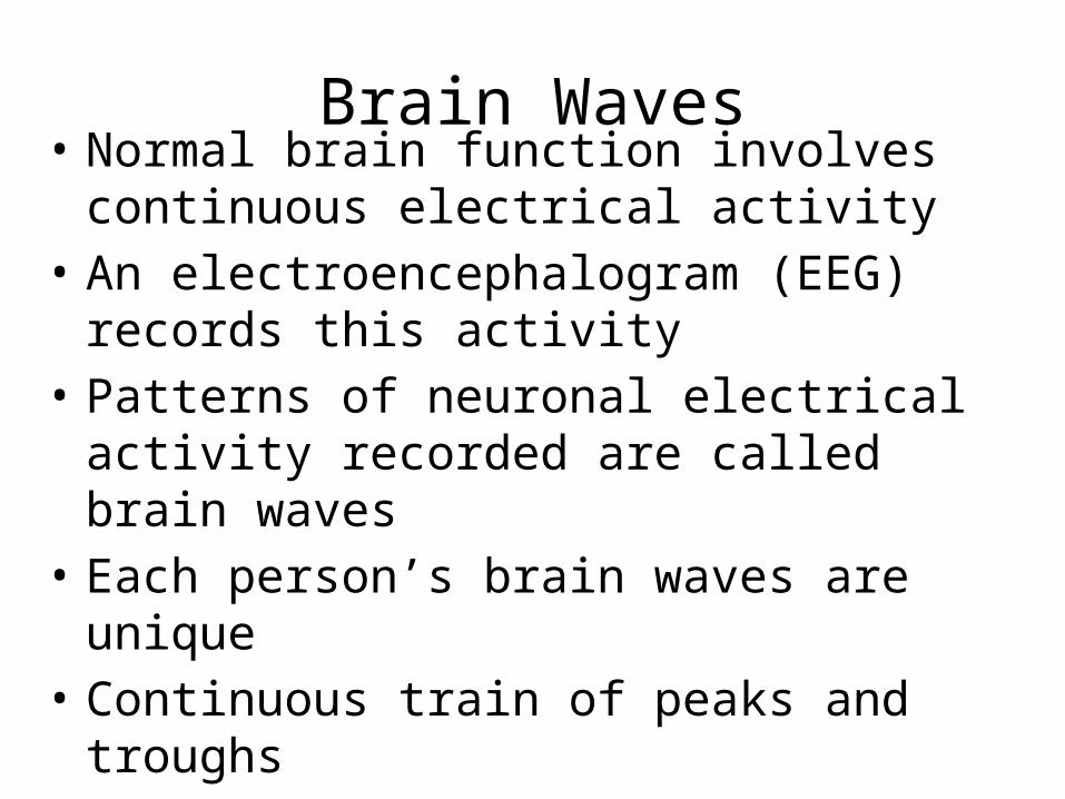
Brain Waves• Normal brain function involves continuous
electrical activity• An electroencephalogram (EEG) records this
activity• Patterns of neuronal electrical activity
recorded are called brain waves• Each person’s brain waves are unique• Continuous train of peaks and troughs• Wave frequency is expressed in Hertz (Hz)
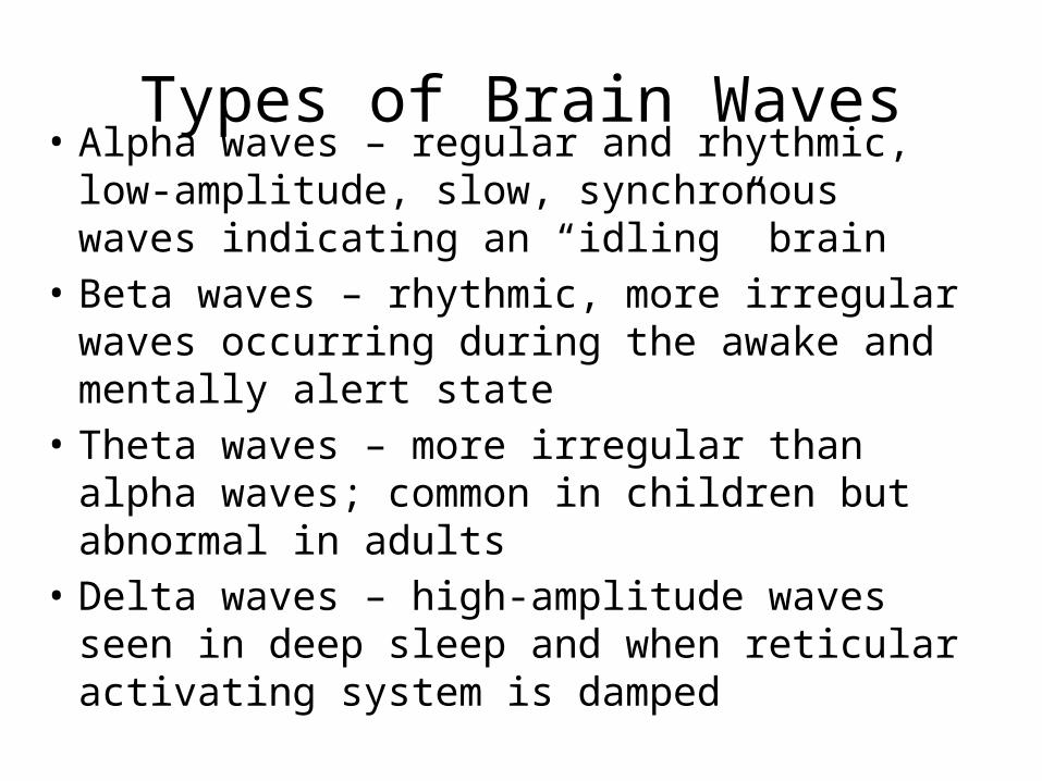
Types of Brain Waves• Alpha waves – regular and rhythmic, low-
amplitude, slow, synchronous waves indicating an “idling” brain
• Beta waves – rhythmic, more irregular waves occurring during the awake and mentally alert state
• Theta waves – more irregular than alpha waves; common in children but abnormal in adults
• Delta waves – high-amplitude waves seen in deep sleep and when reticular activating system is damped

Types of Brain Waves
Figure 12.20b
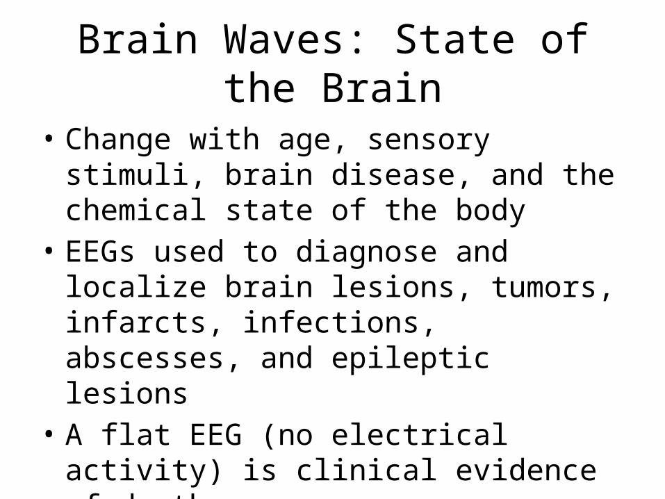
Brain Waves: State of the Brain
• Change with age, sensory stimuli, brain disease, and the chemical state of the body
• EEGs used to diagnose and localize brain lesions, tumors, infarcts, infections, abscesses, and epileptic lesions
• A flat EEG (no electrical activity) is clinical evidence of death
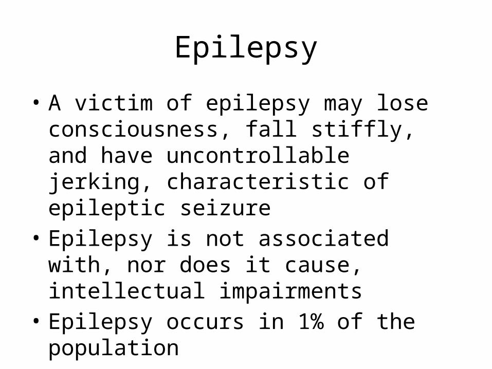
Epilepsy
• A victim of epilepsy may lose consciousness, fall stiffly, and have uncontrollable jerking, characteristic of epileptic seizure
• Epilepsy is not associated with, nor does it cause, intellectual impairments
• Epilepsy occurs in 1% of the population
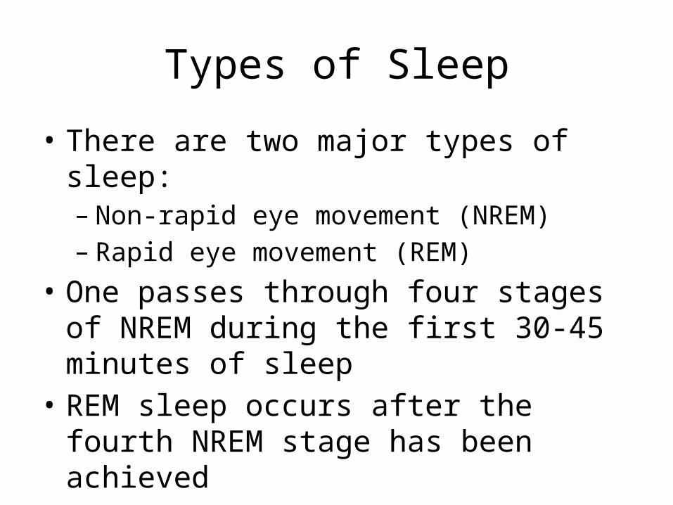
Types of Sleep
• There are two major types of sleep:– Non-rapid eye movement (NREM)– Rapid eye movement (REM)
• One passes through four stages of NREM during the first 30-45 minutes of sleep
• REM sleep occurs after the fourth NREM stage has been achieved

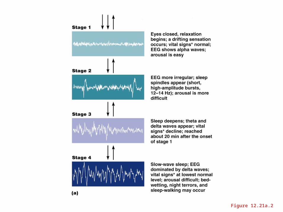
Types and Stages of Sleep: NREM
• NREM stages include:– Stage 1 – eyes are closed and relaxation begins; the
EEG shows alpha waves; one can be easily aroused– Stage 2 – EEG pattern is irregular with sleep spindles
(high-voltage wave bursts); arousal is more difficult– Stage 3 – sleep deepens; theta and delta waves
appear; vital signs decline; dreaming is common– Stage 4 – EEG pattern is dominated by delta waves;
skeletal muscles are relaxed; arousal is difficult
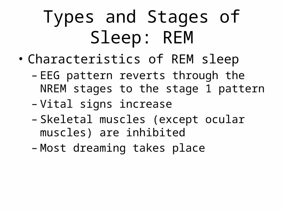
Types and Stages of Sleep: REM
• Characteristics of REM sleep – EEG pattern reverts through the NREM stages to
the stage 1 pattern– Vital signs increase– Skeletal muscles (except ocular muscles) are
inhibited– Most dreaming takes place

Sleep
Figure 12.21a.1
Sleep
Figure 12.21a.2
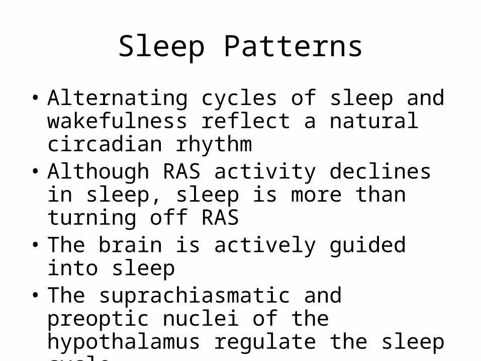
Sleep Patterns
• Alternating cycles of sleep and wakefulness reflect a natural circadian rhythm
• Although RAS activity declines in sleep, sleep is more than turning off RAS
• The brain is actively guided into sleep• The suprachiasmatic and preoptic nuclei of the
hypothalamus regulate the sleep cycle• A typical sleep pattern alternates between
REM and NREM sleep
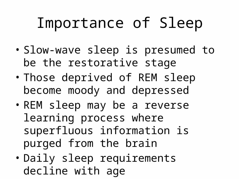
Importance of Sleep
• Slow-wave sleep is presumed to be the restorative stage
• Those deprived of REM sleep become moody and depressed
• REM sleep may be a reverse learning process where superfluous information is purged from the brain
• Daily sleep requirements decline with age
Protection of the Brain
• The brain is protected by bone, meninges, and cerebrospinal fluid
• Harmful substances are shielded from the brain by the blood-brain barrier

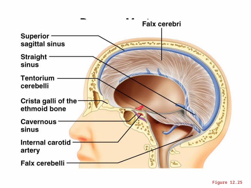
Meninges
• Three connective tissue membranes lie external to the CNS – dura mater, arachnoid mater, and pia mater
• Functions of the meninges– Cover and protect the CNS– Protect blood vessels and enclose venous sinuses– Contain cerebrospinal fluid (CSF)– Form partitions within the skull

Meninges
Figure 12.24a
Dura Mater
• Leathery, strong meninx composed of two fibrous connective tissue layers
• The two layers separate in certain areas and form dural sinuses

Dura Mater
• Three dural septa extend inward and limit excessive movement of the brain– Falx cerebri – fold that dips into the longitudinal
fissure– Falx cerebelli – runs along the vermis of the
cerebellum– Tentorium cerebelli – horizontal dural fold extends
into the transverse fissure
Dura Mater
Figure 12.25
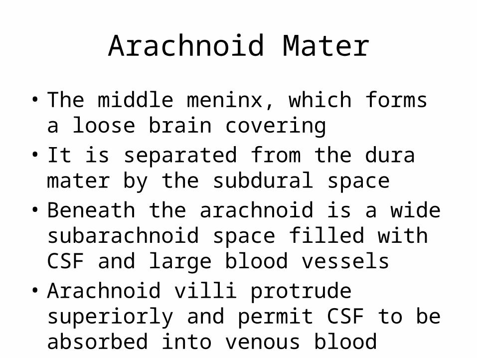
Arachnoid Mater
• The middle meninx, which forms a loose brain covering
• It is separated from the dura mater by the subdural space
• Beneath the arachnoid is a wide subarachnoid space filled with CSF and large blood vessels
• Arachnoid villi protrude superiorly and permit CSF to be absorbed into venous blood

Arachnoid Mater
Figure 12.24a

Pia Mater
• Deep meninx composed of delicate connective tissue that clings tightly to the brain
Cerebrospinal Fluid (CSF)• Watery solution similar in composition to
blood plasma• Contains less protein and different ion
concentrations than plasma• Forms a liquid cushion that gives buoyancy to
the CNS organs
Cerebrospinal Fluid (CSF)
• Prevents the brain from crushing under its own weight
• Protects the CNS from blows and other trauma
• Nourishes the brain and carries chemical signals throughout it

Circulation of CSF
Figure 12.26b
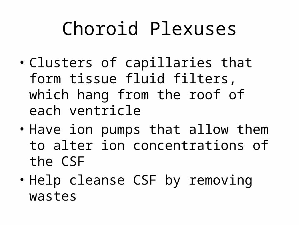
Choroid Plexuses
• Clusters of capillaries that form tissue fluid filters, which hang from the roof of each ventricle
• Have ion pumps that allow them to alter ion concentrations of the CSF
• Help cleanse CSF by removing wastes

Choroid Plexuses
Figure 12.26a

Blood-Brain Barrier
• Protective mechanism that helps maintain a stable environment for the brain
• Bloodborne substances are separated from neurons by:– Continuous endothelium of capillary walls– Relatively thick basal lamina– Bulbous feet of astrocytes
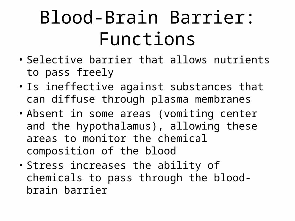
Blood-Brain Barrier: Functions
• Selective barrier that allows nutrients to pass freely
• Is ineffective against substances that can diffuse through plasma membranes
• Absent in some areas (vomiting center and the hypothalamus), allowing these areas to monitor the chemical composition of the blood
• Stress increases the ability of chemicals to pass through the blood-brain barrier
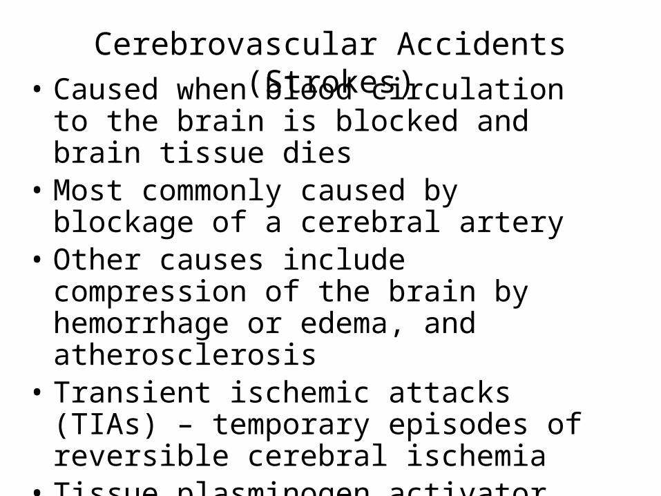
Cerebrovascular Accidents (Strokes)• Caused when blood circulation to the brain is
blocked and brain tissue dies• Most commonly caused by blockage of a
cerebral artery• Other causes include compression of the brain
by hemorrhage or edema, and atherosclerosis• Transient ischemic attacks (TIAs) – temporary
episodes of reversible cerebral ischemia• Tissue plasminogen activator (TPA) is the only
approved treatment for stroke
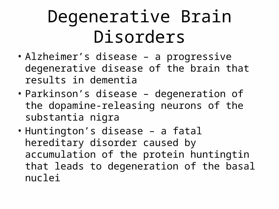
Degenerative Brain Disorders
• Alzheimer’s disease – a progressive degenerative disease of the brain that results in dementia
• Parkinson’s disease – degeneration of the dopamine-releasing neurons of the substantia nigra
• Huntington’s disease – a fatal hereditary disorder caused by accumulation of the protein huntingtin that leads to degeneration of the basal nuclei
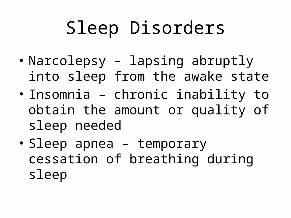
Sleep Disorders
• Narcolepsy – lapsing abruptly into sleep from the awake state
• Insomnia – chronic inability to obtain the amount or quality of sleep needed
• Sleep apnea – temporary cessation of breathing during sleep
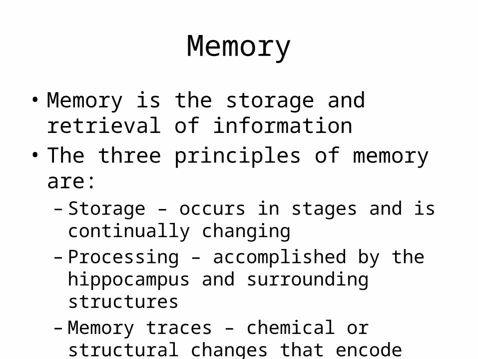
Memory
• Memory is the storage and retrieval of information
• The three principles of memory are:– Storage – occurs in stages and is continually
changing– Processing – accomplished by the hippocampus
and surrounding structures – Memory traces – chemical or structural changes
that encode memory
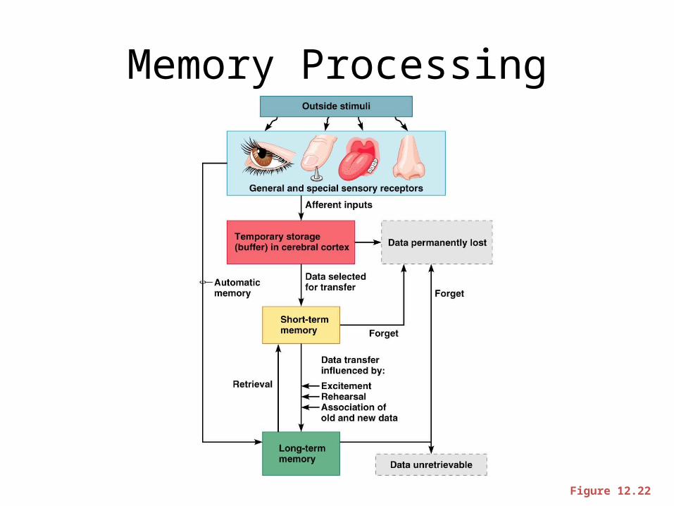
Memory Processing
Figure 12.22
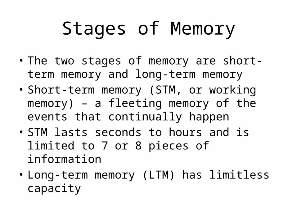
Stages of Memory
• The two stages of memory are short-term memory and long-term memory
• Short-term memory (STM, or working memory) – a fleeting memory of the events that continually happen
• STM lasts seconds to hours and is limited to 7 or 8 pieces of information
• Long-term memory (LTM) has limitless capacity

Epileptic Seizures
• Absence seizures, or petit mal – mild seizures seen in young children where the expression goes blank
• Grand mal seizures – victim loses consciousness, bones are often broken due to intense convulsions, loss of bowel and bladder control, and severe biting of the tongue

Control of Epilepsy
• Epilepsy can usually be controlled with anticonvulsive drugs
• Valproic acid, a nonsedating drug, enhances GABA and is a drug of choice
• Vagus nerve stimulators can be implanted under the skin of the chest and can keep electrical activity of the brain from becoming chaotic
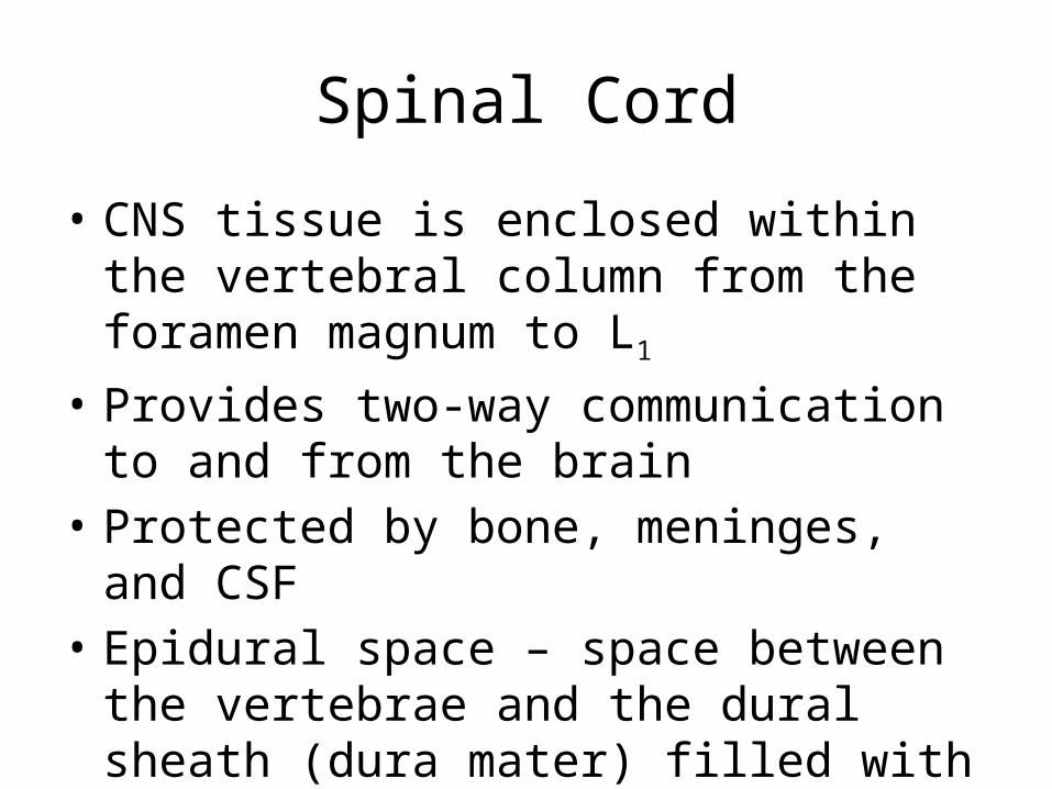
Spinal Cord
• CNS tissue is enclosed within the vertebral column from the foramen magnum to L1
• Provides two-way communication to and from the brain
• Protected by bone, meninges, and CSF• Epidural space – space between the vertebrae
and the dural sheath (dura mater) filled with fat and a network of veins
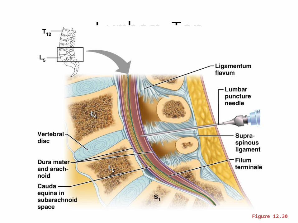
Lumbar Tap
Figure 12.30

Spinal Cord
Figure 12.29a

Spinal Cord
• Conus medullaris – terminal portion of the spinal cord
• Filum terminale – fibrous extension of the pia mater; anchors the spinal cord to the coccyx
• Denticulate ligaments – delicate shelves of pia mater; attach the spinal cord to the vertebrae
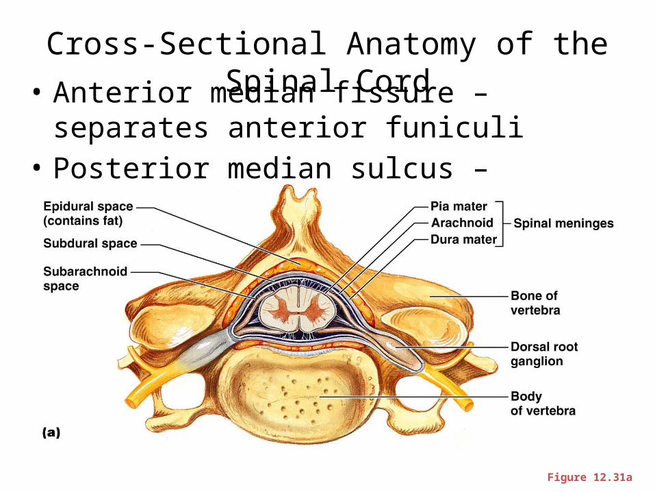
Cross-Sectional Anatomy of the Spinal Cord• Anterior median fissure – separates anterior
funiculi• Posterior median sulcus – divides posterior
funiculi
Figure 12.31a
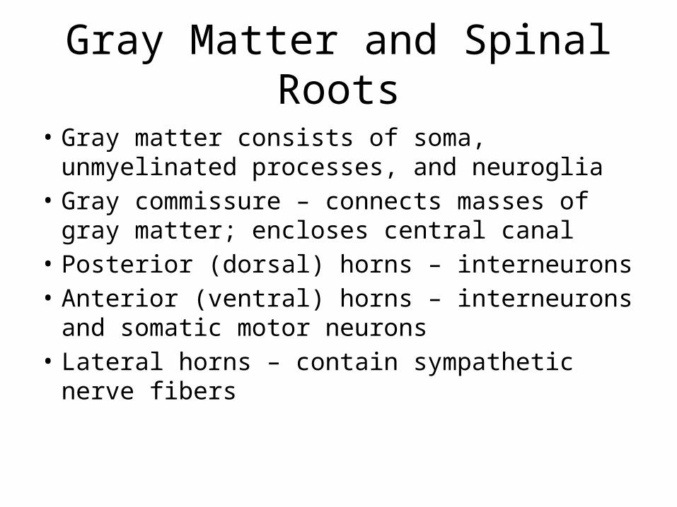
Gray Matter and Spinal Roots
• Gray matter consists of soma, unmyelinated processes, and neuroglia
• Gray commissure – connects masses of gray matter; encloses central canal
• Posterior (dorsal) horns – interneurons• Anterior (ventral) horns – interneurons and
somatic motor neurons• Lateral horns – contain sympathetic nerve fibers

Gray Matter and Spinal Roots
Figure 12.31b

Gray Matter: Organization
• Dorsal half – sensory roots and ganglia• Ventral half – motor roots• Dorsal and ventral roots fuse laterally to form
spinal nerves • Four zones are evident within the gray matter
– somatic sensory (SS), visceral sensory (VS), visceral motor (VM), and somatic motor (SM)
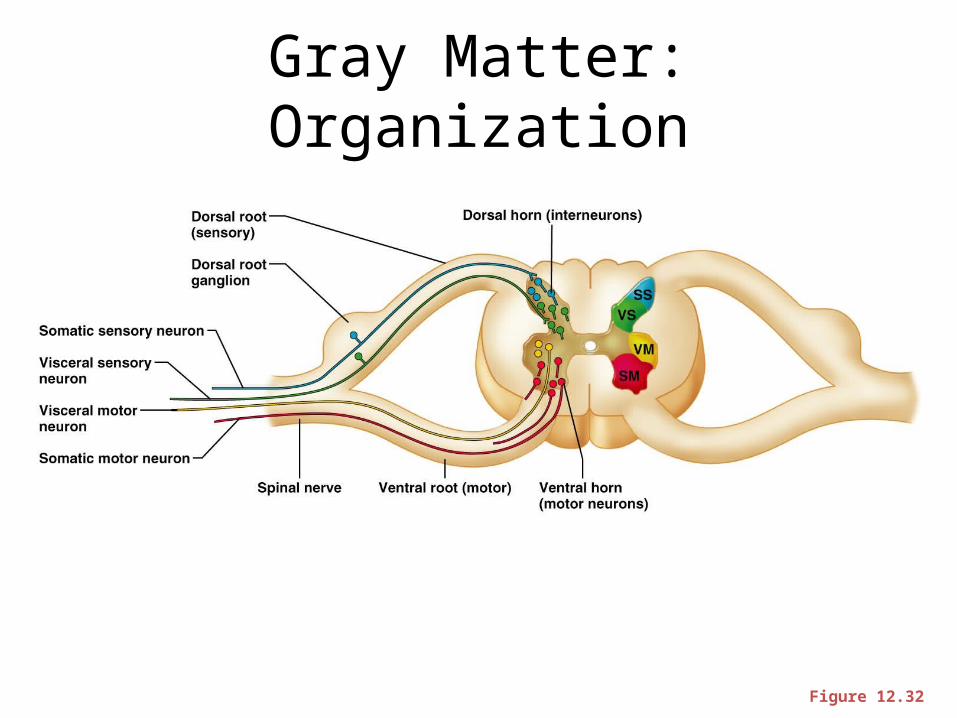
Gray Matter: Organization
Figure 12.32
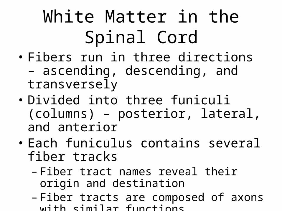
White Matter in the Spinal Cord
• Fibers run in three directions – ascending, descending, and transversely
• Divided into three funiculi (columns) – posterior, lateral, and anterior
• Each funiculus contains several fiber tracks– Fiber tract names reveal their origin and
destination– Fiber tracts are composed of axons with similar
functions
White Matter: Pathway Generalizations
• Pathways decussate• Most consist of two or three neurons• Most exhibit somatotopy (precise spatial
relationships)• Pathways are paired (one on each side of the
spinal cord or brain)
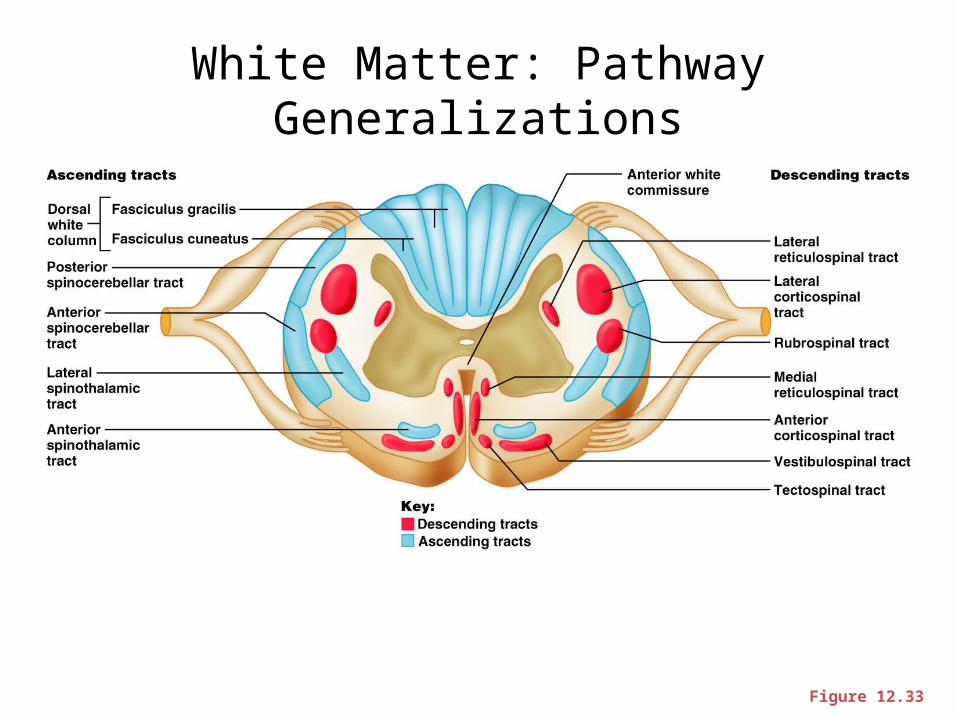
White Matter: Pathway Generalizations
Figure 12.33
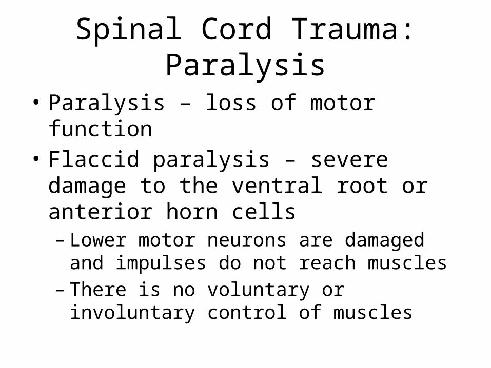
Spinal Cord Trauma: Paralysis
• Paralysis – loss of motor function• Flaccid paralysis – severe damage to the
ventral root or anterior horn cells– Lower motor neurons are damaged and impulses
do not reach muscles– There is no voluntary or involuntary control of
muscles

Spinal Cord Trauma: Paralysis
• Spastic paralysis – only upper motor neurons of the primary motor cortex are damaged– Spinal neurons remain intact and muscles are
stimulated irregularly– There is no voluntary control of muscles
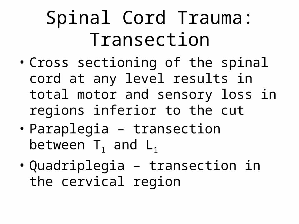
Spinal Cord Trauma: Transection
• Cross sectioning of the spinal cord at any level results in total motor and sensory loss in regions inferior to the cut
• Paraplegia – transection between T1 and L1
• Quadriplegia – transection in the cervical region

Poliomyelitis
• Destruction of the anterior horn motor neurons by the poliovirus
• Early symptoms – fever, headache, muscle pain and weakness, and loss of somatic reflexes
• Vaccines are available and can prevent infection
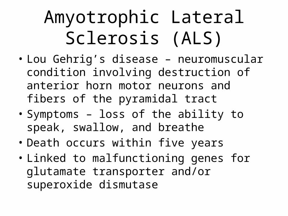
Amyotrophic Lateral Sclerosis (ALS)
• Lou Gehrig’s disease – neuromuscular condition involving destruction of anterior horn motor neurons and fibers of the pyramidal tract
• Symptoms – loss of the ability to speak, swallow, and breathe
• Death occurs within five years• Linked to malfunctioning genes for glutamate
transporter and/or superoxide dismutase

Cranial Nerves
• 12 pairs of nerves that mostly serve the head and neck
• Numbered in order, front to back• Most are mixed nerves, but three are sensory
only
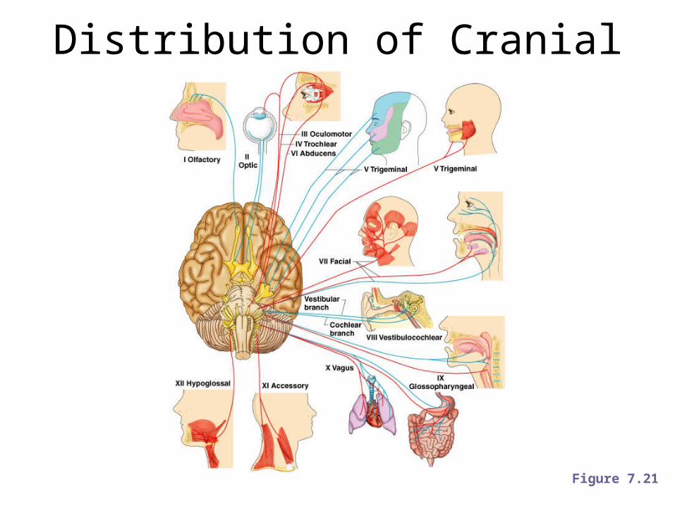
Distribution of Cranial Nerves
Figure 7.21

Cranial Nerves
• I Olfactory nerve – sensory for smell• II Optic nerve – sensory for vision• III Oculomotor nerve – motor fibers to eye
muscles• IV Trochlear – motor fiber to eye muscles
Cranial Nerves
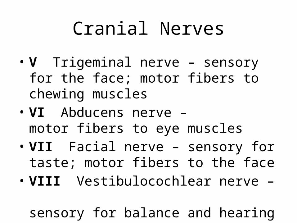
• V Trigeminal nerve – sensory for the face; motor fibers to chewing muscles
• VI Abducens nerve – motor fibers to eye muscles
• VII Facial nerve – sensory for taste; motor fibers to the face
• VIII Vestibulocochlear nerve – sensory for balance and hearing