Chapter 12. NERVE ENDINGS FREE NERVE ENDING SPECIAL RECEPTOR CELL distance.stcc.edu...
128
Chapter 12 Chapter 12
-
Upload
clarence-rodney-butler -
Category
Documents
-
view
227 -
download
4
Transcript of Chapter 12. NERVE ENDINGS FREE NERVE ENDING SPECIAL RECEPTOR CELL distance.stcc.edu...

Chapter 12Chapter 12

NERVE ENDINGSNERVE ENDINGS
FREE NERVEENDING
SPECIAL RECEPTOR CELL
distance.stcc.edu
starsandseas.com

RECEPTOR TYPESRECEPTOR TYPES
CHEMORECEPTORSCHEMORECEPTORS
NOCICEPTORSNOCICEPTORS
THERMORECEPTORSTHERMORECEPTORS
MECHNORECEPTORSMECHNORECEPTORS
PHOTORECEPTORSPHOTORECEPTORS

CHEMORECEPTORSCHEMORECEPTORS
CHEMICAL CONCENTRATIONSCHEMICAL CONCENTRATIONS SMELL, TASTESMELL, TASTE
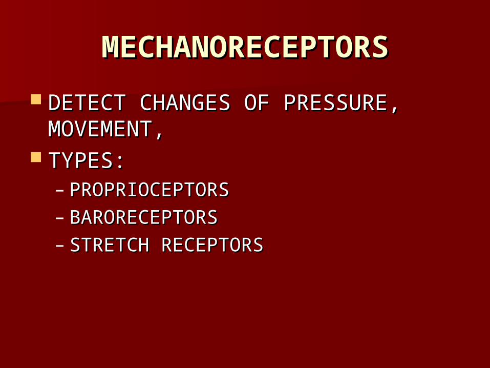
MECHANORECEPTORSMECHANORECEPTORS
DETECT CHANGES OF PRESSURE, DETECT CHANGES OF PRESSURE, MOVEMENT,MOVEMENT,
TYPES:TYPES:– PROPRIOCEPTORSPROPRIOCEPTORS– BARORECEPTORSBARORECEPTORS– STRETCH RECEPTORSSTRETCH RECEPTORS

Meissner’s corpuscle
Pacinian corpuscle 5
Ruffini corpuscle
6
4
Free endings
Peri-trichal (around hair follicle)
1“TOUCH” - hair displacement
2
Merkel cell3
TOUCH
TOUCH COLD PAIN
VIBRATION
TOUCH
RECEPTOR MODALITIES
CT DISPLACEMENT*
* slowly adaptingwberesford.hsc.wvu.eduMeisner’s corpuscle

PACINIAN CORPUSCLEPACINIAN CORPUSCLE
d-mis-web.ana.bris.ac.uk

MEISNER’S CORPUSCLEMEISNER’S CORPUSCLE
d-mis-web.ana.bris.ac.uk

BARORECEPTORBARORECEPTOR
.cvphysiology.com
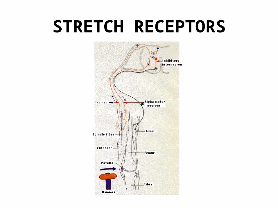
STRETCH RECEPTORS
users.rcn.com/jkimball.ma.ultranet/BiologyPages

PROPRIOCEPTORSPROPRIOCEPTORS
CHANGE IN LENGTH OF MUSCLE, CHANGE IN LENGTH OF MUSCLE, MUSCLE TENSIONMUSCLE TENSION
PRESSUREPRESSURE
GRAVITYGRAVITY
http://courses.washington.edu/conj/bess/spindle/proprioceptors.html

THERMORECEPTORSTHERMORECEPTORS
TEMPERATURE CHANGETEMPERATURE CHANGE
HEAT: HEAT: RUFFINI’S END ORGANRUFFINI’S END ORGAN
LOSS OF HEAT: LOSS OF HEAT: KRAUSE CELLKRAUSE CELL

KRAUSE CELLSKRAUSE CELLS
COLDCOLD
starsandseas.com
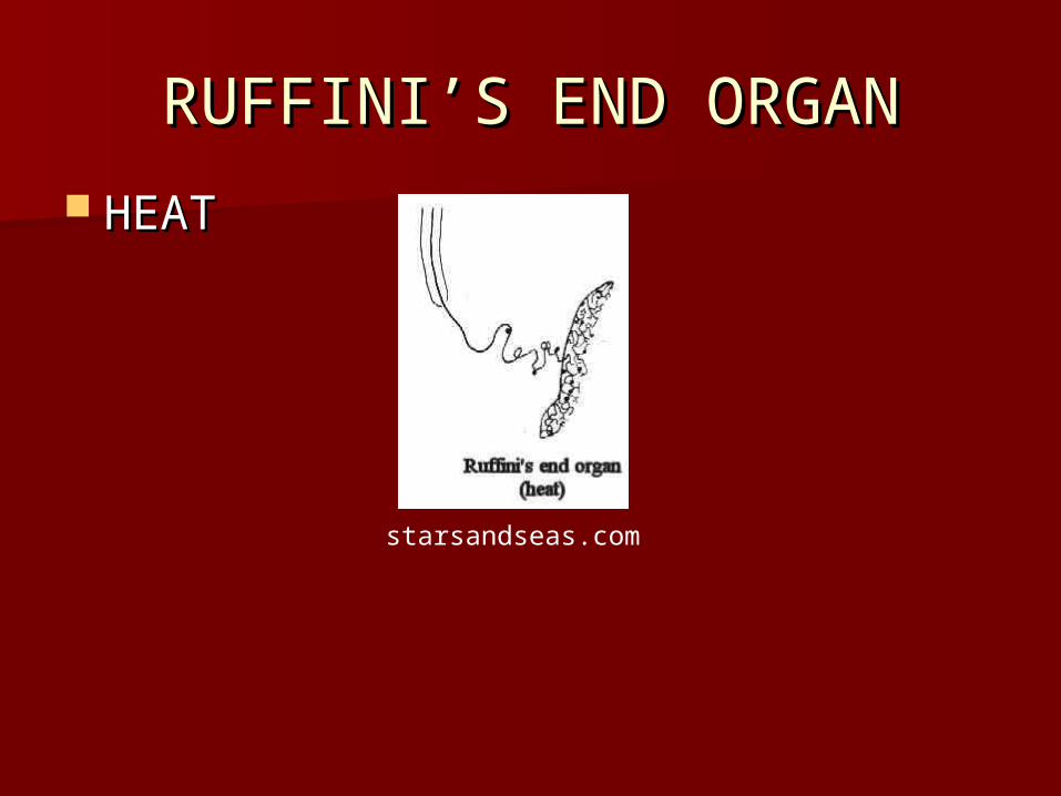
RUFFINI’S END ORGANRUFFINI’S END ORGAN
HEATHEAT
starsandseas.com

NOCICEPTORSNOCICEPTORS
PAINPAIN
TISSUE DAMAGE DUE TO: EXCESSIVE TISSUE DAMAGE DUE TO: EXCESSIVE MECHANICAL, ELECTRICAL, MECHANICAL, ELECTRICAL, THERMAL, OR CHEMICAL ENERGYTHERMAL, OR CHEMICAL ENERGY
FREE NERVE ENDINGFREE NERVE ENDING

NOCICEPTORSNOCICEPTORS
starsandseas.com
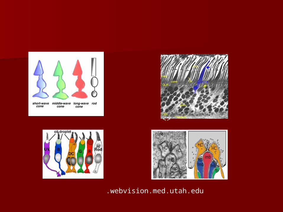
PHOTRECEPTORSPHOTRECEPTORS
LIGHTLIGHT RODS AND CONESRODS AND CONES
<>
.webvision.med.utah.edu

PAIN PATHWAYSPAIN PATHWAYS
ACUTE PAIN/ FAST PAIN:ACUTE PAIN/ FAST PAIN:– THIN, MYELINATED FIBERS, Aδ FIBERSTHIN, MYELINATED FIBERS, Aδ FIBERS– 6-30 M/SEC, DETECTED WITHIN A TENTH 6-30 M/SEC, DETECTED WITHIN A TENTH
OF A SECONDOF A SECOND– SHARP, PRICKLING PAINSHARP, PRICKLING PAIN– MECHANICAL AND THERMAL PAINMECHANICAL AND THERMAL PAIN
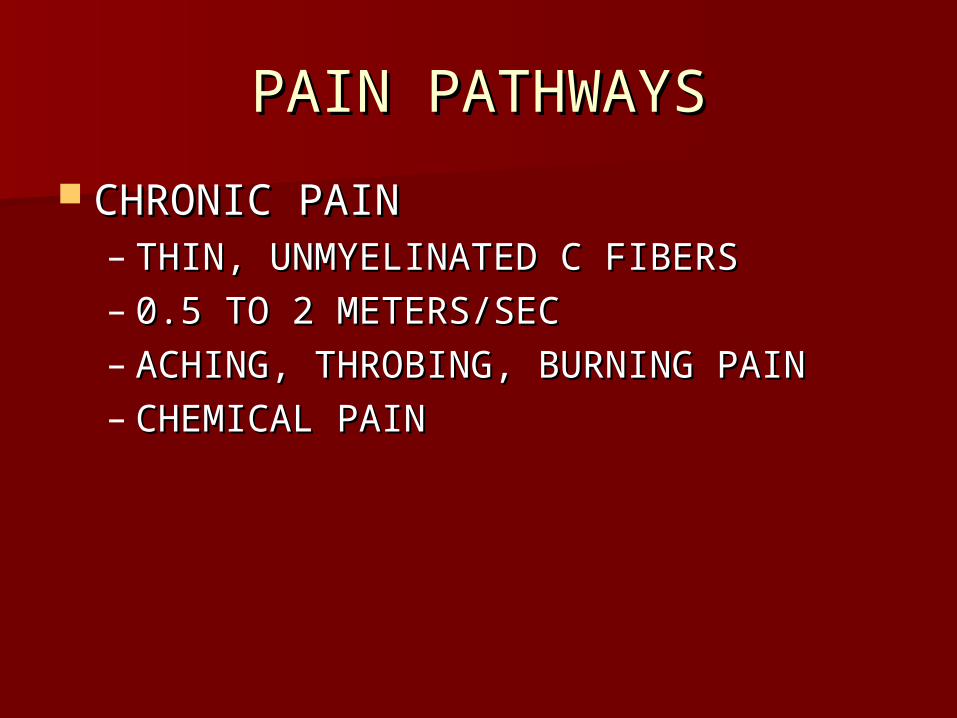
PAIN PATHWAYSPAIN PATHWAYS
CHRONIC PAINCHRONIC PAIN– THIN, UNMYELINATED C FIBERSTHIN, UNMYELINATED C FIBERS– 0.5 TO 2 METERS/SEC0.5 TO 2 METERS/SEC– ACHING, THROBING, BURNING PAINACHING, THROBING, BURNING PAIN– CHEMICAL PAINCHEMICAL PAIN

SEROTONINSEROTONIN
digital-fx.ca/cme/neuro

PULMONARY AND CARDIAC PULMONARY AND CARDIAC STRETCH RECEPTORSSTRETCH RECEPTORS
.lib.mcg.edu

MUSCLE SPINDLEMUSCLE SPINDLE
.lib.mcg.edu

MUSCLE SPINDLEMUSCLE SPINDLE
NEAR JUNCTIONS WITH TENDONSNEAR JUNCTIONS WITH TENDONS– INTRAFUSAL FIBERS: MODIFIED INTRAFUSAL FIBERS: MODIFIED
SKELETAL MUSCLE FIBERSSKELETAL MUSCLE FIBERS– COVERED BY A CONNECTIVE TISSUE COVERED BY A CONNECTIVE TISSUE
SHEATHSHEATH– CENTER: NONSTRIATED WITH A CENTER: NONSTRIATED WITH A
DENDRITE WRAPPED AROUND ITDENDRITE WRAPPED AROUND IT

MUSCLE SPINDLE FUNCTIONMUSCLE SPINDLE FUNCTION
– STRIATED PORTIONS CONTARACT: STRIATED PORTIONS CONTARACT: SPINDLE RELAXESSPINDLE RELAXES
– WHOLE MUSCLE RELAXES: SPINDLE WHOLE MUSCLE RELAXES: SPINDLE STRETCHES AND SENDS IMPULSE TO STRETCHES AND SENDS IMPULSE TO SPINAL CORD TO MOTOR NEURON TO SPINAL CORD TO MOTOR NEURON TO MUSCLEMUSCLE
– MUSCLE CONTRACTS AND OPPOSES MUSCLE CONTRACTS AND OPPOSES GRAVITATIONAL PULL: STRETCH REFLEXGRAVITATIONAL PULL: STRETCH REFLEX
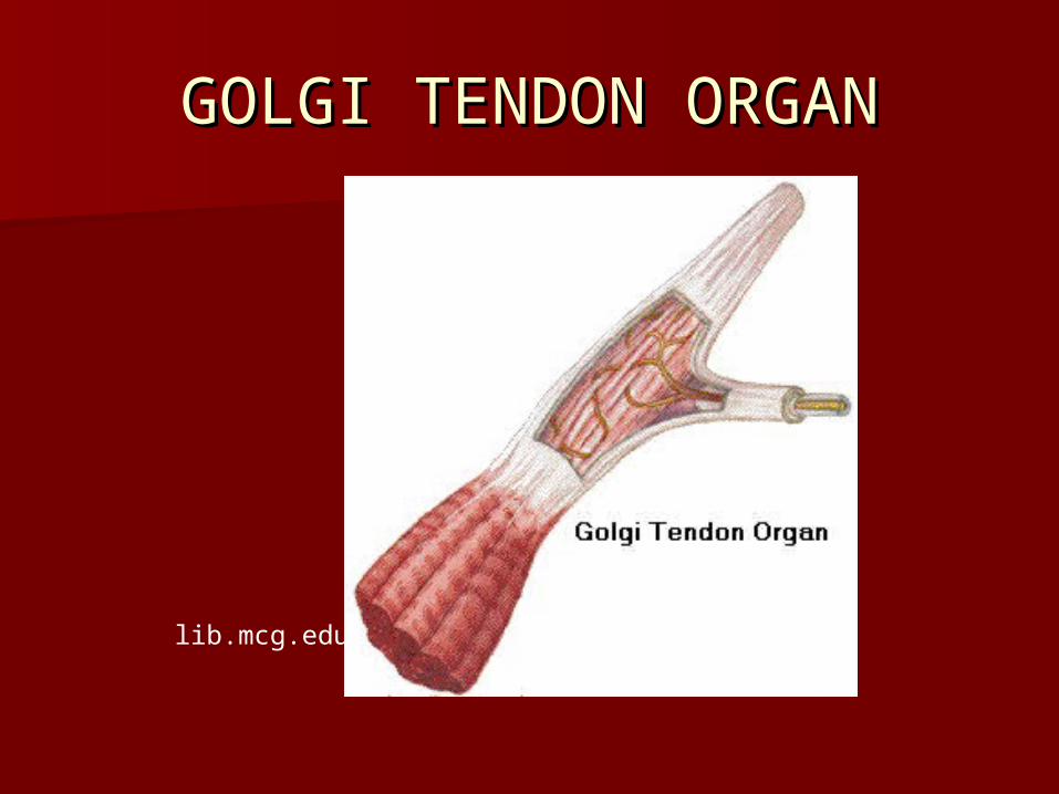
GOLGI TENDON ORGANGOLGI TENDON ORGAN
lib.mcg.edu
GOLGI TENDON ORGANGOLGI TENDON ORGAN
IN TENDONS; CLOSE TO MUSCLE IN TENDONS; CLOSE TO MUSCLE ATTACHMENTATTACHMENT– CONNECTED TO A SET OF SKELETAL CONNECTED TO A SET OF SKELETAL
MUSCLE FIBERSMUSCLE FIBERS– HIGH THRESHOLDHIGH THRESHOLD– STIMULATED BY INCREASED TENSIONSTIMULATED BY INCREASED TENSION

GOLGI TENDON ORGAN GOLGI TENDON ORGAN FUNCTIONFUNCTION
– STIMULATED BY INCREASED TENSIONSTIMULATED BY INCREASED TENSION– INHIBITS CONTRACTION OF MUSCLESINHIBITS CONTRACTION OF MUSCLES– OPPOSITE OF STRETCH REFLEXOPPOSITE OF STRETCH REFLEX– HELPS MAINTAIN POSTUREHELPS MAINTAIN POSTURE– PROTECTS AGAINST MUSCLE PROTECTS AGAINST MUSCLE
ATTACHMENTS BEING PULLED AWAY ATTACHMENTS BEING PULLED AWAY FROM INSERTIONSFROM INSERTIONS

SPECIAL SENSESSPECIAL SENSES
RECEPTOR: RECEPTOR: – INDIVIDUAL CELL OR AN EYE OR AN EARINDIVIDUAL CELL OR AN EYE OR AN EAR
MEMBRANE RECEPTOR: MEMBRANE RECEPTOR: – PROTEIN ON PLASMA MEMBRANEPROTEIN ON PLASMA MEMBRANE
SMELLSMELL
OLFACTORY RECEPTORS FOUND IN THE OLFACTORY RECEPTORS FOUND IN THE OLFACTORY EPITHELIUM OF NASAL OLFACTORY EPITHELIUM OF NASAL CAVITY: CHEMORECEPTORSCAVITY: CHEMORECEPTORS
1000 GENES CODE FOR THE 1000 GENES CODE FOR THE RECEPTORSRECEPTORS
A RECEPTOR CELL HAS ONLY ONE TYPE A RECEPTOR CELL HAS ONLY ONE TYPE OF RECEPTOR WHICH CAN ONLY OF RECEPTOR WHICH CAN ONLY DETECT A FEW NUMBER OF ODORS DETECT A FEW NUMBER OF ODORS

OLFACTORY RECEPTORSOLFACTORY RECEPTORS
THE RECEPTORS HAVE 10-20 CILLIA THE RECEPTORS HAVE 10-20 CILLIA WHICH STICK INTO THE CAVITY AND WHICH STICK INTO THE CAVITY AND ARE THE SENSITIVE AREAARE THE SENSITIVE AREA
THERE ARE ABOUT 40 RECEPTORS; THERE ARE ABOUT 40 RECEPTORS; BIPOLAR NEURONSBIPOLAR NEURONS
ALSO COLUMNAR EPITHELIUM AND ALSO COLUMNAR EPITHELIUM AND MUCOUS CELLSMUCOUS CELLS

OLFACTORY RECEPTOROLFACTORY RECEPTOR
faculty.washington.edu

OLFACTORY RECEPTOROLFACTORY RECEPTOR
faculty.washington.edu
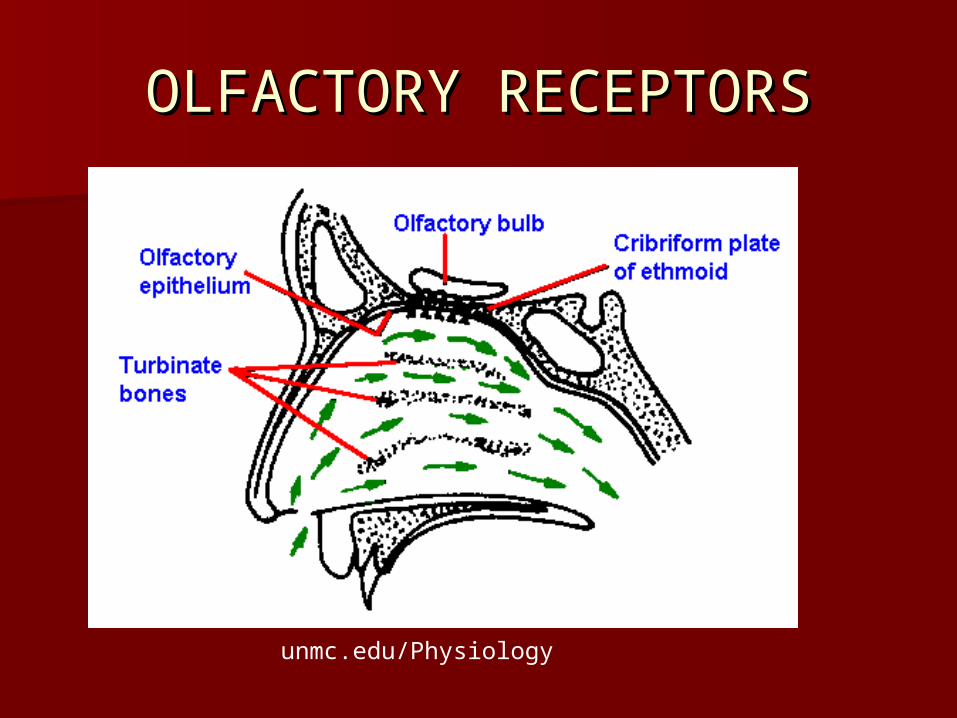
OLFACTORY RECEPTORSOLFACTORY RECEPTORS
unmc.edu/Physiology

OLFACTORY RECEPTORSOLFACTORY RECEPTORS
unmc.edu/Physiology
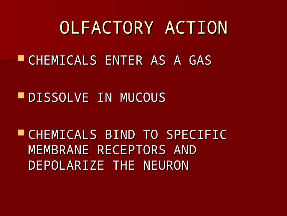
OLFACTORY ACTIONOLFACTORY ACTION
CHEMICALS ENTER AS A GASCHEMICALS ENTER AS A GAS
DISSOLVE IN MUCOUSDISSOLVE IN MUCOUS
CHEMICALS BIND TO SPECIFIC CHEMICALS BIND TO SPECIFIC MEMBRANE RECEPTORS AND MEMBRANE RECEPTORS AND DEPOLARIZE THE NEURONDEPOLARIZE THE NEURON
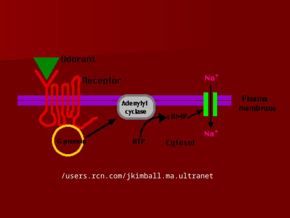
/users.rcn.com/jkimball.ma.ultranet

faculty.washington.edu

faculty.washington.edu

OLFACTORY PATHWAYOLFACTORY PATHWAY
OLFACTORY RECEPTOR IS OLFACTORY RECEPTOR IS STIMULATED AND FORMS AN IMPULSESTIMULATED AND FORMS AN IMPULSE
IMPULSE TRAVELS ALONG THE AXON IMPULSE TRAVELS ALONG THE AXON TO THE MITRAL CELLS OF THE TO THE MITRAL CELLS OF THE OLFACTORY BULBOLFACTORY BULB
TRAVELS BY OLFACTORY TRACT TO TRAVELS BY OLFACTORY TRACT TO LIMBIC SYSTEM DEEP IN CEREBRAL LIMBIC SYSTEM DEEP IN CEREBRAL CORTEX OF TEMPORAL AND FRONTAL CORTEX OF TEMPORAL AND FRONTAL LOBESLOBES

OLFACTORY PATHWAY OLFACTORY PATHWAY CONT.CONT.
IMPULSES FROM THE OLFACTORY IMPULSES FROM THE OLFACTORY RECEPTORS ARE TRANSLATED BY THE RECEPTORS ARE TRANSLATED BY THE BRAIN AS AN OLFACTORY CODEBRAIN AS AN OLFACTORY CODE
RAPID SENSORY ADAPTATION; 50% IN 1 RAPID SENSORY ADAPTATION; 50% IN 1 SECOND, INSENSITIVE WITHIN 1 MINUTESECOND, INSENSITIVE WITHIN 1 MINUTE
IN DIRECT CONTACT WITH ENVIRONMENT IN DIRECT CONTACT WITH ENVIRONMENT SO OFTEN DESTROYED AND NOT USUALLY SO OFTEN DESTROYED AND NOT USUALLY REPLACED; COULD LOSE 1%/YEARREPLACED; COULD LOSE 1%/YEAR

SMELLSMELL
MOST PEOPLE CAN DETECT MOST PEOPLE CAN DETECT BETWEEN 3,000 AND 10,000 ODORS BETWEEN 3,000 AND 10,000 ODORS
TASTETASTE
TASTE BUDS CONTAIN GUSTATORY TASTE BUDS CONTAIN GUSTATORY CELLSCELLS
TASTE BUDS FOUND ON PAPILLAE OF TASTE BUDS FOUND ON PAPILLAE OF TONGUE, ROOF OF MOUTH, LINING TONGUE, ROOF OF MOUTH, LINING OF CHEEKS, WALLS OF PHARYNXOF CHEEKS, WALLS OF PHARYNX
RECEPTORS: MODIFIED EPITHEILIAL RECEPTORS: MODIFIED EPITHEILIAL CELLSCELLS
10,000 TASTE BUDS WITH 50-100 10,000 TASTE BUDS WITH 50-100 RECEPTOR CELLS EACHRECEPTOR CELLS EACH
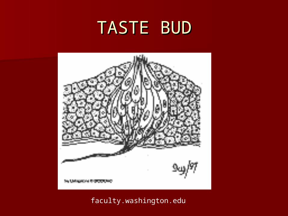
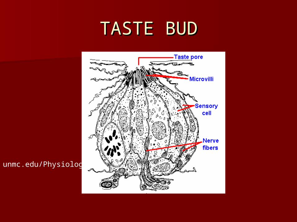
TASTE BUDTASTE BUD
faculty.washington.edu
TASTE BUDTASTE BUD
unmc.edu/Physiology

TASTE CELLTASTE CELL
faculty.washington.edu
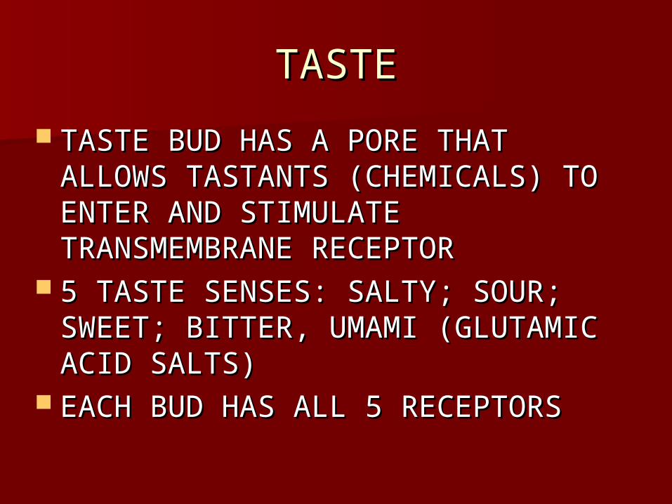
TASTETASTE
TASTE BUD HAS A PORE THAT TASTE BUD HAS A PORE THAT ALLOWS TASTANTS (CHEMICALS) TO ALLOWS TASTANTS (CHEMICALS) TO ENTER AND STIMULATE ENTER AND STIMULATE TRANSMEMBRANE RECEPTORTRANSMEMBRANE RECEPTOR
5 TASTE SENSES: SALTY; SOUR; 5 TASTE SENSES: SALTY; SOUR; SWEET; BITTER, UMAMI (GLUTAMIC SWEET; BITTER, UMAMI (GLUTAMIC ACID SALTS)ACID SALTS)
EACH BUD HAS ALL 5 RECEPTORSEACH BUD HAS ALL 5 RECEPTORS
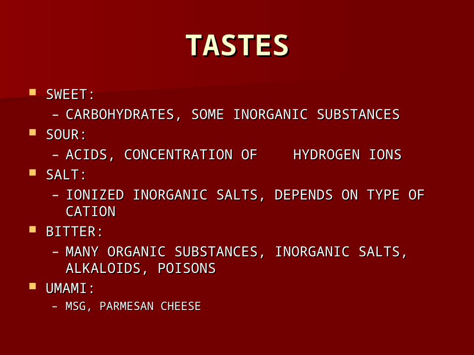
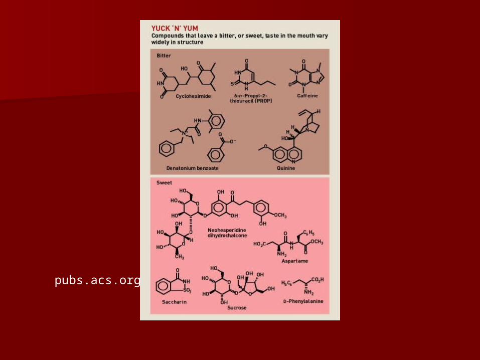
TASTESTASTES SWEET: SWEET:
– CARBOHYDRATES, SOME INORGANIC SUBSTANCESCARBOHYDRATES, SOME INORGANIC SUBSTANCES SOUR: SOUR:
– ACIDS, CONCENTRATION OF ACIDS, CONCENTRATION OF HYDROGEN IONSHYDROGEN IONS SALT: SALT:
– IONIZED INORGANIC SALTS, DEPENDS ON TYPE OF IONIZED INORGANIC SALTS, DEPENDS ON TYPE OF CATIONCATION
BITTER: BITTER: – MANY ORGANIC SUBSTANCES, INORGANIC SALTS, MANY ORGANIC SUBSTANCES, INORGANIC SALTS,
ALKALOIDS, POISONSALKALOIDS, POISONS UMAMI: UMAMI:
– MSG, PARMESAN CHEESEMSG, PARMESAN CHEESE
pubs.acs.org
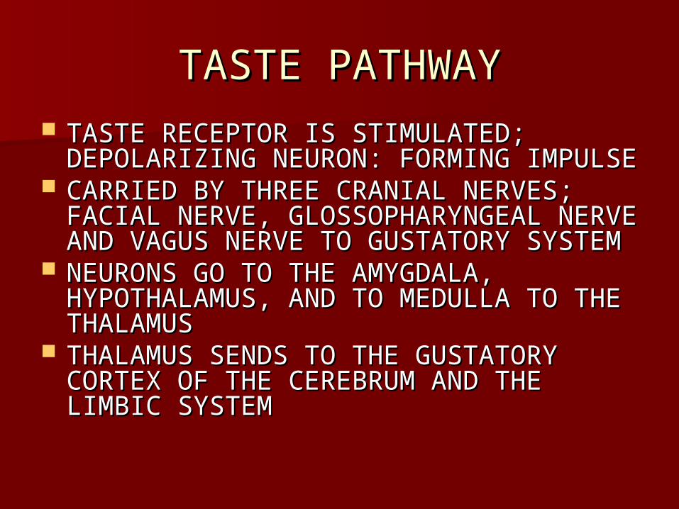
TASTE PATHWAYTASTE PATHWAY TASTE RECEPTOR IS STIMULATED; TASTE RECEPTOR IS STIMULATED;
DEPOLARIZING NEURON: FORMING IMPULSEDEPOLARIZING NEURON: FORMING IMPULSE CARRIED BY THREE CRANIAL NERVES; CARRIED BY THREE CRANIAL NERVES;
FACIAL NERVE, GLOSSOPHARYNGEAL FACIAL NERVE, GLOSSOPHARYNGEAL NERVE AND VAGUS NERVE TO GUSTATORY NERVE AND VAGUS NERVE TO GUSTATORY SYSTEMSYSTEM
NEURONS GO TO THE AMYGDALA, NEURONS GO TO THE AMYGDALA, HYPOTHALAMUS, AND TO MEDULLA TO THE HYPOTHALAMUS, AND TO MEDULLA TO THE THALAMUSTHALAMUS
THALAMUS SENDS TO THE GUSTATORY THALAMUS SENDS TO THE GUSTATORY CORTEX OF THE CEREBRUM AND THE CORTEX OF THE CEREBRUM AND THE LIMBIC SYSTEMLIMBIC SYSTEM
FAST ADAPTATIONFAST ADAPTATION CELLS REPLACED EVERY THREE DAYS CELLS REPLACED EVERY THREE DAYS
SO DOESN’T DECREASE WITH AGESO DOESN’T DECREASE WITH AGE
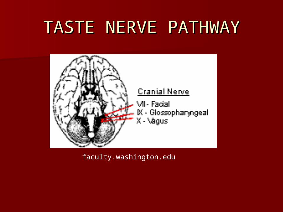
TASTE NERVE PATHWAYTASTE NERVE PATHWAY
faculty.washington.edu

TASTE BUD MAP: MYTHTASTE BUD MAP: MYTH
unmc.edu/Physiology

mona.uwi.edu

mona.uwi.edu

FLAVORFLAVOR
SMELL, TASTE, TEXTURE, SMELL, TASTE, TEXTURE, TEMPERATURETEMPERATURE
HEARINGHEARING
faculty.washington.edu

sirinet.net
EARDRUMEARDRUM
human-body.net

HEARINGHEARING
Humans can hear between 20 to Humans can hear between 20 to 20,000 decibels, greatest sensitivity 20,000 decibels, greatest sensitivity at 2,00 to 3,000 vibrations/secondat 2,00 to 3,000 vibrations/second
Muscles:Muscles:– Tensor tympani: holds malleus inward Tensor tympani: holds malleus inward
and to wall, involved in tympanic reflex and to wall, involved in tympanic reflex to muffle loud sounds for protectionto muffle loud sounds for protection
– Stapedius: holds stapes in placeStapedius: holds stapes in place

HEARINGHEARING
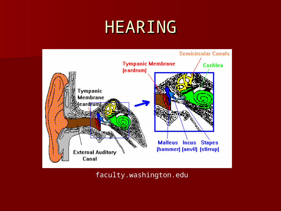
Auricle funnels sound waves to Auricle funnels sound waves to auditory canal to eardrumauditory canal to eardrum
Sound waves changed to vibrations, Sound waves changed to vibrations, passed through middle ear bones, passed through middle ear bones, stapes magnifies vibrationsstapes magnifies vibrations
Stapes vibrates oval window, Stapes vibrates oval window, vibrations to liquid waves in vibrations to liquid waves in perilymphperilymph

sirinet.net

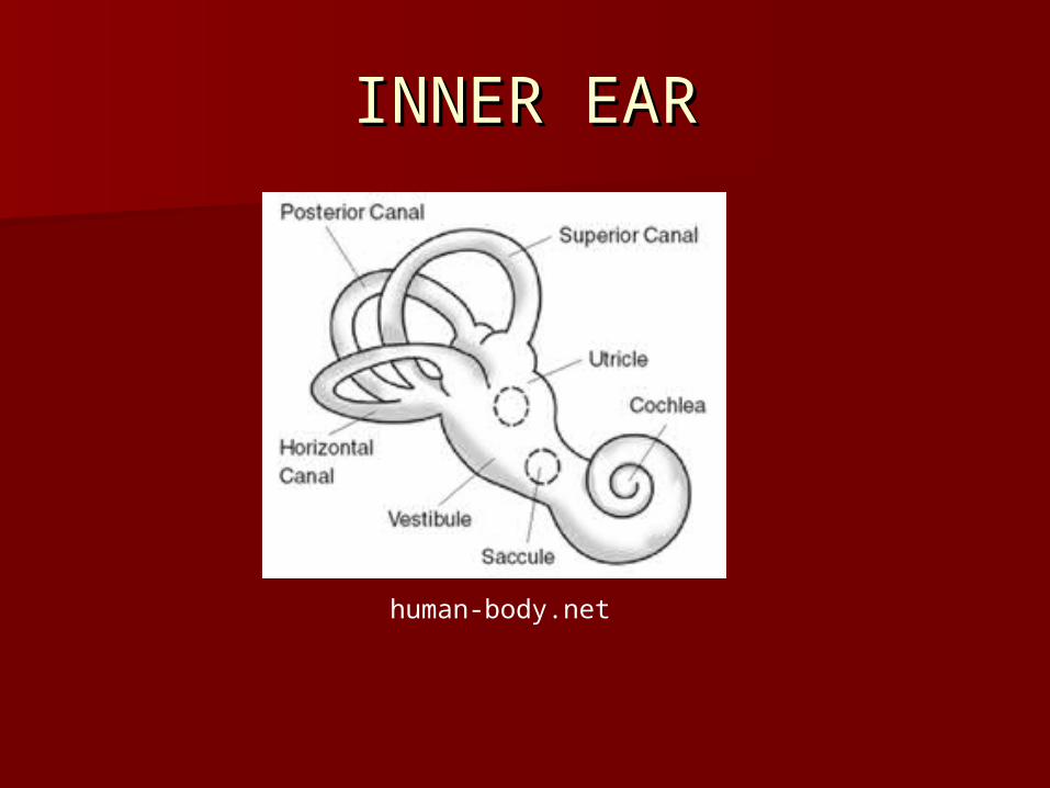
INNER EARINNER EAR
human-body.net

Vibrations pass from perilymph of scala Vibrations pass from perilymph of scala vestibuli through vestibular membrane to vestibuli through vestibular membrane to endolymph of scala media and through basilar endolymph of scala media and through basilar membrane to perilymph of scala tympani and membrane to perilymph of scala tympani and out to air at round windowout to air at round window
Organ of Corti has 16,000 hearing receptor Organ of Corti has 16,000 hearing receptor cells on surface of basilar membrane in scala cells on surface of basilar membrane in scala mediamedia
Each receptor cell (epithelial cells) has 4 hair Each receptor cell (epithelial cells) has 4 hair cells with parallel cells of many stereocillia cells with parallel cells of many stereocillia (micro villi) which release neurotransmitters to (micro villi) which release neurotransmitters to stimulate associated neuronstimulate associated neuron
CONTINUEDCONTINUED
Specific frequencies vibrate specific Specific frequencies vibrate specific subsets of hairs of specific receptor subsets of hairs of specific receptor cells (epithelial cells) depolarizes the cells (epithelial cells) depolarizes the cellcell
Calcium enters causing Calcium enters causing neurotransmitters to be released neurotransmitters to be released which stimulate the neuronwhich stimulate the neuron
INNER EARINNER EAR
human-body.net
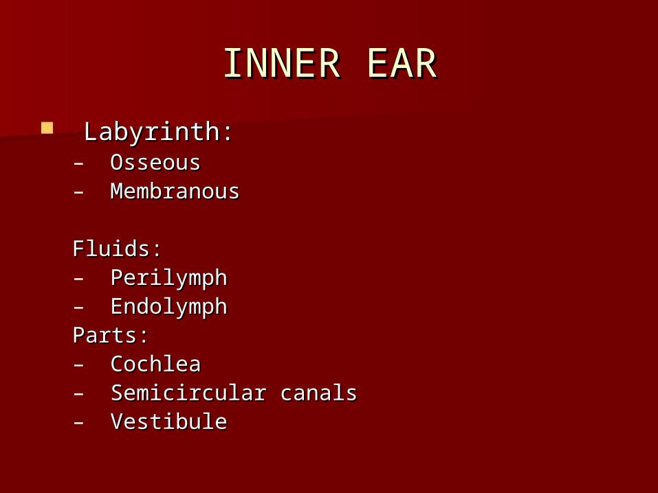
INNER EARINNER EAR
Labyrinth:Labyrinth:– OsseousOsseous– MembranousMembranous
Fluids:Fluids:– PerilymphPerilymph– EndolymphEndolymphParts: Parts: – CochleaCochlea– Semicircular canalsSemicircular canals– VestibuleVestibule
AUDITORY NERVE AUDITORY NERVE PATHWAYS PATHWAYS
Cochlear branch of VestibulocochlearCochlear branch of Vestibulocochlear
cells to auditory neurons, to cells to auditory neurons, to medulla medulla to midbrain to thalamus to to midbrain to thalamus to auditory auditory cortices of temporal lobescortices of temporal lobes
Impulse goes to both sides of the Impulse goes to both sides of the cerebrumcerebrum

EQUILIBRIUMEQUILIBRIUM
2 senses2 senses– StaticStatic– DynamicDynamic
StaticStatic– In vestibule: membranous labyrinthIn vestibule: membranous labyrinth
UtricleUtricle SacculeSaccule
STATIC EQUILIBRIUMSTATIC EQUILIBRIUM
Contain macula: hair cells and Contain macula: hair cells and supporting cells covered by otolithic supporting cells covered by otolithic membrane with otoliths (calcium membrane with otoliths (calcium carbonate crystals) for increased carbonate crystals) for increased sensitivitysensitivity
Head upright: Head upright: – Utricle hairs verticalUtricle hairs vertical– Saccule hairs horizontalSaccule hairs horizontal
STATIC EQUILIBRIUMSTATIC EQUILIBRIUM
Hair cells have nerve fiber wrapped Hair cells have nerve fiber wrapped around the basearound the base
Neuron goes to vestibular portion of Neuron goes to vestibular portion of vestibulocochlear nervevestibulocochlear nerve
When head moves macula (of one or When head moves macula (of one or both) sags due to gravity bending both) sags due to gravity bending hairs and depolarizing cellhairs and depolarizing cell
Neurotransmitters released Neurotransmitters released stimulating neuronstimulating neuron

STATIC EQUILIBRIUM CONT.STATIC EQUILIBRIUM CONT.
Impulse travels up Vestibulocochlear Impulse travels up Vestibulocochlear nerve to midbrain to cerebrumnerve to midbrain to cerebrum
Cerebrum translates and analyzes Cerebrum translates and analyzes impulse and sends appropriate impulse and sends appropriate impulse along motor nerves to impulse along motor nerves to skeletal muscles to maintain postureskeletal muscles to maintain posture

DYNAMIC EQUILIBRIUMDYNAMIC EQUILIBRIUM
Macula also involved some: when Macula also involved some: when head or body is thrust forward or head or body is thrust forward or backward otolithic membrane lags backward otolithic membrane lags behind and stimulates hair cells: behind and stimulates hair cells: falling, walkingfalling, walking
Semicircular canalsSemicircular canals– 3: superior & posterior (lateral)3: superior & posterior (lateral)– lateral (horizontal)lateral (horizontal)– Body planesBody planes
DYNAMIC EQUILIBRIUMDYNAMIC EQUILIBRIUM
Semicircular canal ends in swelling= Semicircular canal ends in swelling= ampulla; communicates with utricleampulla; communicates with utricle
Ampulla has crista ampullaris Ampulla has crista ampullaris containing hair cells in a dome containing hair cells in a dome shaped gelatinous mass= cupulashaped gelatinous mass= cupula
Hair cells have nerve fibers wrapped Hair cells have nerve fibers wrapped around the base connected to around the base connected to vestibulocochlear nervevestibulocochlear nerve

DYNAMIC EQUILIBRIUMDYNAMIC EQUILIBRIUM
Rapid turns of head moves but not Rapid turns of head moves but not the fluid so the cupula of 1 or more the fluid so the cupula of 1 or more semicircular canals bend stimulating semicircular canals bend stimulating the hair cells forming impulse in the hair cells forming impulse in neuronneuron
Vestibulocochlear nerve carries Vestibulocochlear nerve carries impulse to cerebellum for impulse to cerebellum for interpretation to maintain balanceinterpretation to maintain balance

DYNAMIC EQUILIBRIUMDYNAMIC EQUILIBRIUM
Other sensory structures:Other sensory structures:– ProprioceptorsProprioceptors– EyesEyes
Motion sicknessMotion sickness

sirinet.net

Cochlear ImplantCochlear Implant
http://www.nidcd.nih.gov/health/hearing/pages/coch.aspx

Cochlear Implant PartsCochlear Implant Parts
MicrophoneMicrophone Speech processor: selects and arranges Speech processor: selects and arranges
soundssounds Transmitter and receiver/stimulator: Transmitter and receiver/stimulator:
convert signals to electricconvert signals to electric Electrode array: group of electrodes Electrode array: group of electrodes
that collects the impulses from the that collects the impulses from the stimulator and sends them to different stimulator and sends them to different regions of the auditory nerve regions of the auditory nerve
Cochlear Implant continuedCochlear Implant continued
Bypass the part of the ear that is Bypass the part of the ear that is damageddamaged
Directly stimulate auditory nerveDirectly stimulate auditory nerve Nerve sends impulses to proper area Nerve sends impulses to proper area
of brainof brain Not like normal hearing but allows Not like normal hearing but allows
person to pick up on soundsperson to pick up on sounds

SIGHTSIGHT
EYE PARTSEYE PARTS
Eyelid: PalpebraEyelid: Palpebra– 4 layers: 4 layers:
Skin: thinnestSkin: thinnest Muscle: orbicularis oculi; levator palpebrae Muscle: orbicularis oculi; levator palpebrae
superiorissuperioris Connective tissue: tarsal glandsConnective tissue: tarsal glands Conjunctiva: mucous membraneConjunctiva: mucous membrane
Lacrimal apparatus: lacrimal gland, superior Lacrimal apparatus: lacrimal gland, superior and inferior canaliculi, lacrimal sac, and inferior canaliculi, lacrimal sac, nasolacrimal duct, nasal cavitynasolacrimal duct, nasal cavity

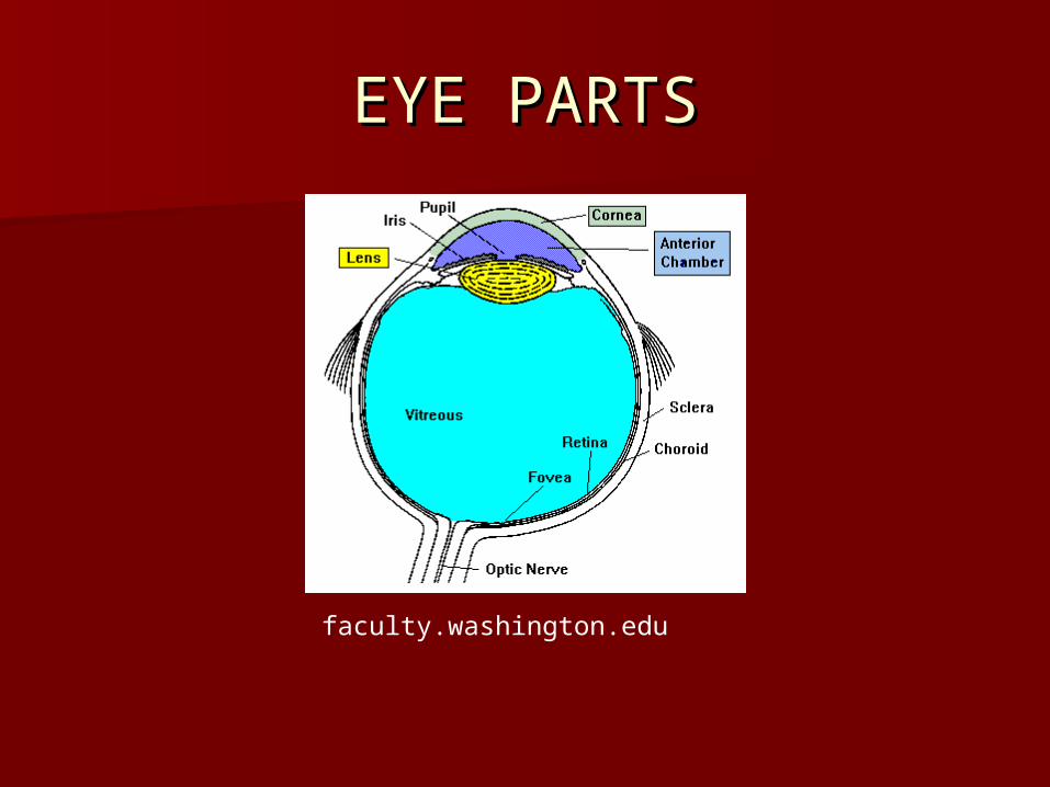
EYE PARTSEYE PARTS
Conjunctiva: glandular cells, lysozymeConjunctiva: glandular cells, lysozyme Extrinsic musclesExtrinsic muscles 3 layers:3 layers:
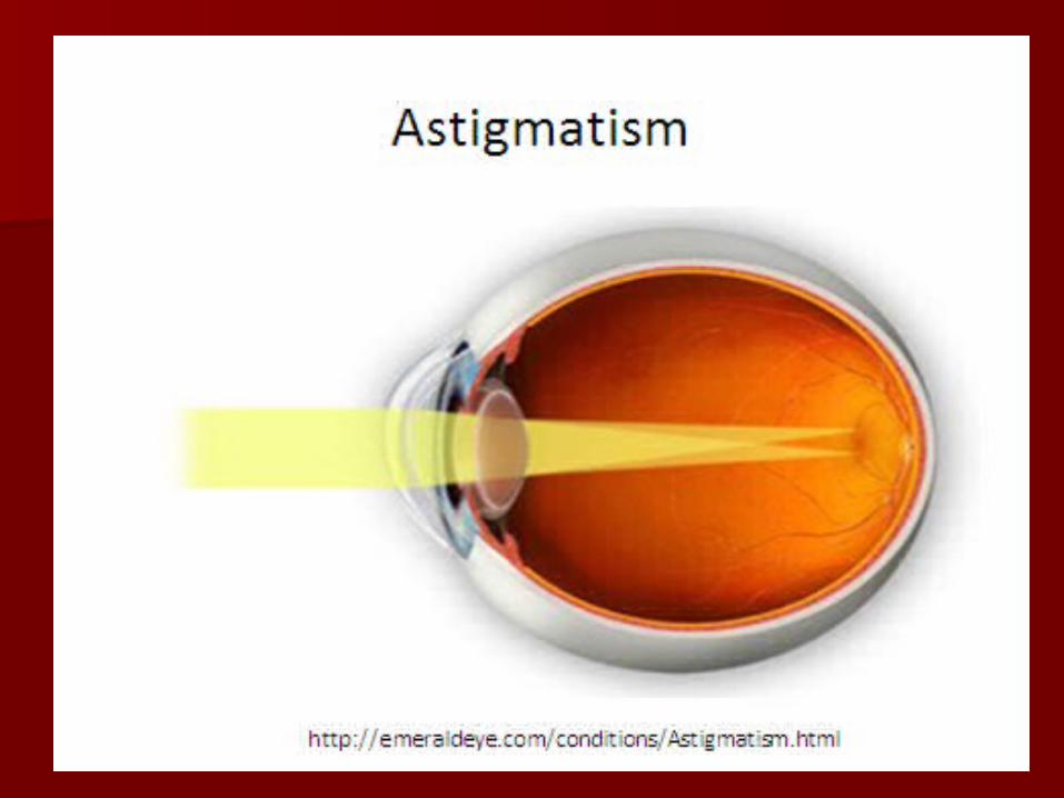
– Fibrous tunic, vascular tunic, nervous tunicFibrous tunic, vascular tunic, nervous tunic
Fibrous: cornea, sclera, Fibrous: cornea, sclera,
Vascular/uveal layer: choroid coat, ciliary body, Vascular/uveal layer: choroid coat, ciliary body, suspensory ligaments, lens, iris, suspensory ligaments, lens, iris,
Nervous tunic: retina, macula lutea, fovea Nervous tunic: retina, macula lutea, fovea centralis, optic disk, photoreceptors, vitreous centralis, optic disk, photoreceptors, vitreous humor, vitreous bodyhumor, vitreous body

en.wikipedia.org

IRISIRIS Controlled by circular and radial set of musclesControlled by circular and radial set of muscles Parasympathetic nerves stimulate the circular Parasympathetic nerves stimulate the circular
muscles to constrict iris (bright light)muscles to constrict iris (bright light) Sympathetic nerves control stimulate radial Sympathetic nerves control stimulate radial
muscles to dilate iris muscles to dilate iris Color: very complex, melanin is pigment, Color: very complex, melanin is pigment,
genes for blue (recessive), green and brown; genes for blue (recessive), green and brown; no pigment= light blue, also texture, fibrous no pigment= light blue, also texture, fibrous tissue, blood vessels and selective absorption tissue, blood vessels and selective absorption and reflection of biological compoundsand reflection of biological compounds
EYE PARTSEYE PARTS
faculty.washington.edu

RETINARETINA
webvision.med.utah.edu

webvision.med.utah.edu

PATHWAYPATHWAY
http://webvision.med.utah.edu/http://webvision.med.utah.edu/movies/irisedu.movmovies/irisedu.mov

RETINARETINA
webvision.med.utah.edu
webvision.med.utah.edu

webvision.med.utah.edu
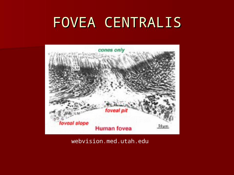
FOVEA CENTRALISFOVEA CENTRALIS
webvision.med.utah.edu

RETINARETINA
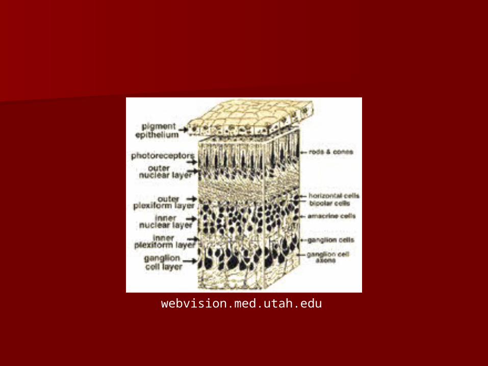
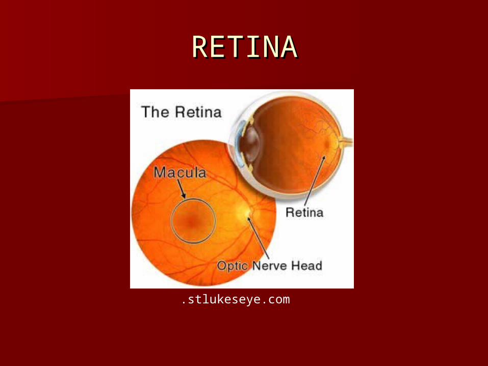
PhotoreceptorsPhotoreceptors Pigmented epitheliumPigmented epithelium
– absorbs light; stores vitamin Aabsorbs light; stores vitamin A NeuronsNeurons Nerve fibersNerve fibers Limiting membranesLimiting membranes

RETINARETINA
Retinal neuronsRetinal neurons– Receptor cellsReceptor cells– Bipolar neuronsBipolar neurons– Ganglion cellsGanglion cells– Horizontal cellsHorizontal cells– Amacrine cellsAmacrine cells
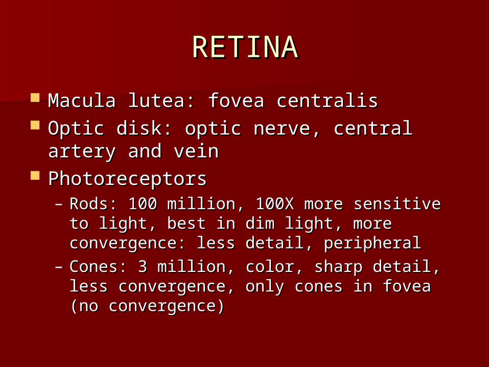
RETINARETINA
Macula lutea: fovea centralisMacula lutea: fovea centralis Optic disk: optic nerve, central artery and Optic disk: optic nerve, central artery and
veinvein PhotoreceptorsPhotoreceptors
– Rods: 100 million, 100X more sensitive to light, Rods: 100 million, 100X more sensitive to light, best in dim light, more convergence: less best in dim light, more convergence: less detail, peripheraldetail, peripheral
– Cones: 3 million, color, sharp detail, less Cones: 3 million, color, sharp detail, less convergence, only cones in fovea (no convergence, only cones in fovea (no convergence)convergence)
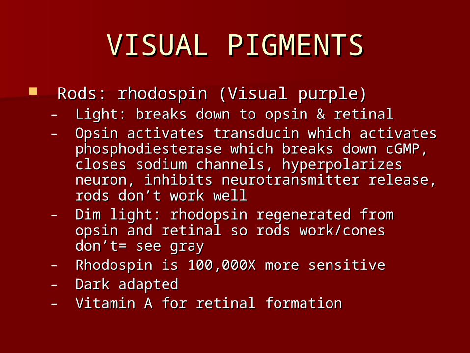
VISUAL PIGMENTSVISUAL PIGMENTS Rods: rhodospin (Visual purple)Rods: rhodospin (Visual purple)
– Light: breaks down to opsin & retinalLight: breaks down to opsin & retinal– Opsin activates transducin which activates Opsin activates transducin which activates
phosphodiesterase which breaks down cGMP, phosphodiesterase which breaks down cGMP, closes sodium channels, hyperpolarizes closes sodium channels, hyperpolarizes neuron, inhibits neurotransmitter release, rods neuron, inhibits neurotransmitter release, rods don’t work welldon’t work well
– Dim light: rhodopsin regenerated from opsin Dim light: rhodopsin regenerated from opsin and retinal so rods work/cones don’t= see grayand retinal so rods work/cones don’t= see gray
– Rhodospin is 100,000X more sensitiveRhodospin is 100,000X more sensitive– Dark adaptedDark adapted– Vitamin A for retinal formationVitamin A for retinal formation

IODOSPINSIODOSPINS
Light sensitive pigments of conesLight sensitive pigments of cones– Erythrolabe: red lightErythrolabe: red light– Chlorolabe: greenChlorolabe: green– Cyanolabe: blueCyanolabe: blue– All 3= whiteAll 3= white– None= blackNone= black
Color blindness: Green: most common, sex Color blindness: Green: most common, sex linked (more in males), red weakness: sex linked (more in males), red weakness: sex linked; Blue: rare, not sex linked= equal in linked; Blue: rare, not sex linked= equal in male & femalemale & female
STEROPSISSTEROPSIS
Distance, depth, height, and widthDistance, depth, height, and width Eyes are 6-7 cm apart: so Eyes are 6-7 cm apart: so
superimposed+ 3-D in visual cortexsuperimposed+ 3-D in visual cortex
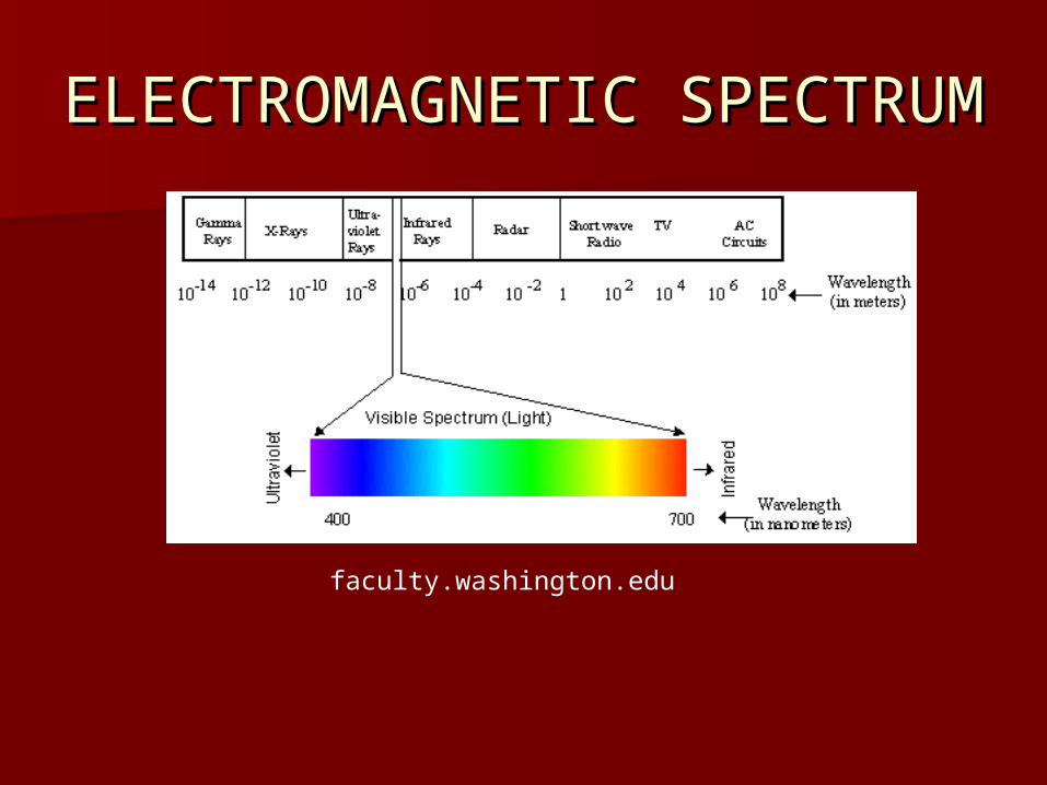
ELECTROMAGNETIC ELECTROMAGNETIC SPECTRUMSPECTRUM
faculty.washington.edu

EYE ANATOMYEYE ANATOMY
.stlukeseye.com
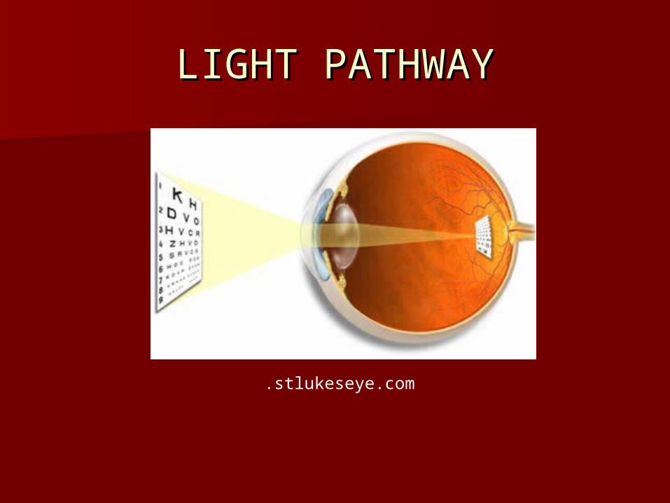
LIGHT PATHWAYLIGHT PATHWAY
.stlukeseye.com

LIGHT PATHWAYLIGHT PATHWAY
Refraction: cornea (75%), lens and liquidsRefraction: cornea (75%), lens and liquids Cornea refracts light, iris controls amount, Cornea refracts light, iris controls amount,
lens focuses and flips image onto retina, lens focuses and flips image onto retina, photoreceptors form impulse to ganglion photoreceptors form impulse to ganglion cells to optic nerve to optic chiasma, some cells to optic nerve to optic chiasma, some impulses cross to thalamus via optic impulses cross to thalamus via optic radiators to visual cortex of occipital lobesradiators to visual cortex of occipital lobes
Some impulses to brain stem to control Some impulses to brain stem to control head & eye movement to track moving head & eye movement to track moving object; & control movements of both eyesobject; & control movements of both eyes
RETINARETINA
.stlukeseye.com

WAVELENGTHSWAVELENGTHS
webvision.med.utah.edu

BLUE CONEBLUE CONE
webvision.med.utah.edu

Demonstration of the blind Demonstration of the blind spotspot
O X
Look at an eye diagram, what causes the blind spot?Where the optic nerve enters/ no receptor cellsWhy don’t we see a black dot where the blind spot is?The brain fills in the space with info from the surrounding area and the other eye

• It was first thought that the optic It was first thought that the optic nerve entrance should have the nerve entrance should have the greatest visual acuitygreatest visual acuity
• This hypothesis was disproved in This hypothesis was disproved in 1660 by Edme Mariotte of France1660 by Edme Mariotte of France
• It is located 1.5◦ below the horizon It is located 1.5◦ below the horizon and is about 7.5◦ by 5.5◦and is about 7.5◦ by 5.5◦
• Not found in all animals Not found in all animals Why not?Why not?
Blind spot Blind spot
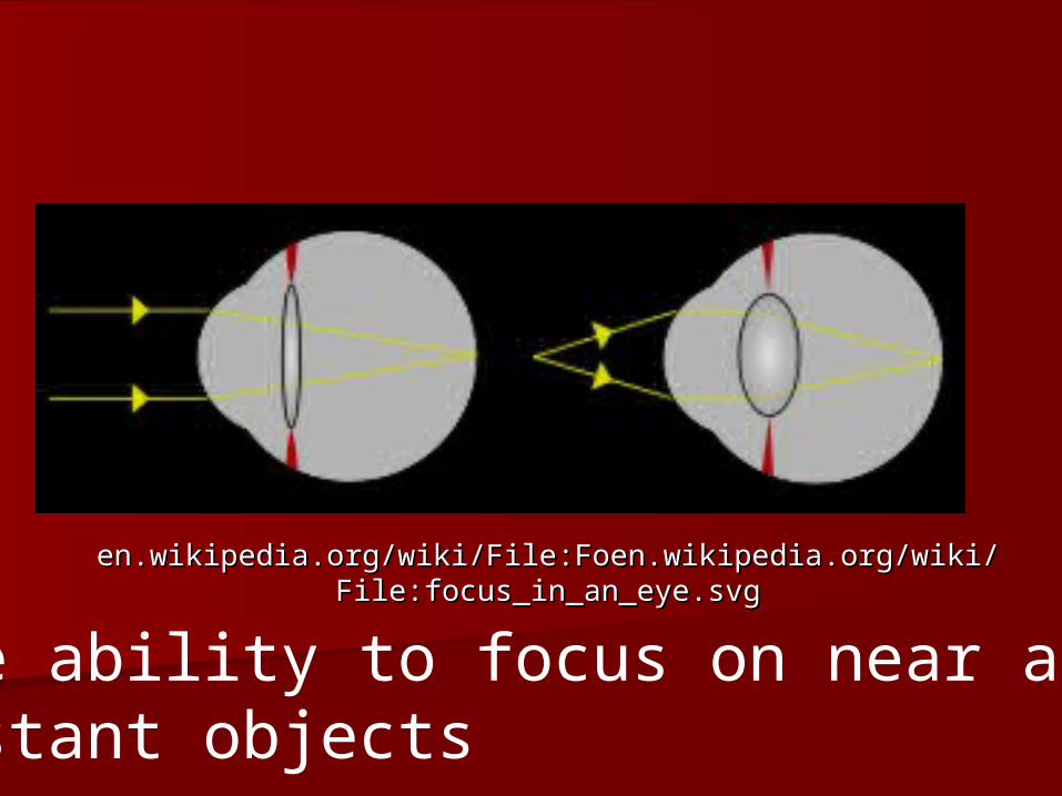
en.wikipedia.org/wiki/File:Foen.wikipedia.org/wiki/en.wikipedia.org/wiki/File:Foen.wikipedia.org/wiki/File:focus_in_an_eye.svgFile:focus_in_an_eye.svg
The ability to focus on near and distant objects

What is happening? What can be seen correctly?Near objects
Wikipedia.org
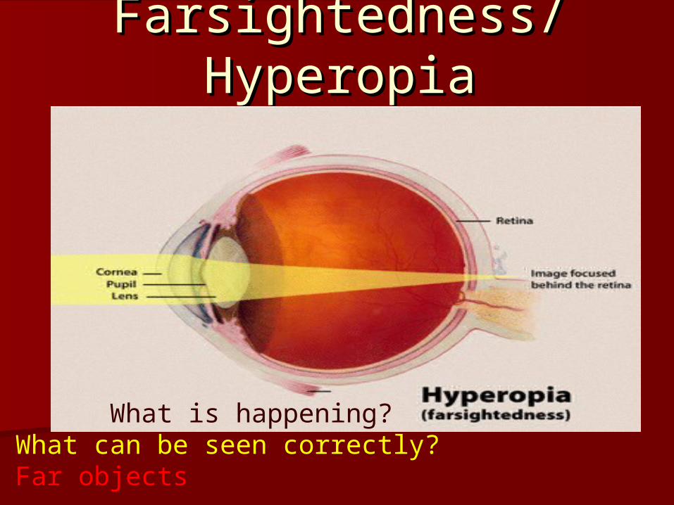
Farsightedness/Farsightedness/HyperopiaHyperopia
What is happening? What can be seen correctly?Far objects

Cortical ImplantCortical Implant
Dr. William Dobelle produced the Dr. William Dobelle produced the "Dobelle Eye“ which uses a video "Dobelle Eye“ which uses a video camera wired by a computer to camera wired by a computer to platinum electrodes implanted on the platinum electrodes implanted on the visual cortex (brain)visual cortex (brain)

Corneal TransplantCorneal Transplant
Just the cornea is transplantedJust the cornea is transplanted

Pink EyePink Eye
Conjunctivitis/ inflammation of the Conjunctivitis/ inflammation of the conjunctivaconjunctiva
Symptoms: itching, burning or Symptoms: itching, burning or stinging, discharge, swelling, stinging, discharge, swelling, wateringwatering
Allergic: not contagiousAllergic: not contagious Viral, bacterial: contaigous Viral, bacterial: contaigous
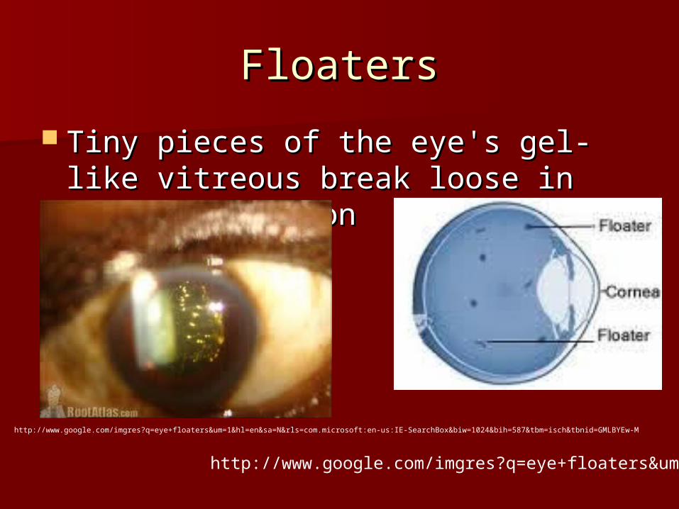
FloatersFloaters
Tiny pieces of the eye's gel-like Tiny pieces of the eye's gel-like vitreous break loose in the back vitreous break loose in the back portionportion
http://www.google.com/imgres?q=eye+floaters&um=1&hl=en&sa=N&rls=com.microsoft:en-us:IE-SearchBox&biw=1024&bih=587&tbm=isch&tbnid=GMLBYEw-M
http://www.google.com/imgres?q=eye+floaters&um=

Life Span ChangesLife Span Changes
Often first age related change noticedOften first age related change noticed 4040ss: lack of accomodation: lack of accomodation 5050ss: smell and taste: anosmia: : smell and taste: anosmia:
– Loss of olfactory receptorsLoss of olfactory receptors 60: 25% have significant hearing loss60: 25% have significant hearing loss
– Damage to hair cellsDamage to hair cells 65-75: 1/365-75: 1/3 85: 50% can not hear well: especially 85: 50% can not hear well: especially
high pitches and certain lettershigh pitches and certain letters
Life Span continuedLife Span continued
– Also presbycusis: degeneration of nerve Also presbycusis: degeneration of nerve pathwayspathways
– Tinnitus: ringing of the earsTinnitus: ringing of the ears– Hearing aidsHearing aids
Vision declines:Vision declines:– ““dry eyes”dry eyes”– Increased floatersIncreased floaters– Vitreous humor shrinks and pulls away from Vitreous humor shrinks and pulls away from
retina: flashesretina: flashes– Presbyopia: lens loses elasticity, can’t focus Presbyopia: lens loses elasticity, can’t focus ??

Life span continuedLife span continued
– 70: iris is inelastic and doesn’t let in as 70: iris is inelastic and doesn’t let in as much lightmuch light
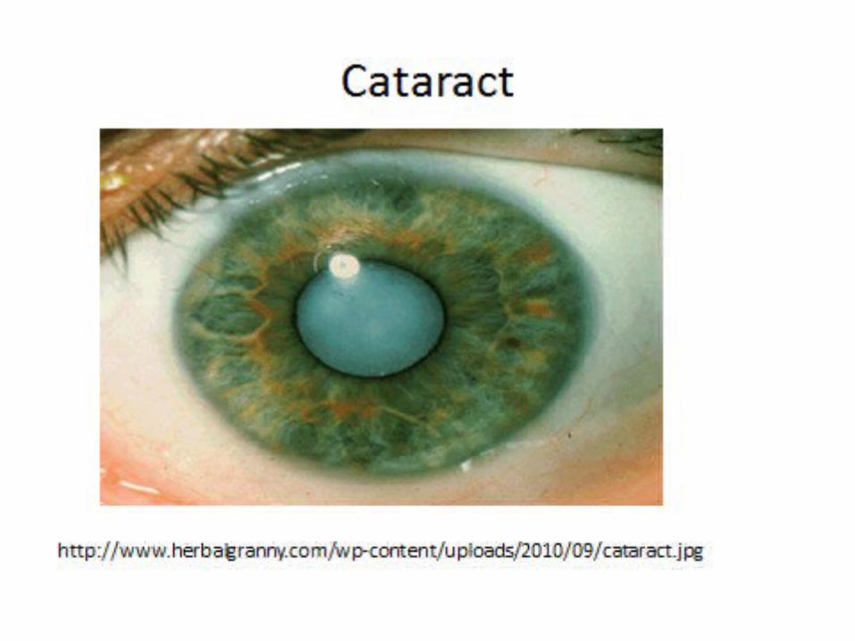
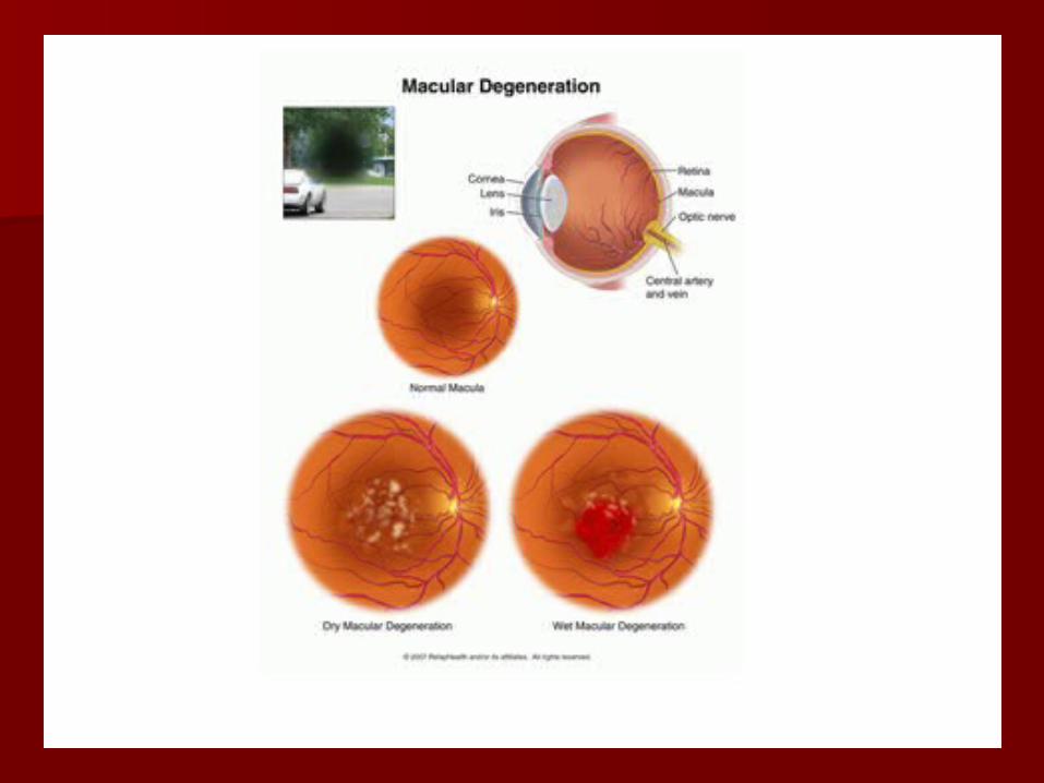
– Glaucoma: build up of pressure Glaucoma: build up of pressure ??– Aqueous humor: shuts blood vessels Aqueous humor: shuts blood vessels ??– Cataract: lens or capsule becomes opaque Cataract: lens or capsule becomes opaque ??– Laser surgery and implantLaser surgery and implant– Macular degeneration: macula cones Macular degeneration: macula cones
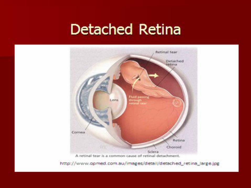
degeneratedegenerate– Detached retina: pulls away from choroid Detached retina: pulls away from choroid
coat coat ??