Chapter 11 The Sensory System - Amazon S3 · aqueous humor kinesthesia semicircular canal ... Pop...
84
Copyright © 2015 Wolters Kluwer Health | Lippincott Williams & Wilkins Chapter 11 The Sensory System
Transcript of Chapter 11 The Sensory System - Amazon S3 · aqueous humor kinesthesia semicircular canal ... Pop...
Copyright © 2015 Wolters Kluwer Health | Lippincott Williams & Wilkins
Chapter 11
The Sensory System
Copyright © 2015 Wolters Kluwer Health | Lippincott Williams & Wilkins
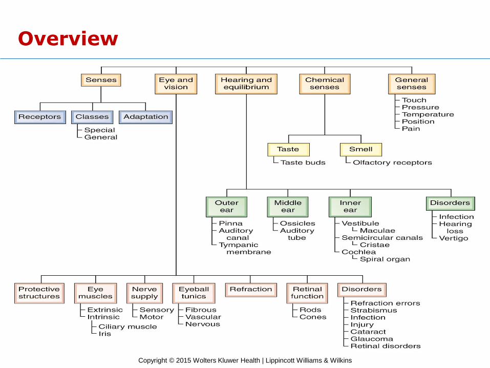
Overview
Copyright © 2015 Wolters Kluwer Health | Lippincott Williams & Wilkins
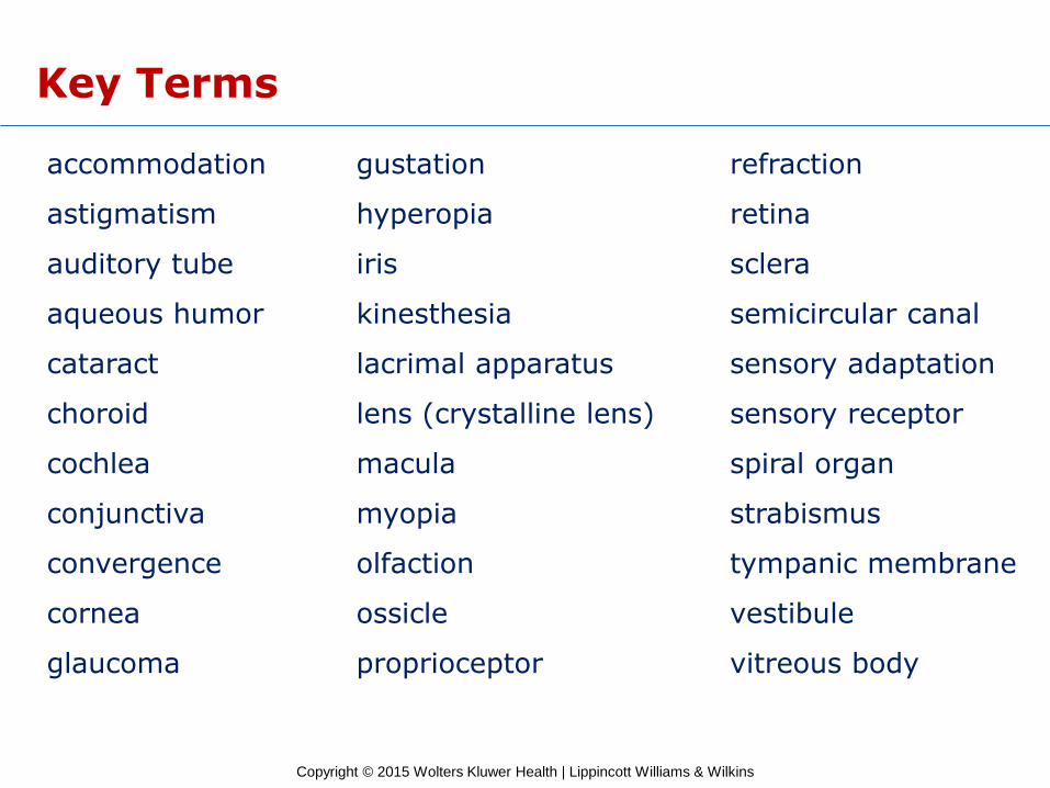
Key Terms
accommodation gustation refraction
astigmatism hyperopia retina
auditory tube iris sclera
aqueous humor kinesthesia semicircular canal
cataract lacrimal apparatus sensory adaptation
choroid lens (crystalline lens) sensory receptor
cochlea macula spiral organ
conjunctiva myopia strabismus
convergence olfaction tympanic membrane
cornea ossicle vestibule
glaucoma proprioceptor vitreous body

Copyright © 2015 Wolters Kluwer Health | Lippincott Williams & Wilkins
The Senses
Learning Objectives
1. Describe the functions of the sensory system.
2. Differentiate between the different types of sensory receptors, and give examples of each.
3. Describe sensory adaptation, and explain its value.

Copyright © 2015 Wolters Kluwer Health | Lippincott Williams & Wilkins
The Eye and Vision
Learning Objectives
4. List and describe the structures that protect the eye.
5. Cite the location and the purpose of the extrinsic eye muscles.
6. Identify the three tunics of the eye.
7. Describe the processes involved in vision.
8. Differentiate between the rods and the cones of the eye.
9. List seven disorders of the eye and vision.
Copyright © 2015 Wolters Kluwer Health | Lippincott Williams & Wilkins
The Ear
Learning Objectives
10. Describe the three divisions of the ear.
11. Describe the receptor for hearing, and explain how it functions.
12. Compare the locations and functions of the equilibrium receptors.
13. Describe three disorders that involve the ear.
Copyright © 2015 Wolters Kluwer Health | Lippincott Williams & Wilkins
Other Special Sense Organs
Learning Objective
14. Discuss the locations and functions of the sense organs for taste and smell.
Copyright © 2015 Wolters Kluwer Health | Lippincott Williams & Wilkins
The General Senses
Learning Objectives
15. Describe five general senses.
16. List five approaches for treatment of pain.

Copyright © 2015 Wolters Kluwer Health | Lippincott Williams & Wilkins
Case Study
Learning Objective
17. Referring to the case study, discuss the purpose and mechanism of a cochlear implant.

Copyright © 2015 Wolters Kluwer Health | Lippincott Williams & Wilkins
Word Anatomy
Learning Objective
18. Show how word parts are used to build words related to the sensory system.
Copyright © 2015 Wolters Kluwer Health | Lippincott Williams & Wilkins
The Senses
• The sensory system provides awareness of our external and internal environments.
– Environmental change (stimulus) initiates nerve impulse.
– Stimulus interpreted by the cerebral cortex.
– Sensation experienced.
Copyright © 2015 Wolters Kluwer Health | Lippincott Williams & Wilkins
The Senses (cont.)
Sensory Receptors
• Receptor classification based on structure
– Free dendrite
– End organ
– Specialized cell
• Receptor classification based on stimulus
– Chemoreceptor
– Photoreceptor
– Thermoreceptor
– Mechanoreceptor
Copyright © 2015 Wolters Kluwer Health | Lippincott Williams & Wilkins
The Senses (cont.)
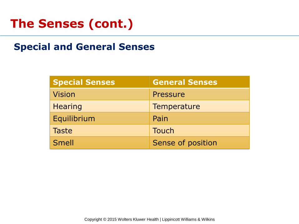
Special and General Senses
Special Senses General Senses
Vision Pressure
Hearing Temperature
Equilibrium Pain
Taste Touch
Smell Sense of position
Copyright © 2015 Wolters Kluwer Health | Lippincott Williams & Wilkins
The Senses (cont.)
Sensory Adaptation
• Receptors often become less sensitive to a continuous unimportant stimulus.
Copyright © 2015 Wolters Kluwer Health | Lippincott Williams & Wilkins
The Senses (cont.)
Checkpoints
11-1 What is a sensory receptor?
11-2 What are some categories of sensory receptors based on type of stimulus?
11-3 How do the special and general senses differ in location?
11-4 What happens when a sensory receptor adapts to a stimulus?
Copyright © 2015 Wolters Kluwer Health | Lippincott Williams & Wilkins
The Senses (cont.)
Pop Quiz
11.1 Which of the following is a special sense?
A) Pressure
B) Taste
C) Touch
D) Proprioception
Copyright © 2015 Wolters Kluwer Health | Lippincott Williams & Wilkins
The Senses (cont.)
Pop Quiz Answer
11.1 Which of the following is a special sense?
A) Pressure
B) Taste
C) Touch
D) Proprioception
Copyright © 2015 Wolters Kluwer Health | Lippincott Williams & Wilkins
The Eye and Vision
Learning Objectives
4. List and describe the structures that protect the eye.
5. Cite the location and the purpose of the extrinsic eye muscles.
6. Identify the three tunics of the eye.
7. Describe the processes involved in vision.
8. Differentiate between the rods and the cones of the eye.
9. List seven disorders of the eye and vision.
Copyright © 2015 Wolters Kluwer Health | Lippincott Williams & Wilkins
The Eye and Vision (cont.)
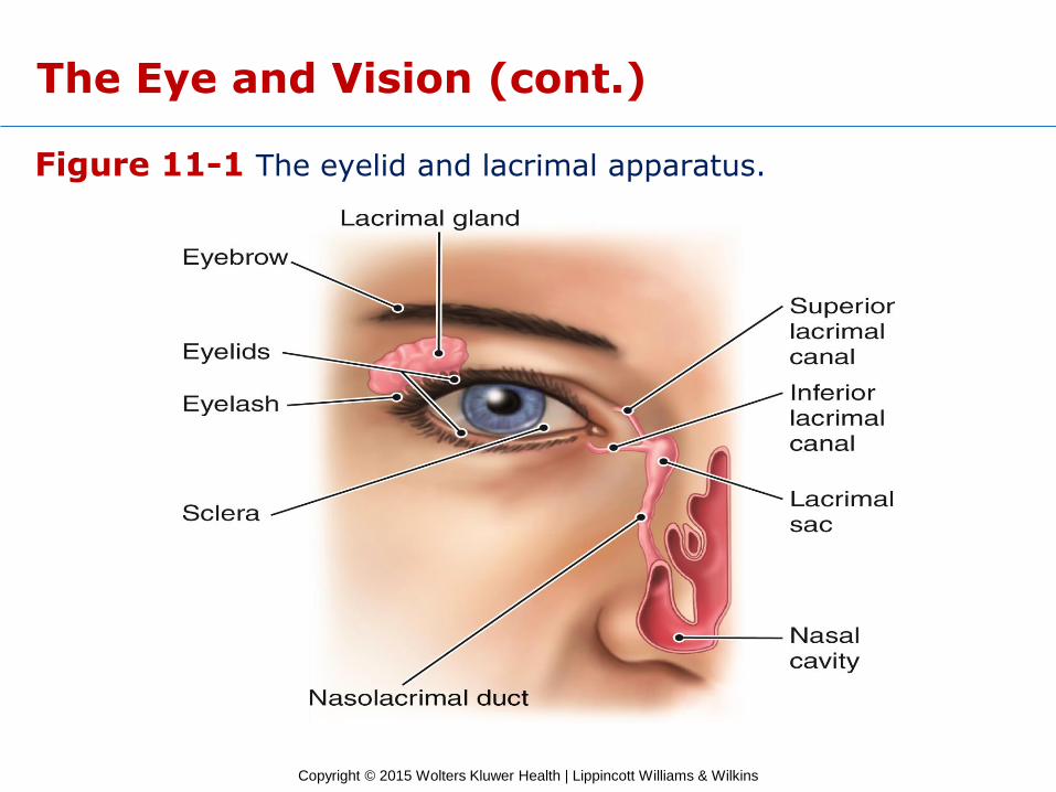
Protective Structures of the Eye
• Bony orbit
• Eyelids
• Eyelashes
• Eyebrows
• Lacrimal glands
• Conjunctiva
Copyright © 2015 Wolters Kluwer Health | Lippincott Williams & Wilkins
Figure 11-1 The eyelid and lacrimal apparatus.
The Eye and Vision (cont.)
Copyright © 2015 Wolters Kluwer Health | Lippincott Williams & Wilkins
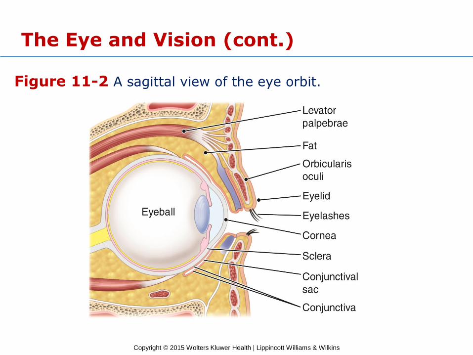
Figure 11-2 A sagittal view of the eye orbit.
The Eye and Vision (cont.)
Copyright © 2015 Wolters Kluwer Health | Lippincott Williams & Wilkins
The Eye and Vision (cont.)
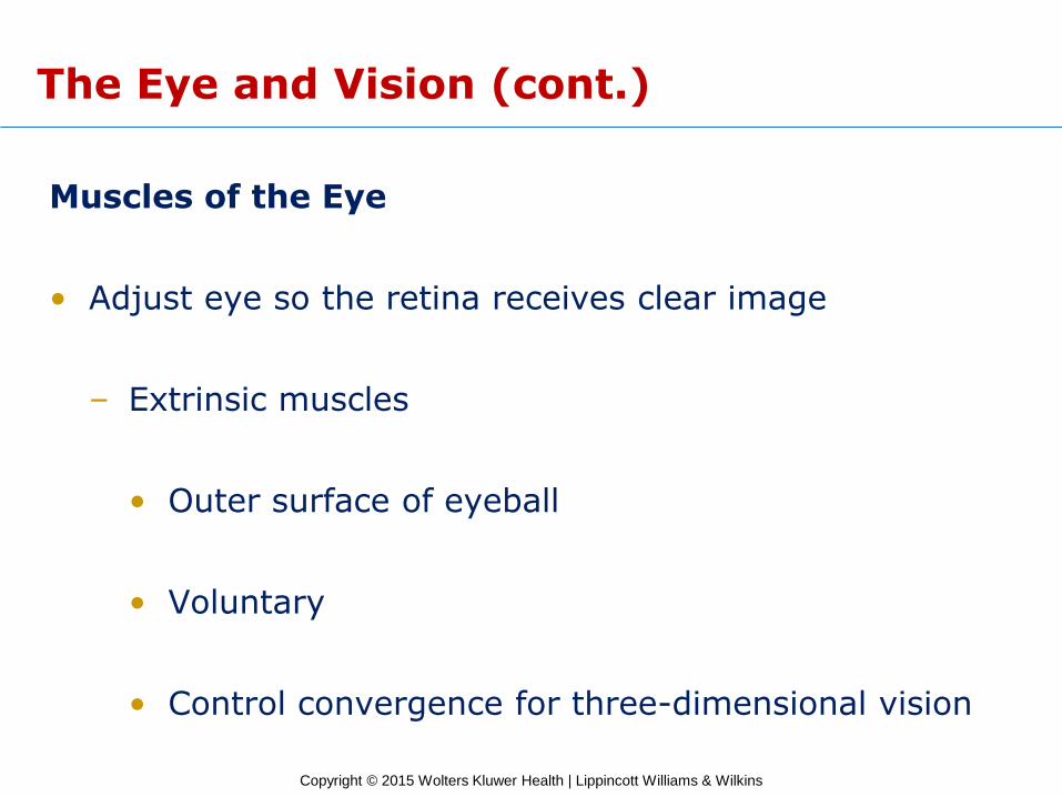
Muscles of the Eye
• Adjust eye so the retina receives clear image
– Extrinsic muscles
• Outer surface of eyeball
• Voluntary
• Control convergence for three-dimensional vision
Copyright © 2015 Wolters Kluwer Health | Lippincott Williams & Wilkins
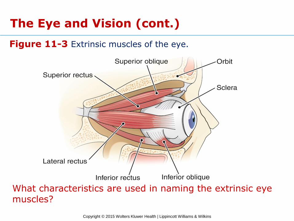
Figure 11-3 Extrinsic muscles of the eye.
What characteristics are used in naming the extrinsic eye muscles?
The Eye and Vision (cont.)
Copyright © 2015 Wolters Kluwer Health | Lippincott Williams & Wilkins
The Eye and Vision (cont.)
Nerve Supply to the Eye
• Sensory
– Optic nerve (CN II)
– Ophthalmic branch of trigeminal nerve (CN V)
• Motor
– Oculomotor nerve (CN III)
– Trochlear (CN IV)
– Abducens (CN VI)
Copyright © 2015 Wolters Kluwer Health | Lippincott Williams & Wilkins
The Eye and Vision (cont.)
Checkpoints
11-5 What are five structures that protect the eye?
11-6 What is the function of the extrinsic eye muscles?
11-7 Which cranial nerve carries impulses from the retina to the brain?
Copyright © 2015 Wolters Kluwer Health | Lippincott Williams & Wilkins
The Eye and Vision (cont.)
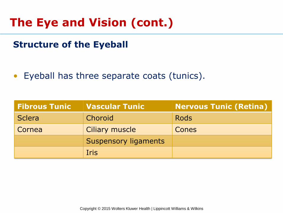
Structure of the Eyeball
• Eyeball has three separate coats (tunics).
Fibrous Tunic Vascular Tunic Nervous Tunic (Retina)
Sclera Choroid Rods
Cornea Ciliary muscle Cones
Suspensory ligaments
Iris
Copyright © 2015 Wolters Kluwer Health | Lippincott Williams & Wilkins
The Eye and Vision (cont.)
Pop Quiz
11.2 The middle, pigmented layer of the eye is the:
A) Sclera
B) Conjunctiva
C) Retina
D) Choroid
Copyright © 2015 Wolters Kluwer Health | Lippincott Williams & Wilkins
The Eye and Vision (cont.)
Pop Quiz Answer
11.2 The middle, pigmented layer of the eye is the:
A) Sclera
B) Conjunctiva
C) Retina
D) Choroid
Copyright © 2015 Wolters Kluwer Health | Lippincott Williams & Wilkins
The Eye and Vision (cont.)
Pathway of Light Rays and Refraction
• Transparent parts of the eye that refract light
– Cornea
– Aqueous humor
– Crystalline lens
– Vitreous body
Copyright © 2015 Wolters Kluwer Health | Lippincott Williams & Wilkins
The Eye and Vision (cont.)
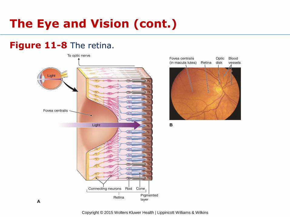
Function of the Retina
• Rod cells
– Function in dim light
– Not sensitive to color
– Blurred images
• Cone cells
– Function in bright light
– Color-sensitive
– Sharp images
Copyright © 2015 Wolters Kluwer Health | Lippincott Williams & Wilkins
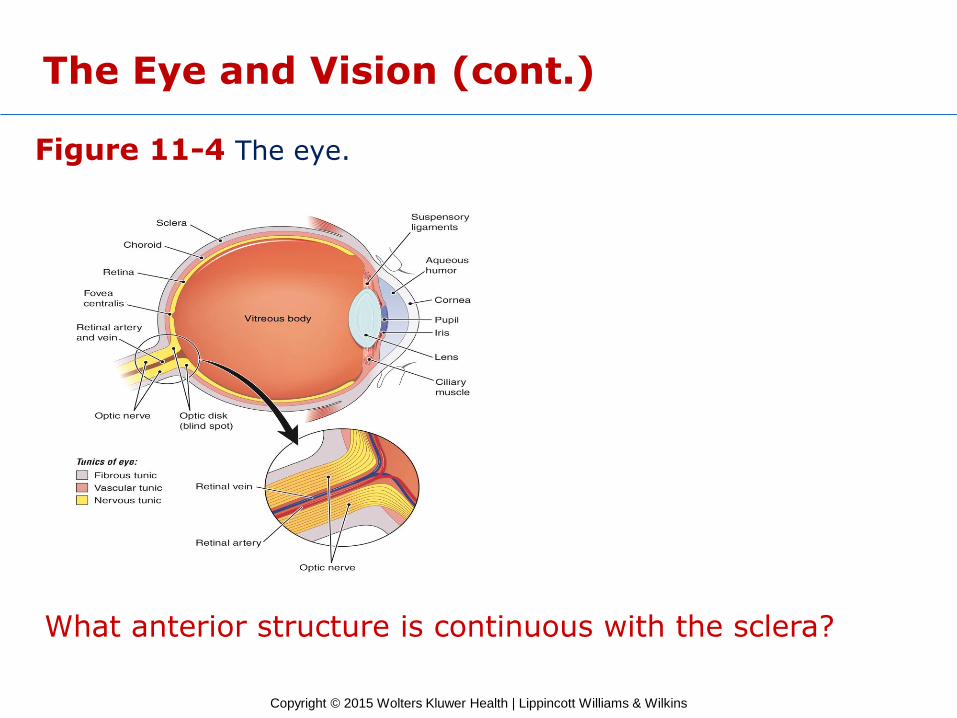
Figure 11-4 The eye.
What anterior structure is continuous with the sclera?
The Eye and Vision (cont.)
Copyright © 2015 Wolters Kluwer Health | Lippincott Williams & Wilkins
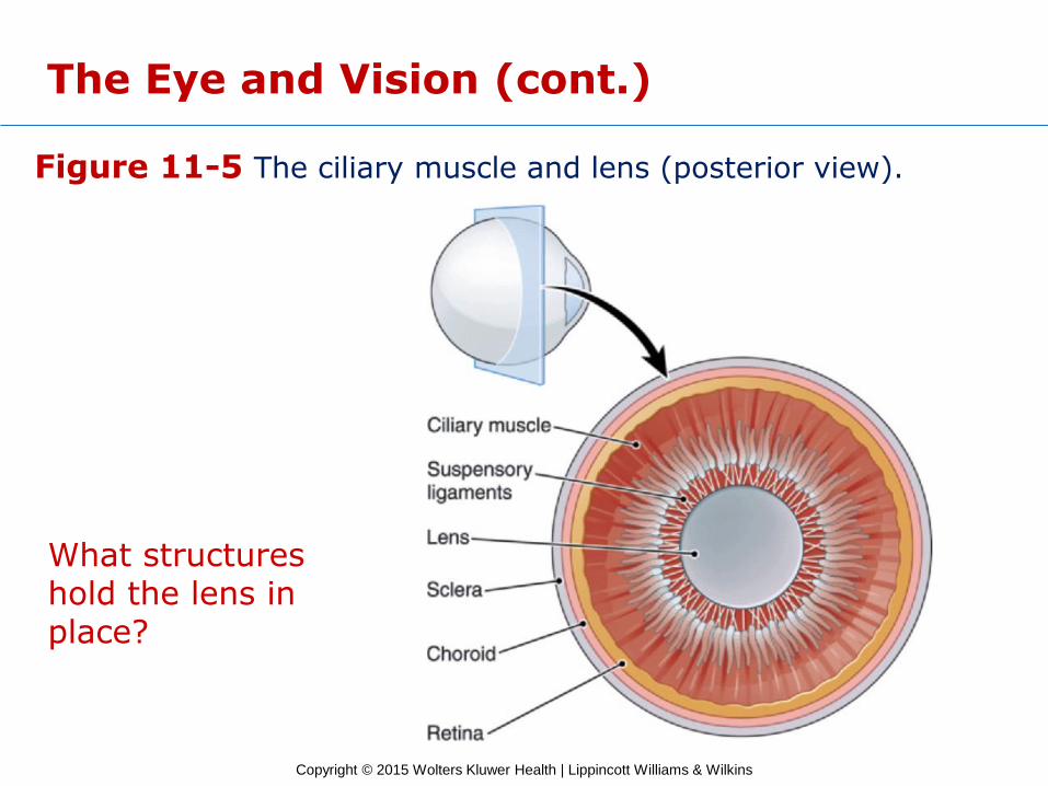
Figure 11-5 The ciliary muscle and lens (posterior view).
What structures hold the lens in place?
The Eye and Vision (cont.)
Copyright © 2015 Wolters Kluwer Health | Lippincott Williams & Wilkins
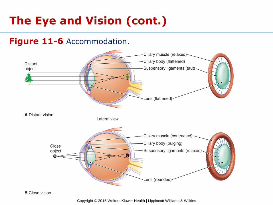
Figure 11-6 Accommodation.
The Eye and Vision (cont.)
Copyright © 2015 Wolters Kluwer Health | Lippincott Williams & Wilkins
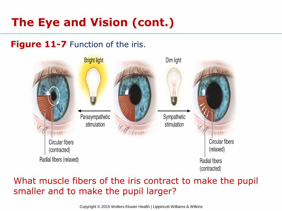
Figure 11-7 Function of the iris.
What muscle fibers of the iris contract to make the pupil smaller and to make the pupil larger?
The Eye and Vision (cont.)
Copyright © 2015 Wolters Kluwer Health | Lippincott Williams & Wilkins
Figure 11-8 The retina.
The Eye and Vision (cont.)
Copyright © 2015 Wolters Kluwer Health | Lippincott Williams & Wilkins
The Eye and Vision (cont.)
Checkpoints
11-8 What are the three tunics of the eyeball?
11-9 What are the structures that refract light as it passes through the eye?
11-10 What is the function of the ciliary muscle?
11-11 What is the function of the iris?
11-12 What are the receptor cells of the retina?
Copyright © 2015 Wolters Kluwer Health | Lippincott Williams & Wilkins
The Eye and Vision (cont.)
Disorders of the Eye and Vision
• Errors of refraction
– Hyperopia
– Myopia
– Presbyopia
– Astigmatism
• Strabismus
– Convergent
– Divergent
– Amblyopia
Copyright © 2015 Wolters Kluwer Health | Lippincott Williams & Wilkins
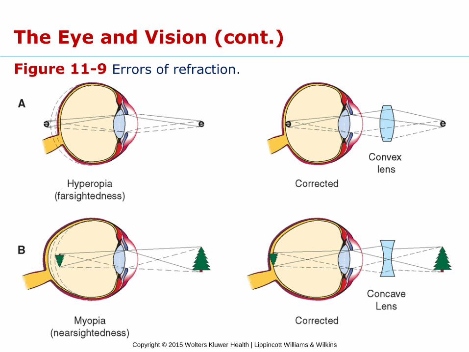
Figure 11-9 Errors of refraction.
The Eye and Vision (cont.)
Copyright © 2015 Wolters Kluwer Health | Lippincott Williams & Wilkins
The Eye and Vision (cont.)
Disorders of the Eye and Vision (cont.)
• Infections
– Conjunctivitis
– Inclusion conjunctivitis
– Neonatal conjunctivitis (ophthalmia neonatorum)
• Injuries
– Corneal laceration
– Enucleation

Copyright © 2015 Wolters Kluwer Health | Lippincott Williams & Wilkins
The Eye and Vision (cont.)
Disorders of the Eye and Vision (cont.)
• Cataract
• Glaucoma
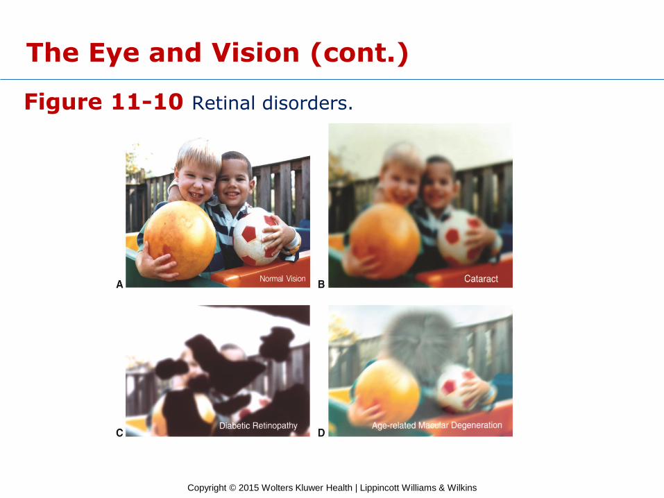
• Retinal disorders
– Diabetic retinopathy
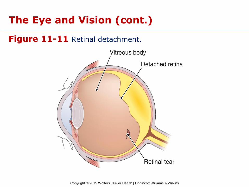
– Retinal detachment
– Macular degeneration
Copyright © 2015 Wolters Kluwer Health | Lippincott Williams & Wilkins
Figure 11-10 Retinal disorders.
The Eye and Vision (cont.)
Copyright © 2015 Wolters Kluwer Health | Lippincott Williams & Wilkins
Figure 11-11 Retinal detachment.
The Eye and Vision (cont.)
Copyright © 2015 Wolters Kluwer Health | Lippincott Williams & Wilkins
The Eye and Vision (cont.)
Checkpoints
11-13 What are four errors of refraction?
11-14 What is cloudiness of the lens called?
11-15 What disorder is caused by excess fluid pressure in the eye?
Copyright © 2015 Wolters Kluwer Health | Lippincott Williams & Wilkins
The Ear
Learning Objectives
10. Describe the three divisions of the ear.
11. Describe the receptor for hearing, and explain how it functions.
12. Compare the locations and functions of the equilibrium receptors.
Copyright © 2015 Wolters Kluwer Health | Lippincott Williams & Wilkins
The Ear (cont.)
• Sense organ for hearing and equilibrium
• Components
– Outer ear
– Middle ear
– Inner ear

Copyright © 2015 Wolters Kluwer Health | Lippincott Williams & Wilkins
The Ear (cont.)
The Outer Ear
• Structure
– Pinna (auricle)
– External auditory canal (meatus)
– Ceruminous glands
– Tympanic membrane
• Function
– Collects sound waves and transmits sounds to the middle ear
Copyright © 2015 Wolters Kluwer Health | Lippincott Williams & Wilkins
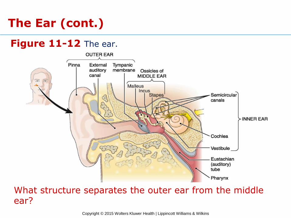
Figure 11-12 The ear.
What structure separates the outer ear from the middle ear?
The Ear (cont.)
Copyright © 2015 Wolters Kluwer Health | Lippincott Williams & Wilkins
The Ear (cont.)
Middle Ear
• Structure
– Ear ossicles
• Malleus
• Incus
• Stapes
– Auditory tube
• Function
– Amplifies sound waves and transmits sounds to the inner ear
Copyright © 2015 Wolters Kluwer Health | Lippincott Williams & Wilkins
The Ear (cont.)
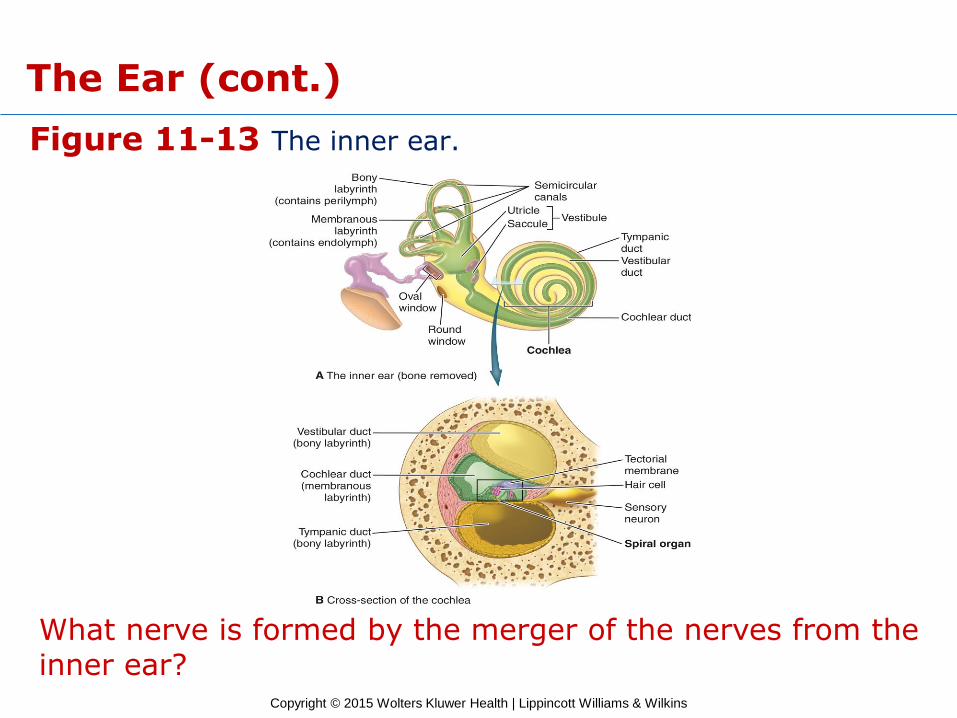
Inner Ear
• Structure
– Bony labyrinth; contains perilymph
• Divisions
• Vestibule
• Semicircular canals
• Cochlea
– Membranous labyrinth; contains endolymph
• Function
– Transduces sound waves into nerve impulses
Copyright © 2015 Wolters Kluwer Health | Lippincott Williams & Wilkins
Figure 11-13 The inner ear.
What nerve is formed by the merger of the nerves from the inner ear?
The Ear (cont.)
Copyright © 2015 Wolters Kluwer Health | Lippincott Williams & Wilkins
The Ear (cont.)
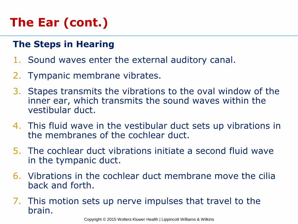
The Steps in Hearing
1. Sound waves enter the external auditory canal.
2. Tympanic membrane vibrates.
3. Stapes transmits the vibrations to the oval window of the inner ear, which transmits the sound waves within the vestibular duct.
4. This fluid wave in the vestibular duct sets up vibrations in the membranes of the cochlear duct.
5. The cochlear duct vibrations initiate a second fluid wave in the tympanic duct.
6. Vibrations in the cochlear duct membrane move the cilia back and forth.
7. This motion sets up nerve impulses that travel to the brain.
Copyright © 2015 Wolters Kluwer Health | Lippincott Williams & Wilkins
The Ear (cont.)
Equilibrium
• Ciliated equilibrium sensory receptors are located in vestibule and semicircular canals.
• Types of receptors
– Maculae
– Cristae
• Nerve supply via vestibular nerve
Copyright © 2015 Wolters Kluwer Health | Lippincott Williams & Wilkins
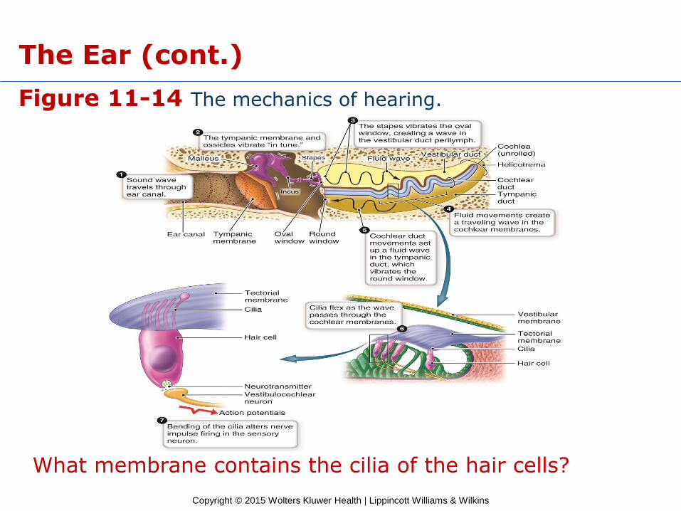
Figure 11-14 The mechanics of hearing.
What membrane contains the cilia of the hair cells?
The Ear (cont.)
Copyright © 2015 Wolters Kluwer Health | Lippincott Williams & Wilkins
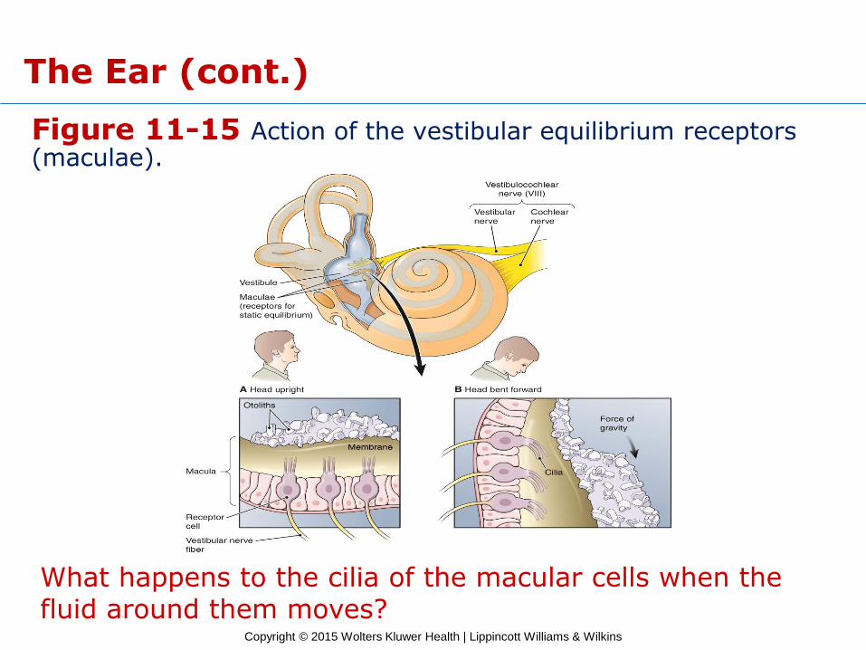
Figure 11-15 Action of the vestibular equilibrium receptors (maculae).
What happens to the cilia of the macular cells when the fluid around them moves?
The Ear (cont.)
Copyright © 2015 Wolters Kluwer Health | Lippincott Williams & Wilkins
The Ear (cont.)
Checkpoints
11-16 What are the three divisions of the ear?
11-17 What are the names of the ear ossicles, and what do they do?
11-18 What are the two fluids found in the inner ear, and where are they located?
11-19 What is the name of the hearing organ, and where is it located?
11-20 Where are the receptors for equilibrium located?
Copyright © 2015 Wolters Kluwer Health | Lippincott Williams & Wilkins
The Ear (cont.)
Disorders of the Ear
• Ear infection
– Otitis media
– Otitis externa
• Hearing loss
– Conductive hearing loss
– Sensorineural hearing loss
– Presbycusis
• Equilibrium disturbances
– Vertigo
Copyright © 2015 Wolters Kluwer Health | Lippincott Williams & Wilkins
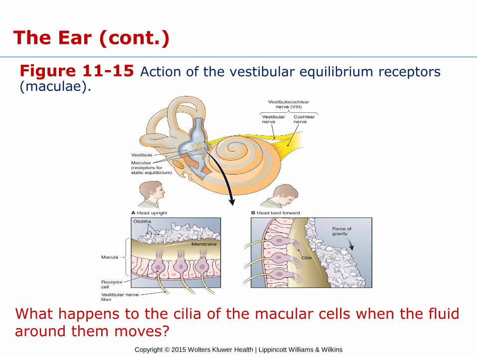
Figure 11-15 Action of the vestibular equilibrium receptors (maculae).
What happens to the cilia of the macular cells when the fluid around them moves?
The Ear (cont.)
Copyright © 2015 Wolters Kluwer Health | Lippincott Williams & Wilkins
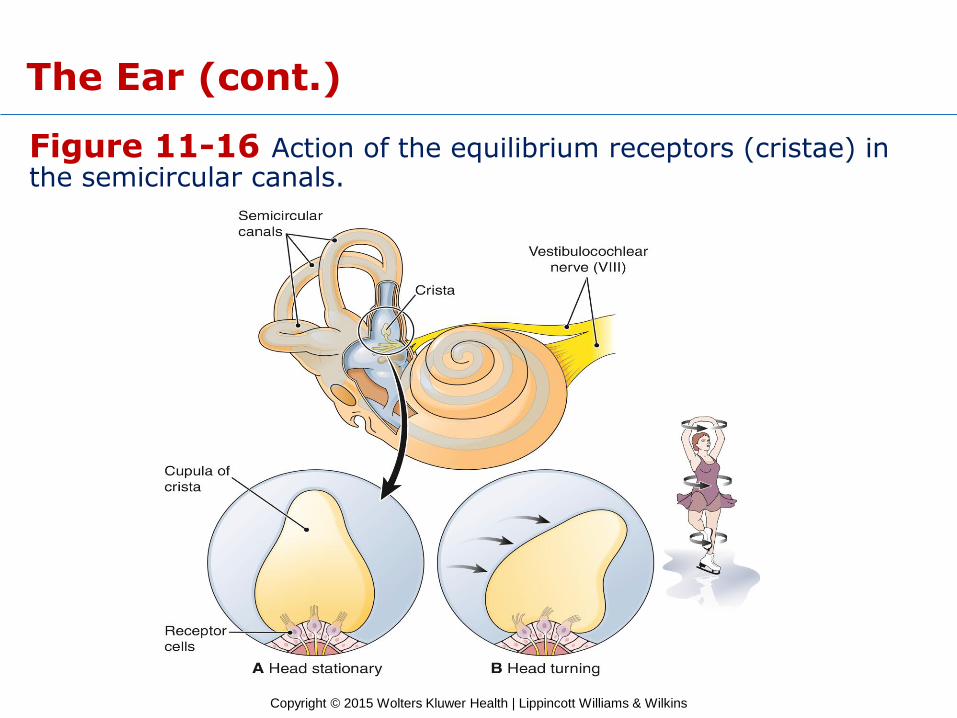
Figure 11-16 Action of the equilibrium receptors (cristae) in the semicircular canals.
The Ear (cont.)
Copyright © 2015 Wolters Kluwer Health | Lippincott Williams & Wilkins
The Ear (cont.)
Checkpoints
11-21 What are the two types of hearing loss?
11-22 What is the term for an abnormal sensation of spinning?
Copyright © 2015 Wolters Kluwer Health | Lippincott Williams & Wilkins
The Ear (cont.)
Pop Quiz
11.3 The ear ossicle that is in contact with the tympanic membrane is the:
A) Malleus
B) Incus
C) Meatus
D) Stapes
Copyright © 2015 Wolters Kluwer Health | Lippincott Williams & Wilkins
The Ear (cont.)
Pop Quiz Answer
11.3 The ear ossicle that is in contact with the tympanic membrane is the:
A) Malleus
B) Incus
C) Meatus
D) Stapes
Copyright © 2015 Wolters Kluwer Health | Lippincott Williams & Wilkins
Other Special Sense Organs
Learning Objective
14. Discuss the locations and functions of the sense organs for taste and smell.
Copyright © 2015 Wolters Kluwer Health | Lippincott Williams & Wilkins
Other Special Sense Organs (cont.)
• Taste and smell sense organs respond to chemical stimuli.
Copyright © 2015 Wolters Kluwer Health | Lippincott Williams & Wilkins
Other Special Sense Organs (cont.)
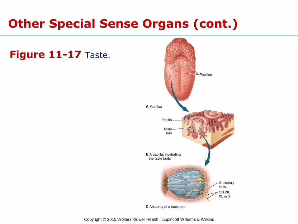
Sense of Taste (Gustation)
• Receptors (taste buds)
– Sweet
– Salty
– Sour
– Bitter
– Umami
• Nerve supply
– Facial nerve
– Glossopharyngeal nerve
Copyright © 2015 Wolters Kluwer Health | Lippincott Williams & Wilkins
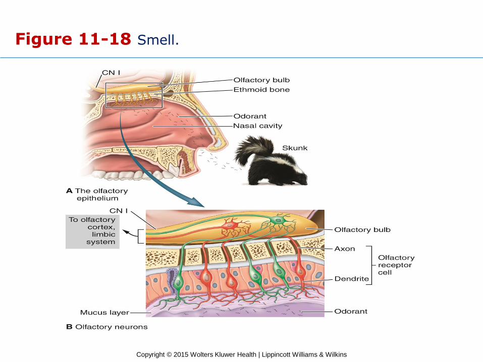
Sense of Smell (Olfaction)
• Smell receptors in the nasal cavity
– Stimulated by substances in solution in nasal fluids.
– Smells stimulate appetite and flow of digestive juices.
• Olfactory nerve (cranial nerve I)
Other Special Sense Organs (cont.)
Copyright © 2015 Wolters Kluwer Health | Lippincott Williams & Wilkins
Figure 11-17 Taste.
Other Special Sense Organs (cont.)
Copyright © 2015 Wolters Kluwer Health | Lippincott Williams & Wilkins
Figure 11-18 Smell.
Copyright © 2015 Wolters Kluwer Health | Lippincott Williams & Wilkins
Other Special Sense Organs (cont.)
Checkpoint
11-23 What are the special senses that respond to chemical stimuli?
Copyright © 2015 Wolters Kluwer Health | Lippincott Williams & Wilkins
The General Senses
Learning Objectives
15. Describe five general senses.
16. List five approaches for treatment of pain.
Copyright © 2015 Wolters Kluwer Health | Lippincott Williams & Wilkins
The General Senses (cont.)
• Receptors scattered throughout the body sense
• Touch
• Pressure
• Heat
• Cold
• Position
• Pain
Copyright © 2015 Wolters Kluwer Health | Lippincott Williams & Wilkins
The General Senses (cont.)
Sense of Pain
• Pain receptors
– Are free nerve endings
– Are found in skin, muscles, joints, and (to a lesser extent) in most internal organs
• Pain relief
– Analgesic drugs
– Anesthetics
– Endorphins
– Heat or cold
– Relaxation or distraction techniques
Copyright © 2015 Wolters Kluwer Health | Lippincott Williams & Wilkins
Sensory Adaptation
• Occurs when receptors are exposed to continuous stimulus.
• Some receptors can adjust themselves, so sensation becomes less acute.
• Receptors adapt at different rates.
• Pain receptors do not adapt.
Copyright © 2015 Wolters Kluwer Health | Lippincott Williams & Wilkins
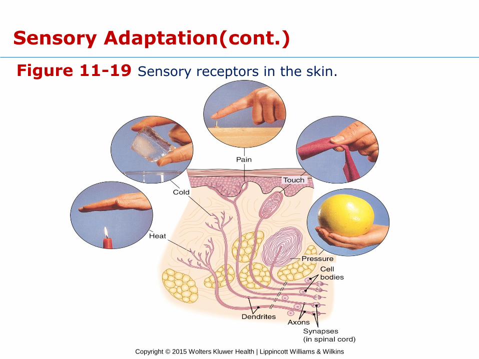
Figure 11-19 Sensory receptors in the skin.
Sensory Adaptation(cont.)
Copyright © 2015 Wolters Kluwer Health | Lippincott Williams & Wilkins
The General Senses
Checkpoints
11-24 What are five examples of general senses?
11-25 What are proprioceptors, and where are they located?
Copyright © 2015 Wolters Kluwer Health | Lippincott Williams & Wilkins
The General Senses (cont.)
Pop Quiz
11.4 If you hold your arm motionless in the air, which receptors are most important in informing you of your hand position?
A) Proprioceptors
B) Pressure receptors
C) Tactile corpuscles
D) Free nerve endings
Copyright © 2015 Wolters Kluwer Health | Lippincott Williams & Wilkins
The General Senses (cont.)
Pop Quiz Answer
11.4 If you hold your arm motionless in the air, which receptors are most important in informing you of your hand position?
A) Proprioceptors
B) Pressure receptors
C) Tactile corpuscles
D) Free nerve endings
Copyright © 2015 Wolters Kluwer Health | Lippincott Williams & Wilkins
Case Study
Learning Objective
17. Referring to the case study, discuss the purpose and mechanism of a cochlear implant.
Copyright © 2015 Wolters Kluwer Health | Lippincott Williams & Wilkins
Case Study (cont.)
Cochlear Implant ● A small surgically implanted electronic device that can
help to provide a sense of sound to someone with sensorineural hearing loss and who is profoundly deaf or severely hard of hearing.
● Implant candidacy takes into account a person's hearing history, cause of hearing loss, amount of residual hearing, speech recognition ability, health status, and family commitment to aural habilitation/rehabilitation.
● Does not cure deafness but rather may be considered a prosthetic substitute for hearing.
Copyright © 2015 Wolters Kluwer Health | Lippincott Williams & Wilkins
Case Study (cont.)
Diagnosis ● The inner part of the ear contains tiny hair cells (cilia)
that change sounds into electric signals. The nerves carry these signals to the brain.
● Sensorineural hearing loss is caused by damage to these special cells in the inner ear.
● Vibrations in the cochlear duct membrane move the cilia back and forth, which set up the nerve impulses for hearing that travel to the brain.
● Bacterial meningitis destroyed the hair cells in Evan’s cochlea causing the hearing impairment.
● The strong drugs used to treat the meningitis may have been ototoxic contributing to further destruction of the hair cells.

Copyright © 2015 Wolters Kluwer Health | Lippincott Williams & Wilkins
Case Study (cont.)
Treatment
Cochlear implant
− Stimulates the cochlear nerve directly, bypassing the damaged
receptor cells
− Three components:
• Internal receiver with electrodes going to the inner ear
• Transmitter
• Sound processor
− Three components allow the brain to interpret the frequency of
sound as if the hair cells were functioning properly.
Postimplant follow-up with a speech therapist and audiologist
Copyright © 2015 Wolters Kluwer Health | Lippincott Williams & Wilkins
Word Anatomy
Learning Objective
18. Show how word parts are used to build words related to the sensory system.
Copyright © 2015 Wolters Kluwer Health | Lippincott Williams & Wilkins
Word Anatomy (cont.)
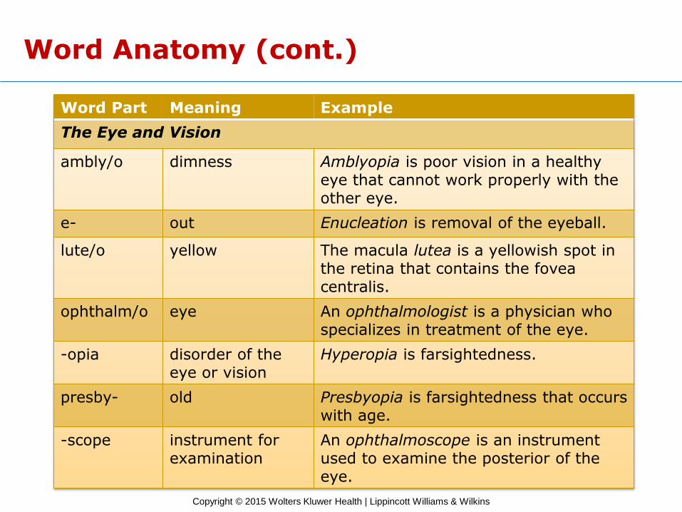
Word Part Meaning Example
The Eye and Vision
ambly/o dimness Amblyopia is poor vision in a healthy eye that cannot work properly with the other eye.
e- out Enucleation is removal of the eyeball.
lute/o yellow The macula lutea is a yellowish spot in the retina that contains the fovea centralis.
ophthalm/o eye An ophthalmologist is a physician who specializes in treatment of the eye.
-opia disorder of the eye or vision
Hyperopia is farsightedness.
presby- old Presbyopia is farsightedness that occurs with age.
-scope instrument for examination
An ophthalmoscope is an instrument used to examine the posterior of the eye.
Copyright © 2015 Wolters Kluwer Health | Lippincott Williams & Wilkins
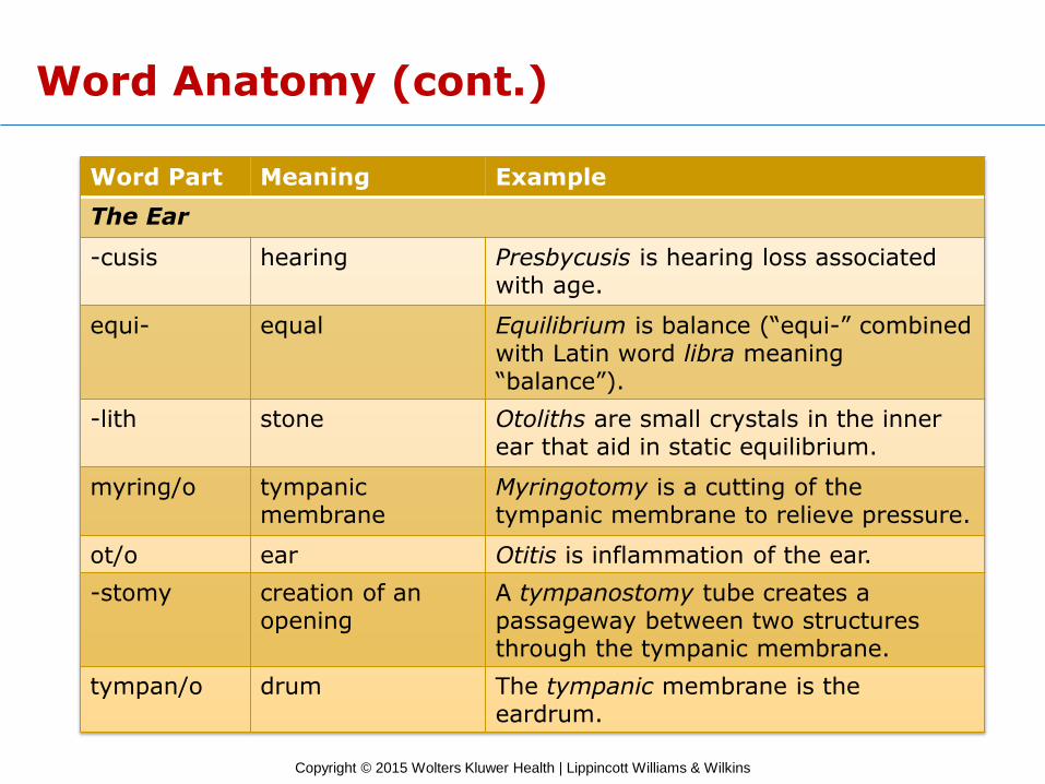
Word Anatomy (cont.)
Word Part Meaning Example
The Ear
-cusis hearing Presbycusis is hearing loss associated with age.
equi- equal Equilibrium is balance (“equi-” combined with Latin word libra meaning “balance”).
-lith stone Otoliths are small crystals in the inner ear that aid in static equilibrium.
myring/o tympanic membrane
Myringotomy is a cutting of the tympanic membrane to relieve pressure.
ot/o ear Otitis is inflammation of the ear.
-stomy creation of an opening
A tympanostomy tube creates a passageway between two structures through the tympanic membrane.
tympan/o drum The tympanic membrane is the eardrum.
Copyright © 2015 Wolters Kluwer Health | Lippincott Williams & Wilkins
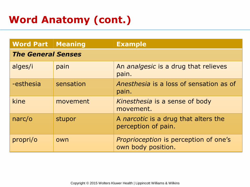
Word Anatomy (cont.)
Word Part Meaning Example
The General Senses
alges/i pain An analgesic is a drug that relieves pain.
-esthesia sensation Anesthesia is a loss of sensation as of pain.
kine movement Kinesthesia is a sense of body movement.
narc/o stupor A narcotic is a drug that alters the perception of pain.
propri/o own Proprioception is perception of one’s own body position.