CHAPTER 10: THE SKELETAL SYSTEM The Rigid Framework of the Body.
68
CHAPTER 10: CHAPTER 10: THE THE SKELETAL SYSTEM SKELETAL SYSTEM The Rigid Framework The Rigid Framework of the Body of the Body
-
Upload
gordon-davidson -
Category
Documents
-
view
232 -
download
3
Transcript of CHAPTER 10: THE SKELETAL SYSTEM The Rigid Framework of the Body.
CHAPTER 10:CHAPTER 10: THE THE SKELETAL SYSTEMSKELETAL SYSTEM
The Rigid FrameworkThe Rigid Frameworkof the Bodyof the Body
Skeletal AnatomySkeletal Anatomy
Skeletal SystemSkeletal System
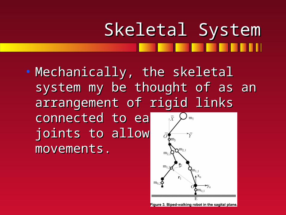
• Mechanically, the skeletal system my be Mechanically, the skeletal system my be thought of as an arrangement of rigid links thought of as an arrangement of rigid links connected to each other at joints to allow connected to each other at joints to allow specific movements.specific movements.

Knowledge ofthe skeletalsystem is
important forALL
movementanalysts

Composition and Structure of Bone Composition and Structure of Bone TissueTissue
• Mechanical functions of boneMechanical functions of bone• provides a rigid skeletal framework provides a rigid skeletal framework
to support and protect other tissues. to support and protect other tissues. • forms a system of rigid levers (links) forms a system of rigid levers (links)
that can be moved by forces from the that can be moved by forces from the attached muscles (attached muscles (rotated by torques rotated by torques from the attached musclesfrom the attached muscles).).

Divisions of the Skeletal System (239):Divisions of the Skeletal System (239):
• Central or axial skeleton Central or axial skeleton • skull, vertebrae, sternum, and skull, vertebrae, sternum, and
ribsribs
• Peripheral or appendicular Peripheral or appendicular skeleton skeleton • bones of the arms and legsbones of the arms and legs


Bone ShapeBone Shape
• The mechanical stresses imposed a bone The mechanical stresses imposed a bone and its function determine its shape.and its function determine its shape.
Three point bending test of an intact rat femur. The resulting force-deflection curve from a three point bend test provides the biomechanical properties of bone

Types of BonesTypes of Bones
• Short bones Short bones • Limited gliding motions and shock absorption. Limited gliding motions and shock absorption. • Small, cubical structures (e.g., carpals, tarsals)Small, cubical structures (e.g., carpals, tarsals)
Types of bonesTypes of bones
• Short bonesShort bones
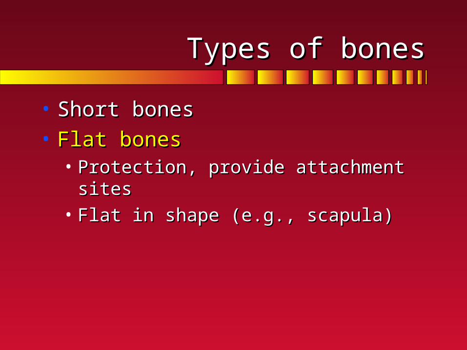
• Flat bones Flat bones • Protection, provide attachment sitesProtection, provide attachment sites• Flat in shape (e.g., scapula)Flat in shape (e.g., scapula)
Types of bonesTypes of bones
• Short bonesShort bones
• Flat bones Flat bones
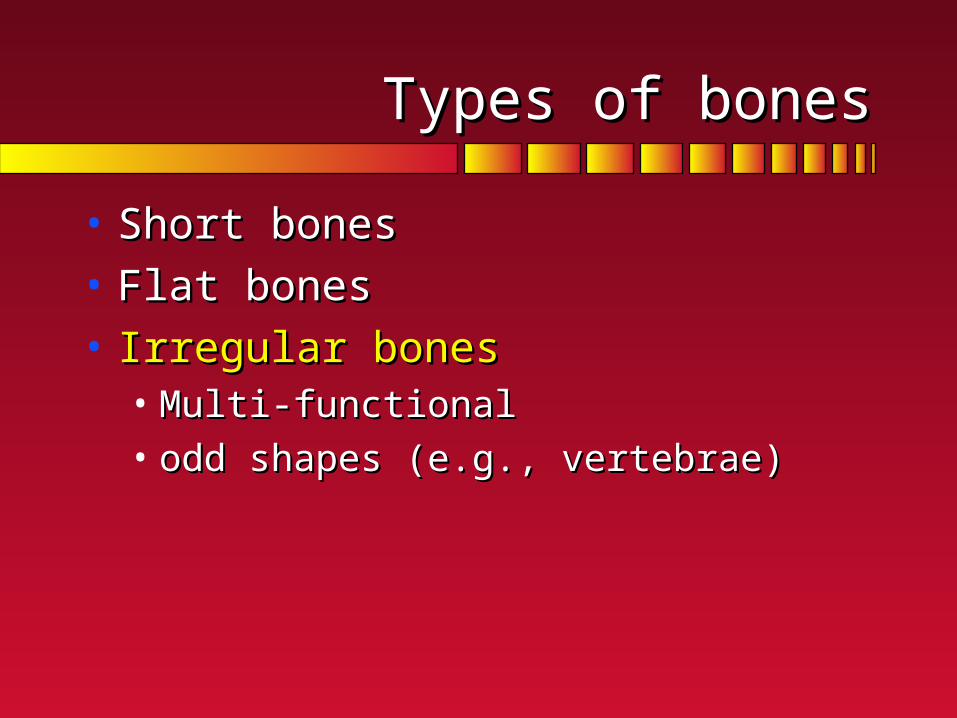
• Irregular bones Irregular bones • Multi-functionalMulti-functional• odd shapes (e.g., vertebrae)odd shapes (e.g., vertebrae)

Types of bonesTypes of bones
• Short bonesShort bones• Flat bones Flat bones • Irregular bonesIrregular bones• Long bones Long bones
• long shaft and bulbous heads (condyles, tubercles, or long shaft and bulbous heads (condyles, tubercles, or tuberosities)tuberosities)
• serve as levers for movement (e.g., tibia, femur, serve as levers for movement (e.g., tibia, femur, humerus, radius, ulna, clavicle, fibula, metatarsals, and humerus, radius, ulna, clavicle, fibula, metatarsals, and the phalanges)the phalanges)

Material Constituents:Material Constituents:
• Calcium carbonateCalcium carbonate
• calcium phosphate calcium phosphate
• collagencollagen
• waterwater
60 to 70%of mass
25 to 30% of bone mass

Material Constituents:Material Constituents:
• Calcium carbonateCalcium carbonate
• calcium phosphate calcium phosphate
• collagencollagen
• waterwater
•stiffness•compressive strength
• flexibility (tensile strength)
• tensile & compressive strength
Varies from person to person
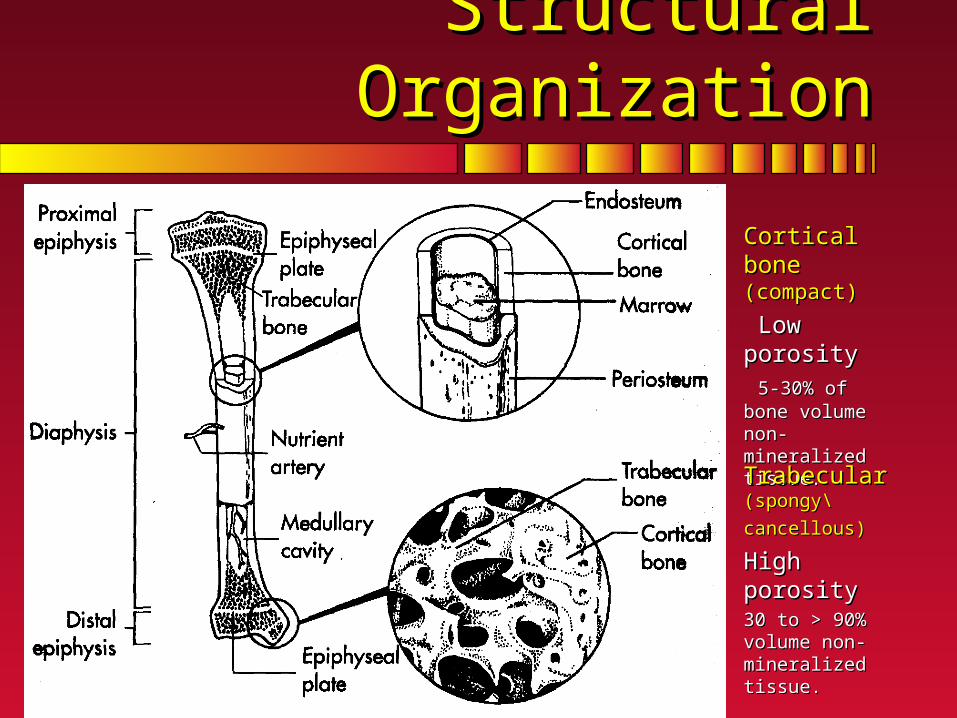
Structural OrganizationStructural Organization
Cortical bone Cortical bone (compact)(compact)
Low porosity Low porosity
5-30% of bone 5-30% of bone volume non-volume non-mineralized tissue.mineralized tissue.
Trabecular Trabecular (spongy\cancellous)(spongy\cancellous)
High porosityHigh porosity30 to > 90% volume 30 to > 90% volume non-mineralized non-mineralized tissue.tissue.

Load and ResponseLoad and Response
• StressStress• force per unit force per unit
areaarea
• StrainStrain• deformation deformation
• amount of amount of deformation deformation divided by divided by original lengthoriginal length

Life is an ongoing cycle of repeated applied external stresses, interrupted by applications of isolated stresses of varying magnitudes.

GenericGeneric Stress-Strain RelationshipStress-Strain Relationship
Strain (deformation)
Str
ess
(loa
d)El
astic
Reg
ion
Elastic Limit
Plastic Region

BoneBone Stress-Strain RelationshipStress-Strain Relationship
Strain (deformation)
Str
ess
(loa
d)E
last
ic R
egio
n FractureThreshold
Plastic Region

Relative Bone StrengthRelative Bone StrengthS
tres
s to
Fra
ctu
re
Load Type
Com
pres
sion
Ten
sion
She
arFractures: with excessive loads, bone tends to Fractures: with excessive loads, bone tends to fracture on the side loaded in tension.fracture on the side loaded in tension.


Bone Growth and DevelopmentBone Growth and Development
• Living bone is dynamicLiving bone is dynamic• continually changes throughout lifespan. continually changes throughout lifespan.
• Longitudinal growthLongitudinal growth• lengthlength increases occur at the epiphyses increases occur at the epiphyses
• epiphyseal plates. epiphyseal plates.
• produce new bone tissue until closing during produce new bone tissue until closing during adolescence or early adulthood. adolescence or early adulthood.
• Circumferential growth Circumferential growth • Bones alter Bones alter diameterdiameter throughout lifespan throughout lifespan
• most rapid change before adulthood. most rapid change before adulthood.

• OsteoblastsOsteoblasts
• form new form new bonebone
• OsteoclastsOsteoclasts
• resorb resorb existing boneexisting bone
Critical factor in bone modelling/remodelling:balance of their action

Bone Response to StressBone Response to Stress
• Wolff's law (1892)Wolff's law (1892)• tissue adapts to level of imposed stresstissue adapts to level of imposed stress
• increased stressincreased stress• hypertrophy (increase strength)hypertrophy (increase strength)
• decreased stressdecreased stress• atrophy (decrease strength)atrophy (decrease strength)
• SHAPE REFLECTS FUNCTIONSHAPE REFLECTS FUNCTION• Genetics, Body weight, physical activity, diet, Genetics, Body weight, physical activity, diet,
lifestyle (lifestyle (see note clippingssee note clippings))
(review the stress continuum)(review the stress continuum)

Protecting our Bones in SportProtecting our Bones in Sport

The pattern oftrabecular bone in thegreater trochanterneck of the femurhead of the femur reflects femur’s roles:muscle attachmentflexibilityweight transfersupport

Atrophy in BoneAtrophy in Bone
• Weight & strength decreaseWeight & strength decrease• Calcium content diminishesCalcium content diminishes
• reduced BMDreduced BMD
• trabecular integrity is losttrabecular integrity is lost
Bone stimulating factorsBone stimulating factors
Rate of loadingRate of loadingMagnitudeMagnitudeFrequencyFrequency

BMD and walking
Quartiles based on miles walked/week
Krall et al, 1994, Walking is related to bone density and rates of bone loss. AJSM, 96:20-26
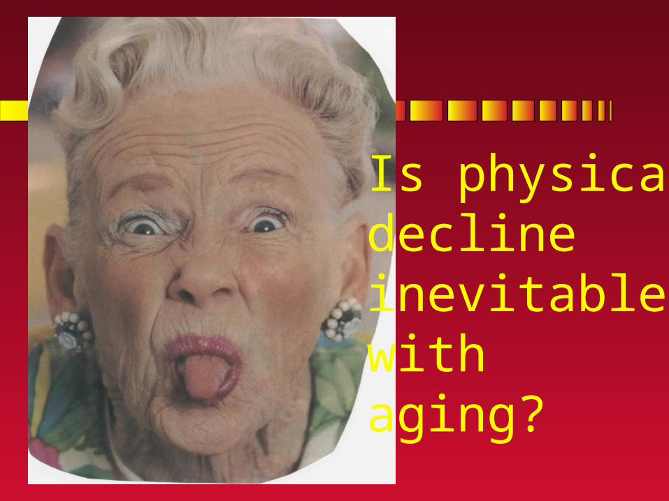
Is physicaldeclineinevitable withaging?
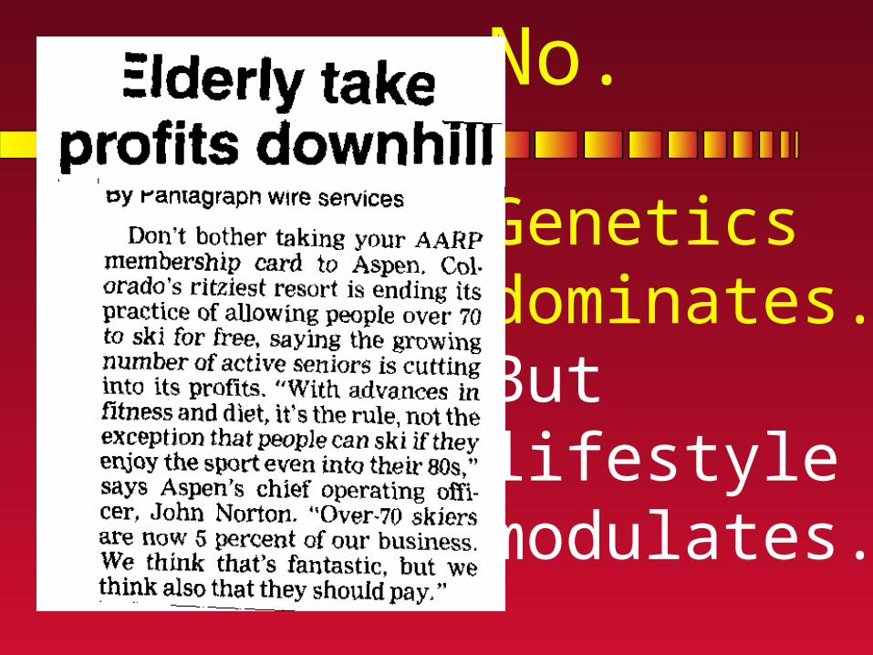
No.
Genetics dominates.But lifestylemodulates.

Changing concept of old age.Changing concept of old age.

How muchHow muchactivity do activity do
we need?we need?

The synovial jointThe synovial joint
You should be able to draw and label this diagram.

Joint Architecture & Joint Architecture & ClassificationClassification
• Synarthoses (immovable)Synarthoses (immovable)
• Amphiarthroses (slightly movable)Amphiarthroses (slightly movable)
• Diarthroses or Diarthroses or synovialsynovial (freely (freely movable)movable)• Get our attention Get our attention
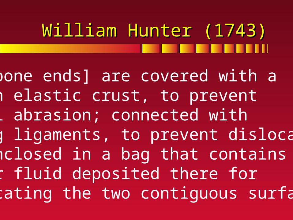
William Hunter (1743)William Hunter (1743)
[The bone ends] are covered with a smooth elastic crust, to preventmutual abrasion; connected with string ligaments, to prevent dislocation;and enclosed in a bag that contains a proper fluid deposited there for lubricating the two contiguous surfaces.

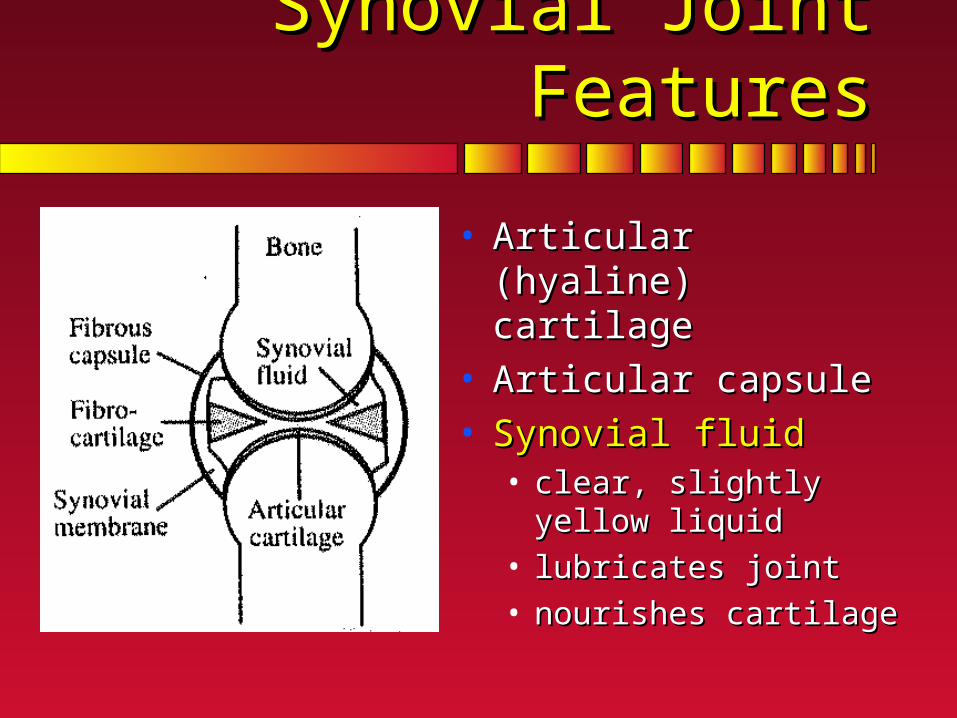
Synovial Joint FeaturesSynovial Joint Features
• Articular (hyaline) cartilage Articular (hyaline) cartilage • covers articulating surfacescovers articulating surfaces
• no blood vesselsno blood vessels
• no nervesno nerves
• Serves 3 purposes:Serves 3 purposes:• reduces friction reduces friction
• increases articulating area to increases articulating area to reduce stressreduce stress
• shock absorption shock absorption

Synovial Joint FeaturesSynovial Joint Features
• Articular (hyaline) cartilageArticular (hyaline) cartilage • Articular (fibrous/joint) Articular (fibrous/joint)
capsule capsule • double layer membrane double layer membrane
surrounds synovial jointsurrounds synovial joint
• outer connects bonesouter connects bones
• inner secretes synovial fluidinner secretes synovial fluid
• may have definite may have definite ligamentsligaments
Synovial Joint FeaturesSynovial Joint Features
• Articular (hyaline) cartilageArticular (hyaline) cartilage • Articular capsuleArticular capsule• Synovial fluid Synovial fluid
• clear, slightly yellow liquid clear, slightly yellow liquid
• lubricates joint lubricates joint
• nourishes cartilagenourishes cartilage
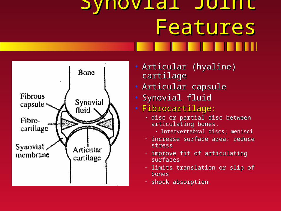
Synovial Joint FeaturesSynovial Joint Features
• Articular (hyaline) cartilageArticular (hyaline) cartilage • Articular capsuleArticular capsule• Synovial fluidSynovial fluid• FibrocartilageFibrocartilage: :
• disc or partial disc between disc or partial disc between articulating bones. articulating bones.
• Intervertebral discs; menisciIntervertebral discs; menisci• increase surface area: reduce stressincrease surface area: reduce stress• improve fit of articulating surfacesimprove fit of articulating surfaces• limits translation or slip of bones limits translation or slip of bones • shock absorptionshock absorption

Synovial Joint FeaturesSynovial Joint Features
• Articular (hyaline) cartilageArticular (hyaline) cartilage • Articular capsuleArticular capsule• Synovial fluidSynovial fluid• FibrocartilageFibrocartilage• Tendon sheaths Tendon sheaths
• surround tendons located surround tendons located close to bonesclose to bones
• reduce stress on tendonreduce stress on tendon
• maintain low friction maintain low friction

Synovial Joint FeaturesSynovial Joint Features
• Articular (hyaline) cartilageArticular (hyaline) cartilage
• Articular capsuleArticular capsule
• Synovial fluidSynovial fluid
• FibrocartilageFibrocartilage
• Tendon sheathsTendon sheaths
• BursaeBursae• small synovial fluid filled capsulessmall synovial fluid filled capsules
• separate tendon from bone to separate tendon from bone to reduce frictionreduce friction

Mobility is a very precious gift.
More complex thanthe space shuttle.

Role of MenisciiRole of Meniscii

Meniscii effect on mechanical stressMeniscii effect on mechanical stress
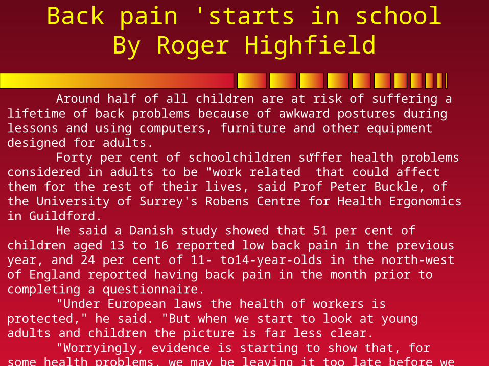
Back pain 'starts in schoolBy Roger Highfield
Around half of all children are at risk of suffering a lifetime of back problems because of awkward postures during lessons and using computers, furniture and other equipment designed for adults.
Forty per cent of schoolchildren suffer health problems considered in adults to be "work related” that could affect them for the rest of their lives, said Prof Peter Buckle, of the University of Surrey's Robens Centre for Health Ergonomics in Guildford.
He said a Danish study showed that 51 per cent of children aged 13 to 16 reported low back pain in the previous year, and 24 per cent of 11- to14-year-olds in the north-west of England reported having back pain in the month prior to completing a questionnaire.
"Under European laws the health of workers is protected," he said. "But when we start to look at young adults and children the picture is far less clear.
"Worryingly, evidence is starting to show that, for some health problems, we may be leaving it too late before we start helping."A study found that those reporting low back pain in school were more likely to report low back pain as adults.
(Filed: 10/09/2002) © Copyright of Telegraph Group Limited 2002.
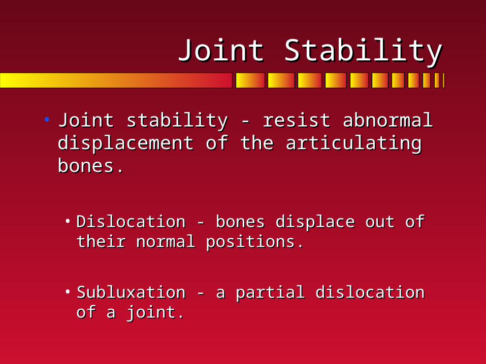
Joint StabilityJoint Stability
• Joint stability - resist abnormal Joint stability - resist abnormal displacement of the articulating bones.displacement of the articulating bones.
• Dislocation - bones displace out of their normal Dislocation - bones displace out of their normal positions.positions.
• Subluxation - a partial dislocation of a joint.Subluxation - a partial dislocation of a joint.

Joint StabilityJoint Stability
• Dislocation - bones displace out of their Dislocation - bones displace out of their normal positions.normal positions.
Impingement Subluxation Dislocation
Joint StabilityJoint Stability
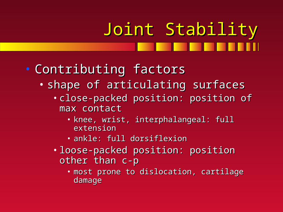
• Contributing factorsContributing factors• shape of articulating surfacesshape of articulating surfaces
• close-packed position: position of max contactclose-packed position: position of max contact• knee, wrist, interphalangeal: full extensionknee, wrist, interphalangeal: full extension• ankle: full dorsiflexionankle: full dorsiflexion
• loose-packed position: position other than c-ploose-packed position: position other than c-p• most prone to dislocation, cartilage damagemost prone to dislocation, cartilage damage

Joint StabilityJoint Stability
• Fatigue or improper use of the joints are major Fatigue or improper use of the joints are major contributing factors.contributing factors.
• Muscles add to joint stability.Muscles add to joint stability.
Joint StabilityJoint Stability
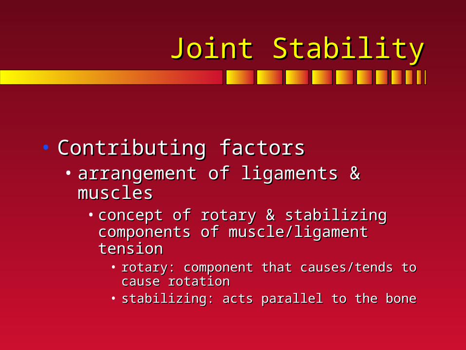
• Contributing factorsContributing factors• arrangement of ligaments & musclesarrangement of ligaments & muscles
• concept of rotary & stabilizing components of concept of rotary & stabilizing components of muscle/ligament tensionmuscle/ligament tension
• rotary: component that causes/tends to cause rotationrotary: component that causes/tends to cause rotation• stabilizing: acts parallel to the bonestabilizing: acts parallel to the bone

Joint StabilityJoint Stability
• Rotary componentRotary component - perpendicular - perpendicular component of a muscle force.component of a muscle force.
• Stabilizing componentStabilizing component - parallel component - parallel component of a muscle force acting toward the joint of a muscle force acting toward the joint center. center.
• Dislocating componentDislocating component - parallel - parallel component of a muscle force acting away component of a muscle force acting away from the joint center.from the joint center.

Joint StabilityJoint Stability
• FasciaFascia - fibrous connective tissue that - fibrous connective tissue that surrounds muscles and the bundles of surrounds muscles and the bundles of muscle fibers within muscles, providing muscle fibers within muscles, providing protection and support. protection and support. • Iliotibial band.Iliotibial band.
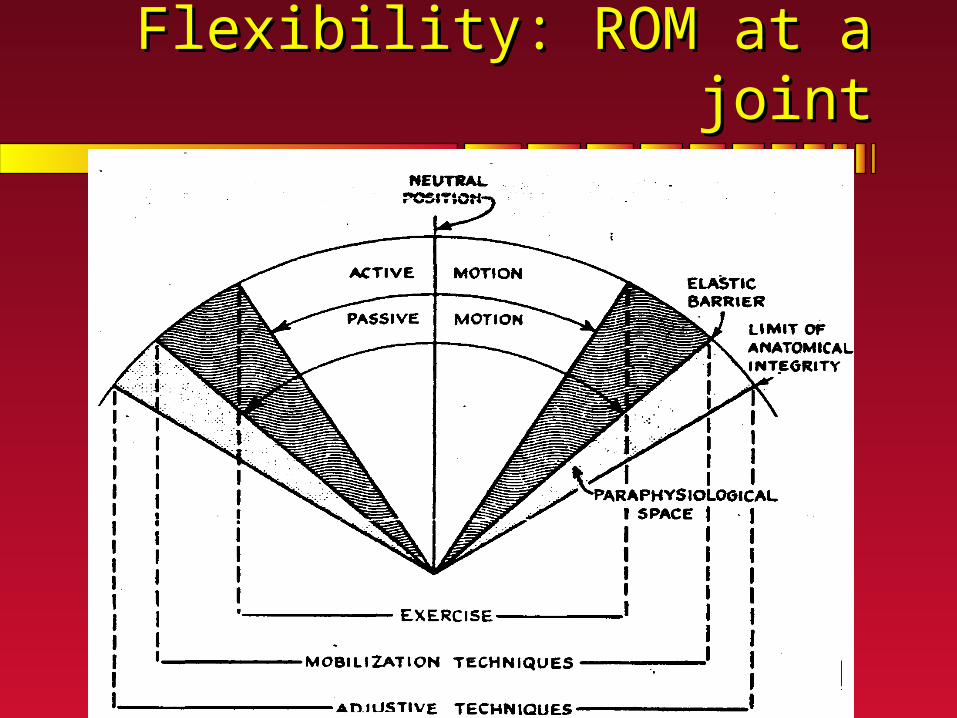
Flexibility: ROM at a jointFlexibility: ROM at a joint

Flexibility and InjuryFlexibility and Injury
• Risk of injury is heightened when joint Risk of injury is heightened when joint flexibility is extremely low, extremely high, flexibility is extremely low, extremely high, or significantly imbalanced between or significantly imbalanced between dominant and non-dominant sides of the dominant and non-dominant sides of the body. body.
• Although people usually become less Although people usually become less flexible with age, a large part is due to flexible with age, a large part is due to inactivity.inactivity.

Joint FlexibilityJoint Flexibility
• Factors influencing joint flexibility:Factors influencing joint flexibility:• Shape of articulating bonesShape of articulating bones• other soft tissue: stiffness & massother soft tissue: stiffness & mass
• muscle: current ‘tone”muscle: current ‘tone”• ligaments: arranged in direction of expected pullligaments: arranged in direction of expected pull• fatty tissuefatty tissue
• temperature: warmer = more plianttemperature: warmer = more pliant• past injury: collagen alignment integritypast injury: collagen alignment integrity• clothingclothing• AGE??? vs inactivityAGE??? vs inactivity

Why is flexibility important?Why is flexibility important?
• Basic component of a fitness profile.Basic component of a fitness profile.

Why is flexibility important?Why is flexibility important?
• Basic component of a fitness profile.Basic component of a fitness profile.• allows for greater choice of movement patternsallows for greater choice of movement patterns
• slides of gymnastsslides of gymnasts
• elderly shoulder ROM & independenceelderly shoulder ROM & independence• Osteoarthroses Osteoarthroses
• contractures (ie cerrebral palsy)contractures (ie cerrebral palsy)
• sprain ankle & inflammationsprain ankle & inflammation

Why is flexibility important?Why is flexibility important?
• Basic component of a fitness profile.Basic component of a fitness profile.• allows for greater choice of movement patternsallows for greater choice of movement patterns• reduce risk of injuryreduce risk of injury
• absorb energy over a greater distance (time)absorb energy over a greater distance (time)
• CAVEAT: Risk of injury increased with ROM high, CAVEAT: Risk of injury increased with ROM high, or low or low
• slide & next overheadslide & next overhead

From Cowan et al, 1988, ref #304
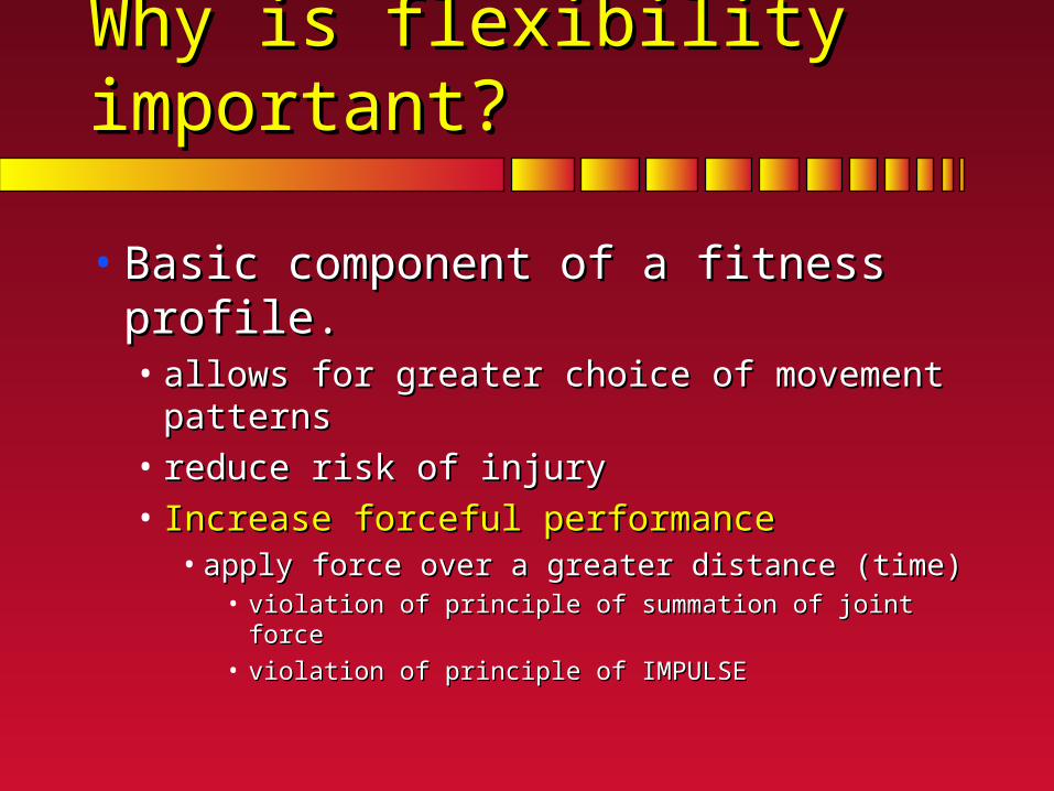
Why is flexibility important?Why is flexibility important?
• Basic component of a fitness profile.Basic component of a fitness profile.• allows for greater choice of movement patternsallows for greater choice of movement patterns• reduce risk of injuryreduce risk of injury• Increase forceful performanceIncrease forceful performance
• apply force over a greater distance (time)apply force over a greater distance (time)• violation of principle of summation of joint forceviolation of principle of summation of joint force
• violation of principle of IMPULSEviolation of principle of IMPULSE

Techniques for increasing Techniques for increasing joint flexibilityjoint flexibility
Best Advice:Use It
Don’t Lose It
How best to stretch?How best to stretch?
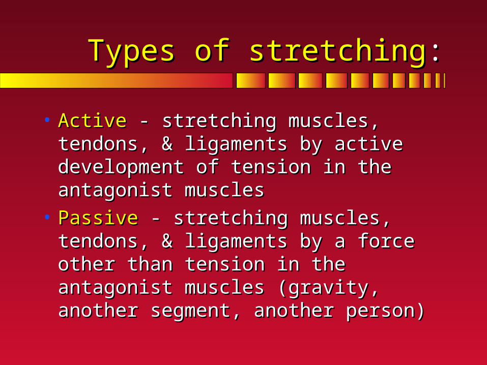
Types of stretchingTypes of stretching::
• Active Active - stretching muscles, tendons, & - stretching muscles, tendons, & ligaments by active development of tension ligaments by active development of tension in the antagonist musclesin the antagonist muscles
• PassivePassive - stretching muscles, tendons, & - stretching muscles, tendons, & ligaments by a force other than tension in ligaments by a force other than tension in the antagonist muscles (gravity, another the antagonist muscles (gravity, another segment, another person)segment, another person)

Types of stretchingTypes of stretching
• BallisticBallistic - a series of quick, bouncing - a series of quick, bouncing movements.movements.
• StaticStatic - a slow controlled stretch held over - a slow controlled stretch held over time (10-30s, 3 to 4 reps)time (10-30s, 3 to 4 reps)
• Proprioceptive Neuromuscular Facilitation Proprioceptive Neuromuscular Facilitation - - alternating contraction and relaxation of the alternating contraction and relaxation of the muscles being stretched. muscles being stretched. • Contract-relax& pull-contractContract-relax& pull-contract

Techniques for increasing Techniques for increasing joint flexibilityjoint flexibility
• Review neural Review neural innervationinnervation• Golgi tendon organsGolgi tendon organs
• located in junctions located in junctions between muscles and between muscles and tendonstendons
• responsive to tension in responsive to tension in tendontendon
• inhibits tension inhibits tension development in active development in active musclemuscle

Techniques for increasing Techniques for increasing joint flexibilityjoint flexibility
• Review neural innervationReview neural innervation• Golgi tendon organsGolgi tendon organs• Muscle spindlesMuscle spindles
• located parallel to the muscle fibers in the belly of located parallel to the muscle fibers in the belly of the musclethe muscle
• responsive to lengthening of fibers (rate & length) responsive to lengthening of fibers (rate & length) Stretch ReflexStretch Reflex
• activate stretched muscle, inhibit antagonist activate stretched muscle, inhibit antagonist (reciprocal inhibition)(reciprocal inhibition)

Techniques for increasing Techniques for increasing joint flexibilityjoint flexibility
• Review neural innervationReview neural innervation• Golgi tendon organsGolgi tendon organs• Muscle spindlesMuscle spindles
• Flexibility training goalFlexibility training goal• do not invoke stretch reflex (do not activate the do not invoke stretch reflex (do not activate the
muscle group to be stretched) muscle group to be stretched) HOW???HOW???• activate golgi tendon organs (further inhibit the activate golgi tendon organs (further inhibit the
muscle group to be stretched (reduce tonus)) muscle group to be stretched (reduce tonus)) HOW???HOW???