Chapter 10 10 Shock . Introduction ... transfusion is not enough to save patient . ... –Patients...
68
Chapter 10 Shock
Transcript of Chapter 10 10 Shock . Introduction ... transfusion is not enough to save patient . ... –Patients...
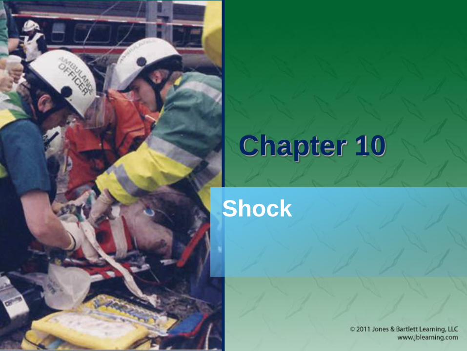
Chapter 10
Shock
Introduction (1 of 3)
• Shock (hypoperfusion) means a state of
collapse and failure of the cardiovascular
system.
– In the early stages, the body attempts to
maintain homeostasis.
– As shock progresses, blood circulation slows
and eventually ceases.
Introduction (2 of 3)
• Shock can occur because of medical or
traumatic events.
– Heart attack
– Severe allergic reaction
– Automobile crash
– Gunshot wound

Introduction (3 of 3)
• As an EMT, you cannot go wrong assuming
that every patient is in shock or may go into
shock.
Pathophysiology (1 of 9)
• Perfusion is the circulation of an adequate
amount of blood to meet the cells’ current
needs.
– The body is perfused via the circulatory system.
– Organs, tissues, and cells must have adequate
oxygenation or they may die.
Pathophysiology (2 of 9)
• Shock refers to a state of collapse and
failure of the cardiovascular system that
leads to inadequate circulation.
– Shock is an unseen life threat caused by a
medical disorder or traumatic injury.
– If the symptoms of shock are not promptly
addressed, the patient will soon die.
Pathophysiology (3 of 9)
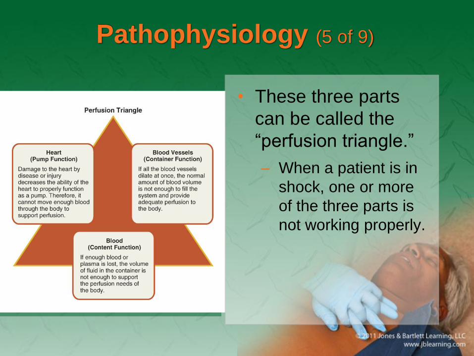
• Cardiovascular system has three parts:
– Pump (heart)
– Set of pipes (blood vessels and arteries)
– Contents (the blood)

Pathophysiology (4 of 9)
Pathophysiology (5 of 9)
• These three parts
can be called the
“perfusion triangle.”
– When a patient is in
shock, one or more
of the three parts is
not working properly.
Pathophysiology (6 of 9)
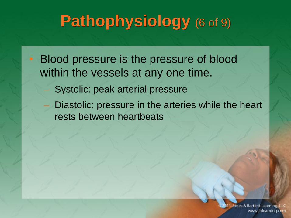
• Blood pressure is the pressure of blood
within the vessels at any one time.
– Systolic: peak arterial pressure
– Diastolic: pressure in the arteries while the heart
rests between heartbeats

Pathophysiology (7 of 9)
• Blood flow through the capillary beds is
regulated by the capillary sphincters.
– Under the control of the autonomic nervous
system
– Sphincters respond to other stimuli:
• Heat
• Cold
• The need for oxygen and waste removal

Pathophysiology (8 of 9)
• Perfusion requires more than just having a
working cardiovascular system.
– Adequate oxygen exchange in the lungs
– Adequate nutrients in the form of glucose in the
blood
– Adequate waste removal, primarily through the
lungs

Pathophysiology (9 of 9)
• Mechanisms are in place to help support
the respiratory and cardiovascular systems
when the need for perfusion of vital organs
is increased.
– Mechanisms include the autonomic nervous
system and hormones.

Causes of Shock (1 of 2)
• Shock can result from bleeding, respiratory
failure, acute allergic reactions, and
overwhelming infection.
– Damage occurs because of insufficient
perfusion of organs and tissues.

Causes of Shock (2 of 2)
Cardiogenic Shock (1 of 3)
• Caused by inadequate function of the heart
• A major effect is the backup of blood into
the lungs.
• Resulting buildup of pulmonary fluid is
called pulmonary edema

Cardiogenic Shock (2 of 3)
• Edema is the
presence of
abnormally large
amounts of fluid
between cells in
body tissues,
causing swelling.
Cardiogenic Shock (3 of 3)
• Cardiogenic shock develops when the heart
cannot maintain sufficient output to meet
the demands of the body.

Obstructive Shock (1 of 3)
• Obstructive shock occurs when conditions
that cause mechanical obstruction of the
cardiac muscle also affect the pump
function
• Common examples include cardiac
tamponade and tension pneumothorax.

Obstructive Shock (2 of 3)
• Cardiac tamponade
– Collection of fluid between the pericardial sac
and the myocardium
– Caused by blunt or penetrating trauma
– Can progress rapidly
– Signs and symptoms are referred to as Beck’s
triad.

Obstructive Shock (3 of 3)
• Tension pneumothorax
– Caused by damage to lung tissue
– The air normally held within the lung escapes
into the chest cavity.
– This air applies pressure to the organs,
including the heart.
Distributive Shock (1 of 11)
• Results from widespread dilation of small
arterioles, venules, or both
• The circulating blood volume pools in the
expanded vascular beds.
• Tissue perfusion decreases.

Distributive Shock (2 of 11)
• Septic shock
– Occurs as a result of severe infections in which
toxins are generated by bacteria or by infected
body tissues
– Toxins damage vessel walls, causing increased
cellular permeability.
– Vessel walls leak and are unable to contract
well.
Distributive Shock (3 of 11)
• Septic shock (cont’d)
– Almost always a complication of a very serious
illness, injury, or surgery.

Distributive Shock (4 of 11)
• Neurogenic shock
– Usually a result of injury to the part of the
nervous system that controls the size and
muscle tone of the blood vessels
– Causes include damage to the spinal cord,
brain conditions, tumors, pressure on the spinal
cord, and spina bifida.

Distributive Shock (5 of 11)
• Neurogenic shock
(cont’d)
– Muscles in the
blood vessel walls
are cut off from
nerve impulses
that cause them to
contract.
Distributive Shock (6 of 11)
• Anaphylactic shock
– Occurs when a person reacts violently to a
substance to which he or she has been
sensitized
– Sensitization means becoming sensitive to a
substance that did not initially cause a reaction.
– Each subsequent exposure tends to produce a
more severe reaction.

Distributive Shock (7 of 11)
• Anaphylactic shock (cont’d)
– Common causes:
• Injections (tetanus antitoxin, penicillin)
• Stings (honeybee, wasp, yellow jacket,
hornet)
• Ingestion (shellfish, fruit, medication)
• Inhalation (dust, pollen)

Distributive Shock (8 of 11)
• Anaphylactic shock (cont’d)
– Develops within minutes or even seconds of
contact with substance
– Signs are very distinct.
– Cyanosis (bluish color of skin) is a late sign.

Distributive Shock (9 of 11)

Distributive Shock (10 of 11)
• Psychogenic shock
– Caused by a sudden reaction of the nervous
system
– Produces temporary vascular dilation
– Results in fainting (syncope)
– Serious causes include irregular heartbeat and
brain aneurysm.
Distributive Shock (11 of 11)
• Psychogenic shock (cont’d)
– Non–life-threatening causes include receiving
bad news or seeing something unpleasant such
as blood.

Hypovolemic Shock (1 of 2)
• Result of an inadequate amount of fluid or
volume in the system
• Hemorrhagic causes and nonhemorrhagic
causes
• Occurs with severe thermal burns
– Intravascular plasma is lost.
Hypovolemic Shock (2 of 2)
• Dehydration, the loss of water or fluid from
body tissues, can cause or aggravate
shock.
– Fluid loss may be a result of severe vomiting
and/or diarrhea.
Respiratory Insufficiency (1 of 2)
• A patient with a severe chest injury may be
unable to breathe in an adequate amount of
oxygen.
– An insufficient concentration of oxygen in the
blood can produce shock as rapidly as vascular
causes.
Respiratory Insufficiency (2 of 2)
• Certain types of poisoning may affect the
ability of cells to metabolize or carry
oxygen:
– Carbon monoxide poisoning
– Cyanide poisoning
• Anemia occurs when there is an abnormally
low number of red blood cells.
The Progression of Shock (1 of 5)
• Three stages in the progression of shock:
– Compensated shock: early stage when the body
can still compensate for blood loss
– Decompensated shock: late stage when blood
pressure falls
– Irreversible shock: terminal stage when
transfusion is not enough to save patient

The Progression of Shock (2 of 5)
• Signs and
symptoms

The Progression of Shock (3 of 5)
• Blood pressure may be the last
measureable factor to change in shock.
– When a drop in blood pressure is evident, shock
is well developed.
– Particularly true in infants and children

The Progression of Shock (4 of 5)
• Use caution when caring for elderly
patients.
• Treating a pediatric or geriatric patient in
shock is no different than treating other
shock patients.
• Expect shock in many emergency medical
situations.
The Progression of Shock (5 of 5)
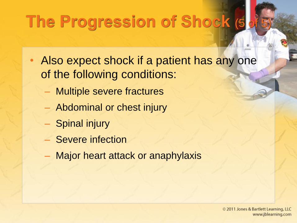
• Also expect shock if a patient has any one
of the following conditions:
– Multiple severe fractures
– Abdominal or chest injury
– Spinal injury
– Severe infection
– Major heart attack or anaphylaxis
Patient Assessment for Shock
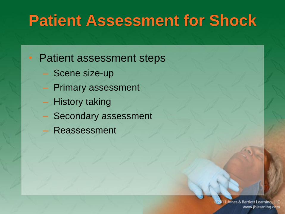
• Patient assessment steps
– Scene size-up
– Primary assessment
– History taking
– Secondary assessment
– Reassessment
Scene Size-Up
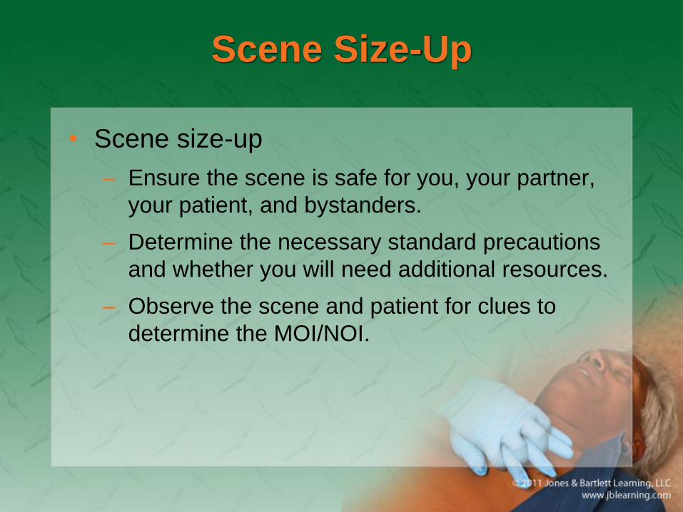
• Scene size-up
– Ensure the scene is safe for you, your partner,
your patient, and bystanders.
– Determine the necessary standard precautions
and whether you will need additional resources.
– Observe the scene and patient for clues to
determine the MOI/NOI.
Primary Assessment (1 of 3)
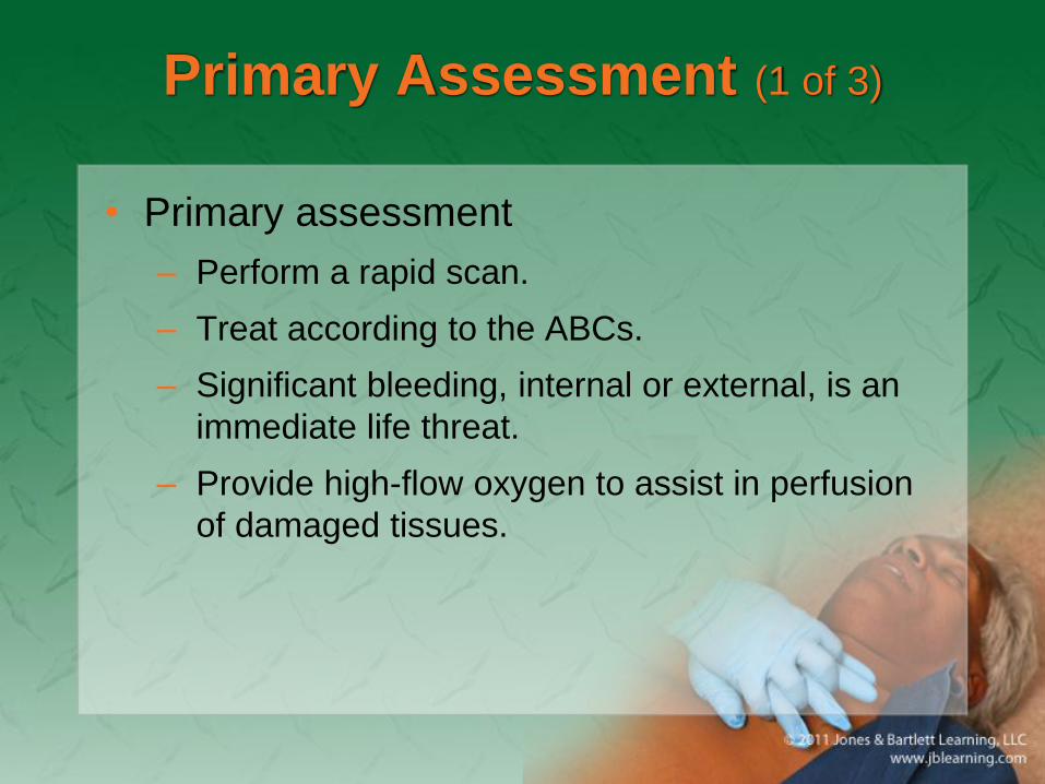
• Primary assessment
– Perform a rapid scan.
– Treat according to the ABCs.
– Significant bleeding, internal or external, is an
immediate life threat.
– Provide high-flow oxygen to assist in perfusion
of damaged tissues.

Primary Assessment (2 of 3)
• Primary assessment (cont’d)
– Form a general impression.
– Assess the airway to ensure it is patent.
– Assess breathing.
– An increased respiratory rate is often an early
sign of impending shock.
– Check for a distal pulse.

Primary Assessment (3 of 3)
• Primary assessment (cont’d)
– A rapid pulse suggests compensated shock.
– In compensated shock, the skin may be cool,
clammy, or ashen.
– Trauma patients with shock, or a suspicious
MOI, generally should go to a trauma center.

History Taking
• History taking
– Investigate the chief complaint.
– Obtain a SAMPLE history.

Secondary Assessment
• Secondary assessment
– Physical examination with a full-body scan
– Assess the respiratory system, neurologic
system, musculoskeletal system, and all
anatomic regions.
– Obtain a complete set of baseline vital signs.
– Use monitoring devices.

Reassessment
• Reassessment
– Determine what interventions are needed.
– Patients who are in decompensated shock will
need rapid interventions to restore adequate
perfusion.
– Determine whether your patient is in
compensated or decompensated shock.
– Document these findings.
Emergency Medical Care for Shock
• As soon as you recognize shock, begin
treatment.
– See Skill Drill 10-1.
– Do not give the patient anything by mouth, no
matter how urgently you are asked.
– Accurately record the patient’s vital signs
approximately every 5 minutes throughout
treatment and transport.
Treating Cardiogenic Shock (1 of 3)
• Patient does not require a transfusion of
blood, IV fluids, or elevation of legs.
• Chronic lung disease will aggravate
cardiogenic shock.
• Patient is able to breathe better in a sitting
or semisitting position.

Treating Cardiogenic Shock (2 of 3)
• Before administering nitroglycerin, consult
with medical control.
• Patients usually have a low blood pressure,
weak/irregular pulse, cyanosis, anxiety, and
nausea.
• Place the patient in a position that eases
breathing as you give high-flow oxygen.
Treating Cardiogenic Shock (3 of 3)
• Assist ventilations as necessary and have
suction nearby in case the patient vomits.
• Provide prompt transport.
• Approach a patient with a suspected heart
attack with calm reassurance.
Treating Obstructive Shock (1 of 2)
• In cardiac tamponade:
– Increasing cardiac output is the priority.
– Surgery is the only definitive treatment.
– Apply high-flow oxygen.
– The key treatment is rapid transport or ALS
management.

Treating Obstructive Shock (2 of 2)
• In tension pneumothorax:
– Apply high-flow oxygen to prevent hypoxia.
– Decompress the injured side of the chest
– The key treatment is rapid transport or ALS
management.

Treating Septic Shock
• Hospital management is required.
• Use standard precautions.
• Transport as promptly as possible.
• Use high-flow oxygen during transport.
• Ventilatory support may be necessary.
• Use blankets to conserve body heat.
Treating Neurogenic Shock (1 of 2)
• For the spinal cord injury patient, use a
combination of all known supportive
measures.
• Hospitalization will be required for a long
time.
• Keep the patient as warm as possible.

Treating Neurogenic Shock (2 of 2)
• Emergency treatment:
– Obtain and maintain a proper airway.
– Provide spinal immobilization.
– Assist inadequate breathing.
– Conserve body heat.
– Provide the most effective circulation.
– Transport promptly.
Treating Anaphylactic Shock
• Administer epinephrine.
• Promptly transport the patient.
• Provide supplemental oxygen and
ventilatory assistance en route.
• A mild reaction may worsen suddenly.
• Consider requesting ALS backup, if
available.
Treating Psychogenic Shock (1 of 2)
• In uncomplicated fainting, once the patient
collapses, circulation to the brain is
restored.
• Psychogenic shock can worsen other types
of shock.
• If the patient falls, check for injuries.

Treating Psychogenic Shock (2 of 2)
• If after regaining consciousness, the patient
is unable to walk normally, suspect head
injury.
– Transport the patient promptly.
– Record all initial observations of vital signs and
level of consciousness.
Treating Hypovolemic Shock
• Control all obvious external bleeding.
• Splint any bone and joint injuries.
• Secure and maintain an airway, and provide
respiratory support.
• Transport as rapidly as possible.

Treating Respiratory Insufficiency
• Secure and maintain the airway.
• Clear the mouth and throat of obstructions.
• If necessary, provide ventilations with a
bag-mask device.
• Give supplemental oxygen.
• Transport the patient promptly.

Summary (1 of 5)
• Perfusion requires an intact cardiovascular
system and a functioning respiratory
system.
• Most types of shock are caused by
dysfunction in the heart, blood vessels, or
volume of blood.
Summary (2 of 5)
• Shock is the collapse and failure of the
cardiovascular system, when blood
circulation slows and eventually stops.
• Blood is the vehicle for carrying oxygen and
nutrients through the vessels to the capillary
beds to tissue cells, where these supplies
are exchanged for waste products.

Summary (3 of 5)
• Blood contains red blood cells, white blood
cells, platelets, and a liquid called plasma.
• The systolic pressure is the peak arterial
pressure.
• The diastolic pressure is the pressure
maintained within the arteries while the
heart rests between heartbeats.

Summary (4 of 5)
• The various types of shock are cardiogenic,
obstructive, septic, neurogenic,
anaphylactic, psychogenic, and
hypovolemic.
• If there is any question on your part, treat
for shock. It is never wrong to treat for
shock.
Summary (5 of 5)
• Remember, by the time a drop in blood
pressure is detected, shock is usually in an
advanced stage.
• Treating a pediatric or geriatric patient in
shock is no different than treating any other
shock patient.