Chapter 1 Gold Nanoparticles Synthesis, Optical Properties...
39
Chapter 1 Gold Nanoparticles ― Synthesis, Optical Properties and Applications Abstract Nanoparticles with controlled size and shape are important from both fundamental and technological viewpoints. For a long time there had been much interest in preparing nanoparticles of various sizes and shapes. Metallic nanoparticles are an important class of nanomaterials with fascinating optical, electronic and magnetic properties. Among the metallic nanomaterials gold nanoparticles are particularly interesting due to their easy methods of synthesis, the ability to adjust properties through size and shape and their stability in a wide variety of solvents and pH conditions. This chapter describes in detail the synthesis, optical properties and applications of gold nanoparticles.
Transcript of Chapter 1 Gold Nanoparticles Synthesis, Optical Properties...

Chapter 1
Gold Nanoparticles ― Synthesis, Optical
Properties and Applications
Abstract
Nanoparticles with controlled size and shape are important from both
fundamental and technological viewpoints. For a long time there had been
much interest in preparing nanoparticles of various sizes and shapes.
Metallic nanoparticles are an important class of nanomaterials with
fascinating optical, electronic and magnetic properties. Among the metallic
nanomaterials gold nanoparticles are particularly interesting due to their
easy methods of synthesis, the ability to adjust properties through size and
shape and their stability in a wide variety of solvents and pH conditions. This
chapter describes in detail the synthesis, optical properties and applications
of gold nanoparticles.

2 Chapter 1
1.1 Introduction
In recent years nanotechnology has become one of the most important and
exciting forefront fields in Physics, Chemistry, Engineering and Biology. It
shows great promise for providing us with many breakthroughs that will
change the direction of technological advances in a wide range of
applications. The research area of nanotechnology is interdisciplinary,
covering a wide variety of subjects ranging from the chemistry of the
catalysis of nanoparticles, to the physics of the quantum dot laser. As a result
researchers in any one particular area need to reach beyond their expertise in
order to appreciate the broader implications of nanotechnology and learn
how to contribute to this exciting new field. Particles with sizes in the range
of 1-100 nm are called nanoparticles, whether they are dispersed in gaseous,
liquid, or solid media [1-2]. Because the NPs are larger than individual
atoms and molecules but are smaller than the bulk solid, materials in the
nanometer size regime show behavior that is intermediate between that of a
macroscopic solid and that of an atomic or molecular system.
Nanotechnology is based on the recognition that particles less than the size
of 100 nm impart to nanostructures built from them new properties and
behavior. The electronic structure, conductivity, reactivity, melting
temperature and mechanical properties have all been observed to change
when particles become smaller than a critical size. The dependence of the
behavior on the particle sizes can allow one to engineer their properties.
The properties of materials with nanometer dimensions are significantly
different from those of atoms and bulk materials. There are three major
factors that are responsible for these differences: high surface-to-volume
ratio, quantum size effect and electrodynamic interactions. All nanoparticles
regardless of their chemical constituents have surface area to volume ratios

Gold Nanoparticles – Synthesis, Optical Properties … 3
that are extremely high. Thus, many of the physical properties of the
nanoparticles such as solubility and stability are dominated by the nature of
the NP surface. One of the direct effects of reducing the size of materials to
the nanometer range is the appearance of quantization effects due to the
confinement of the movement of electrons. This leads to discrete energy
levels depending on the size of the structure. Following these line, artificial
structures with properties different from those of the corresponding bulk
materials can be created. Control over dimensions as well as composition of
structures thus makes it possible to tailor material properties to specific
applications.
Among different nanomaterials employed in research, metallic NPs have
been proved to be the most convenient and suitable. Metallic NPs possess
unique optical, electronic, chemical and magnetic properties that are
strikingly different from those of the individual atoms as well as their bulk
counterparts. It is known that the intrinsic properties of metal NPs are mainly
governed by their size, shape, composition, crystallinity and structure. In
principle, one could control any one of these parameters to fine-tune the
properties of these NPs. If such tiny particles are allowed to coalesce in a
controlled fashion, their color can be systematically varied [3-4] from pink
through violet to blue. The dimension of the particles in the nanometer size
regime makes them ideal candidates for nanoengineering of surfaces and the
fabrication of functional nanostructures [5-6].
Colloidal gold nanoparticles (AuNPs) are particularly interesting because of
its easy preparation and high stability. For biological applications, gold is an
important candidate due to its chemical inertness. The use of AuNPs has
been predominantly found in the work of artists and craftsman because of its
vivid visible colors. However, through research, the size, shape, surface

4 Chapter 1
chemistry and optical properties of AuNPs are the parameters which are
under control and have opened new doors to some very unique and exciting
capabilities. The interest in gold sols has occurred after innumerable
advances in our understanding of various concepts in physics and chemistry.
It includes: (1) the quantum mechanics and the effects associated with
nanoscopic systems, (2) the nature of interaction between colloidal particles,
(3) the statistical mechanics or thermodynamic behavior of aggregates that
can be formed by colloidal particles, (4) the mathematics and physics
associated with pattern formation, pattern characterization and how interplay
of kinetic and thermodynamic effects creates assemblies, (5) the mechanism
of micelle formation, (6) biological physics and (7) numerical methods and
simulation protocols. The corresponding advances in technology related to
optical and electron microscopy, optics, lasers, computing facilities for data
acquisition and data analysis as well as simulations and instruments designed
with better precision and options imply that the corresponding research for
colloidal metal particles can really become useful for applications to lab on a
chip devices, electronic, photonic and sensing applications based on
plasmonics in the field such as cancer treatment, etc. The latest gold rush is
likely to revolutionize the field of biosensing and chemical sensing by
allowing development of technologies that help in identification of chemical
or biological strains with high accuracy, by using highly environmentally
sensitive nature of plasmon resonance as well as other optical effects
associated with the gold and other noble metal particles.
1.2 A brief history of the advances in gold colloids
The use of colloidal gold as a colorant can be traced back at the least to 5th
B.C. for its use in making ruby glass and providing reddish tinge to ceramics.
The definitive study on the nature of gold particles in hydrosols – their

Gold Nanoparticles – Synthesis, Optical Properties … 5
synthesis by reduction of dilute gold chloride using phosphorus, size
dependent optical properties and coagulation behavior were carried out by
Faraday [7]. He obtained relatively unstable colloidal sols, with colors purple
red to sometimes blue and showed that electrical “collidation” of gold in air
or hydrogen gave a precipitate on glass or quartz with the same red or blue
color as present in the sols. Faraday found that the ruby glass was colored
so, because of the presence of finely dispersed gold particles. He showed
that gold chloride can be reduced by heat alone or by reaction with many
different reagents including organic matter, phosphorus, tartaric acid, etc. He
[7] had used many of the reducing agents of his time to produce gold sols.
He also examined other metals like platinum, palladium, rhodium, silver, tin,
lead, zinc, iron, mercury and arsenic. Faraday provided physical and
chemical arguments to emphasize that in both ruby fluid and ruby glass,
metallic gold was present in finely dispersed state. In the context of his
experiments, he attempted to study the optical properties as well as remarked
on the aggregation and sedimentation [7].
For nearly forty years, Faraday’s work remained unnoticed and even the
scientists who worked on the ruby glass were not aware of it. Thereafter
Zsigmondy [8] began his investigations into the color of ruby glass and
formulated a method for preparing colloidal gold by reducing dilute, slightly
alkaline solution of gold chloride with boiling formaldehyde. Using
Faraday’s methods, especially reduction using phosphorus, he combined
both the synthesis techniques to arrive at a two step synthesis method. This
method is referred to as the seed-mediated method in the contemporary
literature and was called ‘nuclear method’ in the early days [8]. Also the
nanoparticles were typically described as ultramicroscopic particles and in
the place of nanometers (nm) as a unit, the equivalent unit used was

6 Chapter 1
ultramicrons )( µµ . The dependence of optical properties on their shape was
apparent to researchers including Zsigmondy who reported difference in
color observed by using polarized light parallel and perpendicular to the
anisotropic particles oriented by spreading out on a gelatin film [8].
Zsigmondy invented the ultramicroscope [9] which allowed the visualization
of colloidal gold particles (i.e. nanoparticles), showing that colloidal matter
consisted of dispersion of particles of measurable size. Zsigmondy was able
to make some of the first particle tracking studies to determine the diffusion
behavior of the nanoparticles. While new generation transmission electron
microscopy can be employed to determine the size and shape of the
nanoparticles, typically dried onto a substrate, ultramicroscopy presents the
option of looking at the particles in their dispersed state. The advances in
optical techniques that have taken place in the past decades and the
expertise developed in the theoretical and experimental aspects of quantum
dot [10-12], make ultramicroscopy an ideal candidate for revisiting colloidal
dispersions of metallic nanoparticles. For example, in principle one can
follow the growth kinetics of particles in situ by visualization through the
ultramicroscope. Zsigmondy also investigated the stability of colloidal gold
in the presence of ions, biomolecules, gums and gelatin, etc., and noted that
certain proteins and substances displayed a ‘‘protective action’’ [8]. The
color of a red gold sol when coagulated with NaCl, changes to blue.
Zsigmondy used this property to define the so called gold number [8] as the
number of milligrams of the hydrophile colloid per 10 cm3 of gold sol that is
sufficient to prevent the coagulation and hence color change to occur when 1
cm3
of 10 per cent NaCl solution is added. The studies were used in
characterizing different proteins as well as in detecting changes in the
composition of liquids containing different proteins. The role of

Gold Nanoparticles – Synthesis, Optical Properties … 7
supersaturation in determining the nucleation and growth of gold particles,
both in condensation growth from solution and in vapor deposition, was also
reported by Svedberg [13-14] and Zsigmondy [8, 15] in their pioneering
studies. Another scientist who played a central role in early studies of gold
sols was Svedberg. He pioneered the use of electrochemical methods for the
synthesis of gold particles. He used every conceivable reducing agent
available at his time to produce colloidal gold from hydrochloroauric acid
[13]. For the early developments of gold colloid the contributions given by
Ostwald’s is also remarkable. He presented experimental and theoretical
principles through a series of demonstrations and it embodied several
principles useful for the synthesis of gold sols [16]. Ostwald pointed out the
importance of pH in the synthesis and expounded how this is useful in
changing the final product from being a red to blue dispersion.
The properties of colloids depend, to a large extent, on the movement of
particles and this movement consists of translational and rotational Brownian
motion. The advances in theoretical understanding of Brownian motion
brought about by Einstein [17-19] and Langevin [20] in the beginning of
twentieth century provided the requisite understanding to describe the
continuous motion of particles as well to understand the size dependence of
their stability and sedimentation behavior. Einstein [18-19] reasoned that
suspended particles behave quite like solute molecules and therefore an
osmotic pressure should be ascribed to the suspended particles. By applying
van’t Hoff’s law to suspensions and by assuming that dissipative force
described by Stokes law balances the force due to osmotic pressure, Einstein
was able to describe Brownian motion as a diffusion process. Further by
formulating the statistical analysis for Brownian motion, he laid a basis for
testing the reality of ‘molecular kinetic theory’ of matter. Einstein showed

8 Chapter 1
that mean squared displacement of particles scales linearly with time and
postulated that these results could be used to determine molecular
dimensions [17-19]. Langevin [20] outlined a simpler derivation for the time
dependence of the displacement of Brownian particles by introducing a
fluctuating random force and counteracting it with Stokesian drag and the
modern treatment of Brownian motion is typically based on it [21].
Perrin carried out meticulous experiments and also used a distribution
function to calculate Avogadro’s number and to establish the equivalence
between a colloidal particle and a molecule as required by the molecular
kinetic theory [22-23]. Perrin’s careful experiments on translational and
rotational Brownian motion not only led support to the theories of Brownian
motion but also established the reality of molecules and established the
statistical nature of thermodynamics. Brownian motion and thermal forces
set the rules for structure, dynamics and function of soft matter [24-25]
(polymers, liquid crystals, emulsions and colloidal dispersions) and the
analysis or theories of Brownian motion apply to stochastic problems in
systems ranging from single cells to galaxies [21, 26]. Another pioneer
Smoluchowski’s theories for diffusion and coagulation [21, 27] are central to
our understanding of collision and coalescence issues in colloidal sols. He
computed the rate at which a diffusing particle arrives in a ‘sphere of
influence’ of another particle. The assumption is that if the diffusing particle
moves about in the region outside this sphere of influence, it moves
unaffected, but if it enters the region, it sticks to the other particle [21]. Thus
Brownian motion, together with the interparticle attractive and repulsive
forces that form the physico-chemical basis for the sphere of influence,
determines the phase behavior and stability of colloids.

Gold Nanoparticles – Synthesis, Optical Properties … 9
1.3 Nanofluids ― general methods of synthesis
Preparing a stable and durable nanofluid is a prerequisite for optimizing its
thermal and optical properties. Therefore, many combinations of a material is
used for particular applications, namely: nanoparticles of metals, oxides,
nitrides, metal carbides and other nonmetals which can be dispersed into
fluids such as water, ethylene glycol or oils [28]. In the stationary state, the
sedimentation velocity of small spherical particles in a liquid follows the
Stokes law [29]
g)(R
V LP ρρµ
−=9
22
(1.1)
where V is the particle’s sedimentation velocity, R is the spherical
particle’s radius, µ is the liquid medium viscosity, Pρ and Lρ are the
particle and the liquid medium density respectively and g is the acceleration
due to gravity. This equation reveals a balance of the gravity, buoyancy force
and viscous drag that are acting on the suspended nanoparticles. According
to Eq. 1.1, the following measures can be taken to decrease the speed of NPs
sedimentation in nanofluids and henceforth to produce an improvement for
the stability of the nanofluids: (1) reducing R, the NPs size; (2) increasing µ ,
the base fluid viscosity and (3) lessening the difference of density between
the nanoparticles and the base fluid )( LP ρ−ρ . Reducing the particle size
should remarkably decrease the sedimentation speed of the NPs and improve
the stability of nanofluids, since V is proportional to the square of R.
According to the theory in colloid chemistry, when the size of particle
decreases to a critical size, ,R c no sedimentation will take place because of
the Brownian motion of NPs (diffusion). However, smaller NPs have a
higher surface energy, increasing the possibility of the nanoparticle

10 Chapter 1
aggregation. Thus, the stable nanofluids preparation strongly link up with
applying smaller nanoparticles to prevent the aggregation process
concurrently [30]. Two different techniques apply to produce nanofluids
namely: single-step and two-step method.
(i) Single step technique
In this method nanoparticle manufacturing and nanofluid preparation are
done concurrently. The single-step method is a process combining the
preparation of nanoparticles with the synthesis of nanofluids, for which the
NPs are directly prepared by physical vapor deposition (PVD) technique or a
liquid chemical method. In this method drying, storage, transportation and
dispersion of nanoparticles are avoided, so the agglomeration of
nanoparticles is minimized and the stability of the nanofluids is increased. A
disadvantage of this method is that it is impossible to scale it up for great
industrial functions and is applicable only for low vapor pressure host fluids
[31-32].
(ii) Two-step technique
In this method, dry NPs are first produced and then they are dispersed in a
suitable liquid host, but as NPs have a high surface energy, aggregation and
clustering are unavoidable and will appear suddenly. Afterward, the particles
will clog and sediment at the bottom of the container. Thus, making a
homogeneous dispersion by two step method remains a challenge. However,
there exist some techniques like high shear and ultrasound to minimise this
problem. Nanofluids containing oxide particles and carbon nanotubes are
produced by this method. This method works well for oxide NPs and is
especially attractive for the industry due to its simple preparation method.
But its disadvantage due to quickly agglomerated particles brings about

Gold Nanoparticles – Synthesis, Optical Properties … 11
many challenges nowadays. As nanoparticles disperse partially, dispersion is
poor and sedimentation happens, so a high volume concentration is needed
increasing the heat transfer (10 times of single step) and accordingly the cost
would be as much as loading [33]. The two-step method is useful for
application with particle concentrations greater than 20 vol % but it is less
successful with metal NPs. However, some surface treated nanoparticles
showed excellent dispersion [34]. The first materials tried for nanofluids
preparation were oxide particles, mainly because of its easy preparation
methods [35].
1.3.1 Synthesis of gold nanofluids
In the synthesis of AuNPs, or more specifically speaking, gold colloidal
dispersion, various types of precursors, reduction reagents, other chemicals
and methods where used to promote or control the reduction reaction, the
initial nucleation and subsequent growth of initial nuclei. The aim of such
synthesis is the preparation of nanoparticles of controlled composition, shape
and size. The simplest and the most commonly used preparation for gold
nanoparticles (AuNPs) is the aqueous reduction of HAuCl4 by sodium citrate
at reflux [36]. Although sodium citrate is the most common reducing agent,
metal nanoparticles can also be synthesized by the use of borohydride and
other reducing agents [37-40]. Particles synthesized by citrate reduction are
nearly monodisperse spheres of a size, controlled by the initial reagent
concentrations [40]. They have negative surface charge as a consequence of
weakly bound citrate coating and are easily characterized by their plasmon
absorbance. Smaller NPs may be formed in the gas phase [41] or by ablation
using high peak power laser pulses [42-43], electrodeposited [44], or
synthesized directly onto surfaces [45-49]. The following section describes

12 Chapter 1
the synthesis of AuNPs with size ranging from 15–80 nm using citrate
reduction method.
(i) Synthesis of nanofluid having 15 nm gold nanoparticles
The AuNPs having a size of 15 nm are synthesized by the citrate reduction of
HAuCl4 in water [50]. A 250 mL double neck round bottom flask was
cleaned in aqua regia (3 HCl: 1 HNO3) and rinsed with distilled water. 100
ml of 1 mM HAuCl4 solution was heated to boiling refluxed while being
stirred. Then, 10 ml of a 38.8 mM sodium citrate solution is added quickly.
The color of the solution turned from yellow to black and then to deep red.
After the color changed, the solution was refluxed for an additional 15
minutes. Then, the heater was turned off and the solution was stirred until it
reached to room temperature.
(ii) Synthesis of nanofluid having particle size from 30 to 80 nm
50 ml of 0.01% HAuCl4 solution was heated to boiling while being stirred in
a 100 ml conical flask. Then a few hundred µl of 1% sodium citrate solution
is quickly added to the auric solution.
6 Au3+
+ C6H5O7 3-
+ 15 OH- 6 Au + 6 CO2 + 10 H2O (1.2)
The color of the solution changed from yellow to black and then to red or
purple color depending on the sizes of the nanoparticles. After the color
change, the solution was stirred for an additional 15 minutes. The color change
for larger nanoparticles was slower compared to smaller nanoparticles. The
amount of citrate solution determines the size of the nanoparticles
synthesized. The photograph of gold nanofluid having particle size from 30
to 80 nm is shown in Fig. 1.1. This method produces very stable gold
nanoparticles with a quite narrow size distribution.

Gold Nanoparticles – Synthesis, Optical Properties … 13
Increasing Particle size
Fig 1.1 Gold nanofluid having particle size from 30 to 80 nm
The advantage of using trisodium citrate is that it can act both as reducing as
well as stabilizing agent. This method presents, with respect to other routes,
several advantages, mainly related to i) easy synthesis procedure,
ii) reproducibility of the method and iii) stability of the prepared sol. Another
advantage is that the medium (water) solvates both reagents very well.
Turkevich et al. [51] have extensively studied the effect of various
parameters, such as the temperature, amount of citrate added or the dilution
of the solution, on the formation of colloidal gold. The amount of citrate
added or the dilution of the solution can dramatically affect the average size
and size distribution of the AuNPs. The latter is additionally depending on
the relative rates of nucleation and growth [52-54]. Gold colloid solutions are
kept in clean, brown glass bottles away from heat or light (alternately, clear
glass bottles may be wrapped in aluminum foil). If Gold colloid solutions are
to be stored for long periods, they can be kept in the refrigerator to prevent
bacterial and fungal growth.
14 Chapter 1
1.4 Optical properties of gold nanoparticles
Although bulk gold and silver are widely known for their lustrous surfaces
and colors, there is a drastic color difference when the metal reduces in
dimensions. Gold and silver NPs are responsible for transmitting the brilliant
colored light through stained glass windows, yet these colors do not resemble
the characteristic yellow or bluish reflective surfaces of the bulk metals.
Even though the artisans did not know it at that time, the mixing of the metal
chlorides with molten glass led to the formation of metallic NPs of different
shape and size, hence the physical dimensions of the metal nanoparticles had
interesting interactions with light and produced visibly beautiful colors.
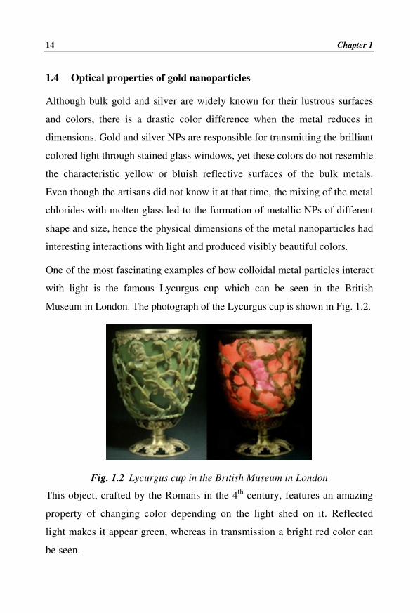
One of the most fascinating examples of how colloidal metal particles interact
with light is the famous Lycurgus cup which can be seen in the British
Museum in London. The photograph of the Lycurgus cup is shown in Fig. 1.2.
Fig. 1.2 Lycurgus cup in the British Museum in London
This object, crafted by the Romans in the 4th
century, features an amazing
property of changing color depending on the light shed on it. Reflected
light makes it appear green, whereas in transmission a bright red color can
be seen.

Gold Nanoparticles – Synthesis, Optical Properties … 15
Another excellent example is the stained glass windows from any European
Gothic Cathedral during the 15th
century as shown in Fig. 1. 3. The artisans
of the day achieved the brilliant blues, reds, yellows and greens in the glass
windows by mixing metal chlorides into molten glass before pouring it. The
metal chlorides nucleated and formed nanoparticles in the molten glass
before cooling, making art, one of the first uses for nanotechnology.
Fig. 1.3 Photograph of a stained glass windows from a European Gothic
Cathedral
A light beam passing through a colloidal dispersion of metal NPs gets
attenuated by the combined contribution of absorption and scattering, as
given by
)zCnexp(I)z(I ext00 −= (1.3)

16 Chapter 1
Here I0 is the intensity of the incident beam, I(z) is the intensity of the beam
after traveling path length z within the sample, 0n is the number density of
particles and scaabsext CCC += is the extinction cross section of a single
particle and is the sum total of the absorption and scattering cross sections
respectively. The product ext0Cn is often termed as the extinction coefficient
α and it has units of reciprocal length. The absorption spectrum determined
by UV–Vis spectroscopy is a measure of attenuation caused by a dispersion
of gold nanoparticles and is thus related to the absorption cross section.
1.4.1 The physical phenomenon of plasmons
(i) Plasmons
Plasmons are quantized waves in a collection of mobile electrons that are
produced when large numbers of these electrons are disturbed from their
equilibrium positions [55-56]. Electrons present in classic gaseous plasmas
can support plasmons, hence the name of these waves. The collection of
mobile electrons in metals is referred to as quantum plasma [56]. This is
composed of delocalized electrons that bathe the metal nuclei and their
localized core electrons, as described by the free-electron theory of metals
[55]. This metallic bonding has been used to explain many metallic
properties. Metals that best exhibit this free-electron plasma behavior include
the alkali metals, Mg, Al, and noble metals such as Cu, Ag and Au [55].
Plasmons can exist within the bulk of metals and their existence was used to
explain energy losses associated with electrons beamed into bulk metals [57].
For a bulk metal of infinite size, the frequency of oscillation, pω , of the
plasmons can be described by the expression,
( ) 210
2 /ep meN εω = (1.4)

Gold Nanoparticles – Synthesis, Optical Properties … 17
where N is the number density of mobile electrons, 0ε is the dielectric
constant of vacuum, e is the charge of an electron and em is the effective
mass of an electron. Surface plasmons (SPs) are a type of plasmon associated
with the surfaces of metals. They are significantly lower in frequency (and
energy) than bulk plasmons and can interact, under certain conditions, with
visible light in a phenomenon called surface plasmon resonance (SPR).
A macroscopic physical analogy that has been used to describe surface
plasmons is that of seaweed floating in water near a shoreline. The waves of
water lapping along the shoreline represent the electric fields and the
seaweed sloshed back and forth by these waves represent electrons [58]. SPs
have immense use to physicists and chemists. For example, the electric fields
of SPs amplify optical phenomena such as Raman scattering [55, 59]. There
are at least three types of SPs: propagating SPs, which occur on extended
metal surfaces, localized SPs, which occur in small volumes such as metal
particles, acoustic SPs, which are predicted to exist for some structures and
metal surfaces and are the subject of continued study [57].
(ii) Propagating surface plasmons
The combination of photons and SPs produce electromagnetic excitations on
extended surfaces known as surface plasmon polaritons (SPPs) or
propagating surface plasmons (PSPs) [55, 60]. PSPs have lower frequencies
and energies than bulk plasmons; metal surfaces in contact with a vacuum
have PSPs with a theoretical frequency of 2pω . Figure 1.4 shows a
representation of PSPs.

18 Chapter 1
Fig. 1.4 Schematic representation of propagating surface plasmons
As the waves of electron density travel along the surface, alternating regions
of positive and negative charges are produced. The electric fields, produced
by these regions of differing charge, decay exponentially away from the
metal surface [60].
Though PSPs can be produced with light, simply incidenting light on a
smooth metal surface in air is not sufficient because the momentum of the
light does not match that of the SPs [57]. Various methods are employed to
enable light to produce and to be produced by PSPs. Some methods involve
passing the light through a medium with a higher refractive index than air,
such as glass, before it comes near to the metal surface [57, 60]. To better
explain the role of refractive index, consider a beam of light which passes
from a medium with a higher index into a medium with a lower index. In
many cases, part of the incident beam is reflected from the interface and part
of the incident beam passes into the medium with the lower refractive index
(and is refracted in the process).
However, when the angle of the incident light exceeds a critical angle, no
portion of the light leaves the medium with higher refractive index and is
completely reflected. This is the condition of total internal reflection. Many

Gold Nanoparticles – Synthesis, Optical Properties … 19
fiber optic cables utilize the same principle to transmit light down curved
strands: the refractive index of the core of the optical fiber is higher than that
of its cladding layer, and the condition of total internal reflectance keeps the
light in the fiber [61]. Under the conditions of total internal reflectance, light
waves that strike the interface between two materials with differing
refractive indices produce new light waves that propagate from the interface
a very short distance into the medium with the lower refractive index. The so
called evanescent waves decay exponentially as the distance from the
interface increases. At a particular angle of incident light beyond the critical
angle, evanescent waves can propagate PSPs along a thin layer of metal
placed at the interface between the media with differing refractive indices
[58, 60, 62]. This SP resonance angle is susceptible to the refractive index of
the media near the metal film, making it sensitive to chemical changes that
involve changes in refractive index (or its square, the dielectric constant).
Other methods to enable light to access a metal surface and produce PSPs
involve roughening the surface, thereby changing its momentum. If light
shines on a metal diffraction grating, with parallel, linear features, it can be
diffracted by that grating. At the same time, however, PSPs will be
propagated along the surface of the grating in the direction perpendicular to
the linear features [60, 63].
(iii) Localized surface plasmon resonance
The answer to the question how do the nanoparticles interact with light, lies in
the understanding of surface localized plasmon resonance (LSPR) phenomena,
which are deeply affected by the nanometal shape and environment. Localized
surface plasmons (LSPs) are the collective electron oscillations in small
volumes. LSPs also have lower frequencies and energies than bulk plasmons;
metal particles in contact with a vacuum have LSPs with a theoretical

20 Chapter 1
frequency of 3pω [55, 57]. LSP resonances are produced in a somewhat
different fashion from PSPs. A schematic of the interaction between light and
the electrons of a metal particle is shown in Fig. 1.5 [55, 64].
Fig 1.5 Electronic cloud displacements in metal nanoparticle under the
effect of an electromagnetic wave. Electric fields are represented by
arrows
For this phenomenon to occur, the particle must be much smaller than the
wavelength of incident light. The electric field of the incident light can
induce an electric dipole in the metal particle by displacing many of the
delocalized electrons in one direction away from the rest of the metal particle
and thus producing a net negative charge on one side. Since the rest of the
metal particle is effectively a cationic lattice of nuclei and localized core
electrons, the side opposite the negative charge has a net positive charge.
LSPs have also been referred to as dipole plasmons, but the oscillating field
of the incident light can induce quadrupole as well as dipole resonances,
especially for particles greater than 30 nm in diameter [64-65]. If a particle
with a dipole can be considered to have a positively charged pole and a
negatively charged pole, then a particle with a quadrupole can be considered

Gold Nanoparticles – Synthesis, Optical Properties … 21
to have two positively charged poles and two negatively charged poles.
Smaller nanoparticles, (quantum dimension < 2nm), do not display this
phenomenon, as their electrons exist in discrete energy levels, and bulk has a
continuous absorbance in the UV/ Vis/IR region, which effectively collapsed
into the single plasmon absorbance in the case of the nanoparticle.
1.4.2 Factors affecting the surface plasmon resonance
The energy of light required to produce LSP resonance depends on a number
of factors, including the size, shape and composition of the particles, as well
as the composition of the surrounding media.
(i) Size and shape
The surface plasmon band position, bandwidth and intensity are affected by
the size and shape of the NPs. Many theories have been reported, to correlate
the size to the surface plasmon band position, some predicting a blue shift,
some a red one and others no shift at all. Generally speaking, as the particle
size increases, the plasmon resonant frequency decreases (shifts to longer
wavelengths) [66]. This result was subsequently rationalized by Liebsch
[67]. Small Au particles (with diameters less than 20 nm) exhibit extinction
that is primarily due to absorption; larger particles tend to exhibit much
stronger scattering [55]. Particles of different shapes have different plasmon
properties. For example, rod-shaped Ag or Au particles exhibit two
plasmons: a longitudinal plasmon, corresponding to the long axis of the rod,
and a transverse plasmon, corresponding to the short axis of the rod. Keeping
the diameters of the rods constant while increasing their lengths result in the
transverse plasmon remaining essentially constant in frequency (or energy)
while the longitudinal plasmon decreases in frequency [55, 68]. Hollow
metal particles tend to have lower plasmon resonant frequencies than solid

22 Chapter 1
metal particles [69]. Aggregation of colloidal metal particles can also lower
their plasmon frequencies [70].
(ii) Effect of the dielectric environment
The dielectric constant of the surrounding medium plays a predominant role
in determining both the plasmon peak position and intensity [1, 71].
Changing the medium surrounding the NPs, for another medium having a
different refractive index, strongly alters the plasmon behavior of the NPs.
Typically, a higher refractive index (or dielectric constant) of the medium
produces a lower plasmon frequency. This is, for instance, evidenced by the
strong shifts induced by transferring NPs from water or ethanol to a
transparent oxide matrix [72]. A macroscale physical analogy to this
phenomenon is using an oscillating weighted spring to represent the electric
field of the LSP. A high dielectric medium can be represented by a viscous,
higher-friction medium like oil. A spring in a vacuum will oscillate with a
higher frequency than the spring in the oil.
(iii) Electronic effects
The plasmon band may be displaced upon adding or subtracting electrons to
the overall metallic core. Oxidation processes were generally carried out
chemically [73] by the addition of free radicals [74] or under the action of
dioxygen [75]. Chemical reductions similarly proceeded via the action of
nucleophiles [76] or common reducing agents [77]. In all cases the increase
(respectively, decrease) in electron density resulted in the postulated
hypsochromic (respectively, bathochromic) shifts. Direct oxidation and
reduction using electrochemical methods have been performed more recently
[78-79]. The resulting devices are very promising for electro-optical
applications.

Gold Nanoparticles – Synthesis, Optical Properties … 23
1.4.3 Mie theory
It was the color variation of colloidal gold with particle size that motivated
Mie to frame work on the general solution of the diffraction problem of a
single sphere of arbitrary material and hence to apply the general theory of
light extinction to small particles [80]. This theory predicts, what fraction of
light impinging upon colloidal metal particles will be absorbed and what
fraction will be scattered. The sum of absorption and scattering is the
extinction of light due to the particles. This is what is measured when one
places a colloidal metal suspension into a UV–Vis spectrometer. Mie applied
Maxwell’s equations with appropriate boundary conditions in spherical
coordinates using multipole expansions of the incoming electric and magnetic
fields and offered an exact electrodynamic calculation of the interaction of
light with spherical metallic nanoparticles. The theory describes the extinction
(absorption and scattering) of spherical particles of arbitrary sizes. Most
standard colloidal preparations yield particles that are approximately spherical,
and most of the optical methods for characterizing nanoparticle spectra probe a
large ensemble of these particles. This leads to results that can be modeled
reasonably well using Mie theory. The main assumption of Mie’s theory is that
the particle and its surrounding medium are homogeneous and describable by
their bulk optical dielectric functions [81-82].
Mie attempted to calculate the optical response of large isolated, that is, single,
metal particles following classical electrodynamics. This model gives a
qualitative account of the variation of the optical properties with the size or the
surrounding medium. Moreover, it is assumed that the individual particles are
noninteracting and separated from one another. Therefore, the electric field
created around the particle by the excitation of surface plasmon resonance is
not felt by the other surrounding particles. In general, when the particle size

24 Chapter 1
(2R) is small enough (assumed to be spherical in shape) compared with the
wavelength of light λ )R2( λ< and also when the particle concentration is
very low, an absorption peak would result due to the excitation of dipole
plasma mode (n=1), and the optical extinction spectra can be described well by
Mie theory [82, 83-85]. Thus, although the Mie theory is valid for spheres of
any size, the limitation of the theory is that the dielectric constant of a small
particle is different from that of the bulk [86].
1.4.4 Maxwell Garnett effective medium theory
The surface plasmon oscillation in metallic nanoparticles is drastically
changed if the particles are densely packed in the reaction medium so that the
individual particles are electronically coupled to each other. It has been seen
theoretically and experimentally found that when the individual spherical
gold particles come into close proximity to one another, electromagnetic
coupling of clusters becomes effective for cluster-cluster distances smaller
than five times the cluster radius ( R5d ≤ , where, d is the center-to-center
distance and R is the radius of the particles) and may lead to complicated
extinction spectra depending on the size and shape of the formed cluster
aggregate by a splitting of single cluster resonance [87-88]. As a
consequence, their plasmon resonance is red-shifted by up to 300 nm [89].
This effect is negligible if d > 5R but becomes increasingly important at
smaller distances [90]. Aggregation causes a coupling of the gold
nanoparticle’s plasma modes, which results in a red shift and broadening of
the longitudinal plasma resonance in the optical spectrum [91]. The
wavelength at which absorption due to dipole-dipole interactions occurs may
be varied from 520 nm (effectively isolated particles) through 750 nm
(particles that are separated by only 0.5 nm), and the resulting spectra are a
composite of the conventional plasmon resonance due to single spherical

Gold Nanoparticles – Synthesis, Optical Properties … 25
particles and the new peak due to particle-particle interactions [92-93].
Since the inter particle coupling is stronger than the coupling within the
surrounding medium, the Mie’s theory developed for very dilute solutions
and isolated particles fails to describe the optical absorption spectrum.
However, the effective-medium theories, dating back to 1904, predicted by J.
C. Maxwell Garnett [94] have been successfully applied to this problem to
account for the optical absorbance behavior of the metal nanoparticles
present in a closely packed assembly. The Maxwell Garnett theory is strictly
valid in the quasistatic limit )R2( λ<< along with very small inter particle
distances but can be generalized to various shapes of the particles. The
Maxwell Garnett theory is an effective-medium theory [95-97].
1.5 Applications of gold nanoparticles
AuNPs are highly modifiable in size, shape and surface chemistry. Also they
are stable in a wide variety of environments, are inert, non-toxic and have
controllable optical-electronic properties. The main target of AuNPs is for
their application in catalysts, biomedical applications, nanosensors and drug
delivery which is discussed in detail in the following sections.
(i) Gold nanoparticles as a sensor
AuNPs are a very attractive material for biosensor, chemisensor, genosensor
and immunosensor production. Additionally, the unique physical and
chemical properties of gold nanoparticles provide excellent prospects for the
realization of this aim. AuNPs can be used as passive labels or as active
sensors [98]. Gold nanoparticles have been widely used to construct
biosensors because of their excellent ability to immobilize biomolecules.
Many kinds of biosensors, such as enzyme sensor, immunosensor and DNA
sensor, have been prepared based on the application of AuNPs [99].

26 Chapter 1
Developing rapid DNA-detection method is important for life science
research.
(ii) Catalytic properties of gold nanoparticles
The discovery in 1987 by Haruta and coworkers that AuNPs with size of <5
nm supported on the metal oxides, manifested a high catalytic activity, has
opened a new route in catalytic science [100]. AuNPs participated in a series
of important processes including hydrocarbon selective oxidation,
hydrogenation and water-gas shift reaction. The new gold catalyst system,
consisting of nanoparticulate gold on oxide support, is used for low-
temperature CO oxidation. It was shown that catalytical activity of AuNPs in
CO oxidation is strongly dependent on the support materials. From the
practical point of view, alumina (Al2O3) would be a preferable support for
Au catalyst, because it is cheap and possesses a high and thermally stable
surface area. Extraordinarily high catalytic activity of supported AuNPs for
oxidation of CO at room temperature arises from the reaction of CO
adsorbed on the step, edge and corner sites of metallic gold particles. Many
of the heterogeneous catalysts used in industry today consist of nanoparticles
of a catalytically active material anchored on a support. Modern
nanotechnology methods offer the synthesis of heterogeneous catalysts.
(iii) Utilization of gold nanoparticles in biomedical applications
AuNPs have numerous promising applications in nanomedicine field. These
applications include biosensing, bioimaging and bioassay. Gold nanoparticles
are used to produce thermal tumor ablation and new therapeutic agents. Also,
AuNP can be used for the detection of an antigen in conjugation with an
antibody. Most of these applications use the unique optical properties of

Gold Nanoparticles – Synthesis, Optical Properties … 27
AuNPs such as SPR, photoluminescence and surface-enhanced Raman
scattering (SERS) effect.
AuNPs with a well-controlled size are suitable for a colorimetric indicator
because the color can be changed in terms of SPR, which is strongly
influenced by the particle size and agglomeration. The detection of trace
amounts of biomolecules, critical for early imaging and diagnosis of cancer,
will be facilitated by the imaging molecule-dense AuNPs. For cancer
therapy, selective delivery and targeting of NPs to tumors is a key to
overcome the problems of toxicity and to increase therapeutic effects. For
tumor-selective delivery of gold, nanoparticles–antigens hybrid on tumor
cells can be used [101]. Tumor antigens include growth factors and their
receptors, hormones and glycol conjugates. Antibodies conjugated with
AuNPs were fabricated through modifying AuNPs by cysteamine and
conjugating the amine-functional group with an antibody. Conjugation of
antibody onto AuNP surface induced the increase in average diameter of
nanoparticles. Most biomedical applications of AuNPs and nanorods are
based on the gold conjugates. AuNPs contains hundreds to thousands of
surface active gold atoms that are able to connect through the Au–S dative
bond oligonucleotides, antibodies, peptides, and carbohydrates. Such
nanostructures are called bioconjugates [102-103]. Another important
application of AuNPs in the biomedical field is for photothermal therapy.
The irradiation by laser beam to AuNPs causes the absorption and
conversion of photon energy into thermal energy. As a result, the local
temperature of AuNPs dramatically increases. The dramatic increase in
temperature can cause a sudden release of heat to the surrounding
environment. It can be used for photothermal destruction of cancer cells.

28 Chapter 1
Gold nanocarriers provided a new group of target-specific deliveries of
therapeutic agents. Fig. 1.6 shows the loading of drug on nanoparticle
surface. The therapeutic agent could be small drug molecules or large
biomolecules, such as DNA, RNA and proteins. The AuNPs are essentially
inert and nontoxic. They are able to penetrate the cell to facilitate cellular
internalization and connective tissue permeation, thus enabling the drugs to
be delivered efficiently to the targeted cell without clogging capillaries.
Fig. 1.6 Loading of drug on the nanoparticle surface
There are two main categories of drug delivery systems. The first group
consists of the capsulation. The capsulation has an ability to contain a
relatively large amount of drug within the capsule. The second group of drug
delivery systems involves the attachment of the drugs to the carriers.
Targeting of drugs is a central goal of the delivery system. One way to
achieve it could be the conjugation of the drug delivery particle with a ligand
that specifically recognizes the target (cell). The special protein can be
employed as a targeting ligand [104]. The monodisperse AuNPs with the size
range from 1.5 to 15 nm can form the core. It was recognized that the
accumulation of AuNPs in various tissues was found to be dependent on
particle size. AuNPs having 15 nm showed higher distribution in tissues
compared to larger particles [105]. AuNPs provide attractive candidates for
gene delivery. A more sophisticated approach is to modify the surface of

Gold Nanoparticles – Synthesis, Optical Properties … 29
AuNPs by the addition of either an antibody or ligands with affinity for the
desired target. These involve coating the AuNPs with a self-assembled layer
of a thiolated PEG (poly-ethyleneglycol) or liposome. It may raise the
potential application of these AuNPs in the relative biomedical and
bioengineering areas.
(iv) Surface enhanced Raman spectroscopy
One of the most recognizable applications of LSP resonance shown by
metallic nanoparticles is in the surface enhanced Raman Spectroscopy
(SERS). Resonance of LSPs amplifies electric fields, E, near the particle
surfaces. The |E|2 of the plasmon electric field can be 10
2 to 10
4 of the |E|
2 of
the incident light [106]. The electric fields of plasmons can amplify the
Raman signals of chemical species near the metal surface, thus enabling the
technique of surface-enhanced Raman spectroscopy (SERS) [56]. Raman
spectroscopy is a type of vibrational spectroscopy that is sensitive to
polarizable bonds within molecules [107]. The electric field interacts with
polarizable molecules to produce dipoles, described by the equation [108]
Ep α= (1.5)
where α is polarizability of the molecule, E is the applied electric field
and p is the electric dipole induced in the molecule. As Eq. 1.5 shows, p
can be made larger by increasing either α or E , but plasmons tend to
enhance E . As the intensity of the induced dipoles increases, the intensity of
the Raman signals from those molecules increase. This surface enhancement
of Raman signals was first observed from pyridine adsorbed onto a
roughened Ag substrate [109]. Reproducible control of the degree of
enhancement has been problematic. It appears that many of the roughened
substrates have “hot spots” with exceptionally good signal enhancement

30 Chapter 1
[110]. Precise control of the nature of the surface roughness (e.g., by
patterning techniques) allows for better control of the degree of Raman
enhancement and of the wavelength required to access the surface plasmons
[106]. The Raman signal of the adsorbed species can be dramatically
enhanced with colloids as well. Enhancement of the electric fields of both the
incident and Raman scattered light by 104 produces an overall theoretical
enhancement of 108 [106]. The regions where colloidal particles come into
close proximity appear to be the location of intense plasmon electric fields
[111]. The fields that can arise between two particles that are very close (on
the order of one nanometer) to one another have enabled the detection of
SERS signals from single molecules located between these particles [112].
Conclusions
To conclude the synthesis of nanostructured materials with useful and
tunable properties are central to the development of nanoscale science and
technology. Bottom-up approaches based on self-assembly and self-
organization are especially appealing because of intrinsically low overhead
for large scale production. Gold nanoparticles, which have been known for
years, are the subject of an exponentially growing number of reports and are
full of promises for optical, electronic, magnetic, catalytic and biomedical
applications in the 21st century. The reason for the present excitement in
gold nanoparticle research is due to the stability of gold nanoparticles, the
extraordinary diversity of its modes of preparation, its size and shape
dependent properties and its role in nanoscience and future nanotechnology.
On the one hand we are still fascinated by the colors of metallic colloidal
suspensions, like Faraday, stained glass makers of the middle age and even
Roman craftsmen; on the other hand, they may be a real breakthrough in
prospect. After a long period devoted to understanding the physical

Gold Nanoparticles – Synthesis, Optical Properties … 31
principles rationalizing the surface plasmon resonance phenomenon,
chemists and physicists have started to use surface plasmon resonance for
their own purposes. New breakthroughs are likely to come from the use of
the surface plasmon resonance as a tool for nanosynthesis.
32 Chapter 1
References
[1] U. Kreibig, M. Vollmer, Optical Properties of Metal Clusters,
Springer., Berlin, 1995
[2] G. Schmid, Clusters and Colloids – From Theory to Applications,
VCH. Weinheim, Germany, 1994
[3] J.A. Creighton, C.G. Blatchford, M.G. Albrecht, J. Chem. Soc.
Faraday Trans. 75, 790 (1979)
[4] K.U. Von Raben, R.K. Chang, B.L. Laube, Chem. Phys. Lett. 79, 465
(1981)
[5] A.A. Lazarides, G.C. Schatz, J. Chem. Phys. 112, 2982 (2000)
[6] K. Drukker, G. Wu, G.C. Schatz, J. Chem. Phys. 114, 579 (2001)
[7] M. Faraday, Philosophical Transactions of the Royal Society of
London 147, 36 (1857)
[8] R. Zsigmondy, The Chemistry of Colloids, John Wiley & Sons, Inc.,
New York, 1917
[9] R. Zsigmondy, Colloids and the Ultramicroscope, John Wiley &
Sons, Inc., New York, 1909
[10] X. Michalet, F.F. Pinaud, L.A. Bentolila, J.M. Tsay, S. Doose, J.J. Li,
G. Sundaresan, A.M. Wu, S.S. Gambhir, S. Weiss, Science 307, 538
(2005)
[11] S.M. Nie, S.R. Emery, Science 275, 1102 (1997)
[12] A.P. Bartko, R.M. Dickson, J. Phy. Chem. B 103, 11237 (1999)
[13] T. Svedberg, The Formation of Colloids, D. Van Nostrand Company,
Inc., New York, 1921

Gold Nanoparticles – Synthesis, Optical Properties … 33
[14] T. Svedberg, A. Tiselius, Colloid Chemistry, The Chemical Catalog
Company, Inc., New York, 1928
[15] R. Zsigmondy, Colloids and the Ultramicroscope, John Wiley &
Sons, Inc., New York, 1909
[16] W. Ostwald, An Introduction to Theoretical and Applied Colloid
Chemistry, John Wiley & Sons, Inc., New York, 1917
[17] A. Einstein, Annalen Der Physik 17, 549 (1905)
[18] A. Einstein, Investigation on the Theory of Brownian Movement,
Dover Publications, Inc., New York, 1956
[19] A. Einstein, Annalen Der Physik 19, 371(1906)
[20] P. Langevin, Comptes Rendus Hebdomadaires des Seances de L
Academie des Sciences 146, 530 (1908)
[21] S. Chandrasekhar, Revs. Modern Phys. 15, 1 (1943)
[22] J. Perrin, Annales de Chimie et de Physique 18, 5 (1909)
[23] M.J. Nye, The Question of the Atom, Tomash Publishers, Los
Angeles, 1984
[24] M.E. Cates, M.R. Evans, Soft and Fragile Matter, Institute of Physics
Publishing, Bristol, 2000
[25] T.A. Witten, P.A. Pincus, Structured Fluids: Polymers, Colloids,
Surfactants, Oxford University Press, New York, 2004
[26] H.C. Berg, Random Walks in Biology, Princeton University Press,
Princeton, 1983
[27] M. von Smoluchowski, Kolloid-Zeitschrift 21, 98 (1917)

34 Chapter 1
[28] P. Keblinski, J.A. Eastman, D.G. Cahill, Mater. Today 8, 36 (2005)
[29] P.C. Hiemenz, M. Dekker, Principles of Colloid and Surface
Chemistry, New York, 1986
[30] D. Wu, H. Zhu, L. Wang, L. Liua, Curr. Nanosci. 5, 103 (2009)
[31] L. Godson, B. Raja, D. Mohan Lal, S. Wongwises, Sustain. Energy
Rev. 14, 629 (2010)
[32] Y. Li, J.E. Zhou, S. Tung, E. Schneider, S. Xi, Powder Technol. 196,
89 (2009)
[33] S.K. Das, S.U.S. Choi, W.H. Yu, T. Pradeep, Nanofluid: Science and
Technology, John Wiley & Sons Inc. 2007
[34] E.J. Swanson, J. Tavares, S. Coulombe, IEEE Trans. Plasma Sci. 36,
886 (2008)
[35] S.K. Das, S.U.S. Choi, H.E. Patel, Heat Transfer Eng. 27, 3 (2006)
[36] J. Turkevich, G. Garton, P.C. Stevenson, J. colloid Sci. 9, 26 (1954)
[37] T.S. Ahmadi, Z.L. Wang, T.C. Green, Science 272, 1924 (1996)
[38] C.K. Yee, R. Jordan, A. Ulman, H. White, A. King, Langmuir 15,
3486 (1999)
[39] T. Teranishi, M. Hosoe, T. Tanaka, M. Miyake, J. Phys. Chem. 103,
3818 (1999)
[40] M.J. Hostetler, J.E. Wingate, C.J. Zhong, J.E Harris, Langmuir 14,
17 (1998)
[41] R.P. Andres, J.D. Bielefeld, J.I. Henderson, D.B. Janes, Science 273,
1690 (1996)

Gold Nanoparticles – Synthesis, Optical Properties … 35
[42] G. Chumanov, K. Sokolov, B.W. Gregory, J. Phys. Chem. 99, 9466
(1995)
[43] J. Neddersen, G. Chumanov, T.M. Cotton, Appl. Spectrosc. 47, 1959
(1993)
[44] S.E. Gilbert, O. Cavalleri, K. Kern, J. Phys. Chem. 100, 12123 (1996)
[45] R.S. Urquhart, D.N. Furlong, T. Gengenbach, N.J. Geddes, Langmuir
11, 1127 (1995)
[46] G. Sberveglieri, L.E. Depero, P. Nelli, C. Perego, Adv. Mat. 8, 334
(1996)
[47] M.T. Reetz, M. Winter, J. Am. Chem. Soc. 119, 4539 (1997)
[48] M. Nagteggal, R. Seshadri, W. Tremel, Chem. Commun. 19, 2139
(1998)
[49] J.R. Heath, R. S. Williams, J.J. Shiang, J. Phys. Chem. 100, 3144
(1996)
[50] G. Frens, Nat. Phys. Sci. 241, 20 (1973)
[51] J. Turkevich, J. Hillier, Anal. Chem. 21, 475 (1949)
[52] G. Schmid, Chem. Rev. 92, 1709 (1992)
[53] J. Kimling, M. Maier, B. Okenve, V. Kotaidis, H. Ballot, A. Plech, J.
Phys. Chem. B 110, 15700 (2006)
[54] J. Turkevich, P.C. Stevenson, J. Hillier, Discuss. Faraday Soc. 11, 55
(1951)
[55] Y. Xia, N. Halas, MRS Bull. 30, 338 (2005)

36 Chapter 1
[56] T.L. Ferrell, Plasmon. In Concise Encyclopedia of Physics;
McGraw–Hill: New York, 2005
[57] J.M. Pitarke, V.M. Silkin, E.V. Chulkov, P.M. Echenique, J. Opt. A:
Pure Appl. Opt. 7, S73 (2005)
[58] J. Cuy, Biomaterials Tutorial: Surface Plasmon Resonance (SPR)
http://www.uweb.engr.washington.edu/research/tutorials/
plasmon.html
[59] A. Campion, P. Kambhampati, Chem. Soc. Rev. 27, 241(1998)
[60] A.V. Zayats, I.I. Smolyaninov, J. Opt. A: Pure Appl. Opt. 5, S16
(2003)
[61] A.B. Ellis, M.J. Geselbracht, B.J. Johnson, G.C. Lisensky, W.R.
Robinson, Teaching General Chemistry: A Materials Science
Companion; Oxford University Press: Oxford, 1993
[62] D. Nedelkov, R.W. Nelson, Trends Biotechnol. 7, 301 (2003)
[63] M. Moskovits, Rev. Mod. Phys. 57, 783 (1985)
[64] K.L. Kelly, E. Coronado, L.L. Zhao, G.C. Schatz, J. Phys. Chem. B
107, 668 (2003)
[65] E. Hutter, J. H. Fendler, Adv. Mater. 16, 1685 (2004)
[66] U. Kreibig, C.V. Fragstein, Z. Phys. 224, 307 (1969)
[67] A. Liebsch, Phys. Rev. B, Condens. Matter. 48, 11317 (1993)
[68] C. J. Murphy, T. K. Sau, A. Gole, Orendorff, C. J. MRS Bull. 30, 349
(2005)
[69] N. Halas, MRS Bull. 30, 362 (2005)

Gold Nanoparticles – Synthesis, Optical Properties … 37
[70] T.J. Jr. Norman, C.D. Grant, D. Magana, J.Z. Zhang, J. Liu, D. Cao,
F. Bridges, A.Van Buren, J. Phys. Chem. B. 106, 7005 (2002)
[71] S. Underwood, P. Mulvaney, Langmuir 10, 3427 (1994)
[72] F. Goettmann, A. Moores, C. Boissiere, P. Le Floch, C. Sanchez,
Small 1, 636 (2005)
[73] A. Henglein, J. Phys. Chem. 97, 5457 (1993)
[74] A. Henglein, P. Mulvaney, T. Linnert, Faraday Discuss. 92, 31
(1991)
[75] A. Henglein, Chem. Mater. 10, 444 (1998)
[76] T. Linnert, P. Mulvaney, A. Henglein, J. Phys. Chem. 97, 679 (1993)
[77] V.V. Vodnik, J. M. Nedeljkovic, J. Serb. Chem. Soc. 65, 195 (2000)
[78] R. Chapman, P. Mulvaney, Chem. Phys. Lett. 349, 358 (2001)
[79] G.C. Lica, B.S. Zelakiewicz, M. Constantinescu, Y.Y. Tong, J. Phys.
Chem. B. 108, 19896 (2004)
[80] G. Mie Ann. Phys. 25, 377(1908)
[81] V. Bonacic-Koutecky, P. Fantucci, J. Koutecky, Chem. Rev. 91, 1035
(1991)
[82] K.P. Charl, W. Schulze, Ber. Bunsenges. Phys. Chem. 88, 350 (1984)
[83] P. Mulvaney, Langmuir 12, 788 (1996)
[84] U. Kreibig, U. Genzel, Surf. Sci. 156, 678 (1985)
[85] G.W. Arnold, J.A. Borders, J. Appl. Phys. 48, 1488 (1997)
[86] M. Kubo, S. Nakamura, Bull. Chem. Soc. Jpn. 26, 318 (1953)
38 Chapter 1
[87] M. Quinten, U. Kreibig, Appl. Opt. 32, 6173(1993)
[88] U. Kreibig, A. Althoff, H.Pressmann, Surf. Sci. 106, 308 (1991)
[89] T. Ung, L.M. Liz-Marzan, P. Mulvaney, Colloids Surf. A 202, 119
(2002)
[90] J.M. Gerardy, M. Ausloos, Phys. Rev. B 27, 6446 (1983)
[91] A.N. Shipway, M. Lahav, R. Gabai, I. Willner, Langmuir 16, 8789
(2000)
[92] W.Y. Shih, J. Liu, W.H. Shih, I.A. Aksay, J. Stat. Phys. 62, 961
(1991)
[93] K.S. Mayya, V. Patil, M. Sastry, Bull. Chem. Soc. Jpn. 73, 1757
(2000)
[94] J.C. Maxwell Garnett, Philos. Trans. R. Soc. 203, 805 (1904), J. C.
Maxwell Garnett, Philos. Trans. R. Soc. 205, 237 (1906)
[95] G.L. Hornyak, C.J. Patrissi, C.R. Martin, J. Phys. Chem. B 101,
1458 (1997)
[96] C.A. Foss Jr. , G.L. Hornyak, J.A. Stockert, C.R. Martin, J. Phys.
Chem. B 96, 7497 (1992)
[97] C.A. Foss Jr., G.L. Hornyak, J.A. Stockert, C.R. Martin, J. Phys.
Chem. 98, 2963 (1994)
[98] A.R. Sperling, R.P.Gil, F. Zhang, M. Zanella, J.W. Parak, Chem.
Soc. Rev. 37, 1896 (2008)
[99] Q.A. Huo, Colloids Surf. B 50, 1 (2007)
[100] M. Haruta, K. Tanaka, A. Ueda, M. R. Torres Sanchez, J. Catal. 168,
125 (1997)

Gold Nanoparticles – Synthesis, Optical Properties … 39
[101] H.H Cai, P.H Yang, J.Y. Cai, Sens. Actuators B: Chem. 135, 603
(2009)
[102] B. Khlebtsov, V. Zharov, A. Melnikov, V. Tuchin, N. Khlebtsov,
Nanotechnology 17, 5167 (2006)
[103] G.N. Khlebtsov, A.L. Dykman, J. Quant. Spectrosc. Radiact. Transf.
111, 1 (2010)
[104] G. Han, P. Ghosh, M. De, M.V. Rotello, Nanobiotechnology 3, 40
(2007)
[105] G. Sonavane, K. Tomoda, K. Makino, Surf B: Biointerfaces 66, 274
(2008)
[106] A. J.Haes, C.J. Haynes, A.D. McFarland, G.C. Schatz, R.P.Van
Duyne, S. Zou, MRS Bull. 30, 368 (2005)
[107] P.W. Atkins, Physical Chemistry, 3rd ed.; W. H. Freeman and
Company: New York, 1986
[108] G.C. Weaver, K. Norrod, J. Chem. Educ. 75, 621 (1998)
[109] M. Fleischmann, P.J. Hendra, A. McQuillan, J. Chem. Phys. Lett. 26,
163 (1974)
[110] N. Halas, MRS Bull. 30, 362 (2005)
[111] C.J. Murphy, T.K. Sau, A. Gole, C.J. Orendorff, MRS Bull. 30, 349
(2005)
[112] J. Jiang, K. Bosnick, M. Mallaird, L. Brus, J. Phys. Chem. B 107,
9964 (2003)