Chap1-6 · Title: Chap1-6 Author: Ruth Subject: Chap1-6 Created Date: 20020228080700Z

The copyright of this thesis rests with the University of Cape Town. No
quotation from it or information derived from it is to be published
without full acknowledgement of the source. The thesis is to be used
for private study or non-commercial research purposes only.
Univers
ity of
Cap
e Tow
n

An investigation into the potential mutagenicity of South African traditional medicinal plants
Amatheni Nair
Thesis presented in fulfilment of the requirements for the degree of Master of Science Medical Biochemistry
in the
Faculty of Medicine University of Cape Town
Supervisor Prof MI Parker
Co-supervisor Prof V Sewram
26 March 2010
Univers
ity O
f Cap
e Tow
n

DeclarationDeclarationDeclarationDeclaration
I, __________________________, hereby declare that the work on which this thesis is based is
my original work (except where acknowledgements indicate otherwise), and that neither the whole
work nor any part of it has been, is being, or is to be submitted for another degree in this or any
other university.
I empower the University of Cape Town to reproduce for the purpose of research either the whole
or any portion of the contents in any manner whatsoever.
__________________
__________________
Univers
ity O
f Cap
e Tow
n

Dedicated to Paramadayalan, Dayalan and Ashaylin for their overwhelming support and encouragement without which I would never
have overcome the hurdles encountered in this thesis.
Univers
ity O
f Cap
e Tow
n

CONTENTS
PAGE
LIST OF ABBREVIATIONS (i)
ACKNOWLEDGEMENT (ii)
ABSTRACT (iii)
CHAPTER 1: INTRODUCTION 1
1.1 Background 1
1.2 Phytochemicals 1
1.2.1 Beneficial effects 1
1.2.2 Adverse effects 3
1.2.2.1 Acute effects 3
1.2.2.2 Chronic effects 3
1.3 Metabolism of chemical carcinogens 4
1.4 Plant extract-induced DNA mutations and genotoxicities 5
1.4.1 Point mutations 5
1.4.2 Chromosomal alteration 5
1.4.3 Other genotoxicities 6
1.5 Mechanism of DNA damage 6
1.6 Known plant carcinogens 7
1.6.1 Pyrrolizidine alkaloids 7
1.6.2 Aristolochic acid 8
1.7 Phytochemical content of the five plants investigated 10
1.7.1 Pteridium aquilinum 10
1.7.1.1 Animal carcinogenicity, in vitro studies and in vivo studies 10
1.7.1.2 Human carcinogenicity 11
1.7.1.3 Structure of ptaquiloside, key metabolites and related carcinogenicity 12
1.7.2 Zantedeschia aethiopica 14
1.7.2.1 Active ingredients and pharmacological effects 15
1.7.3 Acokanthera oppositifolia 15
1.7.3.1 Active ingredients and pharmacological effects 16
1.7.4 Rumex lanceolatus 17
1.7.4.1 Active ingredients and pharmacological effects 18
1.7.5 Pelargonium sp. 19
1.7.5.1 Active ingredients and pharmacological effects 20
1.8 In vitro mutagenicity assays 20
1.8.1 Salmonella reverse mutation assay 20
1.8.2 Chromosomal aberration test 22
Univers
ity O
f Cap
e Tow
n

1.8.2.1 The distinction between gap junction and chromosome breaks 23
1.9 Overview of the study 24
CHAPTER 2: MATERIALS AND METHOD 26
2.1 Collection of plants 26
2.2 Preparation of plant extracts 26
2.3 Preparation of S9 28
2.3.1 Induction of rat liver enzymes 28
2.3.2 Preparation of liver homogenate S9 fraction 28
2.3.3 Protein concentration of S9 29
2.3.4 CYP P450 concentration of S9 29
2.4 Verification of genotypes of Salmonella tester strains 29
2.4.1 Histidine dependence 30
2.4.2 Biotin and histidine dependence 30
2.4.3 Biotin dependence 30
2.4.4 rfa marker 30
2.4.5 uvrB deletion 32
2.4.6 Testing for the presence of plasmid pKM101 33
2.4.7 Spontaneous mutation frequency 33
2.5 Preparation working culture and frozen permanents 34
2.6 Dose-response of tester strain to mutagen 34
2.7 Salmonella reverse mutation assay 34
2.7.1 Statistical analyses 36
2.8 Chromosomal aberration test 36
2.8.1 Cultures 36
2.9 DNA content (flow cytometry) 37
2.9.1 Cultures 37
2.9.2 Staining cells for FACS analysis 37
CHAPTER 3: RESULTS 39
3.1 Salmonella reverse mutation assay 39
3.1.1 The CYP P450 concentration from difference spectra 39
3.1.2 Dose-response and range-finding study 40
3.1.3 Salmonella reverse mutation assay 42
3.1.4 Acokanthera oppositifolia (Lam.) and Zantedeschia aethiopica (L.) 43
3.1.5 Pelargonium sp. cf. inquinans (L.) L’ Herit 43
3.1.6 Pteridium aquilinum subsp aquilinum 48
3.4.5 Rumex lanceolatus Thunb. 49
Univers
ity O
f Cap
e Tow
n

3.2 Chromosomal aberration test 49
3.2.1 Pteridium aquilinum subsp aquilinum 51
3.2.2 Rumex lanceolatus Thunb. 54
3.2.3 Zantedeschia aethiopica (L.) 57
3.3 Flow cytometry (DNA content) 59
CHAPTER 4: DISCUSSION 62
4.1 R. Lanceolatus 63
4.2 Pelargonium sp 65
4.3 P. aquilinum 66
4.4 A. oppositifolia 67
4.5 Z. aethiopica 68
4.6 Conclusion and future prospects 68
APPENDIX A (Materials: Salmonella reverse mutation assay) 70
APPENDIX B (Materials: Flow activated cell sorting) 75
APPENDIX C (Salmonella tester strain query) 76
REFERENCES 78
Univers
ity O
f Cap
e Tow
n

LIST OF ABBREVIATIONS AND SYMBOLS ABBREVIATIONS
AA Aristolochic acid
2-AAF 2-acetamidofluorene
CHO Chinese hamster ovary cells
CHP Cumene hydroperoxide
CPA Cyclophosphamide
CTA Chromatid aberration
CSA Chromosome aberration
CYP cytochrome
DCM Dichloromethane
DHP 6,7 dihydro-7-hydroxy-1-(hydroxymethyl)-5H-pyrrolizine
DMSO Dimethyl sulfoxide
FACS Flow activated cell sorting
GSH Glutathione
LPS Lipopolysaccharide layer
MGA Minimum glucose agar
MMS Methylmethanosulphate
PCB Polychlorinated biphenyl
PHA Phytohaemagglutinin
RBCs Red blood cells
S.D. Standard deviation
SULTS Sulfotransferases
VOD Veno-occlusive liver disease
WBCs White blood cells
i
Univers
ity O
f Cap
e Tow
n

ACKNOWLEDGEMENT
I acknowledge the following people for their contribution to this study:
• Prof I Parker and Prof V Sewram for their guidance during the writing of this thesis and for
lending their expertise when required.
• Prof WCA Gelderblom at MRC-PROMEC for teaching me the skills of S9 preparation.
• Dr L van der Merwe at the MRC-Biostatistics Unit for the statistics done on the Ames data
• Mr R Smart (currently retired), Ms G Schutte and Ms M McCormack at Cytogenetics-NHLS
Laboratory, Groote Schuur Hospital (formerly Cytogenetics Laboratory-Dept of Human
Genetics) for sharing their cytogenetic skills for the chromosomal aberration test
• Sisters P. Legg and D. Sclar for blood collection toward the chromosomal aberration test
• Mrs L de Vries from IMP Innovative Solutions for the microscope she made available to the
study
• Mr R. Dreyer – UCT FACS facility.
ii
Univers
ity O
f Cap
e Tow
n

ABSTRACT
The potential mutagenicity and clastogenicity (ability to cause chromosomal damage) of five South
African traditional medicinal plants: Acokanthera oppositifolia (Lam.); Pelargonium sp. cf. inquinans
( L.) L’ Herit; Pteridium aquilinum subsp aquilinum; Rumex lanceolatus Thunb. and Zantedeschia
aethiopica (L.) Sg, were investigated using two in vitro tests in both a bacterial and a mammalian
cell system. The Salmonella reverse mutation assay and chromosomal aberration test, two
frequently used and accepted pharmacological bioassays, were selected for the investigation. The
rat liver extract (S9), containing CYP P450 and other liver enzymes, was added to the in vitro cell
system to detect pro-mutagens that require metabolic activation in order to exert mutagenicity or
clastogenicity – directly acting mutagens do not require metabolic activation.
A significant mutagenic potential (p≤0.01) was evident with the Salmonella reverse mutation assay
for three of the aqueous plant extracts: (i) R. lanceolatus (in strains TA97a, TA98, TA100 and
TA102) with and without metabolic activation (S9), (ii) P. aquilinum in TA100 with S9 and in TA102
without S9 and (iii) Pelargonium (in TA102) without S9. Furthermore, R. lanceolatus and P.
aquilinum were clastogenic in the chromosomal aberration test but this effect was reduced with S9.
Z. aethiopica demonstrated clastogenicity, which was reduced with S9, but the extract was not
mutagenic.
Since the chromosomal aberration test is dependent on cells entering the cell cycle (Go-G1, S and
G2-M) and chromosome visibility with light microscopy only occurs at metaphase, the clastogenicity
of A. oppositifolia and Pelargonium could not be detected because these extracts inhibited mitosis
(M). A DNA analysis of cultures treated with A. oppositifolia and Pelargonium by Flow Activated
Cell Sorting (FACS) indicated a blockage in the Go/G1 phase of the cell cycle.
The in vitro mutagenicity and clastogenicity tests served as a preliminary investigation into the
safety of five traditional plants. In addition to mutagenicity testing, it is suggested that further
scientific evaluation, validation, standardisation and regulation of South African traditional medicine
is essential in order to prevent the adverse acute and chronic effects of plant ingestion.
iii
Univers
ity O
f Cap
e Tow
n
CHAPTER 1
INTRODUCTION
1.1 Background
A major part of the population of both urban and rural communities in South Africa is reliant on
traditional medicine as opposed to westernised medicine (Van Wyk et al., 1997 and Eldeen et al.,
2005). This is because herbal medicines are an important part of the culture and tradition of African
people and are generally more accessible and affordable. However, many investigations have
shown that plants used as food or in traditional medicine have mutagenic and carcinogenic effects
in in vitro and in vivo assays as well as in humans (Stickel and Seitz, 2000, Zhang et al., 2004,
Zhou et al., 2004, Meinl et al., 2006 and Reid et al., 2006).
The Salmonella reverse mutation assay (Maron and Ames, 1983) is widely used in determining
possible gene mutations caused by extracts (Reid et al., 2006). A chemical that is mutagenic in
Salmonella is presumed to be a rodent carcinogen (and potential human carcinogen) (Zeiger,
2001). The chromosome aberration test is commonly used as a biomarker for the early effects of
genotoxic carcinogens.
Plant extracts are prepared from the aerial parts of the plant or roots and the most commonly used
solvents are water, dichloromethane and methanol. Such extracts contain only the soluble fractions
of the plant material (usually about 20% of the total weight) and the non-soluble (fibrous) residues
(about 80%) are discarded (Gurib-Fakim, 2006). A plant extract that produces a mutagenic or
clastogenic response is a result of one or more phytochemicals in the extract.
1.2 Phytochemicals
Phytochemicals that are produced from primary metabolism are associated with the fundamental
life processes common to all plants, such as photosynthesis, the pentose cycle, glycolysis and the
citric acid cycle. The secondary plant metabolites contribute to the taxonomic and biochemical
differentiation, and diversity within the plant kingdom. Furthermore, secondary plant metabolites
evolved as chemical defences to repel, stun, poison or kill other species or predators; therefore it
would be naive to think that every plant extract is necessarily safe for human consumption. The
beneficial and adverse effects of phytochemicals are discussed in the following sections.
1.2.1 Beneficial effects
Traditional plants have been used for dietary and medicinal purposes for centuries and continue to
be popular. There are many examples of the beneficial effects of traditional plants such as their
chemopreventative activity when herbal constituents form reactive intermediates capable of
irreversibly inhibiting various cytochrome P450s and their antibacterial and anti-inflammatory
properties (Zhou et al., 2004, Eldeen et al., 2005, and Moshi and Mbwambo, 2005) and a variety of
1
Univers
ity O
f Cap
e Tow
n

other beneficial effects (Table 1). The pharmacological screening of medicinal plants is very
important because it provides a scientific basis for the continued use of plants and could be
potential sources of new, effective and safe drugs (Eldeen et al., 2005).
Table 1: Plant compounds and extracts with beneficial medicinal effects
Chemical compound/extract
Botanical source Biological activity Reference
Scopolamine (alkaloid)
Datura stramonium Anti-muscarinic agent, smooth muscle relaxant and anti-nauseant
Van Wyk et al., 1997, 2002, and Dictionary of Natural Products, 2003
Resperine Rauwolfia serpentina Used for the treatment of high blood pressure
Craig, 1999
Taxol Taxus brevifolia A chemotherapeutic agent Craig, 1999 Atropine (alkaloid) Datura stramonium Reduces rigidity in
parkinsonism and is used as an antidote to poisoning with parasympathomimetic agents e.g. nerve gases and organophosphorous insecticides
Dictionary of Natural Products, 2003
Morphine and codeine (opium alkaloids)
Papaver somniferum (Poppy plant)
Narcotic analgesic (pain relief) and treatment of coughs.
Vinblastine and vincristine (alkaloids)
Catharanthus roseus (Periwinkle)
Anti-cancer drugs that interfere with the turbulines of the mitotic and meiotic spindle that inhibit cell division.
EPs®7630, Umckaloabo® (root extract)
Pelargonium sidoides
In vitro antibacterial, antiviral, and immunomodulatory properties in several studies. Efficacy has been proved in numerous clinical trials. Therapeutic effect in acute bronchitis, tonsillopharyngitis, sinusitis and symptoms of the common cold.
Bereznoy et al., 2003, Beil and Kilian, 2007, Agbabiaka et al., 2008 and Brendler and van Wyk, 2008
Rooibos, Honeybush (leaf extracts)
Aspalathus linearis, Cyclopia intermedia
(i) In vitro antimutagenicity, (ii) in vivo antioxidant, immune-modulating and chemopreventative actions.
Van der Merwe et al., 2006 and McKay and Blumberg, 2006
Despite the beneficial effects of plant compounds and extracts many adverse effects continue to
be reported (Stickel and Seitz, 2000, Zhang et al., 2004, Zhou et al., 2004, Meinl et al., 2006 and
Reid et al., 2006).
2
Univers
ity O
f Cap
e Tow
n

1.2.2 Adverse effects
Although a few plant extracts and numerous bioactive compounds that have been isolated from
plants have medicinal value, the ingestion of some plants and/or their extracts has been linked to
acute (short-term) or chronic (long-term) effects.
1.2.2.1 Acute effects
The symptoms of acute effects are toxicity (mainly acute hepato-toxicity), poisoning and acute
veno-occlusive liver disease (VOD), which is a non-thrombotic obliteration of small hepatic veins
leading to cirrhosis and, eventually, liver failure, which is diagnosed from a liver biopsy and
histopathology. Acute effects usually result from the ingestion of large quantities of plant material
over a short period:
(i) Datura stramonium, traditionally used for the treatment of gout, boils, wounds and asthma
and to reduce pain, is extremely toxic because of two main alkaloids, namely atropine and
scopolamine, which have been linked to deaths and poisoning for centuries (van Wyk et al.,
1997, Miraldi et al., 2001, Steenkamp et al., 2004 and Friedman, 2004). However, both
atropine and scopolamine have pharmaceutical significance (refer to Table 1).
(ii) VOD is related to the ingestion of plants containing various pyrrolizidine alkaloids, such as
Symphytum officinale, commonly referred to as comfrey (Stickel and Seitz, 2000). In South
Africa many VOD cases have been reported and genera known to contain pyrrolizidine
alkaloids are Senecio and Crotalaria (Steenkamp et al., 2000).
(iii) The high oxalic acid content of Rumex sp (leaves and root) used in traditional medicine has
been implicated in oxalic intoxication mainly in children. However, when oxalic acid is used
medicinally as a 5% solution in 5% malonic acid it is beneficial as a haemostatic agent (The
review of natural products, 2002, and van Wyk and Wink, 2004).
1.2.2.2 Chronic effects
A chronic effect results from the ingestion of small quantities of plant material over a long period.
This can cause organ damage such as in the liver and kidneys. Carcinogenesis can also result
from the ingestion of plant material.
(i) A case-control study on the ‘usual consumption’ of the plant Solanum nigrum in the
Transkei has been associated with an irritating effect on the oesophageal mucosal lining
(Sammon, 1992) and oesophageal cancer (IARC Scientific Publication 2003, No.153).
(ii) The use of Comfrey leaves (Symphytum officinale) has been recognised as a substantial
health hazard, with hepatic toxicity in humans and carcinogenic potential in rodents
resulting from its pyrrolizidine alkaloids: lasiocarpine and symphytine, and their related N-
oxides (Stickel and Seitz, 2000). The South African traditional medicine, Senecio latifolius,
which is known to contain toxic pyrrolizidine alkaloids, is a potential teratogen and
carcinogen, as observed from the long-term low-dose treatment of human HuH-7 cells
3
Univers
ity O
f Cap
e Tow
n
(Steenkamp et al., 2001). Pyrrolizidine alkaloids are ubiquitous in the plant kingdom but to
date only the retronecine, heliotridine and otonecine types exhibit high toxicity and
tumourigenicity in experimental animals (the metabolism of these three types are the most
studied) (Fu et al., 2004).
(iii) The use of herbal preparations of Aristocholia sp led to progressive nephropathy and
urothelial cancer in humans owing to the formation of DNA adducts in human target tissue
(Zhang et al., 2004; Zhou et al., 2004 and Meinl et al., 2006).
(iv) The use of Pteridium aquilinum has been related to mutagenicity and carcinogenicity in in
vitro, in vivo and epidemiological studies owing to its bioactive compound, ptaquiloside, and
its metabolic intermediate, pteridienone, elaborated in section 1.7.1. Metabolic activation
may influence the mutagenic potential of a phytochemical, as in the case of pteridienone
which was found to have more pronounced activity over ptaquiloside (Matsuoka et al.,
1989). Therefore, mutagenicity tests are generally performed with and without metabolic
activation by rat liver microsomes (S9).
1.3 Metabolism of chemical carcinogens
The liver is the major detoxification centre in the body where chemicals undergo biotransformation
to a more “excretable” (i.e. water-soluble) form. Cytochrome P450 and liver enzymes
(transferases) have critical roles in the metabolism of steroids and eicosanoids, and other
biosynthetic processes, and are involved in activation of pro-carcinogens as the first step to their
removal. Pro-carcinogens or pro-mutagens are not carcinogenic/mutagenic on their own but
require metabolic activation to intermediates in order to exert their effect. Mutagenicity tests
incorporate CYP P450 in the form of rat liver homogenate S9 for the detection of pro-mutagens in
plant extracts. The metabolism of drugs or xenobiotics in the liver occurs in two steps known as
Phase I and Phase II reactions.
Phase I reactions: Cytochrome P450 enzymes, 1A1, 1A2, 1B1, 2A6, 2E1 and 3A4, play a
prominent role in the metabolism of carcinogens, both in bioactivation and detoxification
(Guengerich and Shimada, 1991 and Guengerich, 2001). Carcinogens have reactive electrophilic
groups that can bind to DNA directly and cause mutations and cancer. However, pro-carcinogens
are enzymatically activated by CYP P450 into reactive electrophiles (e.g. ptaquiloside to
pteridienone) that can bind to DNA and also cause mutations and cancer. It is therefore imperative
that in vitro mutagenicity tests incorporate CYP P450 or liver microsomes for the detection of pro-
mutagens that are potential pro-carcinogens (confirmed by a two-year in-vivo study). Most Phase II
enzymes deactivate pro-carcinogens. Plant compounds may initiate the carcinogenic process
through DNA mutations and a variety of other genotoxicities, which are discussed in the following
sections.
4
Univers
ity O
f Cap
e Tow
n
1.4 Plant extract-induced DNA mutations and genotoxicities
Plant extracts or compounds may cause DNA damage through point mutations, chromosomal
alterations and other genotoxicities (e.g. DNA adducts, DNA crosslinking). Point mutations involve
one or more nucleotides (e.g. frameshift mutations and base pair substitutions) while chromosomal
alterations encompass several nucleotides.
1.4.1 Point mutations
A variety of traditional plant extracts and compounds produce point mutations:
(i) Aristolochic acid, the active compound from Aristolochia species, is mutagenic with the
Salmonella reverse mutation assay in strains TA98 and TA100, indicative of frameshift and
base-pair substitution mutations respectively, and in the mouse lymphoma assay (Zhang et
al., 2004).
(ii) Two extraction procedures were used in a sequential manner to extract polar (methanol-
water soluble) and apolar (dichloromethane) compounds from Cyrtanthus falcatus and
Cyrtanthus suaveolens (Family: Amaryllidaceae), which are commonly used in SA
traditional medicine (Elgorashi and van Staden, 2004). The dichloromethane (DCM)
extracts of the leaves and roots of C. falcatus, and the bulbs/roots and leaves of C.
suaveolens induced mutation in the Salmonella reverse mutation assay in strain TA98,
while none of the 90% methanol extracts of the different plant parts of the two species
induced mutagenicity in TA98. Since the DCM extracts of C. falcatus and C. suaveolens
induced frameshift mutations detected by TA98, the traditional medicine should be used
with caution since rigorous toxicological investigations are required.
(iii) Helichrysum simillimum, Helichrysum herbaceum and Helichrysum rugulosum, extensively
used in SA traditional medicine in the treatment of coughs, colds, fever, infections,
headache and menstrual pain (Van Wyk et al., 1997), showed mutagenicity in the
Salmonella reverse mutation assay in strains TA98 and TA100 with the 90% methanol
extracts, which is indicative of frameshift and base-pair substitution mutations respectively
(Reid et al., 2006).
Plant extractions using both polar and apolar solvents would add to scientific knowledge; however,
the validation of the plant extract would be compromised because traditional healers mainly use
water as a solvent in the preparation of plant extracts, mostly as decoctions and/or infusions.
1.4.2 Chromosomal alteration
(i) South African traditional medicinal plant extracts, Afzelia quanzensis Welw.; Bersama
lucens (Hochst.) Szyszyl; Ocotea bullata; Siphonochilus aethiopicus and Tetradenia riparia
(Hochst.) Codd (Lamiaceae), induced DNA damage as detected in the comet assay (Taylor
et al., 2003).
5
Univers
ity O
f Cap
e Tow
n

(ii) Extracts of Antidesma venosum E. Mey. Ex Tul.; Balanities maughamii Sprague,
Catunaregan spinosa (Thunb.) Tirveng.; Chaetacme aristata, Croton sylvaticus Hochst.;
Diospyros whyteana (Hiern) F. White; Euclea divinorum Hiern; and Gardenia volkensii K.
Schum caused both DNA damage and chromosomal aberrations in human white blood
cells as detected by the comet assay and micronucleus tests respectively (Taylor et al.,
2003). The micronucleus test detects chromosome breakage and/or chromosome loss.
(iii) The dichloromethane extracts of twigs/bark of Gardenia volkensii and Spirostachys africana
common South African traditional medicinal plants, were found to be genotoxic in the
micronucleus test (Verschaeve et al., 2004).
(iv) Aristolochic acid from the Aristolochia species caused an increase in chromosomal
aberrations in Chinese hamster ovary (CHO) cells, with and without metabolic activation by
S9 (Zhang et al., 2004).
1.4.3 Other genotoxicities
(i) Pyrrolizidine alkaloids are naturally occurring phytochemicals that are common constituents
of hundreds of plant species worldwide. Some pyrrolizidine alkaloids exhibit a variety of
genotoxicities, including DNA binding, DNA cross-linking and DNA-protein cross-linking
(Reed et al., 1988, Hincks et al., 1991 and Coulombe et al., 1999).
(ii) Aristolochic acid, the active compound from Aristolochia species, produces DNA adducts in
human target tissue. Aristolochic acids undergo reduction of the nitro group by hepatic
cytochrome P450 (CYP1A/2) or peroxidases in extrahepatic tissues to reactive cyclic
nitrenium ion that are capable of reacting with DNA and proteins, has been shown to
activate the H-ras oncogene (Zhou et al., 2004).
Several plants used in South African traditional medicine have the potential to cause long-term
damage to genetic material in patients when administered as medicinal preparations (Fennell et
al., 2004). The generation of DNA damage, which can arise from a direct or indirect mechanism, is
considered an important initial event in carcinogenesis. Mutagenicity tests generally detect those
chemicals that cause direct DNA damage.
1.5 Mechanism of DNA damage
Direct mechanisms refer to DNA damage arising from point mutations, chromosome or genome
alteration, while indirect mechanisms refer to the interaction of chemical compounds with non-DNA
targets leading to genotoxic effects such as lipid peroxidation and protein adducts (Kirsch-Volders
et al., 2003). Carcinogenicity caused by Aristolochia sp is induced by direct and indirect DNA
damaging mechanisms. The metabolic intermediate of aristolochic acid produces DNA adducts
(direct mechanism) and protein adducts (indirect mechanism). Examples of chemical compounds
and extracts shown to damage DNA via direct mechanisms are presented in Table 2.
6
Univers
ity O
f Cap
e Tow
n

Table 2: Plant extracts and chemical compounds shown to cause direct DNA damage
Extract/Compound Direct DNA damage
Reference Point Mutation
Chromosome Genome DNA-adducts
P. aquilinum, extract (bioactive compound ptaquiloside)
eMutagenic in the Salmonella reverse mutation assay with TA100 and TA98
aPeritoneal and bone marrow cells of Swiss mice showed structural chromosome aberrations
aPeritoneal and bone marrow cells of Swiss mice showed numerical chromosome aberrations
bOrgan specific ptaquiloside-DNA alkylations in vivo
eMatoba et al., 1987, bAlonso-Amelot and Avendano, 2002 and aAlmeida Santos et al., 2006.
Riddelliine, a pyrrolizidine alkaloid
- - - Riddelliine induced DNA adduct formation in vitro and in vivo
Yang et al.,2001a, 2001b
C. suaveolens extract
Mutagenic in the Salmonella reverse mutation assay with strain TA98, indicating a frameshift mutation
- - - Elgorashi and van Staden, 2004
Aristocholia sp (bioactive compound aristolochic acid)
cMutagenicity in the Salmonella reverse mutation assay with TA98 & TA100, indicating frameshift and base-pair substitution mutations
cChromosomal aberrations in CHO cells
- dForms DNA adducts in human target tissue
cZhang et al., 2004, dZhou et al., 2004 and dMeinl et al., 2006
1.6 Known plant carcinogens
Many plant carcinogens have been identified from phytochemical and related studies; however,
only pyrrolizidine alkaloids and aristolochic acid are discussed in this section.
1.6.1 Pyrrolizidine alkaloids
Pyrrolizidine alkaloids are categorised into retronecine (e.g. riddelliine, senecionine), heliotridine
(e.g. lasiocarpine, heliotrine) and otonecine (e.g. clivorine) types. These are metabolised by the
cytochrome P450 CYP3A subfamily, in particular CYP3A4 isoenzyme, which is the primary
7
Univers
ity O
f Cap
e Tow
n

metabolising enzyme responsible for the metabolic activation by human liver microsomes.
Metabolic pathways involving N-oxidation and hydrolysis of these pyrrolizidine alkaloid types
produce chemical end products that are reactive toward protein or DNA to form
dehydropyrrolizidine (DHP)-derived adducts (Fu et al., 2004). Male rats were reported to be more
susceptible than female rats to pyrrolizidine alkaloids, with a higher mortality when riddelliine was
administered at 1mg/kg/day, five days per week leading to the termination of the study at 72
weeks, whereas the same study in female rats lasted for 104 weeks (Chan 2001).
The reasons for the marked gender difference in rats was ascribed to: (i) the production of toxic
metabolites of riddelliine, such as pyrrolic esters, bound pyrrole and DHP-derived DNA adducts in
the male rat; (ii) male-specific isoenzymes CYP3A and CYP2C11 that catalyse biotransformations
in senecionine toxicity; and (iii) the lack of CYP3A1 and CYP3A2 activities in female rats leading to
a significantly low metabolic activation rate of clivorine, making them less susceptible to
intoxication (Williams et al., 1989, Chan 1993, Chan et al., 1994 and Chan 2001).
There are no reports on a gender difference in the susceptibility of humans to pyrrolizidine alkaloid
intoxication; it is unlikely that such a gender difference occurs in humans because there is no
evidence exhibiting significant variations of human CYP3A4 expression (Fu et al., 2004). However,
the abundance of CYP3A4 in the liver varies significantly between individuals and in different
ethnic groups, which may result in marked variations in the metabolic profile and thus different
susceptibilities to pyrrolizidine alkaloid intoxication (Fu et al., 2004).
1.6.2 Aristolochic acid
The Aristolochia species contain the active compound aristolochic acid, an alkaloid that plays a
major role in DNA adduct formation in human target tissues. Products containing aristolochic acid
were withdrawn from the market in the early 1980s because it was found to be a potent carcinogen
(Zhang et al., 2004). The cyclic nitrenium ion metabolic intermediate produced from aristolochic
acid metabolism (Fig 1.) interacts with DNA and the use of herbal preparations containing
Aristolochia species has led to progressive nephropathy and urothelial cancer in humans (Meinl et
al., 2006).
Only a minority of the users of Aristolochia-containing products develop nephropathy and cancer,
suggesting differences in individual susceptibility. Human sulfotransferases (SULTS) reinforce the
mutagenic activity of aristolochic acids through activation by SULT1A1 and SULT1B1 (Meinl et al.,
2006). A SULT1A1 inhibitor reduces the mutagenic effect of aristolochic acid in a mammalian cell
system. SULT1A1 and SULT1B1 are expressed in the human kidney but at levels substantially
lower than in liver, and it is therefore conceivable that reactive sulphuric acid conjugates may be
8
Univers
ity O
f Cap
e Tow
n

produced in other tissues and transferred to the target organs such as the kidney (Meinl et al.,
2006).
Aristolochic acid has demonstrated mutagenicity (i) in the Salmonella reverse mutation assay in
strains TA98 and TA100 indicating frameshift and base-pair substitution mutations, (ii) in the
mouse lymphoma assay and (iii) an increase in chromosomal aberrations in Chinese hamster
ovary cells was observed, with and without S9 (Zhang et al., 2004).
Fig1. Proposed bioactivation of aristolochic acids (AA). The reduction of the nitro group of AAI (R = H) and AAII (R = OCH3) to reactive cyclic nitrenium ions is catalysed by oxidative enzymes (such as hepatic microsomal CYP1A1/2, NADPH:CYP reductase, DT-diaphorase, cyclooxygenase-1 and other peroxidases). The cyclic nitrenium ions are capable of reacting with DNA and/or proteins, leading to adduct formation. The formation of DNA adducts has been found to decrease or increase through the addition of inhibitors or inducers of CYP1A1/2 respectively. DNA-aristolactam adducts [e.g. (7-deoxyadenosin-N6-yl) aristolactam I or II and (7-deoxyguanosin-N2-yl) aristolactam I or II] have been detected in the kidney and ureter tissues of patients taking herbs containing AAs several months or years after cessation of the herbal consumption (Zhou et al., 2004).
The use of simplified in vitro tests: (i) Salmonella reverse mutation assay (Ames et al., 1975 and
Maron and Ames, 1983); (ii) mouse lymphoma mutagenicity test (Clive et al., 1987) and (iii)
chromosomal aberration test (Galloway et al., 1994), has demonstrated that the genotoxicity of
aristolochic acid would have been easily detected if simple screening versions of the in vitro
9
Univers
ity O
f Cap
e Tow
n
genotoxicity assays had been used during early product development (Zhang et al., 2004).
Furthermore, such screening tests provide a rapid and economical way of obtaining preliminary
genotoxicity profiles of new substances or products as an aid to decision making for further
development.
1.7 Phytochemical content of the five plants investigated
The plants currently used in traditional medicine and investigated in the current study were
Pteridium aquilinum subsp aquilinum, Zantedeschia aethiopica (L.), Acokanthera oppositifolia
(Lam.), Rumex lanceolatus Thunb. and Pelargonium sp. cf. inquinans (L.) L’ Herit.
1.7.1 Pteridium aquilinum
P. aquilinum, commonly known as bracken fern, belongs to the Family: Dennstaedtiaceae. It is
found throughout the southern and eastern part of South Africa, and is common in moist regions
and at high altitudes, where it often forms dense stands, particularly after a fire (Van Wyk et al.,
2002). Bracken poisoning in bovine, caprine and equine livestock occurs when the plant is
interspersed in grazing fields or is a contaminant in fodder. Young fronds and rhizomes are cooked
and eaten by the Zulu and Tswana (Roberts, 1990).
Fig 2. P. aquilinum is a fern with numerous firm-textured leaves, which are called fronds. Such fronds are generally 1 to 2 feet long and 1 to 2 feet wide. Young leaves are characteristically coiled in a bud (appear as hook-shaped tips) which uncurls during leaf development. Ferns are flowerless, seedless vascular plants that reproduce by means of spores, have roots that emanate from a rhizome and fronds that uncurl upward (van Wyk et al., 2002).
1.7.1.1 Animal carcinogenicity, in vitro studies and in vivo studies
P. aquilinum contains: (i) thiaminase I, which gives rise to a neurological disorder in horses and
sheep; (ii) a carcinogen responsible for neoplasia particularly of the urinary bladder of cattle; and
(iii) a cyanide-producing glycoside, prunasin (Kofod and Eyjolfsson, 1966 and Kellerman et al.,
1988). After several years of investigating the components of the carcinogenic fraction of bracken,
10
Univers
ity O
f Cap
e Tow
n

the first of these, named ptaquiloside, was finally isolated simultaneously in Japan and The
Netherlands (Niwa et al., 1983 and Van der Hoeven et al., 1983). Bracken is most often implicated
as a cause of haemorrhagic tendencies and bone marrow suppression in cattle.
Ptaquiloside destroys bone marrow, therefore it reduces: (i) the production of blood platelets which
leads to internal bleeding; and (ii) white blood cells, which makes the animal susceptible to
infections (Vahrmeijer, 1981). It is possible to reproduce the following: (i) acute bracken poisoning
in calves; (ii) induce papillary, anaplastic and adenocarcinomas in mammary glands and ileum of
Sprague-Dawley rats; (iii) induce mutations in Drosophila melanogaster and (iv) chromatid
exchange-type chromosomal aberrations in hamster lung fibroblast cells (Hirono et al., 1984a,
1984b; Matsuoka et al., 1989 and Sato et al., 1991). The consumption of bracken fern as food is
associated with a high incidence of cancer in humans and animals (Almeida Santos et al., 2006).
The cytogenetic effects of bracken fern extract (hexane extract-HE, ethanol extract-EE, hot water
extract and cold water extract-CWE) on the chromosomes of the peritoneal and bone-marrow cells
of Swiss mice were investigated by Almeida Santos et al. (2006). The peritoneal cells were found
to be susceptible to structural (clastogenic) chromosome aberrations with all four treatments and
numerical (aneugenic) chromosome aberrations with the EE treatment. On the other hand, bone
marrow cells showed only structural chromosome aberrations with HE and CWE treatments.
Bracken fern extracts can be clastogenic or aneugenic, depending on the tissue cell assay, and it
might be speculated that bracken fern induced cancer can be modulated by chromosome
aberrations (Almeida Santos et al., 2006).
1.7.1.2 Human carcinogenicity
In Japan, the crosiers (young bracken leaves with coiled hook-shaped tips) are eaten with soda
ash (sodium bicarbonate) to remove the bitterness caused by the high tannin content and the
presence of a cyanogenic glucoside, prunasin, in considerable quantity. The culinary name for this
dish is Warabi; the local industrial production is from the crosiers of the toxic latiusculum variety,
which may contain13 to 169kg of pure ptaquiloside per 13000 metric tons of crosiers (Alonso-
Amelot and Avedano, 2002). Although bracken ptaquiloside is sensitive to heat and alkali, it was
possible to show that after such preparation, the crosiers from this treatment still retained part of its
carcinogenic potential in rats (Hirono et al., 1972). A case-control study of upper alimentary tract
cancer, comprising of 98 cases and 480 age-matched controls, revealed that the daily
consumption of Warabi was associated with an almost threefold greater risk of developing
oesophageal cancer (Kamon and Hiryama, 1975 and Hirayama, 1979).
In some regions of Brazil P. aquilinum is consumed as “broto da Samambaia” where it is prepared
by boiling the crosiers three to four times without alkali to remove astringency. In one locality called
11
Univers
ity O
f Cap
e Tow
n

Ouro Preto in Minas Gerais, a case-control study was undertaken in view of the high incidence of
gastric and oesophageal cancers in people living in the area (Marliere et al., 1998, 2000).
Examination by endoscopy of eaters and non-eaters of bracken (86 cases) indicated a risk for the
development of upper alimentary tract neoplasias in those who consumed bracken. Furthermore, a
weak but significant cancer protecting activity of bracken crosiers was found when eaten with a
frequency of one meal per month or less. However such an association would need to be
confirmed in a larger study, with greater power. Nevertheless, the correlations obtained thus far are
sufficiently strong to associate direct bracken consumption by people to the onset of cancer of the
upper alimentary tract (Alonso-Amelot and Avendano, 2002).
1.7.1.3 Structure of ptaquiloside, key metabolites and related carcinogenicity
Ptaquiloside has the structure of an illudane with a glucose moiety attached at carbon C4. The
younger and softer the frond (crosier) the greater the content of this illudane (Fig 3. compound 1).
Ptaquiloside is unstable
(i) when separated from the plant
(ii) under mild base or acid conditions
(iii) in acidic pH, ptaquiloside is converted to the harmless pterosin B (Fig 3. compound 4a).
Under conditions (i) and (ii) above, ptaquiloside (Fig 3. compound 1) converts with the loss of the
glucose moiety into pteridienone, also referred to as illudane-dienone or dienone (Fig 3. compound
2) or to ptaquiloside’s aglycone ptaquilosin (Fig 3. compound 3) (Kigoshi et al., 1989, 1993 and
Padwa et al., 1994). Ptaquilosin is also unstable under the same conditions (i and ii) and converts
into the same dienone, hence suggesting that other driving forces must be contributing to this
transformation (Alonso-Amelot and Avendano, 2002).
Fig 3. Structures of the carcinogenic component of bracken, key metabolites and a synthetic analogue: (1) ptaquiloside; (2) key metabolite is dienone commonly referred to as illudane-dienone or pteridienone; (3) synthetic analogue ptaquilosin (4a) is pterosin B and (4b) is pterosin F, which is possibly an artefact and not a natural bracken pterosin (Alonso-Amelot and Avendano, 2002)
12
Univers
ity O
f Cap
e Tow
n

Pteridienone has a more pronounced activity over ptaquiloside, which is in line with its increased
alkylating capacity (Matsuoka et al., 1989).
Ptaquiloside is designed to cross both lipid and hydrophilic barriers, as its aglycone portion enables
its solubility in chloroform, methylene chloride, acetonitrile and methanol, whereas the glucose
fragment facilitates its dilution in water. It is therefore conceivable that ptaquiloside can penetrate
deeply into tissue and cells to reach the endoplasmic reticulum and nuclear DNA. This has led to
the development of experiments to detect in vivo DNA-ptaquiloside alkylation adducts in the
intestine and urinary bladder of ruminants.
A P1-nuclease enhanced 32P-postlabelling assay revealed that ptaquiloside-DNA alkylation
occurred only in specific organs, such as the ileum and urinary bladder, precisely the organs more
prone to bracken-induced carcinogenesis in cattle (Reddy and Randerath, 1987 and Alonso-
Amelot and Avendano, 2002). The contents of the ileum and bladder are alkaline and provide ideal
conditions for the transformation of ptaquiloside reaching the area into the strongly alkylating
pteridienone. The ptaquiloside induction of carcinogenesis in ruminants occurs in organ-provided
conditions (alkaline) for dienone formation followed by early H-ras mutations, and the true
carcinogen is pteridienone rather than ptaquiloside.
The H-ras gene in the target organ has shown an adenine to pyrimidine mutation on codon 61 (N3
position) and guanines have been alkylated at N7 but at a reduced rate. Intravenous administration
of pteridienone in rats induced tumours and a 32P postlabelling assay indicated that the ileum of all
treated rats contained DNA adducts (Shahin et al., 1998). The genotoxicity and onset of cancer by
P. aquilinum is illustrated in Fig 4. Depurination and DNA fragmentation is a destructive
consequence of ptaquiloside assault that DNA synthetases find difficult to repair (Alonso-Amelot
and Avendano, 2002).
Once the structure of ptaquiloside became known, a reliable and sufficiently sensitive analytical
procedure for its chromatographic detection became available (Agnew and Lauren, 1991). The
HPLC chromatographic method was used to quantify the carcinogen in milk from cows feeding on
bracken. Since the illudane is prone to decomposition by moderate heat, base or acid, the
detection of ptaquiloside in milk from cows fed bracken fronds was accomplished by forcing the
decomposition of ptaquiloside in milk to pterosin B, by treating with dilute alkali followed by mild
acid and then analysing for pterosin B. The pasteurisation of milk may reduce the concentration of
this illudane to harmless levels so milk reaching the general public is likely to be much safer
cancer-wise.
13
Univers
ity O
f Cap
e Tow
n
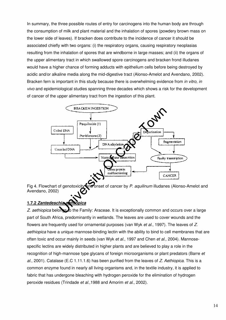
In summary, the three possible routes of entry for carcinogens into the human body are through
the consumption of milk and plant material and the inhalation of spores (powdery brown mass on
the lower side of leaves). If bracken does contribute to the incidence of cancer it should be
associated chiefly with two organs: (i) the respiratory organs, causing respiratory neoplasias
resulting from the inhalation of spores that are windborne in large masses; and (ii) the organs of
the upper alimentary tract in which swallowed spore carcinogens and bracken frond illudanes
would have a higher chance of forming adducts with epithelium cells before being destroyed by
acidic and/or alkaline media along the mid-digestive tract (Alonso-Amelot and Avendano, 2002).
Bracken fern is important in this study because there is overwhelming evidence from in vitro, in
vivo and epidemiological studies spanning three decades which shows a risk for the development
of cancer of the upper alimentary tract from the ingestion of this plant.
Fig 4. Flowchart of genotoxicity and onset of cancer by P. aquilinum illudanes (Alonso-Amelot and Avendano, 2002)
1.7.2 Zantedeschia aethiopica
Z. aethiopica belongs to the Family: Araceae. It is exceptionally common and occurs over a large
part of South Africa, predominantly in wetlands. The leaves are used to cover wounds and the
flowers are frequently used for ornamental purposes (van Wyk et al., 1997). The leaves of Z.
aethiopica have a unique mannose-binding lectin with the ability to bind to cell membranes that are
often toxic and occur mainly in seeds (van Wyk et al., 1997 and Chen et al., 2004). Mannose-
specific lectins are widely distributed in higher plants and are believed to play a role in the
recognition of high-mannose type glycans of foreign microorganisms or plant predators (Barre et
al., 2001). Catalase (E.C 1.11.1.6) has been purified from the leaves of Z. Aethiopica. This is a
common enzyme found in nearly all living organisms and, in the textile industry, it is applied to
fabric that has undergone bleaching with hydrogen peroxide for the elimination of hydrogen
peroxide residues (Trindade et al.,1988 and Amorim et al., 2002).
14
Univers
ity O
f Cap
e Tow
n

1.7.2.1 Active ingredients and pharmacological effects
The active principle varies from species to species but always includes calcium oxalate. The
harmful effects of oxalate produced upon ingestion depend on (i) concentration and (ii) whether
oxalate is present in a soluble or insoluble form. Low to moderate levels of soluble oxalate are less
toxic, however at high levels in the plant, the soluble oxalate is very harmful when eaten since it is
readily absorbed from the digestive tract, reacts with blood calcium, causes ionic imbalance and
decreases the coagulatory properties of the blood leading to internal haemorrhage, nephritis and
renal failure (owing to a rapid accumulation of insoluble calcium oxalate crystals in the kidney
tubules) and eventual death (Blackwell, 1990 and Lewis, 1998). When plants containing the
insoluble form of oxalate are consumed, the crystals may become lodged in the mucous lining of
the mouth and throat causing burning, irritation and swelling with the symptoms disappearing
within a few days (Blackwell, 1990). All parts of Z. aethiopica are toxic and can cause severe
irritation of the mucous membranes, with swelling of the tongue and throat, salivation, nausea,
vomiting and diarrhoea (Lewis, 1998; van Wyk et al., 2002).
Fig 5. Z. aethiopica is commonly referred to as the arum lily and is found from the Western Cape through the Eastern Cape, KwaZulu-Natal, Mpumalanga and into the Northern Province. The flowers are large and are produced in spring, summer and autumn, with a pure white spathe and a yellow spadix, and are often used for ornamental purposes (Van Wyk et al., 1997).
1.7.3 Acokanthera oppositifolia
A. oppositifolia is a member of the family, Apocynaceae, which is well-known for its alkaloidal
content, indole alkaloids derived from tryptamine and steroidal alkaloid (C21 alkaloids) derived from
pregnane, as well as for their potent pharmacological activity (Van Wyk et al., 2002 and Gurib-
Fakim, 2006).
A. oppositifolia is well known throughout Africa as a traditional source of extremely toxic arrow and
spear poisons (van Wyk and Wink, 2004). Secondary poisoning may arise from the consumption of
15
Univers
ity O
f Cap
e Tow
n

meat from an animal that has died from cardiac glycoside poisoning. The poison obtained from
roots, leaves or wood can lead to death in humans within 15 minutes (van Wyk et al., 2002).
A B Fig 6. A. oppositifolia is a medium to large woody shrub with attractive hardy dark green leaves. Clusters of pinkish white, sweetly scented flowers (B) are borne in late winter and spring and are followed by large plum-coloured berry-like fruits (A) which are relished by birds (van Wyk et al., 2002).
The leaves of A. oppositifolia are used in the form of snuff to treat headaches and in infusions for
abdominal pain, convulsions (van Wyk and Gericke, 2000) and snakebite (Watt and Breyer-
Brandwijk, 1962). A. oblongifolia (Hochst.) Codd, a different but related species, prepared by
sequential extraction with dichloromethane followed by 90% methanol induced DNA damage in
human white blood cells using the alkaline comet assay (Taylor et al., 2003).
1.7.3.1 Active ingredients and pharmacological effects
A. oppositifolia are characteristic for their cardiac glycosides, the plants indigenous to South Africa
have the following chemical composition:
(i) seeds: the major component is water-insoluble acovenoside A (Fig 7.), two polar water-soluble
glycosides, acolongifloroside K and acovenoside C, and trace amounts of ouabain (or absent).
(ii) stems and twigs: large amounts of acovenoside A and acolongifloriside K (Neuwinger, 1996).
The amount of the minor component, ouabain (Fig 7.) obtained from Acokanthera sp, varies in
different parts of Africa: trace amounts of ouabain (or absent) from A. oppositifolia in South Africa
and Kenya/Nairobi, whereas a higher content of ouabain is present in A. schimperi from
Kenya/Nairobi (Neuwinger, 1996).
In a study carried out by Wangenheim and Bolcsfoldi (1988), ouabain was positive for mutagenicity
in a mouse lymphoma L5178 thymidine kinase assay in the absence of metabolic activation at a
dose of 0.103-8.22mmol/l; with 2-4 fold increase in the mutation frequency. From their study,
potent mutagenic/carcinogenic compounds tested had greater than 4-fold increases in the
mutation frequency, whilst weak carcinogens or compounds not known to be carcinogenic that was
positive in the assay gave increases of between 2- and 4- fold. In addition, a less than 4-fold
16
Univers
ity O
f Cap
e Tow
n
increase in the mutation frequency was associated with a lower predictivity for carcinogenicity. A
high in vitro cytotoxic activity was obtained for acovenoside A and acolongifloroside K, but no in
vivo activity was obtained for these compounds (Kingsten and Reichstein, 1974).
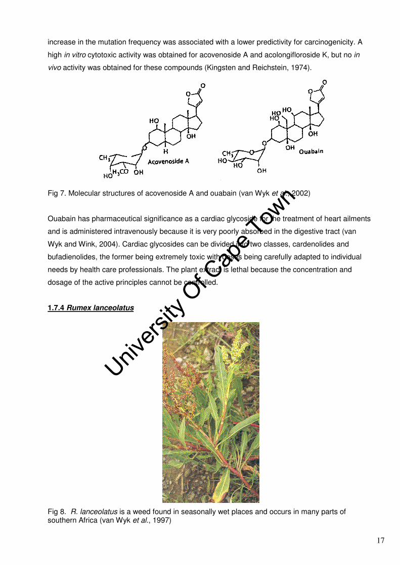
Fig 7. Molecular structures of acovenoside A and ouabain (van Wyk et al., 2002)
Ouabain has pharmaceutical significance as a cardiac glycoside for the treatment of heart ailments
and is administered intravenously because it is very poorly absorbed in the digestive tract (van
Wyk and Wink, 2004). Cardiac glycosides can be divided into two classes, cardenolides and
bufadienolides, the former being extremely toxic with doses being carefully adapted to individual
needs by health care professionals. The plant extract is lethal because the concentration and
dosage of the active principles cannot be controlled.
1.7.4 Rumex lanceolatus
Fig 8. R. lanceolatus is a weed found in seasonally wet places and occurs in many parts of southern Africa (van Wyk et al., 1997)
17
Univers
ity O
f Cap
e Tow
n

Rumex sp (Family: Polygonaceae) occurs widely in southern Africa and the leaves are popular as
a dietary supplement and are served as “sorrel” or cooked with porridge. Both R. lanceolatus and
R. crispus are equally popular as medicinal plants (van Wyk et al., 1997).Infusions and decoctions
of this plant are traditionally used to treat constipation, liver problems and arthritis, with the root
being used as a remedy for internal parasites (tapeworm and roundworm). It is also regarded as a
cleansing herb and used to treat chronic skin problems. The whole plant is said to be widely
employed for vascular disorders and internal bleeding (van Wyk and Wink, 2004). Externally it is
applied to ulcers, boils and tumours.
1.7.4.1 Active ingredients and pharmacological effects
Many Rumex sp contain anthraquinones, with the exception of one species having been found to
date, namely R. induratus (Ferreres et al., 2006). R. lanceolatus is a mild purgative containing
chrysophanic acid, emodin (Fig 9.) and a small amount of volatile oil; neither alkaloid nor glucoside
are present (Watt and Breyer-Brandwijk, 1962). Roots of Rumex sp contain glycosides of emodin
and chrysophanol (such as chrysophanein, Fig 9.) and tannins (van Wyk and Wink, 2004). The
laxative effect of the roots of Rumex sp is due to chrysophanol and related anthracene glycosides.
Fig 9. Molecular structures of chrysophanol, chrysophanein, emodin and oxalic acid (van Wyk et al., 1997 and Lee et al., 2005)
Oxalic acid (Fig 9.) is typical of Rumex sp and the leaves contain large amounts of it (van Wyk et
al., 1997 and van Wyk and Wink, 2004). Oxalic acid is toxic in high concentrations, but is used
18
Univers
ity O
f Cap
e Tow
n

medicinally as a haemostatic agent in the form of a 5% solution in 5% malonic acid (van Wyk and
Wink, 2004).
Rumex species are related to the well-known medicinal plants known as rhubarbs, which are a
group of plants that belong to the genus Rheum in the family Polygonaceae (van Wyk and Wink,
2004). Several monomeric anthraquinones, such as emodin (1,3,8-trihydroxy-6-methyl-
anthraquinone) and related derivatives, are constituents of rhubarb (an oriental medicine for
diarrhoea) that exhibit mutagenicity in the Salmonella/microsomes test and cultured tumour cells
(Morita et al., 1988 and Ueno, 1991). However, emodin from the aerial parts of R. acetosa strongly
inhibits the proliferation of tumour cell lines, and shows antimutagenicity (in strains TA 98 and
TA100) and antigenotoxic activity (Lee et al., 2005).The OH group at the C-6 position in the
anthraquinone nucleus may play an important role for their cytotoxicities, and an introduction of
OH- or OCH3 group at C-6 position is necessary for their antimutagenic activities (Lee et al., 2005).
The rhizome of R. crispus L. contains volatile oil, resin, tannin, rumicin, sulphur, starch, various
other salts, lapathin, oxymethylanthraquinone, emodin and chrysophanic acid (Watt and Breyer-
Brandwijk, 1962). The tannin content of the rhizome is so high that the plant was at one time
considered a possible commercial source of tannin. The phytochemical screening of the powdered
root of Rumex steudlii Hochst shows the presence of polyphenols, pytosterols, O-anthraquinone
glycoside, tannins, hydrolysable tannins and saponins, but the test for alkaloids shows a negative
result (Gebrie et al., 2004).
1.7.5 Pelargonium sp.
Pelargonium sp (Family: Geraniaceae) is used for its commercial and medicinal value. Rose-
scented geranium (Pelargonium species) is a high-value, aromatic plant cultivated for its essential
oil, which is widely used in the fragrance industry, in aromatherapy and for the extraction of
commercial rhodinol (mixture of linalool, citronellol and geraniol) (Rao et al., 2002).
Fig 10. Leaves of Pelargonium sp. cf. inquinans (L.) L’ Herit. Pelargonium sp. are cultivated for their medicinal and commercial value
19
Univers
ity O
f Cap
e Tow
n

The essential oil is obtained using steam or water as well as steam distillation of shoot biomass
(Rao et al., 2002). Root extracts are used in preparations to treat acute and chronic infections of
the nose, ears and chest, and have been found to be particularly effective for bronchitis in children
and as a supportive treatment for tuberculosis and chronic bronchitis (van Wyk and Wink, 2004).
The leaves and roots are also used for the treatment of dysentery and syphilis. Isoquercetrin and
rutin, respectively the major and minor flavonoids obtained from Pelargonium radula (Cav.) L’Herit,
have been shown to have antimicrobial activity (Pepeljnjak et al., 2005).
1.7.5.1 Active ingredients and pharmacological effects
The main ingredients of Pelargonium sp. are coumarins (Fig 11.); mainly 7-hydroxy-5,6-
dimethoxycoumarin, that co-occur with at least seven other coumarins, gallic acid derivatives
(including gallic acid and gallic acid methyl ester), oligomeric proanthocyanidins, and flavonoids:
flavan-3-ols (e.g. catechin) and flavon-3-ol (e.g. quercetin) (Middleton et al., 2000 and van Wyk
and Wink, 2004).
Fig 11. Chemical structure of coumarin (Culvenor and Jago, 1979)
Pelargonium oils contain a variety of monoterpenoids, such as geraniol, (+)-isomethone, citronellol
and phenylethyl alcohol (responsible for the rose smell). The gallic acid derivatives and other
phenolic compounds in the roots have powerful antibacterial and antiviral activity, and together with
coumarins provide a rationale for the proven immunomodulatory activity.
The five South African (SA) plants discussed above have not been previously evaluated for
mutagenicity. In countries where P. aquilinum has been consumed as a food, in-depth scientific
investigations have concluded that ptaquiloside and its metabolically activated intermediate,
pteridienone, play a role in the carcinogenic process. It is inevitable that mutagenicity and
clastogenicity will be anticipated for the SA P. aquilinum, which can be evaluated with the selected
in vitro mutagenicity tests.
1.8 In vitro mutagenicity assays
1.8.1 Salmonella reverse mutation assay
The Salmonella reverse mutation assay is an initial screen to determine the mutagenic potential of
new chemicals and drugs, as there is a high predictive value for rodent carcinogenicity when a
20
Univers
ity O
f Cap
e Tow
n

mutagenic response is obtained. In the Salmonella reverse mutation assay, the test substance is
added to the top agar (minimal medium) containing S9, a bacterial tester strain and a minimal
amount of histidine to support a few cycles of replication, but not enough to permit colonies to form.
The molten top agar is poured onto solid media and incubated (48hrs/37oC). In the presence of the
mutagen, the strain reverts to wild type (his+) thereby allowing colonies to form on the minimal
medium. The number of colonies can be related to controls and to the amount of the test
substance used to give a quantitative result. A twofold rule usually applies when the number of
colonies of the test chemical is twice the amount on the control plate and the test chemical is
mutagenic.
Bacterial strains were originally derived from Salmonella typhimurium LT2. The bacteria carry a
mutation (his-) that makes them nutritionally deficient because they cannot synthesise one of the
enzymes needed to manufacture histidine, which is supplemented in the top agar. The genotypes
and related characteristics of the primary tester strains are indicated in Table 3. The Salmonella
strains used in the test have different mutations in various genes of the histidine operon; each of
these mutations is designed to be responsive to mutagens that act via different mechanisms
(Mortelmans and Zeiger, 2000). TA97a and TA98 detect frameshift mutagens, whilst TA100
detects mutagens that cause base-pair substitutions (Maron and Ames, 1983). TA102 detects
mutagens that cause oxidative DNA damage, and being a DNA repair proficient strain, detects
cross-linking agents such as bleomycin and mitomycin C (Mortelmans and Zeiger, 2000).
Table 3: Genotypes for the primary tester strains used in the Salmonella reverse mutation assay Histidine Mutation
LPS Repair *R-factor plasmid pKM101
hisD6610 hisD3052 hisG46 hisG428 **(pAQ1)
Strain TA97a
Strain TA98 Strain TA100
rfa deletion uvrB deletion
+R
Strain TA102 rfa deletion uvrB intact +R
Reversion event:
Frameshift
Reversion event:
Frameshift
Reversion event:
Base-pair substitution
Reversion event:
Transitions & transversions
(*R-factor plasmid pKM101 confers ampicillin resistance, **pAQ1 confers tetracycline resistance. LPS: lipopolysaccharide layer prevalent in gram negative bacteria, rfa deletion makes the cell wall more permeable to the penetration of chemicals). Source: adapted from Maron and Ames (1983) and Mortelmans and Zeiger (2000)
A chemical can be scrutinised for its ability to achieve one or more kinds of genetic alteration,
resulting in reversion from his- to his+, and is taken as a measure of an agent’s mutagenic and
carcinogenic potential. Since many substances are unable to pass through bacterial cell walls, the
strains used carry a mutation that makes the wall more penetrable to chemicals (rfa). The strains
TA97a, TA98 and TA100 also carry another mutation that causes a defect in the DNA repair
mechanism (uvrB). Therefore bacterial strains (with the exception of TA102) are repair
21
Univers
ity O
f Cap
e Tow
n
incompetent, and permit the easier detection of various kinds of alteration in the DNA, since a
defect will not be repaired.
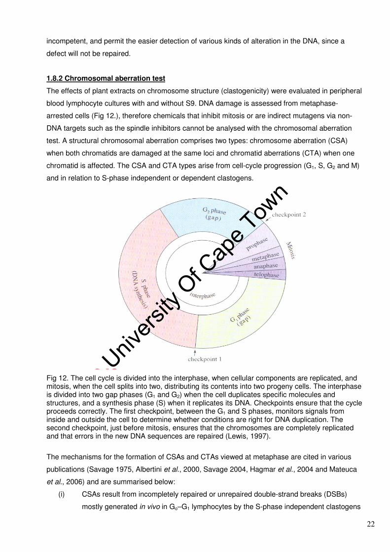
1.8.2 Chromosomal aberration test
The effects of plant extracts on chromosome structure (clastogenicity) were evaluated in peripheral
blood lymphocyte cultures with and without S9. DNA damage is assessed from metaphase-
arrested cells (Fig 12.), therefore chemicals that inhibit mitosis or are indirect mutagens via non-
DNA targets such as the spindle inhibitors cannot be analysed with the chromosomal aberration
test. A structural chromosomal aberration comprises two types: chromosome aberration (CSA)
when both chromatids are damaged at the same loci and chromatid aberrations (CTA) when one
chromatid is affected. The CSA and CTA types arise from cell-cycle progression (G1, S, G2 and M)
and in relation to S-phase independent or dependent clastogens.
Fig 12. The cell cycle is divided into the interphase, when cellular components are replicated, and mitosis, when the cell splits into two, distributing its contents into two progeny cells. The interphase is divided into two gap phases (G1 and G2) when the cell duplicates specific molecules and structures, and a synthesis phase (S) when it replicates its DNA. Checkpoints ensure that the cycle proceeds correctly. The first checkpoint, between the G1 and S phases, monitors signals from inside and outside the cell to determine whether conditions are right for DNA duplication. The second checkpoint, just before mitosis, ensures that the chromosomes are completely replicated and that errors in the new DNA sequences are repaired (Lewis, 1997).
The mechanisms for the formation of CSAs and CTAs viewed at metaphase are cited in various
publications (Savage 1975, Albertini et al., 2000, Savage 2004, Hagmar et al., 2004 and Mateuca
et al., 2006) and are summarised below:
(i) CSAs result from incompletely repaired or unrepaired double-strand breaks (DSBs)
mostly generated in vivo in Go–G1 lymphocytes by the S-phase independent clastogens
22
Univers
ity O
f Cap
e Tow
n

(e.g. ionising radiation). After DNA synthesis and chromosome duplication at S-phase,
the aberrations formed in Go–G1 are seen as chromosome-type breaks in the
metaphase.
(ii) CTAs (e.g. chromatid-type breaks) arise predominantly in vitro during the G2-phase of
the cultured lymphocytes in response to base modifications and single-strand breaks
(SSBs) induced by S-phase-dependent clastogens (e.g. chemicals).
The extensive use of the chromosomal aberration assay over the last 30 years has resulted in the
accumulation of analytical data in many European laboratories and has enabled the examination of
the potential association between previously measured structural chromosomal aberration
frequency and subsequent cancer outcome (Bonassi and Au, 2002). The impact of CSAs and
CTAs from smoking and other environmental carcinogens on human cancer risk has been recently
assessed, because originally the study did not have sufficient power and/or follow-up time for a
conclusive result with respect to cancer predictivity. In the Taiwanese and Nordic cohorts, CSAs
but not CTAs predict cancer risk; however, in the Italian cohort no clear-cut difference in cancer
predictivity between CSA and CTA biomarkers was observed (Liou et al., 1999 and Hagmar et al.,
2004). Evidence from a recent study conducted by Norppa et al. (2006) indicates that both
chromatid-type and chromosome-type aberrations predict cancer; even though some data suggest
that chromosome-type aberrations may have a more pronounced predictive value than chromatid-
type aberrations.
1.8.2.1 The distinction between gap junction and chromosome breaks
Light microscopy is used to detect structural chromosomal aberrations (gaps and breaks) from
metaphase-arrested lymphocytes.
Fig 13. Structural chromosome aberrations by light microscopy of metaphase arrested cells: (a) chromatid constriction; (b) chromatid gap; (c) chromosome constriction; (d) constriction/gap; (e) chromosome gap; (f–h) examples of aligned chromatid breaks; (i–l) examples of dislocated chromatid breaks; (m) chromosome break with complete sister-union (SU); (n) chromosome break with incomplete sister-union (Nup or Nud); (o) chromosome break with no sister-union (Nupd), or a chromosome-type terminal deletion (Savage, 2004)
23
Univers
ity O
f Cap
e Tow
n

The distinguishing features of gaps and breaks applied to the scoring of metaphase cells was
according to Savage (2004). The following features are characteristic of a break: (i) the non-
staining region is larger than the width of a chromatid; (ii) the distance between the broken “ends”
of a chromatid has no visible connection (Fig 13. f–h); and (iii) there is non-alignment, or
dislocation, of the two segments (examples Fig 13. i–l). An aberration is referred to as a gap when
the non-staining region is smaller than the width of a chromatid and little chromatin may be evident.
The visual non-staining region of a gap does not imply that chromatin is absent; in such instances,
chromatin is seen in the non-staining region under the electron microscope.
1.9 Overview of the study
Traditional plants or extracts consumed for their medicinal or dietary value are assumed to be safe
because they occur naturally. This study looked at the potential mutagenicity of five commonly
consumed traditional plants, as there is a huge body of evidence that disproves the theory that
natural equates to safe. In addition, there is a high incidence of late-onset oesophageal cancer
among the population of the Eastern Cape Province of South Africa where traditional
plants/extracts are more frequently consumed. The rationale and justification for this research are
that the five traditional plants in this study are part of the diet in this population; there is no prior
research in this field with the exception of a South African epidemiological study (Sewram, 2006)
which supplements this dissertation.
The research problem (hypothesis) is to identify those traditional plant extracts that damage DNA
(mutagenic agents) and chromosomes (clastogenic agents) using in-vitro quantitative test
methods. The five traditional plants investigated were Acokanthera oppositifolia (Lam.) Codd;
Pelargonium cf. inquinans (L.) L’ Herit; Pteridium aquilinum subsp aquilinum; Rumex lanceolatus
Thunb. and Zantedeschia aethiopica (L.). This is a screening study of the potential mutagenicity of
aqueous extracts and not a phytochemical study. Simplified versions of two of the three in vitro
genotoxicity screening assays currently recommended by the International Conferences on
Harmonisation of Technical requirements for registration of Pharmaceuticals for Human Use (ICH
Guidance S2B): (i) a bacterial mutation screening test (Salmonella reverse mutation assay) and (ii)
a chromosomal aberration screening test in Chinese hamster ovary (CHO) cells (Zhang et al.,
2004) were selected, however in the present study peripheral blood lymphocytes were used
instead of CHO cells. The tests were done with and without metabolic activation (S9 mix).
Limitations of the Salmonella reverse mutation assay include that it cannot detect the mutagenicity
of large molecules that cannot pass through the cell wall of the tester strain and it also fails to
detect those carcinogens (e.g. Fumonisin B1) that are not mutagenic. The chromosomal aberration
test was performed on peripheral blood lymphocytes, the limitations include interference from red
blood cells, and the small size and low concentration of lymphocytes in the blood relative to
24
Univers
ity O
f Cap
e Tow
n

monocytes and granulocytes, which resulted in very few metaphase cells for chromosomal
analysis. The lymphocytes were stimulated to enter the cell cycle (G1, S, G2 and M) using
phytohaemagglutinin followed by metaphase arrest using colcemid. The test was not suitable for
extracts that inhibited the cell cycle, which was an additional limitation. Following Savage (2004),
approximately 50 metaphase cells were scored for chromosomal aberrations (gaps and breaks).
The data were collected by measuring the two endpoints (mutagenicity and clastogenicity) at
increasing concentrations of aqueous plant extracts with and without metabolic activation (refer to
section 1.3). In the Salmonella reverse mutation assay, a reversion to wild type with an associated
three- to fivefold increase in the number of revertant colonies indicated mutagenicity. Four
Salmonella tester strains (TA97a, TA98, TA100 and TA102) were selected, each indicative of a
particular kind of mutation: base-pair substitution, frameshift or oxidative DNA damage. The
baseline revertant colonies for TA102 were the highest (fivefold) relative to the other three tester
strains. Batch TA102 complied with the supplier’s release criteria; the baseline reversion frequency
had increased over the years when compared to that of the original tester strains (email
correspondence with the supplier, Appendix C). The mutagenicity data were analysed by a
professional statistician using gamma statistics, whilst the clastogenicity data were not statistically
analysed owing to the low number of metaphase cells. In light of the evidence provided the
mutagenic potential of five South African plants currently being used in traditional medicine and/or
as dietary supplements were investigated.
25
Univers
ity O
f Cap
e Tow
n
CHAPTER 2
MATERIALS AND METHOD
2.1 Collection of plants
The plants Acokanthera oppositifolia (Lam.); Pelargonium sp. cf. inquinans (L.) L’ Herit;
Pteridium aquilinum subsp aquilinum; Rumex lanceolatus Thunb. and Zantedeschia aethiopica (L.)
Sg. were obtained from Port St Johns in the Eastern Cape, and the Silverglen medicinal plant
nursery in KwaZulu-Natal during November 2002. The identities of the plants were authenticated
by comparison with reference specimens at the Kei Herbarium (Walter Sisulu University) and Natal
Herbarium (National Botanical Institute, KwaZulu-Natal), and voucher specimens (see Table 5)
were deposited in these herbariums for future reference. The leaves of all plants were air-dried,
crushed by hand or cut with an Anvil pruner before they were milled into a fine powder using a
Kenwood blender. A facemask was used to avoid inhalation of the dust during the handling and
milling of plant material. The powdered material was weighed into brown paper bags, labelled and
stored at room temperature.
2.2 Preparation of plant extracts
Milled plant material (30g) was weighed into a conical flask (1L), and then boiled in water (500ml).
The plant material was agitated using a magnetic stirrer and boiled for 1hr. The plant extracts were
cooled to room temperature and filtered twice using a Rundfilter MN617 filter paper. The filtrate
was transferred to a round bottom flask (1L) and refrigerated at -20oC overnight. The frozen filtrate
was thereafter lyophilised and stored at 4 to 5oC in a dessicator; the mass of lyophilisate obtained
is recorded in Table 4.
Table 4. Plant processing and yield of freeze-dried extract (lyophilisate)
Botanical name Part of plant
dried and milled Extraction
75–85oC/1hr
Mass of lyophilised
product %Yield
Pteridium aquilinum subsp.aquilinum
Leaves 30g/500ml 4.33g 14.43
Acokanthera oppositifolia (Lam). Leaves & stems 30g/500ml 5.27g 17.57 Pelargonium L. Herit Leaves & stems 30g/500ml 4.18g 13.93 Rumex lanceolatus Thunb. Leaves & stems 30g/500ml 4.76g 15.87 Zantedeschia aethiopica (L.) Sg Leaves & stems 30g/500ml 5.29g 17.63
The freeze-dried material (0.8g) was dissolved in sterile distilled water (10ml), sonicated for 20min,
centrifuged at 4000rpm for 10min at 0oC and the supernatant decanted into a 25ml beaker. The
supernatant was transferred into a scintered glass filtration apparatus containing a 5-micron
cellulose filter. The filtrate was re-filtered through a 1.2-micron filter. The solution was finally filter
sterilised through a 0.2µm syringe filter into a sterile bijou bottle from which dilutions were made
using sterile water by aseptic handling to avoid test sample contamination. The following dilutions
were prepared: (i) 0.005; 0.01; 0.02 and 0.04g/ml for A. oppositifolia, Pelargonium, P. aquilinum
26
Univers
ity O
f Cap
e Tow
n

27
Univers
ity O
f Cap
e Tow
n

and R. lanceolatus, and (ii) 0.00125; 0.0025; 0.005; 0.01 and 0.02g/ml for Z. aethiopica as a result
of the highly viscous nature of this extract.
2.3 Preparation of S9
2.3.1 Induction of rat liver enzymes
Approval for the experimental protocol was granted by the Ethics Committee for Research on
Animals (ECRA) of the Medical Research Council, Tygerberg, South Africa. Fifteen rats weighing
200 to 300g were administered with Aroclor 1254, a polychlorinated biphenyl (PCB) and a known
carcinogen, based on a procedure by Czygan et al. (1973). Aroclor 1254 was warmed in a 20oC
waterbath, and 3.0g weighed into a 25ml measuring cylinder. Warm sunflower oil was added to the
measuring cylinder to a volume of 15ml and homogenised by aspiration with a pipette. This yielded
a 200mg/ml Aroclor 1254 working stock solution which was dispensed into two 7ml bottles fitted
with rubber bungs and sealed with metal rings. The bottles were covered in foil to avoid
degradation of the light-sensitive PCBs. All pipettes and glassware, including the 25ml measuring
cylinder, were discarded as carcinogenic waste. The viscous Aroclor 1254 working stock was
immersed in warm water during intraperitoneal injection into rats. The Aroclor was administered at
500mg/kg body weight using a 26G needle for intraperitoneal injection. This procedure was
repeated for all fifteen rats.
2.3.2 Preparation of liver homogenate S9 fraction
On the seventh day after induction the rats were sacrificed by cervical dislocation and the livers
removed by midline incision, according to Maron and Ames (1983). Instruments for the dissection
(forceps and scissors) were sterilised by autoclaving (121oC/20min) and iodine was used as a
disinfectant during excision of livers. Preparation of the liver S9 fraction is based on the procedure
of Garner et al. (1972). All steps were carried out on ice and using sterile solutions (cold).
Glassware and centrifuge tubes were autoclaved prior to use. The centrifuge tube caps were
sterilised in 70% ethanol as they were not autoclavable. Frequent disinfection of surfaces during
S9 preparation was attained by application of 70% aqueous ethanol.
The excised livers were washed in pre-weighed 1L beakers containing 400ml cold sterile KCl
(0.15M) to remove haemoglobin, covered with foil and kept on ice. The livers (a total mass of
206.5g) were transferred into a beaker containing 3ml of chilled 0.15M KCl per gram of wet liver,
and cut into smaller pieces. The liver was homogenised for 5min with a Polytron homogeniser (PT
3000, Kinematica AG). The liver homogenate was then filtered through sterile double-layered
cheesecloth into a 500ml beaker to remove larger pieces of tissue while working on ice. The filtrate
collected in the beaker was transferred to an all-glass dounce homogeniser, and dounced by 10
strokes of the plunger to release the microsomes. The homogenate was then centrifuged at 9000g
for 10min at 4oC using a Sorvall Type SS-34 fixed angle rotor. The supernatant (S9 fraction) was
28
Univers
ity O
f Cap
e Tow
n

dispensed in 3ml and 5ml aliquots into sterile bijou bottles and immediately stored at -70oC, and
the pellets were discarded. The S9 was tested for sterility, protein concentration and CYP P450
activity. The absence of colonies on a nutrient broth agar plate after inoculation and incubation at
37oC for 24hrs indicated a sterile S9 preparation.
2.3.3 Protein concentration of S9
The protein concentration was determined by the Bradford method (1976). A standard curve was
constructed using known amounts of bovine serum albumin (BSA). One hundred microlitres of a
1:100 S9 dilution was used for the Bradford test. The BSA standards 8, 16, 24, 32 and 40ug and
the S9 sample were prepared in duplicate, and measured at 595nm with an Uvikon 923
spectrophotometer. The protein concentration of the S9 fraction was 27.1mg/ml.
2.3.4 CYP P450 concentration of S9
The CYP P450 concentration was determined by a method adapted from Omura and Sato (1964).
The S9 was diluted 20x in a volume of 8ml by combining 400µl S9 with 7600µl Tris buffer (0.1M,
pH7.4). The sample was saturated with a stream of carbon monoxide for 1min and placed in two
cuvettes (4ml capacity) marked as reference and sample. The baseline absorbance was read at
500-400nm at a sensitivity of 0.5 and 0.1 with a double beam Beckman spectrophotometer
UV5260.
Sodium dithionite (0.01298g) was added to the sample cuvette and dissolved by inversion several
times. The observed bubbling, which was indicative of CYP P450, stopped before the difference
spectrum was monitored (500nm and 400nm). The CYP P450 activity was determined from the
450nm absorption peak using the following formula and the mmol extinction coefficient (91) of the
compound.
CYP P450 activity = (A450-490 - Correction of baseline)/100 X sensitivity X 1000/91 X 1/mg per ml protein = 75.5 - (-8.6) / 100 X 0.1 X 10.989 X 1/27.1 X 20 dilution factor
= 0.7nmol/mg
The protein concentration (27.1mg/ml) was inserted into the formula. The value for “(A450-490 -
correction of baseline)” was obtained from the difference spectra chromatograph (section 3.1.1).
The CYP P450 activity calculated was 0.7nmol/mg. This was within the recommended range (0.6-
0.8nmol/mg), and therefore suitable for use in the mutagenicity assay.
2.4 Verification of genotypes of Salmonella tester strains
The verification of genotypes was done according to the method prescribed by Maron and Ames
(1983) and Mortelmans and Zeiger (2000). Five microlitres of Salmonella tester strains: TA97a;
TA98; TA100 and TA102 (Xenometrix, CA, USA) was seeded into Oxoid nutrient broth No. 2
29
Univers
ity O
f Cap
e Tow
n

(4.9ml) that was supplemented with antibiotics according to the procedure in Fig 14. Genotype
verification was performed for his-, uvrB, rfa and antibiotic resistance markers. (Note: all solvents
and media are listed in Appendix A.)
Strains TA97a, TA98, TA100 and TA102 contain plasmid pKM101, which (i) confers ampicillin
resistance that serves as a convenient marker for detecting the presence of the plasmid, and (ii)
enhances chemical and UV-induced mutagenesis. In addition to pKM101, strain TA102 contains
multicopy plasmid pAQ1. Plasmid pAQ1 confers tetracycline resistance and is a convenient marker
for detecting the presence of the plasmid. Furthermore, the hisG428 mutation was inserted into
pAQ1 with the aim of amplifying the number of target sites.
2.4.1 Histidine dependence
An aliquot of overnight culture (0.1ml) from each tube at step 4 (Fig 14.) was plated onto MGA
plates supplemented with two drops of 0.01% w/v biotin. Because all Salmonella strains are
histidine dependent, there should be no growth on the plates after incubation at 37oC for 16 to
24hrs. Plates that display growth indicate that the colony from which the broth culture has arisen
(in the masterplate) is unsuitable for the preparation of working cultures for the Salmonella reverse
mutation assay.
2.4.2 Biotin and histidine dependence
MGA plates were supplemented with two drops of 0.01% w/v biotin and two drops of 0.5% w/v
histidine by the spread plate technique described in Fig 14, step 2. A loopful of culture from each of
the ten tubes was streaked onto the MGA plates and incubated at 37oC for 16 to 24hrs. Growth
was expected to be observed with all strains since they are all histidine dependent.
2.4.3 Biotin dependence
MGA plates were supplemented with two drops of 0.5% w/v histidine. A loopful of culture from
each of the ten tubes was streaked onto the MGA plates and incubated at 37oC for 16 to 24hrs.
There should be no growth on the plate except for TA102, which is biotin independent since it has
an intact bio gene, unlike the other tester strains where the uvrB gene deletion extends through the
bio gene making them biotin dependent.
2.4.4 rfa marker
Sterilised low melting top agar (100ml) was supplemented with a sterile 0.5mM histidine/biotin
solution (12ml). Aliquots of top agar (2.5ml) were dispensed into ten test tubes (with spring-loaded
Cap-O-Test) and kept in a 45oC waterbath. A 0.1ml inoculum of culture was added to 2.5ml molten
top agar in each of the test tubes. The top agar was vortexed and poured onto nutrient broth agar
30
Univers
ity O
f Cap
e Tow
n

Fig 14. Flow diagram for genotype verification of Salmonella tester strains Step 1: the tester strains were inoculated into antibiotic-supplemented oxoid nutrient broth. Step 2: individual colonies from the broth culture were isolated on MGA plates supplemented with histidine, biotin and ampicillin (for strains TA97a, TA98 and TA100), and on MGA plates supplemented with histidine, biotin, ampicillin and tetracycline (for strain TA102) by a four-way streak. Step 3: ten colonies were transferred to a designated spot on MGA masterplates M1 and M2 marked “A-J” and into 10ml oxoid nutrient broth tubes marked “A-J”. Step 4: after incubation the broth cultures were used for the genotype test whilst the masterplates were stored at 4oC.
31
Univers
ity O
f Cap
e Tow
n

(NBA) plates using a pour-plate technique. Paper discs (0.6mm) were made from Whatman No. 1
using a paper punch and sterilised by autoclaving at 121oC for 20min. A single disc was placed in
the centre of each plate and 5µl of 0.1% crystal violet were added to the disc. The plates were
incubated at 37oC for 16 to 24hrs. A zone of inhibition indicates a positive result. The rfa deletion
allows large molecules to penetrate the cell wall making it more efficient at detecting mutagens.
Fig 15. The zone of inhibition indicates crystal violet sensitivity. The rfa mutation permits large molecules such as crystal violet to enter and kill the bacteria. Wild-type strains are not inhibited because the crystal violet cannot penetrate the cell. All Salmonella strains should show a zone of growth inhibition surrounding the disc.
2.4.5 uvrB deletion
Fig 16. Strains with uvrB deletion will grow only on the un-irradiated side of the plate as seen for TA97a. The deletion mutation stretches across the bio-uvrB region of the chromosome. It shows that the tester strains are defective in the accurate DNA repair pathway due to the uvrB deletion.
A loopful of inoculum from each of the ten tubes was streaked onto NBA plates. Covering half the
plate with cardboard, irradiation with a 15W UV germicidal lamp (43.5cm in length) was applied for
8 seconds at a distance of 33cm. No growth on the irradiated portion of the plate should be
observed for TA97a, TA98 and TA100, since they do not have an intact uvrB gene. However,
TA102 does have an intact uvrB gene and growth is observed in the UV-exposed region. During
32
Univers
ity O
f Cap
e Tow
n

irradiation, petri plate lids were removed because the UV radiation does not penetrate the plastic.
Plates were incubated at 37oC for 16 to 24hrs.
2.4.6 Testing for the presence of plasmid pKM101
Fig 17. Ampicillin resistance is a convenient marker for testing for the presence of the plasmid pKM101 since specific regions of the pKM101 DNA are essential for enhancing UV and chemical mutagenesis. With the sample on the right, the zone of inhibition indicates loss of the plasmid and thus is unsuitable for working culture preparation.
Ampicillin resistance was used as a marker to test for the presence of plasmid pKM101, since
specific regions of the pKM101 DNA are essential for the enhancement of UV and chemical
mutagenesis. The pour plate technique described in section 2.4.4 was applied. An ampicillin disc
(BBL Sensi-Disc Antimicrobial Susceptibility Test Discs, Ampicillin 10µg) was placed in the centre
of each plate and incubated at 370C for 16 to 24hrs.The absence of a zone of inhibition around the
disc demonstrates a positive result.
2.4.7 Spontaneous mutation frequency
A 0.1ml inoculum from each of the 10 culture tubes was seeded into top agar supplemented with a
0.5mM histidine/biotin solution and overlayed onto MGA plates. The plates were incubated at 37oC
Table 6: The spontaneous revertant laboratory values in comparison to the values stipulated in the literature and by the supplier
Strain Batch no.
Spontaneous revertant colonies
Laboratory value Minus S9
bValues cited in the
literature
aValues from
supplier Minus S9 Plus S9 Minus S9
TA97a Lot 517 220 ± 28 75-200 100-200 196-256
TA98 Lot 518 39 ± 3 20-50 20-50 -
TA100 Lot 519 187 ± 14 75-200 75-200 -
TA102 Lot 520 564 ± 78 100-300 200-400 500-588
a Values from the supplier’s (Xenometrix CA, USA) quality testing
b The literature by Mortelmans and Zieger (2000) is recommended by the supplier. However, the exception is
for TA97a and TA102 where the supplier values exceed the literature values (minus S9). The supplier indicates that the high values for TA97a and TA102 is because the spontaneous revertant frequencies have increased since Maron and Ames (1983).
33
Univers
ity O
f Cap
e Tow
n

for 48hrs. A lawn of bacterial growth was observed at 24hrs and revertant colonies at 48hrs.The
colonies were hand-counted using a Quebec darkfield colony counter. The spontaneous mutation
frequencies of the tester strains cited in the literature (Mortelmans and Zieger, 2000) by the
supplier and the values obtained in the laboratory are tabulated in Table 6.
2.5 Preparation working culture and frozen permanents
A colony that complied with the genotype verification and spontaneous mutant frequency from
each tester strain was removed from the masterplate and seeded into oxoid nutrient broth No. 2
(5.5ml) and incubated at 37oC overnight in an orbital shaker (Culture A). Working cultures were
made by seeding Culture A (1ml) into sterile oxoid nutrient broth No. 2 (50ml) and incubated at
37oC overnight in an orbital shaker. DMSO (5ml) was added to the working culture (50ml) and
aliquots (1ml) were dispensed into sterile cryovials kept on ice and transferred to a -70oC freezer.
For mutagenicity testing an aliquot of working culture (1ml) was thawed, seeded into oxoid nutrient
broth (50ml) and incubated as previously described. Five “frozen permanents” were made by
adding DMSO (0.5ml) to culture A (4.5ml), dispensed into 1ml aliquots kept on ice and stored at
-70oC.
2.6 Dose-response of tester strain to mutagen
The dose-response of tester strains TA97a, TA98 and TA100 to increasing concentrations of 2-
acetamidofluorine (2-AAF) was done in quadruplicate using a standard pour plate technique. The
purpose of the dose response was to (i) select a single concentration from the monotonic region of
the graph that produced a two- to fivefold increase in revertant colonies than the spontaneous
revertants for the purpose of a positive control and (ii) examine the strain’s responsiveness to
mutagens. An overnight working culture (0.1ml) was seeded into histidine/biotin supplemented with
top agar (2ml), followed by 2-AAF (0.1ml) and 4% S9 mix (0.5ml). The top agar was overlayed onto
minimal glucose agar (MGA) plates and incubated at 37oC for 48hrs. Revertant colonies were
hand-counted and the average of quadruplicate plates was plotted (refer to the results in section
3.1.2). The dose-response in TA102 was done with cumene hydroperoxide (recommended by
supplier), which is a directly acting carcinogen and as such requires no metabolic activation (S9).
Dose response for TA102 was done in a similar manner with the exception that 2.5ml top agar was
used instead of 2ml.
2.7 Salmonella reverse mutation assay
The Salmonella reverse mutation assay was performed in quadruplicate with TA97a, TA98, TA100
and TA102 (with and without S9) for the following reasons: (i) maximum detection of mutagenic
compounds; (ii) is favourable for statistical analysis; and (iii) allows for comparisons to be drawn.
The tester strains, TA97a and TA98, are designed to detect frameshift mutations, TA100 for the
detection of base-pair substitutions and TA102 for the detection of oxidative DNA damage, DNA
34
Univers
ity O
f Cap
e Tow
n
crosslinking and transition transversion reactions. The use of four tester strains increased the
likelihood of detecting a test substance capable of mutagenesis by either one or more of the
mechanisms described above. Bacteria are unable to metabolise chemicals via cytochrome P450,
as in mammals and other vertebrates, and a key component for making the bacterial mutagenicity
test useful was the inclusion of an exogenous mammalian metabolic activation system consisting
of an extract of rat liver (S9).
An extract was mutagenic if the average number of revertants per plate (mean of 4 or 3 replicate
plates) was at least doubled over the spontaneous revertant frequency and is in keeping with the
two- to threefold rule. If the revertant count of a dose-response curve of test chemical exceeds
twice or thrice the spontaneous background rate, the two- to threefold rule determines the test
result positive. When the maximum revertant count does not exceed two to three times the
spontaneous background rate, this rule concludes that the test is negative.
A standard volume of 0.1ml test sample per plate was applied across all strains. The test sample
(0.08g/ml) was prepared by dissolving lyophilised extract (0.57g) in 7ml distilled water. The
following concentrations were tested: (i) 0.005, 0.02 and 0.08g/ml for A. oppositifolia, Pelargonium,
P. aquilinum and R. lanceolatus; and (ii) 0.00125; 0.005 and 0.02g/ml for Z. aethiopica as a result
of problems with viscosity. The working culture (1ml) of tester strains TA97a, TA98, TA100 and
TA102 was seeded into sterile oxoid nutrient broth (50ml) and incubated at 37oC overnight on an
orbital shaker. Top agar was supplemented with histidine/biotin (0.5mM) and maintained at 45oC in
a waterbath. The working culture (0.1ml) was added to 2ml top agar, followed by plant extract
(0.1ml) and 4% S9 mix (0.5ml) for the analysis of promutagens (chemicals requiring metabolic
activation). The top agar was overlayed onto minimum glucose agar (MGA) plates and incubated
at 37oC for 48hrs. For the positive control the plant extract was replaced with 2-AAF (in strains
TA97a, TA98 and TA100, with S9) and cumene hydroperoxide (in strain TA102 without S9). The
extracts were tested in the absence of S9 in a similar manner with the exception that 2.5ml top
agar was used instead of 2ml.
The condition of the background lawn is an indicator of toxicity: (i) presence of the background
lawn in agar plates indicates non-toxicity of extracts (Reid et al, 2006) and (ii) when there is
“thinning” or complete absence of the background lawn, this indicates the toxicity of the test
chemical. A decrease in the number of revertant colonies to levels below the spontaneous
reversion level may on occasion be seen together with thinning (Mortelmans and Zeiger, 2000).
Occasionally, numerous small “pinpoint” non-revertant colonies are present on the plate that are
histidine-dependent bacteria, have survived high chemical toxicity and are readily visible to the
naked eye and may be mistaken for revertant colonies (Mortelmans and Zeiger, 2000).
35
Univers
ity O
f Cap
e Tow
n

2.7.1 Statistical analyses
The gamma test was performed on the mean quadruplicate values and Kruskal’s gamma
coefficient (γ) was calculated. In the Salmonella reverse mutation assay the gamma statistic is very
useful for measuring trends and determining significance. A positive γ statistic is indicative of an
upward trend or positive dose-response relationship, while a negative γ statistic is indicative of a
decreased response (due to toxicity). A p-value ≤0.01 is an indicator of significance and the trend
is significant at the 1% level.
2.8 Chromosomal aberration test
Chromosomal aberrations in normal chromosome structure or number (e.g. aneuploidy, polyploidy)
occur spontaneously or as a result of chemical or radiation treatment (Mateuca et al., 2006).
Structural chromosomal aberrations in peripheral blood lymphocytes, as assessed by the
chromosome aberration assay, have been used for over 30 years in occupational and
environmental settings as a biomarker of the early effects of genotoxic carcinogens (Hagmar et al.,
2004). The chromosomal aberration test is a cytogenetic procedure for investigating the
clastogenic effects of chemicals on mammalian cells. Peripheral blood lymphocytes are most
frequently used owing to their easy availability. Normal circulating lymphocytes do not divide under
routine culture conditions, but are stimulated to proliferate by phytohaemagglutinin (PHA).
2.8.1 Cultures
The culture method was adapted from Fonseca et al. (2000). The blood was collected by
venupuncture into lithium heparin tubes (self-donated). Whole blood (0.3ml) was added to 5ml
RPMI-1640 with L-glutamine (Invitrogen Corporation, Gibco) and phytohaemagglutinin (100µl, m-
form, Invitrogen Corporation, Gibco). Blood was cultured at 37oC in 20ml tubes for 48hrs. The
treatment with aqueous extract was done at three concentrations, that is, 0.005, 0.02 and 0.08g/ml
for A. oppositifolia, Pelargonium, P. aquilinum, R. lanceolatus respectively and at 0.00125; 0.005
and 0.02g/ml for Z. aethiopica in duplicate at 37oC for 24hrs. One hour before culturing ended (i.e.
23hrs), Colcemid (10µg/ml, Karyomax, Invitrogen Corporation) was added to a final culture
concentration of 19.6µl/ml for metaphase arrest and incubated at 37oC for a further 1hr. Fixation
was performed by standard cytological procedures. The cells were exposed to a hypotonic shock
(10ml, warmed 75mM KCl solution) for 20min at 37oC. The cells were then fixed in 5ml acetic
acid:methanol (1:5v/v). The fix washes were done four times before a small quantity of fix (0.2ml)
was transferred to the cells. The cell suspension was then dropped from a height of 30cm onto
cold pre-cooled glass slides (pre-washed with fix).The slides were air-dried, incubated at 59oC for
1hr and stained with Giemsa:Buffer solution (1:10v/v, Giemsa, BDH and Buffer, pH6.86, +/- 0.05,
at 20oC, Cameron Chemical Consultants). Slides were coded to ensure a blinded evaluation. As a
positive control, the cells were treated with methylmethanosulphate (10mM, MMS dissolved in
0.5% DMSO). The scoring of fifty metaphases was adequate (Lee et al., 1996; Chang et al., 2005)
36
Univers
ity O
f Cap
e Tow
n

and carried out by the method recommended by Savage (2004). The in vitro tests were also
carried out in the presence of the S9 fraction (4%) for 24hrs. The S9 mix was filtered through a
0.45µm filter prior to use. As a positive control, the cells were treated with cyclophosphamide
(2.5mM dissolved in water).
2.9 DNA content (flow cytometry)
Owing to the absence of metaphase spreads in the chromosomal aberration assay for
Pelargonium and A. oppositifolia extracts, an investigation into cell cycle arrest was initiated.
2.9.1 Cultures
The blood was collected by venupuncture into lithium heparin tubes (self-donated). Whole blood
(1.2ml) was added to RPMI-1640 (5ml, BioWhittaker, Lonza) and phytohaemagglutinin (100µl, m-
form, Invitrogen Corporation, Gibco). Blood was cultured at 37oC in 20ml tubes for 48hrs. The
cultures were treated with 0.08g/ml Pelargonium and A. oppositifolia extracts (0.1ml) and
incubated at 37oC for 24hrs. Untreated culture comprising whole blood (1.2ml) added to 5ml RPMI-
1640 (BioWhittaker, Lonza) and phytohaemagglutinin (100µl, m-form, Invitrogen Corporation,
Gibco) was included as a control. The experiment was performed in duplicate and the in vitro tests
were also carried out in the presence of the S9 fraction (4%) for 24hrs.
The cells were washed twice with cold 1x PBS (10ml aliquot) and the red blood cells (RBCs) were
lysed in RBC lysis buffer (15ml) at room temperature for 15min. Centrifugation was carried out at
2200rpm for 5min at 5oC. The treatment with the RBC lysis buffer was repeated until the pellet was
white to translucent in colour and barely visible. The white blood cells (WBCs) were washed twice
with cold 1x PBS (10ml aliquots) and the WBCs were re-suspended in 1xPBS (1ml) and
transferred to FACS analysis tubes (Falcon 352052, 5ml round bottom, 12 x 75mm non-pyrogenic,
Becton Dickinson). An aliquot (50µl) from each sample was diluted (30x) by the addition of 1x PBS
(1450µl) and counted on a haemocytometer, the WBC count ranged from 55 to 95 X104 cells/ml.
The remaining 9950µl WBC suspension was centrifuged at 2200rpm for 5min at 5oC. The pellet
was treated with 10µg/ml RNaseA (100µl) at 37oC for 15min to remove RNA that would interfere
with DNA analysis.
To determine viability, the 30x diluted WBC sample (10µl) was added to trypan blue (100µl) and
PBS (90µl). This 1:20 dilution was placed on a hemocytometer slide and counted. The viability was
≥95% for Pelargonium and A. oppositifolia treated cells with and without metabolic activation.
2.9.2 Staining cells for FACS analysis
Cells were stained with propidium iodide. The amount of propidium iodide taken up by the cell is
proportional to the amount of DNA present. Twenty minutes before FACS analysis, 900µl staining
37
Univers
ity O
f Cap
e Tow
n

solution (Appendix B) was added to the RNaseA treated white blood cells. Analysis was performed
on a FACS Calibur Becton Dickinson cell sorter.
38
Univers
ity O
f Cap
e Tow
n
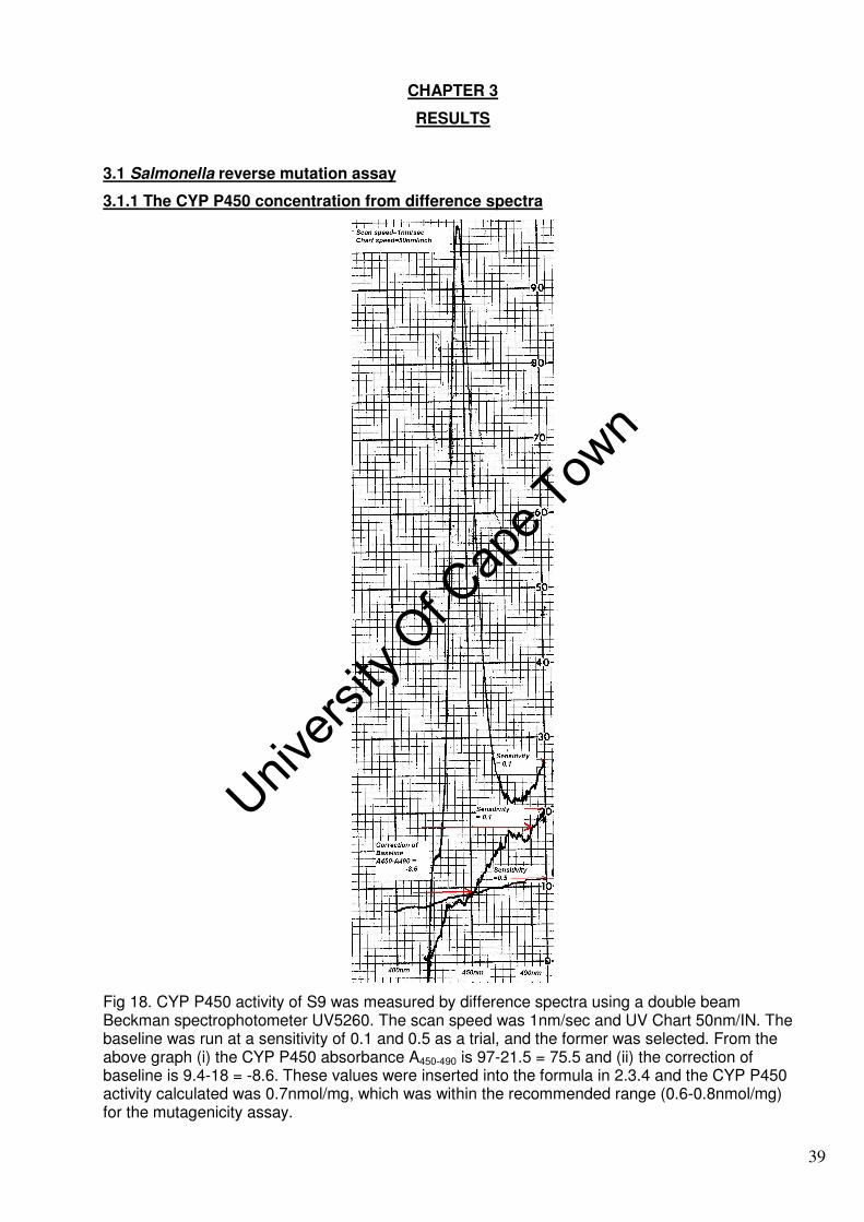
CHAPTER 3
RESULTS
3.1 Salmonella reverse mutation assay
3.1.1 The CYP P450 concentration from difference spectra
Fig 18. CYP P450 activity of S9 was measured by difference spectra using a double beam Beckman spectrophotometer UV5260. The scan speed was 1nm/sec and UV Chart 50nm/IN. The baseline was run at a sensitivity of 0.1 and 0.5 as a trial, and the former was selected. From the above graph (i) the CYP P450 absorbance A450-490 is 97-21.5 = 75.5 and (ii) the correction of baseline is 9.4-18 = -8.6. These values were inserted into the formula in 2.3.4 and the CYP P450 activity calculated was 0.7nmol/mg, which was within the recommended range (0.6-0.8nmol/mg) for the mutagenicity assay.
39
Univers
ity O
f Cap
e Tow
n
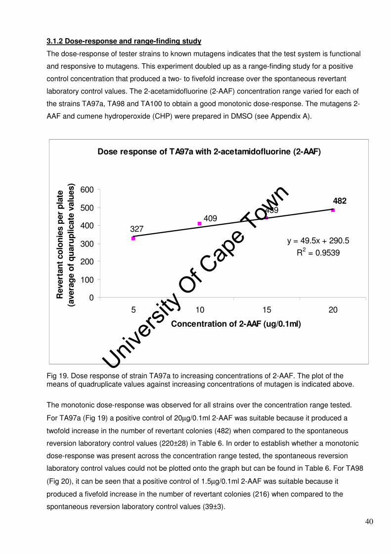
3.1.2 Dose-response and range-finding study
The dose-response of tester strains to known mutagens indicates that the test system is functional
and responsive to mutagens. This experiment doubled up as a range-finding study for a positive
control concentration that produced a two- to fivefold increase over the spontaneous revertant
laboratory control values. The 2-acetamidofluorine (2-AAF) concentration range varied for each of
the strains TA97a, TA98 and TA100 to obtain a good monotonic dose-response. The mutagens 2-
AAF and cumene hydroperoxide (CHP) were prepared in DMSO (see Appendix A).
Dose response of TA97a with 2-acetamidofluorine (2-AAF)
439409
482
327
y = 49.5x + 290.5
R2 = 0.9539
0
100
200
300
400
500
600
5 10 15 20
Concentration of 2-AAF (ug/0.1ml)
Revert
an
t co
lon
ies p
er
pla
te
(avera
ge o
f q
uaru
plicate
valu
es)
Fig 19. Dose response of strain TA97a to increasing concentrations of 2-AAF. The plot of the means of quadruplicate values against increasing concentrations of mutagen is indicated above.
The monotonic dose-response was observed for all strains over the concentration range tested.
For TA97a (Fig 19) a positive control of 20µg/0.1ml 2-AAF was suitable because it produced a
twofold increase in the number of revertant colonies (482) when compared to the spontaneous
reversion laboratory control values (220±28) in Table 6. In order to establish whether a monotonic
dose-response was present across the concentration range tested, the spontaneous reversion
laboratory control values could not be plotted onto the graph but can be found in Table 6. For TA98
(Fig 20), it can be seen that a positive control of 1.5µg/0.1ml 2-AAF was suitable because it
produced a fivefold increase in the number of revertant colonies (216) when compared to the
spontaneous reversion laboratory control values (39±3).
40
Univers
ity O
f Cap
e Tow
n

Dose reponse of TA98 with 2-acetamidofluorine (2-AAF)
725 698
504
380
216
y = 130.9x + 111.9
R2 = 0.9272
0
100
200
300
400
500
600
700
800
900
1.5 3 4.5 6 7.5
Concentration of 2-AAF (ug/0.1ml)
Revert
an
t co
lon
ies p
er
pla
te
(avera
ge o
f q
uad
rup
licate
valu
es)
Fig 20. Dose response of strain TA98 to increasing concentrations of 2-AAF
The number of spontaneous revertants increased over the concentration range tested.
Dose response of TA100 to 2-acetamidofluorine (2-AAF)
301
453403
249
y = 71.4x + 173
R2 = 0.98
0
50
100
150
200
250
300
350
400
450
500
3 6 9 12
Concentration of 2-AAF (ug/0.1ml)
Revert
an
t co
lon
ies p
er
pla
te
(avera
ge o
f q
uad
rup
licate
valu
es)
Fig 21. Dose response of strain TA100 to increasing concentrations of 2-AAF
41
Univers
ity O
f Cap
e Tow
n

A positive control of 9µg/0.1ml 2-AAF was selected for TA100 (Fig 21) because it produced a
twofold increase in the number of revertant colonies (403) when compared to the spontaneous
reversion laboratory control values (187±14).
Dose response of TA102 with cumene hydroperoxide (CHP)
527
11331103
830
973
y = 148.5x + 467.7
R2 = 0.9038
0
200
400
600
800
1000
1200
1400
20 50 80 110 140
Concentration of CHP (ug/0.1ml)
Revert
an
t co
lon
ies p
er
pla
te
(avera
ge o
f q
uad
rup
licate
valu
es)
Fig 22. Dose response of tester strain TA102 to increasing concentrations of CHP, a directly acting mutagen
A positive control of 110µg/0.1ml CHP was selected for TA102 (Fig 22) because it produced a
twofold increase in the number of revertant colonies (1103) when compared to the spontaneous
reversion laboratory control values (564±78). The results of the range-finding study is summarised
in Table 7.
Table 7: Positive control concentrations from the range-finding study
Strain 2-AAF (+S9)
Cumene hydroperoxide (-S9)
TA97a 20µg/0.1ml -
TA98 1.5µg/0.1ml -
TA100 9µg/0.1ml -
TA102 - 110µg/0.1ml
3.1.3 Salmonella reverse mutation assay
Chemicals are judged to be non-mutagenic when they have been tested in at least four Salmonella
tester strains: a positive response in any single strain either with or without metabolic activation is
sufficient for designating a chemical as a mutagen and often causes the substance to be branded
a presumptive carcinogen until definitive results have been obtained from a two-year
carcinogenicity study (Zeiger, 1998; 2001). In the Salmonella reverse mutation assay, gamma
42
Univers
ity O
f Cap
e Tow
n
statistics are very useful for measuring trends and significance. A positive gamma statistic is
indicative of an upward trend and a positive dose response relationship. A negative gamma
statistic is indicative of a decreased response possibly due to (i) toxicity, which may be associated
with thinning or complete absence of the background lawn, or (ii) antibacterial activity in the test
substance. Plant extracts were prepared as described in Chapter 2 on materials and method. It is
important to note that the aqueous extracts were prepared from the aerial part of the plant (leaves),
and are most often consumed in this manner as medicine and dietary supplements. The data,
including standard deviation and statistical significance, are tabulated in Tables 8 and 9.
3.1.4 Acokanthera oppositifolia (Lam.) and Zantedeschia aethiopica (L.)
A. oppositifolia and Z. aethiopica proved to be non-mutagenic with all four tester strains with or
without S9. Although Z. Aethiopica was non-mutagenic, it produced a significant decrease in the
number of revertants in TA97a with S9 (p≤0.01). Visual inspection of the plates showed an intact
background lawn with small pinpoint colonies, easily mistaken for revertant colonies. This indicated
that Z. aethiopica was toxic but only in the presence of S9. Calcium oxalate, a chemical constituent
of Z. aethiopica, is known to produce toxicity. The leaves of Z. aethiopica have a unique mannose-
binding lectin that binds to cell membranes that are often toxic (van Wyk et al., 1997 and Chen et
al., 2004). One can speculate that the toxicity in TA97a with S9 may be attributed to (i) metabolic
intermediates of calcium oxalate and lectins or (ii) phytochemical metabolites of Z. aethiopica that
may have potential antibacterial activity. What is rather alarming is that only one tester strain was
affected in this manner when all strains carry the rfa deletion, making their cell wall more
permeable to the penetration of chemicals. Since it is generally accepted that mutagenicity in any
one of the tester strains with or without S9 is a positive response, based on a similar principle,
toxicity in any one of the tester strains with or without S9 should be sufficient to brand the chemical
as a potential toxicant.
3.1.5 Pelargonium sp. cf. inquinans ( L.) L’ Herit
The results indicate that (i) in TA102 (Fig 23B) phytochemicals in the Pelargonium extract were
mutagenic by one or more of the following mechanisms: transition and transversion reactions, DNA
crosslinkage and oxidative DNA damage and (ii) in TA97a (Fig 23A) the cytoxicity of the
Pelargonium extract in the presence of S9 is undoubtedly due to a phytochemical metabolite.
Furthermore, phytochemical metabolites of Pelargonium may have potential antibacterial activity.
Coumarin, a chemical constituent of Pelargonium sp, may contribute to the cytotoxicity observed at
increasing concentrations. With regards to a potential risk related to the coumarin content, a NOEL
(no observed effect level) value of more than 750mg/kg body weight has been established in dogs
and rats in a Pelargonium sidoides-related toxicological study (Brendler et al., 2008).
43
Univers
ity O
f Cap
e Tow
n

44
Univers
ity O
f Cap
e Tow
n

45
Univers
ity O
f Cap
e Tow
n

46
Univers
ity O
f Cap
e Tow
n

A Number of revertants per plate versus extract
with strain TA97a+ S9
y = -991.98x + 139.04
R2 = 0.7545
0
20
40
60
80
100
120
140
160
180
0 0.02 0.04 0.06 0.08 0.1
Concentration of Pelargonium extract (g/ml)
Nu
mb
er
of
revert
an
ts
B Number of revertants per plate versus extract
with strain TA102
y = 1883.5x + 435.64
R2 = 0.6498
300
350
400
450
500
550
600
650
-0.01 0.01 0.03 0.05 0.07 0.09
Concentration of Pelargonium extract (g/ml)
Nu
mb
er
of
revert
an
ts
Fig 23. Salmonella reverse mutation assay of Pelargonium sp. (A). The significant downward trend at a level of 1% (p≤0.01) with TA97a in the presence of S9 was observed. (B) The upward trend observed for PL in TA102 in the absence of S9 is significant (p≤0.01), indicating strong mutagenicity.
P. sidoides root extracts have a track record of success as a plant-based medicine and the plant is
currently commercialised as EPs®7630 (Umckaloabo) (refer to Table 2 for its beneficial effects). In
general, Pelargonium sp is used for the manufacture of rose-scented geranium oil that is used
mainly in aromatherapy and in the fragrance industry. Traditionally, the leaves and root of
Pelargonium sp are taken as tea or for the treatment of medical ailments such as dysentery and
syphilis. Further toxicological studies of Pelargonium extracts are required.
47
Univers
ity O
f Cap
e Tow
n

3.1.6 Pteridium aquilinum subsp aquilinum
A Number of revertants per plate versus extract
with TA 100 + S9
y = 586.18x + 287.61
R2 = 0.2873
200
220
240
260
280
300
320
340
360
380
-0.01 0.01 0.03 0.05 0.07 0.09
Concentration of P. aquilinum extract (g/ml)
Nu
mb
er
of
revert
an
ts
B Number of revertants per plate versus extract
with TA 102
y = 968.66x + 426.64
R2 = 0.4569
300
350
400
450
500
550
600
-0.01 0.01 0.03 0.05 0.07 0.09Concentration of P. aquilinum extract (g/ml)
Nu
mb
er
of
revert
an
ts
TA 102
TA 102+
Regression TA 102
Fig 24. Salmonella reverse mutation assay of P. aquilinum indicates a mutagenic reaction with two of the four tester strains (p≤0.01) (A) in TA100 with S9 metabolic activation and (B) TA102 in the absence of S9.
P. aquilinum is mutagenic with (i) TA100 in the presence of S9 and (ii) TA102 in the absence of S9,
which is evidence of promutagens and mutagens respectively. The mutagenicity, clastogenicity,
teratogenicity and carcinogenicity of the major bracken carcinogen, ptaquiloside, a
norsesquiterpene glucoside, has been convincingly demonstrated. Ptaquiloside is quite unstable
once it is separated from the plant or under mild base or acid, and decomposes rapidly with the
48
Univers
ity O
f Cap
e Tow
n

loss of the glucose moiety into pteridienone, which has been found to have more pronounced
activity over ptaquiloside (Matsuoka et al., 1989).
The decomposition of ptaquiloside is pH dependent, and studies have shown that ptaquiloside and
its related compounds produce mutagenicity in the Salmonella reverse mutation assay in TA98 and
TA100 under weakly basic conditions (pH8.5) (Nagao et al., 1989). However, the pH in the current
experiment was not controlled and mutagenicity was produced in (i) TA100 by base-pair
substitution exerted by metabolic intermediates and (ii) in TA102 possibly by the crosslinkage
reactions of ptaquiloside and pteridienone with DNA. These compounds have a strong alkylating
capacity and react with DNA.
3.1.7 Rumex lanceolatus Thunb.
This extract contains compounds that are potent mutagens and promutagens, as detected with all
four tester strains with and without metabolic activation (Fig 25). The mechanisms of DNA damage
are by frameshift mutations, base-pair substitution, transition and transversion mutations, DNA
crosslinkage and oxidative DNA damage.
Rumex species are related to the well-known medicinal plants known as rhubarbs (Rheum
species) (van Wyk and Wink, 2004). Several monomeric anthraquinones, such as emodin (1,3,8-
trihydroxy-6-methyl-anthraquinone) and related derivatives that are constituents of rhubarb (an
oriental medicine for diarrhoea) exhibit mutagenicity in the Salmonella/microsomes test and
cultured tumour cells (Morita et al., 1988 and Ueno, 1991). Emodin is a constituent of R.
lanceolatus (Watt and Breyer-Brandwijk, 1962), which may be responsible for the potent mutagenic
response observed.
3.2 Chromosomal aberration test
The in vitro chromosomal aberration test was used to measure the clastogenicity of aqueous plant
extracts in peripheral blood lymphocytes. The chromosomal aberration test was done in duplicate
with S9 activation and non-activation at three concentrations of plant extract: (i) concentrations of
0.005, 0.02 and 0.08g/ml P. aquilinum and R. lanceolatus and (ii) concentrations of 0.00125; 0.005
and 0.02g/ml for Z. aethiopica because of problems with viscosity. The positive control used in the
non-activation analysis was methanemethanosulphate (MMS), soluble in 0.5% DMSO for which a
solvent control was included. The positive control used with S9 activation was cyclophosphamide
(CPA), which is water soluble. Chromosomal aberrations were scored from metaphase arrested
cells that had been fixed, dropped from a height of 30cm onto microscope slides and Giemsa
stained. However, this method is not suitable for estimating numerical chromosomal aberrations,
as artefactual chromosome loss may occur (Mateuca et al., 2006) and its use for this purpose was
not considered in this investigation. The KARY workstation (microscope, personal computer and
49
Univers
ity O
f Cap
e Tow
n

50
Univers
ity O
f Cap
e Tow
n

printer) facilitated image capture via a microscope mounted video camera, digitisation of the
captured image, storage on the computer’s hard disk or on compact disc (CD) as a
photomicrograph to be imported or printed.
Extensive research has been done by means of light microscopy from which categories of
structural chromosome aberrations for metaphase cells have been established (Fig 13).
Metaphase cells were scored as recommended by Savage (2004) and categorised into
chromosome-type (CSA) and chromatid-type (CTA) aberrations. The CSA and CTA types arise
from cell cycle progression (G1, S, G2 and M) and in relation to S-phase independent or dependent
clastogens. CSAs result from incompletely repaired or unrepaired double-strand breaks mostly
generated in vivo in Go–G1 lymphocytes by S-phase independent clastogens (e.g. ionising
radiation). After DNA synthesis and chromosome duplication, the aberrations formed in Go–G1
were doubled and chromosome-type breaks were seen in metaphase. CTAs (e.g. chromatid type
breaks) arose predominantly in vitro during the G2-phase or late S-phase of the cultured
lymphocytes in response to base modifications and single-strand breaks (SSBs) induced by S-
phase-dependent clastogens (e.g. chemicals) (Savage 1975, 2004; Albertini et al., 2000, Hagmar
et al., 2004 and Mateuca et al., 2006).
3.2.1 Pteridium aquilinum subsp aquilinum
This test was done in duplicate and the average values from scoring approximately 50 metaphases
was tabulated (Table 10). Photomicrographs depict chromatid-type aberrations (CTAs) observed at
metaphase in Fig 26a, b and c. A chromatid aberration results from a single strand DNA break
(SSB) occurring in the G2 phase of the cell cycle by S-phase dependent clastogens in the 0.005
and 0.02g/ml P. aquilinum extracts.
The clastogenicity is attributed to ptaquiloside and pteridienone. Liver enzymes and CYP P450
play a major role in the detoxification of drugs and xenobiotics in the body. This was observed by a
reduction of aberrations to 2% in P. aquilinum treated cultures with S9 under the current in vitro
experimental conditions. The clastogenicity decreased in the presence of S9 as a result of
detoxification; however, metabolically activated clastogens were still evident. A graphical
representation of the data is given in Fig 27.
51
Univers
ity O
f Cap
e Tow
n

Table 10: Chromosomal aberrations from the treatment of cultures with P. aquilinum
Fig 26a. A chromatid gap after treatment of peripheral blood lymphocytes with 0.005g/ml P. aquilinum without S9, implying direct clastogenicity [Slide Z1, vernier 65 X 92.5]
Treatment Dose (g/ml)
% cells with chromatid aberrations (CTAs)
% cells with chromosome aberrations (CSAs)
Gaps Breaks Gaps Breaks
Nonactivation P. aquilinum 0.005 10 2 6 0 0.02 5 0 5 3 0.08 3 3 0 5 Negative control (untreated blood)
- 0 0 0 0
0.5% DMSO 2 4 2 2 10mM MMS 14 4 6 6
S9 activation (24 hr exposure)
P. aquilinum 0.005 0 2 2 0 0.02 2 2 0 0 0.08 0 0 0 0 Negative control (untreated blood)
0 0 0 0
2.5mM CPA 15 10 3 3
52
Univers
ity O
f Cap
e Tow
n

Fig 26b. Chromatid break caused after treatment of lymphocytes with 0.005g/ml P. aquilinum with S9. A reduction in chromosomal aberrations was observed after metabolic activation with S9 owing to detoxification reactions [Slide ZD12, vernier 61 X 93.5, aberration ZD12.5]
Fig 26c Chromatid break caused by treatment of lymphocytes with 0.02g/ml P. aquilinum with S9 [slide ZD14, vernier 57 X 81, aberration ZD14.6]
53
Univers
ity O
f Cap
e Tow
n

A % cells with aberrations versus extract (-S9)
0
2
4
6
8
10
12
14
16
0.005g/ml 0.02g/ml 0.08g/ml Control (-) 0.5%
DMSO
10mM
MMS
Concentration of P. aquilinum and controls
% c
ells w
ith
ab
err
ati
on
chromatid break
chromatid gap
chromosome break
chromosome gap
B % cells with aberrations versus extract (+S9)
0
2
4
6
8
10
12
14
16
0.005g/ml 0.02g/ml 0.08g/ml Control (-) 2.5mM CPA
Concentration of P. aquilinum and control
% c
ells w
ith
ab
err
ati
on
s
chromatid break
chromatid gap
chromosome break
chromosome gap
Fig 27. The treatment of lymphocytes with P aquilinum at three increasing concentrations, 0.005; 0.02 and 0.08g/ml, in (A) the absence of S9 and (B) presence of S9. The reduction in genotoxicity is evident with CYP P450 and liver enzymes. The 10mM methylmethanosuphate (MMS) is soluble in 0.5% DMSO and cyclophosphamide (CPA) is water soluble.
3.2.2 Rumex lanceolatus Thunb.
This test was done in duplicate and the average values from scoring approximately 50 metaphases
was tabulated (Table 11). R. lanceolatus generated CTAs (Fig 28) and CSAs at 0.005, 0.02 and
0.08g/ml without S9. However, only CSAs were generated at 0.005 and 0.02g/ml in the presence
of S9 at a reduced level and no aberrations were detected at 0.08g/ml (Fig 29). The percentage of
54
Univers
ity O
f Cap
e Tow
n

cells with aberrations decreased in the presence of S9 as a result of phase II enzymes that
deactivate the reactive species. However, the chromosome breaks evident at 0.005 and 0.02g/ml
in the presence of S9 are attributed to S-phase independent clastogenic metabolites, possibly 2-
hydroxy-emodin.
Fig 28. A chromatid gap by treatment of lymphocytes with 0.02g/ml R. lanceolatus without S9, indicating direct genotoxicity. The CTA was generated at the G2 phase of the cell cycle by S-phase dependent phytochemical clastogens in the R. lanceolatus extract [Slide Z5, vernier 61 X 103, aberration Z5.1].
Table 11: Chromosomal aberrations from the treatment of cultures with R. lanceolatus
Treatment Dose (g/ml)
% cells with chromatid aberrations (CTAs)
% cells with chromosome aberrations (CSAs)
Gaps Breaks Gaps Breaks
Nonactivation R. lanceolatus 0.005 6 0 0 0 0.02 6 2 0 4 0.08 0 2 0 4 Negative control (untreated blood)
- 0 0 0 0
0.5% DMSO 2 4 2 2 10mM MMS 14 4 6 6
S9 activation (24hr exposure)
R. lanceolatus 0.005 0 0 0 2 0.02 0 0 0 2 0.08 0 0 0 0 Negative control (untreated blood)
0 0 0 0
2.5mM CPA 15 10 3 3
55
Univers
ity O
f Cap
e Tow
n

A % cells with aberrations versus extract (-S9)
0
2
4
6
8
10
12
14
16
0.005g/ml 0.02g/ml 0.08g/ml Control (-) 0.5%
DMSO
10mM MMS
Concentration of R. lanceolatus and controls
% c
ells w
ith
ab
err
ati
on
s
chromatid break
chromatid gap
chromosome break
chromosome gap
B % cells with aberrations versus extract (+S9)
0
2
4
6
8
10
12
14
16
0.005g/ml 0.02g/ml 0.08g/ml Control (-) 2.5mM CPA
Concentration of R. lanceolatus and control
% c
ells w
ith
ab
err
ati
on
s
chromatid break
chromatid gap
chromosome break
chromosome gap
Fig 29. The treatment of lymphocytes with R lanceolatus at three increasing concentrations 0.005, 0.02 and 0.08g/ml in (A) the absence of S9 and (B) the presence of S9 where the chromosomal breaks are reduced from 4 to 2%. Chromosome breaks may translocate or rejoin with a loss of genetic integrity.
56
Univers
ity O
f Cap
e Tow
n

3.2.3 Zantedeschia aethiopica (L.)
Fig 30a. Chromosome gap caused by treatment of lymphocytes with 0.005g/ml Z. aethiopica in the absence of S9 [slide X2, vernier73 X 93, aberration X2.1]
Fig 30b. Chromatid break caused by treatment with 0.005g/ml Z aethiopica in the absence of S9 [slide X23, vernier 63 X 86.5]
57
Univers
ity O
f Cap
e Tow
n

Table 12: Chromosomal aberrations from the treatment of cultures with Z. aethiopica
Treatment Dose (g/ml)
% cells with chromatid aberrations (CTAs)
% cells with chromosome aberrations (CSAs)
Gaps Breaks Gaps Breaks Nonactivation
Z. aethiopica 0.00125 0 0 0 0 0.005 2 2 1 0 0.02 2 4 2 0 Negative control (untreated blood)
- 0 0 0 0
0.5% DMSO 2 4 2 2 10mM MMS 14 4 6 6 S9 activation (24hr
exposure)
Z. aethiopica 0.00125 0 0 0 0 0.005 0 0 0 0 0.02 0 4 0 0 Negative control (untreated blood)
0 0 0 0
2.5mM CPA 15 10 3 3
The chromosome-type aberrations (CSA) seen in Fig 30a above were generated in the Go-G1
phase of the cell cycle from an S-phase independent phytochemical clastogen. The chromatid-type
aberration (CTA) seen in Fig 30b above was generated in S or G2 of the cell cycle from an S-phase
dependent phytochemical clastogen. CTAs and CSAs were generated over the concentration
range tested, but were reduced in the presence of S9 (Fig 31a,b).
A % cells with aberrations versus extract (-S9)
0
2
4
6
8
10
12
14
16
0.00125g/ml 0.005g/ml 0.02g/ml Control (-) 0.5% DMSO 10mM MMS
Concentration of Z. aethiopica and controls
% c
ells w
ith
ab
err
ati
on
s
chromatid break
chromatid gap
chromosome break
chromosome gap
Fig 31a. A dose related increase in chromatid breaks and chromosome gaps was observed with 0.005g/ml and 0.02g/ml Z. aethiopica in the absence of S9
58
Univers
ity O
f Cap
e Tow
n

B % cells with aberrations versus extract (+S9)
0
2
4
6
8
10
12
14
16
0.00125g/ml 0.005g/ml 0.02g/ml Control (-) 2.5mM CPA
Concentration of Z. aethiopica and control
% c
ells w
ith
ab
err
ati
on
schromatid break
chromatid gap
chromosome break
chromosome gap
Fig 31b. A reduction in genotoxicity in the presence of S9 was observed for Z. aethiopica
3.3 Flow cytometry (DNA content)
A. oppositifolia and Pelargonium inhibited mitosis in the chromosomal aberration test and produced
no scorable metaphases. Cell cycle arrest was investigated by flow cytometry. Experiments for
DNA content were carried out in duplicate at the highest concentration, 0.08g/ml. Since
lymphocytes are quiescent (i.e. in Go phase they are not actively dividing cells), the cultures were
stimulated with phytohaemagglutinin (PHA) to enter the cell cycle (G1, S, G2-M). After 72hrs at
37oC, the red blood cells were lysed and the DNA content of the white blood cells was determined
by flow cytometry. In both treated and untreated cells, ≥95% viable white blood cells remained at
the end of the 24hr treatment period. Three distinct populations of white blood cells based on their
difference in size lymphocytes<monocytes<granulocytes were visualised. The instrument was
configured to measure the DNA content of lymphocytes and the data tabulated in Table 13.
Table 13. DNA content of lymphocytes (average of duplicate values is indicated)
Microculture PHA %Go-G1 %S %G2-M
Blood + PHA (control) 100µl 81±1 19±1 0.1±0
A. oppositifolia 0.08g/ml, -S9 100µl 86±0 14±0 0
A. oppositifolia 0.08g/ml, +S9 100µl 99±1 2±1 0
Pelargonium 0.08g/ml, -S9 100µl 98±1 2±1 0
Pelargonium 0.08g/ml, +S9 100µl 89±1 11±1 0
59
Univers
ity O
f Cap
e Tow
n
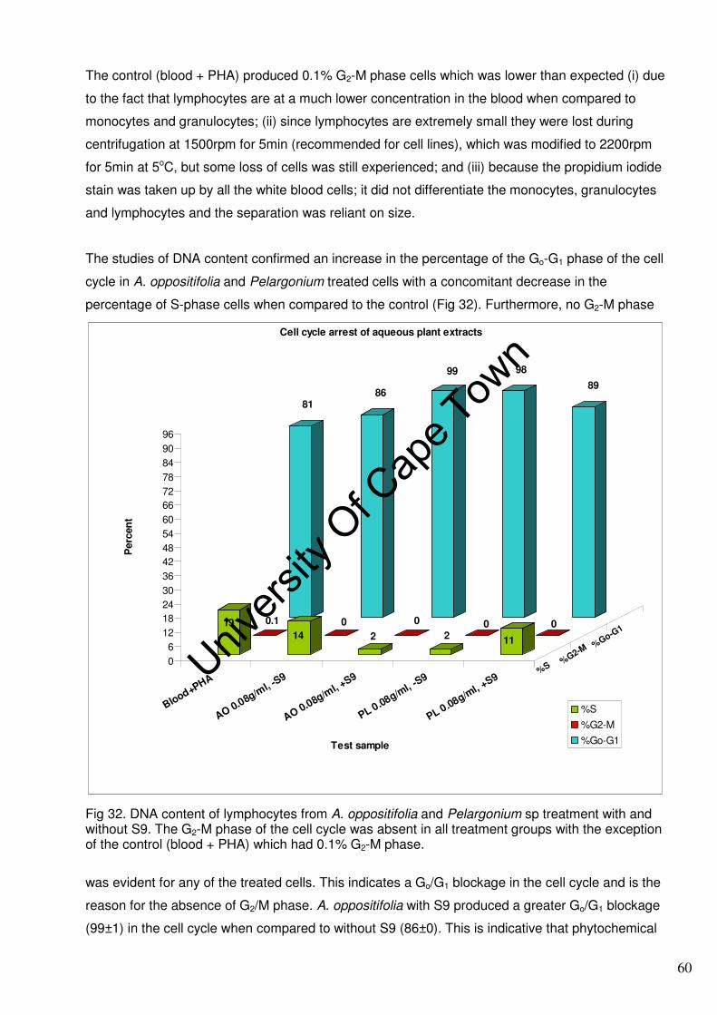
The control (blood + PHA) produced 0.1% G2-M phase cells which was lower than expected (i) due
to the fact that lymphocytes are at a much lower concentration in the blood when compared to
monocytes and granulocytes; (ii) since lymphocytes are extremely small they were lost during
centrifugation at 1500rpm for 5min (recommended for cell lines), which was modified to 2200rpm
for 5min at 5oC, but some loss of cells was still experienced; and (iii) because the propidium iodide
stain was taken up by all the white blood cells; it did not differentiate the monocytes, granulocytes
and lymphocytes and the separation was reliant on size.
The studies of DNA content confirmed an increase in the percentage of the Go-G1 phase of the cell
cycle in A. oppositifolia and Pelargonium treated cells with a concomitant decrease in the
percentage of S-phase cells when compared to the control (Fig 32). Furthermore, no G2-M phase
8186
99 98
89
0.1 0 0 0 019
14 2 2 11
0
6
12
18
24
30
36
42
48
54
60
66
72
78
84
90
96
Perc
en
t
Blood+PHA
AO 0.08g/ml, -
S9
AO 0.08g/ml, +
S9
PL 0.08g/ml, -
S9
PL 0.08g/ml, +
S9%
S%
G2-M%
Go-G1
Test sample
Cell cycle arrest of aqueous plant extracts
%S
%G2-M
%Go-G1
Fig 32. DNA content of lymphocytes from A. oppositifolia and Pelargonium sp treatment with and without S9. The G2-M phase of the cell cycle was absent in all treatment groups with the exception of the control (blood + PHA) which had 0.1% G2-M phase.
was evident for any of the treated cells. This indicates a Go/G1 blockage in the cell cycle and is the
reason for the absence of G2/M phase. A. oppositifolia with S9 produced a greater Go/G1 blockage
(99±1) in the cell cycle when compared to without S9 (86±0). This is indicative that phytochemical
60
Univers
ity O
f Cap
e Tow
n

metabolites of A. oppositifolia are more potent cell cycle arresting substances. Pelargonium without
S9 produced a greater Go/G1 blockage (98±1) when compared to with S9 (89±1), indicating that
phytochemicals in the extract are potential cell cycle arresting substances. These plants should be
further investigated for their cancer chemopreventative properties. The studies of DNA content in
A. oppositifolia and Pelargonium confirmed the absence of the G2-M phase of the cell cycle,
indicating a blockage in the Go/G1 phase of the cell cycle.
61
Univers
ity O
f Cap
e Tow
n

CHAPTER 4
DISCUSSION
The Salmonella reverse mutation assay was used as a primary screen for measuring the ability of
plant extracts or metabolites to induce DNA mutations. The extract was judged to be non-
mutagenic when tested in at least four Salmonella tester strains. A positive response in any single
strain either with or without metabolic activation is considered sufficient for designating the extract
as a mutagen and a presumptive carcinogen until definitive carcinogenicity studies are performed.
The Salmonella reverse mutation assay identifies chemicals that react with cellular DNA to
produce mutations, whereas the carcinogenicity test identifies only those chemicals that can
induce tumour cells that survive and replicate to form visible tumours.
Structural chromosomal aberrations in the form of gaps and breaks were detected in metaphase
cells and comprise both chromosome aberrations (CSAs) and chromatid aberrations (CTAs).
These aberrations arise from cell cycle progression and in relation to S-phase-independent or
dependent clastogens as follows: CSAs result from incompletely repaired or unrepaired double-
strand breaks mostly generated in vivo in Go–G1 lymphocytes by S-phase-independent clastogens
(e.g. ionising radiation) (Mateuca et al., 2006). After DNA synthesis and chromosome duplication,
the aberrations formed in Go–G1 are replicated and chromosome-type breaks (CSAs) are seen in
metaphase, chromatid-type breaks (CTAs) arise predominantly in vitro during the G2 or late S-
phase of the cultured lymphocytes in response to base modifications and single-strand breaks
(SSBs) induced by S-phase-dependent clastogens (e.g. chemicals) (Albertini et al., 2000 and
Hagmar et al., 2004).
Evidence from a recent study conducted by Norppa et al. (2006) indicates that both chromatid-type
and chromosome-type aberrations predict cancer, even though some data suggest that
chromosome-type aberrations may have a more pronounced predictive value than chromatid-type
aberrations. The relationship between the presence of a high frequency of chromosome
aberrations and a predisposition to cancer has been well established in chromosome instability
syndromes (e.g. ataxia telangiectasia, Fanconi’s anaemia and Bloom’s syndrome) (Barrios et al.,
1991). Furthermore, structural and numerical chromosome aberrations are evident in bladder
carcinomas (gaps or breaks = 25.37%, major structural chromosome abnormalities = 67.16% and
numerical chromosome aberrations = 7.46%) (Barrios et al., 1990).
Although peripheral blood lymphocytes are frequently used in the chromosomal aberration assay,
the use of whole blood cultures has many disadvantages. The initial concentration of the different
cell types in whole blood is unknown at the time of seeding. Red blood cells cause interference
during flow cytometry, and the several lysis steps required to remove them result in a decrease in
62
Univers
ity O
f Cap
e Tow
n

the concentration of lymphocytes. In a “whole blood culture environment”, there is potential
interference between blood components and the extract. In addition, the donor’s genetic
predisposition to chromosomal aberration frequency will impact on the results. However, although
this was not controlled in the current experiment, it is recommended that the test be performed with
blood from different donors. Supporting evidence comes from studies showing a decrease in
chromosomal aberration frequency in individuals with homozygous genotype for the XPD 751Gln
variant allele (Vodicka et al, 2004 and Mateuca, 2006). Therefore, an individual’s susceptibility to
chromosomal aberrations may impact on the test, as peripheral blood lymphocytes from such
individuals will be “resilient” to genotoxic agents. Since a high frequency of structural chromosomal
aberrations in lymphocytes is predictive of increased cancer risk, it is advisable to use blood from
more than one source for the chromosomal aberration test to circumvent the problem encountered
with genetic resilience. Cell lines, such as Chinese hamster ovary cells, which were used for the
detection of clastogenicity in Aristolochia sp. (Zhang et al., 2004) would have averted the many
problems encountered. The lack of automated systems for colony counting in the Salmonella
reverse mutation assay and scoring metaphase cells in the chromosomal aberration test presented
difficulty. The results of the mutagenicity and clastogenicity studies under the current experimental
conditions are discussed in the following sections.
4.1 R. lanceolatus
The aqueous extract prepared from the aerial parts of R. lanceolatus contains potent mutagens
and pro-mutagens. Mutagenicity was detected with all four tester strains TA97a, TA98, TA100 and
TA102 in the presence and absence of S9 (p≤0.01). Metabolic activation produces strong
electrophiles that have a high affinity for DNA, resulting in DNA damage. The phytochemicals and
metabolites of R. lanceolatus cause DNA damage through a variety of mechanisms: (i) frameshift
mutations detected by TA97a and TA98; (ii) base-pair substitutions detected by TA100; and (iii)
oxidative DNA damage, transition and transversion reactions (Fig 33) and cross-linking reactions
detected by TA102. The compounds present in the R. lanceolatus extract are presumptive rodent
carcinogens (and potential human carcinogens).
Emodin, a monomeric anthraquinone constituent of R. lanceolatus (Watt and Breyer-Brandwijk,
1962), may be responsible for the mutagenicity observed in TA97a, TA98, TA100 and TA102 in
this study. Supporting evidence comes from extensive studies conducted in the 1980s, which show
that many monomeric anthraquinones, such as emodin (1,3,8-trihydroxy-6-methyl-anthraquinone)
and related derivatives that are constituents of Rheum (rhubarb), exhibit mutagenicity in the
Salmonella reversion/microsome tests and cultured tumour cells (Morita et al., 1988 and Ueno,
1991). The genus Rumex (sorrel, dock) and Rheum belong to the same family, Polygonaceae.
Emodin is a promutagen and is converted to its active form 2-hydroxy-emodin in the presence of
63
Univers
ity O
f Cap
e Tow
n

microsomes (Masuda and Ueno, 1983 and Masuda et al., 1985). CYP P450 catalyses this
biotransformation (Ueno 1985 and Masuda et al., 1985).
Fig 33. Base substitution reactions by transitions and transversion. Transitions are interchanges of purines (A G) or pyrimdines (C T); they therefore involve bases of similar shape. Transversions are interchanges between purine and pyrimidine bases, which involve the exchange of one-ring and two-ring structures.
2-Hydroxy-emodin has a strong electron-spin resonance in a mixture with DNA, which indicates the
participation of free radicals derived from anthraquinone in DNA damage (Kodama et al., 1987)
and may account for the oxidative DNA damage observed with TA102 in the current investigation.
Since hydroxy-radical-forming agents selectively hydroxylate deoxyguanosine residues of DNA at
C-8 in in vivo and in vitro systems, it was presumed that the anthraquinones cause genotoxicity by
their potential for hydroxy radical formation (Hayatsu, 1990). These anthraquinones show
characteristic colours in solutions and on TLC (thin layer chromatography) plates and this physical
property is applied for identification and quantitation in TLC (Ueno, 1982). Furthermore, dimeric
anthraquinones, such as luteoskyrin and rugulosin, are hepatotoxic and hepatocarcinogenic in
mice (Uraguchi et al., 1972 and Ueno et al., 1980).
In the chromosomal aberration test, R. lanceolatus extract produced CTAs and CSAs over the
concentration range tested in the presence and absence of S9; however there was a reduction of
chromosomal aberrations in the presence of S9, which is ascribed to detoxification. Since CTAs
64
Univers
ity O
f Cap
e Tow
n

occur at Go/G1 and CSAs at G2 or the late S-phase of the cell cycle, R. lanceolatus extract has
both S-phase-dependent and independent clastogens. The study by Schwartz (1989) indicates
that alkylating agents are S-phase-dependent clastogenic agents, that is, chromosomal aberrations
are not observed unless the treated cells have first undergone a round of replicative DNA
synthesis. It can be speculated that CTAs were generated by S-phase-independent chemical
compounds originating in the extract or from metabolism. A decreased percentage of cells with
aberrations in the presence of S9 indicated the detoxification of clastogenic phytochemicals by
CYP P450. In the presence of S9, the 2% CSAs produced at 0.005g/ml and 0.02g/ml is due
probably to S-phase-dependent clastogenic metabolites, possibly 2-hydroxy-emodin. Supporting
evidence comes from the fact that 2-hydroxy-emodin caused strand breaks in circular phage DNA
(Kodama et al., 1987) and may be the cause of the clastogenicity observed in the presence of S9
at 0.005 and 0.02g/ml. However, in light of the results from the study by Lee et al. (2005), which
demonstrated that emodin from the aerial parts of Rumex acetosa inhibited the proliferation of
tumour cells, was antimutagenic and antigenotoxic, the R. lanceolatus extract tested requires
further investigation.
The beneficial effects of Rumex induratus as a good dietary source of nutrients owing to its
agreeable taste, and a high phenolic content cannot be ruled out; however, it has to be
approached with caution owing to its high oxalic acid content. No high doses can be consumed
because oxalic acid has a predicted LD50 value (the mg/kg dose required to kill 50% of a test
population) of 375mg/kg body weight (Ferreres et al., 2006).
Other effects of Rumex sp. are anti-fertility action (R. steudelii), antibacterial and anti-inflammatory
activities (Rumex nervosus and Rumex abyssinicus), antidiarrheal effects (Rumex maritimus) and
antiviral activities (Rumex bequaerti) (Getie et al., 2002; Rouf et al., 2002; Cos et al., 2002 and
Gebrie et al., 2004). The lessons from the “Aristolochia incident” in the early 1980s indicate that
mutagenicity tests, such as the Salmonella reverse mutation assay and chromosomal aberration,
are good indicators of potential carcinogenicity. Extracts of R. lanceolatus have produced both
mutagenic and clastogenic effects under the current experimental conditions and must be further
investigated.
4.2 Pelargonium sp.
Perlargonium extract proved to be mutagenic with TA102 in the absence of S9 (p≤0.01),
implicating directly acting mutagens in DNA damage by any of the following: transition/transversion
reactions, oxidative DNA damage or cross-linking reactions. Perlargonium is known to contain
coumarins that interfere with excision repair processes in UV-damaged DNA and with host cell
65
Univers
ity O
f Cap
e Tow
n

reactivation of UV-irradiated phage T1 in E coli (Grigg, 1972). TA102 has an intact excision repair
system unlike the other three Salmonella tester strains, TA7a, TA98 and TA100, which may have
been targeted by coumarin in the Perlargonium extract. Toxicity was observed at a level of 1%
(p≤0.01) with TA97a in the presence of S9, and was attributed to a metabolically activated
phytochemical. Alternatively, this effect may have arisen from antibacterial activity since
isoquercetrin and rutin, respective major and minor flavonoids obtained from Pelargonium radula
(Cav.) L’Herit, were shown to have antimicrobial activity (Pepeljnjak et al., 2005). Under the current
experimental conditions Pelargonium is a presumptive carcinogen (mutagenic with TA102), and
showed in vitro cytotoxicity and/or antibacterial activity.
Rose-scented geranium oil from Pelargonium sp. is of high value in the fragrance industry, while P
sidoides root extract commercialised as EPs®7630 (Umckaloabo) has several benefits (refer to
Table 2). In a Pelargonium sidoides related toxicological study on potential risk related to the
coumarin content, a NOEL (no observed effect level) value of more than 750mg/kg body weight
was established in dogs and rats (Brendler et al., 2008). NOEL values are generated from the
dose-response curves of various agents by plotting “percent of population affected versus dose
(mg/kg)”. When examining such graphs, there is often a minimum concentration (or threshold) of
dose below which there is no observable effect in the test population (e.g. dogs, rats) and this is
referred to as the NOEL. The NOEL dose information obtained from animal experimentation is
often used to establish safe levels for humans. Similar toxicological studies are warranted for use
of Pelargonium as a traditional medicine in South Africa due to the toxicity observed with TA97a.
In the chromosomal aberration test, Pelargonium caused cell-cycle inhibition of peripheral blood
lymphocytes. The studies of DNA content in Pelargonium confirmed the absence of G2-M phase of
the cell cycle, indicating a blockage in the Go/G1 phase of the cell cycle, therefore the clastogenicity
of this extract should be investigated with other test methods. Since the aqueous extract of the
aerial parts of this plant caused cell-cycle inhibition further studies for its potential as a cancer
chemotherapeutic agent should be undertaken.
4.3 P. aquilinum
P. aquilinum produced highly significant mutagenicity with TA102 in the absence of S9 (p≤0.01),
implicating directly acting mutagens in DNA damage by any of the following mechanisms:
transition/transversion reactions (Fig 33), oxidative DNA damage and cross-linking reactions.
Furthermore P. aquilinum proved to be mutagenic with TA100 in the presence of S9 (p≤0.01),
implicating pro-mutagens in DNA damage by means of a mechanism of base-pair substitution. The
toxicity observed in TA97a with S9 was significant (p≤0.01), and since this toxicity was not evident
in TA97a in the absence of S9, it was attributed to a chemical that was metabolically activated.
66
Univers
ity O
f Cap
e Tow
n

The experimental results for P. aquilinum (bracken fern) indicate both mutagenic and promutagenic
activity. Ptaquiloside is responsible for adenine to pyrimidine transversion in the H-ras gene on
codon 61 (N3 position) (Alonso-Amelot and Avendano, 2002), and therefore it is conceivable that
ptaquiloside carries out mutagenicity by purine to pyrimidine transversions in TA102 or by cross-
linkage reactions, since both ptaquiloside and pteridienone in bracken fern are alkylating agents.
Pteridienone was found to have more pronounced alkylating activity over ptaquiloside (Matsuoka et
al., 1989). Other chemical constituents of bracken fern, hypolosides and illudins M and S,
produced mutagenicity in the Salmonella reversion test with TA98 and TA100 (Nagao et al., 1989).
It was found that P. aquilinum generated CTAs and CSAs in the presence and absence of S9.
Schwartz (1989) reported that alkylating agents are S-phase-dependent clastogenic agents, that is,
chromosomal aberrations are not observed unless the treated cells have first undergone a
replicative DNA synthesis. Therefore, CSAs must be attributed to ptaquiloside and pteridienone,
which are alkylating agents. In the presence of S9 the aberrations were reduced by CYP P450
detoxification; however, metabolically activated S-phase-independent clastogens are still evident.
Supporting evidence comes from the structural chromosomal aberrations produced in the
peritoneal cells of female Swiss mice treated with a variety of bracken fern extracts, including hot
and cold water extracts (Almeida Santos et al., 2006). Hypoloside B and C are clastogenic at a
dose of 4.5µg/ml, and illudin M and S are potent clastogens and induced aberrations at much
lower doses than ptaquiloside (Matsuoka et al., 1989). The clastogenicity, mutagenicity,
carcinogenicity and teratogenicity of P. aquilinum has been convincingly demonstrated in prior
studies (Kofod and Eyjolfsson, 1966; Kamon and Hiryama, 1975; Hirayama, 1979; Hirono et al.,
1984a,1984b; Reddy and Randerath, 1987; Kellerman et al., 1988; Matsuoka et al., 1989; Sato et
al., 1991; Shahin et al., 1998; Marliere et al., 1998, 2000; Alonso-Amelot and Avendano, 2002 and
Almeida Santos et al., 2006). There is a potential risk for the development of cancer of the upper
alimentary tract if this plant is ingested.
4.4 A. oppositifolia
A. oppositifolia proved to be non-mutagenic with all four Salmonella tester strains (TA97a, TA98,
TA100 and TA102) in the presence and absence of S9. In a study conducted by Wangenheim and
Bolcsfoldi (1988), ouabain proved to be mutagenic in a mouse lymphoma assay, however the
quantity of ouabain varies in Acokanthera sp.; it is present in trace amounts or completely absent
in the South African A. oppositifolia (Neuwinger, 1996), explaining the non-mutagenicity observed
in the current study.
The clastogenicity of A. oppositifolia was not established owing to the cell-cycle inhibition of
peripheral blood lymphocytes in the chromosomal aberration test. The studies of DNA content in A.
oppositifolia confirmed the absence of G2-M phase of the cell cycle, indicating a blockage in the
67
Univers
ity O
f Cap
e Tow
n

Go/G1 phase of the cell cycle, therefore the clastogenicity of this extract should be investigated with
other test methods.
Since the South African A. oppositifolia contains trace amounts of ouabain (or completely absent);
the water-soluble acolongifloriside K showed no in vivo activity in the study conducted by
Wangenheim and Bolcsfoldi (1988); the aqueous extract of the aerial parts of this plant is non-
mutagenic and inhibits mitosis as observed in the current study, this plant extract is a good
candidate for further studies into its cancer chemotherapeutic value.
4.5 Z. aethiopica
Z. aethiopica proved to be non-mutagenic with all four strains in the presence and absence of S9.
Z. aethiopica was toxic with TA97a in the presence of S9 and the trend was significant at the 1%
level (p≤0.01). Since this toxicity was not evident in the absence of S9, it was attributed to a
chemical that was metabolically activated. Z. aethiopica was clastogenic and produced CTAs and
CSAs which were reduced in the presence of S9; however, metabolically activated S-phase-
dependent clastogens, giving rise to a level of 2% chromatid breaks, were still evident with S9.
Z. aethiopica has characteristic lectins that have the ability to bind to cell membranes, which
probably accounts for the precipitation or agglutination of red blood cells observed in the cultures.
Further toxicological studies are required since toxicity was observed with TA97a.
4.6 Conclusion and future prospects
It was found that R. lanceolatus and P. aquilinum are mutagenic in the Salmonella reverse
mutation assay in the presence and absence of metabolic activation (p≤0.01), with R. lanceolatus
showing the highest dose-response. Pelargonium, meanwhile, was found to be mutagenic in the
absence of metabolic activation (p≤0.01). Definitive carcinogenicity studies should be conducted
on these three extracts, since a positive response in any single strain, either with or without
metabolic activation, is considered sufficient for designating the extract a mutagen and a
presumptive carcinogen. In vitro cytoxicity was observed for Z. aethiopica and Pelargonium with
TA97a in the presence of metabolic activation (S9), therefore toxicology studies would seem to be
indicated. R. lanceolatus, P. aquilinum and Z. aethiopica were all found to be clastogenic in the
chromosomal aberration test with peripheral blood lymphocytes in the presence and absence of
S9; however, a reduction in the number of aberrations was observed in the presence of S9 as a
result of detoxification reactions. Both chromatid-type and chromosome-type aberrations are
predictive of cancer. Although the number of metaphase cells counted was inadequate for
statistical analysis, Z. aethiopica should also be included in the carcinogenicity study.
Pelargonium and A. oppositifolia could not be examined for clastogenicity in the chromosomal
aberration test since they inhibited the cell-cycle; therefore the clastogenicity of this extract should
68
Univers
ity O
f Cap
e Tow
n
be investigated with other test methods. The studies of DNA content of peripheral blood
lymphocytes treated with the A. oppositifolia and Pelargonium extracts confirmed the absence of
the G2-M phase and a blockage in the Go/G1 phase of the cell cycle. The South African A.
oppositifolia is known to contain traces of ouabain (which can be completely absent); in addition,
the plant is non-mutagenic and inhibits mitosis as observed in the current study. Other researchers
have found no in vivo activity in the water-soluble acolongifloriside K, therefore this plant extract is
a good candidate for further studies on its potential as a cancer chemotherapeutic agent.
69
Univers
ity O
f Cap
e Tow
n

APPENDIX A (Materials: Salmonella reverse mutation assay)
1. 0.5mM Histidine/Biotin solution,
D-Biotin 0.12215g
L-Histidine 0.07758g
Distilled water to final volume of 1000ml
2. Top Agar
Bacto difco agar 1.5g
Sodium chloride (NaCl) 1.25g
Distilled water 250ml
3. Oxoid nutrient broth
Oxoid nutrient broth No. 2 1.25g
Distilled water 50ml
4. 0.1M NADP
NADP-sodium salt , Sigma (C21H27N7O17P3Na) 0.7466g or
NADP-disodium salt, Roche (C21H26N7O17P3N2) 0.7880g
Cold sterile distilled water 10ml
The solution was filter sterilised in a laminar flow cabinet with a 0.22µm syringe filter. Aliquots
of 2ml and 0.4ml were dispensed into cryovials and stored at -20oC.
5. 25X Vogel Bonner Medium E (VB salts), used in minimal agar
Magnesium sulphate 7 hydrate, MgSO4.7H2O (BDH) 5g
Citric acid monohydrate, C6H8O7.H2O (BDH) 50g
Di-potassium hydrogen phosphate, anhydrous, HK204P 250g
Ammonium sodium hydrogen orthophosphate
NH4NaHPO4.4H2O 87.5g
Warmed distilled water (45oC) to final volume of 1000ml
Dispense two 500ml aliquots into two 1L Schott bottles, and autoclave at 121oC for 20min.
6. 40% Glucose solution
D-(+)-glucose, BDH 400g
Distilled water to final volume of 1000ml
70
Univers
ity O
f Cap
e Tow
n
Dissolve; divide into two 500ml aliquots using 1L Schott bottles and autoclave at 121oC for
20min.
7. Minimal glucose agar plates (MGA)
Bacto Difco agar 15g
25x VB salts 40ml
40% glucose 50ml
Distilled water to 1L
Autoclave at 121oC for 20min, cool slightly, add 40ml 25X VB salts and 50ml 40% glucose.
Ensure that the solution is thoroughly stirred. Pour 30ml into each 90mm petri plate. Allow to
set and store at 4oC.
8. 1M Glucose-6-phosphate (used in the preparation of S9 mix)
D-glucose-6-phosphate (disodium salt hydrate, anhydrous
m.wt 304) 2.82g
sterile distilled water 10ml
The solution was filter sterilised with a 0.22µm syringe filter. Aliquots of 1ml and 2ml were
dispensed into cryovials and transferred to storage at -4oC.
9. 0.2M Sodium phosphate buffer pH7.4
9.1) 0.2M sodium dihydrogen phosphate, NaH2PO4.H20
(m.wt=137g/mol, BDH) 13.8g
9.2) 0.2M disodium hydrogen orthophosphate, anhydrous
Na2HPO4 (m.wt=142) 14.2g
Distilled water to final volume of 1000ml
The two solutions were prepared separately and then combined as follows: 60ml of 9.1 and
440ml of 9.2. The pH of the solution was adjusted to 7.4 by adding 9.1, transferred to a 1L
Schott bottle and autoclaved at 121oC for 20min. Store the cooled solution in a refrigerator.
10. 2-Acetamidofluorene (2-AAF)
2-AAF 0.00506g
DMSO 2.53ml
71
Univers
ity O
f Cap
e Tow
n
Reagent
2-AAF working standards for mutagenicity assay TA97a
(20µµµµg/0.1ml)
TA98 (1.5µµµµg/0.1ml) TA100 (9µµµµg/0.1ml)
2-AAF stock (0.002g/ml)
400µl 30µl 180µl
DMSO 3.6ml 3.97ml 3.82ml
Total volume 4ml 4ml 4ml
11. Cumene hydroperoxide
Cumene hydroperoxide (80% solution in cumene, Sigma) 100µl
DMSO to final volume of 50ml
Reagent
CHP working standards for dose response verification
20 µµµµg/0.1ml 50 µµµµg/0.1ml 80 µµµµg/0.1ml 110
µµµµg/0.1ml
140
µµµµg/0.1ml
CHP stock 1.6mg/ml 0.13ml 0.31ml 0.5ml 0.69ml 0.88ml
DMSO 0.87ml 0.69ml 0.5ml 0.31ml 0.12ml
Total volume 1ml 1ml 1ml 1ml 1ml
12. Salt solution (1.65M KCl + 0.4M MgCl2), used in S9 mix for mutagenicity assay
Potassium chloride (KCl) 61.5g
Magnesium chloride (MgCl2.6H2O) 40.7g
Distilled water to final volume of 500ml
Autoclave at 121oC for 20min and store at 4oC.
13. Preparation of S9 mix (standard S9 mix, 4%)
Salt solution MgCl2.KCl 1ml
1M Glucose-6-phosphate 0.25ml
0.2M Phosphate buffer pH7.4 25ml
Sterile distilled water 19.75ml
0.1M NADP 2ml
Rat liver S9 2ml
Add the ingredients in the order indicated so that the rat liver S9 will be added to a buffered
solution. Thaw a 2ml aliquot of S9 and keep on ice. Preparation is done in a laminar flow hood
and all ingredients should be chilled, whilst the S9 mix is prepared on crushed ice. Any left over
S9 or S9mix should be discarded. Never refreeze S9.
72
Univers
ity O
f Cap
e Tow
n
14. Nutrient broth agar plates (NBA)
Oxoid nutrient broth No. 2 25g
Bacto difco agar 15g
Distilled water 1000ml
Autoclave at 121oC for 20min, dispense into 90mm petri plates, cool and store at 4oC.
15. Crystal violet solution (0.1%, w/v)
Crystal violet 100mg
Distilled water 100ml
Dissolve the crystal violet in the 100ml of water. Mix well and store at 4oC in a brown glass
bottle to protect against light.
16. Ampicillin solution (0.025% w/v)
Ampicillin 2.5mg
Distilled water 10ml
Dissolve the ampicillin in warm (65oC) water. Filter sterilise using a 0.2µm filter and store at
4oC.
18. Ampicillin solution 0.8%, w/v
Ampicillin 0.00295g
Warm sterile distilled water 36.875ml
Filter sterilise using a 0.2µm filter and store at 4oC. Minimal glucose agar (MGA) plates are
supplemented with two drops of 0.8% w/v ampicillin solution by spread plating.
17. Tetracycline solution (0.022% w/v)
Tetracycline (Sigma) 0.0022g
Hydrochloric acid (0.02N) 10ml
Oxoid nutrient broth No. 2 (4850ul) was supplemented with 50ul 0.022% w/v tetracycline and
100ul 0.025% w/v ampicillin solution
18. Tetracycline solution (0.8%, w/v)
Tetracycline 0.00140g
Hydrochloric acid (0.02N) 17.5ml
73
Univers
ity O
f Cap
e Tow
n

Dissolve the tetracycline in the 0.02N HCl, filter sterilise with a 0.2µm syringe filter into a sterile
bottle, wrap with foil and store at 4oC (tetracycline is light sensitive).
19. Biotin solution (0.01%, w/v)
D-biotin 10mg
Distilled water 100ml
Filter sterilise using a 0.45µm filter and store at 4oC.
20. Histidine solution (0.5% w/v)
L-Histidine 500mg
Distilled water 100ml
Dissolve the histidine in the water, autoclave for 15min at 121oC and store at 4oC.
74
Univers
ity O
f Cap
e Tow
n

APPENDIX B (Materials: Flow activated cell sorting)
1. Rnase Working solution (10µµµµg/ml)
A dilution (1:100) of Rnase was made by adding 40µl Rnase stock (10mg/ml) to 3.96ml 1x
PBS. Ensure that there is sufficient Rnase working solution for the number of samples to be
analysed. Aliquots of 0.1ml Rnase working solution is added to the pellet.
2. Phosphate Buffered Saline (10x stock)
Sodium chloride (NaCl) 80g
Potassium chloride (KCl) 2g
Na2HPO4.7H2O 11.5g
KH2PO4 2g
Add water to 1000ml
3. Staining solution
An aliquot of 1ml staining solution per sample is required. Forty millilitre of staining solution is
made as follows:
Stock Stock concentration Volume for 40ml
Triton X-100 100% 40µl
MgCl2 1M 80µl
NaCl 5M 0.8ml
PIPES 0.1M 4ml
PI (propidium iodide) 1mg/ml 0.4ml
Total 5.32ml
H2O 34.68ml
4. 0.4% Trypan Blue stain (for viability)
0.4% trypan blue was prepared in 1x PBS
5. Cell lysis buffer
NH4Cl 8.34g
EDTA 0.037g
NaHCO3 1.00g
Dissolve in 1L distilled water, filter sterilise and store at 4oC.
75
Univers
ity O
f Cap
e Tow
n

APPENDIX C (Salmonella tester strain query)
76
Univers
ity O
f Cap
e Tow
n

77
Univers
ity O
f Cap
e Tow
n

REFERENCES
Agbabiaka T.B., Guo R. and Ernst E. (2008) Pelargonium sidoides for acute bronchitis: a systematic review and meta-analysis. Phytomedicine 15, p378-385 Agnew M.P. and Lauren D.R. (1991) Determination of ptaquiloside in bracken fern (Pteridium esculentum). Journal of Chromatography A. 538, p462-468 Albertini R. J., Anderson D., Douglas G.R., Hagmar L., Hemminki K., Merlo F., Natarajan A.T., Norppa H., Shuker D.E., Tice R., Waters M.D. and Aitio A. (2000) IPCS guidelines for the monitoring of genotoxic effects of carcinogens in humans, International Programme on Chemical Safety, Mutat. Res. 463, p111-172. Almeida Santos Mde F., Dorea J.G. and Luna H. (2006) Bracken-fern extracts can be clastogenic or aneugenic depending on the tissue cell assay. Food and Chemical Toxicology 44, p1845-1848 Alonso-Amelot M.E. and Avendano M. (2002) Human carcinogenesis and brackern fern: a review of the evidence. Current Medicinal Chemistry 9, p675-686 Ames B.N., Mc Cann J. and Yamasaki J. (1975) Methods for detecting carcinogens and mutagens with the Salmonella/Mammalian-microsome mutagenicity test. Mutation Research 31, p347-364 Amorim A.M., Gasques D.G., Andreaus J. and Scharf M. (2002) The application of catalase for the elimination of hydrogen peroxide residues after bleaching of cotton fabrics. Annals of the Brazilian Academy of Sciences (Anais da Academia Brasileira de Ciencias) 74, p433-436 Barre A., Bourne Y., Van Damme E.J.M., Peumans W.J. and Rouge P. (2001) Mannose-binding plant lectins: different structural scaffolds for a common sugar-recognition process. Biochimie 83, p645-651 Barrios L., Miro R., Caballin M.R., Fuster C., Guedea F., Subias A., and Egozcue J. (1990) Chromosome instability in bladder carcinoma patients. Cancer Genet Cytogenet. 49, p107-11 Barrios L., Miro R., Caballin M.R., Miro R., Fuster C., Guedea F., Subias A. and Egozcue J. (1991) Chromosomal instability in breast cancer patients. Human Genet. 88, p39-41 Beil W. and Kilian P. (2007) EPs® 7630, an extract of Pelargonium sidoides roots inhibits adherence of Helicobacter pylori to gastric epithelial cells. Phytomedicine 14, p5-8 Bereznoy V.V., Riley D.S., Wassmer G. and Heger M. (2003) Efficacy of the extract of Pelargonium sidoides in children with acute non-group A beta-hemolytic streptococcus tonsillopharyngitis: a randomized, double-blind, placebo-controlled trial. Alternative Therapies in Health and Medicine 9, p68-79 Blackwell W.H. (1990) Poisonous and Medicinal Plants. Edited by Szyferblatt G. Prentice-Hall, Inc. Chapters 3-4, p46-47; p65-66 Bonassi S and Au W.W. (2002) Biomarkers in molecular epidemiology studies for health risk prediction, Mutat. Res. 511, p73-86. Bradford M.M. (1976) A rapid and sensitive method for the quantitation of microgram quantities of protein utilizing the principle of protein-dye binding. Analytical Biochemistry 72, p248-254 Brendler T. and van Wyk B.-E. (2008) A historical, scientific and commercial perspective on the medicinal use of Pelargonium sidoides (Geraniaceae). Journal of Ethnopharmacology 119, p420-433 Chan P.C. (1993) NTP technical report on toxicity studies of riddelliine (CAS No. 23246-96-0) administered by Gavage to F344/N rats and B6C3F1 mice. NIH Publication 94-3350; U.S. Government Printing Office: Washington D.C. Chan P.C., Mahler J., Bucher J.R., Travlos G.S. and Reid J.B. (1994) Toxicology and carcinogenicity of riddelliine following 13 weeks of treatment to rats and mice. Toxicon 32, p891-908 Chan P.C. (2001) NTP technical report on toxicology and carcinogenesis studies of riddelliine, in F344/N rats and B6C3F1 mice. NTP TR, 508
78
Univers
ity O
f Cap
e Tow
n

Chang J.Y., Komaki R., Sasaki R., Liao Z., Stevens C.W., Lu C., Fossella F.V., Allen P.K., Cox J.D., Spitz M.R. and Wu X. (2005) High mutagen sensitivity in peripheral blood lymphocytes predicts poor overall and disease-specific survival in patients with Stage III non-small cell lung cancer treated with radiotherapy and chemotherapy. Clin Cancer Res. 11, p2894-2898 Chen Z., Sun X. and Tang K. (2004) Genomic cloning and characterization of a novel lectin gene from Zantedeschia aethiopica. Bioscience Reports 24, p225-234 Clive D., Caspary W., Kirby P.E., Krehl R., Moore M., Mayou J. and Oberly T.J. (1987) Guide for performing the mouse lymphoma assay for mammalian cell mutagenicity. Mutation Research 189, p143-156 Cos P., Hermans N., Bruyne T., De Apers S., Sindambiwe J.S., Witvrouw M., Clerq E., De Berghe D.V., Pieters L. and Vlietnick A.J. (2002) Antiviral activity of Rwandan medicinal plants against human immunodeficiency virus type-1 HIV-1. Phytomedicine 9, p62-68. Coulombe R.A. Jr, Drew G.L. and Stermitz F.R. (1999) Pyrrolizidine alkaloids crosslink DNA with actin. Toxicol. Appl. Pharmacol. 154, p198-202 Craig W.J. (1999) Health promoting properties of common herbs. Am J Clin Nutr. 70, p491-499 Culvenor C.C.J. and Jago M.V. (1979) Chapter 6: Carcinogenic Plant Products and DNA. In Chemical Carcinogens and DNA, Volume I, Edited by Grover P.L., CRC Press, Inc. P176 Czygan P., Griem H., Garro A.J., Hutterer F., Schaffner F., Popper H., Rosenthal O. and Cooper D.Y. (1973) Microsomal metabolism of dimethylnitrosamine and the cytochrome P450 dependency of its activation to a mutagen. Cancer Research 33, p2983-2986 Dictionary of Natural Products, CRC Press, CD-ROM, Version 11.2 (2003) Eldeen I.M.S., Elgorashi E.E. and van Staden J. (2005) Antibacterial, anti-inflammatory, anti-cholinesterase and mutagenic effects of extracts obtained from some trees used in South African traditional medicine. Journal of Ethnopharmacology 102, p457-464 Elgorashi E.E. and van Staden J. (2004) Pharmacological screening of six Amaryllidaceae species. Journal of Ethnopharmacology 90, p27-32 Fennell C.W., Lindsey K.L., McGaw L.J., Sparg S.G., Stafford G.I., Elgorashi E.E., Grace O.M. and van Staden J. (2004) Assessing African medicinal plants for efficacy and safety: pharmacological screening and toxicology. Journal of Ethnopharmacology 94, p205-217 Ferreres F., Ribeiro V., Izquierdo A.G., Rodrigues M.N., Seabra R.M., Andrade P.B. and Valento P. (2006) Rumex induratus Leaves: Interesting dietary source of potential bioactive compounds. Journal of Agricultural and Food Chemistry 54, p5782-5789. Fonseca C.A.S, Otto S.S., Paumgartten F.J.R. and Leitao A.C. (2000) Nontoxic, mutagenic, and clastogenic activities of Mate-Chimarroa (Ilex paraguariensis). Journal of Environmental Pathology, Toxicology and Oncology 19, p333-346 Friedman M. (2004) Analysis of biological active compounds in potatoes (Solanum tuberosum), tomatoes (Lycopersicon esculentum), and jimson weed (Datura stramonium) seeds. Journal of Chromatography A 1054, p143-155 Fu P.P., Xia Q., Lin G. and Chou M.W. (2004) Pyrrolizidine Alkaloids: genotoxicity, metabolism enzymes, metabolic activation, and mechanisms. Drug Metabolism Reviews 36, p1-55 Galloway S.M., Aardema M.J., Ishidate M. Jr, Ivett J.L., Kirkland T., Morita T., Mosesso P. and Sofuni T. (1994) Report from working group on in vitro tests for chromosomal aberrations. Mutation Research 312, p241-261 Garner R.C., Miller E.C. and Miller J.A. (1972) Liver microsomal metabolism of aflatoxin B1 to a reactive derivative toxic to Salmonella typhimurium TA1530. Cancer Res. 32, p2058-2066
79
Univers
ity O
f Cap
e Tow
n

Gebrie E., Makonnen E., Debella A. and Zerihun L. (2004) Phytochemical screening and pharmacological evaluations for the antifertility effect of the methanolic root extract of Rumex steudelii. Journal of Ethnopharmacology 96, p139-143 Getie M., Gebre-Mariam T., Rietz R., Hohne C., Huschka C., Schmidtke M., Abate A. and Neubert R.H.H. (2002) Evaluation of the anti-microbial and anti-inflammatory activities of the medicinal plants Dodonaea viscose, Rumex nervosus and Rumex abyssinicus. Fitoterapia 74, p139-143 Grigg G.W. (1972) Effects of coumarins, pyronin Y, 6,9-dimethyl-2-methylthiopurine and caffeine on excision repair and recombination repair in Escherichia coli. J. Gen. Microbiol. 70, p221 Guengerich F.P. and Shimada T. (1991) Oxidation of Toxic and carcinogenic chemicals by human Cytochrome P450 enzymes. Chemical Research in Toxicology 4, p391-407 Guengerich F.P (2001) Forging the links between metabolism and carcinogenesis. Mutation Research 488, p195-209 Gurib-Fakim A. (2006) Medicinal plants: Traditions of yesterday and drugs of tomorrow. Molecular Aspects of Medicine 27, p1-93 Hagmar L., Stromberg U., Bonassi S., Hansteen I.-L, Knudsen L.E., Lindholm C. and Norppa H. (2004) Impact of types of lymphocyte chromosomal aberrations on human cancer risk: results from Nordic and Italian cohorts. Cancer Res. 64, p2258-2263 Hayatsu H. and Sorsa V. (1990) Mutagens in Food: Detection and Prevention, CRC Press, p120-125 Hincks J.R., Kim H.Y., Segall H.J., Molynuex R.J. and Stermitz F.R. (1991) DNA cross linking in mammalian cells by pyrrolizidine alkaloids: structure-activity relationships, Toxicol. Appl. Pharmacol. 111, p90-98 Hirayama T. (1979) Diet and Cancer. Nutrition and Cancer 1, p67 Hirono I., Shibuya C., Shimizu M. and Fushimi K. (1972) Carcinogenic Activity of Processed Bracken Used as Human Food. J. National Cancer Inst 48, p1245-1250 Hirono, I., Kono Y., Takahashi K., Yamada K., Niwa H., Ojika M., Kigoshi H., Niyama K. and Uosaki Y. (1984a) Reproduction of acute bracken poisoning in calf with ptaquiloside, a bracken constituent. Vet Rec. 115, p375 Hirono I., Aiso S., Yamaji T., Mori H., Yamada K., Niwa H., Ojika M., Wakamatsu K., Kigoshi H., Niiyama K., Uosaki Y. (1984b) Carcinogenicity in rats of ptaquiloside isolated from bracken. Gann: Japanese Journal of Cancer Research 75, p833 IARC Scientific Publications No 153 (2003) Cancer in Africa: Epidemiology and Prevention. Edited by Parkin D.M, Ferlay J., Hamdi-Cherif M., Sitas F., Thomas J., Warbinga H. and Whelan S.L., p345-352 Kamon S. and Hirayama T. (1975) Proc. Jpn. Cancer Assoc. 34, p211 Kellerman T.S., Coetzer J.A.W. and Naude T.W. (1988) Plant Poisonings and Mycotoxicoses of Livestock in Southern Africa. Oxford University Press, Cape Town. p62-63; 86 and 204-206 Kigoshi H., Imamura Y., Niwa H. and Yamada K. (1989) Total synthesis of ptaquilosin: the aglycon of ptaquiloside, a potent bracken carcinogen. J. Am. Chem. Soc. 111, p2302-2303 Kigoshi H., Imamura Y, Mizuta K., Niwa H. and Yamada K. (1993) Total synthesis of natural (-)-ptaquilosin, the aglycon of a potent bracken carcinogen ptaquiloside, and the (+)-enantiomer and their DNA cleaving activities. J. Am. Chem. Soc. 115, p3056-3065 Kingston David G.I. and Reichstein T. (1974) Cytotoxic cardenolides from Acokanthera longiflora stapf. and related species. Journal of Pharmaceutical Sciences 63, p462-464 Kirsch-Volders M., Vanhauwaert A., Eichenlaub-Ritter U. and Decordier I. (2003) Indirect mechanisms of genotoxicity. Toxicology Letters 140-141, p63-74.
80
Univers
ity O
f Cap
e Tow
n
Kodama M., Kamioka Y., Nakayama T., Nagata C., Marooka N. and Ueno Y. (1987) Generation of free radicals and hydrogen peroxide from 2-hydroxyemodin, a direct acting mutagen, and DNA strand breaks by active oxygen. Toxicol. Lett. 37, p149. Kofod H. and Eyjolfsson R. (1966) The isolation of cyanogenic glycoside prunasin from Pteridium aquilinum (L.) Kuhn. Tetrahedron Lett. 12, p1289-1291 Lee J.J., Trizna Z., Hsu T.C., Spitz M.R. and Hong W.K. (1996) A statistical analysis of the reliability and classification error in application of the mutagen sensitivity assay. Cancer Epidemiol Biomarkers Prev. 5, p191-197. Lee N.J., Choi J.H., Koo B.S., Ryu S.Y., Han Y.H., Lee S.I. and Lee D.U. (2005) Antimutagenicity and cytotoxicity of the constituents from the aerial parts of Rumex acetosa. Biol Pharm Bull. 28, p2158-2161 Lewis R. (1997) Human Genetics: concepts and applications, 2
nd Ed. Wm. C. Brown Publishers, Editor: E
Sievers, p32 Lewis R.A. (1998) Lewis’ Dictionary of Toxicology. CRC Press, p108 Liou S.H., Lung J.C., Chen Y.H., Yang T., Hsieh L.L., C.J. Chen, Wu T.N. (1999) Increased chromosome-type chromosome aberration frequencies as biomarkers of cancer risk in a blackfoot endemic area. Cancer Res. 59, p1481-1484 Marliere C.A., Santos R.C., Galvao M.A.M, Soares J.F., Evangelista C.L.M. and Gomes R.F.Q. (1998) Rev. Bras. Cancerol. 44, p225 Marliere C.A., Wathern P., Freitas S.N., Castro M.C. and Galvao M.A.M. (2000) In Bracken fern toxicity, biology and control. Edited by Taylor J.A. and Smith R.T. International Bracken Group Special Publication No 4. Manchester, UK, p144 Maron D.M. and Ames B.N. (1983) Revised methods for the Salmonella mutagenicity test. Mutation Research 113, p173-215 Masuda T. and Ueno Y. (1983) Microsomal transformation of emodin into a direct mutagen. Mutation Res 125, p135 Masuda T., Haraikawa K., Marooka N., Nakano S. and Ueno Y. (1985) 2-Hydroxyemodin, an active metabolite of emodin in the hepatic microsomes of rats. Mutation Res.149, p327. Mateuca R., Lombaert N., Aka P.V., Decordier I. and Kirsch-Volders M. (2006) Chromosomal changes: induction, detection methods and applicability in human biomonitoring. Biochimie 88, p1515-1531 Matoba M., Saito E., Saito K., Koyama K., Natori S., Matsushima T. and Takimoto M. (1987) Assay of ptaquiloside, the carcinogenic principle of bracken, Pteridium aquilinum, by mutagenicity testing in Salmonella typhimurium. Mutagenesis 2, p419-423 Matsuoka A., Hirosawa A., Natori S., Iwasaki S., Sofuni T., Ishidate M. Jr (1989) Mutagenicity of ptaquiloside, the carcinogen in bracken, and its related illudane-type sesquiterpene II. Chromosomal aberration tests with cultured mammalian cells. Mutation Research/Fundamental and Molecular Mechanisms of Mutagenesis 215, p179-185 McKay D.L. and Blumberg J.B. (2006) A review of the bioactivity of South African herbal teas: rooibos (Aspalathus linearis) and honeybush (Cyclopia intermedia). Phytotherapy Research 21, p1-16 Meinl W., Pabel U.; Osterioh-Quiroz M.; Hengstler J.G. and Glatt H. (2006) Human sulphotransferases are involved in the activation of aristolochic acids and are expressed in renal target tissue. Int. J. Cancer 118, p1090-1097 Middleton E. Jr, Kandaswami C. and Theoharides T.C. (2000) The effects of Plant Flavonoids on Mammalian Cells: Implications for Inflammation, Heart Disease, and Cancer. Pharmacological Reviews 52, p673-751
81
Univers
ity O
f Cap
e Tow
n

Miraldi E., Masti A., Ferri S. and Comparini I.B. (2001) Distribution of hyoscyamine and scopolamine in Datura stramonium. Fitoterapia 72, p644-648 Morita H., Umeda M., Masuda T. and Ueno Y. (1988) Cytotoxic and mutagenic effects of emodin on cultured mouse carcinoma EM3A cells. Mutation Research 204, p329 Mortelmans K. and Zieger E. (2000) The Ames Salmonella/microsome mutagenicity assay. Mutation Research 455, p29-60 Moshi M.J. and Mbwambo Z.H. (2005) Some pharmacological properties of extracts of Terminalia sericea roots. Journal of Ethnopharmacology 72, p247-263. Nagao T., Saito K., Hirayama E., Uchikoshi K., Koyama K., Natori S., Morisaki N., Iwasaki S. and Matsushima T. (1989) Mutagenicity of ptaquiloside, the carcinogen in bracken, and its related illudane-type sesquiterpenes. I. Mutagenicity in Salmonella typhimurium. Mutat. Res. 215, p173-178 Neuwinger H.D. (1996) African Ethnobotany. Poisons and drugs. Chapman and Hall, London. p78-80 Niwa H., Ojika M., Wakamatsu, K., Yamada K., Hirone I. and Matsushita K. (1983) Ptaquiloside, a novel norsesquiterpene glucoside from bracken. Tetrahedron Letters 24, p4117-4120 Norppa H., Bonassi S., Hansteen I.-L, Hagmar L., Stromberg U., Rossner P., Boffetta P., Lindholm C., Gundy S., Lazutka J., Cebulska-Wasilewska A., Fabianova E., Sram R.J., Knudsen L.E., Barale R. and Fucic A. (2006) Chromosomal aberrations and SCEs as biomarkers of cancer risk. Mutation Research/Fundamental and Molecular Mechanisms of Mutagenesis 600, p37-45 Omura T. and Sato R. (1964) The carbon monoxide-binding pigment of liver microsomes. The Journal of Biological Chemistry 239, p2370-2378 Padwa A., Sandanayaka V.P. and Curtis E.A. (1994) Synthetic studies toward illudins and ptaquilosin. A highly convergent approach via the dipolar cycloaddition of Carbonyl Ylides. J. Am. Chem. Soc. 116, p2667-2668 Pepeljnjak S., Kalodera Z. and Zovko M. (2005) Antimicrobial activity of flavonoids from Pelargonium radula (Cav.) L’ Herit. Acta Pharm. 55, p431-435 Rao Rajeswara B.R., Kaul P.N., Syamasundar K.V. and Ramesh S. (2002) Water soluble fractions of rose-scented geranium (Pelargonium species) essential oil. Bioresour Technol. 84, p243-246 Reddy M.V. and Randerath K. (1987)
32P-postlabelling assay for carcinogen-DNA adducts: nuclease P1-
mediated enhancement of its sensitivity and applications. Environmental Health Perspectives 76, p41-47 Reed R.L., Ahern K.G., Pearson G.D. and Buhler D.R. (1988) Crosslinking of DNA by dehydroretronecine, a metabolite of pyrrolizidine alkaloids. Carcinogenesis 9, p1355-1356 Reid K.A., Maes J., Maes A., van Staden J., De Kimpe N., Mulholland D.A. and Verschaeve L. (2006) Evaluation of the mutagenic and antimutagenic effects of South African plants. Journal of Ethnopharmacology 106, p44-50 Roberts M. (1990) Indigenous Healing Plants. Southern Book Publishers, Halfway House Rouf A.S.S., Islam M.S. and Rahman M.T. (2002) Evaluation of anti-diarrheal activity of Rumex maritimus root. J. Ethnopharmacol. 84, p307-310 Sammon A.M. (1992) A case-control study of diet and social factors in cancer of the esophagus in Transkei. Cancer 69, p860-865 Sato T., Indaba H., Kawai K., Furukawa H., Hirono I., Miyazawa T. (1991) Low-level chemiluminescence from Drosophila melanogaster fed with chemical mutagens polycyclic aromatic hydrocarbon quinines and a carcinogenic bracken fern. Mutation Research/Fundamental and Molecular Mechanisms of Mutagenesis 251, p91-97
82
Univers
ity O
f Cap
e Tow
n

Savage J.R.K. (1975) Classification and relationships of induced chromosomal structural changes. Journal of Medical Genetics 12, p103-122 Savage J.R.K. (2004) On the nature of visible chromosomal gaps and breaks. Cytogenet Genome Res. 104, p46-55 Schwartz J.L. (1989) Monofunctional alkylating agent-induced S-phase dependent DNA damage. Mutat Res. 216, p111-8 Sewram V. (2006) Risk factors for oesophageal cancer in the Eastern Cape Province of South Africa. PhD (Public Health) in the School of Public Health and Family Medicine, Faculty of Health Science, University of Cape Town Shahin M, Smith B.L., Worral S. and Moore M.R. (1998) Bracken fern carcinogenesis: multiple intravenous
doses of activated ptaquiloside induce DNA adducts, monocytosis, increased TNF∝ levels, and mammary gland carcinoma in rats. Biochemical and Biophysical Research Communications 244, p192-197 Steenkamp V., Stewart M.J. and Zuckerman M. (2000) Clinical and analytical aspects of pyrrolizidine poisoning caused by South African traditional medicines. Ther Drug Monit. 22, p302-306 Steenkamp V., Stewart M.J., van der Merwe S., Zuckerman M. and Crowther N.J. (2001) The effect of Senecio latifolius a plant used as a South African traditional medicine, on a human hepatoma cell line. Journal of Ethnopharmacology 78, p51-58 Steenkamp P.A., Harding N.M., van Heerden F.R. and van Wyk B.-E (2004) Fatal Datura poisoning: Identification of atropine and scopolamine by high performance liquid chromatography/photodiode array/mass spectrometry. Forensic Science International 145, p31-39. Stickel F. and Seitz H.K. (2000) The efficacy and safety of comfrey. Public Health Nutrition 3, p501-508 Tadeg H., Mohammed E., Asres K. and Gebre-Mariam T. (2005) Antimicrobial activities of some selected traditional Ethiopian medicinal plants used in the treatment of skin disorders. Journal of Ethnopharmacology 100, p168-175 Taylor J.L.S, Elgorashi E.E., Maes A., Van Gorp U., De Kimpe N., van Staden J. and Verschaeve L. (2003) Investigating the safety of plants used in South African traditional medicine: testing for genotoxicity in the micronucleus and alkaline comet assays. Environmental and Molecular Mutagenesis 42, p144-154 The review of natural products (2002) Facts and comparison s (3
rd Edition): St Louis, MO, p772
Trindade H., Karmali A. and Pais M.S. (1988) One-step purification and properties of catalase from leaves of Zantedeschia aethiopica. Biochimie 70, p 1759-1764 Ueno Y., Sato N., Ito T., Ueno I., Enomoto M. and Tsunodo H. (1980) Chronic toxicity and hepatocarcinogenicity of (+) rugulosin, anthraquinoid mycotoxin from Penicillium sp: Preliminary surveys in mice. J. Toxicol. Sci. 5, p295. Ueno Y. (1982) Thin-layer chromatographic determination of luteoskyrin in rice grains, in Environmental Carcinogens Selected Methods of Analysis, Vol 5, Egan H. Ed. International Agency for Research on Cancer, Lyon, France, 450. Ueno Y. (1985) Biotransformation of mycotoxins in the reconstituted cytochrome P450 system. Proc. Jpn. Assoc. Mycotoxicol. 22, p28. Ueno Y. (1991) Chapter 6.2: Mycotoxins, In Mutagens in Food: Detection and Prevention, Edited by H. Hayatsu, CRC Press, p120-121 Uraguchi K., Saito M., Noguchi Y., Takahashi K., Enomoto M. and Tatsuno T. (1972). Chronic toxicity and carcinogenicity in mice of purified mycotoxins luteoskyrin and cyclochlorotine. Food Cosmet. Toxicol. 10, p193.
Vahrmeijer J. (1981) Poisonous Plants of South Africa that cause stock losses. Tafelberg Publishers Limited, Cape Town. p28-29
83
Univers
ity O
f Cap
e Tow
n
Van der Hoeven, Lagerweij W.J., Posthumus M.A., van Veldhuizen A. and Holterman H.A.J (1983) Aquilide A, a new mutagenic compound isolated from bracken fern (Pteridium aquilinum (L.) Kuhn). Carcinogenesis 4, p1587-1590 Van der Merwe J.D., Joubert E., Richard E.S., Manley M., Snijman P.W., Marnewick J.L. and Gelderblom W.C. (2006) A comparative study on the antimutagenic properties of aqueous extracts of Aspalathus linearis (rooibos), different Cyclopia spp. (honeybush) and Camellia sinensis teas. Mutation Research 611, p42-53 Van Wyk B-E., van Oudstoorn B. and Gericke N. (1997) Medicinal plants of South Africa. 1
st Ed, edited by
Smit J., Briza Publications, p8, 10, 12, 14, 16, 20, 22, 26, 102-103, 218, 280, 288-289 Van Wyk B-E. and Gericke N. (2000) People’s Plants. 1
st Ed, Edited by Smit J., Briza Publications, p74,
132, 202, 238 Van Wyk B.-E.; van Heerden F. and van Oudtshoorn B. (2002) Poisonous Plants of South Africa. 1
st Ed,
edited by Ferreira R., Briza Publications, p6-7, 18-28, 34, 172, 261-271 Van Wyk B.-E and Wink M. (2004) Medicinal Plants of the World. Edited by Ferreira R., Briza Publications. p231, 278, 310, 387, 395-397, 420-425 Verschaeve L., Kerstens V., Taylor J.L.S., Elgorashi E.E., Maes A., Van Puyvelde L., De Kimpe N. and Van Staden J. (2004) Investigation of the antimutagenic effects of selected South African medicinal plant extracts. Toxicology In Vitro 18, p29-35 Vodicka P., Kumar R., Stetina R., Sanyal S., Soucel P., Haufroid V., Dusinska M., Kuricova M., Zamecnikova M., Musak L., Buchancova J., Norppa H., Hirvonen A., Vodickova L., Naccarati A., Matousu Z. and Hemminki K. (2004) Genetic polymorphisms in DNA repair genes and possible links with DNA repair rates, chromosomal aberrations and single-strand breaks in DNA. Carcinogenesis 25, p757-763 Wangenheim J. and Bolcsfoldi G. (1988) Mouse lymphoma L5178Y thymidine kinase locus assay of 50 compounds. Mutagenesis 3, p193-205 Watt J.M. and Breyer-Brandwijk M.G. (1962) The Medicinal and Poisonous Plants of Southern and Eastern Africa. 2
nd Ed. E&S Livingstone LTD, London, U.K., p115-116; 453-455; 867; 1089-1093
Williams D.E., Reed R.L., Kedzierski B., Dannan G.A., Guengerich F.P. and Buhler D.R. (1989) Bioactivation and detoxification of the pyrrolizidine alkaloid senecionine by cytochrome P450 enzymes in rat liver. Drug Metab. Dispos. 17, p387-392 Yang Y.C., Yan J., Doerge D.R., Chan P.C., Fu P.P. and Chou M.W. (2001a) Metabolic activation of the tumorigenic pyrrolizidine alkaloid, riddelliine, leading to DNA adduct formation in vivo. Chem. Res. Toxicol. 14, p101-109 Yang Y.C., Yan J., Churchell M., Berger R., Chan P.C., Doerge D.R., Fu P.P. and Chou M.W. (2001b) Development of a 32P-postlabelling/HPLC method for detection of dehydroretronecine-derived DNA adducts in vivo and in vitro. Chem. Res. Toxicol. 14, p91-100 Zhang H., Cifone M.A., Murli H., Erexson G.L., Mecchi M.S. and Lawlor T.E. (2004) Application of simplified in vitro screening tests to detect genotoxicity of aristolochic acid. Food and Chemical Toxicology 42, p2021-2028 Zhou S., Koh H-L., Gao Y., Gong Z-Y. and Lee E.J.D. (2004) Minireview. Herbal bioactivation: the good, the bad and the ugly. Life Sciences 74, p935-968 Zeiger E. (1998) Identification of rodent carcinogens and non-carcinogens using genetic toxicity tests: premises, promises, and performance. Regul. Toxicol. Pharmacol. 28, p85-95 Zeiger E. (2001) Mutagens that are not carcinogens: faulty theory or faulty tests. Mutation Research 492, p29-38
84
Univers
ity O
f Cap
e Tow
n