Changes of melanocytic lesions induced by Melanotan ... · Changes of melanocytic lesions induced...
4
DERMATOLOGY PRACTICAL & CONCEPTUAL www.derm101.com Observation | Dermatol Pract Concept 2012;2(3):10 53 Case presentation The patient had skin type 3 according to the Fitzpatrick clas- sification and she had more than 50 moles present on her skin. Her mother had had a level 1 malignant melanoma removed from her abdomen in 2008. She had been injecting subcutaneous 0.5 mg of Mela- notan 2 daily over two months. During this time she was attending a solarium two to three times weekly. At each visit she spent at least 10 minutes on the UV tanning bed. (The patient had purchased the Melanotan 2 via the Inter- net and obtained needles and syringes from the local needle- exchange program. An ampule of Melanotan 2 is listed on the Internet as containing 10 mg, which is equivalent to 20 injecting doses.) The patient and her mother had noticed general skin tan- ning and darkening of multiple moles within three weeks of commencing Melanotan 2 injections and using the tanning bed. A mole in her left groin had been darkening and also enlarging. The patient was aware that many moles were dark- ening but that only the one in her groin had been enlarging. The patient was advised to cease injecting Melanotan and to avoid solariums. The patient and her mother were advised in the appropri- ate application of sunscreen. They were both given informa- tion about the FAMMM (familial atypical mole and malig- nant melanoma) syndrome. Due to clinical concern, the history and dermatoscopic appearance (Figures 1 and 2) the enlarging 14 mm x 8 mm pigmented lesion on her left groin was removed (on dermatoscopy, the nevus showed 4 colors, black dots and central blue white structures.). Histopatho- logically the lesion was diagnosed as dysplastic compound nevus (Figure 3). Changes of melanocytic lesions induced by Melanotan injections and sun bed use in a teenage patient with FAMMM syndrome Graham W. Sivyer, MBBS Key words: Melanotan, UV tanning, familial atypical mole and malignant melanoma syndrome, atypical mole, dysplastic nevus Citation: Sivyer GW. Changes of melanocytic lesions induced by Melanotan injections and sun bed use in a teenage patient with FAMMM syndrome. Dermatol Pract Conc. 2012;2(3):10. http://dx.doi.org/10.5826/dpc.0203a10. History: Received: December 10, 2012; Accepted: March 31, 2012; Published: July 31, 2012 Copyright: ©2012 Sivyer. This is an open-access article distributed under the terms of the Creative Commons Attribution License, which permits unrestricted use, distribution, and reproduction in any medium, provided the original author and source are credited. Funding: None. Competing interests: The authors have no conflicts of interest to disclose. All authors have contributed significantly to this publication. Written consent to publish this case study has been given by the patient’s guardian. Corresponding author:. Graham W Sivyer, M.B.B.S. (Hons), Mermaid Beach Medical Centre, 2506 Gold Coast Highway, Mermaid Beach QLD 4218, Australia Email: [email protected]. A 16-year-old girl presented with general skin tanning, multiple dark melanocytic nevi and an enlarg- ing nevus in her left groin following self-injections of Melanotan 2 and attending a UV tanning studio. She had been referred by her GP who was concerned that some of the darkened nevi were potentially malignant. ABSTRACT
Transcript of Changes of melanocytic lesions induced by Melanotan ... · Changes of melanocytic lesions induced...
DERMATOLOGY PRACTICAL & CONCEPTUALwww.derm101.com
Observation | Dermatol Pract Concept 2012;2(3):10 53
Case presentation
The patient had skin type 3 according to the Fitzpatrick clas-
sification and she had more than 50 moles present on her
skin. Her mother had had a level 1 malignant melanoma
removed from her abdomen in 2008.
She had been injecting subcutaneous 0.5 mg of Mela-
notan 2 daily over two months. During this time she was
attending a solarium two to three times weekly. At each
visit she spent at least 10 minutes on the UV tanning bed.
(The patient had purchased the Melanotan 2 via the Inter-
net and obtained needles and syringes from the local needle-
exchange program. An ampule of Melanotan 2 is listed on
the Internet as containing 10 mg, which is equivalent to 20
injecting doses.)
The patient and her mother had noticed general skin tan-
ning and darkening of multiple moles within three weeks of
commencing Melanotan 2 injections and using the tanning
bed. A mole in her left groin had been darkening and also
enlarging. The patient was aware that many moles were dark-
ening but that only the one in her groin had been enlarging.
The patient was advised to cease injecting Melanotan
and to avoid solariums.
The patient and her mother were advised in the appropri-
ate application of sunscreen. They were both given informa-
tion about the FAMMM (familial atypical mole and malig-
nant melanoma) syndrome. Due to clinical concern, the
history and dermatoscopic appearance (Figures 1 and 2) the
enlarging 14 mm x 8 mm pigmented lesion on her left groin
was removed (on dermatoscopy, the nevus showed 4 colors,
black dots and central blue white structures.). Histopatho-
logically the lesion was diagnosed as dysplastic compound
nevus (Figure 3).
Changes of melanocytic lesions induced by Melanotan injections and sun bed use in a teenage patient with FAMMM syndrome
Graham W. Sivyer, MBBS
Key words: Melanotan, UV tanning, familial atypical mole and malignant melanoma syndrome, atypical mole, dysplastic nevus
Citation: Sivyer GW. Changes of melanocytic lesions induced by Melanotan injections and sun bed use in a teenage patient with FAMMM syndrome. Dermatol Pract Conc. 2012;2(3):10. http://dx.doi.org/10.5826/dpc.0203a10.
History: Received: December 10, 2012; Accepted: March 31, 2012; Published: July 31, 2012
Copyright: ©2012 Sivyer. This is an open-access article distributed under the terms of the Creative Commons Attribution License, which permits unrestricted use, distribution, and reproduction in any medium, provided the original author and source are credited.
Funding: None.
Competing interests: The authors have no conflicts of interest to disclose.
All authors have contributed significantly to this publication. Written consent to publish this case study has been given by the patient’s guardian.
Corresponding author:. Graham W Sivyer, M.B.B.S. (Hons), Mermaid Beach Medical Centre, 2506 Gold Coast Highway, Mermaid Beach QLD 4218, Australia
Email: [email protected].
A 16-year-old girl presented with general skin tanning, multiple dark melanocytic nevi and an enlarg-ing nevus in her left groin following self-injections of Melanotan 2 and attending a UV tanning studio. She had been referred by her GP who was concerned that some of the darkened nevi were potentially malignant.
ABSTRACT
54 Observation | Dermatol Pract Concept 2012;2(3):10
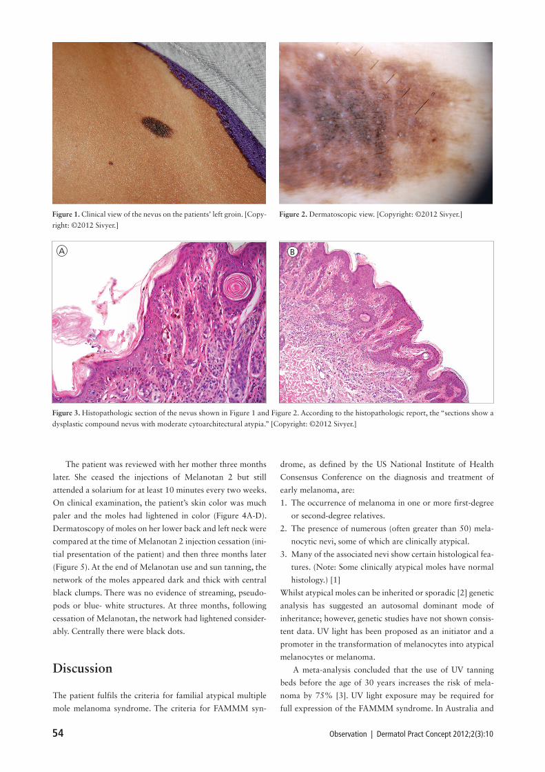
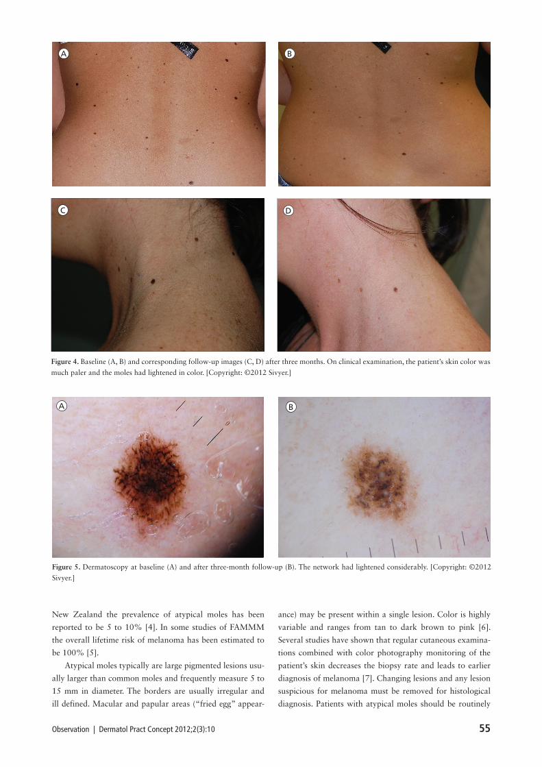
The patient was reviewed with her mother three months
later. She ceased the injections of Melanotan 2 but still
attended a solarium for at least 10 minutes every two weeks.
On clinical examination, the patient’s skin color was much
paler and the moles had lightened in color (Figure 4A-D).
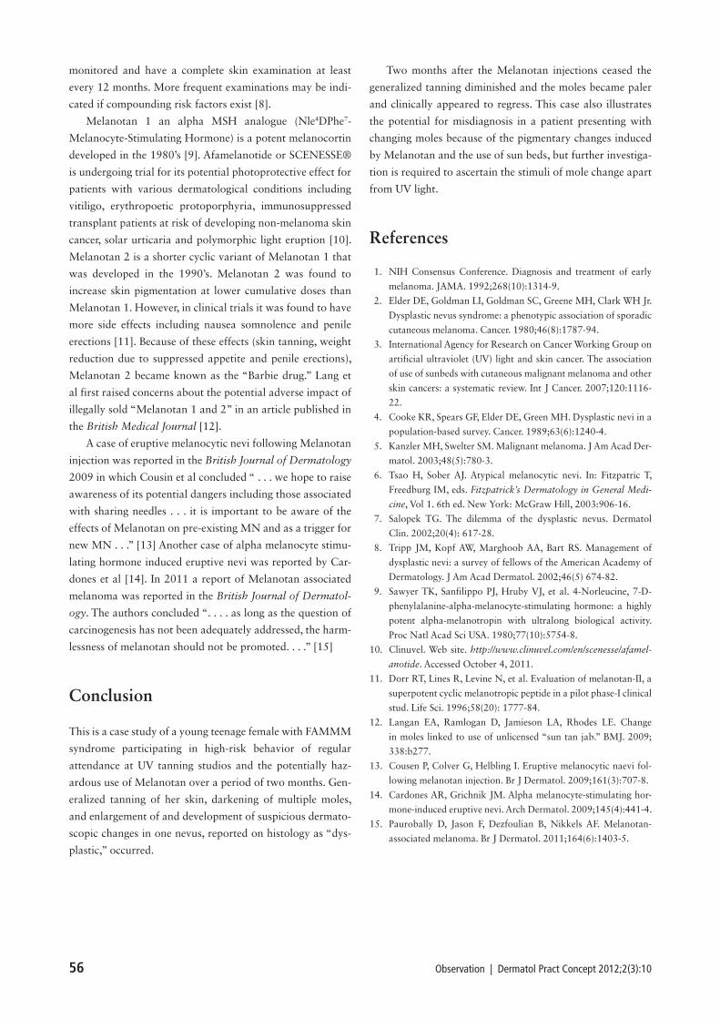
Dermatoscopy of moles on her lower back and left neck were
compared at the time of Melanotan 2 injection cessation (ini-
tial presentation of the patient) and then three months later
(Figure 5). At the end of Melanotan use and sun tanning, the
network of the moles appeared dark and thick with central
black clumps. There was no evidence of streaming, pseudo-
pods or blue- white structures. At three months, following
cessation of Melanotan, the network had lightened consider-
ably. Centrally there were black dots.
Discussion
The patient fulfils the criteria for familial atypical multiple
mole melanoma syndrome. The criteria for FAMMM syn-
drome, as defined by the US National Institute of Health
Consensus Conference on the diagnosis and treatment of
early melanoma, are:
1. The occurrence of melanoma in one or more first-degree
or second-degree relatives.
2. The presence of numerous (often greater than 50) mela-
nocytic nevi, some of which are clinically atypical.
3. Many of the associated nevi show certain histological fea-
tures. (Note: Some clinically atypical moles have normal
histology.) [1]
Whilst atypical moles can be inherited or sporadic [2] genetic
analysis has suggested an autosomal dominant mode of
inheritance; however, genetic studies have not shown consis-
tent data. UV light has been proposed as an initiator and a
promoter in the transformation of melanocytes into atypical
melanocytes or melanoma.
A meta-analysis concluded that the use of UV tanning
beds before the age of 30 years increases the risk of mela-
noma by 75% [3]. UV light exposure may be required for
full expression of the FAMMM syndrome. In Australia and
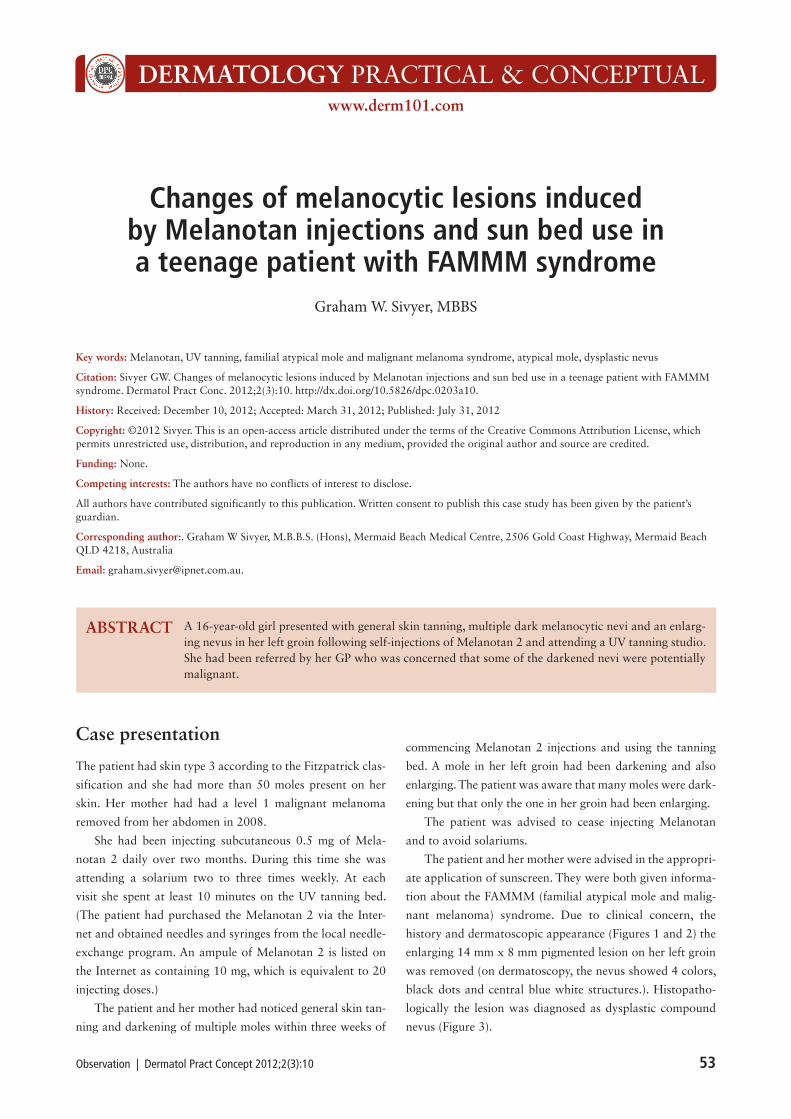
Figure 1. Clinical view of the nevus on the patients’ left groin. [Copy-
right: ©2012 Sivyer.]
Figure 2. Dermatoscopic view. [Copyright: ©2012 Sivyer.]
Figure 3. Histopathologic section of the nevus shown in Figure 1 and Figure 2. According to the histopathologic report, the “sections show a
dysplastic compound nevus with moderate cytoarchitectural atypia.” [Copyright: ©2012 Sivyer.]
A B
Observation | Dermatol Pract Concept 2012;2(3):10 55
New Zealand the prevalence of atypical moles has been
reported to be 5 to 10% [4]. In some studies of FAMMM
the overall lifetime risk of melanoma has been estimated to
be 100% [5].
Atypical moles typically are large pigmented lesions usu-
ally larger than common moles and frequently measure 5 to
15 mm in diameter. The borders are usually irregular and
ill defined. Macular and papular areas (“fried egg” appear-
ance) may be present within a single lesion. Color is highly
variable and ranges from tan to dark brown to pink [6].
Several studies have shown that regular cutaneous examina-
tions combined with color photography monitoring of the
patient’s skin decreases the biopsy rate and leads to earlier
diagnosis of melanoma [7]. Changing lesions and any lesion
suspicious for melanoma must be removed for histological
diagnosis. Patients with atypical moles should be routinely
A B
C D
Figure 4. Baseline (A, B) and corresponding follow-up images (C, D) after three months. On clinical examination, the patient’s skin color was
much paler and the moles had lightened in color. [Copyright: ©2012 Sivyer.]
Figure 5. Dermatoscopy at baseline (A) and after three-month follow-up (B). The network had lightened considerably. [Copyright: ©2012
Sivyer.]
A B
56 Observation | Dermatol Pract Concept 2012;2(3):10
monitored and have a complete skin examination at least
every 12 months. More frequent examinations may be indi-
cated if compounding risk factors exist [8].
Melanotan 1 an alpha MSH analogue (Nle4DPhe7-
Melanocyte-Stimulating Hormone) is a potent melanocortin
developed in the 1980’s [9]. Afamelanotide or SCENESSE®
is undergoing trial for its potential photoprotective effect for
patients with various dermatological conditions including
vitiligo, erythropoetic protoporphyria, immunosuppressed
transplant patients at risk of developing non-melanoma skin
cancer, solar urticaria and polymorphic light eruption [10].
Melanotan 2 is a shorter cyclic variant of Melanotan 1 that
was developed in the 1990’s. Melanotan 2 was found to
increase skin pigmentation at lower cumulative doses than
Melanotan 1. However, in clinical trials it was found to have
more side effects including nausea somnolence and penile
erections [11]. Because of these effects (skin tanning, weight
reduction due to suppressed appetite and penile erections),
Melanotan 2 became known as the “Barbie drug.” Lang et
al first raised concerns about the potential adverse impact of
illegally sold “Melanotan 1 and 2” in an article published in
the British Medical Journal [12].
A case of eruptive melanocytic nevi following Melanotan
injection was reported in the British Journal of Dermatology
2009 in which Cousin et al concluded “ . . . we hope to raise
awareness of its potential dangers including those associated
with sharing needles . . . it is important to be aware of the
effects of Melanotan on pre-existing MN and as a trigger for
new MN . . .” [13] Another case of alpha melanocyte stimu-
lating hormone induced eruptive nevi was reported by Car-
dones et al [14]. In 2011 a report of Melanotan associated
melanoma was reported in the British Journal of Dermatol-
ogy. The authors concluded “. . . . as long as the question of
carcinogenesis has not been adequately addressed, the harm-
lessness of melanotan should not be promoted. . . .” [15]
Conclusion
This is a case study of a young teenage female with FAMMM
syndrome participating in high-risk behavior of regular
attendance at UV tanning studios and the potentially haz-
ardous use of Melanotan over a period of two months. Gen-
eralized tanning of her skin, darkening of multiple moles,
and enlargement of and development of suspicious dermato-
scopic changes in one nevus, reported on histology as “dys-
plastic,” occurred.
Two months after the Melanotan injections ceased the
generalized tanning diminished and the moles became paler
and clinically appeared to regress. This case also illustrates
the potential for misdiagnosis in a patient presenting with
changing moles because of the pigmentary changes induced
by Melanotan and the use of sun beds, but further investiga-
tion is required to ascertain the stimuli of mole change apart
from UV light.
References
1. NIH Consensus Conference. Diagnosis and treatment of early
melanoma. JAMA. 1992;268(10):1314-9.
2. Elder DE, Goldman LI, Goldman SC, Greene MH, Clark WH Jr.
Dysplastic nevus syndrome: a phenotypic association of sporadic
cutaneous melanoma. Cancer. 1980;46(8):1787-94.
3. International Agency for Research on Cancer Working Group on
artificial ultraviolet (UV) light and skin cancer. The association
of use of sunbeds with cutaneous malignant melanoma and other
skin cancers: a systematic review. Int J Cancer. 2007;120:1116-
22.
4. Cooke KR, Spears GF, Elder DE, Green MH. Dysplastic nevi in a
population-based survey. Cancer. 1989;63(6):1240-4.
5. Kanzler MH, Swelter SM. Malignant melanoma. J Am Acad Der-
matol. 2003;48(5):780-3.
6. Tsao H, Sober AJ. Atypical melanocytic nevi. In: Fitzpatric T,
Freedburg IM, eds. Fitzpatrick’s Dermatology in General Medi-
cine, Vol 1. 6th ed. New York: McGraw Hill, 2003:906-16.
7. Salopek TG. The dilemma of the dysplastic nevus. Dermatol
Clin. 2002;20(4): 617-28.
8. Tripp JM, Kopf AW, Marghoob AA, Bart RS. Management of
dysplastic nevi: a survey of fellows of the American Academy of
Dermatology. J Am Acad Dermatol. 2002;46(5) 674-82.
9. Sawyer TK, Sanfilippo PJ, Hruby VJ, et al. 4-Norleucine, 7-D-
phenylalanine-alpha-melanocyte-stimulating hormone: a highly
potent alpha-melanotropin with ultralong biological activity.
Proc Natl Acad Sci USA. 1980;77(10):5754-8.
10. Clinuvel. Web site. http://www.clinuvel.com/en/scenesse/afamel-
anotide. Accessed October 4, 2011.
11. Dorr RT, Lines R, Levine N, et al. Evaluation of melanotan-II, a
superpotent cyclic melanotropic peptide in a pilot phase-I clinical
stud. Life Sci. 1996;58(20): 1777-84.
12. Langan EA, Ramlogan D, Jamieson LA, Rhodes LE. Change
in moles linked to use of unlicensed “sun tan jab.” BMJ. 2009;
338:b277.
13. Cousen P, Colver G, Helbling I. Eruptive melanocytic naevi fol-
lowing melanotan injection. Br J Dermatol. 2009;161(3):707-8.
14. Cardones AR, Grichnik JM. Alpha melanocyte-stimulating hor-
mone-induced eruptive nevi. Arch Dermatol. 2009;145(4):441-4.
15. Paurobally D, Jason F, Dezfoulian B, Nikkels AF. Melanotan-
associated melanoma. Br J Dermatol. 2011;164(6):1403-5.