
Changes in Coronal Alignment of the Knee Joint after ...
8
Research Article Changes in Coronal Alignment of the Knee Joint after Supramalleolar Osteotomy Dong-Il Chun , 1 Jahyung Kim , 2 Sung Hun Won , 2 Jaeho Cho , 3 Jeongku Ha , 4 Minkyu Kil, 4 and Young Yi 5 1 Department of Orthopedic Surgery, Foot and Ankle Center, Soonchunhyang University Seoul Hospital, 59, Daesagwan-ro, Yongsan- gu, Seoul, Republic of Korea 2 Department of Orthopedic Surgery, Soonchunhyang University Seoul Hospital, 59, Daesagwan-ro, Yongsan-gu, Seoul, Republic of Korea 3 Department of Orthopaedic Surgery, Chuncheon Sacred Heart Hospital, Hallym University, 77, Sakju-ro, Chuncheon-si, Gangwon-do, Republic of Korea 4 Department of Orthopaedic Surgery, Sports Medical Center and Sports Medical Research Institute, Inje University Seoul Paik Hospital, 85, 2-ga, Jeo-dong, Jung-gu, Seoul, Republic of Korea 5 Department of Orthopaedic Surgery, Seoul Foot and Ankle Center, Inje University Seoul Paik Hospital, 85, 2-ga, Jeo-dong, Jung-gu, Seoul, Republic of Korea Correspondence should be addressed to Young Yi; [email protected] Received 1 November 2020; Revised 24 December 2020; Accepted 11 February 2021; Published 19 February 2021 Academic Editor: Hyuk-Soo Han Copyright © 2021 Dong-Il Chun et al. This is an open access article distributed under the Creative Commons Attribution License, which permits unrestricted use, distribution, and reproduction in any medium, provided the original work is properly cited. Background. Assessing knee joint orientation changes after SMO may help clinical advancement in managing patients with ipsilateral ankle and knee joint arthritis. However, knee joint changes after supramalleolar osteotomy (SMO) have not been reported. We investigated changes in coronal alignment of the knee joint after SMO. Methods. In this multicentre study, from January 2014 to December 2018, 47 ankles with varus osteoarthritis treated with SMO were retrospectively identified. Ankle joint changes were assessed using the tibiotalar angle, talar tilt angle, and lateral distal tibial angle (LDTA); knee joint changes using the medial proximal tibial angle (MPTA), medial and lateral joint space widths (mJSW and lJSW, respectively), and medial and lateral joint line convergence angles (JLCA); and lower limb alignment changes using mechanical axis deviation angle (MADA) and the hip-knee-ankle (HKA) angle measured on full-length anteroposterior radiographs of the lower extremity. Correlation analysis and binary logistic regression analysis were performed. Results. Postoperatively, LDTA (p <0:001) and tibiotalar angle (p <0:001) significantly changed, indicating meaningful improvement in the ankle joint varus deformity. Regarding the knee joint changes, JLCA significantly changed into valgus direction (p =0:044). As for lower limb alignment changes, MADA significantly decreased (p <0:001), whereas the HKA angle significantly increased (p <0:001). In univariate and multivariate logistic regression analyses, changes in the MADA (p <0:001) and the HKA angle (p <0:001) were significantly correlated with the correction angle. Conclusions. SMO remarkably improves ankle joint varus deformity, followed by significant lower limb alignment changes. Despite meaningful changes in JLCA, the relationship between the amount of osteotomy near the ankle joint and improvement in knee joint radiographic parameters was not significant. Radiographic parameters of the knee joint would less likely be changed following SMO. 1. Introduction Supramalleolar osteotomy (SMO) of the ankle has been sug- gested to be a suitable treatment modality for varus ankle arthrosis by simultaneously redistributing the weight-bearing portion of the ankle joint and adjusting the varus alignment through lateral translation of the mechanical axis, followed by improved patient outcomes [1–4]. Similarly, high tibial osteotomy (HTO) is a useful treatment option to redistribute the knee joint load from areas of degeneration to intact Hindawi BioMed Research International Volume 2021, Article ID 6664279, 8 pages https://doi.org/10.1155/2021/6664279
Transcript of Changes in Coronal Alignment of the Knee Joint after ...
Research ArticleChanges in Coronal Alignment of the Knee Joint afterSupramalleolar Osteotomy
Dong-Il Chun ,1 Jahyung Kim ,2 Sung Hun Won ,2 Jaeho Cho ,3 Jeongku Ha ,4
Minkyu Kil,4 and Young Yi 5
1Department of Orthopedic Surgery, Foot and Ankle Center, Soonchunhyang University Seoul Hospital, 59, Daesagwan-ro, Yongsan-gu, Seoul, Republic of Korea2Department of Orthopedic Surgery, Soonchunhyang University Seoul Hospital, 59, Daesagwan-ro, Yongsan-gu,Seoul, Republic of Korea3Department of Orthopaedic Surgery, Chuncheon Sacred Heart Hospital, Hallym University, 77, Sakju-ro, Chuncheon-si,Gangwon-do, Republic of Korea4Department of Orthopaedic Surgery, Sports Medical Center and Sports Medical Research Institute, Inje University SeoulPaik Hospital, 85, 2-ga, Jeo-dong, Jung-gu, Seoul, Republic of Korea5Department of Orthopaedic Surgery, Seoul Foot and Ankle Center, Inje University Seoul Paik Hospital, 85, 2-ga, Jeo-dong, Jung-gu,Seoul, Republic of Korea
Correspondence should be addressed to Young Yi; [email protected]
Received 1 November 2020; Revised 24 December 2020; Accepted 11 February 2021; Published 19 February 2021
Academic Editor: Hyuk-Soo Han
Copyright © 2021 Dong-Il Chun et al. This is an open access article distributed under the Creative Commons Attribution License,which permits unrestricted use, distribution, and reproduction in any medium, provided the original work is properly cited.
Background. Assessing knee joint orientation changes after SMO may help clinical advancement in managing patients withipsilateral ankle and knee joint arthritis. However, knee joint changes after supramalleolar osteotomy (SMO) have not beenreported. We investigated changes in coronal alignment of the knee joint after SMO. Methods. In this multicentre study, fromJanuary 2014 to December 2018, 47 ankles with varus osteoarthritis treated with SMO were retrospectively identified. Anklejoint changes were assessed using the tibiotalar angle, talar tilt angle, and lateral distal tibial angle (LDTA); knee joint changesusing the medial proximal tibial angle (MPTA), medial and lateral joint space widths (mJSW and lJSW, respectively), andmedial and lateral joint line convergence angles (JLCA); and lower limb alignment changes using mechanical axis deviationangle (MADA) and the hip-knee-ankle (HKA) angle measured on full-length anteroposterior radiographs of the lower extremity.Correlation analysis and binary logistic regression analysis were performed. Results. Postoperatively, LDTA (p < 0:001) andtibiotalar angle (p < 0:001) significantly changed, indicating meaningful improvement in the ankle joint varus deformity. Regardingthe knee joint changes, JLCA significantly changed into valgus direction (p = 0:044). As for lower limb alignment changes, MADAsignificantly decreased (p < 0:001), whereas the HKA angle significantly increased (p < 0:001). In univariate and multivariatelogistic regression analyses, changes in the MADA (p < 0:001) and the HKA angle (p < 0:001) were significantly correlated with thecorrection angle. Conclusions. SMO remarkably improves ankle joint varus deformity, followed by significant lower limb alignmentchanges. Despite meaningful changes in JLCA, the relationship between the amount of osteotomy near the ankle joint andimprovement in knee joint radiographic parameters was not significant. Radiographic parameters of the knee joint would lesslikely be changed following SMO.
1. Introduction
Supramalleolar osteotomy (SMO) of the ankle has been sug-gested to be a suitable treatment modality for varus anklearthrosis by simultaneously redistributing the weight-bearing
portion of the ankle joint and adjusting the varus alignmentthrough lateral translation of the mechanical axis, followedby improved patient outcomes [1–4]. Similarly, high tibialosteotomy (HTO) is a useful treatment option to redistributethe knee joint load from areas of degeneration to intact
HindawiBioMed Research InternationalVolume 2021, Article ID 6664279, 8 pageshttps://doi.org/10.1155/2021/6664279

articular surfaces in patients with varus knee alignment andmedial compartment knee arthritis [5–7].
Although osteotomies are generally performed near thesymptomatic joints, subsequent alignment changes can alsoinfluence the ipsilateral joints. Multiple studies have proventhat varus ankle osteoarthritis in concurrence with ipsilateralvarus knee osteoarthritis improved after HTO [4, 8, 9].Takeuchi et al. [9] reported significant improvement in bothclinical and radiologic outcomes of ankle osteoarthritis inpatients who underwent HTO on the same limb. Afterosteotomy, reductions in both valgus inclination of the distaltibial joint surface and medial inclination of the talus weredetected, followed by complete resolution of knee and anklejoint pain in all patients.
Similarly, the main goal of SMO is to realign the alteredmechanical axis in order to normalize the joint loadingwithin the ankle [10]. Haraguchi et al. [11] reported changesin the mechanical axis, including the hindfoot, amongpatients who underwent SMO, and Yi et al. [4] stated lateraldeviation of the mechanical axis after SMO. Furthermore,Choi et al. [8] reported that the ankle joint orientationbecomes parallel to the ground after HTO, and symptomsare affected by subsequent coronal alignment changes.
Consequently, defining changes in knee joint orientationafter SMO may bring out significant clinical advancement inmanaging patients with ipsilateral ankle and knee jointarthritis, such as setting the order of priority between HTOand SMO or choosing adequate treatment modality to coverboth joints. However, to our knowledge, no study has focusedon knee joint changes after SMO. Therefore, we hypothesisedthat SMO would influence the alignment and function of theknee joint and such a change may affect knee joint orienta-tion. This study is aimed at investigating changes in coronalalignment of the knee joint after SMO.
2. Material and Methods
This study was approved by the Institutional Review Board atour institution.
2.1. Patient Selection. This is a multicentre study performedby three surgeons at three different institutions under thesame operation technique and rehabilitation program. FromJanuary 2014 to December 2018, 158 consecutive ankles (129patients) with varus osteoarthritis treated with SMO wereretrospectively identified. The indications for SMO includedsymptomatic Takakura IIIa varus ankle arthritis charac-terised with medial gutter narrowing and varus lower limbalignment. Contraindications were Takakura IIIb varus anklearthritis and end-stage ankle arthritis. Among these patients,87 patients who were concurrent with a standing whole-leganteroposterior radiograph including bilateral knee andankle joints were evaluated. 11 patients were lost during fol-low-up, and 39 patients were excluded following the exclu-sion criteria. Finally, 47 ankles in 45 patients (17 men and28 women) were chosen for the study (Figure 1). The meanage was 56.3 years (range, 44 to 75 years). Comparative anal-ysis was performed using preoperative and postoperativeradiographic measurements among all included patients.
2.2. Operative Procedure. A 7 cm longitudinal incision ismade along the anteromedial aspect of the distal tibia. TwoKirschner wires are inserted about 5 cm above the ankle mor-tise, just proximal to the tibiofibular syndesmosis, under fluo-roscopic image intensifier in order to guide the osteotomy.Then, a periosteal incision of less than 1 cm is made alongthe site to perform the osteotomy, in an effort to maintainas many soft tissue attached to the distal bone fragment aspossible. The osteotomy is performed parallel to the anklemortise, and the lateral cortex at the apex of the distal partof the tibia must be preserved so that it can be used as a hinge.After completing the osteotomy, the distal osteotomizedfragment is shifted inferiorly by introducing an osteotomethrough the medial aspect of the osteotomized site. Intraop-erative visualization and fluoroscopy are used to evaluatethe adequacy of the correction angle and lower limb align-ment. Then, the osteotomy is fixed with 7-hole dynamiccompression plate, in order to place three holes proximaland three holes distal to the osteotomy site. The gap is filledwith autologous or allogenic bone graft.
2.3. Radiological Evaluation. The weight-bearing anteropos-terior ankle, knee radiographs, and full-length anteroposter-ior radiographs were taken in all patients at preoperativeand final follow-up period of at least 1 year after the surgery.
158 ankles undergoing supramalleolar osteotomy (129 patients)were screened_
From January 2014~December 2018
Inclusion criteria (87 ankles were included)Radiographic evaluation performed with knee, ankle, and
lower limb alignment.
Follow-up loss: 11 ankles excluded
Exclusion criteria (39 ankles were excluded)1. Ipsilateral realignment sugery before SMO
2. Total knee arthroplasty
(i) Proximal and distal femoral OT(ii) High tibial osteotomy
(iii) Lateral column lengthening(iv) Medial displacement calcaneal OT
3. The patient has underlying disease of
4. Insufficient radiograph
(i) Malunion of tibial shaft fracture(ii) Neurologic impairment
(iii) Syndesmotic instability
Patient group :47 ankles with supramalleolar osteotomy were studied
Figure 1: Flowchart showing the inclusion and exclusion criteria forthe patients in this study.
2 BioMed Research International
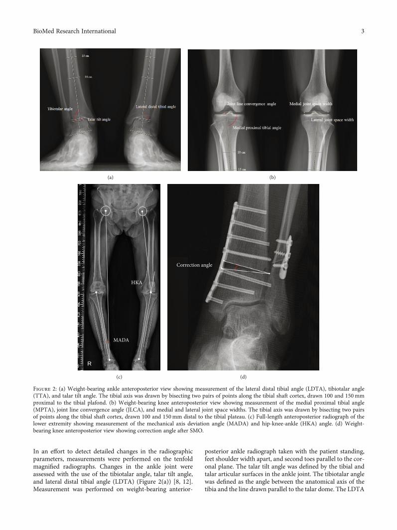
In an effort to detect detailed changes in the radiographicparameters, measurements were performed on the tenfoldmagnified radiographs. Changes in the ankle joint wereassessed with the use of the tibiotalar angle, talar tilt angle,and lateral distal tibial angle (LDTA) (Figure 2(a)) [8, 12].Measurement was performed on weight-bearing anterior-
posterior ankle radiograph taken with the patient standing,feet shoulder width apart, and second toes parallel to the cor-onal plane. The talar tilt angle was defined by the tibial andtalar articular surfaces in the ankle joint. The tibiotalar anglewas defined as the angle between the anatomical axis of thetibia and the line drawn parallel to the talar dome. The LDTA
(a) (b)
HKA
MADA
(c)
Correction angle
(d)
Figure 2: (a) Weight-bearing ankle anteroposterior view showing measurement of the lateral distal tibial angle (LDTA), tibiotalar angle(TTA), and talar tilt angle. The tibial axis was drawn by bisecting two pairs of points along the tibial shaft cortex, drawn 100 and 150mmproximal to the tibial plafond. (b) Weight-bearing knee anteroposterior view showing measurement of the medial proximal tibial angle(MPTA), joint line convergence angle (JLCA), and medial and lateral joint space widths. The tibial axis was drawn by bisecting two pairsof points along the tibial shaft cortex, drawn 100 and 150mm distal to the tibial plateau. (c) Full-length anteroposterior radiograph of thelower extremity showing measurement of the mechanical axis deviation angle (MADA) and hip-knee-ankle (HKA) angle. (d) Weight-bearing knee anteroposterior view showing correction angle after SMO.
3BioMed Research International
was defined as the angle between the tibial anatomical axisand the distal tibial articular surface on the standing ankleanteroposterior radiographs.
In addition, changes in the knee joint were assessed withmedial proximal tibial angle (MPTA), medial and lateraljoint space widths (mJSW and lJSW, respectively) [13], andmedial and lateral joint line convergence angles (JLCA)[14]. Measurement was performed on weight-bearing ante-roposterior knee radiographs taken with the patient standing,feet shoulder width apart, and both patella heading forward(Figure 2(b)). MPTA was defined by the angle between thetibial mechanical axis and the articular surface line of theproximal tibia. mJSW and lJSW were measured as the short-est distance between the distal femur and the proximal tibiafor the medial and lateral joint spaces of each knee. JLCAwas measured as the angle formed between a line tangentialto the distal femoral condyle and the tibial plateau.
Moreover, changes in lower limb alignment were assessedwith mechanical axis deviation angle (MADA) [12] and hip-knee-ankle (HKA) angle [8] measured on full-length antero-posterior radiograph of the lower extremity. MADA wasdefined as the angle formed by the line connecting the hipcentre to the ankle centre and the tibial mechanical axis, whilethe HKA angle was the angle between the mechanical axes ofthe femur and the tibia (Figure 2(c)).
Lastly, the correction angle of SMO was measured as theangle between the proximal and distal osteotomized ends(Figure 2(d)).
2.4. Statistical Analysis. Patient demographics and pre-operative and postoperative radiographic measurements aredescribed as average value ± standard deviation. Althoughall the radiographic measurements were measured to threedecimal points, the angle measurements were described toone decimal point. Changes in the radiographic parametersof the ankle, knee, and lower limb alignment before and afterthe surgery were analysed using paired t-test or signed ranktest. Shapiro-Wilk normality test was performed on all mea-sured data. Correlation analysis of the correction angle afterSMO and preoperative and postoperative changes in theradiographic parameters was performed with Pearson corre-lation coefficient and Spearman’s rank correlation coefficient.Binary logistic regression analysis with stepwise selectionmethod was performed over values with significant correla-tion. Statistical analyses were performed using SPSS version20.0 (IBM Corp., Armonk, NY, USA), and p values < 0.05were considered statistically significant.
3. Results
Interobserver and intraobserver reliability was determined bycalculating the interclass correlation coefficients for continu-ous data, and the reliability of all radiographic parameterswas above 0.8 (Table 1). The target correction angle of theSMO was to rectify LDTA into 87°, and the valgus angulationdifference was an average of 7:3° ± 6:0°.
Preoperative and postoperative changes in radiographicparameters of the ankle were as follows. The LDTA signifi-cantly changed from 94:7° ± 4:1° to 87:5° ± 5:7° with an aver-
age of 7:2° ± 6:0° valgus (p < 0:001). The tibiotalar angle alsosignificantly improved from 95:4° ± 3:6° to 89.5° (87.4°, 93.4°)with 4.1° (-9.1°, -0.4°) valgus (p < 0:001). The talar tilt anglechanged from an average of 2° (1.1°, 3.2°) to 1.8° (1.1°, 2.9°)with no significant difference.
Changes in the radiographic parameters of the knee jointwere as follows. The MPTA changed from an average of 88.1°
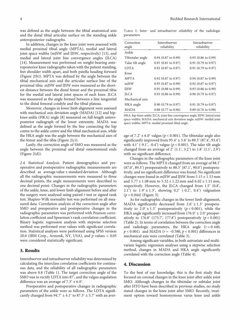
(87.0°, 89.3°) preoperatively to 88.3° (87.1°, 90.0°) postopera-tively, and no significant difference was found. No significantchanges were found in mJSW and lJSW from 5:15 ± 1:13mmand 5:77 ± 1:08mm to 5:32 ± 1:22mm and 6:02 ± 1:51mm,respectively. However, the JLCA changed from 1.5° (0.8°,2.1°) to 1:9° ± 1:3°, showing 0.2° (-0.2°, 0.4°) valgisation(p = 0:044) (Figure 3).
As for radiographic changes in the lower limb alignment,MADA significantly decreased from 2:6° ± 1:3° preopera-tively to 2:0° ± 1:5° postoperatively (p < 0:001), while theHKA angle significantly increased from 176:0° ± 2:0° preoper-atively to 176.8° (175.7°, 177.8°) postoperatively (p < 0:001)(Table 2). In terms of correlation between the correction angleand radiologic parameters, the HKA angle (r = 0:440,p < 0:001) and MADA (r = −0:580, p < 0:001) differences inmechanical axis were correlated (Table 3).
Among significant variables, in both univariate and multi-variate logistic regression analyses using a stepwise selectionmethod, changes in MADA and HKA angle significantlycorrelated with the correction angle (Table 4).
4. Discussion
To the best of our knowledge, this is the first study thatfocused on coronal changes in the knee joint after ankle jointSMO. Although changes in the tibiotalar or subtalar jointafter HTO have been described in previous studies, no studystated changes in the knee joint after SMO. Recently, treat-ment option toward homonymous varus knee and ankle
Table 1: Inter- and intraobserver reliability of the radiologicparameters.
Correctionangle
Interobserverreliability
Intraobserverreliability
Ankle
Tibiotalar angle 0.94 (0.87 to 0.99) 0.93 (0.86 to 0.99)
Talar tilt angle 0.91 (0.81 to 0.97) 0.91 (0.79 to 0.97)
LDTA 0.92 (0.87 to 0.97) 0.91 (0.79 to 0.97)
Knee
MPTA 0.92 (0.87 to 0.97) 0.94 (0.87 to 0.99)
mJSW 0.95 (0.87 to 0.99) 0.92 (0.87 to 0.97)
lJSW 0.95 (0.88 to 0.99) 0.93 (0.86 to 0.99)
JLCA 0.93 (0.86 to 0.99) 0.90 (0.79 to 0.97)
Mechanical axis
HKA angle 0.90 (0.79 to 0.97) 0.91 (0.79 to 0.97)
MADA 0.88 (0.77 to 0.96) 0.89 (0.76 to 0.98)
HKA: hip-knee-ankle; JLCA: joint line convergence angle; lJSW: lateral jointspace widths; MADA: mechanical axis deviation angle; mJSW: medial jointspace widths; MPTA: medial proximal tibial angle.
4 BioMed Research International

osteoarthritis has been reported in the literature [9].Although it is commonly recommended to approach theproximal degenerative joint ahead, defining changes in theuntouched joint induced by the operated joint could be animportant issue because the two joints are in line with eachother based on the mechanical axis.
In this study, changes in the knee joint and lower limbalignments were measured in addition to those in the anklejoint, and significant correlation was found between the cor-rection angle and limb alignment. That is, as the correctionangle increased after SMO, mechanical deviation decreasedand the HKA angle changed. However, when JLCA wasexcluded, no significant correlations were found among otherradiographic parameters of the knee joint.
Several studies have reported the correlation between theHTO and coronal lower limb alignment [15–17]. Lee et al.
Preoperative Postoperative
JLCA = 3.71 JLCA = 0.42
Figure 3: Example of a 57-year-old woman who underwent supramalleolar osteotomy. Preoperative and postoperative anteroposteriorradiographs showing the JLCA changed from 3.71° to 0.42° after surgery (JLCA= joint line congruence angle).
Table 2: Changes and significance in radiologic parameters of pre- and postoperative ankle, knee, and lower limb alignment.
Preoperative Postoperative Change (post-pre)p valueMean ± SD or median (IQR)
Ankle
LDTA 94:7 ± 4:1 87:5 ± 5:7 −7:2 ± 6:0 <0.001
Talar tilt angle 2 (1.1, 3.2) 1.8 (1.1, 2.9) -0.6 (-1.4, 0.6) 0.1214
Tibiotalar angle 95:4 ± 3:6 89.5 (87.4, 93.4) -4.1 (-9.1, -0.4) <0.001
Knee
MPTA 88.1 (87.0, 89.3) 88.85 (87.1, 89.5) 0 (-0.2, 0.5) 0.3542
mJSW 5:15 ± 1:13 5:32 ± 1:22 0:17 ± 0:74 0.1203
lJSW 5:77 ± 1:08 6:02 ± 1:51 0.06 (-0.59, 0.78) 0.2665
JLCA 1.5 (0.8, 2.1) 1:9 ± 1:3 0.2 (-0.2, 0.4) 0.0444
Mechanical axis
HKA angle 176:0 ± 2:0 176.8 (175.7, 177.8) 0.5 (0.2, 1.3) <0.001
MADA 2:6 ± 1:3 2:0 ± 1:5 -0.7 (-1, -0.1) <0.001
IQR: interquartile range; HKA: hip-knee-ankle; JLCA: joint line convergence angle; lJSW: lateral joint space widths; MADA: mechanical axis deviation; mJSW:medial joint space widths; MPTA: medial proximal tibial angle.
Table 3: Correlation between the correction angle and radiologicparameters.
Correlation with correction angler p value
Knee
MPTA difference -0.080 0.587
mJSW difference 0.080 0.590
lJSW difference 0.235 0.108
JLCA difference 0.115 0.436
Mechanical axis
HKA angle difference 0.440 <0.001MADA difference -0.580 <0.001
HKA: hip-knee-ankle; JLCA: joint line convergence angle; lJSW: lateral jointspace widths; MADA: mechanical axis deviation angle; mJSW: medial jointspace widths; MPTA: medial proximal tibial angle.
5BioMed Research International

[16] compared the JLCA among patients divided into threedifferent groups after HTO: undercorrection (weight-bearingline (WBL) ratio, <57%), overcorrection (WBL ratio, >67%),and acceptable correction (WBL ratio, 57–67%) groups. TheJLCA was measured before and after surgery and comparedbetween the three groups. Difference between the pre‐ andpostoperative values of the JLCA showed a stronger correla-tion than those of the MAD. Such differences in the JLCAafter HTO are related to an increase in the knee medial jointwidth and a decrease in the lateral joint width. Changes inMPTA were also found in the case of open-wedge HTObecause of alteration in the morphology of the proximal tibia.
In this study, a significant change in the JLCA wasdetected after SMO, probably because of the fact that theknee joint becomes affected by the valgus force after medialopen-wedge osteotomy performed at its distal level [18].However, although statistically significant, such change wasas minimal as 0.2 degrees and was not significantly correlatedwith the correction angle in the regression analysis. Similarly,no significant change was detected on the MPTA or mJSWand lJSW after surgery, while these factors were not relatedto the correction angle in the regression analysis.
Variety of explanations could be made concerning suchinsignificant changes in the knee joint following SMO. First,as osteotomy is performed on the distal portion of the tibia inSMO, no direct structural change around the knee joint canbe achieved. Although changes in the JLCA formed by thedistal femur and proximal tibia were found, operative correc-tion cannot be performed either. Second, the knee jointbecomes less corrected after SMO because the more distalthe osteotomy is performed, that is, at the level of the meta-physis of distal tibia, the lesser the mechanical axis changes.It is depicted in Figure 4 showing inferior alteration inmechanical axis in SMO compared with HTO, even withthe same correction angle (Figure 4). Third, the knee jointis surrounded by relatively abundant soft tissue includingmedial collateral ligament, articular cartilage, or meniscus.Owing to the untouched, normal integrity of these structuresacting as restraints to valgus forces around the knee joint,change in the mechanical axis caused by SMO, whichbecomes larger with the amount of correction angle, couldeventually be counterbalanced [19]. Lastly, other factors
including patients’ weight or hindfoot compensation mighthave buffered the substantial changes in the knee joint [15, 17].
Another possible explanation would be the difference inthe moment arms between two surgeries. Many studies havereported that the ankle joint has a longer moment arm thanthe knee joint when HTO is performed [8, 20]. Therefore,changes could be more prominent in the ankle joint than inthe knee joint. However, in the case of SMO, given theshorter moment arm than the ankle joint, the influence ofosteotomy would be inferior in the knee joint.
This study has some limitations. First, patients with rela-tively early stage of knee osteoarthritis were included, as nooperation was performed on the affected knee at the time ofSMO. For this reason, direct influence of SMO on theadvanced knee osteoarthritis could not be analysed. How-ever, as changes in lower limb alignment had minimal effecton the knee joint, insignificant change in advanced arthriticknee joint could be assumed in the same manner. Second,external factors such as administration of painkillers or pro-longed period of nonweight bearing related to cast applica-tion could not be controlled after surgery. Nevertheless, aspartial weight bearing was allowed in 4 weeks, full weightbearing in 8 weeks, and an attempt to return to the normalactivity initiated within 3 months after surgery, we thoughtthat the follow-up period of 1 year would be long enoughto neutralise the non-weight-bearing period. Third, clinicaloutcomes were not evaluated simultaneously. Although nosignificant correlation was found between the correctionangle and radiographic parameters of the knee joint, changesin lower limb alignment might positively influence the clini-cal symptoms. However, as additional pain control usingmedications was performed during the 1-year follow-upperiod, we thought it would be awkward to establish a clearrelationship between clinical outcomes and radiographicparameters. As a result, we thought that radiographicanalysis would be sufficiently meaningful. Fourth, this is aretrospective study with a relatively small sample size of 47cases. When we performed a post hoc power analysis uponall the variables in the final regression model, it turned outthat more sample size would be needed in order to verifythe HKA angle in the succeeding study. Last, as this studysolely focused on radiographic parameters in the coronal
Table 4: Binary regression analysis of the radiologic parameters.
Univarable analysis Multivariable analysis
Beta95% CI
p value Beta95% CI
p valueLower Upper Lower Upper
Knee
MPTA -0.128 -0.460 0.204 0.441
mJSW 0.198 -0.536 0.931 0.590
lJSW 0.162 -0.210 0.534 0.385
JLCA -0.059 -0.397 0.278 0.726
Mechanical axis
HKA angle 0.001 -0.335 0.336 0.996 -0.301 -0.614 0.012 0.049
MADA -0.94 -1.421 -0.459 <0.001 -1.157 -1.676 -0.638 <0.001HKA: hip-knee-ankle; JLCA: joint line convergence angle; lJSW: lateral joint space widths; MADA: mechanical axis deviation angle; mJSW: medial joint spacewidths; MPTA: medial proximal tibial angle.
6 BioMed Research International

plane, the possible influence of changes in the other planescould have been neglected. In addition, the relationshipbetween changes in the subtalar joint and knee joint couldnot be evaluated. These factors would have to be additionallyconsidered in the future study.
In this study, our hypothesis was dismissed, as no signif-icant change was found in the knee joint after SMO. Asmechanical axis transition in association with the amountof correction angle was detected, a substantial amount ofosteotomy near the ankle joint has to be performed to suffi-ciently rotate the mechanical axis to affect both the ankleand knee joints. Nevertheless, correction through osteotomywithout limit is impossible to perform, and both functionalmaintenance and bony union of the osteotomy site have tobe taken into account.
5. Conclusion
SMO can lead to a remarkable improvement in the anklejoint varus deformity, followed by significant changes inlower limb alignment. Although meaningful changes werealso detected in terms of JLCA, the relationship betweenthe amount of osteotomy near the ankle joint and improve-ment in radiographic parameters of the knee joint was notsignificant. Therefore, radiographic parameters of the kneejoint would less likely be changed following SMO.
Data Availability
These data were available to us as staffs of three differentinstitutions including Seoul Paik Hospital of Inje University,
Correction angle
(a)
Correction angle
(b)
Figure 4: Schematic illustration of alterations in lower limb alignment. (a) Changes in the mechanical axis deviation after high tibialosteotomy on the lower limb alignment. (b) Changes in the mechanical axis deviation after supramalleolar osteotomy on the lower limbalignment (the same amount of correction angle was made in both surgeries).
7BioMed Research International
Soonchunhyang Seoul Hospital, and Chuncheon SacredHeart Hospital Hallym University. These data are protectedby the Ministry of Health and Welfare and patient privacylaws in Korea; no public links are available to these protectedhealth information datasets. These data will be made avail-able to others after appropriate data privacy and human sub-ject approvals needed by the institution. Requests should besent to [email protected].
Disclosure
An earlier version of this work was presented as an abstract atThe 64th Annual Congress of the Korean Orthopaedic Asso-ciation, 2020.
Conflicts of Interest
The authors declare that there is no conflict of interestregarding the publication of this paper.
Authors’ Contributions
Dong-Il Chun and Jahyung Kim contributed equally to thiswork.
Acknowledgments
This work was supported by Soonchunhyang UniversityResearch Fund.
References
[1] T.-K. Ahn, Y. Yi, J.-H. Cho, and W.-C. Lee, “A cohort study ofpatients undergoing distal tibial osteotomy without fibularosteotomy for medial ankle arthritis with mortise widening,”Journal of Bone and Joint Surgery, vol. 97, no. 5, pp. 381–388,2015.
[2] W.-C. Lee, J.-S. Moon, K. Lee, W. J. Byun, and S. H. Lee, “Indi-cations for supramalleolar osteotomy in patients with ankleosteoarthritis and varus deformity,” Journal of Bone and JointSurgery, vol. 93, no. 13, pp. 1243–1248, 2011.
[3] Y. Tanaka, Y. Takakura, K. Hayashi, A. Taniguchi, T. Kumai,and K. Sugimoto, “Low tibial osteotomy for varus-type osteo-arthritis of the ankle,” The Journal of Bone and Joint Surgery.British volume, vol. 88-B, no. 7, pp. 909–913, 2006.
[4] Y. Yi and W. Lee, “Peri-talar re-alignment osteotomy for jointpreservation in asymmetrical ankle osteoarthritis,” EFORTOpen Reviews, vol. 2, no. 7, pp. 324–331, 2017.
[5] M. Lind, J. McClelland, J. E. Wittwer, T. S. Whitehead, J. A.Feller, and K. E. Webster, “Gait analysis of walking beforeand after medial opening wedge high tibial osteotomy,” KneeSurgery, Sports Traumatology, Arthroscopy, vol. 21, no. 1,pp. 74–81, 2013.
[6] S. Lustig, C. J. Scholes, A. J. Costa, M. J. Coolican, and D. A.Parker, “Different changes in slope between the medial and lat-eral tibial plateau after open-wedge high tibial osteotomy,”Knee Surgery, Sports Traumatology, Arthroscopy, vol. 21,no. 1, pp. 32–38, 2013.
[7] T. Majima, K. Yasuda, R. Katsuragi, and K. Kaneda, “Progres-sion of joint arthrosis 10 to 15 years after high tibial osteot-
omy,” Clinical Orthopaedics and Related Research, vol. 381,pp. 177–184, 2000.
[8] G. W. Choi, J. H. Yang, J. H. Park et al., “Changes in coronalalignment of the ankle joint after high tibial osteotomy,” KneeSurgery, Sports Traumatology, Arthroscopy, vol. 25, no. 3,pp. 838–845, 2017.
[9] R. Takeuchi, T. Saito, and T. Koshino, “Clinical results of a val-gus high tibial osteotomy for the treatment of osteoarthritis ofthe knee and the ipsilateral ankle,” The Knee, vol. 15, no. 3,pp. 196–200, 2008.
[10] B. Hintermann, M. Knupp, and A. Barg, “Supramalleolarosteotomies for the treatment of ankle arthritis,” The Journalof the American Academy of Orthopaedic Surgeons, vol. 24,no. 7, pp. 424–432, 2016.
[11] N. Haraguchi, K. Ota, N. Tsunoda, K. Seike, Y. Kanetake, andA. Tsutaya, “Weight-bearing-line analysis in supramalleolarosteotomy for varus-type osteoarthritis of the ankle,” The Jour-nal of Bone and Joint Surgery. American Volume, vol. 97, no. 4,pp. 333–339, 2015.
[12] Y. Yi, J.-H. Cho, J.-B. Kim, J.-Y. Kim, S.-Y. Park, and W. C.Lee, “Change in talar translation in the coronal plane aftermobile-bearing total ankle replacement and its associationwith lower-limb and hindfoot alignment,” Journal of Boneand Joint Surgery, vol. 99, no. 4, article e13, 2017.
[13] C. H. Park, D. K. Bae, K. I. Kim, J. W. Lee, and S. J. Song,“Serial changes in the joint space width and joint line conver-gence angle after closed-wedge high tibial osteotomy,” TheAmerican Journal of Sports Medicine, vol. 45, no. 14,pp. 3254–3261, 2017.
[14] O.-S. Lee, S. H. Lee, and Y. S. Lee, “Does coronal knee andankle alignment affect recurrence of the varus deformity afterhigh tibial osteotomy?,” Knee Surgery & Related Research,vol. 30, no. 4, pp. 311–318, 2018.
[15] M. P. Jansen, G. S. van der Weiden, R. J. Custers, S. C. Mast-bergen, and F. P. Lafeber, “Long-term benefit and survival ofknee joint distraction as treatment of severe knee osteoarthri-tis,” Osteoarthritis and Cartilage, vol. 26, pp. S282–S283, 2018.
[16] D.-H. Lee, S.-C. Park, H.-J. Park, and S.-B. Han, “Effect of softtissue laxity of the knee joint on limb alignment correction inopen-wedge high tibial osteotomy,” Knee Surgery, Sports Trau-matology, Arthroscopy, vol. 24, no. 12, pp. 3704–3712, 2016.
[17] G. C. Lonza, M. G. Gardner-Morse, P. M. Vacek, and B. D.Beynnon, “Radiographic-based measurement of tibiofemoraljoint space width and magnetic resonance imaging derivedarticular cartilage thickness are not related in subjects at riskfor post traumatic arthritis of the knee,” Journal of OrthopaedicResearch, vol. 37, no. 5, pp. 1052–1058, 2019.
[18] E. M. Suero, Y. Sabbagh, R. Westphal et al., “Effect of medialopening wedge high tibial osteotomy on intraarticular kneeand ankle contact pressures,” Journal of Orthopaedic Research,vol. 33, no. 4, pp. 598–604, 2015.
[19] D. Sato, E. Kondo, K. Yabuuchi et al., “Assessment of valguslaxity after release of the medial structure in medial open-wedge high tibial osteotomy: an in vivo biomechanical studyusing quantitative valgus stress radiography,” BMC Musculo-skeletal Disorders, vol. 20, no. 1, p. 481, 2019.
[20] Y. S. Lee, M. G. Kim, H. W. Byun, S. B. Kim, and J. G. Kim,“Reliability of the imaging software in the preoperative plan-ning of the open-wedge high tibial osteotomy,” Knee Surgery,Sports Traumatology, Arthroscopy, vol. 23, no. 3, pp. 846–851, 2015.
8 BioMed Research International