Changes in Activities of Antioxidant Enzymes and Their ... · Relationship to Genetic and...
10
Plant Physiol. (1 997) 11 4: 695-704 Changes in Activities of Antioxidant Enzymes and Their Relationship to Genetic and Paclobutrazolhnduced Chilling Tolerance of Maize Seedlings' Reena Grittle Pinhero, Mulpuri V. Rao, Copinadhan Paliyath*, Dennis P. Murr, and R. Austin Fletcher Department of Horticultural Science (R.G.P., M.V.R., G.P., D.P.M.), and Department of Environmental Biology (R.A.F.), University of Guelph, Guelph, Ontario, Canada N1 G 2W1 The potential role of antioxidant enzymes in protecting maize (Zea mays 1.) seedlings from chilling injury was examined by ana- lyzing enzyme activities and isozyme profiles of chilling-susceptible (CO 316) and chilling-tolerant (CO 328) inbreds. Leaf superoxide dismutase (SOD) activity in CO 316 was nearly one-half that of CO 328, in which the high activity was maintained during the chilling and postchilling periods. Activity of glutathione reductase (CR) was much higher in roots than in leaves. CO 328 also possessed a new CR isozyme that was absent in roots of CO 316. Ascorbate perox- idase (APX) activity was considerably lower in leaves of CO 328 than in CO 316, and nearly similar in roots. Paclobutrazol treatment of CO 316 induced several changes in the antioxidant enzyme profiles and enhanced their activities, especially those of SOD and APX, along with the induction of chilling tolerance. These results suggest that increased activities of SOD in leaves and CR in roots of CO 328, as well as SOD and APX in leaves and roots of paclobutrazol-treated CO 31 6, contribute to their enhanced chill- ing tolerance. Many crop species of the temperate regions are subjected to chilling stress during early stages of their growth. Maize (Zea mays L.) seedlings are exposed to chilling tempera- tures especially during germination and early seedling growth. Sensitivity of maize to chilling varies depending on the cultivar (Miedema et al., 1987).Damage from expo- sure to chilling stress along with illumination may be me- diated by AOS such as superoxide radicals, singlet oxygen, H,O,, and hydroxyl radicals (Wise and Naylor, 1987). The generation of AOS is a common event in growth and developmental processes (Thompson et al., 1987; Paliyath and Droillard, 1992; McKersie and Leshem, 1994). During normal conditions of growth and development plants are invariably exposed to several forms of stress, such as drought, heat, chilling, pollutants, and UV radiation. AOS are commonly generated under these conditions (Scan- dalios, 1993; Allen, 1995; Anderson et al., 1995; Rao et al., 1996). By virtue of their chemical properties, AOS are This research was supported by a Canadian Commonwealth Scholarship awarded to R.G.P. and research grants from the Nat- ural Sciences and Engineering Research Council of Canada and from the Ontario Ministry of Agriculture, Food, and Rural Affairs to G.P. and D.P.M. * Corresponding author; e-mail [email protected]; fax 1-519 -767- 0755. highly reactive and have the potential to damage mem- brane lipids, proteins, chlorophyll, and nucleic acids, thus disrupting the homeostasis of the organism (Shaaltiel and Gressel, 1986; Scandalios, 1993). Plants have evolved several mechanisms to prevent or alleviate the damage from AOS. These mechanisms include scavenging the AOS by natural antioxidants such as ascor- bate and a-tocopherol, and the use of an enzymatic anti- oxidant system that includes SOD, CAT, POX, APX, and GR, many of which act in tandem (Scandalios, 1993; Foyer et al., 1994; Allen, 1995; Anderson et al., 1995; Rao et al., 1996).The existence of multiple molecular forms of antiox- idant enzymes, their location within tissues, cells, or or- ganelles, and any changes they may undergo in response to various environmental signals or development imply po- tential roles for these isozymes in the detoxification of AOS. Severa1 isozymes of SOD, CAT, GR, POX, and APX are present in plants and their relative compositions change during exposure to stress. Acclimation and chilling of maize (Scandalios, 1990; Anderson et al., 1995), air- adaptation of submerged rice seedlings (Ushimaru et al., 1995), and exposure of Arabidopsis to UV-B light (280-320 nm) or ozone (Rao et al., 1996)have been shown to result in changes in antioxidant isozyme composition. Additionally, increases in the activity of different antioxidant enzymes under stress appear to occur simultaneously. For example, in tobacco and maize subjected to paraquat treatment, an increase in SOD activity is accompanied by an increase in GR activity (Malan et al., 1990). Reports also suggest that overexpression of SOD results in improved oxidative stress tolerance (Bowler et al., 1991; McKersie et al., 1993; Sen Gupta et al., 1993; Pitcher and Zilinskas, 1996). Paclobutrazol, which is a member of the triazole family of plant growth regulators, has been found to protect sev- era1 crops from various environmental stresses, including drought, chilling, heat, and UV-B radiation (280-320 nm) (Davis and Curry, 1991; Lurie et al., 1994; Pinhero and Fletcher, 1994; Kraus et al., 1995; Paliyath and Fletcher, Abbreviations: AOS, active oxygen species; APX, ascorbate peroxidase; ASA, reduced ascorbate; CAT, catalase; DHA, dehy- droascorbate; F,/ F, variable to maximal chlorophyll fluores- cence ratio; GR, glutathione reductase; MDHA, monodehy- droascorbate; P, paclobutrazol[(2RS,3RS)-l-(4-chlorophenyl)- -4,4-dimethy1-2-(1,2,4-triazolyl) pentan-3-01]; POX, guaiacol per- oxidase; SOD, superoxide dismutase. I 695 www.plantphysiol.org on July 26, 2018 - Published by Downloaded from Copyright © 1997 American Society of Plant Biologists. All rights reserved.
Transcript of Changes in Activities of Antioxidant Enzymes and Their ... · Relationship to Genetic and...

Plant Physiol. (1 997) 11 4: 695-704
Changes in Activities of Antioxidant Enzymes and Their Relationship to Genetic and Paclobutrazolhnduced Chilling
Tolerance of Maize Seedlings'
Reena Grittle Pinhero, Mulpuri V. Rao, Copinadhan Paliyath*, Dennis P. Murr, and R. Austin Fletcher
Department of Horticultural Science (R.G.P., M.V.R., G.P., D.P.M.), and Department of Environmental Biology (R.A.F.), University of Guelph, Guelph, Ontario, Canada N1 G 2W1
The potential role of antioxidant enzymes in protecting maize (Zea mays 1.) seedlings from chilling injury was examined by ana- lyzing enzyme activities and isozyme profiles of chilling-susceptible (CO 316) and chilling-tolerant (CO 328) inbreds. Leaf superoxide dismutase (SOD) activity in CO 316 was nearly one-half that of CO 328, in which the high activity was maintained during the chilling and postchilling periods. Activity of glutathione reductase (CR) was much higher in roots than in leaves. CO 328 also possessed a new CR isozyme that was absent in roots of CO 316. Ascorbate perox- idase (APX) activity was considerably lower in leaves of CO 328 than in CO 316, and nearly similar in roots. Paclobutrazol treatment of CO 316 induced several changes in the antioxidant enzyme profiles and enhanced their activities, especially those of SOD and APX, along with the induction of chilling tolerance. These results suggest that increased activities of SOD in leaves and CR in roots of CO 328, as well as SOD and APX in leaves and roots of paclobutrazol-treated CO 31 6, contribute to their enhanced chill- ing tolerance.
Many crop species of the temperate regions are subjected to chilling stress during early stages of their growth. Maize (Zea mays L.) seedlings are exposed to chilling tempera- tures especially during germination and early seedling growth. Sensitivity of maize to chilling varies depending on the cultivar (Miedema et al., 1987). Damage from expo- sure to chilling stress along with illumination may be me- diated by AOS such as superoxide radicals, singlet oxygen, H,O,, and hydroxyl radicals (Wise and Naylor, 1987). The generation of AOS is a common event in growth and developmental processes (Thompson et al., 1987; Paliyath and Droillard, 1992; McKersie and Leshem, 1994). During normal conditions of growth and development plants are invariably exposed to several forms of stress, such as drought, heat, chilling, pollutants, and UV radiation. AOS are commonly generated under these conditions (Scan- dalios, 1993; Allen, 1995; Anderson et al., 1995; Rao et al., 1996). By virtue of their chemical properties, AOS are
This research was supported by a Canadian Commonwealth Scholarship awarded to R.G.P. and research grants from the Nat- ural Sciences and Engineering Research Council of Canada and from the Ontario Ministry of Agriculture, Food, and Rural Affairs to G.P. and D.P.M.
* Corresponding author; e-mail [email protected]; fax 1-519 -767- 0755.
highly reactive and have the potential to damage mem- brane lipids, proteins, chlorophyll, and nucleic acids, thus disrupting the homeostasis of the organism (Shaaltiel and Gressel, 1986; Scandalios, 1993).
Plants have evolved several mechanisms to prevent or alleviate the damage from AOS. These mechanisms include scavenging the AOS by natural antioxidants such as ascor- bate and a-tocopherol, and the use of an enzymatic anti- oxidant system that includes SOD, CAT, POX, APX, and GR, many of which act in tandem (Scandalios, 1993; Foyer et al., 1994; Allen, 1995; Anderson et al., 1995; Rao et al., 1996). The existence of multiple molecular forms of antiox- idant enzymes, their location within tissues, cells, or or- ganelles, and any changes they may undergo in response to various environmental signals or development imply po- tential roles for these isozymes in the detoxification of AOS. Severa1 isozymes of SOD, CAT, GR, POX, and APX are present in plants and their relative compositions change during exposure to stress. Acclimation and chilling of maize (Scandalios, 1990; Anderson et al., 1995), air- adaptation of submerged rice seedlings (Ushimaru et al., 1995), and exposure of Arabidopsis to UV-B light (280-320 nm) or ozone (Rao et al., 1996) have been shown to result in changes in antioxidant isozyme composition. Additionally, increases in the activity of different antioxidant enzymes under stress appear to occur simultaneously. For example, in tobacco and maize subjected to paraquat treatment, an increase in SOD activity is accompanied by an increase in GR activity (Malan et al., 1990). Reports also suggest that overexpression of SOD results in improved oxidative stress tolerance (Bowler et al., 1991; McKersie et al., 1993; Sen Gupta et al., 1993; Pitcher and Zilinskas, 1996).
Paclobutrazol, which is a member of the triazole family of plant growth regulators, has been found to protect sev- era1 crops from various environmental stresses, including drought, chilling, heat, and UV-B radiation (280-320 nm) (Davis and Curry, 1991; Lurie et al., 1994; Pinhero and Fletcher, 1994; Kraus et al., 1995; Paliyath and Fletcher,
Abbreviations: AOS, active oxygen species; APX, ascorbate peroxidase; ASA, reduced ascorbate; CAT, catalase; DHA, dehy- droascorbate; F,/ F,, variable to maximal chlorophyll fluores- cence ratio; GR, glutathione reductase; MDHA, monodehy- droascorbate; P, paclobutrazol[(2RS,3RS)-l-(4-chlorophenyl)- -4,4-dimethy1-2-(1,2,4-triazolyl) pentan-3-01]; POX, guaiacol per- oxidase; SOD, superoxide dismutase.
I 695 www.plantphysiol.orgon July 26, 2018 - Published by Downloaded from Copyright © 1997 American Society of Plant Biologists. All rights reserved.

696 Pinhero et al. Plant Physiol. Vol. 114, 1997
1995). Even though paclobutrazol-induced stress tolerance is reported to be due to increased antioxidant enzymes (Upadhyaya et al., 1989; Kraus and Fletcher, 1994; Lurie et al., 1994; Paliyath and Fletcher, 1995), no detailed studies have been conducted to delineate the isozyme profiles of antioxidant enzymes and their potential relation to stress tolerance. In a previous study evaluating the chilling tol- erance of the maize inbred lines CO 328 and CO 316, it was observed that paclobutrazol treatment did not provide any added protection against chilling to the chilling-tolerant CO 328 compared with its untreated control (R.G. Pinhero, G. Paliyath, R.Y. Yada, and D.P. Murr, unpublished data). However, the same treatment protected the chilling- sensitive CO 316 from chilling injury. This result suggested that paclobutrazol treatment may cause biochemical changes, perhaps at the leve1 of various antioxidant en- zymes, thus affording chilling tolerance to the chilling- sensitive inbred.
The changes in metabolism of AOS in response to expos- ing roots to chilling stress have not been investigated in detail. Under chilling conditions both the plant canopy and roots located close to the soil surface are exposed to chilling temperatures. However, roots at the soil surface are ex- posed to chilling temperatures much longer than leaves because of the high heat capacity of water-saturated soil. Hence, the activities of various antioxidant enzymes such as SOD, POX, CAT, GR, and APX in leaves and roots of maize seedlings were compared between untreated CO 316, paclobutrazol-treated CO 316 (CO 316P), and CO 328. The activities observed were related to chilling tolerance. We also studied the changes in isozyme profiles of the antioxidant enzymes in leaves and roots to ascertain whether new isozymes that might provide an added pro- tective role are synthesized in response to chilling.
MATERIALS A N D METHODS
Plant Material and Growth Conditions
Seeds of maize (Zea mays L.) chilling-susceptible (CO 316) and chilling-tolerant (CO 328) inbreds were allowed to imbibe for 18 h in 100 mL of aerated, distilled water or in 50 mg L-l paclobutrazol, and air-dried for 4 d. Air-dried seeds were sown in a commercial potting medium (Promix PGX, Premier Brands, Red Hill, PA) in flats containing a total of 24 plastic cells, each having a dimension of 17.5 x 13.5 X 5.5 cm. Twenty seeds were sown in each cell. Seed- lings were grown in a growth cabinet maintained at day/ night temperatures of 23 5 2"C/19 ? 2"C, 50 to 70% RH, and a 16-h photoperiod at 200 pE m-' s-' for 11 d. Eleven- day-old seedlings were exposed for 48 h to a cold temper- ature regime of 6"C/2"C (day/night), 50 to 70% RH, and a photoperiod of 16 h at 450 pE m-* s-l in an air-circulated cold room. Six cells containing 120 seedlings were sampled for each time period of treatment. The cells were randomly arranged in the growth cabinet and in the cold room. The experiments described were conducted twice, each with three to five replications. Data from nonchilled controls of CO 316, paclobutrazol-treated CO 316 (CO 316P), and CO 328 were statistically analyzed using analysis of variance as
an independent set. Data were also analyzed within each group of seedlings between nonchilled controls, after 24 and 48 h of chilling, and 24 h postchilling. Means were separated using the Student-Newman-Keuls test.
Assessment of Chilling lnjury
Chilling injury was assessed by measuring F, /F , and electrolyte leakage at different time periods as indicated. Loss of membrane integrity is expressed as percent leakage ([initial conductivity/ total conductivity] X 100) of ions using the second true leaf. The leaves were cut into 2-cm segments, rinsed in distilled water, and placed in a test tube with 15 mL of distilled water at 24°C for 24 h. Initial conductivity of the solution and total ionic conductivity (after boiling for 20 min and cooling to room temperature) were measured using a conductance meter (model 32, Yel- low Springs Instrument Co., Inc., Yellow Springs, OH) (Pinhero and Fletcher, 1994). F, /F , was measured in situ on the second leaf with a portable efficiency analyzer (Han- satech Instrument Ltd., King's Lynn, UK) after subjecting the seedlings to dark adaptation for 30 min (Pinhero and Fletcher, 1994).
Protein Extraction
Second true leaves (2 g), and the whole root system (4 g) were used for enzyme extraction and analyses. Plant tis- sues were homogenized in 2 mL of 100 mM sodium phos- phate (pH 7.5) containing 1 miv EDTA, 1 mM PMSF, and 1% PVP-40. The homogenate was filtered through four layers of cheesecloth and centrifuged at 12,000 rpm (17,000g) for 20 min. The supernatant was collected and 1-mL aliquots of the extract were passed through a Seph- adex G-25 column (PD-10, Pharmacia) equilibrated with 100 mM sodium phosphate (pH 7.5) at 4°C. Proteins were eluted with 100 mM sodium phosphate buffer (pH 7.5). Protein samples were stored at -80°C for further analyses. Protein extraction for determining APX activity was per- formed essentially as described above, except that the ho- mogenization buffer contained 5 mM ascorbate. Protein content was determined using BSA as a standard, accord- ing to the method of Bradford (1976).
Enzyme Analyses
Activities of various antioxidant enzymes were deter- mined as described in detail by Rao et al. (1996). Activity of SOD (EC 1.15.1.1) was determined by measuring ferricyto- chrome c reduction at 550 nm. One unit of SOD is defined as the amount of enzyme that inhibited the rate of ferricy- tochrome c reduction by 50%. CAT (EC 1.11.1.6) activity was determined by following the consumption of H,O, (extinction coefficient, 39.4 mM-' cm-') at 240 nm for 2 min. POX (EC 1.11.1.7) activity was determined by the oxidation of guaiacol in the presence of H,O, (extinction coefficient, 26.6 mM-' cm-I) at 470 nm. GR (EC 1.6.4.2) activity was determined by following the oxidation of NADPH at 340 nm (extinction coefficient, 6.2 mM-' cm-'). APX (EC 1.11.1.11) activity was determined by following the decrease in A,,, of an assay mixture containing 0.5 mM
www.plantphysiol.orgon July 26, 2018 - Published by Downloaded from Copyright © 1997 American Society of Plant Biologists. All rights reserved.
Antioxidant Enzymes and Chilling Tolerance 697
ascorbate (extinction coefficient, 2.8 mM-' cm-I), and was expressed as nanomoles of ascorbate oxidized per milli- gram of protein per minute.
Native PACE and Activity Staining
Proteins eluted from the Sephadex G-25 columns were concentrated further using ultraspin filters (10 K cutoff, Chromatography Specialities, Mississauga, Ontario, Cana- da). Equal amounts of protein from different treatments (in the linear range of increasing enzyme activity) were sub- jected to discontinuous PAGE under nondenaturing, nonre- ducing conditions, essentially as described by Rao et al. (1996). Electrophoretic separation was performed at 4°C for 4.5 h at a constant current of 35 mA per gel for SOD, POX, and GR. For separating CAT isozymes electrophoresis was performed for 18 h at a constant current of 12.5 mA per gel. SOD activity was localized by a reaction using nitroblue tetrazolium, riboflavin, and tetramethylethylenediamine (Rao et al., 1996). POX isozymes were stained by incubating the gels in sodium acetate buffer (pH 4.5) containing 2 mM benzidine dihydrochloride (dissolved in 50% [v/ v] DMSO in water). CAT activity was localized by incubating the gel in a reaction mixture containing 1% potassium ferricyanide and 1% ferric chloride, based on the methods of Woodbury et al. (1971). GR isozymes were stained in a solution of 0.25 M Tris, pH 7.8, containing 0.24 mM 3-(4,5-dimethylthiazol- 2-yl)-2,5-diphenyltetrazolium bromide, 0.4 mM NADPH, 0.34 mM 2,6-dichlorophenolindophenol, and 3.6 mM GSSG for 1 h in the dark, as described by Anderson et al. (1995). Duplicate gels were also stained in the absence of GSSG to distinguish GR from other nonspecific sources of reduction.
APX isozymes were separated by native PAGE (gels containing 10% glycerol) for 4.5 h at 4°C at a constant current of 35 mA per gel. Ascorbate (2 mM) was included in the carrier buffer. The gels were prerun for 30 minto allow ascorbate present in the carrier buffer to enter the gel prior to the application of samples, according to the method of Rao et al. (1996). Following electrophoretic separation, gels were equilibrated with 50 mM potassium phosphate buffer (pH 7.0) containing 2 mM ascorbate for 30 min. The gels were incubated in the above buffer containing 4 mM ascor- bate and 2 mM H,O, for 20 min. After a brief wash in the buffer for 1 min, the gels were submerged in a solution of 50 mM potassium phosphate buffer (pH 7.8) containing 28 mM tetramethylethylenediamine and 2.45 mM nitroblue tetrazolium, and subjected to gentle agitation. The reaction was continued for 10 to 15 min and stopped by a brief wash in water. For a comparison between treatments gels were loaded with equal amounts of protein. Activity staining of the enzymes was repeated three times, and the photo- graphs and gel-scanning data presented are from one rep- resentative experiment.
Intensities of isozymes were calculated with image anal- ysis software (Northern Exposure, Empix Imaging Inc., Mississauga, Ontario, Canada) using the line-scan function. During line-scanning of photographs background subtrac- tion was automatically adjusted to get the actual intensity of the bands. Data are presented only for SOD and APX isozymes of leaves and roots.
RESULTS
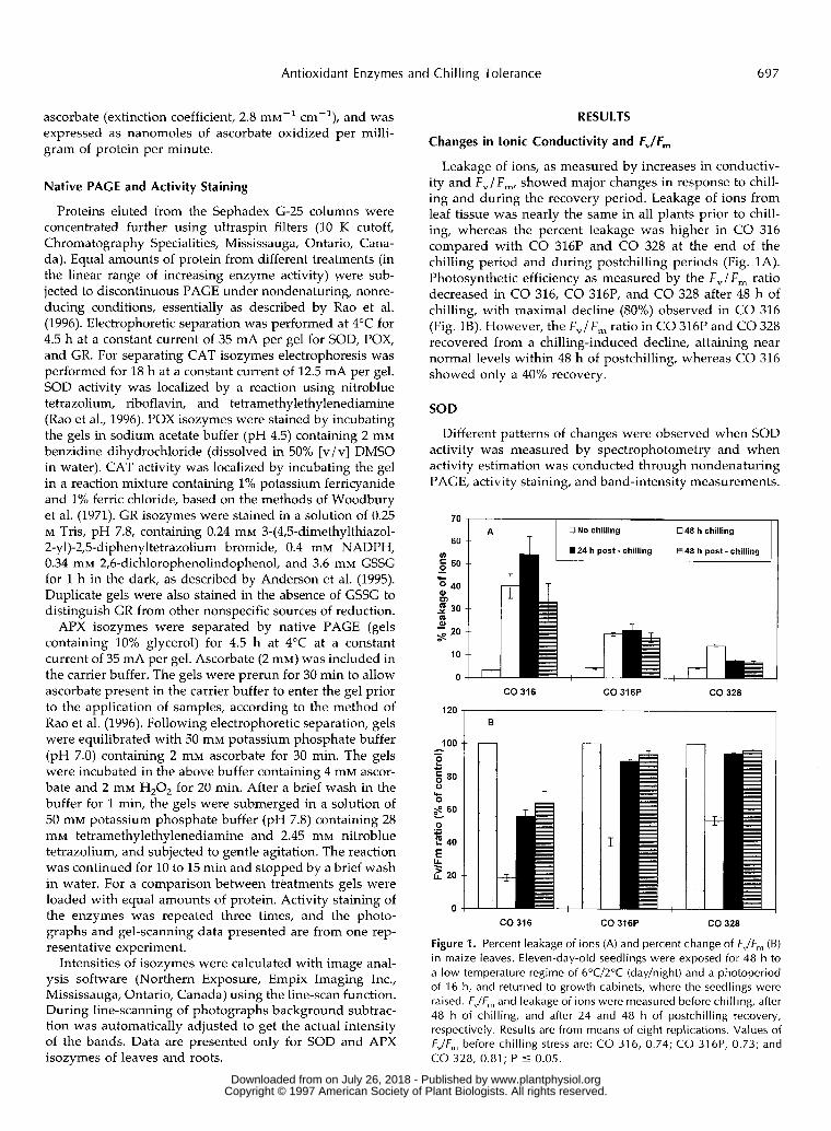
Changes in lonic Conductivity and FJF,
Leakage of ions, as measured by increases in conductiv- ity and F,/F,, showed major changes in response to chill- ing and during the recovery period. Leakage of ions from leaf tissue was nearly the same in all plants prior to chill- ing, whereas the percent leakage was higher in CO 316 compared with CO 316P and CO 328 at the end of the chilling period and during postchilling periods (Fig. 1A). Photosynthetic efficiency as measured by the F,l F , ratio decreased in CO 316, CO 316P, and CO 328 after 48 h of chilling, with maximal decline (80%) observed in CO 316 (Fig. 1B). However, the F,/F, ratio in CO 316P and CO 328 recovered from a chilling-induced decline, attaining near normal levels within 48 h of postchilling, whereas CO 316 showed only a 40% recovery.
SOD
Different patterns of changes were observed when SOD activity was measured by spectrophotometry and when activity estimation was conducted through nondenaturing PAGE, activity staining, and band-intensity measurements.
70
60
50
E 40 m 2 30
- 20 s 10
O
u)
.- 'c
m 0
CO 316 CO 316P CO 328
B
CO 316
Figure 1. Percent leakage of
CO 316P ;O 328
ns (A) and percent change of FJF, (B) in maize leaves. Eleven-day-old seedlings were exposed for 48 h to a low-temperature regime of 6"C/2"C (day/night) and a photoperiod of 16 h, and returned to growth cabinets, where the seedlings were raised. FJF, and leakage of ions were measured before chilling, after 48 h of chilling, and after 24 and 48 h of postchilling recovery, respectively. Results are from means of eight replications. Values of FJF, before chilling stress are: CO 316, 0.74; CO 316P, 0.73; and CO 328, 0.81 ; P 5 0.05.
www.plantphysiol.orgon July 26, 2018 - Published by Downloaded from Copyright © 1997 American Society of Plant Biologists. All rights reserved.

698 Pinhero et al. Plant Physiol. Vol. 114, 1997
Significant differences were observed in SOD activities inleaves of CO 328, CO 316P, and CO 316 prior to andfollowing chilling when assayed by spectrophotometry(Table I). The activity of SOD was lower in the leaves of CO328 compared with CO 316. Paclobutrazol treatment in-creased SOD activity in leaves of CO 316. During chillingand subsequently, leaf SOD activity decreased in CO 316and CO 316P, while the activity in CO 328 remained nearlythe same compared with initial levels. During the post-chilling period a significant increase in leaf SOD activitywas observed in CO 328. Unlike leaves, no significantdifferences were noticed in SOD activities in roots betweenCO 328, CO 316P, and CO 316 prior to chilling (Table I).SOD activity of roots increased significantly in CO 316 andCO 316P during 24 h of chilling. A significant decrease inSOD activity was noticed during 48 h of chilling in CO 328.
Nondenaturing PAGE coupled with activity localizationrevealed four SOD isozymes in leaves of CO 316, CO 316P,and CO 328 (Fig. 2). Incubation of gels in 2 mM potassiumcyanide or 5 mM H2O2 before staining for SOD activityindicated isozymes SOD-1 and SOD-2 to be Mn-SOD, andisozymes SOD-3 and SOD-4 to be Cu,Zn-SOD (data notshown). No Fe-SOD isozymes were observed. SOD-2 andSOD-3 were more prominent in CO 316P compared withCO 316 and CO 328 prior to chilling (Fig. 2, lanes a, e, andi, and Fig. 3). SOD-4 was the most prominent in all threetreatments. Quantification of the band intensities by line-scanning showed that the total intensity due to SOD bandswas nearly double in CO 316P and CO 328 compared withthat in CO 316 prior to chilling, and remained at similarlevels during chilling and postchilling periods (Fig. 3). Bycontrast, the intensity of SOD-4 in CO 316 increased from3.35 before chilling to 7.80 during the 24 h postchilling. The
Table I. Total activity of SOD in partially purified leaf and roottissue homogenates of maize seedlings
One unit of SOD is defined as the quantity of enzyme required toinhibit the reduction of Cyt c by 50% in a 3-mL reaction volume perminute. Treatments were replicated six times.
a b c d e f g h i j k l
TreatmentTotal Activity of SOD (±SE)
Leaf Root
CO 316Control24 h of Chilling48 h of Chilling24 h Postchilling
CO 316PControl24 h of Chilling48 h of Chilling24 h Postchilling
CO 328Control24 h of Chilling48 h of Chilling24 h Postchilling
42.33 ± 1.9036.63 ± 0.71a
34.99 ± 1.40a
41.37 ± 1.41
50.05 ± 1.1 3b
42.49 ± 3.14a
42.33 ± 1.79a
46.25 ± 2.57'1
33.81 ± 1.21h
30.72 ± 1.2732.91 ± 0.9039.34 ± 2.97a
units
47.79 ± 3.0951.34 ± 1.56"47.14 ± 3.4546.49 ± 2.27
49.77 ± 3.8360.03 ± 4.26a
47.18 ± 3.7853.72 ± 1.42
45.97 ± 2.0146.27 ± 2.9840.55 ± 2.75'43.82 ± 0.94
a Significantly different compared with control values within thetreatment group (P ^ 0.01). b Significantly different between thecontrol values of CO 316, CO 316P, and CO 328.
Figure 2. Native gels stained for the activity of SOD in leaves ofmaize inbreds before, during, and after a chilling exposure for 48 hat 6°C/2°C (day/night) and a photoperiod of 16 h. Equal amounts ofprotein (100 ^g) from leaves of seedlings subjected to differenttreatments were loaded on the gel. Lanes a to d, CO 316 unchilledcontrol, 24 h of chilling, 48 h of chilling, and 24 h postchilling,respectively; lanes e to h, CO 316P unchilled control, 24 h ofchilling, 48 h of chilling, and 24 h postchilling, respectively; lanes ito I, CO 328 unchilled control, 24 h of chilling, 48 and 24 hpostchilling, respectively.
intensities of other isozymes also increased in CO 316during 48 h of chilling and 24 h postchilling, attaining theprechilling levels of SOD activity shown by CO 328 and CO316P.
Five isozymes of SOD were observed in roots (Fig. 4), ofwhich SOD-1, SOD-2, and SOD-3 were identified to beMn-SODs and SOD-4 and SOD-5 as Cu,Zn-SOD (data notshown). Isozyme composition was qualitatively similar inall treatments. Total intensity due to SOD isozyme activityin the root was much higher in CO 316P and CO 316 thanin CO 328 (Fig. 5). The intensities of these isozymes de-clined during 48 h of exposure to chilling and during
• SOD 4OSO03• SOD 21• SOD1
IICont 24 h 48 h 24 h Cent 24 h 48 h 24 h Cont 24 h 48 h 24 h
Ch Ch PC Ch Ch PC Ch Ch PCCO 316 CO316P CO 328
Figure 3. Changes in leaf SOD isozyme activities as estimated bynative PAGE, activity staining, and determination of band intensitiesby line-scanning. A photograph (Fig. 2) depicting SOD isozymeactivities of a leaf was scanned using the line-scanning function of acomputer program. Cont, Unchilled control; 24 h Ch, 24 h of chill-ing; 48 h Ch, 48 h of chilling; and 24 h PC, 24 h postchilling. Thedata are representative of two separate experiments showing similarresults. www.plantphysiol.orgon July 26, 2018 - Published by Downloaded from
Copyright © 1997 American Society of Plant Biologists. All rights reserved.

Antioxidant Enzymes and Chilling Tolerance 699
a b c d e f g h i j k l
Figure 4. Native gels stained for the activity of SOD in roots of maizeinbreds. Conditions are the same as described in Figure 2.
postchilling periods in CO 316. However, the activitiesremained similar during chilling and postchilling periodsin CO 316P, and showed slight increases in CO 328. Majorchanges were noticed in SOD-3, SOD-4, and SOD-5.
GR
Spectrophotometric determination of GR activityshowed that maize seedlings derived from paclobutrazol-treated seeds possessed significantly higher GR activity inleaves compared with CO 328 and CO 316 before chilling(Table II). There was an increase in GR activity in CO 316,CO 316P, and CO 328 during chilling and postchillingperiods compared with their respective prechilling levels.In contrast to GR activity in leaves, GR activity in roots wassignificantly higher in CO 328 than in CO 316 and CO 316Pprior to chilling (Table II). No significant difference wasnoticed in root GR activity in CO 316, whereas a decline inthe activity was observed during chilling in CO 316P andCO 328. GR activity was nearly three times higher initially
• SOD 5DSOD4a SOD 3• SOD 2• SOD1
ill!Cont 24 h
Ch48 hCh
24 h Cont 24 hPC Ch
48 hCh
24 h Cant 24 h 48 h 24 hPC Ch Ch PC
CO 328CO 316 CO316P
Figure 5. Changes in root SOD isozyme activities as estimated bynative PAGE, activity staining, and determination of band intensitiesby line-scanning. A photograph (Fig. 4) depicting the SOD isozymeactivities of a root was scanned using the line-scanning function of acomputer program. Cont, Unchilled control; 24 h Ch, 24 h of chill-ing; 48 h Ch, 48 h of chilling; and 24 h PC, 24 h postchilling. Thedata are representative of two separate experiments showing similarresults.
Table II. Total activity of CR in leaf and root tissue preparations ofmaize seedlings
Treatments were replicated six times. GR activity was monitoredfollowing the decrease in NADPH at A340 nm.
TreatmentTotal Activity of GR (±SE)
Leaf Root
CO 316Control24 h of Chilling48 h of Chilling24 h Postchilling
C0316PControl24 h of Chilling48 h of Chilling24 h Postchilling
CO 328Control24 h of Chilling48 h of Chilling24 h Postchilling
nmol
37.83 ± 1.5752.67 ± 1.62a
63.50 ± 3.1T95.33 ± 4.65a
51.50 ± 1.36b
67.17 ± 2.86a
64.50 ± 2.01"69.67 ± 3.21'1
33.83 ± 1.3050.33 ± 2.67a
54.33 ±3.71a
48.83 ± 3.22a
NADPH
131.17 ± 13.62128.33 ± 7.38127.50 ± 11.24121.67 ± 2.55
140.83 ± 7.21127.67 ± 8.1097.00 ± 7.42"
116.33 ± 8.69
240.17 ± 18.38b
145.50 ± 4.43a
127.67 ± 16.16a
201.83 ±11.25a Significantly different compared with control values within the
treatment group (P £ 0.01). h Significantly different between thecontrol values of CO 316, CO 316P, and CO 328.
in roots of CO 316 and CO 316P compared with theirrespective activities in leaves (Table II). GR activity was7-fold higher in roots of CO 328 compared with the activityin leaves (Table II).
Examination of GR isozyme profiles in leaves revealedthree GR isozymes (Fig. 6). Most of the activity appearedto be due to GR-3, which showed much more intenseactivity than GR-1 and GR-2 (Fig. 6, lanes a-I). Addition-ally, higher staining activity was observed in CO 316P(due to isozyme 3) compared with that in CO 316 and CO328 (Fig. 6, lanes a-1).
Paclobutrazol treatment did not cause any major changesin isozyme profiles or intensities of GR in roots of CO 316(Fig. 7). Three isozymes (GR-1, GR-2, and GR-4) were re-vealed by the activity staining in root preparations of CO316 and CO 316P before chilling (Fig. 7, lanes a and e).Activities due to GR isozymes were considerably lower inCO 316 and CO 316P. Substantially higher GR-stainingactivity could be deciphered due to GR-2 and GR-3 in CO328. GR-3 was predominant prior to, during, and afterchilling in CO 328. Isozyme profiles did not undergo majorchanges during chilling (24 or 48 h) or after chilling in CO316. Additionally, staining activity due to GR-3 could be
a b c d e f g h i j k I
Figure 6. Native gels stained for the activity of GR in leaves of maizeinbreds. Equal amounts of protein (200 ig) from leaves of seedlingssubjected to different treatments were loaded on the gel. The order oflanes is the same as described in Figure 2. www.plantphysiol.orgon July 26, 2018 - Published by Downloaded from
Copyright © 1997 American Society of Plant Biologists. All rights reserved.

700 Pinhero et al. Plant Physiol. Vol. 114, 1997
a b c d e f g h i j k l
Figure 7. Native gels si,lined for the activity of CR in roots of maizeinbreds. Equal amounts of protein (100 /ig) from roots of seedlingssubjected to different treatments were loaded on the gel. The order oflanes is the same as described in Figure 2. Note that isozyme 3,marked by * is noticeable only in CO 328 and in CO 316P duringthe 24-h postchilling period.
observed in CO 316P during the postchilling period. How-ever, this activity could be masked by a nonspecific bandlocated adjacent to it (marked by an arrowhead) duringpre- and postchilling periods (Fig. 7, lanes, e, f, g, and h).Because of higher background staining and nonspecificbands, it was difficult to estimate the band intensities thatcorresponded to specific GR activity by scanning the gelphotographs accurately.
APX
Estimation of APX activity by spectrophotometry alsoshowed a varying pattern compared with the respectiveactivity-stained preparations on gels. Spectrophotometricanalyses showed that APX activity in leaves of CO 316Pwas high compared with that of CO 316 and CO 328 beforechilling (Table III). APX activity increased during and after
Table III. APX activities of leaf and root tissue preparations ofmaize seedlings
Treatments were replicated four times. APX activities were esti-mated by following the oxidation of ascorbate and monitored as theUCLiedSt; Ml A\290-
TreatmentTotal Activity of APX (±SE)
Leaf Rootnmol of ascorbate
CO 316Control24 h of Chilling48 h of Chilling24 h Postchilling
CO 31 6PControl24 h of Chilling48 h of Chilling24 h Postchilling
CO 328Control24 h of Chilling48 h of Chilling24 h Postchilling
169.0 ± 7.T1
220.6 ± 30. 2h
239.9 ± 2.9b
202.3 ± 2.4h
203.2 ± 21. 2'1
249.9 ± 2.4b
275.9 ± 32.8b
278.5 ± 11. 9b
106.2 ± 7.1a
139.3 ± 13. 3b
123.8 ± 8.3b
152.2 ± 3.0b
1307.7 ± 64.81017.0 ± 97.0b
1021.7 ± 7.5h
702.5 ± 30.5b
1475.7 ± 4.6a
1439.0 ± 90.61625.6 ± 26. 2b
1390.7 ± 71.5
1359.0 ± 60.9700.0 ± 10.0b
947.0 ± 90.5b
872.2 ± 19. 1b
chilling in CO 316, CO 316P, and CO 328. Paclobutrazoltreatment resulted in increased APX activity in roots of CO316 compared with that of CO 328 and untreated CO 316(Table III). An increase in APX activity was noticed in CO316P exposed to 48 h of chilling. But APX activity declinedin CO 316 and CO 328 during and after chilling (Table III).Nearly 7- to 12-fold higher activity was observed in APXactivity of roots prior to chilling compared with the activityin leaves of CO 316, CO 316P, and CO 328.
Examination of APX isozymes revealed four isozymes inleaves of CO 316 and CO 316P before chilling exposure(Fig. 8, lanes a and e), whereas only APX-1 was observed inCO 328 before chilling (Fig. 8, lane i). APX activity asrevealed by the staining intensity was generally high in CO316P. Considerable increases in the staining intensities ofall of the isozymes (APX-1, APX-2, APX-3, and APX-4)were observed during and after chilling in CO 316P; thehighest increase was observed in APX-1, APX-3, and APX-4(Fig. 8, lanes e, f, g, and h, and Fig. 9). By contrast, CO 316showed only a marginal increase in activity due to APXisozymes. An increase in staining intensity due to APX-1was noticed in leaves of CO 328 during chilling (Fig. 8,lanes, j, k, and 1, and Fig. 9). APX-2, APX-3, and APX-4appeared during chilling and increased during postchillingin CO 328 (Fig. 8, lanes, i, j, k, and 1, and Fig. 9).
Zymograms of roots of CO 316 and CO 316P revealedfour isozymes that appeared to be similar (Fig. 10, 2-5)before chilling (Fig. 10, lanes a and e), whereas in CO 328five bands were observed before chilling (Fig. 10, lane i,1-5). In general, activity due to APX-2, APX-3, APX-4, andAPX-5 increased during and after chilling in CO 316P (Fig.10, lanes e, f, g, and h, and Fig. 11). APX-5 appeared toincrease in intensity during the postchilling period in CO328 (Fig. 10, lane 1, and Fig. 11). No major changes wereobserved in the isozyme composition of CO 316. CO 316and CO 316P also possessed staining activity, presumablydue to several slow-moving isozymes (Fig. 10, indicated bybroad, open arrow).
In addition to SOD, GR, and APX, the changes in activ-ities and isozyme profiles of POX and CAT in CO 316, CO316P, and CO 328 were studied. In general, POX activities
a b
a Significantly different between the control values of CO 316, CO316P, and CO 328. b Significantly different compared with con-trol values within the treatment group (P £ 0.01).
Figure 8. Native gels stained for the activity of APX in leaves ofmaize inbreds. Equal amounts of protein (200 /ng) from leaves ofseedlings subjected to different treatments were loaded on the gel.The order of lanes are the same as described in Figure 2. Note thatAPX-2, APX-3, and APX-4 are absent in the CO 328 unchilled controland that they appear during chilling. www.plantphysiol.orgon July 26, 2018 - Published by Downloaded from
Copyright © 1997 American Society of Plant Biologists. All rights reserved.

Antioxidant Enzymes and Chilling Tolerance 701
70
£ 60o
f soJ•S 40
01 30
20
IAPX1 DAPX2 + 3 + 4
Cont 24 h 48 h 24 h Conf 24 h 48 h 24 h Cont 24 h 48 h 24 hCh Ch PC Ch Ch PC Ch Ch PC
CO 316 CO316P CO 328
Figure 9. Changes in leaf APX activities as determined by nativePAGE, activity staining, and determination of band intensities byline-scanning. A photograph (Fig. 8) of the native gel showing activ-ities of a leaf was scanned using the line-scanning function of acomputer program. APX-2, APX-3, and APX-4 were analyzed to-gether, since it was difficult to separate activities of individualisozymes in this cluster. Cont, Unchilled control; 24 h Ch, 24 h ofchilling; 48 h Ch, 48 h of chilling; and 24 h PC, 24 h postchilling. Thedata are representative of two separate experiments showing similarresults.
were similar in CO 316, CO 328, and CO 316P. CAT activitywas nearly equal in CO 316 and CO 316P and highercompared with CO 328. There were no marked changes inthe activities of POX and CAT during chilling or postchill-ing periods. Additionally, the isozyme profiles of theseenzymes also remained nearly the same during and afterchilling (data not shown).
DISCUSSION
It has been suggested that a tolerant plant might usethree different mechanisms to survive or even grow at
a b
Figure 10. Native gels stained for the activity of APX in roots ofmaize inbreds. Equal amounts of protein (100 /xg) from roots ofseedlings subjected to different treatments were loaded on the gel.The order of lanes is the same as described in Figure 2. Note thatisozyme 1, marked by *, is present only in CO 328. The broad, openarrow indicates the position of several slow-moving isozymes of APXin CO 316 and CO 316P.
Cant 24 h 48 h 24 h Cont 24 h 48 h 24 h Cont 24 h 48 h 24 hCh Ch PC Ch Ch PC Ch Ch PCCO 316 CO 328
Figure 11. Changes in root APX activities as determined by nativePAGE, activity staining, and determination of band intensities byline-scanning. A photograph (Fig. 10) of the native gel showingactivities of a root was scanned using the line-scanning function of acomputer program. APX-3, APX-4, and APX-5 were analyzed to-gether, since it was difficult to separate activities of individualisozymes in this cluster in the later stages of sampling. Cont, Un-chilled control; 24 h Ch, 24 h of chilling; 48 h Ch, 48 h of chilling;and 24 h PC, 24 h postchilling. The data are representative of twoseparate experiments showing similar results.
chilling temperatures (Steffen, 1991): (a) it may avoid theproduction of AOS, (b) it may protect itself from thedeleterious, degradative reactions associated with AOSgeneration by efficiently scavenging them, or (c) it mayrepair the injury after the degradation has occurred. Thechilling-tolerant tomato (Lycopersicon hirsutum) does notpossess an inherently higher antioxidant potential thanthe chilling-susceptible Lycopersicon esculentum, except inthe activities of GR; this chilling tolerance has been attrib-uted to the control of electron transport during chilling(Walker et al., 1991). This suggests that chilling tolerancecould be achieved through several adaptive mechanisms,and the modulation of antioxidant enzyme levels could beonly a part of the whole mechanism.
Fv/Fm has been widely used as a good criterion to assesschilling sensitivity in various crops (Wilson and Greaves,1990). Analysis of chilling damage and subsequent recov-ery as shown by Fv/Fm and ion leakage indicates that CO316P and untreated CO 328 sustained a lower degree ofchilling damage compared with untreated CO 316. Ourresults show interesting differences in the manner in whichantioxidant enzymes undergo changes in response to chill-ing in these three experimental systems studied. Estimationof SOD activity in partially purified enzyme extracts of CO316 showed a significant decrease in leaf SOD activityduring the chilling period; root SOD activity did not un-dergo major changes during chilling. However, the estima-tion of SOD activity in crude extracts is subject to a higherdegree of variability due to interfering reactions such asCyt oxidase and Cyt peroxidase (Beauchamp and Fridov-ich, 1971). Also, assay of SOD activity in crude leaf extractsof rice, papaya, and tobacco results in an overestimation ofactivity compared with gel electrophoresis of proteins, ac-tivity staining, and densitometric quantitation (Chen and www.plantphysiol.orgon July 26, 2018 - Published by Downloaded from
Copyright © 1997 American Society of Plant Biologists. All rights reserved.

702 Pinhero et al. Plant Physiol. Vol. 114, 1997
Pan, 1996). Therefore, we relied more on the estimation of activity by gel electrophoresis and activity staining than on spectrophotometric estimation of the activity of crude preparations in our interpretation of results. By this method total leaf SOD activity in CO 316 was estimated to be nearly one-half that of CO 328 and CO 316P prior to chilling (Fig. 3). The increase in leaf SOD activity during the latter part of the chilling and postchilling periods in CO 316 appears to be too late to afford any protective role. Higher levels of leaf SOD activity observed in CO 316P and CO 328 throughout the chilling and postchilling periods may contribute to their chilling tolerance. Additionally, root SOD activity showed a decline (Fig. 5) in CO 316 during the chilling and postchilling periods. Root SOD activity was inherently high in CO 316P, which enhanced its chilling tolerance capacity. These results are consistent with previous observations of increased SOD activity and tolerance to oxidative stresses (Bowler et al., 1991; McKer- sie et al., 1993; Pitcher and Zilinskas, 1996).
Synthesis of new isozymes of antioxidant enzymes with altered kinetic properties may be more beneficia1 for the metabolism of AOS than mere enhancement of the activi- ties of existing antioxidant enzymes (Edwards et al., 1994; Rao et al., 1996). Isozyme profiles of SOD varied in CO 316, CO 316P, and CO 328 in response to chilling exposure and during the postchilling period. Constitutive levels of Mn- SOD (SOD-1 and SOD-2) were higher in the leaves of CO 316P (Figs. 2 and 3) compared with those of CO 316 and CO 328. Additionally, SOD-2 increased in CO 328 during the chilling and postchilling periods. In maize Mn-SODs are reported to be localized in mitochondria, whereas Cu,Zn- SODs are associated with the cytosol or chloroplast (Baum and Scandalios, 1979). It has been suggested that during exposure to low temperatures the normal mitochondrial electron transport might be disrupted, causing the produc- tion of AOS (Prasad et al., 1994; Purvis et al., 1995). There- fore, higher levels of Mn-SOD activity in CO 316P and CO 328 are related to the protection of mitochondria during chilling exposure. Similarly, the greater induction of SOD-3 and SOD-4 (Cu,Zn-SOD) in SO 328 during the chilling and postchilling periods could have helped CO 328 to prevent damages due to any AOS formed in chloroplast.
GR is an important enzyme involved in the enzymatic detoxification of AOS (Fig. 12) and contributes to the maintenance of a higher GSH to GSSG ratio. GSH is an efficient scavenger of AOS (Alscher, 1989). Inherently higher levels of GR activity were observed in the roots of CO 328. GR activity was much lower in CO 316, CO 316P, and CO 328 leaves compared with roots. Even though leaf GR activity increased during the chilling and postchilling periods in CO 316 (Table 11), this does not appear to have contributed to the chilling tolerance. This is perhaps due to the limiting SOD activity in the leaves, which showed an increase only during the postchilling period. Also, it is likely that higher levels of GR activity in roots of CO 328 lead to an increased translocation of GSH from the roots to the leaves, which could contribute to enhanced scav- enging of AOS and to providing chilling tolerance (Ren- nenberg, 1982). It has also been suggested that increased
Figure 12. Schematic diagram showing the scavenging pathway of AOS by antioxidant enzymes. The superoxide anion radical (O,-) is dismutated to H,O, by SOD. APX, POX, and CAT metabolize H,O, to water. APX metabolizes H,O, to water by oxidizing ASA to MDHA and/or DHA. MDHA reductase reduces MDHA to ASA, or MDHA is spontaneously converted to DHA. DHA reductase converts DHA to ASA, along with the oxidation of GSH to CSSC. GSH is regenerated by GR, utilizing NADPH as the reductant. Alternate sources of 0,- include the mitochondrial electron transport system and NADP/NADPH oxidase activity.
synthesis of GR isozymes with higher affinities toward GSSG could be an alternate mechanism to keep higher GSH levels in the tissue (Edwards et al., 1994; Anderson et al., 1995). Potentially, higher levels of GR activity in leaves of CO 316P (GR-3) and the presence of an addi- tional isozyme (GR-3) in roots of CO 328 and CO 316P will enhance the adaptive mechanisms that result in chilling tolerance in these systems.
APXs are chloroplastic or cytosolic enzymes that act in tandem with SOD to scavenge H,O, generated through SOD action (Fig. 12). APX activities were higher in roots than in leaves of CO 316, CO 316P, and CO 328. Changes in isozyme composition of APX occurred in a cluster of fast-moving isozymes numbered APX-2, APX-3, and APX-4 in leaves and APX-2, APX-3, APX-4, and APX-5 in roots. The large increase in APX activity in leaves and roots in CO 316P contributed to its chilling tolerance. Surprisingly, leaf APX activity due to APX-2, APX-3, and APX-4 were not detectable in CO 328 before chilling. These isozymes appeared only during the chilling and postchilling periods. Thus, increases in APX activity in conjunction with higher GR activity of roots appear to contribute to the chilling tolerance exhibited by CO 328. CAT and peroxidase activities were generally high in CO 316 and CO 316P and do not appear to be limiting factors in the scavenging of AOS.
Among the leaves and roots tested we found inherently higher activities of SOD, GR, and APX in roots, irrespec- tive of treatments (Tables I, 11, and 111). Like leaves, roots are exposed to environmental stresses such as drought, chilling, flooding, and pathogen attack, which cause the generation of AOS. It has been suggested that in dark- grown seedlings or in nonphotosynthetic tissues, mito- chondria are the primary source of AOS generation (Pun- tarulo et al., 1991). Even though the potential contribution of the roots toward chilling tolerance of the whole plant is not fully understood, roots may influence the chilling
www.plantphysiol.orgon July 26, 2018 - Published by Downloaded from Copyright © 1997 American Society of Plant Biologists. All rights reserved.
Antioxidant Enzymes and Chilling Tolerance 703
tolerance of aerial portions by supplying chemical anti- oxidants such as GSH through increased GR activity (Ren- nenberg, 1982). Increased activities of GR i n roots, espe- cially in CO 328, is i n agreement wi th this contention.
Differential modulation of antioxidant isozymes and their activity are important features that contribute to the chilling tolerance of maize seedlings. It appears that the mechanism of chilling tolerance in the genetically chilling- tolerant C O 328 and chilling-sensitive C O 316P involve the use of similar antioxidant enzyme systems, bu t i n differing proportions. A combination of the GSH cycle and the SOD- APX pathway thus contributes to the chilling-tolerance mechanism of C O 328. The predominance of the SOD and APX activities i n CO 316P suggests that the SOD-APX pathway contributes much toward the chilling tolerance exhibited by these seedlings.
It has also been suggested that the cyanide-insensitive alternative pathway of respiration reduces the potential production of AOS i n mitochondria. Cold-resistant culti- vars and tissues generally develop a greater potential of electron flux through alternative pathways than the cold- sensitive cultivars and tissues (Purvis, 1985; Purvis e t al., 1995). In addition, it is possible that other mechanisms responsible for stabilizing the membranes may contribute to the chilling tolerance of CO 328.
ACKNOWLEDCMENTS
The authors wish to thank Dr. Bob Hamilton of Plant Research Centre, Agriculture and Agri-Food Canada, Ottawa, for kindly supplying the seeds of the two inbreds; Brad Smith of Zeneca Corp., Stony Creek, Ontario, Canada, for a generous supply of paclobutrazol; and Dr. Bryan McKersie, Department of Crop Sci- ence, University of Guelph, for providing facilities to scan the gel photographs.
Received December 9, 1996; accepted March 4, 1997. Copyright Clearance Center: 0032-0889/97/ 114/0695/ 10.
LITERATURE ClTED
Allen RD (1995) Dissection of oxidative stress tolerance using transgenic plants. Plant Physiol 107: 1049-1054
Alscher RG (1989) Biosynthesis and antioxidant function of glu- tathione in plants. Physiol Plant 77: 457-464
Anderson MD, Prasad TK, Stewart CR (1995) Changes in isozyme profiles of catalase, peroxidase, and glutathione reductase dur- ing acclimation to chilling in mesocotyls of maize seedlings. Plant Physiol 109: 1247-1257
Baum JA, Scandalios JG (1979) Developmental expression and intracellular localization of superoxide dismutases in maize. Differentiation 13: 133-140
Beauchamp C, Fridovich I (1971) Superoxide dismutase: im- proved assays and an assay applicable to acrylamide gels. Anal Biochem 44: 276-287
Bowler C, Slooten L, Vandenbranden S, De Rycke R, Botterman J, Sybesma C, Van Montagu M, Inze D (1991) Manganese superoxide dismutase can reduce cellular damage mediated by oxygen radicals in transgenic tobacco. Plant Physiol97: 452457
Bradford MM (1976) A rapid and sensitive method for the quan- titation of microgram quantity of protein utilizing the principle of protein-dye binding. Anal Biochem 72: 248-254
Chen CN, Pan SM (1996) Assay of superoxide dismutase activity by combining electrophoresis and densitometry. Bot Bull Acad Sin 37: 107-111
Davis TD, Curry EA (1991) Chemical regulation of vegetative growth. Crit Rev Plant Sci 1 0 204-216
Edwards EA, Enard C, Creissen GP, Mullineaux PM (1994) Syn- thesis and properties of glutathione reductase in stressed peas. Planta 192: 137-143
Foyer CH, Descourvieres P, Kunert KJ (1994) Protection against oxygen radicals: an important defence mechanism studied in transgenic plants. Plant Cell Environ 17: 507-523
Kraus TE, Evans RC, Fletcher RA, Pauls KP (1995) Paclobutrazol enhances tolerance to increased levels of UV-B radiation in soybean (Glycine max) seedlings. Can J Bot 73: 797-806
Kraus TE, Fletcher RA (1994) Paclobutrazol protects wheat seed- lings from heat and paraquat injury: is detoxification of active oxygen involved? Plant Cell Physiol 35: 45-52
Lurie S, Lipsker RZ, Aloni B (1994) Effects of paclobutrazol and chilling temperatures on lipids, antioxidants and ATPase activ- ity of plasma membrane isolated from green bell pepper fruits. Physiol Plant 91: 593-598
Malan C, Greyling MM, Gressel J (1990) Correlation between Cu,Zn superoxide dismutase and glutathione reductase and environmental and xenobiotic stress tolerance in maize inbreds. Plant Sci 69: 157-166
McKersie BD, Chen Y, Debeus M, Bowley SR, Bowler C, Inze D, D’Halluin K, Botterman, K (1993) Superoxide dismutase en- hances tolerance to freezing stress in transgenic alfalfa (Medicago sativa L.). Plant Physiol 103: 1155-1163
McKersie BD, Leshem YY (1994) Stress and stress coping in cultivated plants. Kluwer Academic Publishers, Dordrecht, The Netherlands, pp 79-100
Miedema P, Post J, Groot, PJ (1987) The Effects of Low Temper- ature on Seedling Growth of Maize Genotypes. Centre for Ag- ricultural Publishing and Documentation, Wageningen, The Netherlands, pp 30-68
Paliyath G, Droillard MJ (1992) The mechanism of membrane deterioration and disassembly during senescence. Plant Physiol Biochem 30: 789-812
Paliyath G, Fletcher RA (1995) Paclobutrazol treatment alters peroxidase and catalase activities in heat-stressed maize coleop- tiles. Physiol Mo1 Biol Plants 1: 171-178
Pinhero RG, Fletcher RA (1994) Paclobutrazol and ancymidol protect corn seedlings from high and low temperature stresses. Plant Growth Reg 15: 47-53
Pitcher LH, Zilinskas BA (1996) Overexpression of copper/zinc superoxide dismutase in the cytosol of transgenic tobacco con- fers partia1 resistance to ozone-induced foliar necrosis. Plant Physiol 110: 583-588
Prasad TK, Anderson MD, Martin BA, Stewart CR (1994) Evi- dence for chilling-induced oxidative stress in maize seedlings and a regulatory role for hydrogen peroxide. Plant Cell 6: 65-74
Puntarulo S, Galleano M, Sanchez RA, Boveris A (1991) Super- oxide anion and hydrogen peroxide metabolism in soybean embryonic axes during germination. Biochim Biophys Acta 1074: 277-283
Purvis AC (1985) Low temperature induced azide-insensitive ox- ygen uptake in grapefruit flavedo tissue. J Am SOC Hortic Sci 110: 782-785
Purvis AC, Shewfelt RL, Gegogeine JW (1995) Superoxide pro- duction by mitochondria isolated from green bell pepper fruit. Physiol Plant 94: 743-749
Rao MV, Paliyath G, Ormrod DP (1996) Ultraviolet-B- and ozone- induced biochemical changes in antioxidant enzymes of Avabi- dopsis thaliana. Plant Physiol 110: 125-136
Rennenberg H (1982) Glutathione metabolism and possible bio- logical roles in higher plants. Phytochemistry 21: 2771-2781
Scandalios JG (1990) Response of plant antioxidant defence genes to environmental stress. Adv Genet 28: 1-41
Scandalios JG (1993) Oxygen stress and superoxide dismutases. Plant Physiol 101: 7-12
Sen Gupta A, Heinen JL, Holaday AS, Burke JJ, Allen RD (1993)
www.plantphysiol.orgon July 26, 2018 - Published by Downloaded from Copyright © 1997 American Society of Plant Biologists. All rights reserved.

704 Pinhero et al. Plant Physiol. Vol. 114, 1997
Increased resistance to oxidative stress in transgenic plants that overexpress chloroplastic Cu / Zn superoxide dismutase. Proc Natl Acad Sci USA 90: 1629-1633
Shaaltiel Y, Gressel J (1986) Multienzyme oxygen radical detoxi- fying system correlated with paraquat resistance in Conyza bonaviensis. Pestic Biochem Physiol 26: 22-28
Steffen KL (1991) Avoidance of photooxidative stress: balancing energy flux within the chloroplast. In E Pell, KL Steffen, eds, Active Oxygen/Oxidative Stress and Plant Metabolism. Current Topics in Plant Physiology, Vol 6. American Society of Plant Physiologists, Rockville, MD, pp 119-130
Thompson JE, Legge RL, Barber RF (1987) The role of free radicals in senescence and wounding. New Phytol 105: 317-344
Upadhyaya A, Davis TD, Walser RH, Galbraith AB, Sankhla N (1989) Uniconazole-induced alleviation of low temperature damage in relation to antioxidant activity. Hortic Sci 24: 955-957
Ushimaru T, Ogawa K, Ishida N, Shibasaka M, Kanematsu S,
Asada K, Tsuji H (1995) Changes in organelle superoxide dis- mutase isozymes during air adaptation of submerged rice seed- lings: differential behaviour of isozymes in plastids and mito- chondria. Planta 196: 606-613
Walker MA, McKersie BD, Pauls KP (1991) Effects of chilling on the biochemical and functional properties of thylakoid mem- branes. Plant Physiol 97: 663-669
Wilson JM, Greaves JA (1990) Assessment of chilling sensitivity by chlorophyll fluorescence analysis. In CY Wang, ed, Chilling Injury of Horticultural Crops. CRC Press, Boca Raton, FL, pp 129-141
Wise RR, Naylor AW (1987) Chilling-enhanced photooxidation. . The peroxidative destruction of lipids during chilling injury to photosynthesis and ultrastructure. Plant Physiol 83: 272-277
Woodbury W, Spencer AK, Stahmann MA (1971) An improved procedure using ferricyanide for detecting catalase isozymes. Ana1 Biochem 4 4 301-305
www.plantphysiol.orgon July 26, 2018 - Published by Downloaded from Copyright © 1997 American Society of Plant Biologists. All rights reserved.