1 2 Chromosome Structure 3 Chromosome Number 4 Prokaryotic Cell Division.
Upload
pratheep-sandrasaigaranCategory
view
130download
3
Copyright © 2009 Pearson Education, Inc.
2.0 Chromosome structure
Prepared by Pratheep SandrasaigaranLecturer at Manipal International University
Copyright © 2009 Pearson Education, Inc.
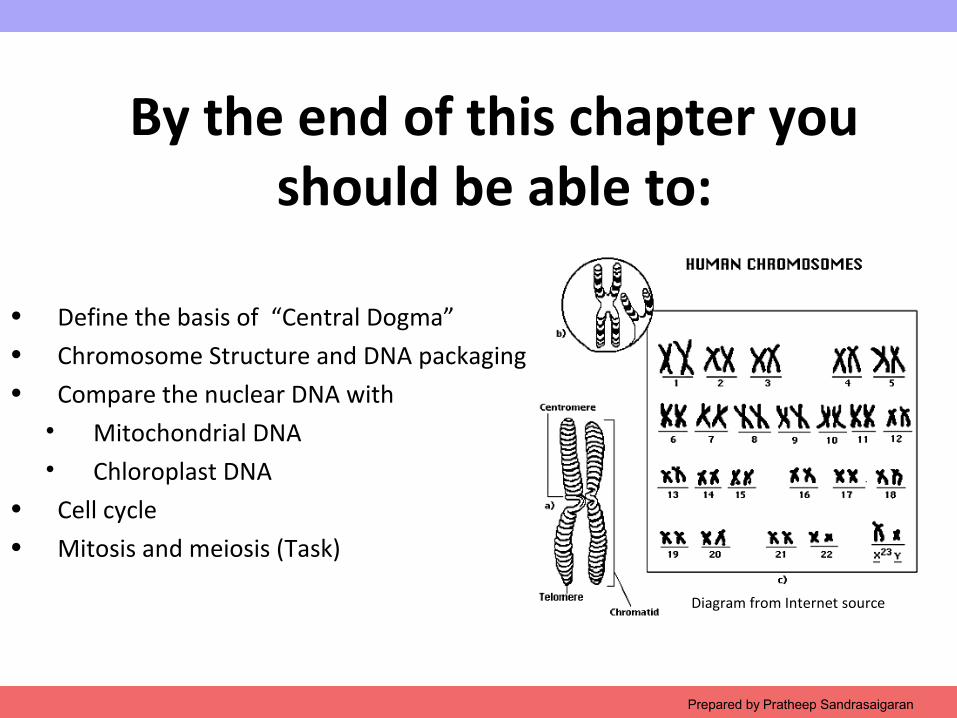
By the end of this chapter you should be able to:
• Define the basis of “Central Dogma”• Chromosome Structure and DNA packaging• Compare the nuclear DNA with
• Mitochondrial DNA• Chloroplast DNA
• Cell cycle • Mitosis and meiosis (Task)
Prepared by Pratheep Sandrasaigaran
Diagram from Internet source
Copyright © 2009 Pearson Education, Inc.
2.1 The Central Dogma
Copyright © 2009 Pearson Education, Inc.
The “Central Dogma”• DNA functions primarily by directing the production
of proteins.
• Each DNA molecule can carry thousands of genes which in return plan for building a particular protein, or part of a particular protein.
• The type of produced decides everything about a cell and eventually the human body as a whole:• The color of the hair and skin• Propensity to certain diseases• Unique ability• Fat, thin, tall, short, bold, hairy, even for the
matter of ‘long or short’….
Copyright © 2009 Pearson Education, Inc.
The “Central Dogma”
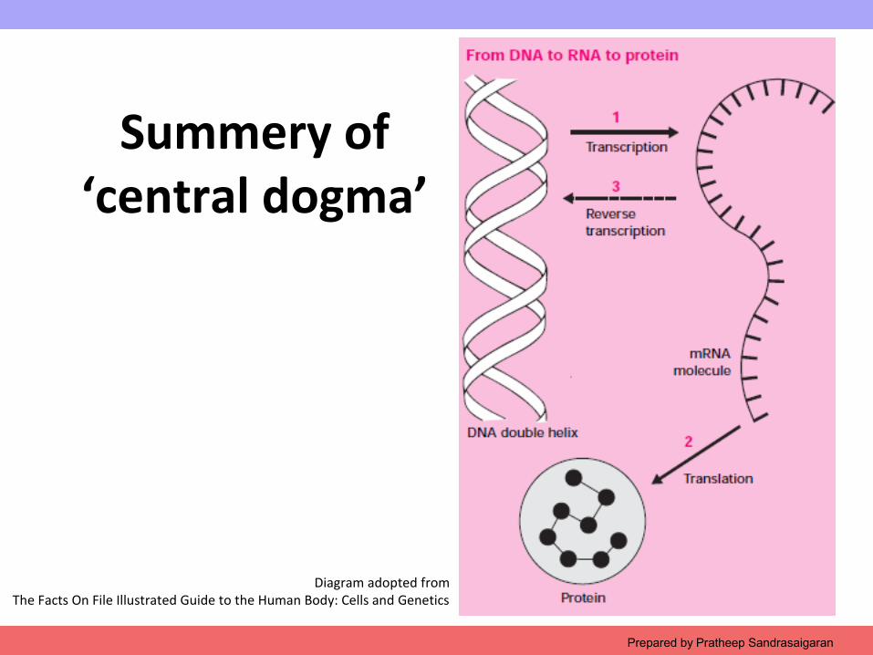
• The “Central dogma” describes the flow of genetic information from DNA to protein.
• DNA makes ribonucleic acid (RNA), which coordinate polypeptide/ protein synthesis.
• This involves two main stages
a. Transcription (mRNA)
b. Translation (Polypeptide)
Copyright © 2009 Pearson Education, Inc.
Transcription
• The genetic information, the nucleotide sequence carried by one strand of the DNA helix is transcribed onto an mRNA (messenger RNA).
• The process of ‘making’ RNA from DNA
Copyright © 2009 Pearson Education, Inc.
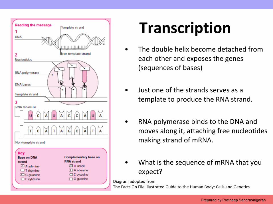
Transcription• The double helix become detached from
each other and exposes the genes (sequences of bases)
• Just one of the strands serves as a template to produce the RNA strand.
• RNA polymerase binds to the DNA and moves along it, attaching free nucleotides making strand of mRNA.
• What is the sequence of mRNA that you expect?
Diagram adopted fromThe Facts On File Illustrated Guide to the Human Body: Cells and Genetics
Copyright © 2009 Pearson Education, Inc.
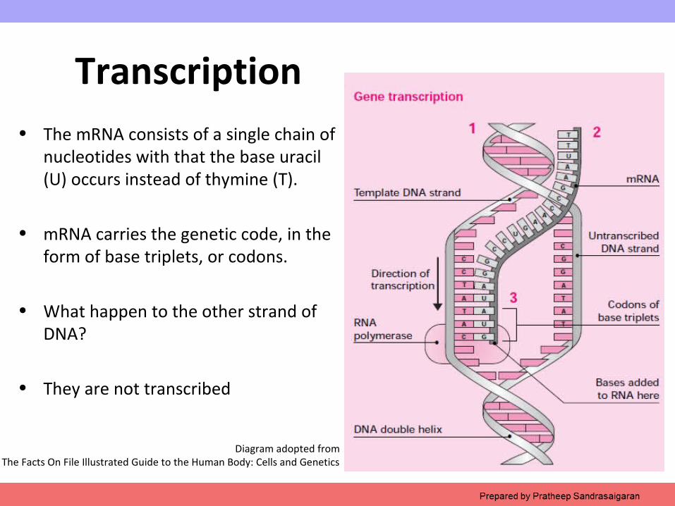
Transcription• The mRNA consists of a single chain of
nucleotides with that the base uracil (U) occurs instead of thymine (T).
• mRNA carries the genetic code, in the form of base triplets, or codons.
• What happen to the other strand of DNA?
• They are not transcribed
Diagram adopted fromThe Facts On File Illustrated Guide to the Human Body: Cells and Genetics
Copyright © 2009 Pearson Education, Inc.Prepared by Pratheep Sandrasaigaran
Translation
• The base sequence of each mRNA molecule is “read” by a ribosome.
• What is Ribosome?
• A third type of RNA, called transfer RNA (tRNA), brings amino acids to the assembly site.
• As the mRNA molecule feeds through the ribosome, amino acids are added to the end of the growing polypeptide chain.
Copyright © 2009 Pearson Education, Inc.
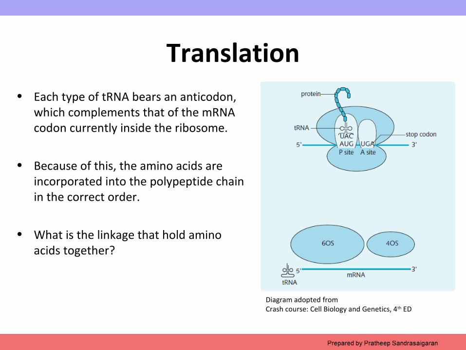
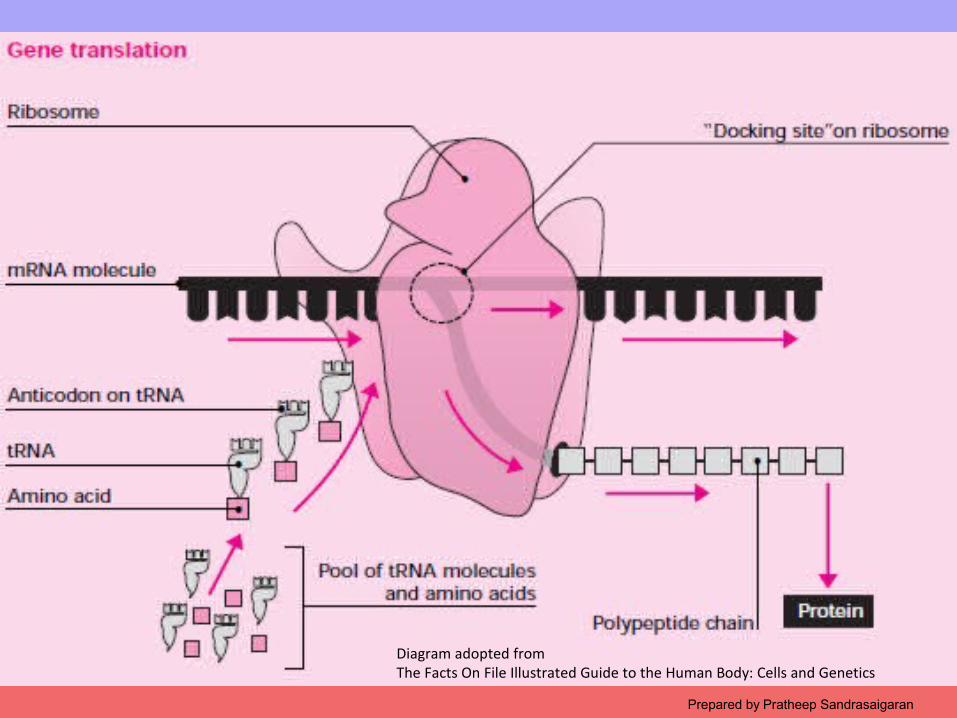
Translation• Each type of tRNA bears an anticodon,
which complements that of the mRNA codon currently inside the ribosome.
• Because of this, the amino acids are incorporated into the polypeptide chain in the correct order.
• What is the linkage that hold amino acids together?
Diagram adopted fromCrash course: Cell Biology and Genetics, 4th ED
Copyright © 2009 Pearson Education, Inc.
Diagram adopted fromThe Facts On File Illustrated Guide to the Human Body: Cells and Genetics
Prepared by Pratheep Sandrasaigaran
Copyright © 2009 Pearson Education, Inc.
Summery of ‘central dogma’
Prepared by Pratheep Sandrasaigaran
Diagram adopted fromThe Facts On File Illustrated Guide to the Human Body: Cells and Genetics
Copyright © 2009 Pearson Education, Inc.
TEST YOUR KNOWLEDGE 1
Prepared by Pratheep Sandrasaigaran
Copyright © 2009 Pearson Education, Inc.
Define the process of transcription. Where does this process fit into the central dogma of molecular genetics (DNA makes RNA makes protein)?
Prepared by Pratheep Sandrasaigaran
Copyright © 2009 Pearson Education, Inc.Prepared by Pratheep Sandrasaigaran

Copyright © 2009 Pearson Education, Inc.
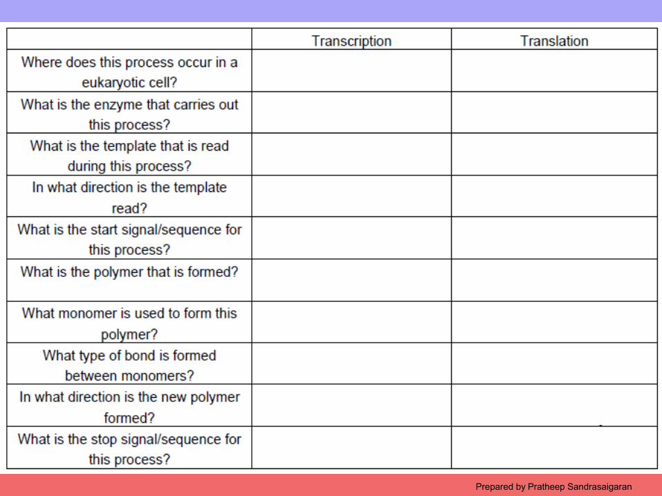
What are the similarities and differences between transcription and DNA replication?
Prepared by Pratheep Sandrasaigaran
Copyright © 2009 Pearson Education, Inc.
2.2 Chromosome Structure and DNA packaging
Prepared by Pratheep Sandrasaigaran
Copyright © 2009 Pearson Education, Inc.
Introduction
• A gene provides each cell in the body with instructions for making (synthesizing) a particular protein (or part of a protein).
• Each protein, either directly or indirectly, determines a particular measurable characteristic or cell function.
• Proteins are formed either as enzymes or structural proteins.
• What is the function of enzyme and structural protein?
Prepared by Pratheep Sandrasaigaran
Copyright © 2009 Pearson Education, Inc.
Where and when genes are active?
• Except for sex cells, all human cells contain a set of about 30,000 genes.
• Which genes manufacture proteins depends on where in the body a cell is situated.
• Where is the gene is located in the body and how does such vast information can be coordinated?
Prepared by Pratheep Sandrasaigaran
Copyright © 2009 Pearson Education, Inc.
Basic structure of a chromosome
• Genes are organized into structures called chromosomes, which serve as vehicles for transmitting genetic information.
• In eukaryotes, nuclear chromosomes are packaged by proteins into a condensed structure called chromatin.
• This allows the very long DNA molecules to fit into the cell nucleus.
Prepared by Pratheep Sandrasaigaran
Copyright © 2009 Pearson Education, Inc.
Basic structure of a chromosome
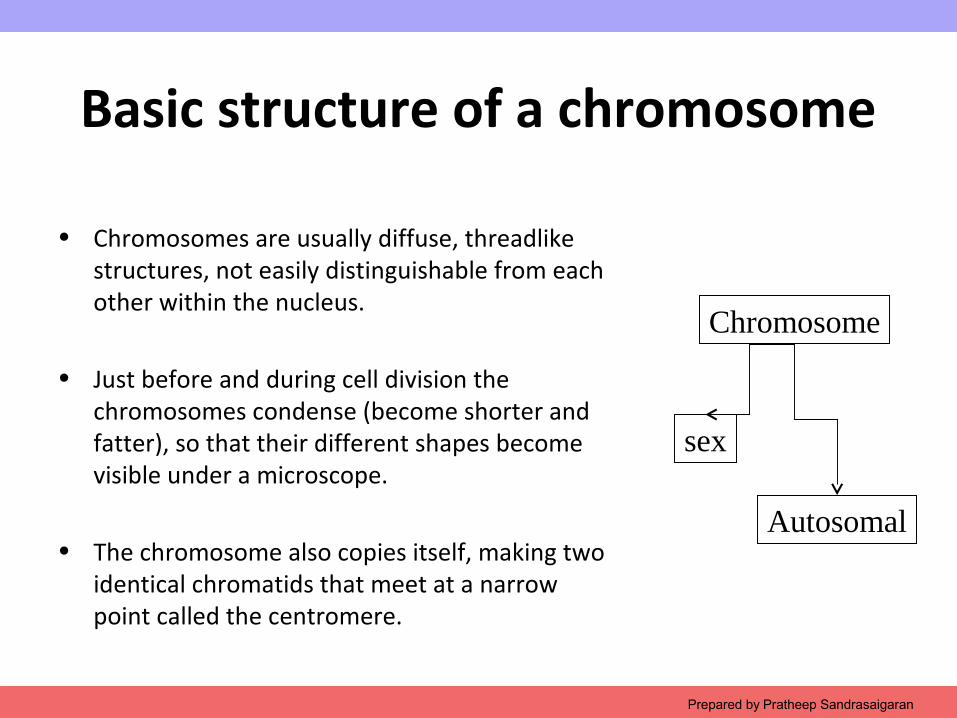
• Chromosomes are usually diffuse, threadlike structures, not easily distinguishable from each other within the nucleus.
• Just before and during cell division the chromosomes condense (become shorter and fatter), so that their different shapes become visible under a microscope.
• The chromosome also copies itself, making two identical chromatids that meet at a narrow point called the centromere.
Prepared by Pratheep Sandrasaigaran
Chromosome
sex
Autosomal
Copyright © 2009 Pearson Education, Inc.
Sex chromosomes
• These chromosomes determine gender.
• Human cells contain two sex chromosomes.
• If you’re female, you have two X chromosomes, and if you’re male, you have an X and a Y chromosome.
Prepared by Pratheep Sandrasaigaran
Copyright © 2009 Pearson Education, Inc.
Autosomal chromosomes
• Autosomal simply refers to non-sex chromosomes.
• So, sticking with the human example, do the math, and you can see that humans have 44 autosomal chromosomes.
• In humans, chromosomes come in pairs.
• How many pairs of chromosome that you will find in your cells?
Prepared by Pratheep Sandrasaigaran
Copyright © 2009 Pearson Education, Inc.Prepared by Pratheep Sandrasaigaran
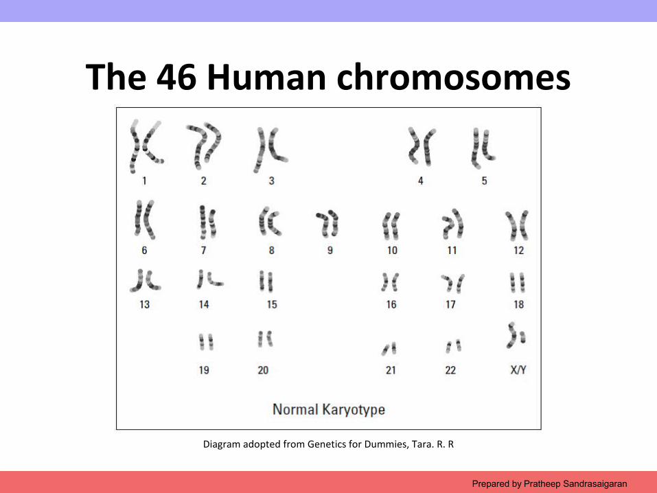
Diagram adopted from Genetics for Dummies, Tara. R. R
The 46 Human chromosomes
Copyright © 2009 Pearson Education, Inc.
DNA packaging in the chromosomes
• In the nucleus of a normal human cell, there are 46 chromosomes each containing 48–240 million bases of DNA.
• Watson and Crick’s double helix model predicts that each chromosome would have a contour length of 1.6–8.2 cm
• While the total length of the DNA would be about 3 m.
Prepared by Pratheep Sandrasaigaran
Copyright © 2009 Pearson Education, Inc.
Diagram from Internet source
DNA packaging in the chromosomes
• However, the average nucleus has a diameter of approximately 5 mm.
• How it is possible?
• A high degree of organization is needed to fit this amount of DNA into the nucleus…!
Prepared by Pratheep Sandrasaigaran
Copyright © 2009 Pearson Education, Inc.Prepared by Pratheep Sandrasaigaran
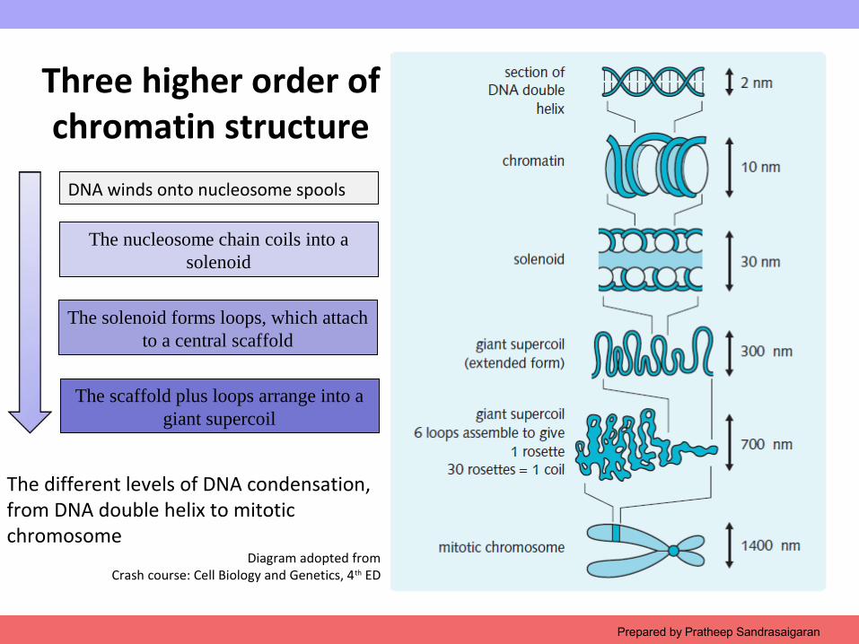
The different levels of DNA condensation, from DNA double helix to mitotic chromosome
Diagram adopted fromCrash course: Cell Biology and Genetics, 4th ED
Three higher order of chromatin structure
DNA winds onto nucleosome spools
The nucleosome chain coils into a solenoid
The solenoid forms loops, which attach to a central scaffold
The scaffold plus loops arrange into a giant supercoil
Copyright © 2009 Pearson Education, Inc.
First order: The chromatin
• Chromatin is the collective name for the long strands of DNA, RNA and their associated nucleoproteins.
• During interphase of the cell cycle, chromatin is dispersed throughout the nucleus, becoming more compact during mitosis or meiosis
• Two types of chromatin can be seen with electron microscopy.• Heterochromatin• Euchromatin
Prepared by Pratheep Sandrasaigaran
Copyright © 2009 Pearson Education, Inc.
Heterochromatin
• Is an electron dense and distributed around the periphery of the nucleus and in discrete masses within the nucleus.
• The DNA is in close association with nucleoproteins, and it is not active in RNA synthesis.
• Serves as structural purpose during the chromosomal stages.
Prepared by Pratheep Sandrasaigaran
Copyright © 2009 Pearson Education, Inc.
Euchromatin
• Is an electron lucent and represents DNA that is actually or potentially active in RNA synthesis.
• Example, the protein coding gene region
Prepared by Pratheep Sandrasaigaran
Copyright © 2009 Pearson Education, Inc.
Nucleosomes
• A nucleosome is formed by 146 bp of DNA wound twice around an octamer (Eight) of histone proteins.
• The octamer consists of two copies each of the histone proteins H2A, H2B, H3, and H4.
• Histone proteins are conserved throughout eukaryotic evolution.
• They contain a high proportion of positively charged amino-acid residues that can form ionic bonds with the negatively charged DNA.
Prepared by Pratheep Sandrasaigaran
Copyright © 2009 Pearson Education, Inc.
Nucleosomes
• This interaction does not depend on DNA sequence and theoretically histones can bind with any piece of DNA.
• However, in vivo, the position of histone binding is influenced by:• AT content (bends more easily than GC)• The presence of other tightly bound proteins.
• Nucleosome bound regions of DNA are separated by a region of linker DNA that varies from 0–80 bp in length.
Prepared by Pratheep Sandrasaigaran
Copyright © 2009 Pearson Education, Inc.Prepared by Pratheep Sandrasaigaran
Diagram adopted fromCrash course: Cell Biology and Genetics, 4th ED
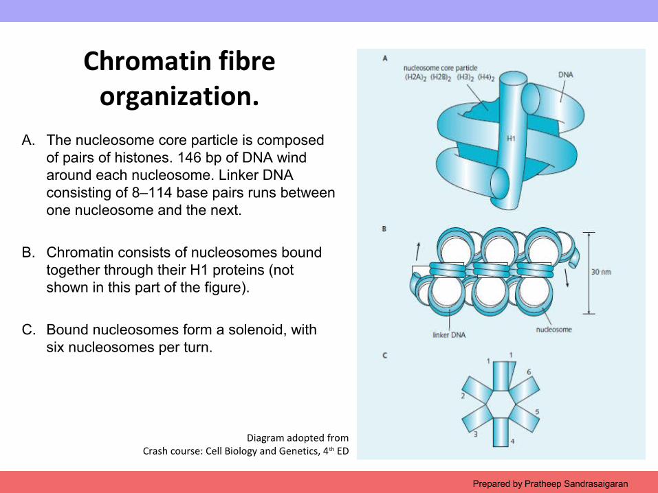
Chromatin fibre organization.
A. The nucleosome core particle is composed of pairs of histones. 146 bp of DNA wind around each nucleosome. Linker DNA consisting of 8–114 base pairs runs between one nucleosome and the next.
B. Chromatin consists of nucleosomes bound together through their H1 proteins (not shown in this part of the figure).
C. Bound nucleosomes form a solenoid, with six nucleosomes per turn.
Copyright © 2009 Pearson Education, Inc.
Second order: Solenoid formation
• The second level of DNA packing is mediated by histone HI, binding together adjacent nucleosomes to condense DNA into the supercoiled 30-nm fibre.
• This fibre is also known as the solenoid.
• The solenoid exhibits six to eight nucleosomes per turn of the spiral, corresponding to heterochromatin.
Prepared by Pratheep Sandrasaigaran
Copyright © 2009 Pearson Education, Inc.
Third order: Giant supercoil
• The third level of organization is thought to involve the formation of transcriptional units of DNA loops radiating from a central scaffold of non-histone proteins…. WHY?
• Histone binding protects DNA from degradation with DNaseI (endonuclease that breaks the internal phosphodiester bonds in DNA irrespective of its base sequence).
• If there is histone bind to DNA, no transcription.
Prepared by Pratheep Sandrasaigaran
Copyright © 2009 Pearson Education, Inc.
Chromosome
•What is chromosome?
•At metaphase, chromatin is maximally condensed and forms 1400-nm fibres.
•After cell staining these structures are visible as chromosomes under light microscopy.
Prepared by Pratheep Sandrasaigaran
Copyright © 2009 Pearson Education, Inc. Figure 2.13Prepared by Pratheep Sandrasaigaran
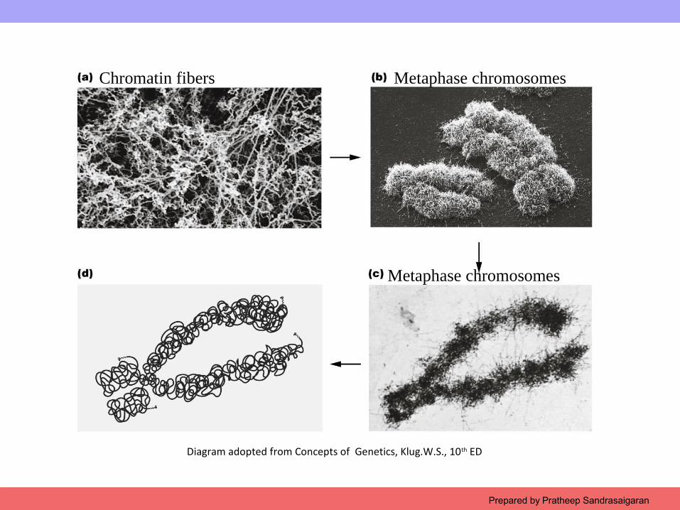
Diagram adopted from Concepts of Genetics, Klug.W.S., 10th ED
Chromatin fibers Metaphase chromosomes
Metaphase chromosomes

Copyright © 2009 Pearson Education, Inc.
Centromeres
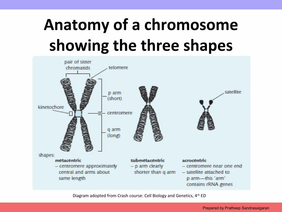
• Each metaphase chromosome is composed of two identical sister chromatids.
• Chromatids are connected at a central region called the centromere
• Centromeres consist of hundreds of kilobases of repetitive DNA and are responsible for the movement of chromosomes at cell division.
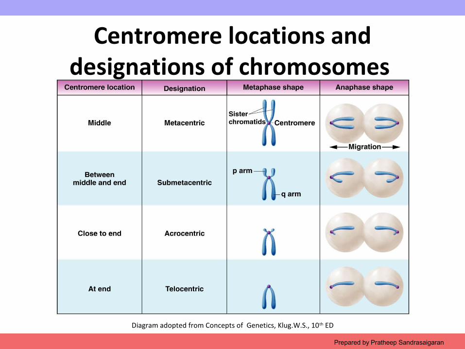
• Each centromere divides the chromosome into short (p) and long (q) arms.
Prepared by Pratheep Sandrasaigaran
Copyright © 2009 Pearson Education, Inc.Prepared by Pratheep Sandrasaigaran
Anatomy of a chromosome showing the three shapes
Diagram adopted from Crash course: Cell Biology and Genetics, 4th ED
Copyright © 2009 Pearson Education, Inc. Figure 2.3Prepared by Pratheep Sandrasaigaran
Diagram adopted from Concepts of Genetics, Klug.W.S., 10th ED
Centromere locations and designations of chromosomes
Copyright © 2009 Pearson Education, Inc.
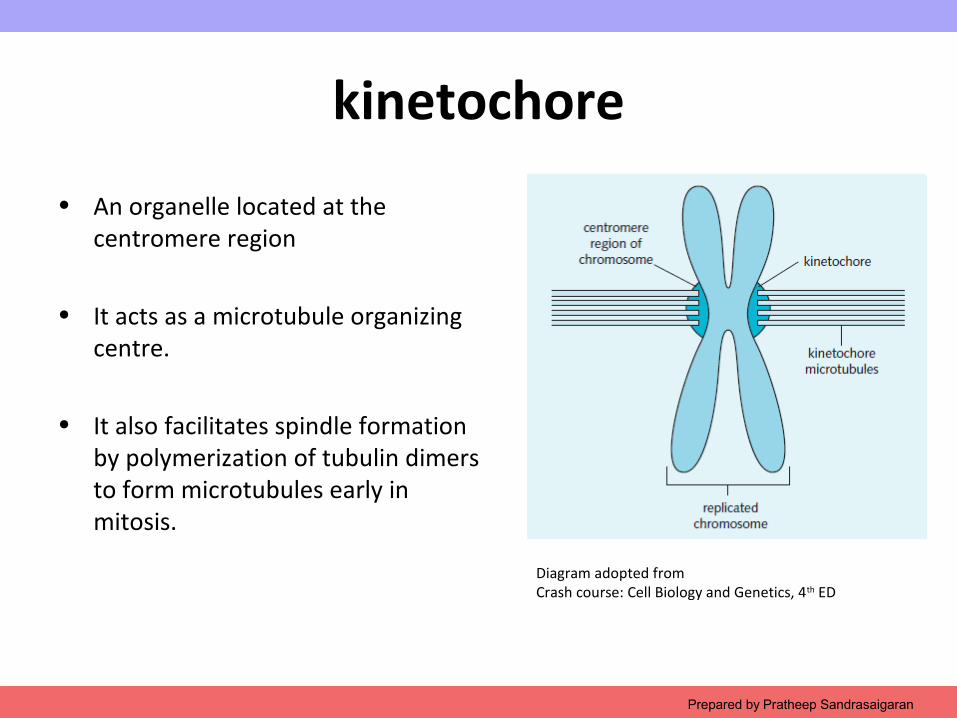
kinetochore
• An organelle located at the centromere region
• It acts as a microtubule organizing centre.
• It also facilitates spindle formation by polymerization of tubulin dimers to form microtubules early in mitosis.
Prepared by Pratheep Sandrasaigaran
Diagram adopted from Crash course: Cell Biology and Genetics, 4th ED
Copyright © 2009 Pearson Education, Inc.
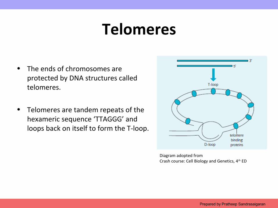
Telomeres
• The ends of chromosomes are protected by DNA structures called telomeres.
• Telomeres are tandem repeats of the hexameric sequence ‘TTAGGG’ and loops back on itself to form the T-loop.
Prepared by Pratheep Sandrasaigaran
Diagram adopted from Crash course: Cell Biology and Genetics, 4th ED
Copyright © 2009 Pearson Education, Inc.
Telomeres
• Telomeres have several functions in preserving chromosome stability:
• Preventing abnormal end-to-end fusion of chromosomes
• Protecting the ends of chromosomes from degradation
• Ensuring complete DNA replication• Having a role in chromosome pairing
during meiosis.
Prepared by Pratheep Sandrasaigaran
Copyright © 2009 Pearson Education, Inc.Prepared by Pratheep Sandrasaigaran
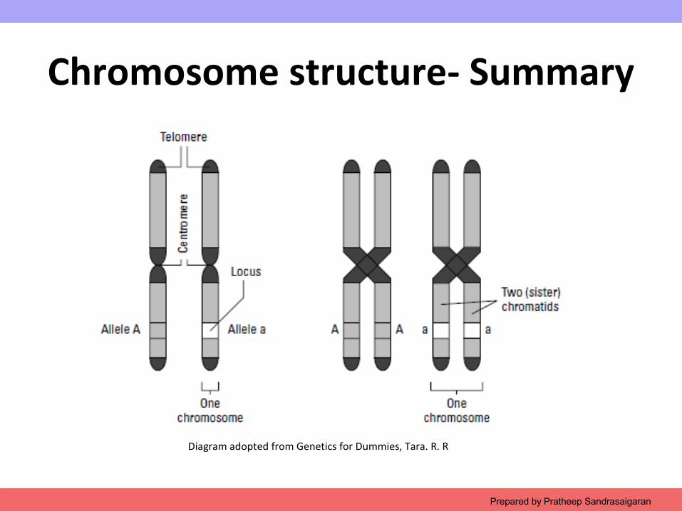
Chromosome structure- Summary
Diagram adopted from Genetics for Dummies, Tara. R. R
Copyright © 2009 Pearson Education, Inc.
TEST YOUR KNOWLEDGE 2
Prepared by Pratheep Sandrasaigaran
Copyright © 2009 Pearson Education, Inc.Prepared by Pratheep Sandrasaigaran
Copyright © 2009 Pearson Education, Inc.Prepared by Pratheep Sandrasaigaran
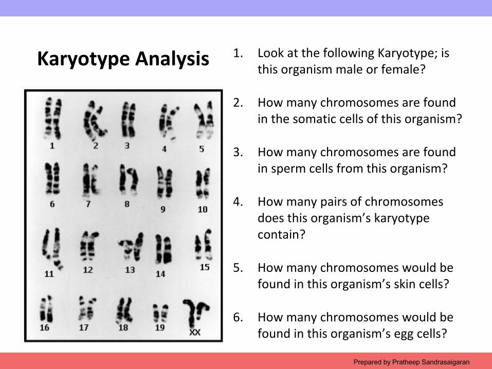
1. Look at the following Karyotype; is this organism male or female?
2. How many chromosomes are found in the somatic cells of this organism?
3. How many chromosomes are found in sperm cells from this organism?
4. How many pairs of chromosomes does this organism’s karyotype contain?
5. How many chromosomes would be found in this organism’s skin cells?
6. How many chromosomes would be found in this organism’s egg cells?
Karyotype Analysis
Copyright © 2009 Pearson Education, Inc.
Define these terms
• Gene
A discrete unit of hereditary information consisting of a specific nucleotide sequence in DNA (or RNA, in some viruses)
• Locus
A specific place along the length of a chromosome where a given gene is located
• Gamete
A haploid reproductive cell, such as an egg or sperm. Gametes unite during sexual reproduction to produce a diploid zygote.
Prepared by Pratheep Sandrasaigaran
Copyright © 2009 Pearson Education, Inc.
Define these terms
• Male gamete
Sperm
• Female gamete
Eggs
• Asexual reproduction
The generation of offspring from a single parent that occurs without the fusion of gametes (by budding, division of a single cell, or division of the entire organism into two or more parts). In most cases, the offspring are genetically identical to the parent.
Prepared by Pratheep Sandrasaigaran
Copyright © 2009 Pearson Education, Inc.
1. What is a somatic cell? Give examples of two human somatic cell types
A somatic cell is any cell in a multicellular organism except a sperm or egg or their precursors. Examples may vary but could include bone cells, skin cells, blood cells, etc.
2. How does a somatic cell compare to a gamete in terms of chromosome number?
Unlike somatic cells, gametes contain a single set of chromosomes. Such cells are called haploid cells, and each has a haploid number of chromosomes (n). For humans, the haploid number is 23.
Prepared by Pratheep Sandrasaigaran
Copyright © 2009 Pearson Education, Inc.
2.3 Compare the nuclear DNA
The Mitochondrial DNA
Prepared by Pratheep Sandrasaigaran
Copyright © 2009 Pearson Education, Inc.
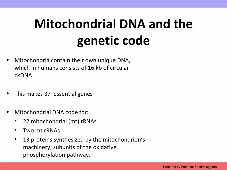
Mitochondrial DNA and the genetic code
• Mitochondria contain their own unique DNA, which in humans consists of 16 kb of circular dsDNA
• This makes 37 essential genes
• Mitochondrial DNA code for:• 22 mitochondrial (mt) tRNAs• Two mt rRNAs• 13 proteins synthesized by the mitochondrion’s
machinery; subunits of the oxidative phosphorylation pathway.
Prepared by Pratheep Sandrasaigaran
Copyright © 2009 Pearson Education, Inc.Prepared by Pratheep Sandrasaigaran
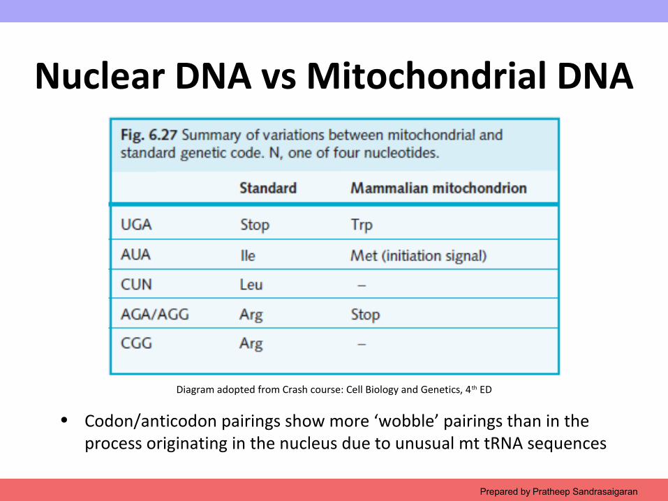
• Codon/anticodon pairings show more ‘wobble’ pairings than in the process originating in the nucleus due to unusual mt tRNA sequences
Nuclear DNA vs Mitochondrial DNA
Diagram adopted from Crash course: Cell Biology and Genetics, 4th ED
Copyright © 2009 Pearson Education, Inc.
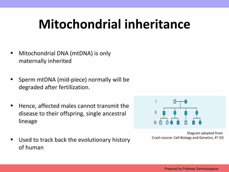
Mitochondrial inheritance
• Mitochondrial DNA (mtDNA) is only maternally inherited
• Sperm mtDNA (mid-piece) normally will be degraded after fertilization.
• Hence, affected males cannot transmit the disease to their offspring, single ancestral lineage
• Used to track back the evolutionary history of human
Prepared by Pratheep Sandrasaigaran
Diagram adopted from Crash course: Cell Biology and Genetics, 4th ED
Copyright © 2009 Pearson Education, Inc.
Mitochondrial inherited disease
• Leber hereditary optic neuropathy- an inherited form of vision loss
• Mitochondrial encephalomyopathy, lactic acidosis and stroke-like syndrome (MELAS)
• Myoclonus with epilepsy and with ragged red fibres (MERRF).
Prepared by Pratheep Sandrasaigaran
Copyright © 2009 Pearson Education, Inc.
2.3 Compare the nuclear DNA
The Chloroplast DNA
Prepared by Pratheep Sandrasaigaran
Copyright © 2009 Pearson Education, Inc.
Chloroplast DNA and the genetic code
• Chloroplast DNA (cpDNA) has between 100 and 225 kb in length.
• It is circular and double stranded.
• The size of cpDNA is much larger than that of mtDNA, hence account for a larger number of genes.
• Most cpDNA are non coding and duplications of same DNA sequences
Prepared by Pratheep Sandrasaigaran
Copyright © 2009 Pearson Education, Inc.
Chloroplast DNA and the genetic code
• Numerous gene products encoded by chloroplast DNA function during translation within the organelle.
• Genes specific to the photosynthetic function have also been identified.
• Mutations in these genes may inactivate photosynthesis.
Prepared by Pratheep Sandrasaigaran
Copyright © 2009 Pearson Education, Inc.
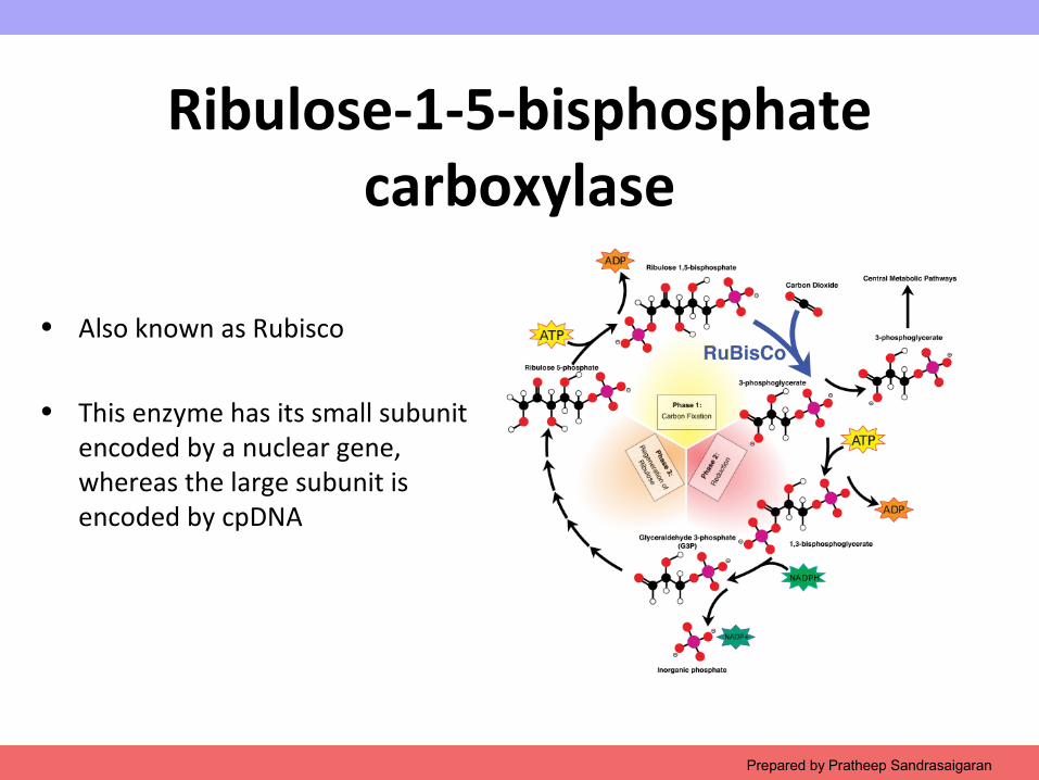
Ribulose-1-5-bisphosphate carboxylase
• Also known as Rubisco
• This enzyme has its small subunit encoded by a nuclear gene, whereas the large subunit is encoded by cpDNA
Prepared by Pratheep Sandrasaigaran
Copyright © 2009 Pearson Education, Inc.
2.4 Cell cycle
Prepared by Pratheep Sandrasaigaran
Copyright © 2009 Pearson Education, Inc.
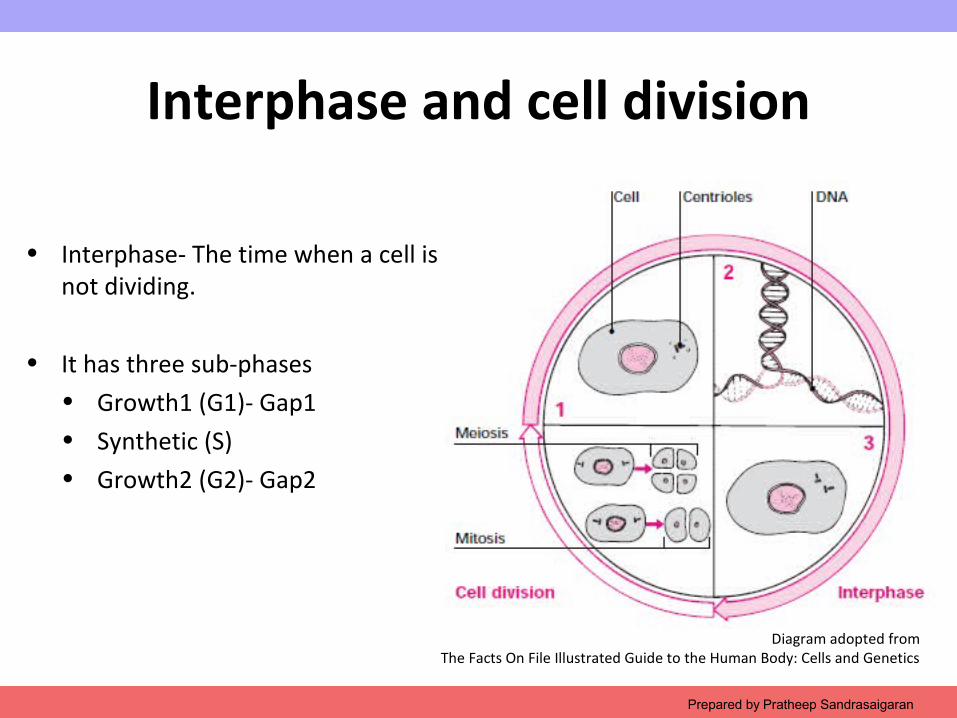
Interphase and cell division
• Interphase- The time when a cell is not dividing.
• It has three sub-phases• Growth1 (G1)- Gap1 • Synthetic (S)• Growth2 (G2)- Gap2
Prepared by Pratheep Sandrasaigaran
Diagram adopted fromThe Facts On File Illustrated Guide to the Human Body: Cells and Genetics
Copyright © 2009 Pearson Education, Inc.
Centrioles
Diagram adopted fromThe Facts On File Illustrated Guide to the Human Body: Cells and Genetics
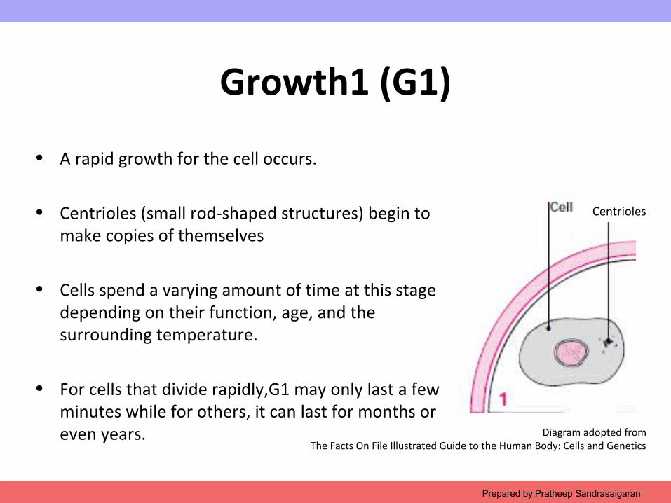
Growth1 (G1)
• A rapid growth for the cell occurs.
• Centrioles (small rod-shaped structures) begin to make copies of themselves
• Cells spend a varying amount of time at this stage depending on their function, age, and the surrounding temperature.
• For cells that divide rapidly,G1 may only last a few minutes while for others, it can last for months or even years.
Prepared by Pratheep Sandrasaigaran
Copyright © 2009 Pearson Education, Inc.
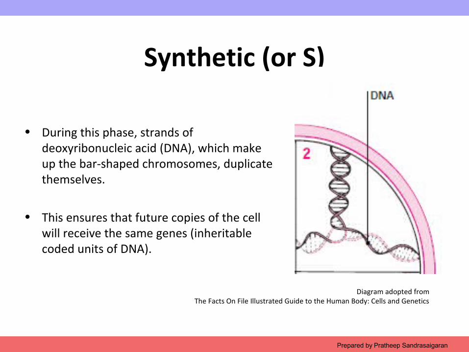
Synthetic (or S)
• During this phase, strands of deoxyribonucleic acid (DNA), which make up the bar-shaped chromosomes, duplicate themselves.
• This ensures that future copies of the cell will receive the same genes (inheritable coded units of DNA).
Prepared by Pratheep Sandrasaigaran
Diagram adopted fromThe Facts On File Illustrated Guide to the Human Body: Cells and Genetics
Copyright © 2009 Pearson Education, Inc.
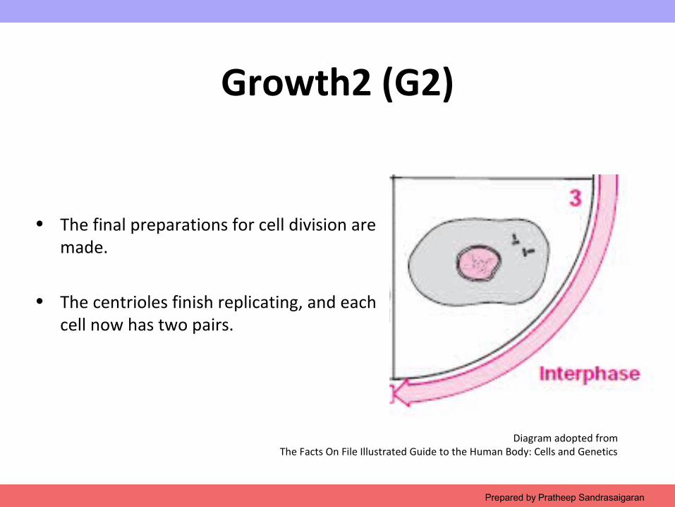
Growth2 (G2)
• The final preparations for cell division are made.
• The centrioles finish replicating, and each cell now has two pairs.
Prepared by Pratheep Sandrasaigaran
Diagram adopted fromThe Facts On File Illustrated Guide to the Human Body: Cells and Genetics
Copyright © 2009 Pearson Education, Inc.Prepared by Pratheep Sandrasaigaran
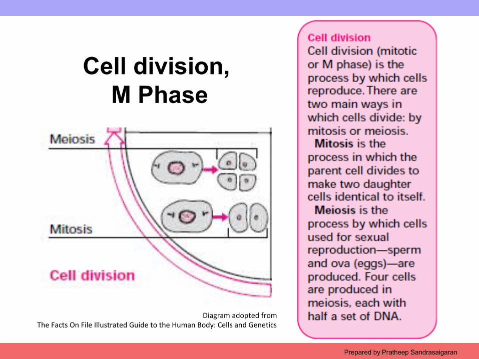
Cell division, M Phase
Diagram adopted fromThe Facts On File Illustrated Guide to the Human Body: Cells and Genetics
Copyright © 2009 Pearson Education, Inc.
Controlling the cell cycle
• Breakdown in the control mechanisms can lead to• Abnormal mitosis and cell division• Uncontrolled proliferation of cells• Produces cancerous tumors
• What control the mechanism?
• Control is exerted by a small group of enzymes called cyclin-dependent kinases (Cdks)
Prepared by Pratheep Sandrasaigaran
Copyright © 2009 Pearson Education, Inc.
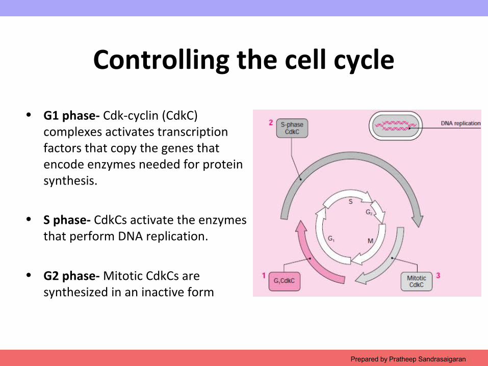
Controlling the cell cycle
• G1 phase- Cdk-cyclin (CdkC) complexes activates transcription factors that copy the genes that encode enzymes needed for protein synthesis.
• S phase- CdkCs activate the enzymes that perform DNA replication.
• G2 phase- Mitotic CdkCs are synthesized in an inactive form
Prepared by Pratheep Sandrasaigaran
Copyright © 2009 Pearson Education, Inc.Prepared by Pratheep Sandrasaigaran
Cell cycle checkpoints
Copyright © 2009 Pearson Education, Inc.
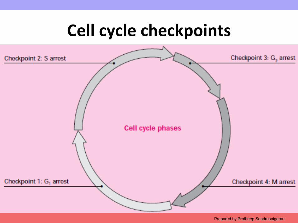
Cell cycle checkpoints
Checkpoint 1 •Damage to DNA causes G1 arrest and allows cell to replace damaged or lost DNA nucleotides before they are copied during S phase.
Checkpoint 2•The presence of unreplicated DNA causes S arrest and allows cell to complete DNA replication before mitosis.
Prepared by Pratheep Sandrasaigaran
Copyright © 2009 Pearson Education, Inc.
Cell cycle checkpoints
Checkpoint 3 •Damaged DNA causes G2 arrest, enabling the cell to repair double strand breaks in DNA that otherwise.
Checkpoint 4 •Delayed or faulty assembly of the mitotic spindle causes M arrest
•Mitosis is delayed, and chromosomes remain in a condensed state.
•This prevents abnormal segregation of chromosomes.
Prepared by Pratheep Sandrasaigaran

Copyright © 2009 Pearson Education, Inc.
TEST YOUR KNOWLEDGE 3
Prepared by Pratheep Sandrasaigaran
Copyright © 2009 Pearson Education, Inc.
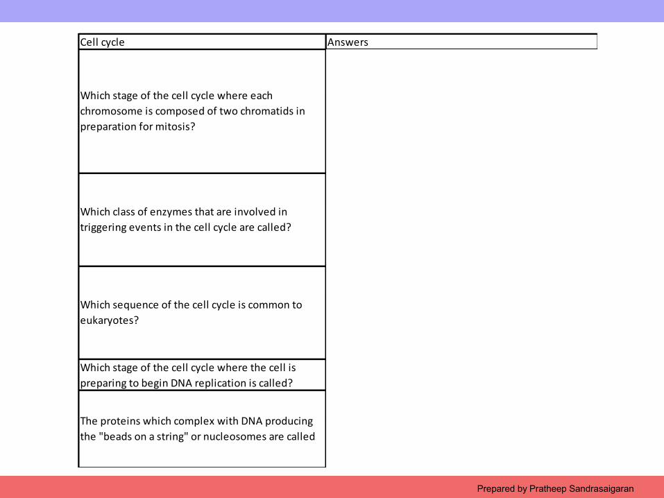
Cell cycle Answers
G2
S resulted in the duplication of each chromatid. Since there is only one centromere on the sister chromatids, we still call them one chromosome. When completed, the cells are in G2 and preparing for M
Kinaseskinases add phosphate to molecules, and the modification can serve as a "switch" to turn events in the cell on or off. Cdk or cyclin dependent kinases regulate the cell cycle.
G1 to S to G2 to M to cytokinesis
G1 as a preparation for S and G2 as the time between the completion of S and entry into M. Cytokinesis occurs after the other stages to create two daughter cells.
Which stage of the cell cycle where the cell is preparing to begin DNA replication is called? G1 cells are preparing for S
Histoneshistones form structural complexes with DNA. In the electron microscope, these can take the appearance of beads on a string
Which stage of the cell cycle where each chromosome is composed of two chromatids in preparation for mitosis?
Which class of enzymes that are involved in triggering events in the cell cycle are called?
Which sequence of the cell cycle is common to eukaryotes?
The proteins which complex with DNA producing the "beads on a string" or nucleosomes are called
Prepared by Pratheep Sandrasaigaran
Copyright © 2009 Pearson Education, Inc.
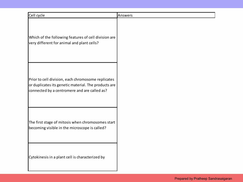
Cell cycle Answers
Cytokinesis
the animal cell can pinch into two by a ring of microfilaments. The plant cell with a rigid wall synthesizes a new cell plate. In both cases, the spindle fibers play a role in determining the site where the cell is split
Sister chromatids
The replication of the chromosome begins with the production of sister chromatids. The complex is considered to be one chromosome since there is only one centromere
Prophase
At the beginning of prophase, the chromosomes condense into structures visible in the light microscope
The formation of a cell plate in the cytoplasm
The spindle fibers are used to bring in vesicles that line up along the cell plate and produce a new cell wall
Which of the following features of cell division are very different for animal and plant cells?
Prior to cell division, each chromosome replicates or duplicates its genetic material. The products are connected by a centromere and are called as?
The first stage of mitosis when chromosomes start becoming visible in the microscope is called?
Cytokinesis in a plant cell is characterized by
Prepared by Pratheep Sandrasaigaran
Copyright © 2009 Pearson Education, Inc.
Mitosis and Meiosis
Prepared by Pratheep Sandrasaigaran